
HEART AND NECK VESSEL ASSESSMENT
40
HEART AND NECK VESSEL ASSESSMENT 18 351 ● STRUCTURE AND FUNCTION The cardiovascular system is a highly complex system that includes the heart and a closed system of blood vessels. To collect accurate data and correctly interpret that data, the examiner must have an understanding of the structure and function of the heart, the great vessels, the electrical conduction system of the heart, the cardiac cycle, the production of heart sounds, cardiac output, and the neck vessels. This information helps the examiner to differen- tiate between normal and abnormal findings as they relate to the cardiovascular system. HEART AND GREAT VESSELS The heart is a hollow, muscular, four-chambered (left and right atria and left and right ventricles) organ located in the middle of the thoracic cavity between the lungs in the space called the mediastinum. It is about the size of a clenched fist and weighs approximately 255 g (9 oz) in women and 310 g (10.9 oz) in men. The heart extends vertically from the left second to the left fifth intercostal space (ICS) and horizontally from the right edge of the sternum to the left midclavicular line (MCL). The heart can be described as an inverted cone. The upper portion, near the left second ICS, is the base and the lower por- tion, near the left fifth ICS and the left MCL, is the apex. The anterior chest area that overlies the heart and great vessels is called the precordium (Fig. 18-1). The right side of the heart pumps blood to the lungs for gas exchange (pulmonary circulation); the left side of the heart pumps blood to all other parts of the body (systemic circulation). The large veins and arteries leading directly to and away from the heart are referred to as the great vessels. The superior and inferior vena cava return blood to the right atrium from the upper and lower torso respec- tively. The pulmonary artery exits the right ventricle, bifurcates, and carries blood to the lungs. The pulmonary veins (two from each lung) return oxygenated blood to the left atrium. The aorta transports oxygenated blood from the left ventricle to the body (Fig. 18-2). Heart Chambers and Valves The heart consists of four chambers or cavities: two upper chambers, the right and left atria, and two lower cham- bers, the right and left ventricles. The right and left sides of the heart are separated by a partition called the sep- tum. The thin-walled atria receive blood returning to the heart and pump blood into the ventricles. The thicker- walled ventricles pump blood out of the heart. The left ventricle is thicker than the right ventricle because the left side of the heart has a greater workload. The entrance and exit of each ventricle are pro- tected by one-way valves that direct the flow of blood through the heart. The atrioventricular (AV) valves are located at the entrance into the ventricles. There are two AV valves: the tricuspid valve and the bicuspid (mitral) valve. The tricuspid valve is composed of three cusps or flaps and is located between the right atrium and the right ventricle; the bicuspid (mitral) valve is composed of two cusps or flaps and is located between the left atrium and the left ventricle. Collagen fibers, called chor- dae tendineae, anchor the AV valve flaps to papillary mus- cles within the ventricles. Open AV valves allow blood to flow from the atria into the ventricles. However, as the ventricles begin to contract, the AV valves snap shut, preventing the regur- gitation of blood into the atria. The valves are prevented from blowing open in the reverse direction (i.e., toward the atria) by their secure anchors to the papillary muscles of the ventricular wall. The semilunar valves are located
-
Upload
khangminh22 -
Category
Documents
-
view
1 -
download
0
Transcript of HEART AND NECK VESSEL ASSESSMENT

351
HEART AND NECKVESSEL ASSESSMENT
18
351
l STRUCTURE AND FUNCTION
The cardiovascular system is a highly complex system thatincludes the heart and a closed system of blood vessels. Tocollect accurate data and correctly interpret that data, theexaminer must have an understanding of the structureand function of the heart, the great vessels, the electricalconduction system of the heart, the cardiac cycle, theproduction of heart sounds, cardiac output, and the neckvessels. This information helps the examiner to differen-tiate between normal and abnormal findings as they relateto the cardiovascular system.
HEART AND GREAT VESSELSThe heart is a hollow, muscular, four-chambered (left andright atria and left and right ventricles) organ located inthe middle of the thoracic cavity between the lungs in thespace called the mediastinum. It is about the size of aclenched fist and weighs approximately 255 g (9 oz) inwomen and 310 g (10.9 oz) in men. The heart extendsvertically from the left second to the left fifth intercostalspace (ICS) and horizontally from the right edge of thesternum to the left midclavicular line (MCL). The heartcan be described as an inverted cone. The upper portion,near the left second ICS, is the base and the lower por-tion, near the left fifth ICS and the left MCL, is the apex.The anterior chest area that overlies the heart and greatvessels is called the precordium (Fig. 18-1). The right sideof the heart pumps blood to the lungs for gas exchange(pulmonary circulation); the left side of the heart pumpsblood to all other parts of the body (systemic circulation).
The large veins and arteries leading directly to andaway from the heart are referred to as the great vessels.The superior and inferior vena cava return blood to
the right atrium from the upper and lower torso respec-tively. The pulmonary artery exits the right ventricle,bifurcates, and carries blood to the lungs. The pulmonaryveins (two from each lung) return oxygenated blood tothe left atrium. The aorta transports oxygenated bloodfrom the left ventricle to the body (Fig. 18-2).
Heart Chambers and ValvesThe heart consists of four chambers or cavities: two upperchambers, the right and left atria, and two lower cham-bers, the right and left ventricles. The right and left sidesof the heart are separated by a partition called the sep-tum. The thin-walled atria receive blood returning to theheart and pump blood into the ventricles. The thicker-walled ventricles pump blood out of the heart. The leftventricle is thicker than the right ventricle because theleft side of the heart has a greater workload.
The entrance and exit of each ventricle are pro-tected by one-way valves that direct the flow of bloodthrough the heart. The atrioventricular (AV) valves arelocated at the entrance into the ventricles. There are twoAV valves: the tricuspid valve and the bicuspid (mitral)valve. The tricuspid valve is composed of three cusps orflaps and is located between the right atrium and theright ventricle; the bicuspid (mitral) valve is composedof two cusps or flaps and is located between the leftatrium and the left ventricle. Collagen fibers, called chor-dae tendineae, anchor the AV valve flaps to papillary mus-cles within the ventricles.
Open AV valves allow blood to flow from the atriainto the ventricles. However, as the ventricles begin tocontract, the AV valves snap shut, preventing the regur-gitation of blood into the atria. The valves are preventedfrom blowing open in the reverse direction (i.e., towardthe atria) by their secure anchors to the papillary musclesof the ventricular wall. The semilunar valves are located

352 U N I T III • NURSING ASSESSMENT OF THE ADULT
at the exit of each ventricle at the beginning of the greatvessels. Each valve has three cusps or flaps that look likehalf-moons, hence the name “semilunar.” There are twosemilunar valves: the pulmonic valve is located at theentrance of the pulmonary artery as it exits the right ven-tricle and the aortic valve is located at the beginning ofthe ascending aorta as it exits the left ventricle. Thesevalves are open during ventricular contraction and closefrom the pressure of blood when the ventricles relax.Blood is thus prevented from flowing backward into therelaxed ventricles (see Fig. 18-2).
Heart Covering and WallsThe pericardium is a tough, inextensible, loose-fitting,fibroserous sac that attaches to the great vessels and,thereby, surrounds the heart. A serous membrane lining,the parietal pericardium, secretes a small amount of peri-
Apex andapical impulse
Base of heart
Precordium
Figure 18-1 The heart and major blood vessels lie centrally inthe chest behind the protective sternum.
Brachiocephalic arteryLeft common carotid artery
Left subclavian artery
Right pulmonaryartery(branches)
Ascendingaorta
Superior vena cava
Rightpulmonaryveins
Rightatrium
Right AV(tricuspid)valve
Inferiorvena cava
Right ventricle
Aortic arch
Pulmonary trunk
Left pulmonaryartery (branches)
Pulmonary valve
LeftpulmonaryveinsLeft atrium
Aortic valve
Left AV(mitral)valve
Endocardium
Leftventricle
MyocardiumBlood high in oxygen
Blood low in oxygen Epicardium
Apex
Interventricularseptum
Figure 18-2 Heart chambers, valves, and direction of circulatory flow.
C H A P T E R 1 8 • HEART AND NECK VESSEL ASSESSMENT 353
cardial fluid that allows for smooth, friction-free movementof the heart. This same type of serous membrane covers theouter surface of the heart and is known as the epicardium.The myocardium is the thickest layer of the heart andis made up of contractile cardiac muscle cells. The endo-cardium is a thin layer of endothelial tissue that forms theinnermost layer of the heart and is continuous with theendothelial lining of blood vessels (see Fig. 18-2).
ELECTRICAL CONDUCTION OFTHE HEARTCardiac muscle cells have a unique inherent ability. Theycan spontaneously generate an electrical impulse andconduct it through the heart. The generation and con-duction of electrical impulses by specialized sections ofthe myocardium regulate the events associated with thefilling and emptying of the cardiac chambers. The processis called the cardiac cycle (see description below).
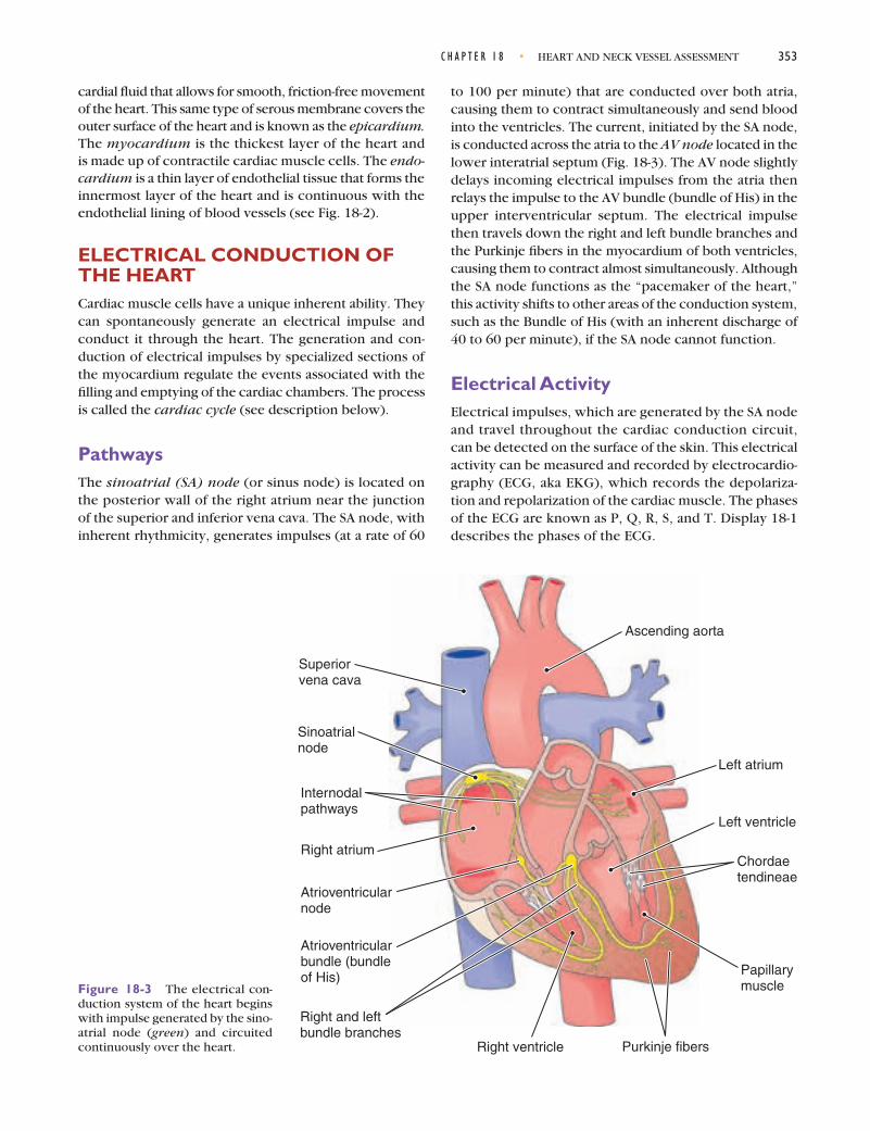
PathwaysThe sinoatrial (SA) node (or sinus node) is located onthe posterior wall of the right atrium near the junctionof the superior and inferior vena cava. The SA node, withinherent rhythmicity, generates impulses (at a rate of 60
to 100 per minute) that are conducted over both atria,causing them to contract simultaneously and send bloodinto the ventricles. The current, initiated by the SA node,is conducted across the atria to the AV node located in thelower interatrial septum (Fig. 18-3). The AV node slightlydelays incoming electrical impulses from the atria thenrelays the impulse to the AV bundle (bundle of His) in theupper interventricular septum. The electrical impulsethen travels down the right and left bundle branches andthe Purkinje fibers in the myocardium of both ventricles,causing them to contract almost simultaneously. Althoughthe SA node functions as the “pacemaker of the heart,”this activity shifts to other areas of the conduction system,such as the Bundle of His (with an inherent discharge of40 to 60 per minute), if the SA node cannot function.
Electrical ActivityElectrical impulses, which are generated by the SA nodeand travel throughout the cardiac conduction circuit,can be detected on the surface of the skin. This electricalactivity can be measured and recorded by electrocardio-graphy (ECG, aka EKG), which records the depolariza-tion and repolarization of the cardiac muscle. The phasesof the ECG are known as P, Q, R, S, and T. Display 18-1describes the phases of the ECG.
Sinoatrialnode
Internodalpathways
Atrioventricularnode
Atrioventricularbundle (bundleof His)
Right and leftbundle branches
Purkinje fibers
Right atrium
Right ventricle
Ascending aorta
Left atrium
Papillarymuscle
Left ventricle
Chordaetendineae
Superiorvena cava
Figure 18-3 The electrical con-duction system of the heart beginswith impulse generated by the sino-atrial node (green) and circuitedcontinuously over the heart.

354 U N I T III • NURSING ASSESSMENT OF THE ADULT
THE CARDIAC CYCLEThe cardiac cycle refers to the filling and emptying of theheart’s chambers. The cardiac cycle has two phases: dias-tole (relaxation of the ventricles, known as filling) and sys-tole (contraction of the ventricles, known as emptying).Diastole endures for approximately two-thirds of the car-diac cycle and systole is the remaining one-third (Fig. 18-4).
DiastoleDuring ventricular diastole, the AV valves are open andthe ventricles are relaxed. This causes higher pressure inthe atria than in the ventricles. Therefore, blood rushes
through the atria into the ventricles. This early, rapid, pas-sive filling is called early or protodiastolic filling. This isfollowed by a period of slow passive filling. Finally, nearthe end of ventricular diastole, the atria contract and com-plete the emptying of blood out of the upper chambersby propelling it into the ventricles. This final active fillingphase is called presystole, atrial systole, or sometimes the“atrial kick.” This action raises left ventricular pressure.
SystoleThe filling phases during diastole result in a large amount ofblood in the ventricles, causing the pressure in the ventri-cles to be higher than in the atria. This causes the AV valves
The phases of the electrocardiogram (ECG), which records depolarization and repolarization of the heart, are assignedletters: P, Q, R, S, and T.
• P wave: Atrial depolarization; conduction of the impulse throughout the atria.• PR interval: Time from the beginning of the atrial depolarization to the beginning of ventricular depolarization,
that is, from the beginning of the P wave to the beginning of the QRS complex.• QRS complex: Ventricular depolarization (also atrial repolarization); conduction of the impulse throughout the
ventricles, which then triggers contraction of the ventricles; measured from the beginning of the Q wave to theend of the S wave.
• ST segment: Period between ventricular depolarization and the beginning of ventricular repolarization.• T wave: Ventricular repolarization; the ventricles return to a resting state.• QT interval: Total time for ventricular depolarization and repolarization, that is, from the beginning of the
Q wave to the end of the T wave; the QT interval varies with heart rate.• U wave: May or may not be present; if it is present, it follows the T wave and represents the final phase of
ventricular repolarization.
DISPLAY 18-1 PHASES OF THE ELECTROCARDIOGRAM

C H A P T E R 1 8 • HEART AND NECK VESSEL ASSESSMENT 355
(mitral and tricuspid) to shut. Closure of the AV valvesproduces the first heart sound (S1), which is the begin-ning of systole. This valve closure also prevents blood fromflowing backward (a process known as regurgitation) intothe atria during ventricular contraction.
At this point in systole, all four valves are closed andthe ventricles contract (isometric contraction). There isnow high pressure inside the ventricles, causing the aorticvalve to open on the left side of the heart and the pulmonicvalve to open on the right side of the heart. Blood is ejectedrapidly through these valves. With ventricular emptying,the ventricular pressure falls and the semilunar valves close.This closure produces the second heart sound (S2), whichsignals the end of systole. After closure of the semilunarvalves, the ventricles relax. Atrial pressure is now higherthan the ventricular pressure, causing the AV valves toopen and diastolic filling to begin again.
HEART SOUNDSHeart sounds are produced by valve closure, as describedabove. The opening of valves is silent. Normal heart sounds,characterized as “lub dubb” (S1 and S2), and, occasionally,extra heart sounds and murmurs can be auscultated with astethoscope over the precordium, the area of the anteriorchest overlying the heart and great vessels.
Normal Heart SoundsThe first heart sound (S1) is the result of closure of the AVvalves: the mitral and tricuspid valves. As mentioned pre-viously, S1 correlates with the beginning of systole (see
Display 18-2 for more information about S1 and variationsof S1). S1 (“lub”) is usually heard as one sound but may beheard as two sounds (see also Fig. 18-4). If heard as twosounds, the first component represents mitral valve clo-sure (M1), and the second component represents tricus-pid closure (T1). M1 occurs first because of increasedpressure on the left side of the heart and because of theroute of myocardial depolarization. S1 may be heard overthe entire precordium but is heard best at the apex (leftMCL, fifth ICS).
The second heart sound (S2) results from closure ofthe semilunar valves (aortic and pulmonic) and corre-lates with the beginning of diastole. S2 (“dubb”) is alsousually heard as one sound but may be heard as twosounds. If S2 is heard as two sounds, the first componentrepresents aortic valve closure (A2) and the second com-ponent represents pulmonic valve closure (P2). A2 occursfirst because of increased pressure on the left side of theheart and because of the route of myocardial depolar-ization. If S2 is heard as two distinct sounds, it is called asplit S2. A splitting of S2 may be exaggerated during inspi-ration and disappear during expiration. S2 is heard best atthe base of the heart. See Display 18-3 for more informa-tion about variations of S2.
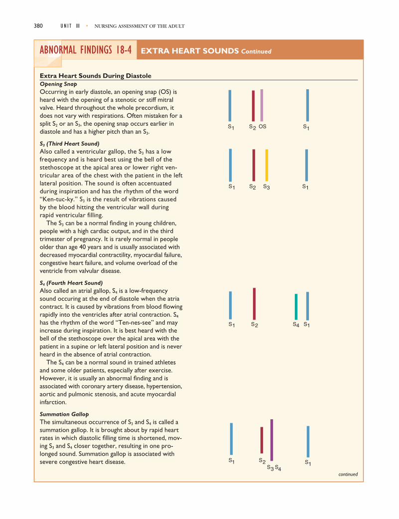
Extra Heart SoundsS3 and S4 are referred to as diastolic filling sounds or extraheart sounds, which result from ventricular vibration sec-ondary to rapid ventricular filling. If present, S3 can beheard early in diastole, after S2 (see Fig. 18-4). S4 also results
Heart Sounds
Electrocardiogram
S3 S4 S1 S2
P
Q
R
S
T
Rapidfilling
(Protodiastolic)
DIASTOLESlowfilling
Presystole
Isom
etric
cont
ract
ion
Isom
etric
rela
xatio
nSYSTOLEEjection
DIASTOLERapidfilling
Figure 18-4 The cardiac cycle con-sists of filling and ejection. Heartsounds S2, S3, and S4 are associatedwith diastole, while S1 is associatedwith systole. The electrical activityof the heart is measured throughoutdiastole and systole by electrocar-diography.
continued on page 362

356 U N I T III • NURSING ASSESSMENT OF THE ADULT
S1, which is the first heart sound, is produced by the atrioventricular (AV) closing. S1 (the “lub” portion of “lub dubb”)correlates with the beginning of systole.
The intensity of S1 depends on the position of the mitral valve at the start of systole, the structure of the valve leaflets,and how quickly pressure rises in the ventricles. All of these factors influence the speed and amount of closure the valveexperiences, which, in turn, determine the amount of sound produced.
Normal variations in S1 are heard at the base and the apex of the heart. S1 is softer at the base andlouder at the apex of the heart. An S1 may be split along the lower left sternal border, where the tricuspid componentof the sound, usually too faint to be heard, can be auscultated. A split S1 heard over the apex may be an S4.
1st Cardiac Beginning of NextAccentuated S1 Cycle Cardiac Cycle
An accentuated S1 sound is louder than an S2. This occurs when themitral valve is wide open and closes quickly. Examples include
• Hyperkinetic states in which blood velocity increases such as fever,anemia, and hyperthyroidism
• Mitral stenosis in which the leaflets are still mobile but increasedventricualr pressure is needed to close the valve
Diminished S1
Sometimes the S1 sound is softer than the S2 sound. This occurs whenthe mitral valve is not fully open at the time of ventricular contractionand valve closing. Examples include
• Delayed conduction from the atria to the ventricles as in first-degree heart block, which allows the mitral valve to drift closedbefore ventricular contraction closes it
• Mitral insufficiency in which extreme calcification of the valve limitsmobility
• Delayed or diminished ventricular contraction arising from force-ful atrial contraction into a noncompliant ventricle as in severepulmonary or systemic hypertension.
Split S1
As named, a split S1 occurs as a split sound. This occurs when the left andright ventricles contract at different times (asynchronous ventricularcontraction). Examples include
• Conduction delaying the cardiac impulse to one of the ventricles asin bundle branch block
• Ventricular ectopy in which the impulse starts in one ventricle,contracting it first, and then spreading to the second ventricle
Varying S1
This occurs when the mitral valve is in different positions when con-traction occurs. Examples include
• Rhythms in which the atria and ventricles are beating independentlyof each other
• Totally irregular rhythm such as atrial fibrillation
DISPLAY 18-2 UNDERSTANDING NORMAL S1 SOUNDS AND VARIATIONS
S1 S2 S1
S1 S2 S1
S1 S2 S1
S1 S2 S2S1

The S2 sound depends on the closure of the aortic and the pulmonic valves. Closure of the pulmonic valve is delayedby inspiration, resulting in a split S2 sound. The components of the split sound are referred to as A2 (aortic valve sound)and P2 (pulmonic valve sound). If either sound is absent, no split sounds are heard. The A2 sound is heard best over thesecond right intercostal space. P2 is normally softer than A2.
1st Cardiac Beginning of NextAccentuated S2 Cycle Cardiac Cycle
An accentuated S2 means that S2 is louder than S1. This occursin conditions in which the aortic or pulmonic valve has ahigher closing pressure. Examples include
• Increased pressure in the aorta from exercise, excite-ment, or systemic hypertension (a booming S2 is heardwith systemic hypertension)
• Increased pressure in the pulmonary vasculature, whichmay occur with mitral stenosis or congestive heart failure
• Calcification of the semilunar valve in which the valve isstill mobile as in pulmonic or aortic stenosis
Diminished S2
A diminished S2 means that S2 is softer than S1. This occurs inconditions in which the aortic or pulmonic valves havedecreased mobility. Examples include
• Decreased systemic blood pressure, which weakens thevalves, as in shock
• Aortic or pulmonic stenosis in which the valves are thick-ened snd calcified, with decreased mobility
Normal (Physiologic) Split S2
A normal split S2 can be heard over the second or third leftintercostal space. It is usually heard best during inspiration anddisappears during expiration. Over the aortic area and apex,the pulmonic component of S2 is usually too faint to be heardand S2 is a single sound resulting from aortic valve closure. Insome patients, S2 may not become single on expiration unlessthe patient sits up. Splitting that does not disappear duringexpiration is suggestive of heart disease.
Wide Split S2
This is an increase in the usual splitting that persists through-out the entire respiratory cycle and widens on expiration. Itoccurs when there is delayed electrical activation of the rightventricle. Example includes
• Right bundle branch block, which delays pulmonic valveclosing
DISPLAY 18-3 VARIATIONS IN S2
C H A P T E R 1 8 • HEART AND NECK VESSEL ASSESSMENT 357
S1 S1S2
S2S1 S1
S1 S2
A2P2
S1 S2
A2 P2
Expiration Inspiration
S1 S2
A2 P2
S2
A2 P2
Expiration Inspiration
mT
S1
mT
continued
358 U N I T III • NURSING ASSESSMENT OF THE ADULT
1st Cardiac Beginning of NextFixed Split S2 Cycle Cardiac Cycle
This is a wide splitting that does not vary with respiration. Itoccurs when there is delayed closure of one of the valves.Example includes
• Atrial septal defect and right ventricular failure, whichdelay pulmonic valve closing
Reversed Split S2
This is a split S2 that appears on expiration and disappears oninspiration—also known as paradoxical split. It occurs whenclosure of the aortic valve is abnormally delayed, causing A2 tofollow P2 in expiration. Normal inspiratory delay of P2 makesthe split disappear during inspiration. Example includes
• Left bundle branch block
Accentuated A2
An accentuated A2 is loud over the right, second intercostal space. This occurs with increased pressure as in sytemichypertension and aortic root dilation because of the closer position of the aortic valve to the chest wall.
Diminished A2
A diminished A2 is soft or absent over the right, second intercostal space. This occurs with immobility of the aorticvalve in calcific aortic stenosis.
Accentuated P2
An accentuated P2 is louder than or equal to an A2 sound. This occurs with pulmonary hypertension, dilated pulmonaryartery, and atrial septal defect. A wide split S2, heard even at the apex, indicates an accentuated P2.
Diminished P2
A soft or absent P2 sound occurs with an increased anteroposterior diameter of the chest (barrel chest), which is asso-ciated with aging, pulmonic stenosis, or COPD (chronic obstructive pulmonary disease).
DISPLAY 18-3 VARIATIONS IN S2 Continued
S1 S2
A2 P2
S1 S2
Expiration Inspiration
S1 S1 S2
A2 P2
S2
A2 P2
Expiration Inspiration
from ventricular vibration but, contrary to S3, the vibrationis secondary to ventricular resistance (noncompliance)during atrial contraction. If present, S4 can be heard latein diastole, just before S1 (see Fig. 18-4). S3 is often termedventricular gallop, and S4 is called atrial gallop. Extra heartsounds are described further in the Physical Assessmentsection of the text and in Display 18-4.
Murmurs
Blood normally flows silently through the heart. There areconditions, however, that can create turbulent blood flowin which a swooshing or blowing sound may be auscul-tated over the precordium. Conditions that contribute toturbulent blood flow include (1) increased blood velocity,

C H A P T E R 1 8 • HEART AND NECK VESSEL ASSESSMENT 359
Most nurses need many hours of practice in auscultating heart sounds to assess a client’s health status and interpret find-ings proficiently and confidently. Practitioners may be able to recognize an abnormal heart sound but may have difficultydetermining what and where it is exactly. Continued exposure and experience increase one’s ability to determine theexact nature and characteristics of abnormal heart sounds. An added difficulty involves palpation, particularly of the api-cal impulse in clients who are obese or barrel chested. These conditions increase the distance from the apex of the heartto the precordium.
Where to Auscultate
Heart sounds can be auscultated in the traditional five areas on the precordium, which is the anterior surface of thebody overlying the heart and great vessels. The traditional areas include the aortic area, the pulmonic area, Erb’s point,the tricuspid area, and the mitral or apical area. The four valve areas do not reflect the anatomic location of the valves.Rather, they reflect the way in which heart sounds radiate to the chest wall. Sounds always travel in the direction ofblood flow. For example, sounds that originate in the tricuspid valve are usually best heard along the left lower sternalborder at the fourth or fifth intercostal space.
Traditional Areas of Auscultation
• Aortic area: Second intercostal spaceat the right sternal border—the baseof the heart
• Pulmonic area: Second or third inter-costal space at the left sternal bor-der—the base of the heart
• Erb’s point: Third to fifth intercostalspace at the left sternal border
• Mitral (apical): Fifth intercostal spacenear the left midclavicular line—theapex of the heart
• Tricuspid area: Fourth or fifth inter-costal space at the left lower sternalborder
Alternative Areas
In reality, the areas described above over-lap extensively and sounds produced bythe valves can be heard all over the pre-cordium. Therefore, it is important to lis-ten to more than just five specific points on the precordium. Keep the fact of overlap in mind and use the names of thechambers instead of Erb’s point, mitral, and tricuspid areas when auscultating over the precordium. “Alternative” (ver-sus the traditional) areas of auscultation overlap and are not as discrete as the traditional areas. The alternative areasare the aortic area, pulmonic area, left atrial area, right atrial area, left ventricular area, and right ventricular area.
Cover the entire precordium. As you auscultate in all areas, concentrate on systematically moving the stethoscopefrom left to right across the entire heart area from the base to the apex (top to bottom) or from the apex to the base(bottom to top).
DISPLAY 18-4 AUSCULTATING HEART SOUNDS
Midsternum
Midclavicular line
Aortic area
Mitral(apical)ar ea
Tri cuspidarea
Erb's point
Pulmonicarea
continued
360 U N I T III • NURSING ASSESSMENT OF THE ADULT
(2) structural valve defects, (3) valve malfunction, and(4) abnormal chamber openings (e.g., septal defect).
CARDIAC OUTPUTCardiac output (CO) is the amount of blood pumped bythe ventricles during a given period of time (usually 1 min)and is determined by the stroke volume (SV) multipliedby the heart rate (HR): SV × HR = CO. The normal adultcardiac output is 5 to 6 L/min.
Stroke VolumeStroke volume is the amount of blood pumped from theheart with each contraction (stroke volume from the leftventricle is usually 70 mL). Stroke volume is influenced byseveral factors:
• The degree of stretch of the heart muscle up to acritical length before contraction (preload); thegreater the preload, the greater the stroke volume.
This holds true unless the heart muscle is stretchedso much that it cannot contract effectively.
• The pressure against which the heart muscle hasto eject blood during contraction (afterload);increased afterload results in decreased strokevolume.
• Synergy of contraction (i.e., the uniform, synchro-nized contraction of the myocardium); conditionsthat cause an asynchronous contraction decreasestroke volume.
• Compliance or distensibility of the ventricles;decreased compliance decreases stroke volume.
• Contractility or the force of contractions of themyocardium under given loading conditions;increased contractility increases stroke volume.
Although cardiac muscle has an innate pattern of con-tractility, cardiac activity is also mediated by the autonomicnervous system to respond to changing needs. The sym-pathetic impulses increase heart rate and, therefore, car-diac output. The parasympathetic impulses, which travel
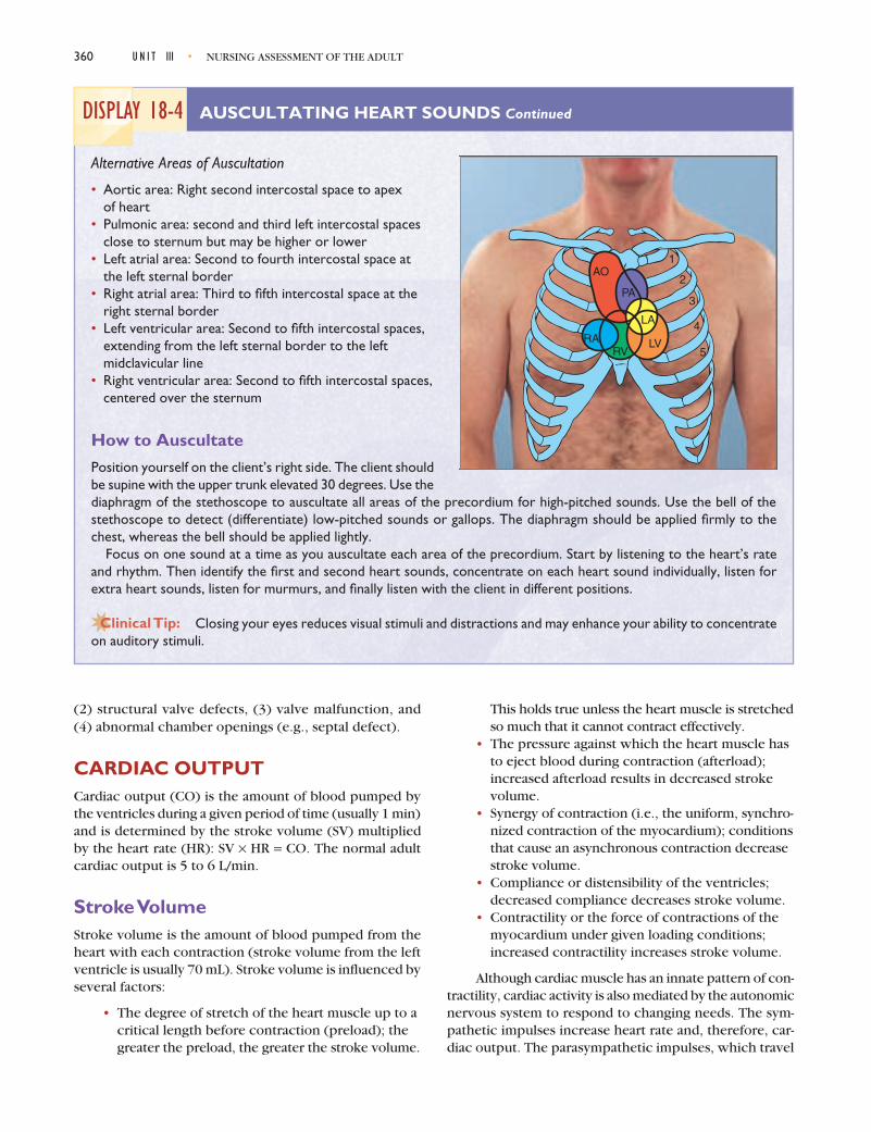
Alternative Areas of Auscultation
• Aortic area: Right second intercostal space to apex of heart
• Pulmonic area: second and third left intercostal spacesclose to sternum but may be higher or lower
• Left atrial area: Second to fourth intercostal space at the left sternal border
• Right atrial area: Third to fifth intercostal space at theright sternal border
• Left ventricular area: Second to fifth intercostal spaces,extending from the left sternal border to the left midclavicular line
• Right ventricular area: Second to fifth intercostal spaces,centered over the sternum
How to Auscultate
Position yourself on the client’s right side. The client shouldbe supine with the upper trunk elevated 30 degrees. Use thediaphragm of the stethoscope to auscultate all areas of the precordium for high-pitched sounds. Use the bell of thestethoscope to detect (differentiate) low-pitched sounds or gallops. The diaphragm should be applied firmly to thechest, whereas the bell should be applied lightly.
Focus on one sound at a time as you auscultate each area of the precordium. Start by listening to the heart’s rateand rhythm. Then identify the first and second heart sounds, concentrate on each heart sound individually, listen forextra heart sounds, listen for murmurs, and finally listen with the client in different positions.
Closing your eyes reduces visual stimuli and distractions and may enhance your ability to concentrateon auditory stimuli.
DISPLAY 18-4 AUSCULTATING HEART SOUNDS Continued
1
2
3
4
5
AO
RARV
LV
PA
LA

C H A P T E R 1 8 • HEART AND NECK VESSEL ASSESSMENT 361
to the heart by the vagus nerve, decrease the heart rateand, therefore, decrease cardiac output.
NECK VESSELSAssessment of the cardiovascular system includes evalua-tion of the vessels of the neck: the carotid artery and thejugular veins (Fig. 18-5). Assessment of the pulses of thesevessels reflects the integrity of the heart muscle.
Carotid Artery PulseThe right and left common carotid arteries extend fromthe brachiocephalic trunk and the aortic arch and arelocated in the groove between the trachea and the rightand left sternocleidomastoid muscles. Slightly below themandible, each bifurcates into an internal and externalcarotid artery. They supply the neck and head, includingthe brain, with oxygenated blood. The carotid arterypulse is a centrally located arterial pulse. Because it isclose to the heart, the pressure wave pulsation coin-cides closely with ventricular systole. The carotid arter-ial pulse is good for assessing amplitude and contour ofthe pulse wave. The pulse should normally have a smooth,rapid upstroke that occurs in early systole and a moregradual downstroke.
Jugular Venous Pulse and PressureThere are two sets of jugular veins: internal and external.The internal jugular veins lie deep and medial to the ster-nocleidomastoid muscle. The external jugular veins are
more superficial; they lie lateral to the sternocleidomastoidmuscle and above the clavicle. The jugular veins returnblood to the heart from the head and neck by way of thesuperior vena cava.
Assessment of the jugular venous pulse is importantfor determining the hemodynamics of the right side ofthe heart. The level of the jugular venous pressure reflectsright atrial (central venous) pressure and, usually, rightventricular diastolic filling pressure. Right-sided heart fail-ure raises pressure and volume, thus raising jugular venouspressure.
Decreased jugular venous pressure occurs withreduced left ventricular output or reduced blood volume.The right internal jugular vein is most directly connectedto the right atrium and provides the best assessment ofpressure changes. Components of the jugular venouspulse follow:
a wave—reflects rise in atrial pressure that occurswith atrial contraction
x descent—reflects right atrial relaxation anddescent of the atrial floor during ventricularsystole
v wave—reflects right atrial filling, increased volume,and increased atrial pressure
y descent—reflects right atrial emptying into theright ventricle and decreased atrial pressure
Figure 18-6 illustrates the jugular venous pulse.
l HEALTH ASSESSMENT
COLLECTING SUBJECTIVE DATA:THE NURSING HEALTH HISTORYSubjective data collected about the heart and neck vesselshelps the nurse to identify abnormal conditions that mayaffect the client’s ability to perform activities of daily livingand to fulfill his role and responsibilities. Data collection
Internalcarotid artery
Externalcarotid artery
Carotid sinus
Thyroidartery and vein
Left commoncarotid artery
Internal jugularvein
Left subclavian artery
Left subclavian vein
External jugularvein
Figure 18-5 Major neck vessels, including the carotid arteriesand jugular veins.
S1 S2 S1 S2
a
xy
v
Figure 18-6 Jugular venous pulse wave reflects pressure levelsin the heart.

362 U N I T III • NURSING ASSESSMENT OF THE ADULT
also provides information on the client’s risk for cardio-vascular disease and helps to identify area where healtheducation is needed. The client may not be aware of thesignificant role that health promotion activities can playin preventing cardiovascular disease.
When compiling the nursing history of current com-plaints or symptoms, personal and family history, andlifestyle and health practices, remember to thoroughlyexplore signs and symptoms that the client brings to yourattention either intentionally or inadvertently.
llä HISTORY OF PRESENT HEALTH CONCERN
Use the COLDSPA mnemonic as a guideline for informa-tion to collect. In addition, the following questions helpelicit important information.
QUESTION RATIONALE
Chest pain can be cardiac, pulmonary, muscular, orgastrointestinal in origin. Angina (cardiac chest pain) isusually described as a sensation of squeezing around theheart; a steady, severe pain; and a sense of pressure. Itmay radiate to the left shoulder and down the left armor to the jaw. Diaphoresis and pain worsened by activ-ity are usually related to cardiac chest pain.
Palpitations may occur with an abnormality of the heart’sconduction system or during the heart’s attempt toincrease cardiac output by increasing the heart rate.Palpitations may cause the client to feel anxious.
Fatigue may result from compromised cardiac output.Fatigue related to decreased cardiac output is worse inthe evening or as the day progresses.
Dyspnea may result from congestive heart failure, pul-monary disorders, coronary artery disease, myocardialischemia, and myocardial infarction. Dyspnea may occurat rest, during sleep, or with mild, moderate, or extremeexertion.
Increased renal perfusion during periods of rest orrecumbency may cause nocturia. Decreased frequencymay be related to decreased cardiac output.
C •O •L •D •S •P •A
Character : Describe the sign or symptom. Howdoes it feel, look, sound, smell, and so forth?
Onset : When did it begin?Locat ion: Where is it? Does it radiate?Durat ion: How long does it last? Does it recur?Sever i ty : How bad is it?Pattern: What makes it better? What makes it worse?Associated Factors : What other symptoms
occur with it?
Chest Pain and PalpitationsDo you experience chest pain? When did it start?Describe the type of pain, location, radiation, duration,and how often you experience the pain. Rate the painon a scale of 0 to 10, with 10 being the worst possiblepain. Does activity make the pain worse? Did you haveperspiration (diaphoresis) with the chest pain?
Do you experience palpitations?
Other SymptomsDo you tire easily? Do you experience fatigue? Describewhen the fatigue started. Was it sudden or gradual? Doyou notice it at any particular time of day?
Do you have difficulty breathing or shortness of breath(dyspnea)?
Do you wake up at night with an urgent need to urinate(nocturia)? How many times a night?
continued on page 367

C H A P T E R 1 8 • HEART AND NECK VESSEL ASSESSMENT 363
Dizziness may indicate decreased blood flow to thebrain due to myocardial damage; however, there areseveral other causes for dizziness such as inner ear syn-dromes, decreased cerebral circulation, and hypoten-sion. Dizziness may put the client at risk for falls.
Edema of the lower extremities may occur as a result ofheart failure.
Cardiac pain may be overlooked or misinterpreted asgastrointestinal problems.
Gastrointestinal pain may occur after meals and is relievedwith antacids, whereas cardiac pain may occur anytime,is not relieved with antacids, and worsens with activity.
Do you experience dizziness?
Do you experience swelling (edema) in your feet, ankles,or legs?
Do you have frequent heart burn? When does it occur?What relieves it? How often do you experience it?
QUESTION Continued RATIONALE Continued
Congenital or acquired defects affect the heart’s abilityto pump, decreasing the oxygen supply to the tissues.
Approximately 40% of people with rheumatic feverdevelop rheumatic carditis. Rheumatic carditis devel-ops after exposure to group A beta-hemolytic strepto-cocci and results in inflammation of all layers of theheart, impairing contraction and valvular function.
Previous heart surgery may change the heart soundsheard during auscultation. Surgery and cardiac ballooninterventions indicate prior cardiac compromise.
A prior ECG allows the health care team to evaluate forany changes in cardiac conduction or previous myo-cardial infarction.
Dyslipidemia presents the greatest risk for the develop-ing coronary artery disease. Elevated cholesterol levelshave been linked to the development of atherosclerosis(Libby, Schoenbeck, Mach, Selwyn & Ganz, 1998).
Clients may have medications prescribed for heart dis-ease but may not take them regularly. Clients may skiptaking their diuretics because of having to urinate fre-quently. Beta-blockers may be omitted because of theadverse effects on sexual energy. Education about med-ications may be needed.
llä PAST HEALTH HISTORY
QUESTION RATIONALE
Have you been diagnosed with a heart defect or amurmur?
Have you ever had rheumatic fever?
Have you ever had heart surgery or cardiac ballooninterventions?
Have you ever had an electrocardiogram (ECG)? Whenwas the last one performed? Do you know the results?
Have you ever had a blood test called a lipid profile?Based on your last test, do you know what your choles-terol levels were?
Do you take medications or use other treatments forheart disease? How often do you take them? Why doyou take them?
continued on page 368

364 U N I T III • NURSING ASSESSMENT OF THE ADULT
A genetic predisposition to these risk factors increasesa client’s chance for development of heart disease.
llä FAMILY HISTORY
QUESTION RATIONALE
Is there a history of hypertension, myocardial infarction(MI), coronary heart disease (CHD), elevated choles-terol levels, or diabetes mellitus (DM) in your family?
Cigarette smoking greatly increases the risk of heart dis-ease (see Risk Factors—Coronary Heart Disease).
Stress has been identified as a possible risk factor forheart disease.
An elevated cholesterol level increases the chance offatty plaque formation in the coronary vessels.
Excessive intake of alcohol has been linked to hyper-tension.
A sedentary lifestyle is a known modifiable risk factorcontributing to heart disease. Aerobic exercise threetimes per week for 30 min is more beneficial thananaerobic exercise or sporadic exercise in preventingheart disease.
Heart disease may impede the ability to perform dailyactivities. Exertional dyspnea or fatigue may indicateheart failure. An inability to complete activities of dailyliving may necessitate a referral for home care.
Many clients with heart disease are afraid that sexualactivity will precipitate chest pain. If the client can walkone block or climb two flights of stairs without expe-riencing symptoms, it is generally acceptable for the
llä LIFESTYLE AND HEALTH PRACTICES
QUESTION RATIONALE
Do you smoke? How many packs of cigarettes per dayand for how many years?
What type of stress do you have in your life? How doyou cope with it?
Describe what you usually eat in a 24-hour period.
How much alcohol do you consume each day/week?
Do you exercise? What type of exercise and how often?
Describe your daily activities. How are they different fromyour routine 5 or 10 years ago? Does fatigue, chest pain,or shortness of breath limit your ability to perform dailyactivities? Describe. Are you able to care for yourself?
Has your heart disease had any effect on your sexualactivity?
Self-monitoring of heart rate or blood pressure is rec-ommended if the client is taking cardiotonic or antihy-pertensive medications respectively. A demonstrationis necessary to ensure appropriate technique.
Do you monitor your own heart rate or blood pressure?
QUESTION Continued RATIONALE Continued
continued on page 369
C H A P T E R 1 8 • HEART AND NECK VESSEL ASSESSMENT 365
client to engage in sexual intercourse. Nitroglycerin canbe taken before intercourse as a prophylactic for chestpain. In addition, the side-lying position for sexual inter-course may reduce the workload on the heart.
If heart function is compromised, cardiac output to thekidneys is reduced during episodes of activity. At rest,cardiac output increases, as does glomerular filtrationand urinary output. Orthopnea (the inability to breathewhile supine) and nocturia may indicate heart failure.In addition, these two conditions may also impede theability to get adequate rest.
A person’s feeling of self-worth may depend on his orher ability to perform usual daily activities and fulfill hisor her usual roles.
How many pillows do you use to sleep at night? Do youget up to urinate during the night? Do you feel rested inthe morning?
How important is having a healthy heart to your abilityto feel good about yourself and your appearance? Whatfears about heart disease do you have?
QUESTION Continued RATIONALE Continued
RISK FACTORSCORONARY HEART DISEASEOverviewThe World Health Organization (WHO) reports that global death rates in 2002 from cardiovasculardiseases numbered 16.7 million/year, of which 7.2 million were from coronary heart disease (CHD).According to the American Heart Association (2001), CHD is the single largest killer of Americans, bothmen and women. In 1998, a total of 459,841 deaths in the United States were from CHD. Moreover,about 12.4 million people alive today have a history of heart attack, chest pain, or both. The rates aredeclining, however. There was a 28.4% decline in CHD deaths between 1988 and 1998. However, in2001 about 1.1 million Americans had a new or recurrent coronary attack, with more than 40% dyingas a result. Of those who died, 85% were age 65 or older and 80% of deaths in those under age 65occurred during the first attack. The lifetime risk of developing CHD after age 40 is 49% for men and32% for women. About 25% of men and 38% of women will die within 1 year after an initial recog-nized heart attack.
Risk Factors (AHA, 2001; WHO, 2004; except as noted) Risk Reduction Teaching Tips
• Age: Male over age 45; female overage 55 (postmenopausal or ovariesremoved and not on estrogen replace-ment therapy)
• Family history: Father or brother hadheart attack before age 55; mother orsister before age 65; close relative hadstroke
• Cigarette smoking or exposure to sec-ond-hand smoke
Young Clients (Misra, 2000)
• Learn about heart and related diseases.
• Maintain ideal body weight.• Exercise regularly.• Avoid smoking and chewing tobacco.• Eat a balanced diet.
continued on page 370

366 U N I T III • NURSING ASSESSMENT OF THE ADULT
Risk Factors (AHA, 2001; WHO, 2004; except as noted) Risk Reduction Teaching Tips
• Cholesterol or high-density lipoprotein(HDL) levels: Total cholesterol >240 mg/dL; HDL <35 mg/dL
• Blood pressure above 140/90• Limited physical activity: Fewer than
30 minutes of moderate activitymost days
• Body weight: 20 or more poundsoverweight; upper body adiposity(Azevedo, Ramos, vonHafe & Barros,1999)
• Diabetes or fasting blood glucoselevel ≥126 mg/dL
• Dietary intake low in antioxidants,especially fruit (Eichholzer, Luthy,Gutzwiller & Stahelin, 2001)
• Low-grade systemic infection/inflammation (elevated C-reactive protein; Rifai & Ridker, 2001)
• Low birth weight (Leeson,Kattenhorn, Morley, Lucas &Deanfield, 2001)
• Stress: Psychological/emotional orphysical stress; family relationshipstresses; burnout; and daily hassles,especially in women (Hallmen, Burell,Setterlind, Oden & Lisspirs, 2001)
Adult Clients
• Have blood pressure checked regularly.• Exercise regularly (three to five times
per week for 20 to 30 minutes)• Avoid smoking or stop smoking ciga-
rettes; avoid second-hand smoke.• Eat a well-balanced diet: low in cho-
lesterol and saturated fats, high infruits and vegetables with moderateamounts of salt and sugar.
• Have regular medical checkups.• Maintain a healthy weight; lose
weight if overweight or obese.• Learn about heart disease and the
signs of heart attack.—Uncomfortable pressure, fullness,
squeezing, or pain in the center ofthe chest that lasts for more than afew minutes
—Pain spreading to the shoulders,neck, or arms
—Chest discomfort with lightheaded-ness, fainting, sweating, nausea, orshortness of breath
Postmenopausal Clients
• Learn about estrogen replacementtherapy.
• Maintain controlled blood glucoselevels.
• Take antihypertensive medications ifprescribed.
• Minimize stress levels whenever possible.
Cultural Considerations
According to the AHA (2001), among American adults age 20 and older, the estimated age-adjusted(2000 standard) prevalence of CHD is 6.9% for non-Hispanic white men and 5.4% for women; 7.1% fornon-Hispanic black men and 9% for women; 7.2% for Mexican-American men and 6.8% for women. Forheart attack (myocardial infarction), the prevalence is 5.2% for non-Hispanic white men and 2% forwomen; 4.3% for non-Hispanic black men and 3.3% for women; and 4.1% for Mexican-American menand 1.9% for women.
Racial differences in incidence of CHD have both genetic and environmental components. For exam-ple, African Americans have higher HDL levels but higher lifestyle risk factors than white Americans.However, both groups have a similar CHD frequency. Blacks also have been noted to have higher ratesof hypertension than whites, and Hispanics have lower rates than either of the other two groups in theUnited States. How much of the variation is due to genetic and how much to cultural and lifestyle differ-ences is not known. Overfield (1995) notes that hypertension in blacks is clinically and biochemically dif-ferent from that in whites. Blood pressure correlates with darker skin color, which may be due to the role

C H A P T E R 1 8 • HEART AND NECK VESSEL ASSESSMENT 367
of melanin as a reservoir for heavy metals such as sodium. Comparisons of blacks and whites of higher edu-cation and socioeconomic levels indicate little difference in hypertension rates (Overfield, 1995). However,rates for blacks remain high and for black women the rates are rising. Compared with U.S. white women,hypertension in U.S. black women has a higher incidence, earlier onset, and longer duration and resultsin higher mortality, which remains among the highest rate in the industrialized world (Gillum, 1996).
Teaching is needed for individuals, families, and communities because of the widespread nature ofCHD in developed countries. Immigrants need instruction on avoiding lifestyle changes that canincrease their risks. It has been suggested that the best method for preventing CHD in the United Statesis a population-based approach, especially educating children to adopt and maintain healthy lifestyles(Berenson & Pickoff, 1995).
COLLECTING OBJECTIVE DATA:PHYSICAL EXAMINATIONA major purpose of this examination is to identify anysign of heart disease and thereby initiate early referral andtreatment. Since 1900, cardiovascular disease (CVD) hasbeen the number one killer in the United States every yearexcept 1918 and more than 2,600 Americans die of CVDevery day, for an average of one death every 33 seconds(AHA, 2000). Some 60.8 million Americans have one ormore types of CVD (AHA, 2001). The National CholesterolEducation Project (NCEP) recommends that all adults age20 years or older have their total cholesterol and HDL cho-lesterol levels checked at least once every 5 years.
Assessment of the heart and neck vessels is an essen-tial part of the total cardiovascular examination. It is impor-tant to remember that additional data gathered duringassessment of the blood pressure, skin, nails, head, thoraxand lungs, and peripheral pulses all play a part in the com-plete cardiovascular assessment. These additional assess-ment areas are covered in Chapters 7, 8, 9, 12, and 19.
The part of the cardiovascular assessment coveredin this chapter involves inspection, palpation, and aus-cultation of the neck and anterior chest area (precordium).Inspection is a fairly easy skill to acquire. However, aus-cultation requires a lot of practice to develop expert pro-ficiency. Novice practitioners may be able to recognize anabnormal heart sound but may have difficulty determin-ing what and where it is exactly. Continued exposure andexperience increase the practitioner’s ability to determinethe exact nature and characteristics of abnormal heartsounds. In addition, it may be difficult to palpate the apicalimpulse in clients who are obese or barrel chested becausethese conditions increase the distance from the apex of theheart to the precordium.
Heart and neck vessel assessment skills are usefulto the nurse in all types of health care settings, includingacute, clinical, and home health care.
When performing a total body systemexamination (see Chapter 26), it is often convenient to assessthe heart and neck vessels immediately after assessment ofthe thorax and lungs.
Preparing the ClientPrepare clients for the examination by explaining thatthey will need to expose the anterior chest. Female clientsmay keep their breasts covered and may simply hold theleft breast out of the way when necessary. Explain to theclient that she will need to assume several different posi-tions for this examination. Auscultation and palpation ofthe neck vessels and inspection, palpation, and auscul-tation of the precordium are performed with the clientin the supine position with the head elevated to about30 degrees. The client will be asked to assume a left lateralposition for palpation of the apical impulse if the examineris having trouble locating the pulse with the client in thesupine position. In addition, the client will be asked toassume a left lateral and a sitting-up and leaning-forwardposition so the examiner can auscultate for the presenceof any abnormal heart sounds. These positions may bringout an abnormal sound not detected with the client in thesupine position. Make sure you explain to the client thatyou will be listening to the heart in a number of placesand that this does not necessarily mean that anything iswrong. Provide the client with as much modesty as pos-sible during the examination, describe the steps of theexamination, and answer any questions the client mayhave. These actions will help to ease any client anxiety.
Equipment• Stethoscope with a bell and diaphragm• Small pillow• Penlight or movable examination light• Watch with second hand• Centimeter rulers (two)
Physical AssessmentRemember these key points during examination:
• Understand the anatomy and function of the heartand major coronary vessels to identify and interpretheart sounds and electrocardiograms accurately.
• Know normal variations of the cardiovascularsystem in the elderly client.
continued on page 390

368 U N I T III • NURSING ASSESSMENT OF THE ADULT
Fully distended jugular veins withthe client’s torso elevated more than45 degrees indicate increased centralvenous pressure that may be the resultof right ventricular failure, pulmonaryhypertension, pulmonary emboli, orcardiac tamponade.
Distention, bulging, or protrusion at 45, 60, or 90 degrees may indicateright-sided heart failure. Document at which positions (45, 60, and/or 90 degrees) you observe distention.
Clients with obstructive pulmonarydisease may have elevated venouspressure only during expiration.
An inspiratory increase in venouspressure, called Kussmaul’s sign, mayoccur in clients with severe constric-tive pericarditis.
PHY S I C A L A S S E S SMENTAssessment Procedure Normal Findings Abnormal Findings
Neck Vessels
Inspection
Observe the jugular venous pulse.Inspect the jugular venous pulse bystanding on the right side of the client.The client should be in a supine posi-tion with the torso elevated 30 to 45 degrees. Make sure the head andtorso are on the same plane. Ask theclient to turn the head slightly to theleft. Shine a tangential light sourceonto the neck to increase visualiza-tion of pulsations as well as shadows.Next inspect the suprasternal notch orthe area around the clavicles for pulsa-tions of the internal jugular veins.
Be careful not to con-fuse pulsations of the carotid arterieswith pulsations of the internal jugularveins.
Evaluate jugular venous pressure.Evaluate jugular venous pressure bywatching for distention of the jugularvein. It is normal for the jugular veinsto be visible when the client is supine;to evaluate jugular vein distention,position the client in a supine posi-tion with the head of the bed ele-vated 30, 45, 60, and 90 degrees. Ateach increase of the elevation, havethe client’s head turned slightly awayfrom the side being evaluated. Usingtangential lighting, observe for disten-tion, protrusion, or bulging.
Note: In acute care settings, invasivecardiac monitors (pulmonary arterycatheters) are used for precisely mea-suring pressures.
The jugular venous pulse is not nor-mally visible with the client sittingupright. This position fully distendsthe vein, and pulsations may or maynot be discernible.
The jugular vein should not be dis-tended, bulging, or protruding at 45 degrees or greater.
continued

C H A P T E R 1 8 • HEART AND NECK VESSEL ASSESSMENT 369
A bruit, a blowing or swishing soundcaused by turbulent blood flowthrough a narrowed vessel, is indica-tive of occlusive arterial disease. How-ever, if the artery is more than two-thirds occluded, a bruit may not beheard.
Pulse inequality may indicate arter-ial constriction or occlusion in onecarotid.
Weak pulses may indicate hypo-volemia, shock, or decreased cardiacoutput.
A bounding, firm pulse may indicatehypervolemia or increased cardiacoutput.
Variations in strength from beat tobeat or with respiration are abnormaland may indicate a variety of prob-lems (Abnormal Findings 18-1).
A delayed upstroke may indicate aor-tic stenosis.
Auscultation and Palpation
Auscultate the carotid arteries.Auscultate the carotid arteries if theclient is middle-aged or older or if yoususpect cardiovascular disease. Placethe bell of the stethoscope over thecarotid artery and ask the client tohold his or her breath for a momentso breath sounds do not conceal anyvascular sounds (Fig. 18-7).
Always auscultate thecarotid arteries before palpating becausepalpation may increase or slow the heartrate, therefore, changing the strengthof the carotid impulse heard.
Palpate the carotid arteries. Palpateeach carotid artery alternately by plac-ing the pads of the index and middlefingers medial to the sternocleidomas-toid muscle on the neck (Fig. 18-8).Note amplitude and contour of thepulse, elasticity of the artery, and anythrills.
If you detect occlu-sion during auscultation, palpate very
No blowing or swishing or othersounds are heard.
Pulses are equally strong; a 2+ ornormal with no variation in strengthfrom beat to beat. Contour is normallysmooth and rapid on the upstrokeand slower and less abrupt on thedownstroke. Arteries are elastic andno thrills are noted.
The strength of the pulse is evaluatedon a scale from 0 to 4 as follows:
Pulse Amplitude Scale0 = Absent1+ = Weak2+ = Normal3+ = Increased4+ = Bounding
Assessment Procedure Normal Findings Abnormal Findings
continued
Figure 18-7 Auscultating the carotid artery. (© B. Proud.)

370 U N I T III • NURSING ASSESSMENT OF THE ADULT
Figure 18-8 Palpating the carotid artery. (© B. Proud.)
Loss of elasticity may indicate arte-riosclerosis. Thrills may indicate anarrowing of the artery.
Pulsations, which may also be calledheaves or lifts, other than the apicalpulsation are considered abnormal andshould be evaluated. A heave or liftmay occur as the result of an enlargedventricle from an overload of work.Abnormal Findings 18-2 describesabnormal ventricular impulses.
lightly to avoid blocking circulation ortriggering vagal stimulation and brady-cardia, hypotension, or even cardiacarrest.
Palpate the carotid arteries individu-ally because bilateral palpation couldresult in reduced cerebral blood flow.
Be cautious with older clients because athero-
sclerosis may have caused obstructionand compression may easily blockcirculation.
Heart (Precordium)
Inspection
Inspect pulsations. With the clientin supine position with the head ofthe bed elevated between 30 and 45 degrees, stand on the client’s rightside and look for the apical impulseand any abnormal pulsations.
The apical impulsewas originally called the point of maxi-mal impulse (PMI). However, this termis not used any more because a maxi-mal impulse may occur in other areasof the precordium as a result of abnor-mal conditions.
Assessment Procedure Normal Findings Abnormal Findings
continued
The apical impulse may or may not bevisible. If apparent, it would be in themitral area (left midclavicular line,fourth or fifth intercostal space). Theapical impulse is a result of the left ven-tricle moving outward during systole.

C H A P T E R 1 8 • HEART AND NECK VESSEL ASSESSMENT 371
The apical impulse may be impossibleto palpate in clients with pulmonaryemphysema. If the apical impulse islarger than 1 to 2 cm, displaced, moreforceful, or of longer duration, sus-pect cardiac enlargement.
A thrill, which feels similar to a purringcat, or a pulsation is usually associatedwith a grade IV or higher murmur.
Bradycardia (less than 60 beats/min)or tachycardia (more than 100 beats/min) may result in decreased cardiac
PalpationPalpate the apical impulse. Remainon the client’s right side and ask theclient to remain supine. Use the pal-mar surfaces of your hand to palpatethe apical impulse in the mitral area(fourth or fifth intercostal space atthe midclavicular line) (Fig. 18-9A).After locating the pulse, use one fin-ger pad for more accurate palpation(see Fig. 18-9B).
If this pulsation can-not be palpated, have the client assumea left lateral position. This displaces theheart toward the left chest wall andrelocates the apical impulse farther tothe left.
Palpate for abnormal pulsations.Use your palmar surfaces to palpatethe apex, left sternal border, and base.
Auscultation
Auscultate heart rate and rhythm.Follow the guidelines given in Display18-4. Place the diaphragm of the
The apical impulse is palpated in themitral area and may be the size of anickel (1 to 2 cm). Amplitude is usu-ally small—like a gentle tap. Theduration is brief, lasting through thefirst two-thirds of systole and oftenless. In obese clients or clients withlarge breasts, the apical impulse maynot be palpable.
In older clients the apicalimpulse may be difficult to
palpate because of increased antero-posterior chest diameter.
No pulsations or vibrations are pal-pated in the areas of the apex, leftsternal border, or base.
Rate should be 60 to 100 beats perminute with regular rhythm. A reg-ularly irregular rhythm, such as
Assessment Procedure Normal Findings Abnormal Findings
continued
A B
Figure 18-9 Locate the apical impulse with the palmar surface (A), then palpate the apicalimpulse with the fingerpad (B). (© B. Proud.)

372 U N I T III • NURSING ASSESSMENT OF THE ADULT
output. Clients with regular irregu-lar rhythms (i.e., premature atrialcontraction or premature ventricularcontractions) and irregular rhythms(i.e., atrial fibrillation and atrial flutterwith varying block) should be referredfor further evaluation. These types ofirregular patterns may predispose theclient to decreased cardiac output,heart failure, or emboli (see AbnormalFindings 18-3).
A pulse deficit (difference betweenthe apical and peripheral/radial pulses)may indicate atrial fibrillation, atrialflutter, premature ventricular contrac-tions, and varying degrees of heartblock.
See Displays 18-2 and 18-3.
Accentuated, diminished, varying, orsplit S1 are all abnormal findings (seeDisplay 18-2).
stethoscope at the apex and listenclosely to the rate and rhythm of theapical impulse.
If you detect an irregular rhythm,auscultate for a pulse rate deficit.This is done by palpating the radialpulse while you auscultate the apicalpulse. Count for a full minute.
Auscultate to identify S1 and S2.Auscultate the first heart sound (S1 or“lub”) and the second heart sound(S2 or “dubb”). Remember these twosounds make up the cardiac cycle ofsystole and diastole. S1 starts systole,and S2 starts diastole. The space, orsystolic pause, between S1 and S2 is ofshort duration (thus S1 and S2 occurvery close together), whereas thespace, or diastolic pause, between S2
and the start of another S1 is of longerduration.
If you are experienc-ing difficulty differentiating S1 from S2,palpate the carotid pulse: the harshsound that occurs with the carotid pulseis S1 (Fig. 18-10).
Listen to S1. Use the diaphragm ofthe stethoscope to best hear S1 (Fig.18-11).
sinus arrhythmia when the heartrate increases with inspiration anddecreases with expiration, may benormal in young adults. Normally thepulse rate in females is 5 to 10 beatsper minute faster than in males. Pulserates do not differ by race or age inadults (Overfield, 1995).
The radial and apical pulse ratesshould be identical.
S1 corresponds with each carotid pul-sation and is loudest at the apex ofthe heart. S2 immediately followsafter S1 and is loudest at the base ofthe heart.
A distinct sound is heard in each areabut loudest at the apex. May becomesofter with inspiration. A split S1 maybe heard normally in young adults atthe left lateral sternal border.
Assessment Procedure Normal Findings Abnormal Findings
continued

C H A P T E R 1 8 • HEART AND NECK VESSEL ASSESSMENT 373
Any split S2 heard in expiration isabnormal. The abnormal split can beone of three types: wide, fixed, orreversed.
Ejection sounds or clicks (e.g., a mid-systolic click associated with mitralvalve prolapse). A friction rub mayalso be heard during the systolicpause. Abnormal Findings 18-4 pro-vides a full description of the extraheart sounds (normal and abnormal)of systole and diastole.
A pathologic S3 (ventricular gallop)may be heard with ischemic heartdisease, hyperkinetic states (e.g., ane-mia), or restrictive myocardial disease.
A pathologic S4 (atrial gallop) towardthe left side of the precordium maybe heard with coronary artery disease,hypertensive heart disease, cardio-myopathy, and aortic stenosis. Apathologic S4 toward the right sideof the precordium may be heardwith pulmonary hypertension andpulmonic stenosis.
S3 and S4 pathologic sounds togethercreate a quadruple rhythm, which iscalled a summation gallop. Openingsnaps occur early in diastole and
Listen to S2. Use the diaphragm ofthe stethoscope. Ask the client tobreath regularly.
Do not ask the clientto hold his or her breath. Breath hold-ing will cause any normal or abnormalsplit to subside.
Auscultate for extra heart sounds.Use the diaphragm first then the bell toauscultate over the entire heart area.Note the characteristics (e.g., location,timing) of any extra sound heard.Auscultate during the systolic pause(space heard between S1 and S2).
Auscultate during the diastolic pause(space heard between end of S2 andthe next S1).
While auscultating,keep in mind that development of apathologic S3 may be the earliest sign ofheart failure.
Distinct sound is heard in each areabut is loudest at the base. A split S2
(into two distinct sounds of its com-ponents—A2 and P2) is normal andtermed physiologic splitting. It is usu-ally heard late in inspiration at thesecond or third left interspaces (seeDisplay 18-3).
Normally no sounds are heard.
Normally no sounds are heard. Aphysiologic S3 heart sound is a benignfinding commonly heard at the begin-ning of the diastolic pause in chil-dren, adolescents, and young adults.It is rare after age 40. The physiologicS3 usually subsides upon standing orsitting up. A physiologic S4 heart soundmay be heard near the end of diastolein well-conditioned athletes and inadults older than age 40 or 50 with noevidence of heart disease, especiallyafter exercise.
Assessment Procedure Normal Findings Abnormal Findings
continued
Figure 18-10 Palpating the carotid pulse whileauscultating S1 and S2. (© B. Proud.)
Figure 18-11 Auscultating S1. (© B. Proud.)

374 U N I T III • NURSING ASSESSMENT OF THE ADULT
indicate mitral valve stenosis. A fric-tion rub may also be heard duringthe diastolic pause (see AbnormalFindings 18-4).
Pathologic midsystolic, pansystolic,and diastolic murmurs. AbnormalFindings 18-5 describes pathologicmurmurs.
An S3 or S4 heart sound or a murmur ofmitral stenosis that was not detectedwith the client in the supine posi-tion may be revealed when the clientassumes the left lateral position.
Murmur of aortic regurgitation maybe detected when the client assumesthis position.
Auscultate for murmurs. A murmuris a swishing sound caused by turbu-lent blood flow through the heartvalves or great vessels. Auscultate formurmurs across the entire heart area.Use the diaphragm and the bell of thestethoscope in all areas of ausculta-tion because murmurs have a varietyof pitches. Also auscultate with theclient in different positions becausesome murmurs occur or subsideaccording to the client’s position (see“Position Changes for Auscultation”immediately below).
Auscultate in with the clientassuming other positions. Ask theclient to assume a left lateral position.Use the bell of the stethoscope andlisten at the apex of the heart.
Ask the client to sit up, lean forward,and exhale. Use the diaphragm ofthe stethoscope and listen over theapex and along the left sternal border(Fig. 18-12).
Normally no murmurs are heard. How-ever, innocent and physiologic mid-systolic murmurs may be present in ahealthy heart.
S1 and S2 heart sounds are normallypresent.
S1 and S2 heart sounds are normallypresent.
Assessment Procedure Normal Findings Abnormal Findings
Figure 18-12 Auscultating at left ster-nal border with client sitting up, leaningforward, and exhaling. (© B. Proud.)

C H A P T E R 1 8 • HEART AND NECK VESSEL ASSESSMENT 375
ABNORMAL FINDINGS 18-1 ABNORMAL ARTERIAL PULSE AND PRESSURE WAVES
A normal pulse, represented below, has a smooth, rounded wave with a notch on the descending slope. The pulseshould feel strong and regular. The notch is not palpable. The pulse pressure (the difference between the systolic anddiastolic pressure) is 30 to 40 mmHg. Pulse pressure may be measured in waveforms, which are produced when a pul-monary artery cathether is used to evaluate arterial pressure.
The arterial pressure waveform consists of five parts: Anacrotic limb, systolic peak, dicrotic limb, dicrotic notch,and end diastole. The initial upstroke, or anacrotic limb, occurs as blood is rapidly ejected from the ventricle throughthe open aortic valve into the aorta. The anacrotic limb ends at the systolic peak, the waveform’s highest point. Arterialpressure falls as the blood continues into the peripheral vessels and the waveform turns downward forming thedicrotic limb. When the pressure in the ventricle is less than the pressure in the aortic root, the aortic valve closes and asmall notch (dicrotic notch) appears on the waveform. The closing of the aortic notch is the beginning of diastole. Thepressure continues to fall in the aortic root until it reaches its lowest point, seen on the waveform as the diastolic peak.
Changes in circulation and heart rhythm affect the pulse and its waveform. Listed below are some of the variationsyou may find.
Small, Weak PulseCharacteristics• Diminished pulse pressure• Weak and small on palpation• Slow upstroke• Prolonged systolic peak
Causes• Conditions causing a decreased stroke volume• Heart failure• Hypovolemia• Severe aortic stenosis• Conditions causing increased peripheral resistance• Hypothermia• Severe congestive heart failure
Large, Bounding PulseCharacteristics• Increased pulse pressure• Strong and bounding on palpation• Rapid rise and fall with a brief systolic peak
Causes• Conditions that cause an increased stroke volume or decreased peripheral resistance• Fever• Anemia• Hyperthyroidism• Aortic regurgitation• Patent ductus arteriosus• Conditions resulting in increased stroke volume due to decreased heart rate• Bradycardia• Complete heart block• Conditions resulting in decreased compliance of the aortic walls• Aging• Atherosclerosis continued

376 U N I T III • NURSING ASSESSMENT OF THE ADULT
ABNORMAL FINDINGS 18-1 ABNORMAL ARTERIAL PULSE AND PRESSURE WAVES Continued
Bisferiens PulseCharacteristics• Double systolic peak
Causes• Pure aortic regurgitation• Combined aortic stenosis and regurgitation• Hypertrophic cardiomyopathy
Pulsus AlternansCharacteristics• Regular rhythm• Changes in amplitude (or strength) from beat to beat
(you may need a sphygmomanometer to detect thedifference)
Causes• Left ventricular failure (usually accompanied by an S3
sound on the left)
Bigeminal PulseCharacteristics• Regular, irregular rhythm (one normal beat followed
by a premature contraction)• Alternates in amplitude (one strong pulse followed by
a quick, weaker one)
Causes• Premature ventricular contractions
Paradoxical PulseCharacteristics• Palpable decrease in pulse amplitude on quiet inspiration• Pulse becomes stronger with expiration• You may need a sphygmomanometer to detect the
change (the systolic pressure will decrease by morethan 10 mmHg during inspiration)
Causes• Pericardial tamponade• Constrictive pericarditis• Obstructive lung disease
C H A P T E R 1 8 • HEART AND NECK VESSEL ASSESSMENT 377
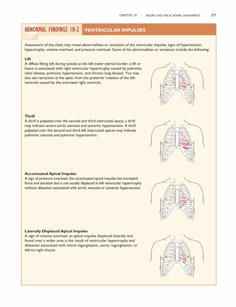
ABNORMAL FINDINGS 18-2 VENTRICULAR IMPULSES
Assessment of the chest may reveal abnormalities or variations of the ventricular impulse, signs of hypertension,hypertrophy, volume overload, and pressure overload. Some of the abnormalities or variations include the following:
LiftA diffuse lifting left during systole at the left lower sternal border, a lift orheave is associated with right ventricular hypertrophy caused by pulmonicvalve disease, pulmonic hypertension, and chronic lung disease. You mayalso see retraction at the apex, from the posterior rotation of the leftventricle caused by the oversized right ventricle.
ThrillA thrill is palpated over the second and third intercostal space; a thrillmay indicate severe aortic stenosis and systemic hypertension. A thrillpalpated over the second and third left intercostal spaces may indicatepulmonic stenosis and pulmonic hypertension.
Accentuated Apical ImpulseA sign of pressure overload, the accentuated apical impulse has increasedforce and duration but is not usually displaced in left ventricular hypertrophywithout dilatation associated with aortic stenosis or systemic hypertension.
Laterally Displaced Apical ImpulseA sign of volume overload, an apical impulse displaced laterally andfound over a wider area is the result of ventricular hypertrophy anddilatation associated with mitral regurgitation, aortic regurgitation, orleft-to-right shunts.

378 U N I T III • NURSING ASSESSMENT OF THE ADULT
ABNORMAL FINDINGS 18-3 ABNORMAL HEART RHYTHMS
Changes in the heart rhythm alter the sounds heardon auscultation.
Premature Atrial or Junctional ContractionsThese beats occur earlier than the next expectedbeat and are followed by a pause. The rhythmresumes with the next beat.
Auscultation Tip: The early beat has an S1 of differentintensity and a diminished S2. S1 and S2 are otherwisesimilar to normal beats.
Premature Ventricular ContractionsThese beats occur earlier than the next expectedbeat and are followed by a pulse. The rhythmresumes with the next beat.
Auscultation Tip: The early beat has an S1 of differentintensity and a diminished S2. Both sounds are usuallysplit.
Sinus ArrhythmiaWith this dysrhythmia, the heart rate speeds upand slows down in a cycle, usually becoming fasterwith inhalation and slower with expiration.
Auscultation Tip: S1 and S2 sounds are usually normal.The S1 may vary with the heart rate.
Atrial Fibrillation and Atrial Flutter withVarying Ventricular Response With this dysrhythmia, ventricular contractionoccurs irregularly. At times, short runs of theirregular rhythm may appear regularly.
Auscultation Tip: S1 varies in intensity.

C H A P T E R 1 8 • HEART AND NECK VESSEL ASSESSMENT 379
ABNORMAL FINDINGS 18-4 EXTRA HEART SOUNDS
Additional heart sounds can be classified by their timing in the cardiac cycle. The presence of the sound during systoleor diastole helps in its identification. Some sounds extend into both systole and diastole.
Extra Heart Sounds During Systole—ClicksHigh-frequency sounds heard just after S1 (ejectionclicks) are produced by a functioning but diseasedvalve. clicks can occur in early or mid-to-late systoleand are best heard through the diaphragm of thestethoscope.
Aortic Ejection ClickHeard during early systole at the second right inter-costal space and apex, the aortic ejection clickoccurs with the opening of the aortic valve and doesnot change with respiration.
Pulmonic Ejection ClickBest heard at the second left intercostal space duringearly systole, the pulmonic ejection click oftenbecomes softer with inspiration.
Midsystolic ClickHeard in middle or late systole, a midsystolic clickcan be heard over the mitral or apical area and is theresult of mitral valve leaflet prolapse during left ven-tricular emptying. A late systolic murmur typicallyfollows, indicating mild mitral regurgitation.
S
Right Left
ES S1
j21
SS S1 12Ej
Right Left
S C SS1
1 21
Right Left
continued
380 U N I T III • NURSING ASSESSMENT OF THE ADULT
ABNORMAL FINDINGS 18-4 EXTRA HEART SOUNDS Continued
Extra Heart Sounds During DiastoleOpening SnapOccurring in early diastole, an opening snap (OS) isheard with the opening of a stenotic or stiff mitralvalve. Heard throughout the whole precordium, itdoes not vary with respirations. Often mistaken for asplit S2 or an S3, the opening snap occurs earlier indiastole and has a higher pitch than an S3.
S3 (Third Heart Sound)Also called a ventricular gallop, the S3 has a lowfrequency and is heard best using the bell of thestethoscope at the apical area or lower right ven-tricular area of the chest with the patient in the leftlateral position. The sound is often accentuatedduring inspiration and has the rhythm of the word“Ken-tuc-ky.” S3 is the result of vibrations causedby the blood hitting the ventricular wall duringrapid ventricular filling.
The S3 can be a normal finding in young children,people with a high cardiac output, and in the thirdtrimester of pregnancy. It is rarely normal in peopleolder than age 40 years and is usually associated withdecreased myocardial contractility, myocardial failure,congestive heart failure, and volume overload of theventricle from valvular disease.
S4 (Fourth Heart Sound)Also called an atrial gallop, S4 is a low-frequencysound occuring at the end of diastole when the atriacontract. It is caused by vibrations from blood flowingrapidly into the ventricles after atrial contraction. S4
has the rhythm of the word “Ten-nes-see” and mayincrease during inspiration. It is best heard with thebell of the stethoscope over the apical area with thepatient in a supine or left lateral position and is neverheard in the absence of atrial contraction.
The S4 can be a normal sound in trained athletesand some older patients, especially after exercise.However, it is usually an abnormal finding and isassociated with coronary artery disease, hypertension,aortic and pulmonic stenosis, and acute myocardialinfarction.
Summation GallopThe simultaneous occurrence of S3 and S4 is called asummation gallop. It is brought about by rapid heartrates in which diastolic filling time is shortened, mov-ing S3 and S4 closer together, resulting in one pro-longed sound. Summation gallop is associated withsevere congestive heart disease.
S S OS S1 2 1
continued
S S S S11 2 3
S S S S12 41
S SSS S
123 4
1
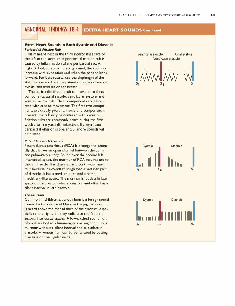
C H A P T E R 1 8 • HEART AND NECK VESSEL ASSESSMENT 381
ABNORMAL FINDINGS 18-4 EXTRA HEART SOUNDS Continued
Extra Heart Sounds in Both Systole and DiastolePericardial Friction RubUsually heard best in the third intercostal space tothe left of the sternum, a pericardial friction rub iscaused by inflammation of the pericardial sac. Ahigh-pitched, scratchy, scraping sound, the rub mayincrease with exhalation and when the patient leansforward. For best results, use the diaphragm of thestethoscope and have the patient sit up, lean forward,exhale, and hold his or her breath.
The pericardial friction rub can have up to threecomponents: atrial systole, ventricular systole, andventricular diastole. These components are associ-ated with cardiac movement. The first two compo-nents are usually present. If only one component ispresent, the rub may be confused with a murmur.Friction rubs are commonly heard during the firstweek after a myocardial infarction. If a significantpericardial effusion is present, S1 and S2 sounds willbe distant.
Patent Ductus ArteriosusPatent ductus arteriosus (PDA) is a congenital anom-ally that leaves an open channel between the aortaand pulmonary artery. Found over the second leftintercostal space, the murmur of PDA may radiate tothe left clavicle. It is classified as a continuous mur-mur because it extends through sytole and into partof diastole. It has a medium pitch and a harsh,machinery-like sound. The murmur is loudest in latesystole, obscures S2, fades in diastole, and often has asilent interval in late diastole.
Venous HumCommon in children, a venous hum is a benign soundcaused by turbulence of blood in the jugular veins. Itis heard above the medial third of the clavicles, espe-cially on the right, and may radiate to the first andsecond intercostal spaces. A low-pitched sound, it isoften described as a humming or roaring continuousmurmur without a silent interval and is loudest indiastole. A venous hum can be obliterated by puttingpressure on the jugular veins.
Ventricular systoleVentricular diastole
Atrial systole
S S S1 12
Systole Diastole
S S S11 2
S S S
Systole Diastole
1 2 1

382 U N I T III • NURSING ASSESSMENT OF THE ADULT
ABNORMAL FINDINGS 18-5 HEART MURMURS
Heart murmurs are typically characterized by turbulent blood flow, which creates a swooshing or blowing soundover the precordium. When listening to the heart, be alert for this turbulance and keep the characteristics of heartmurmurs in mind.
CharacteristicsHeart murmurs are assessed according to various characteristics, which include timing, intensity, pitch, quality, shapeor pattern, location, transmission, and ventilation and position.
TimingA murmur can occur during systole or diastole. In addition to determining when it occurs, it is important to determinewhere it occurs, because a systolic murmur can be present in a healthy heart whereas a diastolic murmur always indicatesheart disease. Systolic murmurs can be divided into three categories: midsystolic, pansystolic, and late systolic. Diastolicmurmurs can be divided into three categories: early diastolic, mid-diastolic, and late diastolic.
IntensitySix grades describe the intensity of a murmur.
Grade 1: Very faint, heard only after the listener has “tuned in”; may not be heard in all positionsGrade 2: Quiet but heard immediately on placing the stethoscope on the chestGrade 3: Moderately loudGrade 4: Loud*Grade 5: Very loud, may be heard with a stethoscope partly off the chest*Grade 6: May be heard with the stethoscope entirely off the chest*
PitchMurmurs can assume a high, medium, or low pitch.
QualityThe sound murmurs make has been described as blowing, rushing, roaring, rumbling, harsh, or musical.
Shape or PatternThe shape of a murmur is determined by its intensity frombeginning to end. There are four different categories ofshape: crescendo (growing louder), decrescendo (growingsofter), crescendo–decrescendo (growing louder and thengrowing softer), and plateau (staying the same throughout).
LocationDetermine where you can best hear the murmur; this isthe point where the murmur originates. Try to be as exactas possible in describing its location. Use the heart land-marks in your description (e.g., the second intercostalspace at the left sternal border).
TransmissionThe murmur may be felt in areas other than the point of origination. If you determine where the murmur transmits,you can determine the direction of blood flow and the intensity of the murmur.
Ventilation and PositionDetermine if the murmur is affected by inspiration, expiration, or a change in body position.
Midsystolic MurmursThe most common type of heart murmurs, midsystolic murmurs occur during ventricular ejection and can be innocent,physiologic, or pathologic. They have a crescendo-decrescendo shape and usually peak near midsystole and stop before S2.
S1 S2 S1 S2
S1 S2
S1 S2
S1 S2
continued

C H A P T E R 1 8 • HEART AND NECK VESSEL ASSESSMENT 383
ABNORMAL FINDINGS 18-5 HEART MURMURS Continued
Innocent MurmurNot associated with any physical abnormality, innocentmurmurs occur when the ejection of blood into the aortais turbulent. Very common in children and young adults,they may also be heard in older people with no evidence ofcardiovascular disease. A patient may have an innocentmurmur and another kind of murmur.
Location: Second to fourth left intercostal spaces between the left sternal border and the apexRadiation: Little radiationIntensity: Grade 1 to 2Pitch: MediumQuality: VariablePosition: Usually disappear when the patient sits
Physiologic MurmurCaused by a temporary increase in blood flow, a physio-logic murmur can occur with anemia, pregnancy, fever, andhyperthyroidism.
Location: Second to fourth left intercostal spaces betweenthe left sternal border and the apexRadiation: Little radiationIntensity: Grade 1 to 2Pitch: MediumQuality: Harsh
Murmur of Pulmonic StenosisA pathologic murmur, the murmur of pulmonic stenosisoccurs from impeded flow across the pulmonic valve andincreased right ventricular afterload. Often occuring as acongenital anomaly, the murmur is commonly found in chil-dren. Pathologic changes in flow across the valve, as inatrial septal defect, may also mimic this condition.
With severe pulmonic stenosis, the S2 is widely split andP2 is diminished. An early pulmonic ejection sound is alsocommon. A right-sided S4 may also be present, and the rightventricular impulse is often stronger and may be prolonged.
Location: Second and third intercostal spacesRadiation: Toward the left shoulder and neckIntensity: Soft to loud (may be associated with a thrill if loud)Pitch: MediumQuality: HarshPosition: Loudest during inspiration
Murmur of Aortic StenosisThe murmur of aortic stenosis occurs when stenosis of theaortic valve impedes blood flow across the valve and increasesleft ventricular afterload. Aortic stenosis may result from a con-genital anomaly, rheumatic disease, or a degenerative process.Conditions that may mimic this murmur include aortic sclero-sis, a bicuspid aortic valve, a dilated aorta, or any condition thatmimics the flow across the valve, such as aortic regurgitation. continued
S1 S2 S1
S1 S2 S1
S1 Ej P2
S2
A2 S1
S1 S2 S1
May be diminished

384 U N I T III • NURSING ASSESSMENT OF THE ADULT
ABNORMAL FINDINGS 18-5 HEART MURMURS Continued
If valvular disease is severe, A2 may be delayed, resulting in an unsplit S2 or a paradoxical split S2. An S4 may occur asa result of decreased left ventricular compliance. An aortic ejection sound, if present, suggests a congenital cause.
Location: Right second intercostal spaceRadiation: May radiate to the neck and down the left sternal border to the apexIntensity: Usually loud, with a thrillPitch: MediumQuality: Harsh, may be musical at the apexPosition: Heard best with the patient sitting and leaning forward, loudest during expiration
Murmur of Hypertrophic CardiomyopathyCaused by unusually rapid ejection of blood from the leftventricle during systole, the murmur of cardiac hypertro-phy results from massive hypertrophy of the ventricularmuscle. There may be a coexisting obstruction to bloodflow. If there is an accompanying distortion of the mitralvalve, mitral regurgitation may result. The patient may alsohave an S3 and an S4. There may be a sustained apicalimpulse with two palpable components.
Location: Third and fourth left intercostal space, decreases with squatting, increases with straining downIntensity: VariablePitch: MediumQuality: Harsh
Pansystolic MurmursOccuring when blood flows from a chamber with high pressure to a chamber of low pressure through an orifice thatshould be closed, pansystolic murmurs are pathologic. Also called holosystolic murmur, these murmurs begin with S1
and continue through systole to S2.
Murmur of Mitral RegurgitationOccuring when the mitral valve fails to close fully in systole,the murmur of mitral regurgitation is the result of bloodflowing from the left ventricle back into the left atrium.Volume overload occurs in the left ventricle, causingdilatation and hypertrophy.
The S1 sound is often decreased, and the apical impulseis stronger and may be prolonged. Left ventricular volumeoverload should be suspected if an apical S3 is heard.
Location: ApexRadiation: To the left axilla, less often to the left sternal borderIntensity: Soft to loud, an apical thrill is associated with loud murmursPitch: Medium to highQuality: BlowingPosition: Heard best with patient in the left lateral decubitus position, does not become louder with inspiration
Murmur of Tricuspid RegurgitationBlood flowing from the right ventricle back into the rightatrium over a tricuspid valve that has not fully closed causesthe murmur of tricuspid regurgitation. Right ventricularfailure with dilatation is the most common cause andusually results from pulmonary hypertension or left ventricular failure.
continued
S1 S2 S1
S1 S2 S3 S1
Diminished

C H A P T E R 1 8 • HEART AND NECK VESSEL ASSESSMENT 385
ABNORMAL FINDINGS 18-5 HEART MURMURS Continued
With this murmur, the right ventricular impulse is stronger and may be prolonged. There may be an S3 along thelower left sternal border and the jugular venous pressure is often elevated with visible v waves.
Location: Lower left sternal borderRadiation: To the right of the sternum, to the xiphoid area, and sometimes to the midclavicular line; there is no radia-tion to the axillaIntensity: VariablePitch: Medium to highQuality: BlowingPosition: May increase slightly with inspiration
Ventricular Septal DefectA congenital abnormality in which blood flows from theleft ventricle into the right ventricle through a hole in theseptum, a ventricular septal defect causes a loud murmurthat obscures the A2 sound. Other findings vary depend-ing on the severity of the defect and any associatedlesions.
Location: Third, fourth, and fifth left intercostal spaceRadiation: Often wideIntensity: Very loud, with a thrillPitch: HighQuality: HarshPosition: Increase with exercise
Diastolic MurmursUsually indicative of heart disease, diastolic murmurs occur in two types. Early decrescendo diastolic murmurs indicateflow through an incompetent semilunar valve, commonly the aortic valve. Rumbling diastolic murmurs in mid- or latediastole indicate valve stenosis, usually of the mitral valve.
Aortic RegurgitationOccuring when the leaflets of the aortic valve fail to closecompletely, the murmur of aortic regurgitation is theresult of blood flowing from the aorta back into the leftventricle. This results in left ventricular volume overload.An ejection sound also may be present. Severe regurgita-tion should be suspected if an S3 or S4 is also present. Theapical impulse becomes displaced downward and laterallywith a widened diameter and increased duration. As thepulse pressure increases, the arterial pulses are often largeand bounding.
Location: Second to fourth left intercostal spaceRadiation: May radiate to the apex or left sternal borderIntensity: Grade 1 to 3Pitch: HighQuality: Blowing, sometime mistaken for breath soundsPosition: Heard best with the patient sitting, leaning forward. Have the patient exhale and then hold his or her breath.
continued
S1 S2 S1
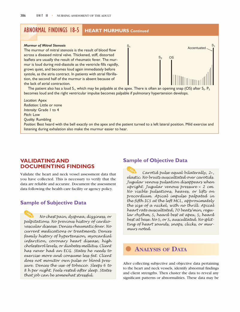
386 U N I T III • NURSING ASSESSMENT OF THE ADULT
ABNORMAL FINDINGS 18-5 HEART MURMURS Continued
Murmur of Mitral StenosisThe murmur of mitral stenosis is the result of blood flowacross a diseased mitral valve. Thickened, stiff, distortedleaflets are usually the result of rheumatic fever. The mur-mur is loud during mid-diastole as the ventricle fills rapidly,grows quiet, and becomes loud again immediately beforesystole, as the atria contract. In patients with atrial fibrilla-tion, the second half of the murmur is absent because ofthe lack of atrial contraction.
The patient also has a loud S1, which may be palpable at the apex. There is often an opening snap (OS) after S2. P2
becomes loud and the right ventricular impulse becomes palpable if pulmonary hypertension develops.
Location: ApexRadiation: Little or noneIntensity: Grade 1 to 4Pitch: LowQuality: RumblingPosition: Best heard with the bell exactly on the apex and the patient turned to a left lateral position. Mild exercise andlistening during exhalation also make the murmur easier to hear.
Accentuated
S2 OS
S1S1
VALIDATING AND DOCUMENTING FINDINGSValidate the heart and neck vessel assessment data thatyou have collected. This is necessary to verify that thedata are reliable and accurate. Document the assessmentdata following the health care facility or agency policy.
Sample of Subjective Data
No chest pain, dyspnea, dizziness, orpalpitations. No previous history of cardio-vascular disease. Denies rheumatic fever. Nocurrent medications or treatments. Deniesfamily history of hypertension, myocardialinfarction, coronary heart disease, highcholesterol levels, or diabetes mellitus. Clienthas never had an ECG. States he needs toexercise more and consume less fat. Clientdoes not monitor own pulse or blood pres-sure. Denies the use of tobacco. Sleeps 6 to 8 h per night. Feels rested after sleep. Statesthat job can be somewhat stressful.
Sample of Objective Data
Carotid pulse equal bilaterally, 2+,elastic. No bruits auscultated over carotids.Jugular venous pulsation disappears whenupright. Jugular venous pressure × 2 cm.No visible pulsations, heaves, or lifts on precordium. Apical impulse palpated inthe fifth ICS at the left MCL, approximatelythe size of a nickel, with no thrill. Apicalheart rate auscultated, 70 beats/min, regu-lar rhythm, S1 heard best at apex, S2 heardbest at base. No S3 or S4 auscultated. No split-ting of heart sounds, snaps, clicks, or mur-murs noted.
l ANALYSIS OF DATA
After collecting subjective and objective data pertainingto the heart and neck vessels, identify abnormal findingsand client strengths. Then cluster the data to reveal anysignificant patterns or abnormalities. These data may be

C H A P T E R 1 8 • HEART AND NECK VESSEL ASSESSMENT 387
used to make clinical judgments about the status of theclient’s heart and neck vessels.
DIAGNOSTIC REASONING:POSSIBLE CONCLUSIONS
Selected Nursing DiagnosesThe following is a listing of selected nursing diagnosesthat you may identify when analyzing data for this part ofthe assessment.
Wellness Diagnoses
• Readiness for enhanced cardiac output• Health-Seeking Behavior: Desired information on
exercise and low-fat diet
Risk Diagnoses
• Risk for Sexual Dysfunction related to misinfor-mation or lack of knowledge regarding sexualactivity and heart disease
• Risk for Ineffective Denial related to smokingand obesity
Actual Diagnoses
• Fatigue related to decreased cardiac output• Activity Intolerance related to compromised
oxygen transport secondary to heart failure• Acute Pain: Cardiac related to an inequality
between oxygen supply and demand• Anxiety• Ineffective Tissue Perfusion: Cardiac related to
impaired circulation
Selective Collaborative ProblemsAfter grouping the data, you may see various collaborativeproblems emerge. Remember that collaborative problemsdiffer from nursing diagnoses in that they cannot be pre-vented by nursing interventions. However, these physio-logic complications of medical conditions can be detectedand monitored by the nurse. In addition, the nurse can usephysician- and nurse-prescribed interventions to minimizethe complications of these problems. The nurse may alsohave to refer the client in such situations for further treat-ment of the problem. Following is a list of collaborativeproblems that may be identified when assessing the heart
D I A G N O S T I C R E A S O N I N G : C A S E S T U D Y
The case study demonstrates how to analyze thoracic and lung assessment
data for a specific client. The critical thinking exercises included in the study
guide/lab manual and interactive product that complement this text also offer
opportunities to analyze assessment data.
Malcolm Winchester is being admitted to the coro-nary care unit (CCU) with a diagnosis of hypertension,angina, R/O MI (myocardial infarction). He is a tall,slender black man who looks younger than his statedage of 45. He is in no acute distress. Mr. Winchestersays, “I don’t know why they brought me here—I guess my wife panicked and called 911. I have thesepains all of the time, but my doc said they were frommy high blood pressure. I don’t hurt now.”
His wife arrives, looking pale and anxious. “Idon’t know what to do with him. I work so hard tokeep him healthy, but he goes out to that fast foodplace and eats burgers and fries. I’m so tired of deal-ing with him when he won’t help himself.” Mr.Winchester grins and says, “I just got to have my junk
food! That low-fat, low-salt diet my doctor put me onis impossible.”
Physical assessment reveals BP 210/110 right armreclining and 200/108 left arm reclining, pulse 88 reg-ular and strong, respirations 16 regular and moder-ately shallow, temperature 36.5°C (97.7°F). His apicalbeat is also 88 and strong; heart sounds: S1 and S2 withno murmurs and clicks, but an S4 is noted. Evaluationof the thorax reveals no heaves or visible pulsation.Neck veins are flat at >45 degrees and no carotidbruits noted. Skin is warm and dry, dark brown withpink nail beds, palms, and oral mucous membranes.Pedal pulses strong; 1 + ankle edema present.
The following concept map illustrates the diag-nostic reasoning process.

388 U N I T III • NURSING ASSESSMENT OF THE ADULT
Accept diagnosis because itmeets defining characteristicsand is validated by client
Confirm, because diagnosismeets defining characteristics
Accept this diagnosis be-cause it is a risk diagnosis
Rule out diagnosis because itdoes not meet the majordefining characteristic. Moredata are needed
Data are insufficient toaccept this diagnosis, although it iscertainly a risk diagnosis given thewife’s verbalization of frustration
Major: Reports unhealthfulpractices (e.g., high-fat, high-salt diet)Minor: None, except possiblycompulsive behavior regardingdiet (“has to have my junkfood”)
Major: Reports pain “all thetime” but not at this timeMinor: None
Major: NoneMinor: Possibly impliedapprehension about thefuture for care receiver'shealth. Also possiblydepressed feelings andanger.
Major: None specificMinor: None specific
• BP 210/110 and 200/108• S4 heart sound
• Confirms eating junk food• Finds low-fat, low-salt diet “impossible”
• “I have pains all the time”• Denies pain at this time
• Wife: “Don't know what to do with him … tired of dealing with him when he won't help himself”• Called 911 when he had pain
Dangerously high bloodpressure with concurrentatrial gallop seen withhypertension. Refer tophysician
Not experiencing paincurrently but has historyof pain related tohypertension
Chooses not to followspecial diet. Unable totolerate special diet
Wife, who perceives herself as a caregiveris frustrated and anxious about client’s illhealth and noncompliance—possiblyburned out
Ineffective Health Main-tenance r/t choice not tofollow prescribed dietarytreatment of hypertension
Ineffective Therapeutic RegimenManagement r/t intolerance oftherapeutic diet and knowledgedeficit of alternative strategiesfor managing hypertension
Risk for Acute Pain: Acutepain (angina) r/t knowledgedeficit of managementstrategies
Ineffective Family Copingr/t strain on family fromclient's illness
• Ineffective Health Maintenance r/t choice not to follow dietary treatment of hypertension• Ineffective Therapeutic Regimen Management r/t intolerance of therapeutic diet and knowledge deficit of alternative strategies for managing hypertension• Risk for Acute Pain: acute pain (angina) r/t knowledge deficit of management strategies
• PC: Cerebrovascular accident• PC: Retinal hemorrhage• PC: Myocardial infarction• PC: Heart failure• PC: Renal failure
Caregiver Role Strain r/tfrustration with client's non-compliant behavior and possibleanxiety over seriousness ofsymptoms
Major: Verbalizes dislike ofand difficulty with integrationof prescribed regimen (diet)for treatment of illnessMinor: Verbalizes he did notinclude treatment in dailyroutine
• “I don't know why they brought me here.”• “I have these pains all of the time”• Denies pain at this time• Has to have junk food—low fat, low-salt diet “impossible”• Wife: “Don't know what to do with him”• Wife: “He eats hamburgers and french fries and forgets to take medication”• Wife: “Tired of dealing with him when he won't
help himself”
• Admitted with angina, R/O MI• BP 210/110 right arm reclining and 200/108 left arm reclining• S4 heart sound• 1 + pedal edema

and neck vessels. These problems are worded as PotentialComplications (or PC) followed by the problem.
• PC: Decreased cardiac output• PC: Dysrhythmias• PC: Hypertension• PC: Congestive heart failure• PC: Angina• PC: Cerebrovascular accident• PC: Cerebral hemorrhage• PC: Renal failure
Medical ProblemsOnce the data are grouped, certain signs and symptoms maybecome evident and may require medical diagnosis andtreatment. Referral to a primary care provider is necessary.
References and Selected Readings
Appel, L. J., Moore, T. J., et al. (1997). A clinical trial of the effects ofdietary patterns on blood pressure. New England Journal ofMedicine, 336, 1–17.
Archbold, R. A., Barakat, K., Magee, P., & Curzen, N. (2001). Screeningfor carotid artery disease before cardiac surgery: Is current clinicalpractice evidence based? Clinical Cardiology, 24(1), 26–32.
Barett, et al. (2004). Mastering cardiac murmurs: The power of repeti-tion. Chest, 126, 470–475.
Carabello, B. A., & Crawford, J. (1997). Valvular heart disease. NewEngland Journal of Medicine, 337, 32.
Chizner, M. (2003). The diagnosis of heart disease. Disease-a month,48(1), 7–98.
Davidson, L. J., Bennett, S. E., Hamera, E. K., & Raines, B. K. (2004).What constitutes advanced assessment? Journal of NursingEducation, 43(9), 421–425.
Daviglus, M. L., Stamler, J., Pirzada, A., Yan, L. L., Garside, D. B., Liu,K., Wade-Dyer, A. R., Lloyd-Jones, D. M., & Greenland, P. (2004).Favorable cardiac risk profile in young women and low risk of car-diovascular and all-cause mortality. Journal of the AmericanMedical Association, 292(13), 1588–1592.
Dulak, S. B. (2004). Hands-on help: Assessing heart sounds. RN, 67(8),241–244.
Fabius, D. B. (2000). Solving the mystery of heart murmurs. Nursing2000, 30(7), 39–44.
Gillett, M., Davis, W. A., Jackson, D., Bruce, D. G., Davis, T. M., &Fremantle, D. (2003). Prospective evaluation of carotid bruit as apredictor of first sign of type 2 diabetes: The Fremantle DiabetesStudy. Stroke, 34(9), 2145–2151.
Jolobe, O. M. P. (2001). Systolic murmurs and aortic stenosis.Quarterly Journal of Medicine, 94(1), 49.
Kirton, C. A. (1997). Assessing a heart murmur. Nursing 1997, 27(9), 51.Kirton, C. A. (2000). Physical assessment. Assessing normal heart
sounds. Nursing 2000, 30(2), 52–54.
Loveridge, M. (2003). Acquiring percussion and auscultation skillsthrough experiential learning. Emergency Nurse, 11(6), 31–37.
Ludwig, L. M. (1998). Cardiovascular assessment for home healthcarenurses. Part 1. Assessing blood pressure and cardiac function. HomeHealthcare Nurse, 16(8), 547–554.
Marshall, K. G. (1998). More techniques of auscultation: General prin-ciples of murmurs. Patient Care, 9(2), S1–S5.
Mehta, M. (2003). Assessing cardiovascular status. Nursing 2003,33(1), 56–58.
Nirav, J., Mehta, M., & Ijaz, A. (2003). Third heart sound: Genesis andclinical importance. International Journal of Cardiology, 97(2),183–186.
Wasserman, A. (2000). Chest pain. Is it life-threatening—or benign?Consultant, 40(7), 1204–1208.
Welsby, P. D., et al. (2003). The stethoscope: Some preliminary investi-gations. Postgraduate Medical Journal, 79, 695–698.
Risk Factors—Coronary Heart Disease
American Heart Association (AHA). (2001). Coronary heart disease.Available online. Author.
———. (1999). Risk factor assessment for heart attack or stroke.Available online. Author.
Azevedo, A., Ramos, E., vonHafe, P., & Barros, H. (1999). Upper-bodyadiposity and risk of myocardial infarction. Journal ofCardiovascular Risk, 6(5), 321–325.
Berenson, G., & Pickoff, A. (1995). Preventive cardiology and itspotential influence on the early natural history of adult heart dis-eases: The Bogalusa Heart Study and the Heart Smart Program.American Journal of the Medical Sciences, 310(Suppl. 1),S1333–S138.
Eichholzer, M., Luthy, J., Gutzwiller, F., & Stahelin, H. (2001). Therole of folate, antioxidant vitamins and other constituents in fruitand vegetables in the prevention of cardiovascular disease: Theepidemiological evidence. International Journal for Vitaminand Nutrition Research, 71(1), 5–17.
Gillum, R. (1996). Epidemiology of hypertension in African Americanwomen. American Heart Journal, 131, 385–395.
Hallmen, T., Burell, G., Setterlind, S., Oden, A., & Lisspers, J. (2001).Psychosocial risk factors for coronary heart disease, their impor-tance compared with other risk factors and gender differences insensitivity. Journal of Cardiovascular Risk, 8(1), 39–49.
Leeson, C. P., Kattenhorn, M., Morley, R., Lucas, A, & Deanfield, J. (2001).Impact of low birth weight and cardiovascular risk factors on endothe-lial function in early adult life. Circulation, 103(9), 1264–1268.
Libby, P., Schoenbeck, V., Mach, F., Selwyn, A., & Ganz, P. (1998).Current concepts in cardiovascular pathology: The role of LDL cho-lesterol in plaque rupture and stabilization. American Journal ofMedicine, 104(24), 145–185.
Misra, A. (2000). Risk factors for atherosclerosis in young individuals.Journal of Cardiovascular Risk, 7(3), 215–219.
Overfield, T. (1995). Biological variation in health and illness: Race,age, and sex differences (2nd ed.). Boca Raton, FL: CRC Press.
Rifai, N. M., & Ridker, P. M. (2001). High sensitivity C-reactive protein:A novel and promising marker of coronary heart disease. ClinicalChemistry, 47(3), 403–411.
W.H.O. (2004). Types of cardiovascular disease. Retrieved November,2004 from http://www.who.int/cardiovasular_diseases/resources/atlas/
C H A P T E R 1 8 • HEART AND NECK VESSEL ASSESSMENT 389