Retinal vessel segmentation on SLO image
9
Retinal vessel segmentation on SLO image Juan Xu [Student Member, IEEE] 1 , Hiroshi Ishikawa 1,2 , Gadi Wollstein 1 , and Joel S. Schuman 1,2 1 UPMC Eye Center, Eye and Ear Institute, Ophthalmology and Visual Science Research Center, Dept. of Ophthalmology, University of Pittsburgh School of Medicine, Pittsburgh, PA 2 Dept. of Bioengineering, Swanson School of Engineering, University of Pittsburgh, Pittsburgh, PA Abstract A scanning laser ophthalmoscopy (SLO) image, taken from optical coherence tomography (OCT), usually has lower global/local contrast and more noise compared to the traditional retinal photograph, which makes the vessel segmentation challenging work. A hybrid algorithm is proposed to efficiently solve these problems by fusing several designed methods, taking the advantages of each method and reducing the error measurements. The algorithm has several steps consisting of image preprocessing, thresholding probe and weighted fusing. Four different methods are first designed to transform the SLO image into feature response images by taking different combinations of matched filter, contrast enhancement and mathematical morphology operators. A thresholding probe algorithm is then applied on those response images to obtain four vessel maps. Weighted majority opinion is used to fuse these vessel maps and generate a final vessel map. The experimental results showed that the proposed hybrid algorithm could successfully segment the blood vessels on SLO images, by detecting the major and small vessels and suppressing the noises. The algorithm showed substantial potential in various clinical applications. The use of this method can be also extended to medical image registration based on blood vessel location. Keywords Vessel segmentation; hybrid algorithm; SLO image; retina I. INTRODUCTION The appearance of a blood vessel, i.e., diameter, color and tortuosity is an important indicator, often used for detection of eye diseases, such as diabetic retinopathy, a leading cause of blindness worldwide. The blood vessels can also be used as a landmark to register the retinal images of the same patient taken at different visits, or even taken with different ophthalmic devices. Spectral domain optical coherence tomography (SD-OCT), a new generation of ophthalmic imaging device, detects the light back-scattered from various layers of retina to generate a two-dimensional, cross-sectional image of ocular tissue and is capable of obtaining three dimensional (3D) images of the scanned region. Although the actual scanning time is less ©2008 IEEE. J. S. Schuman, corresponding author, UPMC Eye Center, Department of Ophthalmology, University of Pittsburgh, 203 Lothrop Street, Eye and Ear Institute, Pittsburgh, PA 15213, 412-647-2205, [email protected]. NIH Public Access Author Manuscript Conf Proc IEEE Eng Med Biol Soc. Author manuscript; available in PMC 2010 July 21. Published in final edited form as: Conf Proc IEEE Eng Med Biol Soc. 2008 ; 2008: 2258–2261. doi:10.1109/IEMBS.2008.4649646. NIH-PA Author Manuscript NIH-PA Author Manuscript NIH-PA Author Manuscript
Transcript of Retinal vessel segmentation on SLO image
Retinal vessel segmentation on SLO image
Juan Xu [Student Member, IEEE]1, Hiroshi Ishikawa1,2, Gadi Wollstein1, and Joel S.Schuman1,21UPMC Eye Center, Eye and Ear Institute, Ophthalmology and Visual Science Research Center,Dept. of Ophthalmology, University of Pittsburgh School of Medicine, Pittsburgh, PA2Dept. of Bioengineering, Swanson School of Engineering, University of Pittsburgh, Pittsburgh,PA
AbstractA scanning laser ophthalmoscopy (SLO) image, taken from optical coherence tomography (OCT),usually has lower global/local contrast and more noise compared to the traditional retinalphotograph, which makes the vessel segmentation challenging work. A hybrid algorithm isproposed to efficiently solve these problems by fusing several designed methods, taking theadvantages of each method and reducing the error measurements. The algorithm has several stepsconsisting of image preprocessing, thresholding probe and weighted fusing. Four differentmethods are first designed to transform the SLO image into feature response images by takingdifferent combinations of matched filter, contrast enhancement and mathematical morphologyoperators. A thresholding probe algorithm is then applied on those response images to obtain fourvessel maps. Weighted majority opinion is used to fuse these vessel maps and generate a finalvessel map. The experimental results showed that the proposed hybrid algorithm couldsuccessfully segment the blood vessels on SLO images, by detecting the major and small vesselsand suppressing the noises. The algorithm showed substantial potential in various clinicalapplications. The use of this method can be also extended to medical image registration based onblood vessel location.
KeywordsVessel segmentation; hybrid algorithm; SLO image; retina
I. INTRODUCTIONThe appearance of a blood vessel, i.e., diameter, color and tortuosity is an importantindicator, often used for detection of eye diseases, such as diabetic retinopathy, a leadingcause of blindness worldwide. The blood vessels can also be used as a landmark to registerthe retinal images of the same patient taken at different visits, or even taken with differentophthalmic devices.
Spectral domain optical coherence tomography (SD-OCT), a new generation of ophthalmicimaging device, detects the light back-scattered from various layers of retina to generate atwo-dimensional, cross-sectional image of ocular tissue and is capable of obtaining threedimensional (3D) images of the scanned region. Although the actual scanning time is less
©2008 IEEE.J. S. Schuman, corresponding author, UPMC Eye Center, Department of Ophthalmology, University of Pittsburgh, 203 LothropStreet, Eye and Ear Institute, Pittsburgh, PA 15213, 412-647-2205, [email protected].
NIH Public AccessAuthor ManuscriptConf Proc IEEE Eng Med Biol Soc. Author manuscript; available in PMC 2010 July 21.
Published in final edited form as:Conf Proc IEEE Eng Med Biol Soc. 2008 ; 2008: 2258–2261. doi:10.1109/IEMBS.2008.4649646.
NIH
-PA Author Manuscript
NIH
-PA Author Manuscript
NIH
-PA Author Manuscript

than 2 seconds, subtle eye motions during the scanning cause enough distortion in theresulting images. This is clinically problematic since longitudinal observation of the samelocation is essential. Scanning laser ophthalmoscopy (SLO) images taken from an SD-OCTdevice can be used as a reference for image registration in order to improve visualization.The SLO image is typically a grey scale image with non-uniform illumination, low global/local contrast and high background noise, which makes vessel segmentation on SLO imagesa challenge.
Niemeijer et al. [1] gave a comparative study of retinal vessel segmentation algorithms onconventional fundus photographs (retinal images). Existing vessel segmentation techniqueson the conventional fundus photographs can be divided into several major categories:matched filter based method [2,3], thresholding based method [4], tracking based method[5], mathematical morphology method [6], and classification based method [7,8]. Thematched filter method is convolving the image with a directional filter designed according tothe vessel profile [3], such as Gaussian [2] and second-order Gaussian filter [3]. Thematched filter can be used as the first step for other segmentation methods. The filterresponse enhances the vessel pattern features, thus improving the performance ofthresholding or tracking process. Hoover and et al. [4] introduced a piecewise thresholdingprobe algorithm on the matched filter response (MFR) image, yielding a 90% true positivedetection rate compared with the manual label ground truth. Staal and et al. [7] described apixel classification method for color retinal image. Feature vectors were first computed foreach pixel and then a kNN-classifier was used to classify the given pixel as either vessel ornon-vessel. It is a supervised learning method, which requires manually labeled images fortraining. Color images provide much more information, which is not available in SLOimages. The algorithms in previous studies did not work perfectly on SLO image due to thelow global/local contrast, non-uniform illumination and noise.
In this paper, a hybrid algorithm is proposed to automatically segment blood vessels on SLOimages, which includes several steps. The original SLO image is first transformed into afeature response image using four methods, which are designed based on differentcombinations of matched filter, contrast enhancement and mathematical morphologyoperators. Threshold probing algorithm [4] is then applied to all the four response images.The final vessel map is generated by taking the weighted majority opinion from the fourvessel segmentation results pixel by pixel, which combines both the original and enhancedimage information to solve the above mentioned problems.
II. METHODOLOGYThe proposed algorithm combines several operations, i.e., matched filter, contrastenhancement, mathematical morphology and thresholding probe. The overall hybrid methodcould be divided into three steps, (1) preprocess the SLO images to generate featureresponse images, (2) apply thresholding probe algorithm on those response images tosegment blood vessels, and (3) generate final vessel map.
Blood vessels on SLO image are difficult to detect by only the single global threshold.Matched filter or morphology operations can efficiently enhance the vessel pattern featuresin order to improve the performance of thresholding method. Adaptive thresholdingperforms well on the images having non-uniform illumination, though the low contrast stillposes a substantial challenge to the algorithm performance. Contrast enhancement canenhance the vessels in the low contrast region, but introduces more noises simultaneously. Asingle process may not solve all the problems of vessel segmentation. The proposed hybridalgorithm takes the advantages of multiple approaches, i.e. enhancing weak vessels and
Xu et al. Page 2
Conf Proc IEEE Eng Med Biol Soc. Author manuscript; available in PMC 2010 July 21.
NIH
-PA Author Manuscript
NIH
-PA Author Manuscript
NIH
-PA Author Manuscript

keeping the original information from low noise image, so that the final vessel map is morestable than each single method.
The methodology section is organized as follows. Sections A to D introduce four differentoperators in the hybrid algorithm; the overall hybrid algorithm is described in section E.
A. Matched filterBlood vessels usually appear darker relative to the background in SLO images due to lowerreflectance compared to the retinal surface [2]. They have small curvatures that may beapproximated by piecewise linear segments [2]. Gang and et al. [3] indicated that thebackground contributed less to the convolution response using second-order Gaussian modelcompared to Gaussian model [2]. Therefore a normalized two dimensional (2D) second-order Gaussian filter template (Fig. 1(a)) is used to approximately represent the patternalong the direction of vessel length, written as (modified based on eq.(10) in [3]):
(1)
where L (the vessel direction) and W are the length and width of filter template respectively,x0 is the center location along the vessel direction, k is the normalized coefficient, and R isthe rotation matrix according to vessel orientation. Directional filters are generated using therotation matrix R with the rotation angle θ. Multiple σ corresponding to different vesselwidths are also applied to generate different filters. All the filters are employed on the SLOimage and the response image takes the maximal value pixel by pixel among thoseresponses according to different σ and θ.
B. Local contrast enhancementLocal enhancement is introduced as follows:
(2)
where f(x,y) is the original image and W is the window size. The intensity values arenormalized to the range of 0 to 255 within the given window. If the window size is enlargedto the image size, it becomes a global enhancement. Weak vessels could be enhanced by thisoperation with consequent increase in the noise level.
C. Mathematical morphologyAccording to Zana and Klein’s study [6], a line structure was selected to generate amorphological filter for vessel segmentation assuming the vessels maintain predominantlylocal linear orientation. Basic morphological operators are erosion, dilation, opening, closingas defined in [6]. The top-hat, bottom-hat, and morphological enhancement operators arewritten as
(3)
Xu et al. Page 3
Conf Proc IEEE Eng Med Biol Soc. Author manuscript; available in PMC 2010 July 21.
NIH
-PA Author Manuscript
NIH
-PA Author Manuscript
NIH
-PA Author Manuscript

Because blood vessels are darker than the background in SLO image, a bottom-hat operatorand enhancement operator are used to generate the feature response images. Morphologicalfilters rotated to different directions are applied on the image. The final filter response is themaximum (measuring pixel by pixel) among those responses of directionally morphologicalfilters with different rotation angles.
D. Thresholding probeThresholding probe [4] is an adaptive thresholding with region growing algorithm for vesselsegmentation. The method can be summarized into a number of steps: (1) Generating a MFRimage by convolving the image with the matched filter, (2) detecting the endpoints of thevessel tree using single global threshold to initialize a probe queue, (3) starting from thepoints in the queue, probe growing with local threshold to generate a vessel tree, (4) re-computing the endpoints of vessel tree to update probe queue, and (5) repeating step 3 and 4until the queue is empty. Optic disc and image borders are masked in this operation (Fig.1(b)).
E. Hybrid algorithmHybrid algorithm includes thresholding probe operator and different preprocessingoperators. Four vessel segmentation methods (written as M1 to M4) are designed by takingdifferent combinations among four introduced operations, as shown in Fig. 2. Byobservation, SLO images usually have low global or local contrast. Therefore the resultedvessel map from method 4 (in which local contrast enhancement is operated on the originalSLO image) is considered having higher contribution to generate the final result (written asM). Let 1 and 0 indicate vessel and non-vessel respectively. Majority opinion is taken pixelby pixel from the four resulted vessel maps to generate the final vessel map, described as
(4)
where method 4 has higher weight than all the other three methods. The M(x,y) is labeled as1 if the weighted summation is larger than 3, otherwise it is labeled as 0. The hybridalgorithm combines different operations so as to enhance the vessel pattern and suppress theeffect of noises.
III. RESULT AND DISCUSSION20 SLO images (668×801 pixels, taken from healthy eyes) acquired at University ofPittsburgh Medical Center Eye Center were tested by the proposed hybrid algorithm ofblood vessel segmentation. All the images were resized to 334×400 pixels in order to reducethe computation time. Fig. 3 illustrates an example of the entire segmentation procedure.Feature response images before thresholding probe operation generated from four designedmethods are shown in Fig. 3(c)–(f). The vessel maps after employing the thresholding probeare illustrated in Fig. 3(g)–(j). The final vessel map was obtained by taking the weightedmajority opinion (Fig. 3(b)), in which the weak vessels were remained and the segmentationerrors from each single method were removed.
Different operations have their own advantages and disadvantages. Thresholding on thefeature response image much improved the performance compared to applying it directly onthe original image. Matched filter and mathematical morphology operations enhanced thevessel pattern features and suppressed the noises. However, it was difficult to obtain a highvessel response with both methods in regions with low contrast as shown in the right regionof Fig. 3(g)(i). Global or local contrast enhancement operation solved the low contrast
Xu et al. Page 4
Conf Proc IEEE Eng Med Biol Soc. Author manuscript; available in PMC 2010 July 21.
NIH
-PA Author Manuscript
NIH
-PA Author Manuscript
NIH
-PA Author Manuscript

problem, but introduced more noise as illustrated in Fig. 3(h)(j). The proposed hybridalgorithm effectively took those advantages and reduced the probability of errormeasurements.
Taking the weighted majority opinion is a simple and fast method to fuse the vessel mapsand generate a final result. Advanced methods, such as machine learning, could be furtherstudied to fuse the vessel maps, which may have a more robust output.
The proposed hybrid algorithm was also applied on the OCT enface images (generated byaveraging 3D OCT data along z axis) and obtained similar results. This indicated that theproposed algorithm is not limited in the SLO images, which can be expanded to multipleapplications.
IV. CONCLUSIONA hybrid algorithm of retinal vessel segmentation has been proposed in this paper, usingmatched filter, mathematical morphology, contrast enhancement and thresholding probe.Weighted majority opinion was taken to generate the final segmentation result. Experimentresults showed that the proposed algorithm is a more efficient way to segment the vessels,suppress the noises and remove error measurements compared to every individual method.The proposed hybrid algorithm showed a substantial potential in various clinicalapplications, such as qualification of blood vessel appearance and medical imageregistration based on blood vessel location.
AcknowledgmentsFinancial support: Supported in part by National Institute of Health contracts R01-EY13178-08, P30-EY08098(Bethesda, MD), The Eye and Ear Foundation (Pittsburgh, PA) and an unrestricted grant from Research to PreventBlindness, Inc. (New York, NY)
REFERENCES1. Niemeijer, M.; Staal, J. Comparative study of retinal vessel segmentation methods on a new publicly
available database; Proc. SPIE Conf. on Medical Imaging; 2004. p. 648-656.2. Chaudhuri S, Chatterjee S, Katz N, Nelson M, Goldbaum M. Detection of blood vessels in retinal
images using two-dimensional matched filters. IEEE Transactions on Medical Imaging 1989Sep.;vol. 8(no. 3):263–269. [PubMed: 18230524]
3. Gang L, Chutatape O, Krishnan SM. Detection and measurement of retinal vessels in fundus imagesusing amplitude modified second-order Gaussian filter. IEEE Transactions on BiomedicalEngineering 2002 Feb.;vol. 49(No. 2):168–172. [PubMed: 12066884]
4. Hoover A, Kouznetsova V, Goldbaum M. Locating blood vessels in retinal images by piecewisethreshold probing of a matched filter response. IEEE Transactions on Medical Imaging 2000;vol.19(No. 3):203–210. [PubMed: 10875704]
5. Can A, Shen H, Turner JN, Tanenbaum HL, Roysam B. Rapid Automated Tracing and FeatureExtraction from Live High-Resolution Retinal Fundus Images Using Direct ExploratoryAlgorithms. IEEE Transactions on Information Technology in Biomedicine 1999 June;vol. 3(No.2):125–138. [PubMed: 10719494]
6. Zana F, Klein J. Segmentation of vessel-like patterns using mathematical morphology and curvatureevaluation. IEEE Transactions on Image Processing 2001;vol. 10(no. 7):1010–1019. [PubMed:18249674]
7. Staal JJ, Abramoff MD, Niemeijer M, Viergever MA, van Ginneken B. Ridge based vesselsegmentation in color images of the retina. IEEE Transactions on Medical Imaging 2004;vol.23:501–509. [PubMed: 15084075]
Xu et al. Page 5
Conf Proc IEEE Eng Med Biol Soc. Author manuscript; available in PMC 2010 July 21.
NIH
-PA Author Manuscript
NIH
-PA Author Manuscript
NIH
-PA Author Manuscript

8. Sinthanayothin C, Boyce JF, Cook HL, Williamson TH. Automated localization of the optic disc,fovea and retinal blood vessels from digital color fundus images. Br. J. Opthalmol 1999 Aug.;vol.83:231–238.
Xu et al. Page 6
Conf Proc IEEE Eng Med Biol Soc. Author manuscript; available in PMC 2010 July 21.
NIH
-PA Author Manuscript
NIH
-PA Author Manuscript
NIH
-PA Author Manuscript
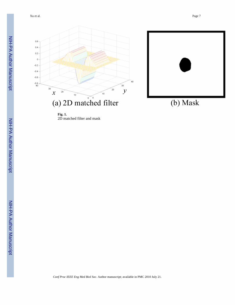
Fig. 1.2D matched filter and mask
Xu et al. Page 7
Conf Proc IEEE Eng Med Biol Soc. Author manuscript; available in PMC 2010 July 21.
NIH
-PA Author Manuscript
NIH
-PA Author Manuscript
NIH
-PA Author Manuscript

Fig. 2.Hybrid method of blood vessel segmentation
Xu et al. Page 8
Conf Proc IEEE Eng Med Biol Soc. Author manuscript; available in PMC 2010 July 21.
NIH
-PA Author Manuscript
NIH
-PA Author Manuscript
NIH
-PA Author Manuscript

Fig. 3.An example of hybrid algorithm of retinal vessel segmentation. (a) SLO image taken fromSD-OCT, (b) Final vessel map generated through weighted majority opinion, (c)–(f) Featureresponse images of method 1 to method 4 before applying thresholding probe operation,(g)–(i) Vessel maps of method 1 to method 4.
Xu et al. Page 9
Conf Proc IEEE Eng Med Biol Soc. Author manuscript; available in PMC 2010 July 21.
NIH
-PA Author Manuscript
NIH
-PA Author Manuscript
NIH
-PA Author Manuscript