
QUARTZ : Quantitative Analysis of Retinal Vessel Topology ...
40
Accepted Manuscript QUARTZ : Quantitative Analysis of Retinal Vessel Topology and size An au- tomated system for quantification of retinal vessels morphology M.M. Fraz, R.A. Welikala, A.R. Rudnicka, C.G. Owen, D.P. Strachan, S.A. Barman PII: S0957-4174(15)00350-4 DOI: http://dx.doi.org/10.1016/j.eswa.2015.05.022 Reference: ESWA 10041 To appear in: Expert Systems with Applications Please cite this article as: Fraz, M.M., Welikala, R.A., Rudnicka, A.R., Owen, C.G., Strachan, D.P., Barman, S.A., QUARTZ : Quantitative Analysis of Retinal Vessel Topology and size An automated system for quantification of retinal vessels morphology, Expert Systems with Applications (2015), doi: http://dx.doi.org/10.1016/j.eswa. 2015.05.022 This is a PDF file of an unedited manuscript that has been accepted for publication. As a service to our customers we are providing this early version of the manuscript. The manuscript will undergo copyediting, typesetting, and review of the resulting proof before it is published in its final form. Please note that during the production process errors may be discovered which could affect the content, and all legal disclaimers that apply to the journal pertain.
-
Upload
khangminh22 -
Category
Documents
-
view
4 -
download
0
Transcript of QUARTZ : Quantitative Analysis of Retinal Vessel Topology ...
Accepted Manuscript
QUARTZ : Quantitative Analysis of Retinal Vessel Topology and size An au-
tomated system for quantification of retinal vessels morphology
M.M. Fraz, R.A. Welikala, A.R. Rudnicka, C.G. Owen, D.P. Strachan, S.A.
Barman
PII: S0957-4174(15)00350-4
DOI: http://dx.doi.org/10.1016/j.eswa.2015.05.022
Reference: ESWA 10041
To appear in: Expert Systems with Applications
Please cite this article as: Fraz, M.M., Welikala, R.A., Rudnicka, A.R., Owen, C.G., Strachan, D.P., Barman, S.A.,
QUARTZ : Quantitative Analysis of Retinal Vessel Topology and size An automated system for quantification of
retinal vessels morphology, Expert Systems with Applications (2015), doi: http://dx.doi.org/10.1016/j.eswa.
2015.05.022
This is a PDF file of an unedited manuscript that has been accepted for publication. As a service to our customers
we are providing this early version of the manuscript. The manuscript will undergo copyediting, typesetting, and
review of the resulting proof before it is published in its final form. Please note that during the production process
errors may be discovered which could affect the content, and all legal disclaimers that apply to the journal pertain.

QUARTZ : Quantitative Analysis of Retinal Vessel Topology and size
An automated system for quantification of retinal vessels morphology
M. M. Fraza*, R.A. Welikalab, A.R. Rudnickac, C.G. Owenc, D.P. Strachanc, S.A. Barmanb
aSchool of Electrical Engineering and Computer Science, National University of Sciences and Technology,
Islamabad, Pakistan bFaculty of Science Engineering and Computing, Kingston University London, United Kingdom
cPopulation Health Research Institute, St. George’s, University of London, United Kingdom a*[email protected], b [email protected], c [email protected],
Abstract
Retinal vessels are easily and non-invasively imaged using fundus cameras. Growing evidence
including longitudinal evidence, suggests morphological changes in retinal vessels are early physio-
markers of cardio-metabolic risk and outcome (as well as other disease processes). However, data
from large population based studies are needed to examine the nature of these morphological
associations. Several retinal image analysis (RIA) systems have been developed. While these
provide a number of retinal vessel indices, they are often restricted in the area of analysis, and have
limited automation, including the ability to distinguish between arterioles and venules. With the aim
of developing reliable, automated, efficient retinal image analysis (RIA) software, generating a rich
quantification of retinal vasculature in large volumes of fundus images, we present QUARTZ
(Quantitative Analysis of Retinal Vessel Topology and size), a novel automated system for processing
and analysing retinal images. QUARTZ consists of modules for vessel segmentation, width
measurement and angular change at each vessel centreline pixel with sub-pixel accuracy, computing
local vessel orientation, optic disc localization, arteriole/venule classification, tortuosity
measurement, and exporting the quantitative measurements in various output file formats. The
performance metrics of the algorithms incorporated in QUARTZ are validated on a number of
publically available retinal databases (including DRIVE, STARE, CHASE_DB1, INSPIRE-AVR, and
DIARETDB1). QUARTZ performs well in terms of segmentation accuracy, calibre measurement, optic
disc and arteriole/venule recognition. The system provides a rich quantification of retinal vessel

morphology, which has potential medical applications in identifying those at high risk, so that
prophylactic measure can be initiated before onset of overt disease.
Keywords: Retinal Image Processing; Automated Analysis; Retinal Vessel Morphology; Vessel
Quantification; Feature Extraction; Epidemiological studies; Screening programs; Large population
studies
1 Introduction
Medical imaging has revolutionized healthcare procedures, allowing professionals to detect and
diagnose disease at the earliest and most treatable stages, thus improving patient outcomes with
appropriate and effective care. An accurate diagnosis in medical imaging depends on the successful
acquisition of the image as well as on the successful interpretation of the image. The advances in
image capture hardware and the unrelenting development in computational efficiency, coupled with
increasingly sophisticated image analysis and machine learning techniques, have provided the
platform for acquiring minute details of biological tissues in regions such as the retina, and
interpretation of the image to aid a physician in detecting possibly subtle abnormalities. With the
development of digital imaging and computational efficiency, medical image processing, analysis and
modelling techniques are increasingly used in all fields of medical sciences, particularly in
ophthalmology and retinal image analysis.
The blood vessel structure in retinal images is unique in the sense that it is the only part of the blood
circulation system that can be directly observed non-invasively and can be easily imaged using
Fundus cameras. The morphological characteristics of retinal vessels are associated with
cardiovascular and systemic disease. Cardiovascular disease (CVD) accounts for almost a third of
deaths in both men and women, responsible for nearly 200,000 deaths in the UK per year (Statistics,
2012). Coronary heart disease (CHD), stroke and heart failure account for most of these deaths with
CHD making the largest contribution. CVD is responsible for a substantial burden of morbidity and
disability, accentuated by an ageing population and rising survival rates following myocardial

infarction. Diabetes is a strong risk factor for CVD both in middle and later life (SR, S, & A, 2011). The
UK prevalence of diabetes, particularly type 2 diabetes (T2D) has more than doubled over 30 years
(González, Johansson, Wallander, & Rodríguez, 2009; Thomas, et al., 2009). Diabetic precursors
(particularly insulin resistance), as well as other blood markers, are important determinants of
cardiovascular and metabolic risk (SR, et al., 2011). These precursors, along with other patient
characteristics / phenotypes, are used in primary prevention to estimate future risk of cardiovascular
disease, providing indications for medical / lifestyle interventions to alter disease trajectory (Collins
& Altman, 2010) . Early detection and prevention of disease outcome is key, especially as morbidity
and mortality are so much higher in those with CVD compared to those without. In addition to vessel
features pathognomonic of overt disease (e.g., micro-aneurysms and diabetes, artery-vein nicking
and hypertension), accurate measurement / monitoring of vascular morphology may be an
important marker of early vascular disease, which may be important in risk prediction.
Abnormalities of retinal vessels have been prospectively associated with CVD outcomes in adult life,
including coronary heart disease (CHD), stroke and cardiovascular mortality (Wong, et al., 2002). In
particular, narrowing of retinal arterioles has been related to CHD, and cardiovascular mortality
(Wong, et al., 2002). Changes in retinal vessel calibre in later life have also been associated with
established risk factors for cardiovascular disease. Narrow arterioles have been linked with the
presence of hypertension and raised blood pressure (Ikram, et al., 2006; Leung, et al., 2003).
Changes in retinal vessel calibre in later life have also been associated with other established risk
factors for cardiovascular disease; narrow arterioles being linked to obesity and higher HDL
cholesterol (Cheung, et al., 2007; Ikram, et al., 2004). Wider arterioles have been associated with
higher levels of blood glucose, total cholesterol, triglycerides and inflammatory markers (Wong, et
al., 2006). Associations of venular width with blood pressure have been less conclusive. Wider
venules seem to be associated with diabetes, elevated glycosylated haemoglobin, lower levels of
high density lipoprotein, inflammatory markers, smoking and obesity (Wang, et al., 2006; Wong, et
al., 2006).

Some of these associations with vessel morphology (particularly with obesity and blood pressure)
have been observed in childhood, and retinal vessel tortuosity has been associated with a number of
established cardiovascular risk markers in the first decade of life (Owen, et al., 2011). This suggests
life course patterning of vascular development and that retinal vessel morphology may be an
important early marker of vascular health. Hence, accurate assessment of retinal vessel morphology
(in both arterioles and venules) may be an important physio-marker of vascular health, which might
predict those at high risk of disease in middle and later life (Abràmoff, Garvin, & Sonka, 2010).
Screening programs and large population based studies produce a large number of images to deal
with, which brings specific challenges. The inter-expert variability which in-turn is the repeatability
between the experts is a desirable feature. Different conclusions could be reached by two experts
when they are provided with the same set of images. This may be due to the varying image
conditions, difficulty related to the data analysed, observer training for this particular task or even
the subjective difference in perception. Moreover, the manual segmentation, Arteriole/Venule (A/V)
labelling, width marking and optic disc localization is a tedious and slow task. This inevitably results
in performance decline over time for the human grader that is the challenge of intra-expert
variability. Finally, with the objective of finding epidemiological associations in the images acquired
from the large screening programs and population based studies, it is impossible to derive the
quantitative measures of vessel morphology for each of the vessel segments in all of the retinal
images. These quantitative measures may include the width measurement and the local orientation
angle at each centreline pixel, the tortuosity of the vessel segment, A/V classification, the branching
index of the vessel and many more.
1.1 Motivation
Epidemiological objective of retinal imaging include the following:-.

To deliver automatic and semi-automatic image analysis for generating quantitative
measures from retinal vessel morphology by establishing a common repeatable procedure,
therefore increasing the reliability and performance of the analysis.
Help to extract the quantitative measures from a large number of images acquired which
can be used to find epidemiological associations.
Therefore an automated system is required which can process and analyze the large amount of data;
and extract useful quantitative information from vessel morphology which helps epidemiologists and
other medical experts in identifying those at high risk of disease (Trucco, et al., 2013).
There are some software systems that have been released recently for automatic and semi-
automatic analysis for retinal images. This includes Retinopathy Image Search and Analysis
(RISA) (Mirsharif, Tajeripour, & Pourreza) system that uses a content-based image retrieval method
to perform rapid analysis and diagnosis of diabetic retinopathy from digital retinal imagery through a
telemedicine model. The RoPtool (Rothaus, Jiang, & Rhiem, 2009) and RoPnet (Dashtbozorg,
Mendonca, & Campilho, 2013) which are designed for the evaluation and analysis of retinopathy of
prematurity in infancy. ROPnet (Dashtbozorg, et al., 2013) is an interactive tool for semi-automatic
tracking of retinal vessels and computation of tortuosity index in narrow-field images, whereas
RoPtool (Rothaus, et al., 2009) traces retinal blood vessels and calculates width (expressed as
dilation index) and tortuosity (expressed as tortuosity index). CAIAR program (Owen, et al., 2009)
developed in python and Pearl, is designed for measuring retinal vessel width and has been used to
calculate tortuosity in the retinal images of school children.
Several software packages to analyse adult retinal images have been developed, including the
System for the Integration of Retinal Image Understanding Services (SIRUS), Interactive Vessel
Analysis (IVAN), the Vascular Assessment and Measurement Platform for Images of the Retina
system (VAMPIRE), and the Singapore ‘I’ Vessel Assessment program (SIVA). SIVA(Vázquez, et al.,
2013) developed by the Singapore Eye Research Institute is designed for extraction of the retinal
vascular structure and derives quantitative measures from retinal images to describe the retinal

vessels' characteristics. IVAN (Grisan & Ruggeri, 2003) is another software tool used for obtaining
clinical indexes of AVR, but the time for the analysis of a single image is approximately 20 minutes,
too long to allow its use in screening studies or to become a standard in clinical practice (Huang,
Zhang, & Huang, 2012). SIRIUS (Ortega, et al., 2010) is a web-based system for retinal image analysis
which provides a collaborative framework for experts. SIRIUS consists of a web based client user
interface, a web application server for service delivery and the service module for the analysis of
retinal microcirculation using a semi-automatic methodology for the computation of the arteriolar-
to-venular ratio (AVR). The RIVERS (Retinal Image Vessel Extraction and Registration System) project
(Stewart & Roysam) (Tsai, et al., 2008) can also be considered as an initiative in this direction.
Automated Retinal Image Analyser (ARIA) software is designed to facilitate fast, accurate and
repeatable measurements of retinal vessel diameters in a variety of retinal image types. VAMPIRE
(Perez-Rovira, et al., 2011) (Vascular Assessment and Measurement Platform for Images of the
REtina) is a software application for semi-automatic quantification of retinal vessel properties. The
system aims to provide efficient and reliable detection of retinal landmarks (optic disc, retinal zones,
main vasculature), and quantify key parameters used frequently in investigative studies which
includes vessel width, vessel branching coefficients and tortuosity measures. The creation of ground
truths for vessel segmentation is a crucial task which entails training and skill. Live-Vessel (Kelvin,
Ghassan, & Rafeef, 2007) is a semi-automatic and interactive medical image segmentation software
tool for locating vessels and vascular trees in 2D color medical images.
The above discussed software packages provide a number of indices for describing the morphology
of retinal vessels, they have several important limitations. In particular, they are restricted to
analysis of limited areas around the optic disc, have limited automated ability to discriminate retinal
arterioles from venules and provide evidence on a limited number of parameters; mainly vessel
width with limited information on vessel tortuosity. Typically central retinal artery / or vein
equivalent vessel widths are computed and these two summary measures do not capture variance in
measures across an image, are highly dependent on the number of vessels measured and the
method of obtaining the real size. Many involve extended processing times for a single image (e.g.,
IVAN takes 20 minutes per image), and some charge for their use.
The rationale behind most of these systems is to focus on research and advancement of image
analysis techniques and methodologies. They are not developed to run automatically on large image
sets. Moreover, for retinal image analysis, there is no solution which allows epidemiologists to
extract the quantitative measures from retinal vessel morphology in very large image sets
automatically. In this environment, an automated computer system fulfilling the previously
described features is needed. We present QUARTZ, a software system that provides epidemiologists
with a framework for extracting quantitative measures of retinal vessel morphology from the images
obtained from large population based studies.
Our goal is to provide fully automated software which will include:- (i) segmentation of retinal
vessels, (ii) measurement of retinal vessels (including sub-pixel measures of width and tortuosity), (ii)
recognition of arteriole and venule status, (iii) identification of right and left eye (by automated
identification of the optic disc), (iv) derivation of information from the whole retina, not simply
concentric areas centred on the optic disc.
The rest of the paper is structured as follows: Section 2 introduces the architecture and
implementation of the framework. Section 3 introduces a case study for the framework, integrating
the AVR computation service into it. Section 4 validates the AVR service and the framework by
evaluating its performance and functionality in several real case scenarios where the application has
been used. Section 5 contains some discussion about the obtained results. Section 6 offers final
conclusions and future work on the web-based tool.
2 QUARTZ Overview
QUARTZ (QUantitative Analysis of Retinal vessel Topology and size) is developed to provide a tool for
automated processing of large numbers of retinal images and obtain quantitative measures from
vessel morphology, which will be used in epidemiological studies. It is developed with the aim to

allow multilevel data analyses allowing for multiple measures in the same individual, with right and
left eye measures correlated.
Figure 1: Quantitative measures of retinal vessel morphology
The quantitative measures derived from vessel morphology which are illustrated in Figure 1 are
summarized as;
Person / Image Identifier
Left or right eye, which can be identified with the position of the optic disc in the macula
centred retinal images.
Classification of vessels into arterioles and venules.
Vessel segments identification
The centreline coordinates of vessel segments [(X1,Y1) , (X2,Y2) ,…., (Xn,Yn) ]
Local orientation angle at each centreline coordinate. [ Ɵ as shown in Figure 1]
Angular change at each vessel segment centreline coordinate, ΔƟ
Width of vessel segment at each centreline coordinate.
Tortuosity of vessel segment
Therefore, in order to obtain the quantitative measures mentioned above, the QUARTZ software
has incorporated the following modules
Retinal blood vessel tree segmentation
Vessel segments extraction

Vessel width measurement
Local angle computation
Arteriole / Venule classification
Optic disc localization
3 QUARTZ Algorithms
The QUARTZ software is developed in Matlab R2014a using object oriented programming (OOP).
This allows the software to be structured into modules which includes blood vessel segmentation,
vessel analysis module, optic disc (OD) localization module and arteriole/venule (a/v) classification
component. The algorithm details of these modules are presented in this section.
3.1 Vessel Segmentation
Automated segmentation of retinal vasculature is considered as the first step in the development of
computer assisted diagnostic system for eye related disease. A comprehensive review of blood
vessel segmentation methodologies is available in literature (M. M. Fraz, Remagnino, et al., 2012a).
Recently, a trainable COSFIRE filters is presented for retinal vessel segmentation (Azzopardi,
Strisciuglio, Vento, & Petkov, 2015) and localization of bifurcations and crossovers (Azzopardi &
Petkov, 2013).
The retinal vasculature is composed of arterioles and venules, appearing as piecewise linear
features, with variation in width and their branches visible within the retinal image. Automatic
segmentation of retinal vessels is the first step in the development of a computer aided / assisted
diagnostic system for ophthalmologic studies (M.M. Fraz & Sarah A Barman, 2014; M. M. Fraz,
Remagnino, et al., 2012a). There is an array of supervised and unsupervised retinal vessel
segmentation algorithms developed within the research group (M. M. Fraz & Sarah A Barman, 2014;
M. M. Fraz, Barman, et al., 2012; M. M. Fraz, Basit, & Barman, 2012; M. M. Fraz, Remagnino, et al.,
2012b; M. M. Fraz, Rudnicka, Owen, & Barman, 2013). Supervised methods exploit some prior

labelling information to decide whether a pixel belongs to a vessel or not, while unsupervised
methods perform the vessel segmentation without any prior labelling knowledge. The performance
of supervised methods is better in general (M.M. Fraz & Sarah A Barman, 2014) but their
prerequisite is the availability of the already classified ground truth data, which may not be available
in real life applications. The ability to quantify the morphological features of the retinal vasculature
for large population based studies is one of the design features of the QUARTZ software. Therefore
the supervised method is not an optimal choice due to its inherent difficulties. Therefore in QUARTZ,
we have implemented an unsupervised vessel segmentation algorithm based on multi-scale line
detector and hysteresis morphological reconstruction.
A measure of vessel-ness for each pixel in the retinal image is computed by combining multi-scale
line detection. In this procedure, the average pixel intensity is measured along lines of a particular
length passing through the pixel under consideration at 12 different orientations spaced by 15
degrees each. The line with the highest average pixel intensity is selected. The line strength of a pixel
is calculated by computing the difference in the average grey values of a square sub-window centred
at the target pixel with the average intensity of the selected line. This concept was first introduced
by (Ricci & Perfetti, 2007) and has also been employed elsewhere (M. M. Fraz, Remagnino, et al.,
2012b). We have used a generalized multi-scale line detector (Nguyen, Bhuiyan, Park, &
Ramamohanarao, 2012) (MLD), which uses a variable length “Ln” of aligned lines in a fixed square
sub-window “W”, for calculating the line strength measures for the pixels in the images containing a
central vessel reflex. Figure 2 illustrates the application of the MLD on a portion of vessel exhibiting
the central reflex. It can be observed in Figure 2(c) that the MLD with longer lengths “Ln” in the fixed
square sub-window W (where n <= W) performs better in computing the vessel-ness measure of the
pixels belonging to the central reflex but it generates false responses for background pixels which
are in close vicinity to each other. The MLD with shorter lines is effective in highlighting the vessel
structure but it contributes to the background noise and it does not perform well with the central
reflex; as illustrated in Figure 2(b). The final measure of vessel-ness for each pixel is computed by a

linear combination of responses obtained with the MLD at different scales i.e. different line lengths,
thus exploiting the strength and eliminating the limitation of each individual line detector. The size
of fixed sub-window W is selected to be twice the size of a typical vessel width in the image
database. We have experimentally chosen W = 25 and n = [11,15,19,23] (M. M. Fraz, Remagnino, et
al., 2012b) i.e. the MLD is used at four scales Ln. In the line strength image (LSI), each value
corresponds to the confidence measure of each pixel to be a part of the vessel or not. The LSI, as
illustrated in Figure 3(b), is often considered as a greyscale image, where bright pixels indicate a
higher probability of being a vessel pixel.
(a)
(b)
(c)
(d)
Figure 2: Line strength image; (a) Retinal image part; (b-d) Vessel-ness images; (b) MLD response with Ln ,n= 11,
and (c) MLD response with Ln ,n= 23; (d) Linear combination of all MLD responses
A hysteresis thresholding based morphological reconstruction is applied to the line strength image.
The details of this procedure were reported by the authors elsewhere (M. M. Fraz, Rudnicka, Owen,
Strachan, & Barman, 2014). This procedure employs a bi-threshold procedure such that the LSI
which is considered as an intensity image, is thresholded for two ranges of grey values, one being
included in the other. The image is first segmented by a narrow threshold range which concedes

only high confidence object pixels and thus also contains many false negatives. This image is termed
a marker image. The mask image is generated by applying a wide threshold range to the greyscale
image. These threshold values are derived from the intensity histogram of the non-null pixels; each
one of these thresholds; T1 for the marker image and T2 for the mask image, is defined as the
highest intensity value such that the number of pixels with intensities above this limit is greater or
equal to a predefined percentage. This percentage value is empirically selected for T1 and T2 as 90%
and 95% respectively. The marker image is used as a seed for the morphological reconstruction using
the mask image. Figure 3(c-e) shows the marker, mask and segmented vessels image respectively.
(a) (b) (c) (d) (e)
Figure 3: (a) Coloured retinal image, (b) Line strength image, (c) Marker Image, (d) Mask Image, (e) segmented
vasculature
3.2 Width Measurement and Quantitative Analysis
The centrelines of the blood vessel are found by applying a morphological thinning operation to the
segmented vascular tree, which iteratively removes the exterior pixels from the detected vessel thus
resulting in the vessel centreline image.
The centreline image is analysed for the bifurcations and crossovers. At a bifurcation point, the
blood vessel splits in to two smaller vessels. The centreline pixel at this point has three 8-connected
neighbours. At a crossover point, two different blood vessels which are in general one arteriole and
one venule coincide with each other at different depth levels. The centreline pixel at this point has
four or more 8-connected neighbours. The bifurcations and crossovers are then removed from the
centreline image thus dividing the vascular tree in to different vessel segments for further analysis.
This step is necessary for two reasons. The vessels’ widths are not well-defined at the branching
points. Moreover, there is less amount of blood flow through the vessel after bifurcations due to a

change in vessel diameter. The vessel width measured before a significant branch point cannot be
directly compared with the one measured afterwards.
The centreline image is cleaned in order to remove the centreline segments with very short (<15
pixels) length and spurs. Furthermore the distance transform of the binary vascular tree image is
calculated in order to find the coarse estimate of vessel width. The distance transform gives the
Euclidean distance of each vessel pixel from its closest non-vessel pixel. The estimate of vessel
diameter at the widest point of the vessel segment can be found by doubling the maximum value of
the distance transform along the centreline pixels. The centreline segment which is shorter in length
than its estimated width will also be cleaned from the centreline image.
The local orientation of a vessel is estimated by fitting a least square cubic spline in piecewise
polynomial form to the centrelines. The centripetal scheme (Lee, 1989) of defining a parametric
spline curve for obtaining smooth centrelines with appropriate parameterization is utilized. The
derivatives of the spline curve are evaluated to compute the vessel orientation (local angle with
respect to x-axis) at centreline pixels. This scheme has also been utilized by (Bankhead, Scholfield,
McGeown, & Curtis, 2012) for estimation of local vessel angles. Diameter of the vessel segment is
the distance between the locations of edge points of the vessel segment orthogonal to the vessel
centreline orientation, calculated at each centreline pixel.
The vessel profiles in retinal images resemble Gaussian functions. The profile of a normal blood
vessel is modelled with a 2-D Gaussian function whereas the Dual-Gaussian function is used to
model the profile of a vessel with a central reflex (M. M. Fraz, Remagnino, et al., 2013; M. M. Fraz,
Rudnicka, et al., 2013). The 2-D model is fitted to a local section of vessel segment within a
rectangular region of interest (ROI) using BFGS Quasi-Newton. The ROI along the centreline is
extracted as twice the coarse estimate of vessel diameter obtained through the distance transform
previously. The inflection points of the optimized Gaussian curve are calculated which corresponds
with the vessel edges. The distance between the inflection points is the vessel diameter. The
detailed description of the width estimation procedure is given by the authors elsewhere (M. M.

Fraz, Remagnino, et al., 2013). The vessel diameter, the local orientation angle, the vessel
centrelines and the vessel edges are marked on the normal vessel segments as well as on the
segments with central reflex in Figure 4.
(a) (b) (c)
Figure 4: Demonstration of vessel diameters; (a – 1st column) magnified snippet of retinal image, (b – 2
nd column)
Vessel edges and centreline marking overlaid on the magnified retinal image, (c-3rd
column) Vessel width and local
orientation angle marking in the vessel of interest.
3.3 Arteriole/Venule Classification
The classification of the retinal vessel is a two class classification problem where each pixel in the
image either belongs to an arteriole (A) or to a venule (V). Our research group has presented an

automated method for classification of a vessel segment in to arterioles and venules based on colour
features using the ensemble classifier of bagged decision trees (M. M. Fraz, et al., 2014).
For each centreline pixel in the vessel segment, the feature vector is computed using pixel based
features, profile based features and vessel segment based features of the RGB and HSI colour
spaces, and finally each centreline pixel is assigned an artery or vein label by a decision tree based
ensemble classifier.
The pixel based features are the centreline pixel intensity values taken from the respective RGB and
HSI colour channels. The profile based features are the mean and variance of the intensity values
across a vessel profile for each centreline. The vessel segment based features are the mean and
variance of the pixel intensities calculated for the entire vessel segment. The feature importance
index and out-of-bag classification error computed during training of the classifier is helpful in
determining the optimal number of features as well as the number of decision trees used to
construct the ensemble classifier.
Let us consider a set of observations “xn” from the feature vector with a known class label “y” as a
training set, where y [A, V]. The objective is to predict the class label “y” for the given
observations. The classifier assigns soft labels to the centreline pixel labels, which can be regarded as
a vote for the label of the complete vessel segment, and the mean of these votes is assigned as the
label for the entire vessel segment. The classification of vessel segments into arterioles and venules
is shown in Figure 6(a) and (b) for DRIVE and INSPIRE-AVR database images respectively, where red
coloured segments are arterioles and blue coloured segments are venules.

(a)
(b)
Figure 5: Classification of arterioles and venules; (a) DRIVE database Image; (b) INSPIRE-AVR database image
3.4 Optic Disc localization
The Optic Disc (OD) localization and boundary extraction method (Basit & Fraz, 2015) recently
published by the authors, is based on morphological operations, regional properties and the marker
controlled watershed transform. After the segmentation of the main blood vessels, the green
channel of the RGB image is used for OD localization. The green channel is smoothed with a median
filter and a location of maximum intensity value in this smoothed image is found. This maximum
intensity value location is checked for two properties that it should not be near the boundary of the
image and it must be in the neighbourhood of an extracted main blood vessel. These conditions are
applied to ensure that the OD is not centred at the boundary (within 50 pixels) of the image and
blood vessels enter through it. A candidate location fulfilling these conditions is regarded as a point
within the OD. In case of failure, the process is repeated iteratively for the next maximum intensity
value from the smoothed image until the condition is satisfied and OD location is obtained. This
algorithm overcomes the problem of false OD detection and makes the method robust and efficient.
The initial maxima, not satisfying the above conditions, is not within the desired location so these
are eliminated repeatedly and maxima is shifts towards the OD. The location of this point is used in
the subsequent boundary detection and plays an important role in the modification of the gradient
image which is to be used in a watershed transformation (Gonzalez & Woods, 2002).

After the detection algorithm, the OD boundary extraction is carried out by the marker controlled
watershed transformation. Two types of markers are used for modification of the gradient image: an
internal marker and an external marker. The detected OD point is used as an internal marker and a
circle of a predefined size is used as an external marker. The red channel of the original RGB image is
more suitable for OD boundary extraction because the blood vessel effect is not so severe in this
channel. Morphological operations are performed on the red channel to remove the vessel effect
and large peaks. The red channel is first closed with an octagonal structuring element to further
reduce the effect of vessels on the OD. Then an opening is performed with the octagonal structuring
element to remove large peaks. The opened image is reconstructed to recover boundary shape and
obtain the morphological gradient image of the reconstructed image. Coordinates of the detected
OD point and a circle of predefined size are utilized to make a marker image. The image is
reconstructed by taking the marker image and the morphological gradient as the mask. Next, the
minimum imposition method modifies the gradient image which is further applied with the
watershed transformation (Gonzalez & Woods, 2002) to estimate the boundary of the OD. The optic
disc localization and boundary identification is illustrated in Figure 6(a) and Figure 6(b) respectively
on the retinal images from DIARETDB1database.
(a)
(b)
Figure 6: (a) Optic disc localization; (b) Optic Disc boundary extraction

4 QUARTZ User Interface
The QUARTZ software is developed with the aim to extract quantifiable measures from retinal vessel
morphology in larger population based epidemiological studies. Most of the functionality of the
QUARTZ software can be accessed through the main screen. This section explains the interface and
usability of QUARTZ.
4.1 QUARTZ Main Screen
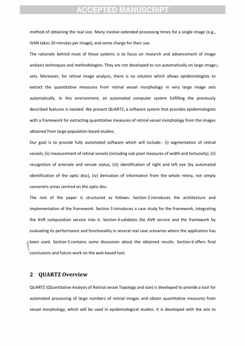
The main screen of QUARTZ is illustrated in Figure 7. The processing options are grouped into two
categories; vessel segmentation and vessel analysis. As the name indicates, the blood vessel tree is
extracted in the vessel segmentation task. The vessel analysis consists of calculation of the
quantifiable measures of vessel morphology, which includes vessel segment generation,
measurement of diameters in the vessel segments, computing local orientation of vessel segments,
optic disc localization, a/v classification and tortuosity measurement.
There is an option available to select the working directory for the images to be processed. All the
images in the working directory are loaded in the software and the names of the images are shown
in a selectable tabular view. This tabular view has four columns. The first column shows the index
count of the retinal image, the name is shown in the second column. The last two columns named as
“S” for segmentation and “W” for Analysis; depicts the processing progress of the particular retinal
image in check boxes. The “S” column is checked if the segmentation result is available and the “W”
column is shown checked when the vessel analysis of the image is completed. The selected retinal
image can be previewed in the Image-Preview area.
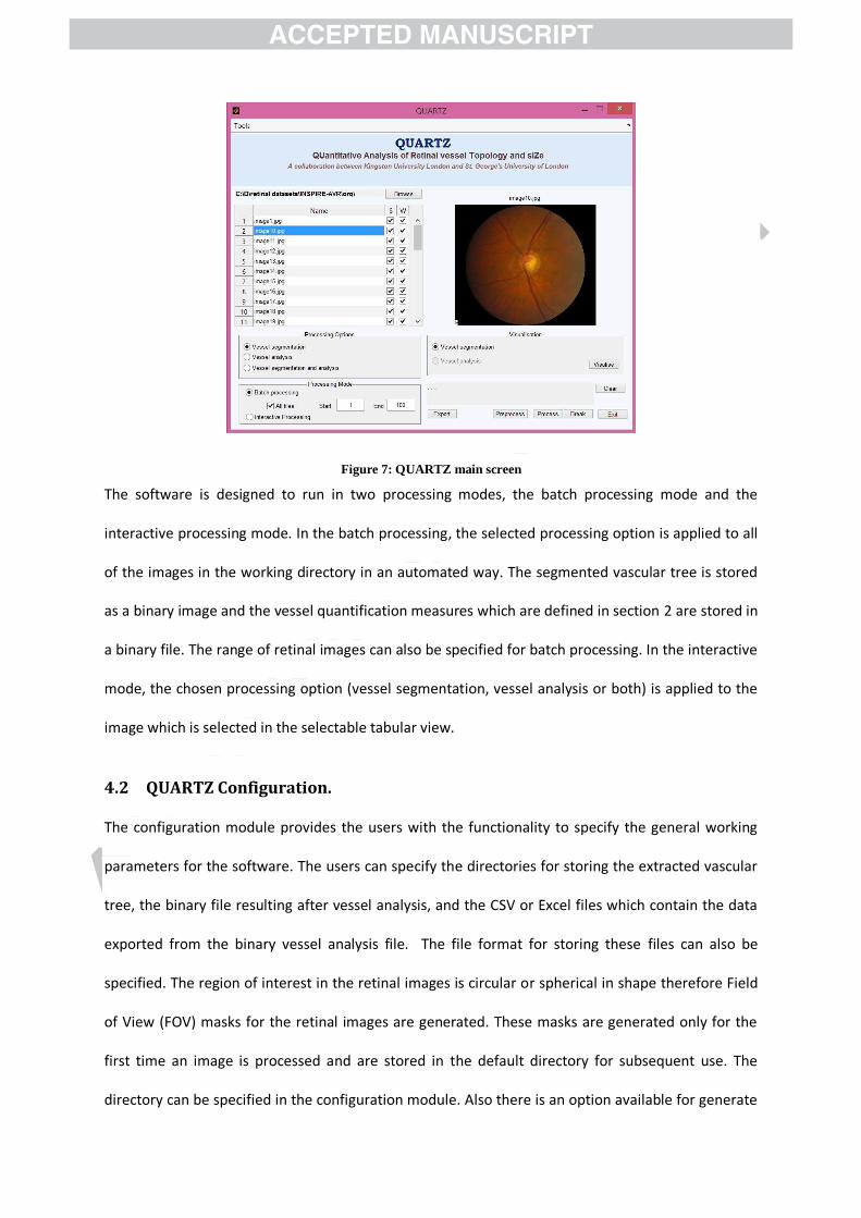
Figure 7: QUARTZ main screen
The software is designed to run in two processing modes, the batch processing mode and the
interactive processing mode. In the batch processing, the selected processing option is applied to all
of the images in the working directory in an automated way. The segmented vascular tree is stored
as a binary image and the vessel quantification measures which are defined in section 2 are stored in
a binary file. The range of retinal images can also be specified for batch processing. In the interactive
mode, the chosen processing option (vessel segmentation, vessel analysis or both) is applied to the
image which is selected in the selectable tabular view.
4.2 QUARTZ Configuration.
The configuration module provides the users with the functionality to specify the general working
parameters for the software. The users can specify the directories for storing the extracted vascular
tree, the binary file resulting after vessel analysis, and the CSV or Excel files which contain the data
exported from the binary vessel analysis file. The file format for storing these files can also be
specified. The region of interest in the retinal images is circular or spherical in shape therefore Field
of View (FOV) masks for the retinal images are generated. These masks are generated only for the
first time an image is processed and are stored in the default directory for subsequent use. The
directory can be specified in the configuration module. Also there is an option available for generate

the FOV mask each time the image is processed. The quantitative measures can be exported as a
CSV or Excel file, there is an option available for writing all the quantitative data in one file or
generate separate files for each image under consideration. The configuration module screenshot is
illustrated in Figure 8.
Figure 8: QUARTZ Configuration Module
4.3 Segmented Vasculature Visualization
The segmented vascular tree and the vessel analysis for the selected retinal image can also be
visualized. The vessel segmentation visualization is shown in Figure 9. The segmented vascular tree
can be shown as overlaid on the original RGB coloured image (Figure 9-a) or on the green channel of
RGB (Figure 9-b). The segmentation overlay colour as well as the overlay opacity can also be
customized for better visibility. The z-ordering of the retinal image and vascular tree can also be
changed. The functionality of zoom-in, zoom-out and pan is also provided for segmentation
visualization.

(a) (b)
Figure 9 : Vessel segmentation visualization; (a) RGB retinal image, (b) Green Channel of RGB image
4.4 Vessel Analysis Visualization
The visualization of vessel analysis is shown in Figure 10. The marking of vessel segment edges,
centrelines, diameters, labels and optic disc location can be viewed as overlaid on either the
coloured RGB image or on the green channel of RGB. The visualization options are shown as
highlighted in Figure 10(b), which also shows the zoomed-in view of the retinal image marked with
centrelines and vessel edges.
(a)
(b)
Figure 10: Vessel Analysis Visualization; (a) complete retinal image marked with centrelines and the vessel edges; (b)
Magnified view of retinal image with vessel edges shown in yellow colour.

In Figure 11, the vessel segment labels, the edges, the centrelines and the diameters of a selected
vessel segment are shown in the colour white. The selected diameters are shown in yellow. A list of
centreline coordinates and the diameter at respective coordinates of the selected vessel is shown as
highlighted in Figure 11(a).
The vessel segments can be clicked and selected in the preview area. The following properties of the
selected vessel segment are shown in a table, highlighted in Figure 11(b).
No of diameters
Mean diameter
Standard Deviation (SD) of diameters
Min diameter
Max diameter
Segment length (in pixels)
Diameter/Length ratio
Vessel segment tortuosity
(a)
(b)
Figure 11: Vessel segment labels, diameters, edges, centrelines and selected diameters
The graph of vessel segment diameter across its length is shown in Figure 12. The vessel diameters
are shown in red colour and the selected diameters in the list are visible in yellow colour.
(a)
(b)
Figure 12: The plot of vessel diameter across its length; (a) selected vessel segment
The QUARTZ software system is aimed at the analysis of large data sets containing thousands of
images; therefore the manual interaction with individual images is not feasible. However, the
software also provides manual intervention for correction of vessel segmentation as well as for the
correction of misclassified vessel segments as artery or veins.
4.5 Export of Quantitative Data
The quantification of vessel analysis can be exported as Comma Separated Values (CSV) or as a
Microsoft excel sheet.
The properties related to individual vessel segments are shown in the snapshot of the CSV file in
Figure 13. The vessel segment No 60 is emphasized, which is also shown as the selected vessel in
Figure 12(a). The vessel segment properties include;
Person Identifier: the Image Name
Position of OD which in turn indicates the right/left eye
Segment ID

ProbA: probability that the vessel segment is an arteriole
ProbV: probability that the vessel segment is a venule
Number of width measures in the vessel segment
Mean diameter of the vessel segment
Standard deviation of vessel segment diameters
Min diameter of vessel segment
Max diameter of vessel segment
Length of vessel segment measured as Euclidean distance between vessel segment end
points
Vessel segment diameter to length ratio
Tortuosity of vessel segment
Figure 13 : Vessel segment properties
Figure 14 shows the snapshot of the CSV file that contains the person identifier, segment ID,
centreline coordinates, diameters and local orientation angle. The first 15 diameters of vessel
segment no 60 are highlighted, which is shown in Figure 12(a). The local angle, as shown in the last
column of Figure 14 is the measure of orientation of a perpendicular line passing through the vessel
centreline pixel and joining two vessel edges from the x-axis. The –ve sign indicates that it is 30.06
degrees (last measure of segment 59) counter clockwise.

Figure 14: Vessel segment centreline coordinate diameters and local orientation angles
5 Quantitative Analysis of Results
The results obtained evaluating the usability of QUARTZ software and validation of the algorithms
are summarized in this section.
The QUARTZ software incorporates some of the retinal image processing and quantification
algorithms that were previously described by our group (M.M. Fraz & Sarah A Barman, 2014; M. M.
Fraz & Sarah A Barman, 2014; M. M. Fraz, Barman, et al., 2012; M. M. Fraz, Remagnino, et al., 2013;
M. M. Fraz, Remagnino, et al., 2012a, 2012b; M. M. Fraz, et al., 2014). Each module and algorithm
has been carefully evaluated and the validation results are presented. The performance metrics of
the incorporated algorithms are evaluated and analysed on different retinal image databases
available in the public domain, which includes DRIVE ("DRIVE: Digital Retinal Images for Vessel
Extraction," 2004), STARE ("STARE: STructured Analysis of the Retina," 2000), CHASE_DB1 (M.M.
Fraz & Barman, 2013), INSPIRE-AVR(Niemeijer, et al., 2011), and DIARETDB1(Kauppi, et al., 2007).
The accuracy of the vessel segmentation algorithm on DRIVE ("DRIVE: Digital Retinal Images for
Vessel Extraction," 2004), STARE ("STARE: STructured Analysis of the Retina," 2000) and CHASE_DB1
(M.M. Fraz & Barman, 2013) is found to be 0.948, 0.953 and 0.946 respectively. The sensitivity

(detection rate) and specificity are found to be 0.740, 07554 and 0.722; and 0.980, 0.976 and 0.741
respectively. The details of the evaluation methodology are illustrated in (M. M. Fraz & Sarah A
Barman, 2014; M. M. Fraz, Remagnino, et al., 2012b; M. M. Fraz, Rudnicka, et al., 2013). The average
accuracy values and precision rates obtained by the algorithm are more than the 2nd human
observers for the DRIVE and STARE databases. The specificity values for the algorithm are also higher
than the 2nd human observer for each of the three image databases that indicates the low false
positive rate of the methodology as compared with the 2nd human observer. This, in turn indicates
that the algorithm has identified less numbers of background pixels or pathological area pixels as
part of a vessel than the 2nd human observer.
The diameter measurement algorithm is evaluated on 1605 vessel profiles from different kinds of
vessel segments in the CHASE_DB1 database (M. M. Fraz, Remagnino, et al., 2013). It includes 544
profiles from vessel segments without a central reflex, 488 profiles are from vessel segments with a
central vessel reflex, 264 profiles are from the vessels with normal as well as a central reflex along
their length, 309 profiles are from low contrast vessel segments with uneven background
illumination. The diameters measured by the automated system are compared with the manually
marked vessel widths by two human observers. The mean vessel segment diameter observed by
both of the expert observers is 10.10 and 8.9 pixels respectively. The mean width computed by the
methodology is approximately 7.91 pixels which align more closely with the second observer. The
variance in width measured by both of the observers and estimated by the algorithm is
approximately 2.0. We consider the reference standard as the average of the measures marked by
two expert human observers. The mean and standard deviation of the difference in width measured
by the algorithm and the reference standard is 1.62 and 1.51 respectively. It should be noted that
precision in measures of width, i.e., low variance, might be more important, than absolute measures
of width. Any systematic bias in measures of width may be less important, as long as clinicians
measure widths consistently well, especially if detecting change in width along a vessel segment is
considered important. In contrast, if measures of width fluctuate considerably due to measurement

error then changes in width along a vessel length are unlikely to be detected. The detailed
evaluation of the quantification methodology is presented by the authors in (M. M. Fraz, Remagnino,
et al., 2013).
The a/v classification methodology is tested on DRIVE, INSPIRE-AVR, and images from the EPIC
Norfolk study (EPIC-Norfolk, 2014). The authors have reported the detailed evaluation of a/v
classification on EPIC Norfolk images elsewhere (M. M. Fraz, et al., 2014). The a/v classification on
the images from DRIVE and INSPIRE-AVR database are illustrated in Figure 6. The test dataset
contains 2500 vessel segments from 40 colour fundus images available in the DRIVE database. The
vessel segments are classified as arteriole or venule manually by expert observers. The algorithm is
evaluated by using a two-fold validation methodology. The first twenty images are assigned to set S1
and rest of the twenty images are allocated to set S2. The classifier is then trained on S1 and tested
on S2, followed by training on S2 and testing on S1. The algorithm is evaluated in terms of Detection
Rate / Sensitivity (SNa|v), Specificity (SPa|v), Classification Accuracy (ACCa|v), Classification Error Rate
(CERa|v), Positive Predictive Value (PPVa|v), Negative Predictive Value (NPVa|v) and the Positive and
Negative Likelihood Ratios (PLRa|v and NLRa|v). The ACCa|v is measured by the ratio of the total
number of correctly classified pixels (sum of true positives and true negatives) by the number of
pixels under consideration in the image. SNa|v reflects the ability of an algorithm to detect the true
positives. SPa|v measures the proportion of negatives that are correctly identified. PPVa|v or the
precession rate gives the proportion of vessel pixels with correctly identified positive test results and
NPVa|v is the proportion of vessel pixels with negative test results that are correctly identified. The
predictive values depends on the percentage of a/v in the retina (prevalence), therefore the
likelihood ratios (PLRa|v and NLRa|v) are also computed which are not dependent on prevalence. The
performance metrics are computed separately for arterioles and venules and presented in Table 1.

Table 1: Vessel classification performance metrics on DRIVE database
Measure Arterioles Venules Tested on S1 Tested on S2 Tested on S1 Tested on S2
SNa|v 0.9123 0.8815 0.7838 0.7652
SPa|v 0.7758 0.7829 0.9127 0.8804
ACCa|v 0.8487 0.8344 0.8369 0.8261 CERa|v 0.1512 0.1682 0.1634 0.1742
PPVa|v 0.8462 0.8342 0.8789 0.8621
NPVa|v 0.8773 0.8542 0.8234 0.8340
PLRa|v 3.7481 4.8341 5.5685 6.3718
NLRa|v 0.1135 0.1403 0.2515 0.2416
The similarity in the performance metrics obtained for the sets S1 and S2 indicates the repeatability
of the methodology in classification of vessels.
The working of software can be subdivided in to four modules. (1): Vessel segmentation, (2): Vessel
Analysis, which further includes computation of width measurement and angular change at each
vessel centreline pixel with sub-pixel accuracy, calculating local vessel orientation and tortuosity
measurement, (3): optic disc localization, and (4): arteriole/venule classification. The average
processing time for each module is computed on a set of 20 images randomly picked from the image
dataset. The QUARTZ is evaluated for the processing time on Dell XPS 13 laptop with Corei7
processor and 8GB RAM. The measures are reported in Table 2. However, it should also be noted
that the QUARTZ system is aimed at the analysis of large dataset in batch processing mode,
therefore the processing time is not of very much significance.
Table 2: Average processing time for each Module is QUARTZ
S.No Module Average Processing Time
In seconds 1 Vessel Segmentation 16.57 2 Quantitative Analysis of Segmented Vasculature
Vessel Segments Labeling
Vessel Edges and Centreline pixels identification
Vessel Width computation at each centreline pixel with sub-pixel accuracy
Tortuosity Measurement of Vessel Segment
Local angle computation at each centreline pixel
10.12
3 OD Localization 0.48
4 AV Classification 26.40

The optic disc location is used to identify the right/left eye in the macula centred retinal images. The
OD localization and boundary extraction is illustrated in Figure 6. The algorithm achieves a success
rate of 100% and 98.9% for DRIVE and DIARETDB1 databases respectively. The algorithm achieves an
overlap of 61.88% and 54.69% for DRIVE and DIARETDB1 databases respectively. The detailed
evaluation procedure for OD localization is reported by the authors elsewhere (Basit & Fraz, 2015).
The quantitative comparison shows a close correlation between the automatic and manual location
as well as a high spatial overlap between the OD generated by the manual method, other OD
localization methodologies available in literature (Hsiao, Liu, Yu, Kuo, & Yu, 2012) and the proposed
method.
6 Discussion and Conclusion
The retinal vasculature is the only part of the blood circulation system that can be directly observed
non-invasively and can be easily imaged using fundus cameras. Abnormalities in morphological
characteristics of arterioles and venules have been prospectively associated with a number of
disease outcomes which includes hypertension, coronary heart disease, diabetes, elevated
glycosylated haemoglobin, lower levels of high density lipoprotein. The assessment of the
characteristics of the retinal vascular network may provide important information about early
diagnosis of many systemic and vascular diseases. Epidemiologists and other medical / statistical
experts study the association of retinal vessel abnormalities with other disease by examining the
data gathered in the large population based studies and screening programs. The analysis of the
vessel morphology and extraction of quantifiable measures from large number of images is a tedious
task if performed manually.
With the aim of developing reliable, automated, efficient retinal image analysis software which can
generate a rich quantification of retinal vasculature in large volumes of fundus images, we present
QUARTZ (Quantitative Analysis of Retinal Vessel Topology and size), a novel automated system for
processing and analysing bulk of retinal images. Several software packages to analyse adult retinal

images have been developed. While these provide several indices of retinal vessel morphology, they
have several important limitations. In particular, they are restricted to analysis of limited areas
around the optic disc, have limited automated ability to discriminate retinal arterioles from venules
and provide evidence on a limited number of parameters; mainly vessel width with limited
information on vessel tortuosity. Moreover, these packages are often semi-automated and some
include extended processing times for a single image.
QUARTZ is fully automated software system that has been developed to localize and quantify the
morphological characteristics of blood vessels in the retinal images, including (i) measurement of
retinal vessels (including sub-pixel measures of width and tortuosity), and (ii) recognition of arteriole
and venule status, (iii) automated identification of the optic disc). These measures will derive
information from the whole retina, not simply concentric areas centred on the optic disc.
The automated methods for quantification of retinal vessel morphology and width may be used as
an alternative to the time consuming subjective clinical evaluation for monitoring the progression of
retinopathies and their association with normal and abnormal vascular patterns. This may enable
early diagnosis and treatment, improving prognosis by rapid introduction of clinical health-care.
QUARTZ provides quantifiable measures of retinal vessel morphology, which may enable
epidemiologists / clinicians to detect the likelihood or presence of a disease by observing specific
signs in combination with other external factors e.g. age symptoms and certain clinical features.
The retinal images are placed in a directory and the folder path is specified in the QUARTZ system.
The system automatically loads the images from the specified directory, extract vasculature, convert
it into vessel segments, classify into arteries\veins, compute local angle and tortuosity and localize
optic disc. The quantitative measures can be exported as CSV files or Microsoft Excel Workbooks.
The software is designed to run in two processing modes, the batch processing mode and the
interactive processing mode. In the batch processing, the selected processing option is applied to all
of the images in the working directory in an automated way. The number of retinal images to be
processed in the working directory can also be specified. In the interactive processing mode, the
selected retinal image from the selectable tabular view can be processed according to the chosen
processing option (vessel segmentation, vessel analysis or both).
QUARTZ can be used to identify early retinal vessel changes that may be physiological biomarkers of
disease of cardio-metabolic risk and outcome, such coronary heart disease and stroke. Another
application area is to study the effect of new therapies and drugs on disease e.g. alteration in retinal
vessel measurements with a new treatments for hypertension. The quantifiable measures extracted
from QUARTZ can also be used for examining the association of novel pathways in the natural
history of specific disease e.g. microvascular disease pathways in stroke. It can be used to study the
association between retinal vessel abnormalities and cognitive performance based on gene
expression (Ding, et al., 2008). QUARTZ can assist in longitudinal studies i.e. quantitative study of the
evolution and characterization of a disease, which will assist in treatment planning or investigating
the response of a patient to certain treatments. The performance of the software is demonstrated to
be state-of-the art in terms of segmentation accuracy, calibre measurement, optic disc and
arteriole/venule recognition. In terms of automation with respect to specific large datasets, it is
shown to be leading in the field.
At present, clinical detection and grading of diabetic retinopathy is largely evaluated manually by a
grader who compares the patient’s retinal image with a set of standard photographs and accesses
the severity of retinal pathologies (abnormal blood vessel width, venous beading etc) before
assigning an overall grade. An application of image processing algorithms for computer assisted
analysis of digital fundus images offers a number of advantages over a manual system, including fast,
timely and reliable quantification of abnormalities with a reduction of subjective human error.
Regarding future work, we aim to extend the functionality of this tool in multiple directions. The
quality assessment of retinal image is an important pre-processing step for identifying those images
in large datasets for which the automated analysis procedures may fail. An image is considered as
inadequate when it is difficult or impossible to make a reliable clinical judgment regarding the
presence or absence of disease. In the screening programs, studies (Teng, Lefley, & Claremont, 2002)

have shown that approximately 10% of the mydriatic (pupil dilation) images and 20.8% of non-
mydriatic (no pupil dilation) are of inadequate quality. The major reasons for low quality images
include illumination variability due to small pupil size; lack of contrast and blurriness due to poor
focus, eye movement and imaging of part of the eyelid and eyelash due to blinking. Sufficient image
quality is essential to ensure a reliable extraction of quantitative measures from retinal vessel
morphology.
From usability point of view, an informal feedback has been gathered on the use of the QUARTZ
system by epidemiologists at St. Georges University of London. The QUARTZ system has been
applied to over 16000 retinal images and a more complete evaluation of the user experience of the
software is planned for future work. An algorithm for change detection in OD cup-to-disc diameter
ratio will be incorporated which enables this software to be used in large population based studies
for glaucoma detection. Moreover, the A/V classification module will be extended towards
automatic computation of Arterio-Venous Ratio. The width measurement component together with
the A/V classification will be extended for automatic detection of venous beading and a/v nicking.
Most importantly, the vessel segmentation algorithm will be extended such that it can detect
neovascularization in the retina. This detection of formation of new vessels in the retina is a strong
indicator of proliferative diabetic retinopathy (Ramlugun, Nagarajan, & Chakraborty, 2012). This
enhancement will enable QUARTZ to be utilized for studying the association and linkage of different
phenotypes with proliferative diabetic retinopathy in large population studies. Our research group is
also working in multi-modal and hybrid registration of retinal images and more methodologies for
retinal image analysis are being developed such as quantification of retinal pathologies and drusen
localization, etc. These methodologies have proven very useful for clinicians and epidemiologists,
thus it would be valuable to integrate them into the QUARTZ software.
References
Abràmoff, M. D., Garvin, M. K., & Sonka, M. (2010). Retinal Imaging and Image Analysis. Biomedical
Engineering, IEEE Reviews in, 3, 169-208.

Azzopardi, G., & Petkov, N. (2013). Automatic detection of vascular bifurcations in segmented retinal
images using trainable COSFIRE filters. Pattern Recognition Letters, 34, 922-933.
Azzopardi, G., Strisciuglio, N., Vento, M., & Petkov, N. (2015). Trainable COSFIRE filters for vessel
delineation with application to retinal images. Medical image analysis, 19, 46-57.
Bankhead, P., Scholfield, C. N., McGeown, J. G., & Curtis, T. M. (2012). Fast Retinal Vessel Detection
and Measurement Using Wavelets and Edge Location Refinement. PLoS ONE, 7, e32435.
Basit, A., & Fraz, M. M. (2015). Optic disc detection and boundary extraction in retinal images.
Applied Optics, 54, 3440-3447.
Cheung, N., Saw, S. M., Islam, F. M. A., Rogers, S. L., Shankar, A., De Haseth, K., Mitchell, P., & Wong,
T. Y. (2007). BMI and Retinal Vascular Caliber in Children. Obesity, 15, 209-209.
Collins, G. S., & Altman, D. G. (2010). An independent and external validation of QRISK2
cardiovascular disease risk score: a prospective open cohort study. BMJ, 340.
Dashtbozorg, B., Mendonca, A. M., & Campilho, A. (2013). An Automatic Graph-based Approach for
Artery/Vein Classification in Retinal Images. Image Processing, IEEE Transactions on, PP, 1-1.
Ding, J., Patton, N., Deary, I. J., Strachan, M. W. J., Fowkes, F. G. R., Mitchell, R. J., & Price, J. F.
(2008). Retinal microvascular abnormalities and cognitive dysfunction: a systematic review.
British Journal of Ophthalmology, 92, 1017-1025.
. DRIVE: Digital Retinal Images for Vessel Extraction. In. (2004).
EPIC-Norfolk. (2014). European Prospective Investigation of Cancer (EPIC). In (Vol. 2014).
Fraz, M. M., & Barman, S. A. (2013). CHASE_DB1. In (Vol. 2014). London: Kingston University.

Fraz, M. M., & Barman, S. A. (2014). Computer Vision Algorithms Applied to Retinal Vessel
Segmentation and Quantification of Vessel Caliber. In Image Analysis and Modeling in
Ophthalmology (pp. 49-84): CRC Press.
Fraz, M. M., & Barman, S. A. (2014). Ensemble Classification Applied to Retinal Blood Vessel
Segmentation: Theory and Implementation. In Image Analysis and Modeling in
Ophthalmology (pp. 23-48): CRC Press.
Fraz, M. M., Barman, S. A., Remagnino, P., Hoppe, A., Basit, A., Uyyanonvara, B., Rudnicka, A. R., &
Owen, C. G. (2012). An approach to localize the retinal blood vessels using bit planes and
centerline detection. Computer methods and programs in biomedicine, 108, 600-616.
Fraz, M. M., Basit, A., & Barman, S. A. (2012). Application of Morphological Bit Planes in Retinal
Blood Vessel Extraction. Journal of Digital Imaging, 1-13.
Fraz, M. M., Remagnino, P., Hoppe, A., Basit, A., Rudnicka, A. R., Owen, C. G., & Barman, S. A. (2013).
Quantification of blood vessel calibre in retinal images of multi-ethnic school children using a
model based approach. Computerized Medical Imaging and Graphics, 37, 60-72.
Fraz, M. M., Remagnino, P., Hoppe, A., Uyyanonvara, B., Rudnicka, A. R., Owen, C. G., & Barman, S.
A. (2012a). Blood vessel segmentation methodologies in retinal images – A survey. Computer
methods and programs in biomedicine, 108, 407-433.
Fraz, M. M., Remagnino, P., Hoppe, A., Uyyanonvara, B., Rudnicka, A. R., Owen, C. G., & Barman, S.
A. (2012b). An Ensemble Classification-Based Approach Applied to Retinal Blood Vessel
Segmentation. Biomedical Engineering, IEEE Transactions on, 59, 2538-2548.
Fraz, M. M., Rudnicka, A. R., Owen, C. G., & Barman, S. A. (2013). Delineation of blood vessels in
pediatric retinal images using decision trees-based ensemble classification. International
Journal of Computer Assisted Radiology and Surgery, 1-17.
Fraz, M. M., Rudnicka, A. R., Owen, C. G., Strachan, D. P., & Barman, S. A. (2014). Automated
Arteriole and Venule Recognition in Retinal Images using Ensemble Classification. In 9th
International Conference on Computer Vision Theory and Applications (VISAAP) (Vol. 9th).
Lisbon, Portugal.
González, E. L. M., Johansson, S., Wallander, M.-A., & Rodríguez, L. A. G. (2009). Trends in the
prevalence and incidence of diabetes in the UK: 1996–2005. Journal of Epidemiology and
Community Health, 63, 332-336.
Gonzalez, R. C., & Woods, R. E. (2002). Digital Image Processing (Second ed.): Prentice Hall, Upper
Saddle River, New Jersey.
Grisan, E., & Ruggeri, A. (2003). A divide et impera strategy for automatic classification of retinal
vessels into arteries and veins. In Engineering in Medicine and Biology Society, 2003.
Proceedings of the 25th Annual International Conference of the IEEE (Vol. 1, pp. 890-893
Vol.891).
Hsiao, H.-K., Liu, C.-C., Yu, C.-Y., Kuo, S.-W., & Yu, S.-S. (2012). A novel optic disc detection scheme on
retinal images. Expert Systems with Applications, 39, 10600-10606.
Huang, Y., Zhang, J., & Huang, Y. (2012). An automated computational framework for retinal vascular
network labeling and branching order analysis. Microvascular Research, 84, 169-177.
Ikram, M. K., de Jong, F. J., Vingerling, J. R., Witteman, J. C. M., Hofman, A., Breteler, M. M. B., & de
Jong, P. T. V. M. (2004). Are Retinal Arteriolar or Venular Diameters Associated with Markers
for Cardiovascular Disorders? The Rotterdam Study. Investigative Ophthalmology & Visual
Science, 45, 2129-2134.
Ikram, M. K., Witteman, J. C. M., Vingerling, J. R., Breteler, M. M. B., Hofman, A., & de Jong, P. T. V.
M. (2006). Retinal Vessel Diameters and Risk of Hypertension. Hypertension, 47, 189-194.
Kauppi, T., Kalesnykiene, V., Kamarainen, J.-K., Lensu, L., Sorri, I., Raninen, A., Voutilainen, R., Pietilä,
J., Kälviäinen, H., & Uusitalo, H. (2007). DIARETDB1 diabetic retinopathy database and
evaluation protocol. In Medical Image Understanding and Analysis (MIUA2007) (pp. 61-65).
Aberystwyth, Wales, UK.

Kelvin, P., Ghassan, H., & Rafeef, A. (2007). Live-vessel: extending livewire for simultaneous
extraction of optimal medial and boundary paths in vascular images. In Proceedings of the
10th international conference on Medical image computing and computer-assisted
intervention. Brisbane, Australia: Springer-Verlag.
Lee, E. T. Y. (1989). Choosing nodes in parametric curve interpolation. Computer-Aided Design, 21,
363-370.
Leung, H., Wang, J. J., Rochtchina, E., Tan, A. G., Wong, T. Y., Klein, R., Hubbard, L. D., & Mitchell, P.
(2003). Relationships between Age, Blood Pressure, and Retinal Vessel Diameters in an Older
Population. Investigative Ophthalmology & Vis, 44, 2900-2904.
Mirsharif, Q., Tajeripour, F., & Pourreza, H. Automated characterization of blood vessels as arteries
and veins in retinal images. Computerized Medical Imaging and Graphics.
Nguyen, U. T. V., Bhuiyan, A., Park, L. A. F., & Ramamohanarao, K. (2012). An effective retinal blood
vessel segmentation method using multi-scale line detection. Pattern Recognition.
Niemeijer, M., Xiayu, X., Dumitrescu, A. V., Gupta, P., van Ginneken, B., Folk, J. C., & Abramoff, M. D.
(2011). Automated Measurement of the Arteriolar-to-Venular Width Ratio in Digital Color
Fundus Photographs. Medical Imaging, IEEE Transactions on, 30, 1941-1950.
Ortega, M., Barreira, N., Novo, J., Penedo, M. G., Pose-Reino, A., & Gómez-Ulla, F. (2010). Sirius: A
web-based system for retinal image analysis. International journal of medical informatics,
79, 722-732.
Owen, C. G., Rudnicka, A. R., Mullen, R., Barman, S. A., Monekosso, D., Whincup, P. H., Ng, J., &
Paterson, C. (2009). Measuring Retinal Vessel Tortuosity in 10-Year-Old Children: Validation
of the Computer-Assisted Image Analysis of the Retina (CAIAR) Program. Investigative
Ophthalmology & Visual Science, 50, 2004-2010.
Owen, C. G., Rudnicka, A. R., Nightingale, C. M., Mullen, R., Barman, S. A., Sattar, N., Cook, D. G., &
Whincup, P. H. (2011). Retinal Arteriolar Tortuosity and Cardiovascular Risk Factors in a

Multi-Ethnic Population Study of 10-Year-Old Children; the Child Heart and Health Study in
England (CHASE). Arteriosclerosis, Thrombosis, and Vascular Biology, 31, 1933-1938.
Perez-Rovira, A., MacGillivray, T., Trucco, E., Chin, K. S., Zutis, K., Lupascu, C., Tegolo, D., Giachetti,
A., Wilson, P. J., Doney, A., & Dhillon, B. (2011). VAMPIRE: Vessel assessment and
measurement platform for images of the Retina. In Engineering in Medicine and Biology
Society,EMBC, 2011 Annual International Conference of the IEEE (pp. 3391-3394).
Ramlugun, G. S., Nagarajan, V. K., & Chakraborty, C. (2012). Small retinal vessels extraction towards
proliferative diabetic retinopathy screening. Expert Systems with Applications, 39, 1141-
1146.
Ricci, E., & Perfetti, R. (2007). Retinal Blood Vessel Segmentation Using Line Operators and Support
Vector Classification. Medical Imaging, IEEE Transactions on, 26, 1357-1365.
Rothaus, K., Jiang, X., & Rhiem, P. (2009). Separation of the retinal vascular graph in arteries and
veins based upon structural knowledge. Image and Vision Computing, 27, 864-875.
SR, S., S, K., & A, T. (2011). Diabetes Mellitus, Fasting Glucose, and Risk of Cause-Specific Death. New
England Journal of Medicine, 364, 829-841.
. STARE: STructured Analysis of the Retina. In. (2000).
Statistics, O. f. N. (2012). Deaths Registered in England and Wales in 2010, by Cause. In (Vol. April
2014).
Stewart, C. V., & Roysam, B. RIVERS; Retinal Image Vessel Extraction and Registration System
http://cgi-vision.cs.rpi.edu/cgi/RIVERS/index.php. In.
Teng, T., Lefley, M., & Claremont, D. (2002). Progress towards automated diabetic ocular screening:
A review of image analysis and intelligent systems for diabetic retinopathy. Medical and
Biological Engineering and Computing, 40, 2-13.

Thomas, M. C., Hardoon, S. L., Papacosta, A. O., Morris, R. W., Wannamethee, S. G., Sloggett, A., &
Whincup, P. H. (2009). Evidence of an accelerating increase in prevalence of diagnosed Type
2 diabetes in British men, 1978–2005. Diabetic Medicine, 26, 766-772.
Trucco, E., Ruggeri, A., Karnowski, T., Giancardo, L., Chaum, E., Hubschman, J. P., al-Diri, B., Cheung,
C. Y., Wong, D., Abràmoff, M., Lim, G., Kumar, D., Burlina, P., Bressler, N. M., Jelinek, H. F.,
Meriaudeau, F., Quellec, G., MacGillivray, T., & Dhillon, B. (2013). Validating Retinal Fundus
Image Analysis Algorithms: Issues and a Proposal. Investigative Ophthalmology & Visual
Science, 54, 3546-3559.
Tsai, C. L., Madore, B., Leotta, M. J., Sofka, M., Yang, G., Majerovics, A., Tanenbaum, H. L., Stewart, C.
V., & Roysam, B. (2008). Automated Retinal Image Analysis Over the Internet. Information
Technology in Biomedicine, IEEE Transactions on, 12, 480-487.
Vázquez, S. G., Cancela, B., Barreira, N., Penedo, M. G., Rodríguez-Blanco, M., Pena Seijo, M., Tuero,
G. C., Barceló, M. A., & Saez, M. (2013). Improving retinal artery and vein classification by
means of a minimal path approach. Machine Vision and Applications, 24, 919-930.
Wang, J. J., Taylor, B., Wong, T. Y., Chua, B., Rochtchina, E., Klein, R., & Mitchell, P. (2006). Retinal
Vessel Diameters and Obesity: A Population-Based Study in Older Persons. Obesity, 14, 206-
214.
Wong, T. Y., Islam, F. M. A., Klein, R., Klein, B. E. K., Cotch, M. F., Castro, C., Sharrett, A. R., & Shahar,
E. (2006). Retinal Vascular Caliber, Cardiovascular Risk Factors, and Inflammation: The Multi-
Ethnic Study of Atherosclerosis (MESA). Investigative Ophthalmology & Visual Science, 47,
2341-2350.
Wong, T. Y., Klein, R., Sharrett, A. R., Duncan, B. B., Couper, D. J., Tielsch, J. M., Klein, B. E. K., &
Hubbard, L. D. (2002). Retinal Arteriolar Narrowing and Risk of Coronary Heart Disease in
Men and Women. JAMA: The Journal of the American Medical Association, 287, 1153-1159
http://jama.ama-assn.org/content/1287/1159/1153.abstract.

Highlights
Automated system for interactive / batch processing of large number of retinal images
Extract useful quantifiable measurements of retinal vessel morphology
Modules for vessel segmentation, width, tortuosity measurement at sub-pixel accuracy
Components for Artery / Vein classification, OD localization, tortuosity measurement
Epidemiological study of association of vessel morphology with disease precursor