Mass transfer studies in forming and collapsing droplets - CORE
Upload
independentCategory
view
0download
0
Kidney International, Vol. 63 (2003), pp. 233–239
Familial collapsing glomerulopathy: Clinical, pathological andimmunogenetic features
M. CARMEN AVILA-CASADO, GILBERTO VARGAS-ALARCON, MARIA E. SOTO,GUADALUPE HERNANDEZ, PEDRO A. REYES, and JAIME HERRERA-ACOSTA
Departments of Pathology, Physiology, Immunology, and Nephrology, Instituto Nacional de CardiologiaIgnacio Chavez, Universidad Nacional Autonoma de Mexico, Mexico DF, Mexico
Familial collapsing glomerulopathy: Clinical, pathological and epithelial cell swelling accompanied by tubular cysticimmunogenetic features. dilatation and interstitial fibrosis [1]. Clinically, patients
Background. Collapsing glomerulopathy (CG) is an aggres- present nephrotic syndrome with profuse proteinuriasive form of glomerular injury frequently seen in associationand a rapid course to end-stage renal failure or deathwith HIV infection, although it is also recognized in non-HIVdue to complications of nephrotic syndrome [2]. Thepatients as a primary disease. Until now, the occurrence of CG
in a familial pattern has not been reported. diagnosis of primary CG requires a lack of evidence forMethods. We studied five members of a family (siblings), ad- human immunodeficiency virus (HIV) infection, since
mitted for evaluation of proteinuria and nephrotic syndrome. HIV-nephropathy can be morphologically and clinicallyThey had no other family history of renal disease. Blood sam- indistinguishable [2–8]. The presentation of renal diseaseples for major histocompatibility complex (MHC) analysis were
in a familial pattern has been described in a varietyobtained from the five siblings, both parents and four relatives.of entities that includes: autosomal recessive polycysticResults. Renal biopsy performed in four out of the five sib-
lings revealed capillary collapse and retraction with visceral epi- kidney disease [9], thin basement membrane disease [10]thelial cell swelling and reabsorption droplets, consistent with and Alport disease [11], all of them recognized as heredi-CG. Two of the patients had suggestive symptoms of systemic tary renal diseases, as well as in families with focal andlupus erythematosus, such as arthritis, rash, hair loss, moder-
segmental glomerulosclerosis (FSGS) [12]. Neverthe-ate leukopenia and lymphopenia, low titers of antinuclear anti-less, it has not been reported in patients with collapsingbodies (ANA) and anti-SSA/Ro antibodies, but no immune com-glomerulopathy. We report the clinical, pathological andplex deposition on renal biopsy. IgG serology for parvovirus
B19 (PVB-19) was positive only in two siblings but polymerase immunogenetic findings in a family with collapsing glo-chain reaction (PCR) was negative. Immunogenetic analysis merulopathy.showed that all patients shared the same MHC haplotype inher-ited from the mother.
Conclusions. CG can present in a familial pattern. Since a METHODSsimilar MHC haplotype was observed in affected and non-
Patientsaffected members of the family, we conclude that the environ-ment plays an important role in the development of the disease. Five members of a family (siblings) with no other family
history of renal disease were admitted for clinical evalua-tion. The patients belonged to a Mexican-Mestizo popu-lation and had no evidence of HIV infection or intrave-Collapsing glomerulopathy (CG) is a well-defined formnous drug use. Four of these patients underwent a renalof glomerular injury. In 1986, Weiss et al described sixbiopsy that fulfilled diagnostic criteria for collapsingcases with nephrotic syndrome, progressive irreversibleglomerulopathy. Other family members including bothrenal failure and characteristic histopathological changesparents and four relatives (two grandparents, one uncle,[1]. Histopathological changes in this disease consist ofand one aunt) were screened for abnormal urinary sedi-glomerular capillary retraction or collapse and visceralment and protein excretion; their levels of serum creati-nine, electrolytes, and cholesterol were determined. All
Key words: familial nephrotic syndrome, renal disease, glomerular of the results were either normal or negative. An immuno-injury, inheritance, MHC haplotype, HIV infection. genetic evaluation also was performed.
Received for publication October 26, 2001 Histological diagnosisand in revised form August 8, 2002Accepted for publication August 14, 2002 The diagnosis of CG was based on light microscopy
findings of focal and segmental, or global glomerular capil- 2003 by the International Society of Nephrology
233
Avila-Casado et al: Familial collapsing glomerulopathy234
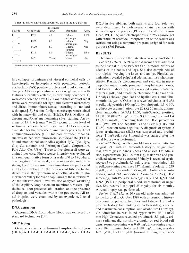
Table 1. Major clinical and laboratory data in the five patients DQB in five siblings, both parents and four relativeswere determined by polymerase chain reaction withProteinuria
Patient Gender/age g/day Symptoms ANA sequence specific primers (PCR-SSP; Pel-Freez, Brown1 F/21 4.8 Edema 1:160 Deer, WI, USA) and electrophoresis in 2% agarose gel
III-7 arthralgias with ethidium bromide. Interpretation of the results was2 F/22 6.5 Edema Neg
carried out using a computer program designed for suchIII-9 arthralgias3 M/20 5.3 Edema Neg purpose (Pel-Freez).
III-11 Fatigue4 F/14 8.0 Edema 1:640 RESULTS
III-8 arthralgias5 M/7 Trace None Neg The clinical history of the patients is presented in Table 1.
III-10 Patient 1 (III-7). A 21-year-old woman was admittedAbbreviations are: ANA, antinuclear antibodies; Neg, negative. to the hospital in June 1997 with an 18-month history of
edema of the hands and legs. She also complained ofarthralgias involving the knees and ankles. Physical ex-amination revealed palpebral edema, hair loss, photosen-
lary collapse, prominence of visceral epithelial cells bysitivity, Raynaud’s phenomenon, and synovitis in meta-
hypertrophy or hyperplasia with prominent periodiccarpophalangeal joints, proximal interphalangeal joints,
acid-Schiff (PAS) positive droplets and tubulointerstitial and knees. Laboratory tests revealed serum creatininechanges. All cases presenting at least one glomerulus with at 0.89 mg/dL, and creatinine clearance at 42.3 mL/min.features of capillary collapse, as previously described [1], Urinalysis showed granular and hyaline casts, and albu-were considered to have CG. Two to three cores of renal minuria 4.8 g/24 h. Other tests revealed cholesterol 232tissue were processed for light and electron microscopy mg/dL, triglycerides 190 mg/dL, lymphopenia 1.3 � 109,and direct immunofluorescence, according to standard erythrocyte sedimentation rate (ESR) 50 mm/h, antinu-techniques [13]. Sections for light microscopy were stained clear antibodies (ANA) 1:160, anti-SSA/Ro positive,with hematoxylin and eosin (H&E), PAS, Mallory tri- CH50 160 (80-320 mg/dl), C3 99 (�75 mg/dL), and C4chrome and Jones’ methenamine silver staining. An av- 13 (�13 mg/dL). Screening tests for HIV, parvoviruserage of 15 � 6 (range 7 to 23) glomeruli per level of B19 (PVB-19), and hepatitis B and C virus (HBV andsection in each sample were examined. All biopsies were HCV) infection were negative. A diagnosis of systemicevaluated for the presence of immune deposits by direct lupus erythematosus (SLE) was suspected and predni-immunofluorescence (IF). One core of frozen renal tis- sone (1 mg/kg/day for 3 months) was started after thesue was stained with fluorescein isothiocyanate (FITC)- renal biopsy was performed.bound monoclonal antibodies against IgG, IgM, IgA, Patient 2 (III-9). A 22-year-old female was admitted inC1q, C3, albumin and fibrinogen (Dako Corporation, August 1997, with an 18-month history of fatigue, hairPalo Alto, CA, USA). Three to five glomeruli were ex- loss, arthralgias in hands, knees and ankles. On admis-amined per case. Fluorescence intensity was evaluated sion, hypertension (150/100 mm Hg), malar rash and gen-in a semiquantitative form on a scale of 0 to 3�, where: eralized edema were detected. Urinalysis revealed eryth-0 � negative, 1� � weak, 2� � moderate, and 3� � rocytes 3�, proteinuria 6.5 g/day, serum creatinine 1.18strong. Electron microscopy examination was performed mg/dL, creatinine clearance 137 mL/min, cholesterol 279in all cases looking for the presence of tubuloreticular mg/dL, and triglycerides 175 mg/dL. Antinuclear anti-structures in the cytoplasm of endothelial cells of glo- bodies, anti-DNA antibodies (Crithidia lucilae), HIVmerular capillary loops and capillaries of the interstitium. screening, anti-PVB-19 serology (IgG and IgM) andAt the ultrastructural level we also analyzed wrinkling DNA (PCR) in peripheral blood, were normal or nega-of the capillary loop basement membrane, visceral epi- tive. She received captopril 25 mg/day for six months.thelial cell foot processes obliteration, and the presence A renal biopsy was performed.of droplets and vacuoles within the podocytes. All re- Patient 3 (III-11). A 20-year-old male was admittednal biopsies were examined by an experienced renal to the hospital in October 1997 with a six-month historypathologist. of edema of pelvic extremities and fatigue. He had a
positive history for smoking (2 packages/day), cocaineDNA extraction and marihuana consumption, and alcoholism (3 oz/day).
Genomic DNA from whole blood was extracted by On admission he was found hypertensive (BP 140/95standard techniques [14]. mm Hg). Urinalysis revealed proteinuria 5.3 g/day, uri-
nary sediment did not show granular or red blood cellMHC typing casts, serum creatinine was 0.89 mg/dL, creatinine clear-
Generic variants of human lymphocyte antigen ance 109 mL/min, cholesterol 194 mg/dL, triglycerides169 mg/dL, C3 117 mg/dL (normal �75 mg/dL), C4 25(HLA)-A, HLA-B, HLA-DR, HLA-DQA and HLA-

Avila-Casado et al: Familial collapsing glomerulopathy 235
mg/dL (nl �13 mg/dL). Antibodies to HIV, HBV and In the collapsed glomeruli, visceral epithelial cells ap-peared prominent and numerous (hypertrophy and hy-HCV were negative. Anti-PVB-19 IgG was positive (5.3)
and anti-PVB-19 IgM negative. He was treated with cap- perplasia) and contained large hyaline droplets (Fig. 1).The rest of the glomeruli showed prominent visceraltopril 50 mg/day for eight months. A renal biopsy was
performed. epithelial cells, the capillary loops were open, and base-ment membranes appeared fine and delicate. InterstitialPatient 4 (III-8). A 14-year-old female was admitted
in January 1998. Her prior medical history was unre- fibrosis was present and scored Grade II (�25 and �40%)with sparse mononuclear cell infiltration. Direct immu-markable; however, two months before admission she
developed cough and hyaline rhinorrhea and was treated nofluorescence against IgG, IgM, IgA, C1q, C3, kappaand lambda was negative for immune-complex depositionwith penicillin for seven days. Two weeks later she no-
ticed frothy urine. Two days before admission she devel- in five non-collapsed glomeruli. One collapsed glomerulusshowed diffuse irregular staining for IgM and vacuolesoped dyspnea and edema of both legs. On admission,
physical examination revealed BP 124/76 mm Hg, rash, of albumin within the visceral epithelial cells. Albuminwas present also in vacuoles within the tubular cells.photosensitivity, and edema of both legs. Laboratory
tests showed proteinuria 8 g/day, serum creatinine 0.85 Patient 2. Renal biopsy had twelve glomeruli per sec-tion by LM. One glomerulus was globally sclerosed. Twomg/dL, creatinine clearance 67 mL/min, lymphopenia
1.3 � 109, and antinuclear antibodies homogeneous and glomeruli showed retraction of the glomerular tuft thatappeared collapsed (Fig. 2). Visceral epithelial cells werein a fibrillar pattern 1:640. PVB-19 serology (IgG and
IgM) and DNA (PCR) were negative. A diagnosis of prominent and contained hyaline droplets and blebs. Therest of the glomeruli showed no abnormalities by lightSLE was suspected, and she was started on prednisone
1 mg/kg/day for one year, and captopril 50 mg/day for microscopy. Interstitial fibrosis was present and scoredGrade II (�25 and �40%) with sparse mononuclearone year. Before starting any treatment, a renal biopsy
was performed. cell infiltration. Direct immunofluorescence against IgG,IgM, IgA, C1q, C3, kappa and lambda was negative forPatient 5 (III-10). A 7-year-old male was admitted to
the hospital in March 1998 because of the family history immune complex deposition in four glomeruli. Albuminwas present in vacuoles in the tubular cells.of renal disease. Medical history was positive for neuro-
dermatitis affecting the neck, arms and legs. He was Patient 3. Renal biopsy contained 23 glomeruli persection at LM. One glomerulus was globally sclerosed.asymptomatic. Urinalysis revealed microscopic hematuria
and trace proteins; urinary sediment showed no cellular Two glomeruli appeared collapsed. One of them withareas of early segmental sclerosis and damage to thecasts; serum creatinine was 0.7 mg/dL, creatinine clear-
ance 97.7 mL/min, cholesterol 190 mg/dL, and triglycer- visceral epithelial cells that appeared swollen and con-tained vacuoles and blebs (Fig. 3). The rest of the glomer-ides 150 mg/dL. Anti-PVB-19 antibodies (IgG) were pos-
itive (5.8), while IgM antibodies and DNA (PCR) were uli showed prominence of visceral epithelial cells. Directimmunofluorescence against IgG, IgM, IgA, C1q, C3,negative. Serology for HIV, HBV and HCV was not
performed. kappa and lambda was negative for immune-complexdeposition in the three non-collapsed glomeruli exam-
Follow-up ined. Two collapsed glomeruli showed equivocal stainingagainst IgM and vacuoles of albumin within the visceralPatients were lost to follow-up in this institution. Clini-
cal data at the last outpatient visit, two years after renal epithelial cells. Tubular cells showed vacuoles of albuminas well.biopsy, showed that all patients had persistent protein-
uria in the nephrotic range (�10 g/day) without progres- Patient 4. Renal biopsy presented seven glomeruli persection at LM. One glomerulus was globally sclerosed.sion to end-stage renal disease (ESRD; serum creatinine
�2 mg/dL). The youngest sibling was seen at the outpa- Two glomeruli were globally collapsed and showed thecharacteristic damage to the visceral epithelial cells thattient visit one year later (April 1999); he had trace pro-
teinuria and microhematuria in the urinalysis. appeared prominent and contained vacuoles and blebs(Fig. 4). Direct immunofluorescence against IgG, IgM
Pathological examination IgA, C1q C3, kappa and lambda was negative.None of the cases presented interstitial tubular cysticRenal biopsy material from four siblings was available.
Histological examination in all cases revealed the pres- dilation.ence of glomerular capillary collapse and retraction of
Electron microscopy examinationthe tuft with obliteration of the capillary lumen andprominence of the visceral epithelial cells. Ultrastructural examination in the four cases revealed
diffuse obliteration of visceral epithelial cell foot pro-Patient 1. Renal biopsy presented ten glomeruli atlight microscopy (LM). Two glomeruli were globally cesses in all glomeruli examined. Collapsed glomeruli
presented wrinkling of the glomerular basement mem-sclerosed and two glomeruli appeared globally collapsed.

Avila-Casado et al: Familial collapsing glomerulopathy236
Fig. 3. Renal biopsy from patient 3. Capillary collapse and retraction.Fig. 1. Renal biopsy of patient 1. Glomerulus shows characteristic cap-Visceral epithelial cells appear prominent and contain vacuoles. A seg-illary collapse and prominence of visceral epithelial cells that containmental area of sclerosis is also seen (PAS stain, �400).blebs and vacuoles (PAS stain, �400).
Fig. 2. Renal biopsy from patient 2. Periodic acid-Schiff positive hya- Fig. 4. Renal biopsy from patient 4 showing capillary collapse andline droplets within hypertrophied visceral epithelial cells can be seen retraction of the glomerular tuft. Visceral epithelial cells appear promi-(�400). nent and contain vacuoles and blebs (�400).
branes. Visceral epithelial cells contained numerous vac- (2) The patients with collapsing glomerulopathy (pa-uoles, blebs and droplets. Electron dense deposits sug- tients 1 and 4; III-7 and III-8, respectively, in Fig. 5)gestive of immune complex deposition were not identified. shared the A74, B8, DR1, DQA1*0101, DQB1*0501The presence of tubuloreticular structures was identified haplotype and both presented several clinical manifesta-in two cases: case 1 and case 4. In these two patients the tions resembling systemic lupus erythematosus (SLE-like).tubuloreticular structures were seldom present within (3) The screening of the mother’s sister (II-6), whothe endothelium lining the capillary loops. had a clinical diagnosis of rheumatoid arthritis (RA),
showed the HLA-A24, B14, DR1, DQA1*0101, DQB1*Immunogenetics 0501 haplotype.
Segregation analysis of MHC haplotypes in the familyshowed the following results:
DISCUSSION(1) The four siblings with CG and the youngest brotherThis study describes a family with renal disease wherewithout renal disease shared the same MHC haplotype
histological examination of their renal biopsies revealed(HLA-A24, B14, DR1, DQA1*0101, DQB1*0501) in-a pattern consistent with collapsing glomerulopathy. Pa-herited from the mother (III-7, III-8, III-9, III-10 and
III-11 in Fig. 5). tients were siblings and they had no previous family his-

Avila-Casado et al: Familial collapsing glomerulopathy 237
Fig. 5. Imunogenetic analysis of eleven membersof the family. Siblings III-7, III-8, III-9 and III-11affected with CG (arrows) and the youngestbrother without CG (III-10) share the same MHChaplotype inherited from the mother (A24, B14,DR1, DQA1*0101, DQB1*0501). Patients III-7and III-8 also have systemic erythematosus-likedisease (SLE-like) and they share the A74, B8,DR1, DQA1*0101, DQB 1*0501 haplotype. Theindividual II-6 (mother’s sister) has rheumatoidarthritis (RA).
tory of renal disease; they were in the age range of 14 sociation of CG with autoimmune diseases has been re-ported previously, especially in primary CG patients [15].to 22 years old and attended the hospital at different
times within a year. Although there are previous reports In a series of 42 non-HIV patients with CG, 13 patientswere classified as having SLE-like disease [15]. It mustof focal and segmental glomerulosclerosis in a familial
pattern [12], to our knowledge the occurrence of CG as be mentioned that in such cases renal damage shouldnot be considered as a manifestation of lupus, since renala familial disease has not been reported previously. As
mentioned earlier, the patients presented with edema damage was not secondary to immune complex deposi-tion. In our patients, no evidence of immune complexand proteinuria within the nephrotic range (�3.5 g/day).
Serum creatinine was normal in all patients at the time deposition was found; direct immunofluorescence wasnegative and no electron dense deposits were found byof diagnosis, although creatinine clearance revealed vari-
able degrees of renal impairment. Serology against HIV, ultrastructural examination. Therefore, renal manifesta-tions in these patients are not related to immune com-HBV and HCV was negative in all patients. Patients had
no known risks for HIV. History of blood transfusions plex-mediated disease. The association of PVB-19 in-fection and SLE-like symptoms, including positive SLEwas negative. Patient number 3 (III-11) had a past history
of drug use (nonintravenous), although the rest of the serology, has been reported [16]. Striking similaritiesbetween SLE and PVB-19 infection may include rash,patients denied contact with drugs. PVB-19 IgG serology
was positive in patients 3 and 5, but IgM levels and DNA fever, photosensitivity, vasculitis, arthropathy, myalgias,cytopenia, hypocomplementemia, ANA, anti-dsDNA anti-by the PCR test were negative. PVB-19 screening by
both methods was negative in the rest of the siblings and bodies, RF and antilymphocyte antibodies [16]. It mustbe mentioned that our patients showing such symptomsin the mother. Patients 1 and 4 (III-7 and III-8, respec-
tively) had clinical symptoms suggestive of SLE, but they (cases 1 and 4) had negative serology for PVB-19 infec-tion and no evidence of viral DNA by PCR in peripheraldid not meet the criteria for this diagnosis. The as-

Avila-Casado et al: Familial collapsing glomerulopathy238
blood specimens in tests performed at the clinical labora- present he has trace proteinuria and microhematuria inthe urinalysis. The rest of the siblings presented withtory (MSB, Quest Diagnostics). However, these two pa-renal manifestations during the second and third decadestients presented tubuloreticular structures within theof life with no previous past history of renal disease inglomerular endothelial cells upon ultrastructural exami-childhood.nation, which are rarely found. It is well known that
The frequency of this haplotype in the normal Mexi-tubuloreticular structures have been described in viralcan-Mestizo population is less than 3%, and in the Mexi-diseases like HIV nephropathy as well as in lupus ne-can-indigenous population such as Mazatecans and Na-phropathy of any type [2]. Since our patients had nohuas its frequency is less than 1% (personal unpublishedknown viral disease, nor the clear diagnostic criteria ofdata). This observation suggests that this haplotype wasSLE, we cannot explain the significance of the presenceacquired by genetic mixture from Caucasian origin, per-of such structures. It has been mentioned also that PVB-haps from a Mediterranean group, since it is one of the19 infection might induce CG or other forms of glomerul-most common haplotypes in the latter [23]. Indeed, theopathies in susceptible individuals, based in a greaterDR-1 allele is likely not a major characteristic of CG,prevalence of PVB-19 DNA in renal biopsies of patientswhich is found mostly in African American groups [4].with primary CG and FSGS [17] or CG de novo in renal
Primary CG can be associated with autoimmune dis-transplant recipients [18]. PBV-19 infection is commoneases [15]. Two of the patients with CG (patients 1 andworldwide at any age, but it is most common in school-4, III-7 and III-8, respectively), also presented severalaged children [16]. Most of the population becomes in-clinical data suggesting the diagnosis of systemic lupusfected at some point, 15% developing infection betweenerythematosus. These particular patients share the A74,one and five years of age, 15% to 60% between 5- andB8, DR1, DQA1*0101, DQB1*0501 haplotype that in-19-years-old, and 30% to 60% in adulthood [19]. How-cludes the HLA-B8 allele. This allele has been associatedever, the development of renal disease is uncommon,with SLE in several Caucasian populations and is in-suggesting that some cases present a genetic susceptibil-cluded in the A1, B8, DR3, SC01 haplotype [24]. Geneticity to the glomerular disease. In our study, patients 3studies on Mexican population have demonstrated thisand 5 presented with IgG serology for PVB-19 infection.haplotype to be rather uncommon (less than 1% haplo-These results indicate that they became infected at onetype frequency) [25]. However, in Mexican SLE patientspoint, but do not signify that they were chronically orthis rises to 10%, suggesting the possibility that this hap-currently infected.lotype is also from Caucasian origin [23]. Therefore, theIt is important to point out that in these particularMHC analysis revealed a haplotype that segregates withpatients with familial CG, the clinical presentation and the disease in this particular family; this haplotype could
course appeared to differ from that in the majority of have been acquired by mixture with a Caucasian popula-sporadic cases with CG. In this regard, the level of pro- tion, because, as mentioned previously, it is found withteinuria at presentation, that is, �10 g/day in all four a very low frequency in Mexican-indigenous population.siblings with biopsy-proven CG compared with �10 We have no explanation for the observation that theg/day in the majority of patients with CG [3], and the mother, grandmother and one aunt (mother’s sister)different rate of progression to ESRD suggests that this failed to develop renal damage despite the fact that theyfamilial form of CG could have a different course from also showed the MHC haplotype (HLA-A24, B14, DR1,sporadic CG. DQA1*0101, DQB1*0501) present in the affected sib-
Human lymphocyte antigen association with some lings. In this regard, we hypothesize that the environmenttypes of glomerulopathy has been reported previously plays an important role in the development of the dis-[20]. HLA-B8 has been associated with steroid-sensitive ease. A viral infection, different from HIV and PVB-19,nephrotic syndrome of childhood [21], and IgA nephrop- has been claimed to be responsible for cases of primaryathy with the presence of HLA-DR4 [22], idiopathic CG in non-HIV patients [2, 3]. Affected members of ourmembranous glomerulopathy with HLA-DR2 [20], mini- family could have been exposed to a non-detected viralmal change disease with HLA-DRw53 [21] and post- infection and/or other unknown factors, while they arestreptococcal glomerulonephritis with DR1 [20] have also susceptible to develop renal disease.been found. In the family of our study, segregation analy- We conclude that primary CG can be present in asis of MHC haplotype demonstrated that all members familial pattern, similar to the occurrence of other causesof the family affected with CG inherited the same MHC of familial nephrotic syndrome such as familial focalhaplotype from the mother: HLA-A24, B14, DR1, and segmental glomerulosclerosis. Primary CG can beDQA1*0101, DQB1*0501. It is important to point out associated with a variety of symptoms commonly seenthat two out of four patients are homozygous for the in autoimmune diseases such as SLE. Renal disease inDR1 allele. The only sibling not affected yet by CG, also patients with CG with SLE-like symptoms cannot be usedpresents this haplotype. He is a seven-year-old boy, and to support a diagnostic criterion for SLE, since in thesewe cannot rule out the possibility that he may be suscepti- specific cases it is not secondary to the deposition of
immune complexes in the glomerulus.ble to developing the disease in the future, although at

Avila-Casado et al: Familial collapsing glomerulopathy 239
11. Rumpelt HJ: Hereditary nephropathy (Alport’s syndrome): corre-ACKNOWLEDGMENTSlation of clinical data with glomerular basement membrane alter-ations. Clin Nephrol 13:203–207, 1980This study was supported in part by The Mexican Council of Science
12. Faubert PF, Porush JG: Familial focal segmental glomerulosclero-and Technology (CONACYT) Project 34751-N. We acknowledge thesis: Nine cases in four families and review of the literature. Am Jphotographic expertise of Dr. Virgilia Soto and the contribution ofKidney Dis 30:265–270, 1997Dr. Fernando Cordera.
13. Zugibe FT (editor): Immunohistochemistry. Fluorescent antibodytechnique, in Diagnostic Histochemistry, St. Louis, C.V. Mosby Com-Reprint requests to Carmen Avila-Casado, M.D., Department of Pa-pany, 1970, pp 321–330thology, Instituto Nacional de Cardiologia Ignacio Chavez, Juan Badi-
14. Davis RW, Thomas M, Cameron J, et al: Rapid DNA isolationano 1, Col. Seccion XVI, Mexico D.F. 14080, Mexico.for enzymatic and hybridization analysis. Methods Enzymol 65:E-mail: [email protected]–411, 1980
15. Laurinavicius A, Hurwitz S, Rennke HG: Collapsing glomer-ulopathy in HIV and non-HIV patients: A clinicopathological andREFERENCESfollow-up study. Kidney Int 56:2203–2213, 1999
1. Weiss MA, Daquioag E, Margolin EG, Pollak VE: Nephrotic 16. Moore TL, Bandlamudi R, Alam SM, Nesher G: Parvovirussyndrome, progressive irreversible renal failure, and glomerular infection mimicking systemic lupus erythematosus in a pediatric“collapse”. A new clinicopathologic entity. Am J Kidney Dis 7:20– population. Semin Arthritis Rheum 28:314–318, 199928, 1986 17. Moudgil A, Nast CC, Bagga A, et al: Association of parvovirus
2. Avila-Casado MC: Collapsing glomerulopathy: A new entity asso- B19 infection with idiopathic collapsing glomerulopathy. Kidneyciated to nephrotic syndrome and chronic renal failure. Rev Invest Int 59:2126–2133, 2001
18. Moudgil A, Shidban H, Nast CC, et al: Parvovirus B19 infection-Clin 51:367–373, 1999related complications in renal transplant recipients. Transplanta-3. Detwiller RK, Falk RJ, Hogan SL, Jennette HC: Collapsingtion 64:1847–1850, 1997glomerulopathy: A clinically and pathologically distinct variant of
19. Torok TJ: Parvovirus B19 and human disease. Adv Intern Medfocal segmental glomerulosclerosis. Kidney Int 45:1416–1424, 199437:431–455, 19924. D’Agati V, Suh JI, Carbone L, et al. Pathology of HIV-associated
20. Naito S, Kohara M, Arakawa K: Association of class II anti-nephropathy: A detailed morphologic and comparative study. Kid-gens of HLA with primary glomerulopathies. Nephron 45:111–ney Int 35:1358–1370, 19891114, 19875. Valeri A, Barisoni L, Appel GB, et al: Idiopathic collapsing focal
21. Swolinska D, Morawska Z, Makulska I, et al: Histocompatibilitysegmental glomerulosclerosis: A clinicopathologic study. Kidneyantigens (HLA) in children with lipoid nephrosis. Pol Tyg Lek 47:Int 50:1734–1746, 1996671–672, 19926. Stokes MB, Davies CL, Alpers CE: Collapsing glomerulopathy
22. Brensilver JM, Mallat S, Scholes S, McCabe R: Recurrentin renal allografts: A morphological pattern with diverse clinico- IgA nephropathy in living-related donor transplantation: Recur-pathologic associations. Am J Kidney Dis 33:658–666, 1999 rence or transmission of familial disease? Am J Kidney Dis 12:147–
7. Stone HD, Appel RG: Human immunodeficiency virus-associated 149, 1988nephropathy: Current concepts. Am J Med Sci 307:212–217, 1994 23. Martinez-Laso J, De Juan D, Martinez-Quiles N, et al: The
8. Meehan SH, Pascual M, Williams WW, et al: De novo collaps- contribution of the HLA-A, -B, -C and -DR. -DQ DNA typinging glomerulopathy in renal allografts. Transplantation 65:1192– to the study of Spaniards and Basques origins. Tissue Antigens1197, 1998 45:237–245, 1995
9. Barth RA, Guillot AP, Capeless EL, Clemmons JJ: Prenatal 24. Schur PH: Genetics of systemic lupus erythematosus. Lupus 4:425–diagnosis of autosomal recessive polycystic kidney disease: Variable 437, 1995outcome within one family. Am J Obstet Gynecol 166:560–561, 1992 25. De Leo C, Castelan N, Lopez M, et al: HLA class I and class
10. Aarons I, Smith PS, Davies RA: Thin membrane nephropathy: II alleles and haplotypes in Mexican Mestizos established fromserological typing of 50 families. Hum Biol 69:809–818, 1997A clinicopathological study. Clin Nephrol 32:151–158, 1989
Copyright © 2022 FDOKUMEN