Focus on Neuronal Differentiation and Pathological Implication
27
Citation: Nothof, S.A.; Magdinier, F.; Van-Gils, J. Chromatin Structure and Dynamics: Focus on Neuronal Differentiation and Pathological Implication. Genes 2022, 13, 639. https://doi.org/10.3390/ genes13040639 Academic Editor: Omer Gilan Received: 21 February 2022 Accepted: 31 March 2022 Published: 2 April 2022 Publisher’s Note: MDPI stays neutral with regard to jurisdictional claims in published maps and institutional affil- iations. Copyright: © 2022 by the authors. Licensee MDPI, Basel, Switzerland. This article is an open access article distributed under the terms and conditions of the Creative Commons Attribution (CC BY) license (https:// creativecommons.org/licenses/by/ 4.0/). genes G C A T T A C G G C A T Review Chromatin Structure and Dynamics: Focus on Neuronal Differentiation and Pathological Implication Sophie A. Nothof 1 , Frédérique Magdinier 1 and Julien Van-Gils 1,2, * 1 Marseille Medical Genetics, Aix Marseille University, Inserm, CEDEX 05, 13385 Marseille, France; [email protected] (S.A.N.); [email protected] (F.M.) 2 Reference Center AD SOOR, AnDDI-RARE, Inserm U 1211, Medical Genetics Department, Bordeaux University, Center Hospitalier Universitaire de Bordeaux, 33076 Bordeaux, France * Correspondence: [email protected] Abstract: Chromatin structure is an essential regulator of gene expression. Its state of compaction contributes to the regulation of genetic programs, in particular during differentiation. Epigenetic processes, which include post-translational modifications of histones, DNA methylation and implica- tion of non-coding RNA, are powerful regulators of gene expression. Neurogenesis and neuronal differentiation are spatio-temporally regulated events that allow the formation of the central ner- vous system components. Here, we review the chromatin structure and post-translational histone modifications associated with neuronal differentiation. Studying the impact of histone modifications on neuronal differentiation improves our understanding of the pathophysiological mechanisms of chromatinopathies and opens up new therapeutic avenues. In addition, we will discuss techniques for the analysis of histone modifications on a genome-wide scale and the pathologies associated with the dysregulation of the epigenetic machinery. Keywords: neuronal differentiation; histone modification; epigenetics; chromatin; acetylation; methylation; chromatinopathies 1. Introduction Unlike prokaryotic organisms, eukaryotic DNA needs to be compacted to facilitate its localization within the cell nucleus. The level of DNA and chromatin compaction is defined as two different states depending on its permissiveness to the transcriptional machinery. Euchromatin is defined as an “open” permissive state of chromatin and heterochromatin as the “closed”, non-permissive state [1]. The open or closed chromatin states are modu- lated by post-translational modifications (PTM) of histones that participate in epigenetic regulation processes. First evoked in 1942, epigenetics defines the processes leading to the modifications of gene expression in the absence of changes in the DNA sequence. Epigenetic processes include DNA methylation, post-translational histone modifications and implicate non-coding RNAs that modulate chromatin accessibility [2]. In the 60s, PTMs of the amino-terminal tails of histones have been identified as the targets of different epigenetic factors dubbed writers, readers and erasers. The epige- netic writers affix epigenetic marks on histones amino acids. Epigenetic erasers remove deposited PTMs, and readers are able recognize specific epigenetic marks and recruit chromatin remodeling complexes able to translate the associated information. Genetic defects in genes encoding a number of epigenetic players lead to pathologies defined as chromatinopathies such as Rubinstein Taybi syndrome (RSTS; OMIM #180849, OMIM #613684), Kabuki syndrome 1 (KS1; OMIM #147920) or SOTOS syndrome (OMIM #277590) among others [3] or are observed in cancers [4]. Post-translational modifications of histones play a key role in gene regulation, in par- ticular during neurogenesis and differentiation of the three neural lineages. Neurogenesis occurs during embryogenesis but also in the adult brain [5]. Methylation and acetylation Genes 2022, 13, 639. https://doi.org/10.3390/genes13040639 https://www.mdpi.com/journal/genes
-
Upload
khangminh22 -
Category
Documents
-
view
1 -
download
0
Transcript of Focus on Neuronal Differentiation and Pathological Implication
�����������������
Citation: Nothof, S.A.; Magdinier, F.;
Van-Gils, J. Chromatin Structure and
Dynamics: Focus on Neuronal
Differentiation and Pathological
Implication. Genes 2022, 13, 639.
https://doi.org/10.3390/
genes13040639
Academic Editor: Omer Gilan
Received: 21 February 2022
Accepted: 31 March 2022
Published: 2 April 2022
Publisher’s Note: MDPI stays neutral
with regard to jurisdictional claims in
published maps and institutional affil-
iations.
Copyright: © 2022 by the authors.
Licensee MDPI, Basel, Switzerland.
This article is an open access article
distributed under the terms and
conditions of the Creative Commons
Attribution (CC BY) license (https://
creativecommons.org/licenses/by/
4.0/).
genesG C A T
T A C G
G C A T
Review
Chromatin Structure and Dynamics: Focus on NeuronalDifferentiation and Pathological ImplicationSophie A. Nothof 1 , Frédérique Magdinier 1 and Julien Van-Gils 1,2,*
1 Marseille Medical Genetics, Aix Marseille University, Inserm, CEDEX 05, 13385 Marseille, France;[email protected] (S.A.N.); [email protected] (F.M.)
2 Reference Center AD SOOR, AnDDI-RARE, Inserm U 1211, Medical Genetics Department,Bordeaux University, Center Hospitalier Universitaire de Bordeaux, 33076 Bordeaux, France
* Correspondence: [email protected]
Abstract: Chromatin structure is an essential regulator of gene expression. Its state of compactioncontributes to the regulation of genetic programs, in particular during differentiation. Epigeneticprocesses, which include post-translational modifications of histones, DNA methylation and implica-tion of non-coding RNA, are powerful regulators of gene expression. Neurogenesis and neuronaldifferentiation are spatio-temporally regulated events that allow the formation of the central ner-vous system components. Here, we review the chromatin structure and post-translational histonemodifications associated with neuronal differentiation. Studying the impact of histone modificationson neuronal differentiation improves our understanding of the pathophysiological mechanisms ofchromatinopathies and opens up new therapeutic avenues. In addition, we will discuss techniquesfor the analysis of histone modifications on a genome-wide scale and the pathologies associated withthe dysregulation of the epigenetic machinery.
Keywords: neuronal differentiation; histone modification; epigenetics; chromatin; acetylation;methylation; chromatinopathies
1. Introduction
Unlike prokaryotic organisms, eukaryotic DNA needs to be compacted to facilitate itslocalization within the cell nucleus. The level of DNA and chromatin compaction is definedas two different states depending on its permissiveness to the transcriptional machinery.Euchromatin is defined as an “open” permissive state of chromatin and heterochromatinas the “closed”, non-permissive state [1]. The open or closed chromatin states are modu-lated by post-translational modifications (PTM) of histones that participate in epigeneticregulation processes. First evoked in 1942, epigenetics defines the processes leading tothe modifications of gene expression in the absence of changes in the DNA sequence.Epigenetic processes include DNA methylation, post-translational histone modificationsand implicate non-coding RNAs that modulate chromatin accessibility [2].
In the 60s, PTMs of the amino-terminal tails of histones have been identified as thetargets of different epigenetic factors dubbed writers, readers and erasers. The epige-netic writers affix epigenetic marks on histones amino acids. Epigenetic erasers removedeposited PTMs, and readers are able recognize specific epigenetic marks and recruitchromatin remodeling complexes able to translate the associated information. Geneticdefects in genes encoding a number of epigenetic players lead to pathologies defined aschromatinopathies such as Rubinstein Taybi syndrome (RSTS; OMIM #180849, OMIM#613684), Kabuki syndrome 1 (KS1; OMIM #147920) or SOTOS syndrome (OMIM #277590)among others [3] or are observed in cancers [4].
Post-translational modifications of histones play a key role in gene regulation, in par-ticular during neurogenesis and differentiation of the three neural lineages. Neurogenesisoccurs during embryogenesis but also in the adult brain [5]. Methylation and acetylation
Genes 2022, 13, 639. https://doi.org/10.3390/genes13040639 https://www.mdpi.com/journal/genes
Genes 2022, 13, 639 2 of 27
are the most studied histone PTMs. These marks are involved in neurogenesis. Theirin vitro and in vivo modulation impact neuronal differentiation [6,7].
In this review, we will discuss the chromatin structure and dynamics, focusing on post-translational modification of histones during neuronal differentiation and the consequencesof defects in the epigenetic machinery in pathologies. Finally, we will discuss the methodsof analysis of these large-scale histone modifications.
2. Chromatin Structure
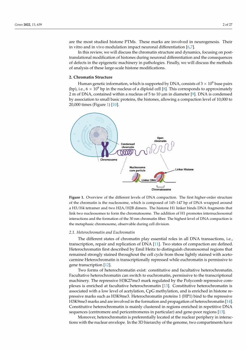
Human genetic information, which is supported by DNA, consists of 3× 10⁹ base pairs(bp), i.e., 6 × 10⁹ bp in the nucleus of a diploid cell [8]. This corresponds to approximately2 m of DNA, contained within a nucleus of 5 to 10 µm in diameter [9]. DNA is condensedby association to small basic proteins, the histones, allowing a compaction level of 10,000 to20,000 times (Figure 1) [10].
Genes 2022, 13, x FOR PEER REVIEW 2 of 27
occurs during embryogenesis but also in the adult brain [5]. Methylation and acetylation are the most studied histone PTMs. These marks are involved in neurogenesis. Their in vitro and in vivo modulation impact neuronal differentiation [6,7].
In this review, we will discuss the chromatin structure and dynamics, focusing on post-translational modification of histones during neuronal differentiation and the conse-quences of defects in the epigenetic machinery in pathologies. Finally, we will discuss the methods of analysis of these large-scale histone modifications.
2. Chromatin Structure Human genetic information, which is supported by DNA, consists of 3 × 10⁹ base
pairs (bp), i.e., 6 × 10⁹ bp in the nucleus of a diploid cell [8]. This corresponds to approxi-mately 2 m of DNA, contained within a nucleus of 5 to 10 µm in diameter [9]. DNA is condensed by association to small basic proteins, the histones, allowing a compaction level of 10,000 to 20,000 times (Figure 1) [10].
Figure 1. Overview of the different levels of DNA compaction. The first higher-order structure of the chromatin is the nucleosome, which is composed of 145–147 bp of DNA wrapped around a H3/H4 tetramer and two H2A/H2B dimers. The histone H1 linker binds DNA fragments that link two nucleosomes to form the chromatosome. The addition of H1 promotes internucleosomal inter-actions and the formation of the 30 nm chromatin fiber. The highest level of DNA compaction is the metaphasic chromosome, observable during cell division.
2.1. Heterochromatin and Euchromatin The different states of chromatin play essential roles in all DNA transactions, i.e.,
transcription, repair and replication of DNA [11]. Two states of compaction are defined. Heterochromatin first described by Emil Heitz to distinguish chromosomal regions that remained strongly stained throughout the cell cycle from those lightly stained with aceto-carmine Heterochromatin is transcriptionally repressed while euchromatin is permissive to gene transcription [12].
Two forms of heterochromatin exist: constitutive and facultative heterochromatin. Facultative heterochromatin can switch to euchromatin, permissive to the transcriptional machinery. The repressive H3K27me3 mark regulated by the Polycomb repressive com-plexes is enriched at facultative heterochromatin [13]. Constitutive heterochromatin is as-sociated with a low level of acetylation, CpG methylation, and is enriched in histone re-pressive marks such as H3K9me3. Heterochromatin proteins 1 (HP1) bind to the repres-sive H3K9me3 marks and are involved in the formation and propagation of heterochro-
Figure 1. Overview of the different levels of DNA compaction. The first higher-order structureof the chromatin is the nucleosome, which is composed of 145–147 bp of DNA wrapped arounda H3/H4 tetramer and two H2A/H2B dimers. The histone H1 linker binds DNA fragments thatlink two nucleosomes to form the chromatosome. The addition of H1 promotes internucleosomalinteractions and the formation of the 30 nm chromatin fiber. The highest level of DNA compaction isthe metaphasic chromosome, observable during cell division.
2.1. Heterochromatin and Euchromatin
The different states of chromatin play essential roles in all DNA transactions, i.e.,transcription, repair and replication of DNA [11]. Two states of compaction are defined.Heterochromatin first described by Emil Heitz to distinguish chromosomal regions thatremained strongly stained throughout the cell cycle from those lightly stained with aceto-carmine Heterochromatin is transcriptionally repressed while euchromatin is permissive togene transcription [12].
Two forms of heterochromatin exist: constitutive and facultative heterochromatin.Facultative heterochromatin can switch to euchromatin, permissive to the transcriptionalmachinery. The repressive H3K27me3 mark regulated by the Polycomb repressive com-plexes is enriched at facultative heterochromatin [13]. Constitutive heterochromatin isassociated with a low level of acetylation, CpG methylation, and is enriched in histone re-pressive marks such as H3K9me3. Heterochromatin proteins 1 (HP1) bind to the repressiveH3K9me3 marks and are involved in the formation and propagation of heterochromatin [14].Constitutive heterochromatin is mainly clustered in regions enriched in repetitive DNAsequences (centromere and pericentromeres in particular) and gene-poor regions [13].
Moreover, heterochromatin is preferentially located at the nuclear periphery in interac-tions with the nuclear envelope. In the 3D hierarchy of the genome, two compartments have
Genes 2022, 13, 639 3 of 27
been defined: the A compartment (euchromatin) enriched in H3K27ac and H3K39me3 andtranscriptionally active, and the B compartment (heterochromatin), associated with a silentstate of transcription, enriched in H3K9me3 [15]. In contrast to this configuration foundin most eukaryotic nuclei, some nuclei display a so-called inverted compartmentalizationwith a dense nuclear center surrounded by a more decondensed region [16]. This type ofnuclear architecture is found, for instance, in the rod photoreceptors involved in the blackand white vision in mammals.
In addition to these two conventional compartments, a compartment I has been de-scribed in colon cells. In the normal colon, this I compartment is located at an intermediateposition between the two canonical A and B compartments, interacts with these two com-partments and is associated with a relatively low level of transcription. It is distinguishedfrom the two other compartments by an enrichment in H3K27me3 [15]. The study of cellswith inverted nuclei (rod photoreceptors, lamin B receptor-null thymocytes) and conven-tional nuclei (non-rod retinal neurons, wild type thymocytes from mice) using polymersimulations to reconcile microscopy and Hi-C data revealed that interactions betweenheterochromatic regions are essential in the A and B compartmentalization of invertednuclei but also of conventional type nuclei.
The living chromatin model, which allows the study of coupling between chromatinfolding and epigenetic regulation, shows that the maintenance of the epigenome is regu-lated by 3D compartmentalization. This compartmentalization allows the local concen-tration of epigenome effectors, increasing their capacity to diffuse an epigenomic signalat a long distance [17]. In addition, lamina-heterochromatin interactions are required toestablish the conventional nuclear architecture [18].
2.2. DNA Compaction
The first fundamental level of higher-order chromatin structure is called the nucleo-some. This structure is composed of an octamer of histones and DNA wrapped aroundit [19–21]. The nucleosome consists of two dimers of H2A-H2B and a tetramer H3-H4 thatallow the left winding, in a helical way, of 145 to 147 bp of DNA [22,23]. This structureforms the nucleosome core particle and has a molecular weight of approximately 205 kDa.The nucleosome also affects the pitch of the helix; the DNA alone, type B, has 10.5 bp/turnof the helix; the DNA wrapped around the histones undergoes a slight twist leading to ahelix pitch of 10.2 bp/turn [22,24].
The nucleosome core particle (NCP) has an axis of symmetry. The axis is in a planeperpendicular to the H2A/H2B dimer and passes through the H3/H4 dimer. The H2A/H2Bdimer contains six amino acids of H2A (Glu56, Glu61, Glu64, Asp90, Glu91, Glu92) andtwo amino acids of H2B (Glu105, Glu113) that create a negatively charged environment.This acid patch is involved in chromatin compaction by allowing interactions with theN-terminal tail of the adjacent histone H4 nucleosome [25,26].
In addition to the nucleosome core particle, the linker histone or internucleosomalhistone H1 forms a new chromatin structure called chromatosome [27]. H1 has a molecularweight of 21 kDa and promotes interaction between adjacent nucleosomes. The linkerhistone is composed of approximately 200 amino acids and binds to about 10 to 60 bp ofDNA (called linker DNA). Electrostatic interactions between lysine or arginine residues ofchromatosome histones and DNA phosphate groups stabilize the nucleosome [10,28–30].
The second level of DNA compaction involves the formation of the 30 nm chromatinfiber [31]. H1 plays an essential role in its formation since it cannot be formed in vitro iflinker histones are depleted. Interactions between the acid patch and the tail of histoneH4 are also involved in its formation. The structure of the 30 nm fiber is still debated, andtwo main models are proposed: the solenoid model and the zigzag model. In the zigzagmodel, odd nucleosomes are stacked on top of each other as are the even nucleosomes.Interactions involve nucleosome N and N + 2. In the solenoid model, the interactions aremade between the N and N + 1 nucleosomes, which allow the nucleosomes to follow oneafter the other [31,32].
Genes 2022, 13, 639 4 of 27
At the 3D level, chromatin fiber can form loops through local interactions that de-limitates topologically associated domains (TADs). DNA sequences inside a TAD interactwith each other more frequently than sequences outside. These TADs are typically delim-ited by the interaction of the anchor CTCF protein and cohesin. These regions delineateenhancer/promoter interactions and thus play a role in gene expression [33]. The CTCFprotein also participates in the stability of adjacent antagonistic epigenomic domains [17].TADs can be found in the three genomic compartments A, B or I and interact preferentiallywith each other within the same compartment [15].
2.3. Histone Structure and Variants
Histones, like other proteins, are synthesized in the cytoplasm. To prevent histonemismatch, so-called histone chaperone proteins bind to neosynthesized histones involvedin the transport of histones to the nucleus [34]. These chaperones also prevent DNA-non-specific bonds and histone degradation. ATP-dependent chromatin remodeling complexesare associated to these histone chaperones and required for nucleosome positioning. Thesecomplexes are involved in the incorporation of histone variants and influence nucleosomespacing, sliding or removal. Four families of chromatin remodelers have been defined andclassified according to the sequence and structure of the ATPase domain: SWI/SNF, ISWI,CHD and INO80 [35].
Histones from the nucleosome core particle comprise a secondary structure knownas the histone fold, amino- and carboxyterminal extensions, an amino-terminal tail and acarboxyterminal tail for H2A. The histone fold is constituted by three helixes connectedby loops according to a α1-L1-α2-L2-α3 model. The α1 and α3 helix are relatively short(9 to 14 amino acids), unlike the α2 helix with an average of 29 amino acids [22,23]. Thesecondary structure of the histone fold is maintained between the four nucleosome coreparticle histones despite low sequence retention. The histone fold promotes protein–proteininteractions that will be used for heterodimerization of H2A/H2B and H3/H4 [36]. TheN-terminal ends do not have a defined secondary structure. The linker histone is composedof unstructured amino- and carboxyterminal tails and an apolar central globular area. TheC-terminal tail is basic [37].
In addition to the conventional canonical histones, several histone variants, defined asnon-allelic isoforms of canonical histones exist [38]. Variants have specific characteristicsthat modify the nucleosome structure with specific functions [39]. Histones H2A and H3have the higher number of variants. Among them, CENP-A, a histone H3 variant specific tocentromeres, plays a key role in the assembly of the kinetochore complex during mitosis [40].The γ-H2AX variant, phosphorylated on serine 139, by the ATR and ATM kinases of thecell cycle checkpoint is recognized by DNA repair effectors and involved in DNA damageresponse [41]. MacroH2A accumulates at double strand DNA breaks. The macroH2A1.1splice variant interacts with the KDM5A lysine demethylase, which is recruited to DNAdamage sites. Subsequently, H3K9me3 demethylation promotes the recruitment of theZMYND8-NuRD complex. This complex causes transcriptional repression and repair ofdouble-strand breaks by homologous recombination [42–44].
3. Histone Post-Translational Modifications
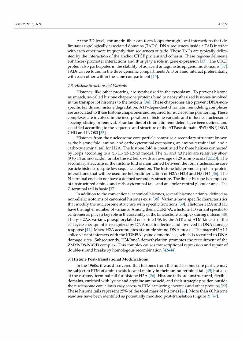
In the 1960s, it was discovered that histones from the nucleosome core particle maybe subject to PTM of amino acids located mainly in their amino-terminal tail [45] but alsoat the carboxy-terminal tail for histone H2A [26]. Histone tails are unstructured, flexibledomains, enriched with lysine and arginine amino acid, and their strategic position outsidethe nucleosome core allows easy access to PTM catalyzing enzymes and other proteins [22].These histone tails represent 25% of the total mass of histones [46]. More than 60 histoneresidues have been identified as potentially modified post-translation (Figure 2) [47].
Genes 2022, 13, 639 5 of 27
Genes 2022, 13, x FOR PEER REVIEW 5 of 27
the nucleosome core allows easy access to PTM catalyzing enzymes and other proteins [22]. These histone tails represent 25% of the total mass of histones [46]. More than 60 histone residues have been identified as potentially modified post-translation (Figure 2) [47].
Figure 2. Structure of a nucleosome and main sites of methylation and acetylation in histones. Post-translational modifications of the histones are mostly performed on the amino-terminal tails of the histones accessible to the epigenetic writer and eraser. Acetylated residues are in pink and methylated ones are in green.
Histone modifications affect chromatin compaction, thus regulating gene transcrip-tion and are therefore associated with epigenetic processes. Enzymes that catalyze PTMs are considered as epigenetic writers; so-called readers are proteins able to read these PTMs, while erasers remove them. Histones undergo different types of PTMs, such as acetylation, methylation, phosphorylation, ubiquination, polyADP-ribosylation, SUMOy-lation, deimination, citrullination, proprionylation, crotonylation, or isomerization of pro-line residues [47–54]. These PTMs form a “histone code” that affects the higher chromatin structure, inter-histone interactions, DNA-histone interactions and the recruitment of non-histone protein [53]. Acetylation and methylation, the most studied PTMs for their role in transcriptional regulation are detailed below. Post-translational modifications in the same histone or between adjacent histones may interact in an antagonistic or syner-gistic manner to modulate cellular processes [55,56].
3.1. Histone Acetylation and Deacetylation The addition of an acetyl group on histones was discovered in 1964 [45]. The reaction
is catalyzed by histones acetyl transferase (HAT) using acetyl CoA as a cofactor allowing the establishment of a covalent bond between an acetyl group and the lateral chain ε-amino of lysine. At a physiological pH, electrostatic interactions are formed between his-tone proteins (positively charged by lysine and arginine) and DNA (negatively charged by phosphate groups), allowing DNA compaction. The loss of the positive charge of lysine weakens the electrostatic bonds, leading to a relaxation of the chromatin, which favors the access of transcription factors to cis-regulatory sequences (Figure 3) [57].
Figure 2. Structure of a nucleosome and main sites of methylation and acetylation in histones. Post-translational modifications of the histones are mostly performed on the amino-terminal tails of thehistones accessible to the epigenetic writer and eraser. Acetylated residues are in pink and methylatedones are in green.
Histone modifications affect chromatin compaction, thus regulating gene transcriptionand are therefore associated with epigenetic processes. Enzymes that catalyze PTMs areconsidered as epigenetic writers; so-called readers are proteins able to read these PTMs,while erasers remove them. Histones undergo different types of PTMs, such as acetyla-tion, methylation, phosphorylation, ubiquination, polyADP-ribosylation, SUMOylation,deimination, citrullination, proprionylation, crotonylation, or isomerization of prolineresidues [47–54]. These PTMs form a “histone code” that affects the higher chromatinstructure, inter-histone interactions, DNA-histone interactions and the recruitment of non-histone protein [53]. Acetylation and methylation, the most studied PTMs for their role intranscriptional regulation are detailed below. Post-translational modifications in the samehistone or between adjacent histones may interact in an antagonistic or synergistic mannerto modulate cellular processes [55,56].
3.1. Histone Acetylation and Deacetylation
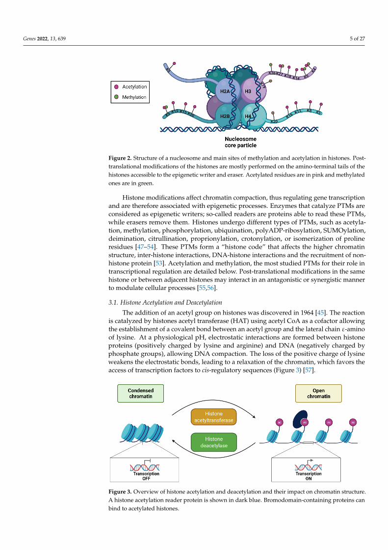
The addition of an acetyl group on histones was discovered in 1964 [45]. The reactionis catalyzed by histones acetyl transferase (HAT) using acetyl CoA as a cofactor allowingthe establishment of a covalent bond between an acetyl group and the lateral chain ε-aminoof lysine. At a physiological pH, electrostatic interactions are formed between histoneproteins (positively charged by lysine and arginine) and DNA (negatively charged byphosphate groups), allowing DNA compaction. The loss of the positive charge of lysineweakens the electrostatic bonds, leading to a relaxation of the chromatin, which favors theaccess of transcription factors to cis-regulatory sequences (Figure 3) [57].
Genes 2022, 13, x FOR PEER REVIEW 6 of 27
Figure 3. Overview of histone acetylation and deacetylation and their impact on chromatin struc-ture. A histone acetylation reader protein is shown in dark blue. Bromodomain-containing pro-teins can bind to acetylated histones.
Since HATs do not exclusively target histones, a more generic name, K-acetyltrans-ferases (KAT) has been given [58]. Six families are distinguished in this KAT superfamily: the Gcn5-related N-acetyltransferase (GNAT) family, the MYST family, the p300/CBP family, the HAT family related to transcription factors, the cytoplasmic KAT family and the nuclear receptor co-activators family (Table 1) [58,59]. Acetylated proteins can be rec-ognized, for example, by bromodomains, Yeats domains and double PHD finger [60]. Acetylation, like other histone PTMs, is a reversible modification. Histone deacetylase (HDAC) catalyze the removal of acetyl groups on histones, generally assimilated to tran-scriptional repression (Figure 3). These enzymes have a low substrate specificity allowing them to target several histone residues [29]. The eighteen HDACs are classified into four classes according to their sequence and mechanism of action. Class I HDACs, which in-clude HDAC1 to 3 and HDAC8, are nuclear proteins. HDAC4 to 7, HDAC9 and HDAC10 belong to Class II. Class III are NAD-dependent and include members of the Sirtuin sub-family (SIRT 1 to 7). HDAC11 is the only member of class IV. Class I, II and IV are zinc-dependent [60].
Table 1. Histone acetylation and deacetylation are performed by KATs and HDACs, respectively. Two types of KATs are defined: type A and B. Type A KATs have a nuclear localization and mod-ify chromatin-associated histones, whereas type B KATs, mainly localized at the cytoplasmic level, modify newly synthesized histones not incorporated in the nucleosome. KATs are classified into six families and HDACs into four.
Histone Acetyltransferase
TYPE B TYPE A Cytoplasmic KAT Family
GNAT Family P300/CBP Family MYST Family Transcription Fac-tor Related Family
Nuclear Receptor Coactivator Family
KAT1 (HAT1) KAT2A
(hGCN5) KAT3A (CBP) KAT5
(TIP60/PLIP) KAT4 (TAF1) KAT13A (SRC1)
KAT2B (PCAF) KAT3B (p300) KAT6A
(MOZ/MYST3) KAT12 (TFIIIC90) KAT13B (ACTR)
KAT9 (ELP3) KAT6B (MORF/MYST4)
KAT13C (P160)
KAT7
(HBO1/MYST2) KAT13D (CLOCK)
KAT8 (HMOF/MYST1)
Histone Deacety-lase
Class I Class II Class III Class IV HDAC1 HDAC4 SIRT1 HDAC11 HDAC2 HDAC5 SIRT2 HDAC3 HDAC6 SIRT3
Figure 3. Overview of histone acetylation and deacetylation and their impact on chromatin structure.A histone acetylation reader protein is shown in dark blue. Bromodomain-containing proteins canbind to acetylated histones.
Genes 2022, 13, 639 6 of 27
Since HATs do not exclusively target histones, a more generic name, K-acetyltransferases(KAT) has been given [58]. Six families are distinguished in this KAT superfamily: the Gcn5-related N-acetyltransferase (GNAT) family, the MYST family, the p300/CBP family, theHAT family related to transcription factors, the cytoplasmic KAT family and the nuclearreceptor co-activators family (Table 1) [58,59]. Acetylated proteins can be recognized, forexample, by bromodomains, Yeats domains and double PHD finger [60]. Acetylation, likeother histone PTMs, is a reversible modification. Histone deacetylase (HDAC) catalyze theremoval of acetyl groups on histones, generally assimilated to transcriptional repression(Figure 3). These enzymes have a low substrate specificity allowing them to target severalhistone residues [29]. The eighteen HDACs are classified into four classes according to theirsequence and mechanism of action. Class I HDACs, which include HDAC1 to 3 and HDAC8,are nuclear proteins. HDAC4 to 7, HDAC9 and HDAC10 belong to Class II. Class III areNAD-dependent and include members of the Sirtuin subfamily (SIRT 1 to 7). HDAC11 isthe only member of class IV. Class I, II and IV are zinc-dependent [60].
Table 1. Histone acetylation and deacetylation are performed by KATs and HDACs, respectively.Two types of KATs are defined: type A and B. Type A KATs have a nuclear localization and modifychromatin-associated histones, whereas type B KATs, mainly localized at the cytoplasmic level,modify newly synthesized histones not incorporated in the nucleosome. KATs are classified into sixfamilies and HDACs into four.
HistoneAcetyltransferase
TYPE B TYPE A
CytoplasmicKAT Family
GNATFamily
P300/CBPFamily MYST Family Transcription Factor
Related FamilyNuclear Receptor
Coactivator Family
KAT1 (HAT1) KAT2A(hGCN5) KAT3A (CBP) KAT5
(TIP60/PLIP) KAT4 (TAF1) KAT13A (SRC1)
KAT2B(PCAF) KAT3B (p300) KAT6A
(MOZ/MYST3) KAT12 (TFIIIC90) KAT13B (ACTR)
KAT9(ELP3)
KAT6B(MORF/MYST4) KAT13C (P160)
KAT7(HBO1/MYST2) KAT13D (CLOCK)
KAT8(HMOF/MYST1)
Histone Deacetylase
Class I Class II Class III Class IV
HDAC1 HDAC4 SIRT1 HDAC11HDAC2 HDAC5 SIRT2HDAC3 HDAC6 SIRT3HDAC8 HDAC7 SIRT4
HDAC9 SIRT5HDAC10 SIRT6
SIRT7
3.2. Methylation and Demethylation of Histones
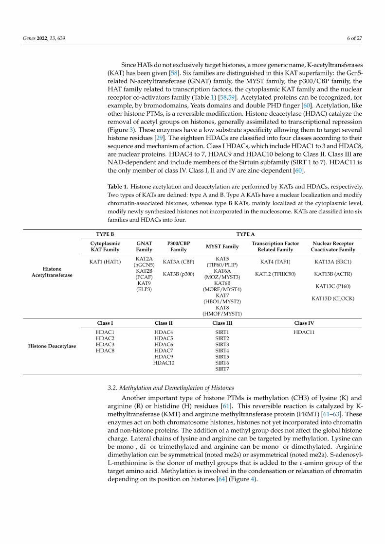
Another important type of histone PTMs is methylation (CH3) of lysine (K) andarginine (R) or histidine (H) residues [61]. This reversible reaction is catalyzed by K-methyltransferase (KMT) and arginine methyltransferase protein (PRMT) [61–63]. Theseenzymes act on both chromatosome histones, histones not yet incorporated into chromatinand non-histone proteins. The addition of a methyl group does not affect the global histonecharge. Lateral chains of lysine and arginine can be targeted by methylation. Lysine canbe mono-, di- or trimethylated and arginine can be mono- or dimethylated. Argininedimethylation can be symmetrical (noted me2s) or asymmetrical (noted me2a). S-adenosyl-L-methionine is the donor of methyl groups that is added to the ε-amino group of thetarget amino acid. Methylation is involved in the condensation or relaxation of chromatindepending on its position on histones [64] (Figure 4).
Genes 2022, 13, 639 7 of 27
Genes 2022, 13, x FOR PEER REVIEW 7 of 27
HDAC8 HDAC7 SIRT4 HDAC9 SIRT5 HDAC10 SIRT6 SIRT7
3.2. Methylation and Demethylation of Histones Another important type of histone PTMs is methylation (CH3) of lysine (K) and ar-
ginine (R) or histidine (H) residues [61]. This reversible reaction is catalyzed by K-methyl-transferase (KMT) and arginine methyltransferase protein (PRMT) [61–63]. These en-zymes act on both chromatosome histones, histones not yet incorporated into chromatin and non-histone proteins. The addition of a methyl group does not affect the global his-tone charge. Lateral chains of lysine and arginine can be targeted by methylation. Lysine can be mono-, di- or trimethylated and arginine can be mono- or dimethylated. Arginine dimethylation can be symmetrical (noted me2s) or asymmetrical (noted me2a). S-adeno-syl-L-methionine is the donor of methyl groups that is added to the ε-amino group of the target amino acid. Methylation is involved in the condensation or relaxation of chromatin depending on its position on histones [64] (Figure 4).
Figure 4. Overview of the mechanism of histone methylation by a methyltransferase. S-adenosyl-L-methionine (SAM) is the methyltransferase cofactor. Following methylation, S-adenosylhomo-cysteine (SAH) is released and the methylated moiety is attached to Nε-Lysine. A methylation reader is represented in purple and corresponds, for example, to a chromodomain or tudor do-main protein.
KMT can be subdivided into two groups: KMT containing the Su(var)3–9 domain, Enhancer of Zeste, trithorax (SET, for example SUV39H1 and SUV39H2) and lysine me-thyltransferases, which do not contain a SET domain (e.g., Disruptor of Telomeric silenc-ing 1 Like or DOT1L). The SET domain is involved in the catalytic activity at the histone tail, while the second group targets the central part of the histone. Methylation readers are composed of different types of domains allowing them to associate with methylated lysine. Examples of characterized domains are: tudor domain, MBT, WD40, ADD, zing finger CW domain, BAH, Ankyrin, PWWP, DCD, TTD and plant homeodomain (PHD) [60].
Lysine demethylases (KDM) are classified into two families: amines oxidase deme-thylases dependent on the flavin adenine dinucleotide (FAD) (example: KMD1A) and the family of the Jumonji domain proteins [60]. LSD1 acts on lysines 4 and 9 of mono- and dimethylated histone H3 [65]. LSD1 is overexpressed in several cancers. In acute lympho-blastic leukemia cells, LSD1 is overexpressed and mono- and dimethylation of lysine 4 of histone H3 is decreased. LSD1 silencing in these cells results in upregulation of H3K4me1 and H3K4me2 associated with induced apoptosis and inhibition of cell proliferation [66]. Members of the family containing the Jumonji domain interact with Fe (II) and α-ketoglu-tarate. Lysines 4, 9, 27 and 36 of histone H3 and lysine 20 of histone H4 are targeted by this family [67].
3.3. Main Active and Repressive Histone Marks The combination of different PTMs deposited on histones defines regulatory func-
tions such as enhancers, active promoters, repressed or chromatin in a bivalent state [68].
Figure 4. Overview of the mechanism of histone methylation by a methyltransferase. S-adenosyl-L-methionine (SAM) is the methyltransferase cofactor. Following methylation, S-adenosylhomocysteine(SAH) is released and the methylated moiety is attached to Nε-Lysine. A methylation reader isrepresented in purple and corresponds, for example, to a chromodomain or tudor domain protein.
KMT can be subdivided into two groups: KMT containing the Su(var)3–9 domain,Enhancer of Zeste, trithorax (SET, for example SUV39H1 and SUV39H2) and lysine methyl-transferases, which do not contain a SET domain (e.g., Disruptor of Telomeric silencing 1Like or DOT1L). The SET domain is involved in the catalytic activity at the histone tail,while the second group targets the central part of the histone. Methylation readers arecomposed of different types of domains allowing them to associate with methylated lysine.Examples of characterized domains are: tudor domain, MBT, WD40, ADD, zing finger CWdomain, BAH, Ankyrin, PWWP, DCD, TTD and plant homeodomain (PHD) [60].
Lysine demethylases (KDM) are classified into two families: amines oxidase demethy-lases dependent on the flavin adenine dinucleotide (FAD) (example: KMD1A) and the fam-ily of the Jumonji domain proteins [60]. LSD1 acts on lysines 4 and 9 of mono- and dimethy-lated histone H3 [65]. LSD1 is overexpressed in several cancers. In acute lymphoblasticleukemia cells, LSD1 is overexpressed and mono- and dimethylation of lysine 4 of histoneH3 is decreased. LSD1 silencing in these cells results in upregulation of H3K4me1 andH3K4me2 associated with induced apoptosis and inhibition of cell proliferation [66]. Mem-bers of the family containing the Jumonji domain interact with Fe (II) and α-ketoglutarate.Lysines 4, 9, 27 and 36 of histone H3 and lysine 20 of histone H4 are targeted by thisfamily [67].
3.3. Main Active and Repressive Histone Marks
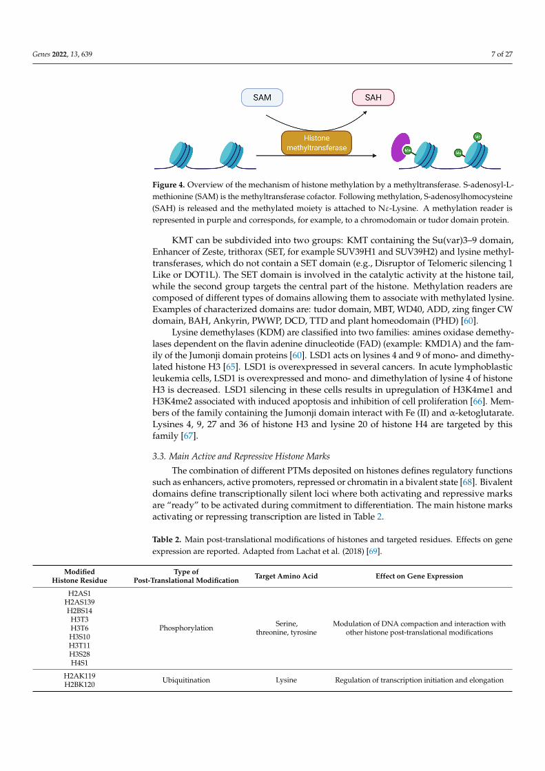
The combination of different PTMs deposited on histones defines regulatory functionssuch as enhancers, active promoters, repressed or chromatin in a bivalent state [68]. Bivalentdomains define transcriptionally silent loci where both activating and repressive marksare “ready” to be activated during commitment to differentiation. The main histone marksactivating or repressing transcription are listed in Table 2.
Table 2. Main post-translational modifications of histones and targeted residues. Effects on geneexpression are reported. Adapted from Lachat et al. (2018) [69].
ModifiedHistone Residue
Type ofPost-Translational Modification Target Amino Acid Effect on Gene Expression
H2AS1
Phosphorylation Serine,threonine, tyrosine
Modulation of DNA compaction and interaction withother histone post-translational modifications
H2AS139H2BS14
H3T3H3T6H3S10H3T11H3S28H4S1
H2AK119 Ubiquitination Lysine Regulation of transcription initiation and elongationH2BK120
Genes 2022, 13, 639 8 of 27
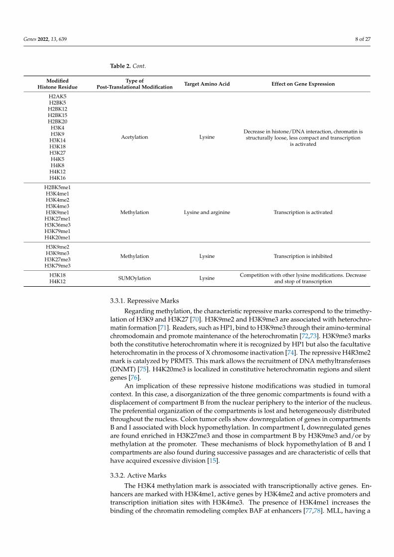
Table 2. Cont.
ModifiedHistone Residue
Type ofPost-Translational Modification Target Amino Acid Effect on Gene Expression
H2AK5
Acetylation LysineDecrease in histone/DNA interaction, chromatin isstructurally loose, less compact and transcription
is activated
H2BK5H2BK12H2BK15H2BK20
H3K4H3K9H3K14H3K18H3K27H4K5H4K8H4K12H4K16
H2BK5me1
Methylation Lysine and arginine Transcription is activated
H3K4me1H3K4me2H3K4me3H3K9me1H3K27me1H3K36me3H3K79me1H4K20me1
H3K9me2
Methylation Lysine Transcription is inhibitedH3K9me3H3K27me3H3K79me3
H3K18 SUMOylation Lysine Competition with other lysine modifications. Decreaseand stop of transcriptionH4K12
3.3.1. Repressive Marks
Regarding methylation, the characteristic repressive marks correspond to the trimethy-lation of H3K9 and H3K27 [70]. H3K9me2 and H3K9me3 are associated with heterochro-matin formation [71]. Readers, such as HP1, bind to H3K9me3 through their amino-terminalchromodomain and promote maintenance of the heterochromatin [72,73]. H3K9me3 marksboth the constitutive heterochromatin where it is recognized by HP1 but also the facultativeheterochromatin in the process of X chromosome inactivation [74]. The repressive H4R3me2mark is catalyzed by PRMT5. This mark allows the recruitment of DNA methyltransferases(DNMT) [75]. H4K20me3 is localized in constitutive heterochromatin regions and silentgenes [76].
An implication of these repressive histone modifications was studied in tumoralcontext. In this case, a disorganization of the three genomic compartments is found with adisplacement of compartment B from the nuclear periphery to the interior of the nucleus.The preferential organization of the compartments is lost and heterogeneously distributedthroughout the nucleus. Colon tumor cells show downregulation of genes in compartmentsB and I associated with block hypomethylation. In compartment I, downregulated genesare found enriched in H3K27me3 and those in compartment B by H3K9me3 and/or bymethylation at the promoter. These mechanisms of block hypomethylation of B and Icompartments are also found during successive passages and are characteristic of cells thathave acquired excessive division [15].
3.3.2. Active Marks
The H3K4 methylation mark is associated with transcriptionally active genes. En-hancers are marked with H3K4me1, active genes by H3K4me2 and active promoters andtranscription initiation sites with H3K4me3. The presence of H3K4me1 increases thebinding of the chromatin remodeling complex BAF at enhancers [77,78]. MLL, having a
Genes 2022, 13, 639 9 of 27
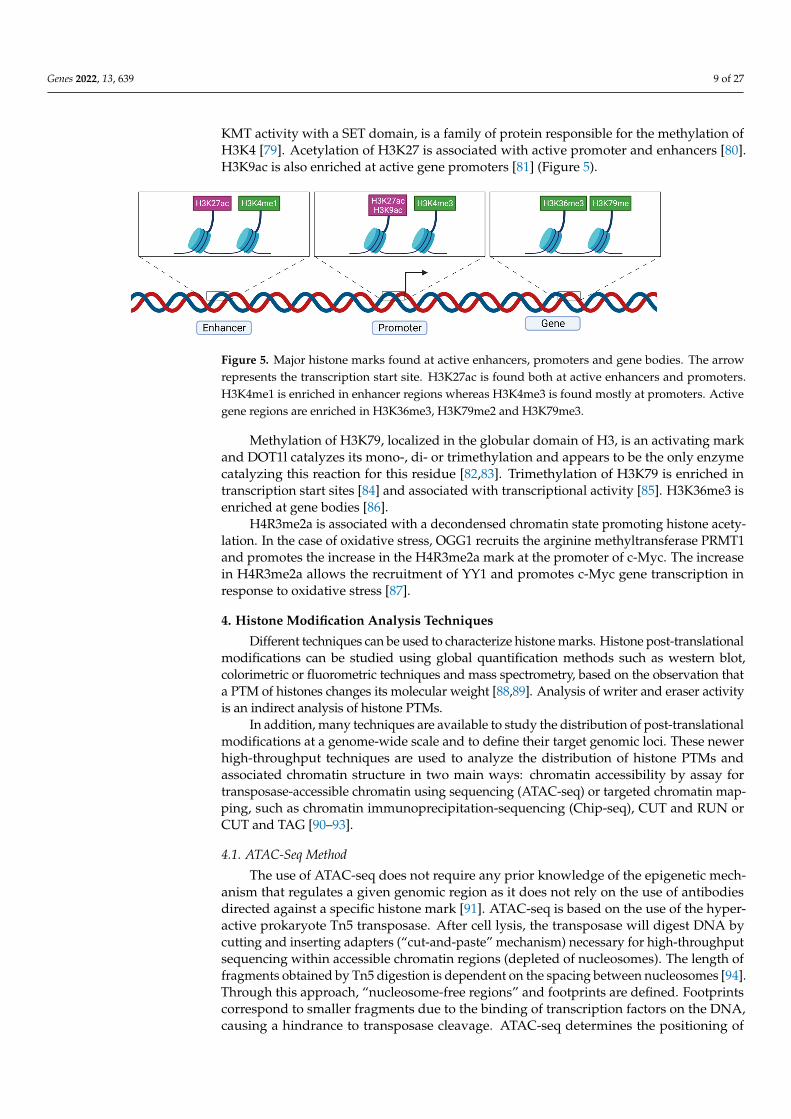
KMT activity with a SET domain, is a family of protein responsible for the methylation ofH3K4 [79]. Acetylation of H3K27 is associated with active promoter and enhancers [80].H3K9ac is also enriched at active gene promoters [81] (Figure 5).
Genes 2022, 13, x FOR PEER REVIEW 9 of 27
terminal chromodomain and promote maintenance of the heterochromatin [72,73]. H3K9me3 marks both the constitutive heterochromatin where it is recognized by HP1 but also the facultative heterochromatin in the process of X chromosome inactivation [74]. The repressive H4R3me2 mark is catalyzed by PRMT5. This mark allows the recruitment of DNA methyltransferases (DNMT) [75]. H4K20me3 is localized in constitutive heterochro-matin regions and silent genes [76].
An implication of these repressive histone modifications was studied in tumoral con-text. In this case, a disorganization of the three genomic compartments is found with a displacement of compartment B from the nuclear periphery to the interior of the nucleus. The preferential organization of the compartments is lost and heterogeneously distributed throughout the nucleus. Colon tumor cells show downregulation of genes in compart-ments B and I associated with block hypomethylation. In compartment I, downregulated genes are found enriched in H3K27me3 and those in compartment B by H3K9me3 and/or by methylation at the promoter. These mechanisms of block hypomethylation of B and I compartments are also found during successive passages and are characteristic of cells that have acquired excessive division [15].
3.3.2. Active Marks The H3K4 methylation mark is associated with transcriptionally active genes. En-
hancers are marked with H3K4me1, active genes by H3K4me2 and active promoters and transcription initiation sites with H3K4me3. The presence of H3K4me1 increases the bind-ing of the chromatin remodeling complex BAF at enhancers [77,78]. MLL, having a KMT activity with a SET domain, is a family of protein responsible for the methylation of H3K4 [79]. Acetylation of H3K27 is associated with active promoter and enhancers [80]. H3K9ac is also enriched at active gene promoters [81] (Figure 5).
Methylation of H3K79, localized in the globular domain of H3, is an activating mark and DOT1l catalyzes its mono-, di- or trimethylation and appears to be the only enzyme catalyzing this reaction for this residue [82,83]. Trimethylation of H3K79 is enriched in transcription start sites [84] and associated with transcriptional activity [85]. H3K36me3 is enriched at gene bodies [86].
H4R3me2a is associated with a decondensed chromatin state promoting histone acet-ylation. In the case of oxidative stress, OGG1 recruits the arginine methyltransferase PRMT1 and promotes the increase in the H4R3me2a mark at the promoter of c-Myc. The increase in H4R3me2a allows the recruitment of YY1 and promotes c-Myc gene transcrip-tion in response to oxidative stress [87].
Figure 5. Major histone marks found at active enhancers, promoters and gene bodies. The arrow represents the transcription start site. H3K27ac is found both at active enhancers and promoters. H3K4me1 is enriched in enhancer regions whereas H3K4me3 is found mostly at promoters. Active gene regions are enriched in H3K36me3, H3K79me2 and H3K79me3.
4. Histone Modification Analysis Techniques Different techniques can be used to characterize histone marks. Histone post-trans-
lational modifications can be studied using global quantification methods such as western
Figure 5. Major histone marks found at active enhancers, promoters and gene bodies. The arrowrepresents the transcription start site. H3K27ac is found both at active enhancers and promoters.H3K4me1 is enriched in enhancer regions whereas H3K4me3 is found mostly at promoters. Activegene regions are enriched in H3K36me3, H3K79me2 and H3K79me3.
Methylation of H3K79, localized in the globular domain of H3, is an activating markand DOT1l catalyzes its mono-, di- or trimethylation and appears to be the only enzymecatalyzing this reaction for this residue [82,83]. Trimethylation of H3K79 is enriched intranscription start sites [84] and associated with transcriptional activity [85]. H3K36me3 isenriched at gene bodies [86].
H4R3me2a is associated with a decondensed chromatin state promoting histone acety-lation. In the case of oxidative stress, OGG1 recruits the arginine methyltransferase PRMT1and promotes the increase in the H4R3me2a mark at the promoter of c-Myc. The increasein H4R3me2a allows the recruitment of YY1 and promotes c-Myc gene transcription inresponse to oxidative stress [87].
4. Histone Modification Analysis Techniques
Different techniques can be used to characterize histone marks. Histone post-translationalmodifications can be studied using global quantification methods such as western blot,colorimetric or fluorometric techniques and mass spectrometry, based on the observation thata PTM of histones changes its molecular weight [88,89]. Analysis of writer and eraser activityis an indirect analysis of histone PTMs.
In addition, many techniques are available to study the distribution of post-translationalmodifications at a genome-wide scale and to define their target genomic loci. These newerhigh-throughput techniques are used to analyze the distribution of histone PTMs andassociated chromatin structure in two main ways: chromatin accessibility by assay fortransposase-accessible chromatin using sequencing (ATAC-seq) or targeted chromatin map-ping, such as chromatin immunoprecipitation-sequencing (Chip-seq), CUT and RUN orCUT and TAG [90–93].
4.1. ATAC-Seq Method
The use of ATAC-seq does not require any prior knowledge of the epigenetic mech-anism that regulates a given genomic region as it does not rely on the use of antibodiesdirected against a specific histone mark [91]. ATAC-seq is based on the use of the hyper-active prokaryote Tn5 transposase. After cell lysis, the transposase will digest DNA bycutting and inserting adapters (“cut-and-paste” mechanism) necessary for high-throughputsequencing within accessible chromatin regions (depleted of nucleosomes). The length offragments obtained by Tn5 digestion is dependent on the spacing between nucleosomes [94].Through this approach, “nucleosome-free regions” and footprints are defined. Footprintscorrespond to smaller fragments due to the binding of transcription factors on the DNA,causing a hindrance to transposase cleavage. ATAC-seq determines the positioning of
Genes 2022, 13, 639 10 of 27
nucleosomes, transcription factor binding sites and gene regulatory regions to be identifiedto the nearest nucleotide [95].
4.2. Targeted Chromatin Mapping Methods4.2.1. ChIP-Seq
Chromatin immunoprecipitation (ChIP), followed by high-throughput sequencing(seq), is now a commonly used method for genome-wide identification of specific post-translational modification-associated loci. ChIP-seq relies on the use of specific antibodiesto determine the binding site of a target protein at a genome-wide scale. The antibodiescan be directed against transcription factors to identify cis-regulatory sequences, but alsoagainst histone PTMs.
ChIP can be performed on native (N-ChIP) or cross-linked (X-ChIP) chromatin. X-ChIPconsists in stabilizing DNA-protein interactions by bridging with chemical agents such asformaldehyde. The chromatin is then fragmented by ultrasound or enzymatic digestion.The accessibility of chromatin to the micrococcal nuclease varies according to the cell type,and different batches of enzymes may present variable activity, which may lead to biases.The size of the fragments obtained, following fragmentation, is important since it correlateswith the resolution of ChIP [90].
4.2.2. CUT and RUN
Cleavage Under Targets and Release Using Nuclease is an another high-throughputtechnique more recent than ChIP or ATAC-seq. This technique maps the interactions be-tween DNA and proteins, but, unlike ChIP-seq, does not require pre-labelled fragmentationof the DNA and tends to better preserve DNA/protein interactions. Therefore, this methodcan be used to analyze the binding of transcription factors to their cis-regulatory regionsbut also to analyze PTMs of histones [92]. No cross-linking of DNA/protein interactionsis required. CUT and RUN uses specific antibodies to which the protein A micrococcalnuclease (pA/Mnase) is attached. This will be activated by the addition of Ca2+ and willallow the cleavage on both sides of the protein binding site. The DNA fragments will besubmitted to sequencing by sequencing adapter ligation [96].
4.2.3. CUT and TAG
This technique also uses antibodies specifically directed against transcription factors orPMTs of histones. The Tn5 transposase, also used in ATAC-seq, is coupled to protein A. Thisfusion protein will guide the Tn5 transposase to the binding site of the antibody directedagainst the protein of interest. The transposase is coupled with adapters for subsequentsequencing [93].
The coupling of different techniques allows the visualization of different activating orrepressive histone marks correlated with the chromatin opening state [97,98].
5. Histones and Neuronal Differentiation
Post-translational modifications of histones in the modulation of chromatin com-paction and access to the transcriptional machinery are involved at the early stages ofdevelopment, and particularly during neuronal differentiation.
5.1. Overview of Neuronal Differentiation
During embryogenesis, the neuroepithelium gradually expands to form the neuraltube. The cells lining the neural tube are neural stem cells (NSC). These NSCs are multipo-tent, i.e., able of self-renew and differentiate. They allow the development of the centralnervous system, including the spinal cord and brain by differentiating to neuronal, astro-cytic and oligodendrocytic lineages [99]. In embryonic mammalian brains, neuroepithelialcells from the cortex will first divide symmetrically and later differentiate into radial glialcells (RGCs) through asymmetrical divisions allowing their self-renewal [100]. RCGs arelocated in the ventricular zone of the cortex, characterized by a loss of tight junctions and
Genes 2022, 13, 639 11 of 27
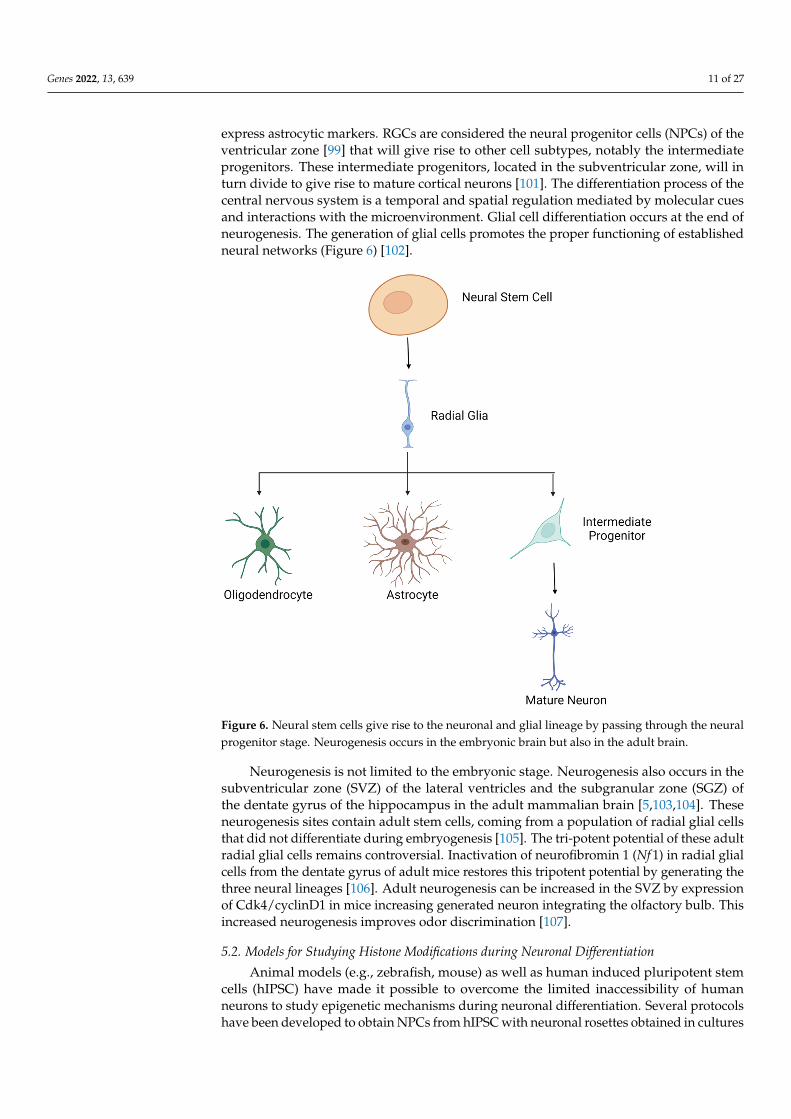
express astrocytic markers. RGCs are considered the neural progenitor cells (NPCs) of theventricular zone [99] that will give rise to other cell subtypes, notably the intermediateprogenitors. These intermediate progenitors, located in the subventricular zone, will inturn divide to give rise to mature cortical neurons [101]. The differentiation process of thecentral nervous system is a temporal and spatial regulation mediated by molecular cuesand interactions with the microenvironment. Glial cell differentiation occurs at the end ofneurogenesis. The generation of glial cells promotes the proper functioning of establishedneural networks (Figure 6) [102].
Genes 2022, 13, x FOR PEER REVIEW 11 of 27
4.2.3. CUT and TAG This technique also uses antibodies specifically directed against transcription factors
or PMTs of histones. The Tn5 transposase, also used in ATAC-seq, is coupled to protein A. This fusion protein will guide the Tn5 transposase to the binding site of the antibody directed against the protein of interest. The transposase is coupled with adapters for sub-sequent sequencing [93].
The coupling of different techniques allows the visualization of different activating or repressive histone marks correlated with the chromatin opening state [97,98].
5. Histones and Neuronal Differentiation Post-translational modifications of histones in the modulation of chromatin compac-
tion and access to the transcriptional machinery are involved at the early stages of devel-opment, and particularly during neuronal differentiation.
5.1. Overview of Neuronal Differentiation During embryogenesis, the neuroepithelium gradually expands to form the neural
tube. The cells lining the neural tube are neural stem cells (NSC). These NSCs are mul-tipotent, i.e., able of self-renew and differentiate. They allow the development of the cen-tral nervous system, including the spinal cord and brain by differentiating to neuronal, astrocytic and oligodendrocytic lineages [99]. In embryonic mammalian brains, neuroep-ithelial cells from the cortex will first divide symmetrically and later differentiate into ra-dial glial cells (RGCs) through asymmetrical divisions allowing their self-renewal [100]. RCGs are located in the ventricular zone of the cortex, characterized by a loss of tight junctions and express astrocytic markers. RGCs are considered the neural progenitor cells (NPCs) of the ventricular zone [99] that will give rise to other cell subtypes, notably the intermediate progenitors. These intermediate progenitors, located in the subventricular zone, will in turn divide to give rise to mature cortical neurons [101]. The differentiation process of the central nervous system is a temporal and spatial regulation mediated by molecular cues and interactions with the microenvironment. Glial cell differentiation oc-curs at the end of neurogenesis. The generation of glial cells promotes the proper func-tioning of established neural networks (Figure 6) [102].
Figure 6. Neural stem cells give rise to the neuronal and glial lineage by passing through the neuralprogenitor stage. Neurogenesis occurs in the embryonic brain but also in the adult brain.
Neurogenesis is not limited to the embryonic stage. Neurogenesis also occurs in thesubventricular zone (SVZ) of the lateral ventricles and the subgranular zone (SGZ) ofthe dentate gyrus of the hippocampus in the adult mammalian brain [5,103,104]. Theseneurogenesis sites contain adult stem cells, coming from a population of radial glial cellsthat did not differentiate during embryogenesis [105]. The tri-potent potential of these adultradial glial cells remains controversial. Inactivation of neurofibromin 1 (Nf 1) in radial glialcells from the dentate gyrus of adult mice restores this tripotent potential by generating thethree neural lineages [106]. Adult neurogenesis can be increased in the SVZ by expressionof Cdk4/cyclinD1 in mice increasing generated neuron integrating the olfactory bulb. Thisincreased neurogenesis improves odor discrimination [107].
5.2. Models for Studying Histone Modifications during Neuronal Differentiation
Animal models (e.g., zebrafish, mouse) as well as human induced pluripotent stemcells (hIPSC) have made it possible to overcome the limited inaccessibility of humanneurons to study epigenetic mechanisms during neuronal differentiation. Several protocolshave been developed to obtain NPCs from hIPSC with neuronal rosettes obtained in cultures
Genes 2022, 13, 639 12 of 27
resembling the neural tube. These NSCs can be differentiated into different types of neurons(glutamatergic, dopaminergic, GABAergic, etc.) using appropriate protocols [108–110]. Inaddition, it is now possible to generate a three-dimensional structure such as organoids orassembloids allowing a better view of cellular processes and cell–cell interactions. Theseapproaches can also be considered to improve the understanding of the pathophysiologyof chromatinopathies as reviewed in [111].
5.3. Histone Changes
The mechanisms of NSCs differentiation are spatio-temporally regulated by changesin the accessibility of transcription factors to the promoter of genes that regulate thedifferentiation to a specific lineage. Changes in chromatin will gradually limit access tomultipotency genes, with downregulation of pluripotency genes (OCT4, NANOG) andpromote access to the specific differentiation program associated with an increase in NPCmarkers (PAX6, SOX1 et OTX2) [112].
5.3.1. Role of Histone Acetylation and Deacetylation
HDAC are essential for the maintenance of self-renewal and proliferation of the neuralstem cell by maintaining, through their catalytic activity, transcriptional suppression ontarget genes. Several studies have been conducted to elucidate the role of HDACs in neu-ronal differentiation. Transient and incomplete HDAC inhibitors such as 2-propylpentanoicacid (called valproic acid or VPA), trichostatin A (TSA), suberoylanilide hydroxamic acid(SAHA) and sodium butyrate (NaB) have been used for this purpose. HDAC inhibition isassociated with histone hyperacetylation and excessive inhibitor concentrations, leading tocell death [113].
The use of VPA induces neuronal differentiation by affecting histone acetylation. Inrat embryonic hippocampus neural progenitor cells, VPA reduces cell proliferation andinitiates neuronal differentiation. This is mediated by an increase in the expression ofNgn1, Math1 and p15 through an increase in H4 acetylation at their promoter. Similarresults were observed in vivo in mice where VPA-treated animals showed an increase inH4 acetylation at the Ngn1 promoter inducing its expression [7]. In NSCs from rat embryohippocampi collected at E16 and treated with VPA, induction of neuronal differentiation isassociated with an increase in the H3K4me3 and H3K9ac active marks at the Ngn1 promotertogether with a decrease in the repressive H3K27me3 and H3K9me3. These modificationsare independent of the AKT/mTOR pathway activated after VPA treatment [114].
VPA also promotes neuronal differentiation in H9 human embryonic stem cells (HUES).Addition of VPA to the differentiation medium increases the expression of neuronal mark-ers (β3-tubulin, MAP2, NEUN) and those specific to the GABAergic and dopaminergicsubtypes. The markers of mature neuron, NEUROD1, and NeuN are expressed 30-timeshigher after VPA addition. This increase is also observed for oligodendrocyte markers(MBP) but not for astrocyte markers (S100β). Differentiation toward the astrocytic lineageis decreased under these conditions, which is usually observed during differentiation tothe neuronal lineage. Length and connections established by neurites are also significantlyincreased by VPA addition [115]. Besides, the use of retinoic acid (NaB) increases the acety-lation of H3K9 at the PAX6 promoter, which promotes the induction of neural progenitorcells [116].
H3K9ac is also involved in the kinetics of neural differentiation. Qiao et al., showedthat during the first four days of neural differentiation of HUES-9, H3K9 acetylation de-creases and then gradually increases between days four to eight. The kinetics of H3K9acetylation at specific promoters allows the silencing of pluripotency genes and the expres-sion of early neural genes. This is supported by experiments on p300 and the use of TSA.Indeed, shRNA depletion of p300, involved in H3K9 acetylation, inhibits the expression ofearly neural genes (PAX6, SOX1, ZIC2 and ZNF521). In addition, the use of TSA inhibitsthe expression of early neural genes, PAX6 and SOX1, between days zero to four but in-creases their expression between days four to eight. This is evidenced by an increase in the
Genes 2022, 13, 639 13 of 27
proportion of NPCs. TSA inhibits HDAC1/5 and 8, which allows the increase in H3K9acand the differentiation to the neural lineage. The expression of early neural genes such asPAX3/6, SOX1, POU3F2 is improved by HDAC1/5 and 8 knockdown, while pluripotencygenes are downregulated. These three enzymes allow an appropriate neural differentiationby preventing an early differentiation [112].
5.3.2. Role of Methylation and Demethylation
During in vitro neuronal differentiation from murine embryonic stem cells to neuralprogenitor cells, significant opposite changes are observed for H3K27ac and H3K79me2through genome-wide acting mechanisms. H3K27ac progressively decreases by a factor oftwo between the two stages, while H3K79me2 is increased four-fold, genome-wide. A gainof H3K79me2 during development is observed for a group of genes with a clear axonogenicsignature and critical for neuronal development. For these genes, H3K79me2 correlates withan increase in gene expression. A decrease in H3K27ac indicates a progressive chromatincondensation during neuronal differentiation in this model. These global opposing changesof histone PTMs might be the consequence of two mechanisms: the accumulation of histonemarks at specific loci with a global shift of the number and magnitude of these marksdepending on the conditions associated with a genome-wide global gain/loss of the signaldue to a balance of accumulation/deletion of histone PTMs [117].
Similar results were also observed when mouse ESCs were transduced into NPCs.The mESCs are in a more open chromatin conformation than differentiated cells with ahigher proportion of active marks such as H3K4me3, H3K9ac and H3K14ac comparedto NPCs [118]. The differentiation of mESCs is associated with an increase in H3K9me3and a decrease in acetylation of H3 and H4 [119]. Neuronal differentiation in vitro is thusaccompanied by a deacetylation associated with a more compact chromatin compared tomESC [117].
On the other hand, genes associated with differentiation and development are foundin a bivalent state with trimethylated marks of H3K27 and H3K4 at the mESC stage. Thesebivalent domains are reduced in differentiated cells and developmental genes acquireeither repressive or activating marks. For genes induced during neural differentiation (e.g.,Nkx2.2, Sox21 and Zfpm2), in a bivalent conformation in mESCs, a resolution of the bivalentstate by methylation of the transcription start site H3K4 is observed [120]. In NSCs, genesthat are specific to the neuronal lineage are kept in a silent state and expressed later duringdifferentiation after the loss of the repressive H3K27me3 mark [121].
DOT1L is currently considered the only enzyme catalyzing H3K79me methylationduring neuronal differentiation [82,83]. Its inhibition in mECSs and neural progenitorsdecreases the H3K79me2 marker and promotes neuronal differentiation in mESC-derivedNPCs. This treatment leads to a decrease in neural stem cell markers in NPCs associatedwith an increase in markers of fully differentiated neurons. DOT1L inhibition alters thedistribution of H3K27ac in ESCs and NPCs. Gene promoters are also affected. Genesassociated with promoters in a repressed state (H3K27me3) recognized by Polycombcomplexes or promoters in a bivalent state (H3K4me3 and H3K27me3) are both upregulated.Conversely, active genes (H3K4me3 and H3K27ac enriched-promoters) are downregulatedfollowing DOT1L inhibition. DOT1L inhibition also reduces SOX2 binding to its targetenhancers in NPCs. SOX2 expression is maintained in both embryonic and adult neuralstem cells until differentiation [117,122]. The decreased binding of SOX2 to its targetenhancers might explain the neuronal differentiation of NPCs following DOT1L inhibition.DOT1L would therefore be involved in the maintenance of NPCs by preventing theirdifferentiation via SOX2 [13,117].
Another KMT, MLL1 plays an important role in neuronal differentiation by methy-lating H3K4 [123] and by being involved in the demethylation of H3K27 by recruitingdemethylases [124]. This role was illustrated by studying the neuronal differentiation of theSVZ olfactory bulb from SVZ (subventricular zone) monolayer NSC cultures derived froman Mll1-deficient mouse model. This model shows normal astrocytic and oligodendrocytic
Genes 2022, 13, 639 14 of 27
cell differentiation but impaired neuronal differentiation with a significant decrease in thetranscription factor DLX2 involved in terminal differentiation of interneurons. MLL1 bindsto the promoter of the Dlx2 gene. In wild-type mice NSCs, activating H3K4me3 marks areenriched at the Dlx2 promoter. In Mll1-deficient mice in which Dlx2 expression is impaired,no decrease in H3K4me3 is reported but a significant increase in H3K27me3 is observed.In these mice, the Dlx2 locus becomes bivalent, and gene transcription is repressed. Mll1thus allows the removal of bivalent marks allowing the induction of neuronal differentia-tion by modulation of gene expression [125]. Studies on whole zebrafish embryos duringthe development of the nervous system have shown that morpholino oligonucleotide-mediated MLL1 protein depletion is associated with a decrease in the number of SOX2positive NPC due to a defect in their proliferation. No proliferation defect was observedin embryonic neurons. However, this decrease in MLL1 is associated with early neuronaldifferentiation and also negatively affects gliogenesis [126]. This leads to a decrease inthe expression of the neural progenitor marker neurogenin1 (Ngn1) and upregulation ofpostmitotic neuron-specific markers.
The EHMT2 methyltransferase is involved in the demethylation of mono- and dimethy-lated H3K9 [127] and also in the methylation of H3K27 residues [128]. This enzyme has twoisoforms that differ by the incorporation or not of exon 10 [129]. EHMT2 activity is essentialfor neuronal differentiation. Indeed, EHMT2 knockdown in the mouse-neural-crest-derivedcell line (N2a) abolishes neurite growth and inhibition of its methyltransferase activitypreventing the acquisition of a fully differentiated phenotype. The essential role of thecatalytic activity of EHMT2 is underlined by the rescue of the phenotype by transfectingwild-type EHMT2, which is not possible with the mutant without any catalytic activity.During neuronal differentiation of N2a, expression of the EHMT2 isoform with exon 10and H3K9 dimethylation are increased. Consistently, knockdown using an siRNA directedagainst exon 10 shows an inhibition of neuronal differentiation indicating that this isoformis necessary for neuronal differentiation [130].
The use of a morpholino antisense oligonucleotide against the Sox19b transcriptionfactor during embryonic development in zebrafish, leads to a decrease in H3K27me3 at theNgn1 and ascl1a promoters, without affecting the histone acetylation state. This decrease isdue to a decreased expression of EZH2, that catalyzes H3K27 trimethylation. Prematureentry of neural tube NSCs into neuronal differentiation is then induced by an increase inthe level of Ngn1 and ascl1a transcription, showing a role for the repressive H3K27me3mark during neuronal differentiation. Thus, the elimination of this repressive markercontrols the balance of self-renewal/differentiation of NSCs in zebrafish. The importance ofa proper functioning of this balance is further highlighted by the neural tube abnormalitiesassociated with a decrease in the surface area of the diencephalon and telencephalon inSOX19B-deficient embryos [131].
Lentiviral-induced overexpression of the neurogenic factor NeuroD1 is able to repro-gram mouse microglia and oligodendrocytes into mature neurons both in vitro and in vivo.These neurons are β3 tubulin-positive and respond to N-methyl-D aspartate (NMDA)stimulation. NeuroD1-mediated conversion proceeds directly from microglia to matureneurons without intermediates such as neural stem cells or Nestin-positive precursors.The generated neurons are predominantly glutamatergic and form excitatory synapses.NeuroD1 initiates this process of neuronal differentiation by binding to bivalent chromatinregions (enriched in H3K4me3 and H3K27me3) present in microglia at genes involved inneuronal development and differentiation such as Bhlhe22, Brn2 or Pou3f 3. Genes upregu-lated after NeuroD1 binding show a decrease in H3K27me3 and an increase in H3K4me3.Furthermore, microglial identity is suppressed by modification of histone marks at the pro-moters of microglia-specific genes. A decrease in H3K4me3 and an increase in H4K27me3on its promoter regions is observed [6].
Genes 2022, 13, 639 15 of 27
5.3.3. Modifications of 3D Architecture of Chromatin during Neuronal Differentiation
As previously discussed, 3D compartmentalization that plays a role in the maintenanceand regulation of the epigenome and by corollary, also influences neuronal differentia-tion processes. Comparison of Drosophila embryonic and neuronal cells indicates thatthree-dimensional organization is modulated during neuronal differentiation with dynamicchanges in enhancer/promoter interactions [132]. A number of TADs that are specific andcommon to both lineages have been identified. Neuron-specific TADs show enrichment inenhancers driven by neuron-specific transcription factors [132]. The regulation of A/B com-partmentalization, TADs and enhancer/promoter loops in neural development is reviewedby Kishi et al., Arzate-Bejía et al. and Ghosh et al. [133–135]. In particular, interactionswithin compartments A and B are modulated during in vitro neuronal differentiation. Adecrease in interactions within compartment A is observed during the first stage frommESCs into NPCs. Then, interactions within compartment B increase during the transitionfrom NPCs to cortical neurons [136].
For a more global approach for understanding neuronal differentiation, it is nowpossible to reconstruct the 3D organization of the genome using predictive methods. Animproved population-based modeling approach and a probabilistic framework to model apopulation of 3D structures of entire diploid genomes was tested on human lymphoblas-toid cells. The predicted 3D structures correctly identified many of the features of thelymphoblastoid genome obtained from imaging experiments, including interchromosomaldistances between gene loci, their interactions and preferred nuclear locations of chromo-somes [137]. Histone PTMs, together with sequence information of CTCF binding sites,are able to predict chromatin structure at a resolution from 5kb. This elaborate modelcombining both bioinformatics analysis with polymer modeling has allowed the characteri-zation of TADs, compartments and chromatin loops [138]. Other methods for modeling the3D structure of the genome are available, notably the data-driven polymer model, whichallows the summarization of the preferential chromosomal positions, the A/B compart-mentalization, to distinguish the inactive X chromosome by a more important compactionand a more inward localization than its active homologue [139].
6. Pathological Involvement of the Epigenetic Machinery
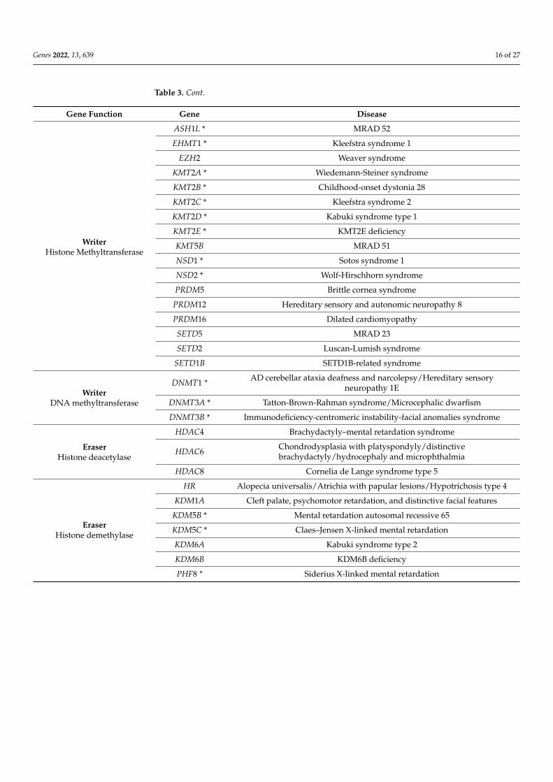
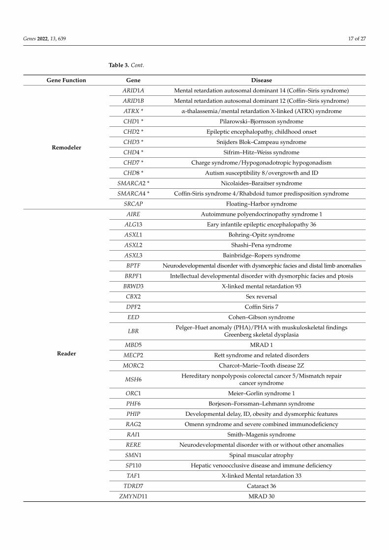
A number of neurodevelopmental disorders and cancers result from mutations incomponents of the epigenetic machinery [4,140–142]. Alterations in the functions of writ-ers, readers and erasers or chromatin remodelers have been reported as associated withMendelian disorders. To date, 86 diseases resulting from mutations in 74 genes encod-ing chromatin-enzymes have been reported [3,143,144]. Such disorders are called “chro-matinopathies” and are listed in Table 3.
Table 3. Mendelian disorders of the epigenetic machinery. Writer, eraser and remodeler enzymes thatalso carry a reader domain are marked by an asterisk. Adapted from [3,143,144].
Gene Function Gene Disease
WriterHistone Acetyltransferase
CREBBP * Rubinstein Taybi syndrome 1
EP300 * Rubinstein Taybi syndrome 2
KANSL1 Koolen-De Vries syndrome
KAT6A * Mental retardation autosomal dominant (MRAD) 32
KAT6B * Say-Barber-Biessecker-Young-Simpson syndrome/Genitopatellar syndrome
Genes 2022, 13, 639 16 of 27
Table 3. Cont.
Gene Function Gene Disease
WriterHistone Methyltransferase
ASH1L * MRAD 52
EHMT1 * Kleefstra syndrome 1
EZH2 Weaver syndrome
KMT2A * Wiedemann-Steiner syndrome
KMT2B * Childhood-onset dystonia 28
KMT2C * Kleefstra syndrome 2
KMT2D * Kabuki syndrome type 1
KMT2E * KMT2E deficiency
KMT5B MRAD 51
NSD1 * Sotos syndrome 1
NSD2 * Wolf-Hirschhorn syndrome
PRDM5 Brittle cornea syndrome
PRDM12 Hereditary sensory and autonomic neuropathy 8
PRDM16 Dilated cardiomyopathy
SETD5 MRAD 23
SETD2 Luscan-Lumish syndrome
SETD1B SETD1B-related syndrome
WriterDNA methyltransferase
DNMT1 * AD cerebellar ataxia deafness and narcolepsy/Hereditary sensoryneuropathy 1E
DNMT3A * Tatton-Brown-Rahman syndrome/Microcephalic dwarfism
DNMT3B * Immunodeficiency-centromeric instability-facial anomalies syndrome
EraserHistone deacetylase
HDAC4 Brachydactyly–mental retardation syndrome
HDAC6 Chondrodysplasia with platyspondyly/distinctivebrachydactyly/hydrocephaly and microphthalmia
HDAC8 Cornelia de Lange syndrome type 5
EraserHistone demethylase
HR Alopecia universalis/Atrichia with papular lesions/Hypotrichosis type 4
KDM1A Cleft palate, psychomotor retardation, and distinctive facial features
KDM5B * Mental retardation autosomal recessive 65
KDM5C * Claes–Jensen X-linked mental retardation
KDM6A Kabuki syndrome type 2
KDM6B KDM6B deficiency
PHF8 * Siderius X-linked mental retardation
Genes 2022, 13, 639 17 of 27
Table 3. Cont.
Gene Function Gene Disease
Remodeler
ARID1A Mental retardation autosomal dominant 14 (Coffin–Siris syndrome)
ARID1B Mental retardation autosomal dominant 12 (Coffin–Siris syndrome)
ATRX * α-thalassemia/mental retardation X-linked (ATRX) syndrome
CHD1 * Pilarowski–Bjornsson syndrome
CHD2 * Epileptic encephalopathy, childhood onset
CHD3 * Snijders Blok–Campeau syndrome
CHD4 * Sifrim–Hitz–Weiss syndrome
CHD7 * Charge syndrome/Hypogonadotropic hypogonadism
CHD8 * Autism susceptibility 8/overgrowth and ID
SMARCA2 * Nicolaides–Baraitser syndrome
SMARCA4 * Coffin-Siris syndrome 4/Rhabdoid tumor predisposition syndrome
SRCAP Floating–Harbor syndrome
Reader
AIRE Autoimmune polyendocrinopathy syndrome 1
ALG13 Eary infantile epileptic encephalopathy 36
ASXL1 Bohring–Opitz syndrome
ASXL2 Shashi–Pena syndrome
ASXL3 Bainbridge–Ropers syndrome
BPTF Neurodevelopmental disorder with dysmorphic facies and distal limb anomalies
BRPF1 Intellectual developmental disorder with dysmorphic facies and ptosis
BRWD3 X-linked mental retardation 93
CBX2 Sex reversal
DPF2 Coffin Siris 7
EED Cohen–Gibson syndrome
LBR Pelger–Huet anomaly (PHA)/PHA with muskuloskeletal findingsGreenberg skeletal dysplasia
MBD5 MRAD 1
MECP2 Rett syndrome and related disorders
MORC2 Charcot–Marie–Tooth disease 2Z
MSH6 Hereditary nonpolyposis colorectal cancer 5/Mismatch repaircancer syndrome
ORC1 Meier–Gorlin syndrome 1
PHF6 Borjeson–Forssman–Lehmann syndrome
PHIP Developmental delay, ID, obesity and dysmorphic features
RAG2 Omenn syndrome and severe combined immunodeficiency
RAI1 Smith–Magenis syndrome
RERE Neurodevelopmental disorder with or without other anomalies
SMN1 Spinal muscular atrophy
SP110 Hepatic venoocclusive disease and immune deficiency
TAF1 X-linked Mental retardation 33
TDRD7 Cataract 36
ZMYND11 MRAD 30

Genes 2022, 13, 639 18 of 27
Chromatinopathies and Neurodevelopment
Among rare diseases, chromatinopathies account for 5% to 10% of Mendelian disor-ders [145]. More specifically, 64 out of the 86 listed diseases (74.4%) are associated withneurodevelopmental disorders, including attention-deficit/hyperactivity disorder, commu-nication disorders, autism spectrum disorder, neurodevelopmental motor disorders andintellectual disability [146]. This illustrates the crucial importance of epigenetic control inthe homeostasis and plasticity of neuronal development. Therefore, a better understandingof these mechanisms represents a real challenge for the improvement of diagnostic yieldand the development of therapeutic tools for a number of these chromatinopathies.
Numerous models have been developed for the study of chromatinopathies and inparticular those leading to neurodevelopmental disorders [147]. Mouse models of Kabukisyndrome 1, mutated in Kmt2d, present an alteration of neurogenesis with a decreasein DCX+ cells, a marker of immature neurons. These mice exhibit memory defects andvisuospatial learning deficits associated with H3K4 hypomethylation in the hippocampus.These defects could be reversed by the use of TAK-418, a KDM1A inhibitor [148]. RegardingRett syndrome (RTT; OMIM #312750,), the Mecp2-mutated mouse model revealed that animprovement of the phenotype is observed after restoration of systemic administration ofMECP2 [149]. Furthermore, these models show that intensive pre-symptomatic trainingcould delay the onset of symptoms, by improving the morphology of the hippocampalgranules and cortical neurons as well as electrophysiological defects [150]. In RubinsteinTaybi syndrome heterozygous Cpb+/− mice, the defect in CREBBP HAT activity results inlong-term memory defects associated with H2B hypoacetylation in the hippocampus [151].Similar results have been observed in lymphoblastoid cells of Rubinstein Taybi syndromepatients with hypoacetylation of histones H2A and H2B [152].
The development of hiPSC-based models represents an interesting alternative to ani-mal models by mimicking steps of human neuronal development [153–155]. The generationof hiPSC-derived neurons from Rubinstein Taybi syndrome patients (iNeuron) revealedmorphological abnormalities with a reduced size but increased number of neurites andhypoexcitability [156]. In this model, RNA-seq transcriptomic analyses reveal disruptedtranscriptional regulation at several levels in RSTS patients compared to controls dur-ing neuronal differentiation with alteration of genes involved in neuronal migration andaxonal and dendritic functions. Finally, a decreased modulation of the overall numberof neural differentiation markers may explain the defects in neuronal differentiation inthese patients [157]. Similarly, a quantitative proteomic analysis of Rett iPSC-derivedneuronal progenitors revealed alterations for proteins involved in dendrite morphologyand synaptogenesis pathways during early neuronal differentiation [158].
The complexity of the human brain and neuronal development cannot be limitedto single layer models. To mediate these issues, 3D models have been developed. Brainorganoids reproduce part of the development of the nervous system and model the se-quential generation of neurons and glial cells [159]. Interestingly, during differentiation ofcerebral organoids, the epigenome is similar to that of the human fetal brain.
Indeed, it was shown that methylated cytosines outside of CG contexts, called mCH,were similar between brain organoids at Day 40 and Day 60 of differentiation and mid-fetalcortex. The accumulation of mCH in cerebral organoids and the fetal cortex is associ-ated with loci that overlap with putative super-enhancers. Corresponding regions areactively expressed during early fetal stages and then become repressed during the neona-tal/postnatal transition. In addition, comparative transcriptomic analyses showed a strongcorrelation between brain organoid differentiation and the human mid-fetal neocortex. Thegenetic pathways are similar to those of the fetal cortex, including NPC proliferation andself-renewal, migration, adhesion, delamination and differentiation suggesting that in vitroorganoids tend to recapitulate embryonic development [160,161]. Organoids from differentbrain regions have been used to model Rett syndrome. Premature development of thecortical plate, together with an increased number of neurons generated, associated with
Genes 2022, 13, 639 19 of 27
an early depletion of the proliferating progenitor pool, are observed in dorsal forebrainorganoids [162].
The large phenotypic overlap between different chromatinopathies (e.g., RSTS, KS,Wiedmann-Steiner (WDSTS; OMIM #605130), Cornelia de Lange (CdLS, OMIM #122470,#300590, #610759, #614701, #300882, #608749)) can be explained by epigenetic modulationsoccurring within interconnected networks [3,163,164]. This results in diagnostic challengesboth for clinical orientation and interpretation of variants of unknown significance (VUS)in genes encoding chromatin-modifying enzymes [165]. The emergence of specific DNAmethylation episignatures provides new diagnostic insights when new generation genomesequencing techniques have not allowed a certainty diagnosis [144,166]. To date, 57 episig-natures have been described for 65 genetic syndromes. Neurodevelopmental disordersare mostly represented with 38 specific signatures for 43 syndromes [167]. These episig-natures are also extended to imprinting disorders such as Prader Wili syndrome [145,168].Regarding chromatinopathies, episignatures focusing on PTMs of histones could be morespecifically developed. Then, the challenge will be to integrate these new biomarkers in thecontext of diagnostic testing.
Based on a better understanding of the pathophysiological mechanisms leading tothese pathologies, new therapeutic perspectives have emerged. HDAC inhibitors haveshown promising results in Rubinstein Taybi syndrome, being able to reverse the memorydefects in mouse models, and to improve morphological and electrophysiological defectsof iNeurons [151,169]. Furthermore, in Kabuki syndrome, defects in H3K4 trimethylation,neurogenesis and memory were normalized by HDAC inhibitor treatment (AR-42) [170].As well as pharmacological therapeutics, gene therapies have been considered for nervoussystem pathologies. Viral-mediated gene therapy vectors such as AAV have the advantagethat some specific serotypes can cross the blood-brain barrier after systemic injection [171].The question of the amount of genetic material provided is to be considered for this kind oftherapy. For example, in Rett syndrome, the duplication of MECP2 leads to neurologicaldisorders such as motor dysfunctions and intellectual disabilities, so the right amount ofprotein must be determined [172]. Therapy using AAV2 ITR-flanked genomes packagedinto AAV9 serotype capsids to transfer wild-type MECP2 improves survival of Mecp2-null mice and delays pathology progression [173]. Furthermore, combined approacheshave been developed for Rett syndrome, using both antisense oligonucleotides directedagainst XIST RNA and a small molecule inhibitor of DNA methyltransferase, to selectivelyreactivate the inactivated X chromosome and report the missing protein amount. MECP2is found reactivated in this context up to 30,000 of Xi levels [174]. This approach couldbe considered for other X-linked diseases due to protein quantity defects. A possiblefuture therapeutic approach is to use the CRISPR tool to directly correct the mutation atthe gene level. A demonstration of the potential of this method has been observed inthe treatment of Duchenne muscular dystrophy (DMD, OMIM #310200), where the useof CRISPR/Cas9 in patient’s myoblasts restored the expression of dystrophin [175]. Thistechnology has also been used to model neurodevelopmental disorders by introducinga specific mutation into a brain organoid model [176]. An alternative version using thedead cas9 nuclease has been used in fragile X syndrome (OMIM #300624, FXS). The deadcas9 fused to Tet1 together with a target sequence allows demethylation of CGG repeatsin the FMR1 locus, which is hypermethylated in this disease. This leads to a silencing ofthe FMR1 gene in IPSCs maintained in derived neurons [177]. This approach could beextended to other neurodevelopmental disorders characterized by hypermethylation at aspecific locus. Because of the reversible nature of epigenetic modifications, all componentsof the epigenetic machinery constitute a potential therapeutic target, however, keeping inmind the possible genome-wide effect of non-targeted epi-therapies.
7. Conclusions and Perspectives
Epigenetic mechanisms have been shown to be essential for modulating gene expres-sion and enabling the acquisition of cellular identity. Here, we focused on the importance
Genes 2022, 13, 639 20 of 27
of epigenetic modulations in neuronal differentiation, but also on the pathological con-sequences associated with deficits in the epigenetic machinery. Neurodevelopmentaldisorders represent about 75% of chromatinopathies [3,166]. A better understanding of theepigenetic mechanisms involved in neuronal differentiation could lead to a reduction inthe diagnostic deadlock in these pathologies. The emergence of DNA methylation episig-natures has already allowed new diagnostic advances, more than half of which concernneurodevelopmental disorders [166].
A multi-omics integration of these episignatures coupled with data from transcrip-tomic and chromatin dynamics high-throughput techniques would allow to refine thepathophysiology and the biological pathways deregulated in these chromatinopathies. Thejoint development of brain organoid models [178] allows new therapeutic perspectives.
Author Contributions: All authors have substantially contributed to the work reported. Individualauthor contributions were as follows: Conceptualization J.V.-G. and F.M.; validation: J.V.-G. andF.M.; data curation: S.A.N.; writing—original draft preparation: S.A.N.; writing—review and editing:S.A.N., J.V.-G. and F.M.; supervision: J.V.-G.; project administration: F.M. All authors have read andagreed to the published version of the manuscript.
Funding: This publication has received funding by the Excellence Initiative of Aix-Marseille University-A*Midex, a French “investissement d’avenir programme” AMX-19-IET-007 through the Marseille MaladiesRares (MarMaRa) and by the Rubinstein-Taybi foundation to J.V.-G.
Institutional Review Board Statement: Not applicable.
Informed Consent Statement: Not applicable.
Acknowledgments: All figures were created with BioRender.
Conflicts of Interest: The authors declare no conflict of interest.
References1. Gross, D.S.; Chowdhary, S.; Anandhakumar, J.; Kainth, A.S. Chromatin. Curr. Biol. 2015, 25, R1158–R1163. [CrossRef] [PubMed]2. Waddington, C.H. The Epigenotype. 1942. Int. J. Epidemiol. 2012, 41, 10–13. [CrossRef] [PubMed]3. Fahrner, J.A.; Bjornsson, H.T. Mendelian Disorders of the Epigenetic Machinery: Tipping the Balance of Chromatin States. Annu.
Rev. Genom. Hum. Genet. 2014, 15, 269–293. [CrossRef] [PubMed]4. Audia, J.E.; Campbell, R.M. Histone Modifications and Cancer. Cold Spring Harb. Perspect. Biol. 2016, 8, a019521. [CrossRef]
[PubMed]5. Ming, G.; Song, H. Adult Neurogenesis in the Mammalian Central Nervous System. Annu. Rev. Neurosci. 2005, 28, 223–250.
[CrossRef]6. Matsuda, T.; Irie, T.; Katsurabayashi, S.; Hayashi, Y.; Nagai, T.; Hamazaki, N.; Adefuin, A.M.D.; Miura, F.; Ito, T.; Kimura, H.; et al.
Pioneer Factor NeuroD1 Rearranges Transcriptional and Epigenetic Profiles to Execute Microglia-Neuron Conversion. Neuron2019, 101, 472–485.e7. [CrossRef]
7. Yu, I.T.; Park, J.-Y.; Kim, S.H.; Lee, J.; Kim, Y.-S.; Son, H. Valproic Acid Promotes Neuronal Differentiation by Induction ofProneural Factors in Association with H4 Acetylation. Neuropharmacology 2009, 56, 473–480. [CrossRef] [PubMed]
8. Hershey, A.D.; Chase, M. Independent Functions of Viral Protein and Nucleic Acid in Growth of Bacteriophage. J. Gen. Physiol.1952, 36, 39–56. [CrossRef] [PubMed]
9. Alberts, B.; Johnson, A.; Lewis, J.; Raff, M.; Roberts, K.; Walter, P. Chromosomal DNA and Its Packaging in the Chromatin Fiber.In Molecular Biology of the Cell, 4th ed.; Garland Science: New York, NY, USA, 2002.
10. Woodcock, C.L.; Ghosh, R.P. Chromatin Higher-Order Structure and Dynamics. Cold Spring Harb. Perspect. Biol. 2010, 2, a000596.[CrossRef] [PubMed]
11. Ehrenhofer-Murray, A.E. Chromatin Dynamics at DNA Replication, Transcription and Repair. Eur. J. Biochem. 2004, 271, 2335–2349.[CrossRef]
12. Passarge, E. Emil Heitz and the Concept of Heterochromatin: Longitudinal Chromosome Differentiation Was Recognized FiftyYears Ago. Am. J. Hum. Genet. 1979, 31, 106–115.
13. Murakami, Y. Phosphorylation of Repressive Histone Code Readers by Casein Kinase 2 Plays Diverse Roles in HeterochromatinRegulation. J. Biochem. 2019, 166, 3–6. [CrossRef]
14. Machida, S.; Takizawa, Y.; Ishimaru, M.; Sugita, Y.; Sekine, S.; Nakayama, J.; Wolf, M.; Kurumizaka, H. Structural Basis ofHeterochromatin Formation by Human HP1. Mol. Cell 2018, 69, 385–397.e8. [CrossRef] [PubMed]
15. Johnstone, S.E.; Reyes, A.; Qi, Y.; Adriaens, C.; Hegazi, E.; Pelka, K.; Chen, J.H.; Zou, L.S.; Drier, Y.; Hecht, V.; et al. Large-ScaleTopological Changes Restrain Malignant Progression in Colorectal Cancer. Cell 2020, 182, 1474–1489.e23. [CrossRef] [PubMed]
Genes 2022, 13, 639 21 of 27
16. Solovei, I.; Kreysing, M.; Lanctôt, C.; Kösem, S.; Peichl, L.; Cremer, T.; Guck, J.; Joffe, B. Nuclear Architecture of Rod PhotoreceptorCells Adapts to Vision in Mammalian Evolution. Cell 2009, 137, 356–368. [CrossRef]
17. Jost, D.; Vaillant, C. Epigenomics in 3D: Importance of Long-Range Spreading and Specific Interactions in Epigenomic Mainte-nance. Nucleic Acids Res. 2018, 46, 2252–2264. [CrossRef] [PubMed]
18. Falk, M.; Feodorova, Y.; Naumova, N.; Imakaev, M.; Lajoie, B.R.; Leonhardt, H.; Joffe, B.; Dekker, J.; Fudenberg, G.; Solovei, I.; et al.Heterochromatin Drives Compartmentalization of Inverted and Conventional Nuclei. Nature 2019, 570, 395–399. [CrossRef][PubMed]
19. Woodcock, C.L.; Frado, L.L.; Hatch, C.L.; Ricciardiello, L. Fine Structure of Active Ribosomal Genes. Chromosoma 1976, 58, 33–39.[CrossRef] [PubMed]
20. Kornberg, R.D. Chromatin Structure: A Repeating Unit of Histones and DNA. Science 1974, 184, 868–871. [CrossRef]21. Finch, J.T.; Lutter, L.C.; Rhodes, D.; Brown, R.S.; Rushton, B.; Levitt, M.; Klug, A. Structure of Nucleosome Core Particles of
Chromatin. Nature 1977, 269, 29–36. [CrossRef] [PubMed]22. Luger, K.; Mäder, A.W.; Richmond, R.K.; Sargent, D.F.; Richmond, T.J. Crystal Structure of the Nucleosome Core Particle at 2.8 Å
Resolution. Nature 1997, 389, 251–260. [CrossRef] [PubMed]23. Arents, G.; Burlingame, R.W.; Wang, B.C.; Love, W.E.; Moudrianakis, E.N. The Nucleosomal Core Histone Octamer at 3.1 A
Resolution: A Tripartite Protein Assembly and a Left-Handed Superhelix. Proc. Natl. Acad. Sci. USA 1991, 88, 10148–10152.[CrossRef]
24. White, C.L.; Suto, R.K.; Luger, K. Structure of the Yeast Nucleosome Core Particle Reveals Fundamental Changes in Internucleo-some Interactions. EMBO J. 2001, 20, 5207–5218. [CrossRef] [PubMed]
25. Kalashnikova, A.A.; Porter-Goff, M.E.; Muthurajan, U.M.; Luger, K.; Hansen, J.C. The Role of the Nucleosome Acidic Patch inModulating Higher Order Chromatin Structure. J. R. Soc. Interface 2013, 10, 20121022. [CrossRef]
26. Davey, C.A.; Sargent, D.F.; Luger, K.; Maeder, A.W.; Richmond, T.J. Solvent Mediated Interactions in the Structure of theNucleosome Core Particle at 1.9Å Resolution††We Dedicate This Paper to the Memory of Max Perutz Who Was ParticularlyInspirational and Supportive to T.J.R. in the Early Stages of This Study. J. Mol. Biol. 2002, 319, 1097–1113. [CrossRef]
27. Simpson, R.T. Structure of the Chromatosome, a Chromatin Particle Containing 160 Base Pairs of DNA and All the Histones.Biochemistry 1978, 17, 5524–5531. [CrossRef] [PubMed]
28. Ye, X.; Feng, C.; Gao, T.; Mu, G.; Zhu, W.; Yang, Y. Linker Histone in Diseases. Int. J. Biol. Sci. 2017, 13, 1008–1018. [CrossRef][PubMed]
29. Fyodorov, D.V.; Zhou, B.-R.; Skoultchi, A.I.; Bai, Y. Emerging Roles of Linker Histones in Regulating Chromatin Structure andFunction. Nat. Rev. Mol. Cell Biol. 2018, 19, 192–206. [CrossRef]
30. Szerlong, H.J.; Herman, J.A.; Krause, C.M.; DeLuca, J.G.; Skoultchi, A.; Winger, Q.A.; Prenni, J.E.; Hansen, J.C. ProteomicCharacterization of the Nucleolar Linker Histone H1 Interaction Network. J. Mol. Biol. 2015, 427, 2056–2071. [CrossRef]
31. Finch, J.T.; Klug, A. Solenoidal Model for Superstructure in Chromatin. Proc. Natl. Acad. Sci. USA 1976, 73, 1897–1901. [CrossRef]32. Wu, C.; McGeehan, J.E.; Travers, A. A Metastable Structure for the Compact 30-Nm Chromatin Fibre. FEBS Lett. 2016, 590, 935–942.
[CrossRef] [PubMed]33. Matthews, B.J.; Waxman, D.J. Computational Prediction of CTCF/Cohesin-Based Intra-TAD Loops That Insulate Chromatin
Contacts and Gene Expression in Mouse Liver. eLife 2018, 7, e34077. [CrossRef]34. Gurard-Levin, Z.A.; Quivy, J.-P.; Almouzni, G. Histone Chaperones: Assisting Histone Traffic and Nucleosome Dynamics. Annu.
Rev. Biochem. 2014, 83, 487–517. [CrossRef] [PubMed]35. Tyagi, M.; Imam, N.; Verma, K.; Patel, A.K. Chromatin Remodelers: We Are the Drivers!! Nucleus 2016, 7, 388–404. [CrossRef]
[PubMed]36. Arents, G.; Moudrianakis, E.N. Topography of the Histone Octamer Surface: Repeating Structural Motifs Utilized in the Docking
of Nucleosomal DNA. Proc. Natl. Acad. Sci. USA 1993, 90, 10489–10493. [CrossRef] [PubMed]37. Ramakrishnan, V.; Finch, J.T.; Graziano, V.; Lee, P.L.; Sweet, R.M. Crystal Structure of Globular Domain of Histone H5 and Its
Implications for Nucleosome Binding. Nature 1993, 362, 219–223. [CrossRef] [PubMed]38. Marzluff, W.F.; Sanders, L.A.; Miller, D.M.; McCarty, K.S. Two Chemically and Metabolically Distinct Forms of Calf Thymus
Histone F3. J. Biol. Chem. 1972, 247, 2026–2033. [CrossRef]39. Maze, I.; Noh, K.-M.; Soshnev, A.A.; Allis, C.D. Every Amino Acid Matters: Essential Contributions of Histone Variants to
Mammalian Development and Disease. Nat. Rev. Genet. 2014, 15, 259–271. [CrossRef]40. Vafa, O.; Shelby, R.D.; Sullivan, K.F. CENP-A Associated Complex Satellite DNA in the Kinetochore of the Indian Muntjac.
Chromosoma 1999, 108, 367–374. [CrossRef]41. Matsuoka, S.; Ballif, B.A.; Smogorzewska, A.; Robert McDonald, E., III; Hurov, K.E.; Luo, J.; Bakalarski, C.E.; Zhao, Z.; Solimini, N.;
Lerenthal, Y.; et al. ATM and ATR Substrate Analysis Reveals Extensive Protein Networks Responsive to DNA Damage. Science2007, 316, 1160–1166. [CrossRef]
42. Gong, F.; Chiu, L.-Y.; Cox, B.; Aymard, F.; Clouaire, T.; Leung, J.W.; Cammarata, M.; Perez, M.; Agarwal, P.; Brodbelt, J.S.; et al.Screen Identifies Bromodomain Protein ZMYND8 in Chromatin Recognition of Transcription-Associated DNA Damage ThatPromotes Homologous Recombination. Genes Dev. 2015, 29, 197–211. [CrossRef]
43. Gong, F.; Clouaire, T.; Aguirrebengoa, M.; Legube, G.; Miller, K.M. Histone Demethylase KDM5A Regulates the ZMYND8–NuRDChromatin Remodeler to Promote DNA Repair. J. Cell Biol. 2017, 216, 1959–1974. [CrossRef]
Genes 2022, 13, 639 22 of 27
44. Kumbhar, R.; Sanchez, A.; Perren, J.; Gong, F.; Corujo, D.; Medina, F.; Devanathan, S.K.; Xhemalce, B.; Matouschek, A.;Buschbeck, M.; et al. Poly(ADP-Ribose) Binding and MacroH2A Mediate Recruitment and Functions of KDM5A at DNA Lesions.J. Cell Biol. 2021, 220, e202006149. [CrossRef]
45. Allfrey, V.G.; Faulkner, R.; Mirsky, A.E. ACETYLATION AND METHYLATION OF HISTONES AND THEIR POSSIBLE ROLE INTHE REGULATION OF RNA SYNTHESIS. Proc. Natl. Acad. Sci. USA 1964, 51, 786–794. [CrossRef] [PubMed]
46. Cutter, A.; Hayes, J.J. A Brief Review of Nucleosome Structure. FEBS Lett. 2015, 589, 2914–2922. [CrossRef] [PubMed]47. Tan, M.; Luo, H.; Lee, S.; Jin, F.; Yang, J.S.; Montellier, E.; Buchou, T.; Cheng, Z.; Rousseaux, S.; Rajagopal, N.; et al. Identification of
67 Histone Marks and Histone Lysine Crotonylation as a New Type of Histone Modification. Cell 2011, 146, 1016–1028. [CrossRef]48. Shiio, Y.; Eisenman, R.N. Histone Sumoylation Is Associated with Transcriptional Repression. Proc. Natl. Acad. Sci. USA
2003, 100, 13225–13230. [CrossRef]49. Martinez-Zamudio, R.; Ha, H.C. Histone ADP-Ribosylation Facilitates Gene Transcription by Directly Remodeling Nucleosomes.
Mol. Cell. Biol. 2012, 32, 2490–2502. [CrossRef] [PubMed]50. Hagiwara, T.; Nakashima, K.; Hirano, H.; Senshu, T.; Yamada, M. Deimination of Arginine Residues in Nucleophosmin/B23 and
Histones in HL-60 Granulocytes. Biochem. Biophys. Res. Commun. 2002, 290, 979–983. [CrossRef]51. Cuthbert, G.L.; Daujat, S.; Snowden, A.W.; Erdjument-Bromage, H.; Hagiwara, T.; Yamada, M.; Schneider, R.; Gregory, P.D.;
Tempst, P.; Bannister, A.J.; et al. Histone Deimination Antagonizes Arginine Methylation. Cell 2004, 118, 545–553. [CrossRef]52. Okanishi, H.; Kim, K.; Masui, R.; Kuramitsu, S. Lysine Propionylation Is a Prevalent Post-Translational Modification in Thermus
Thermophilus. Mol. Cell. Proteom. MCP 2014, 13, 2382–2398. [CrossRef] [PubMed]53. Kouzarides, T. Chromatin Modifications and Their Function. Cell 2007, 128, 693–705. [CrossRef] [PubMed]54. Wang, Y.; Li, M.; Stadler, S.; Correll, S.; Li, P.; Wang, D.; Hayama, R.; Leonelli, L.; Han, H.; Grigoryev, S.A.; et al. Histone Hyper-
citrullination Mediates Chromatin Decondensation and Neutrophil Extracellular Trap Formation. J. Cell Biol. 2009, 184, 205–213.[CrossRef]
55. Strahl, B.D.; Allis, C.D. The Language of Covalent Histone Modifications. Nature 2000, 403, 41–45. [CrossRef] [PubMed]56. Karlic, R.; Chung, H.-R.; Lasserre, J.; Vlahovicek, K.; Vingron, M. Histone Modification Levels Are Predictive for Gene Expression.
Proc. Natl. Acad. Sci. USA 2010, 107, 2926–2931. [CrossRef] [PubMed]57. Fallah, M.S.; Szarics, D.; Robson, C.M.; Eubanks, J.H. Impaired Regulation of Histone Methylation and Acetylation Underlies
Specific Neurodevelopmental Disorders. Front. Genet. 2021, 11, 613098. [CrossRef] [PubMed]58. Allis, C.D.; Berger, S.L.; Cote, J.; Dent, S.; Jenuwien, T.; Kouzarides, T.; Pillus, L.; Reinberg, D.; Shi, Y.; Shiekhattar, R.; et al. New
Nomenclature for Chromatin-Modifying Enzymes. Cell 2007, 131, 633–636. [CrossRef]59. Alaskhar Alhamwe, B.; Khalaila, R.; Wolf, J.; von Bülow, V.; Harb, H.; Alhamdan, F.; Hii, C.S.; Prescott, S.L.; Ferrante, A.;
Renz, H.; et al. Histone Modifications and Their Role in Epigenetics of Atopy and Allergic Diseases. Allergy Asthma Clin. Immunol.Off. J. Can. Soc. Allergy Clin. Immunol. 2018, 14, 39. [CrossRef] [PubMed]
60. Biswas, S.; Rao, C.M. Epigenetic Tools (The Writers, The Readers and The Erasers) and Their Implications in Cancer Therapy. Eur.J. Pharmacol. 2018, 837, 8–24. [CrossRef] [PubMed]
61. Borun, T.W.; Pearson, D.; Paik, W.K. Studies of Histone Methylation during the HeLa S-3 Cell Cycle. J. Biol. Chem.1972, 247, 4288–4298. [CrossRef]
62. Paik, W.K.; Kim, S. Enzymatic Demethylation of Calf Thymus Histones. Biochem. Biophys. Res. Commun. 1973, 51, 781–788.[CrossRef]
63. Murray, K. The Occurrence of Epsilon-N-Methyl Lysine in Histones. Biochemistry 1964, 3, 10–15. [CrossRef] [PubMed]64. Greer, E.L.; Shi, Y. Histone Methylation: A Dynamic Mark in Health, Disease and Inheritance. Nat. Rev. Genet. 2012, 13, 343–357.
[CrossRef] [PubMed]65. Shi, Y.; Lan, F.; Matson, C.; Mulligan, P.; Whetstine, J.R.; Cole, P.A.; Casero, R.A.; Shi, Y. Histone Demethylation Mediated by the
Nuclear Amine Oxidase Homolog LSD1. Cell 2004, 119, 941–953. [CrossRef]66. Zou, Z.-K.; Huang, Y.-Q.; Zou, Y.; Zheng, X.-K.; Ma, X.-D. Silencing of LSD1 Gene Modulates Histone Methylation and Acetylation
and Induces the Apoptosis of JeKo-1 and MOLT-4 Cells. Int. J. Mol. Med. 2017, 40, 319–328. [CrossRef] [PubMed]67. Franci, G.; Ciotta, A.; Altucci, L. The Jumonji Family: Past, Present and Future of Histone Demethylases in Cancer. Biomol.
Concepts 2014, 5, 209–224. [CrossRef] [PubMed]68. Allis, C.D.; Jenuwein, T. The Molecular Hallmarks of Epigenetic Control. Nat. Rev. Genet. 2016, 17, 487–500. [CrossRef] [PubMed]69. Lachat, C.; Boyer-Guittaut, M.; Peixoto, P.; Hervouet, E. Epigenetic Regulation of EMT (Epithelial to Mesenchymal Transition)
and Tumor Aggressiveness: A View on Paradoxical Roles of KDM6B and EZH2. Epigenomes 2018, 3, 1. [CrossRef] [PubMed]70. Margueron, R.; Reinberg, D. The Polycomb Complex PRC2 and Its Mark in Life. Nature 2011, 469, 343–349. [CrossRef]71. Zhang, Y.; Sun, Z.; Jia, J.; Du, T.; Zhang, N.; Tang, Y.; Fang, Y.; Fang, D. Overview of Histone Modification. Adv. Exp. Med. Biol.
2021, 1283, 1–16. [CrossRef]72. Fischle, W.; Tseng, B.S.; Dormann, H.L.; Ueberheide, B.M.; Garcia, B.A.; Shabanowitz, J.; Hunt, D.F.; Funabiki, H.; Allis, C.D.
Regulation of HP1-Chromatin Binding by Histone H3 Methylation and Phosphorylation. Nature 2005, 438, 1116–1122. [CrossRef][PubMed]
73. Bannister, A.J.; Zegerman, P.; Partridge, J.F.; Miska, E.A.; Thomas, J.O.; Allshire, R.C.; Kouzarides, T. Selective Recognition ofMethylated Lysine 9 on Histone H3 by the HP1 Chromo Domain. Nature 2001, 410, 120–124. [CrossRef] [PubMed]
Genes 2022, 13, 639 23 of 27
74. Peters, A.H.F.M.; Mermoud, J.E.; O’Carroll, D.; Pagani, M.; Schweizer, D.; Brockdorff, N.; Jenuwein, T. Histone H3 Lysine 9Methylation Is an Epigenetic Imprint of Facultative Heterochromatin. Nat. Genet. 2002, 30, 77–80. [CrossRef] [PubMed]
75. Zhao, Q.; Rank, G.; Tan, Y.T.; Li, H.; Moritz, R.L.; Simpson, R.J.; Cerruti, L.; Curtis, D.J.; Patel, D.J.; Allis, C.D.; et al. PRMT5-Mediated Methylation of Histone H4R3 Recruits DNMT3A, Coupling Histone and DNA Methylation in Gene Silencing. Nat.Struct. Mol. Biol. 2009, 16, 304–311. [CrossRef] [PubMed]
76. Kourmouli, N.; Jeppesen, P.; Mahadevhaiah, S.; Burgoyne, P.; Wu, R.; Gilbert, D.M.; Bongiorni, S.; Prantera, G.; Fanti, L.;Pimpinelli, S.; et al. Heterochromatin and Tri-Methylated Lysine 20 of Histone H4 in Animals. J. Cell Sci. 2004, 117, 2491–2501.[CrossRef]
77. Local, A.; Huang, H.; Albuquerque, C.P.; Singh, N.; Lee, A.Y.; Wang, W.; Wang, C.; Hsia, J.E.; Shiau, A.K.; Ge, K.; et al.Identification of H3K4me1-Associated Proteins at Mammalian Enhancers. Nat. Genet. 2018, 50, 73–82. [CrossRef]
78. Kim, T.; Buratowski, S. Dimethylation of H3K4 by Set1 Recruits the Set3 Histone Deacetylase Complex to 5′ Transcribed Regions.Cell 2009, 137, 259–272. [CrossRef] [PubMed]
79. Ruthenburg, A.J.; Allis, C.D.; Wysocka, J. Methylation of Lysine 4 on Histone H3: Intricacy of Writing and Reading a SingleEpigenetic Mark. Mol. Cell 2007, 25, 15–30. [CrossRef]
80. Kundaje, A.; Meuleman, W.; Ernst, J.; Bilenky, M.; Yen, A.; Kheradpour, P.; Zhang, Z.; Heravi-Moussavi, A.; Liu, Y.; Amin, V.; et al.Integrative Analysis of 111 Reference Human Epigenomes. Nature 2015, 518, 317–330. [CrossRef] [PubMed]
81. Karmodiya, K.; Krebs, A.R.; Oulad-Abdelghani, M.; Kimura, H.; Tora, L. H3K9 and H3K14 Acetylation Co-Occur at Many GeneRegulatory Elements, While H3K14ac Marks a Subset of Inactive Inducible Promoters in Mouse Embryonic Stem Cells. BMCGenom. 2012, 13, 424. [CrossRef]
82. Feng, Q.; Wang, H.; Ng, H.H.; Erdjument-Bromage, H.; Tempst, P.; Struhl, K.; Zhang, Y. Methylation of H3-Lysine 79 Is Mediatedby a New Family of HMTases without a SET Domain. Curr. Biol. 2002, 12, 1052–1058. [CrossRef]
83. Ng, H.H.; Ciccone, D.N.; Morshead, K.B.; Oettinger, M.A.; Struhl, K. Lysine-79 of Histone H3 Is Hypomethylated at SilencedLoci in Yeast and Mammalian Cells: A Potential Mechanism for Position-Effect Variegation. Proc. Natl. Acad. Sci. USA2003, 100, 1820–1825. [CrossRef] [PubMed]
84. Barski, A.; Cuddapah, S.; Cui, K.; Roh, T.-Y.; Schones, D.E.; Wang, Z.; Wei, G.; Chepelev, I.; Zhao, K. High-Resolution Profiling ofHistone Methylations in the Human Genome. Cell 2007, 129, 823–837. [CrossRef] [PubMed]
85. Wood, K.; Tellier, M.; Murphy, S. DOT1L and H3K79 Methylation in Transcription and Genomic Stability. Biomolecules 2018, 8, 11.[CrossRef] [PubMed]
86. Hahn, M.A.; Wu, X.; Li, A.X.; Hahn, T.; Pfeifer, G.P. Relationship between Gene Body DNA Methylation and Intragenic H3K9me3and H3K36me3 Chromatin Marks. PLoS ONE 2011, 6, e18844. [CrossRef]
87. Wang, W.; Ma, Y.; Huang, M.; Liang, W.; Zhao, X.; Li, Q.; Wang, S.; Hu, Z.; He, L.; Gao, T.; et al. Asymmetrical ArginineDimethylation of Histone H4 by 8-Oxog/OGG1/PRMT1 Is Essential for Oxidative Stress-Induced Transcription Activation. FreeRadic. Biol. Med. 2021, 164, 175–186. [CrossRef]
88. Völker-Albert, M.C.; Schmidt, A.; Forne, I.; Imhof, A. Analysis of Histone Modifications by Mass Spectrometry. Curr. Protoc.Protein Sci. 2018, 92, e54. [CrossRef] [PubMed]
89. Zee, B.M.; Young, N.L.; Garcia, B.A. Quantitative Proteomic Approaches to Studying Histone Modifications. Curr. Chem. Genomics2011, 5, 106–114. [CrossRef]
90. O’Geen, H.; Echipare, L.; Farnham, P.J. Using ChIP-Seq Technology to Generate High-Resolution Profiles of Histone Modifications.Methods Mol. Biol. Clifton NJ 2011, 791, 265–286. [CrossRef]
91. Buenrostro, J.; Wu, B.; Chang, H.; Greenleaf, W. ATAC-Seq: A Method for Assaying Chromatin Accessibility Genome-Wide. Curr.Protoc. Mol. Biol. 2015, 109, 21–29. [CrossRef]
92. Skene, P.J.; Henikoff, J.G.; Henikoff, S. Targeted in Situ Genome-Wide Profiling with High Efficiency for Low Cell Numbers. Nat.Protoc. 2018, 13, 1006–1019. [CrossRef] [PubMed]
93. Kaya-Okur, H.S.; Wu, S.J.; Codomo, C.A.; Pledger, E.S.; Bryson, T.D.; Henikoff, J.G.; Ahmad, K.; Henikoff, S. CUT&Tag forEfficient Epigenomic Profiling of Small Samples and Single Cells. Nat. Commun. 2019, 10, 1930. [CrossRef] [PubMed]
94. Goryshin, I.Y.; Reznikoff, W.S. Tn5 in Vitro Transposition*. J. Biol. Chem. 1998, 273, 7367–7374. [CrossRef] [PubMed]95. Yan, F.; Powell, D.R.; Curtis, D.J.; Wong, N.C. From Reads to Insight: A Hitchhiker’s Guide to ATAC-Seq Data Analysis. Genome
Biol. 2020, 21, 22. [CrossRef] [PubMed]96. Skene, P.J.; Henikoff, S. An Efficient Targeted Nuclease Strategy for High-Resolution Mapping of DNA Binding Sites. eLife
2017, 6, e21856. [CrossRef] [PubMed]97. Jiang, S.; Mortazavi, A. Integrating ChIP-Seq with Other Functional Genomics Data. Brief. Funct. Genom. 2018, 17, 104–115.
[CrossRef]98. Thibodeau, A.; Khetan, S.; Eroglu, A.; Tewhey, R.; Stitzel, M.L.; Ucar, D. CoRE-ATAC: A Deep Learning Model for the Functional
Classification of Regulatory Elements from Single Cell and Bulk ATAC-Seq Data. PLoS Comput. Biol. 2021, 17, e1009670. [CrossRef]99. Martínez-Cerdeño, V.; Noctor, S.C. Neural Progenitor Cell Terminology. Front. Neuroanat. 2018, 12, 104. [CrossRef]100. Agirman, G.; Broix, L.; Nguyen, L. Cerebral Cortex Development: An Outside-in Perspective. FEBS Lett. 2017, 591, 3978–3992.
[CrossRef]101. Noctor, S.C.; Martínez-Cerdeño, V.; Ivic, L.; Kriegstein, A.R. Cortical Neurons Arise in Symmetric and Asymmetric Division
Zones and Migrate through Specific Phases. Nat. Neurosci. 2004, 7, 136–144. [CrossRef]
Genes 2022, 13, 639 24 of 27
102. Yao, B.; Christian, K.M.; He, C.; Jin, P.; Ming, G.; Song, H. Epigenetic Mechanisms in Neurogenesis. Nat. Rev. Neurosci.2016, 17, 537–549. [CrossRef] [PubMed]
103. Kaplan, M.S.; Hinds, J.W. Neurogenesis in the Adult Rat: Electron Microscopic Analysis of Light Radioautographs. Science1977, 197, 1092–1094. [CrossRef] [PubMed]
104. Roy, N.S.; Wang, S.; Jiang, L.; Kang, J.; Benraiss, A.; Harrison-Restelli, C.; Fraser, R.A.; Couldwell, W.T.; Kawaguchi, A.;Okano, H.; et al. In Vitro Neurogenesis by Progenitor Cells Isolated from the Adult Human Hippocampus. Nat. Med.2000, 6, 271–277. [CrossRef] [PubMed]
105. Berg, D.A.; Su, Y.; Jimenez-Cyrus, D.; Patel, A.; Huang, N.; Morizet, D.; Lee, S.; Shah, R.; Ringeling, F.R.; Jain, R.; et al. A CommonEmbryonic Origin of Stem Cells Drives Developmental and Adult Neurogenesis. Cell 2019, 177, 654–668.e15. [CrossRef] [PubMed]
106. Sun, G.J.; Zhou, Y.; Ito, S.; Bonaguidi, M.A.; Stein-O’Brien, G.; Kawasaki, N.; Modak, N.; Zhu, Y.; Ming, G.; Song, H. LatentTri-Lineage Potential of Adult Hippocampal Neural Stem Cells Revealed by Nf1 Inactivation. Nat. Neurosci. 2015, 18, 1722–1724.[CrossRef]
107. Bragado Alonso, S.; Reinert, J.K.; Marichal, N.; Massalini, S.; Berninger, B.; Kuner, T.; Calegari, F. An Increase in Neural StemCells and Olfactory Bulb Adult Neurogenesis Improves Discrimination of Highly Similar Odorants. EMBO J. 2019, 38, e98791.[CrossRef]
108. Lin, L.; Yuan, J.; Sander, B.; Golas, M.M. In Vitro Differentiation of Human Neural Progenitor Cells Into Striatal GABAergicNeurons. Stem Cells Transl. Med. 2015, 4, 775–788. [CrossRef]
109. Gantner, C.W.; Cota-Coronado, A.; Thompson, L.H.; Parish, C.L. An Optimized Protocol for the Generation of Midbrain DopamineNeurons under Defined Conditions. STAR Protoc. 2020, 1, 100065. [CrossRef]
110. Shi, Y.; Kirwan, P.; Livesey, F.J. Directed Differentiation of Human Pluripotent Stem Cells to Cerebral Cortex Neurons and NeuralNetworks. Nat. Protoc. 2012, 7, 1836–1846. [CrossRef]
111. Valencia, A.M.; Pas, ca, S.P. Chromatin Dynamics in Human Brain Development and Disease. Trends Cell Biol. 2022, 32, 98–101.[CrossRef]
112. Qiao, Y.; Wang, R.; Yang, X.; Tang, K.; Jing, N. Dual Roles of Histone H3 Lysine 9 Acetylation in Human Embryonic Stem CellPluripotency and Neural Differentiation. J. Biol. Chem. 2015, 290, 2508–2520. [CrossRef]
113. Salminen, A.; Tapiola, T.; Korhonen, P.; Suuronen, T. Neuronal Apoptosis Induced by Histone Deacetylase Inhibitors. Brain Res.Mol. Brain Res. 1998, 61, 203–206. [CrossRef]
114. Zhang, X.; He, X.; Li, Q.; Kong, X.; Ou, Z.; Zhang, L.; Gong, Z.; Long, D.; Li, J.; Zhang, M.; et al. PI3K/AKT/MTOR SignalingMediates Valproic Acid-Induced Neuronal Differentiation of Neural Stem Cells through Epigenetic Modifications. Stem Cell Rep.2017, 8, 1256–1269. [CrossRef] [PubMed]
115. Fernandes, S.; Vinnakota, R.; Kumar, J.; Kale, V.; Limaye, L. Improved Neural Differentiation of Normal and Abnormal InducedPluripotent Stem Cell Lines in the Presence of Valproic Acid. J. Tissue Eng. Regen. Med. 2019, 13, 1482–1496. [CrossRef] [PubMed]
116. Yang, J.; Tang, Y.; Liu, H.; Guo, F.; Ni, J.; Le, W. Suppression of Histone Deacetylation Promotes the Differentiation of HumanPluripotent Stem Cells towards Neural Progenitor Cells. BMC Biol. 2014, 12, 95. [CrossRef]
117. Ferrari, F.; Arrigoni, L.; Franz, H.; Izzo, A.; Butenko, L.; Trompouki, E.; Vogel, T.; Manke, T. DOT1L-Mediated Murine Neu-ronal Differentiation Associates with H3K79me2 Accumulation and Preserves SOX2-Enhancer Accessibility. Nat. Commun.2020, 11, 5200. [CrossRef] [PubMed]
118. Efroni, S.; Duttagupta, R.; Cheng, J.; Dehghani, H.; Hoeppner, D.J.; Dash, C.; Bazett-Jones, D.P.; Le Grice, S.; McKay, R.D.G.;Buetow, K.H.; et al. Global Transcription in Pluripotent Embryonic Stem Cells. Cell Stem Cell 2008, 2, 437–447. [CrossRef][PubMed]
119. Meshorer, E.; Yellajoshula, D.; George, E.; Scambler, P.J.; Brown, D.T.; Misteli, T. Hyperdynamic Plasticity of Chromatin Proteinsin Pluripotent Embryonic Stem Cells. Dev. Cell 2006, 10, 105–116. [CrossRef]
120. Bernstein, B.E.; Mikkelsen, T.S.; Xie, X.; Kamal, M.; Huebert, D.J.; Cuff, J.; Fry, B.; Meissner, A.; Wernig, M.; Plath, K.; et al. ABivalent Chromatin Structure Marks Key Developmental Genes in Embryonic Stem Cells. Cell 2006, 125, 315–326. [CrossRef]
121. Roidl, D.; Hacker, C. Histone Methylation during Neural Development. Cell Tissue Res. 2014, 356, 539–552. [CrossRef]122. Thiel, G. How Sox2 Maintains Neural Stem Cell Identity. Biochem. J. 2013, 450, e1–e2. [CrossRef] [PubMed]123. Milne, T.A.; Briggs, S.D.; Brock, H.W.; Martin, M.E.; Gibbs, D.; Allis, C.D.; Hess, J.L. MLL Targets SET Domain Methyltransferase
Activity to Hox Gene Promoters. Mol. Cell 2002, 10, 1107–1117. [CrossRef]124. Lee, M.G.; Villa, R.; Trojer, P.; Norman, J.; Yan, K.-P.; Reinberg, D.; Croce, L.D.; Shiekhattar, R. Demethylation of H3K27 Regulates
Polycomb Recruitment and H2A Ubiquitination. Science 2007, 318, 447–450. [CrossRef] [PubMed]125. Lim, D.A.; Huang, Y.-C.; Swigut, T.; Mirick, A.L.; Garcia-Verdugo, J.M.; Wysocka, J.; Ernst, P.; Alvarez-Buylla, A. Chromatin
Remodelling Factor Mll1 Is Essential for Neurogenesis from Postnatal Neural Stem Cells. Nature 2009, 458, 529–533. [CrossRef][PubMed]
126. Huang, Y.-C.; Shih, H.-Y.; Lin, S.-J.; Chiu, C.-C.; Ma, T.-L.; Yeh, T.-H.; Cheng, Y.-C. The Epigenetic Factor Kmt2a/Mll1 RegulatesNeural Progenitor Proliferation and Neuronal and Glial Differentiation. Dev. Neurobiol. 2015, 75, 452–462. [CrossRef] [PubMed]
127. Tachibana, M.; Sugimoto, K.; Nozaki, M.; Ueda, J.; Ohta, T.; Ohki, M.; Fukuda, M.; Takeda, N.; Niida, H.; Kato, H.; et al. G9aHistone Methyltransferase Plays a Dominant Role in Euchromatic Histone H3 Lysine 9 Methylation and Is Essential for EarlyEmbryogenesis. Genes Dev. 2002, 16, 1779–1791. [CrossRef] [PubMed]
Genes 2022, 13, 639 25 of 27
128. Tachibana, M.; Sugimoto, K.; Fukushima, T.; Shinkai, Y. SET Domain-Containing Protein, G9a, Is a Novel Lysine-PreferringMammalian Histone Methyltransferase with Hyperactivity and Specific Selectivity to Lysines 9 and 27 of Histone H3 *. J. Biol.Chem. 2001, 276, 25309–25317. [CrossRef] [PubMed]
129. Brown, S.E.; Campbell, R.D.; Sanderson, C.M. Novel NG36/G9a Gene Products Encoded within the Human and Mouse MHCClass III Regions. Mamm. Genome 2001, 12, 916–924. [CrossRef]
130. Fiszbein, A.; Giono, L.E.; Quaglino, A.; Berardino, B.G.; Sigaut, L.; von Bilderling, C.; Schor, I.E.; Steinberg, J.H.E.; Rossi, M.;Pietrasanta, L.I.; et al. Alternative Splicing of G9a Regulates Neuronal Differentiation. Cell Rep. 2016, 14, 2797–2808. [CrossRef]
131. Li, X.; Zhou, W.; Li, X.; Gao, M.; Ji, S.; Tian, W.; Ji, G.; Du, J.; Hao, A. SOX19b Regulates the Premature Neuronal Differentiation ofNeural Stem Cells through EZH2-Mediated Histone Methylation in Neural Tube Development of Zebrafish. Stem Cell Res. Ther.2019, 10, 389. [CrossRef]
132. Chathoth, K.T.; Zabet, N.R. Chromatin Architecture Reorganization during Neuronal Cell Differentiation in Drosophila Genome.Genome Res. 2019, 29, 613–625. [CrossRef] [PubMed]
133. Arzate-Mejía, R.G.; Recillas-Targa, F.; Corces, V.G. Developing in 3D: The Role of CTCF in Cell Differentiation. Dev. Camb. Engl.2018, 145, dev137729. [CrossRef] [PubMed]
134. Ghosh, R.P.; Meyer, B.J. Spatial Organization of Chromatin: Emergence of Chromatin Structure During Development. Annu. Rev.Cell Dev. Biol. 2021, 37, 199–232. [CrossRef] [PubMed]
135. Kishi, Y.; Gotoh, Y. Regulation of Chromatin Structure During Neural Development. Front. Neurosci. 2018, 12, 874. [CrossRef][PubMed]
136. Bonev, B.; Mendelson Cohen, N.; Szabo, Q.; Fritsch, L.; Papadopoulos, G.L.; Lubling, Y.; Xu, X.; Lv, X.; Hugnot, J.-P.; Tanay, A.; et al.Multiscale 3D Genome Rewiring during Mouse Neural Development. Cell 2017, 171, 557–572.e24. [CrossRef] [PubMed]
137. Tjong, H.; Li, W.; Kalhor, R.; Dai, C.; Hao, S.; Gong, K.; Zhou, Y.; Li, H.; Zhou, X.J.; Le Gros, M.A.; et al. Population-Based 3D Genome Structure Analysis Reveals Driving Forces in Spatial Genome Organization. Proc. Natl. Acad. Sci. USA2016, 113, E1663–E1672. [CrossRef]
138. Qi, Y.; Zhang, B. Predicting three-dimensional genome organization with chromatin states. PLoS Comput. Biol. 2019, 15, e1007024.[CrossRef] [PubMed]
139. Qi, Y.; Reyes, A.; Johnstone, S.E.; Aryee, M.J.; Bernstein, B.E.; Zhang, B. Data-Driven Polymer Model for Mechanistic Explorationof Diploid Genome Organization. Biophys. J. 2020, 119, 1905–1916. [CrossRef]
140. Bartsch, O.; Schmidt, S.; Richter, M.; Morlot, S.; Seemanová, E.; Wiebe, G.; Rasi, S. DNA Sequencing of CREBBP DemonstratesMutations in 56% of Patients with Rubinstein-Taybi Syndrome (RSTS) and in Another Patient with Incomplete RSTS. Hum. Genet.2005, 117, 485–493. [CrossRef]
141. Grangeia, A.; Leão, M.; Moura, C.P. Wiedemann-Steiner Syndrome in Two Patients from Portugal. Am. J. Med. Genet. A2020, 182, 25–28. [CrossRef]
142. Stadelmaier, R.T.; Kenna, M.A.; Barrett, D.; Mullen, T.E.; Bodamer, O.; Agrawal, P.B.; Robson, C.D.; Wojcik, M.H. Neuroimagingin Kabuki Syndrome and Another KMT2D-Related Disorder. Am. J. Med. Genet. A 2021, 185, 3770–3783. [CrossRef] [PubMed]
143. Fahrner, J.A.; Bjornsson, H.T. Mendelian Disorders of the Epigenetic Machinery: Postnatal Malleability and Therapeutic Prospects.Hum. Mol. Genet. 2019, 28, R254–R264. [CrossRef] [PubMed]
144. Sadikovic, B.; Levy, M.A.; Kerkhof, J.; Aref-Eshghi, E.; Schenkel, L.; Stuart, A.; McConkey, H.; Henneman, P.; Venema, A.;Schwartz, C.E.; et al. Clinical Epigenomics: Genome-Wide DNA Methylation Analysis for the Diagnosis of Mendelian Disorders.Genet. Med. 2021, 23, 1065–1074. [CrossRef] [PubMed]
145. Cerrato, F.; Sparago, A.; Ariani, F.; Brugnoletti, F.; Calzari, L.; Coppedè, F.; De Luca, A.; Gervasini, C.; Giardina, E.; Gurrieri, F.; et al.DNA Methylation in the Diagnosis of Monogenic Diseases. Genes 2020, 11, 355. [CrossRef] [PubMed]
146. Morris-Rosendahl, D.J.; Crocq, M.-A. Neurodevelopmental Disorders—the History and Future of a Diagnostic Concept. DialoguesClin. Neurosci. 2020, 22, 65–72. [CrossRef]
147. Harris, J.C. Animal models of neurodevelopmental disorders with behavioral phenotypes. Curr. Opin. Psychiatry 2021, 34, 87–93.[CrossRef]
148. Zhang, L.; Pilarowski, G.; Pich, E.M.; Nakatani, A.; Dunlop, J.; Baba, R.; Matsuda, S.; Daini, M.; Hattori, Y.; Matsumoto, S.; et al.Inhibition of KDM1A Activity Restores Adult Neurogenesis and Improves Hippocampal Memory in a Mouse Model of KabukiSyndrome. Mol. Ther. Methods Clin. Dev. 2021, 20, 779–791. [CrossRef]
149. Garg, S.K.; Lioy, D.T.; Cheval, H.; McGann, J.C.; Bissonnette, J.M.; Murtha, M.J.; Foust, K.D.; Kaspar, B.K.; Bird, A.; Mandel, G.Systemic Delivery of MeCP2 Rescues Behavioral and Cellular Deficits in Female Mouse Models of Rett Syndrome. J. Neurosci. Off.J. Soc. Neurosci. 2013, 33, 13612–13620. [CrossRef]
150. Achilly, N.P.; Wang, W.; Zoghbi, H.Y. Presymptomatic Training Mitigates Functional Deficits in Rett Syndrome Mice. Nature2021, 592, 596–600. [CrossRef]
151. Alarcón, J.M.; Malleret, G.; Touzani, K.; Vronskaya, S.; Ishii, S.; Kandel, E.R.; Barco, A. Chromatin Acetylation, Memory, and LTPAre Impaired in CBP+/−Mice: A Model for the Cognitive Deficit in Rubinstein-Taybi Syndrome and Its Amelioration. Neuron2004, 42, 947–959. [CrossRef]
152. Lopez-Atalaya, J.P.; Gervasini, C.; Mottadelli, F.; Spena, S.; Piccione, M.; Scarano, G.; Selicorni, A.; Barco, A.; Larizza, L. HistoneAcetylation Deficits in Lymphoblastoid Cell Lines from Patients with Rubinstein–Taybi Syndrome. J. Med. Genet. 2012, 49, 66–74.[CrossRef]
Genes 2022, 13, 639 26 of 27
153. Zhao, X.; Bhattacharyya, A. Human Models Are Needed for Studying Human Neurodevelopmental Disorders. Am. J. Hum.Genet. 2018, 103, 829–857. [CrossRef] [PubMed]
154. Deng, B. Mouse Models and Induced Pluripotent Stem Cells in Researching Psychiatric Disorders. Stem Cell Investig. 2017, 4, 62.[CrossRef] [PubMed]
155. Takahashi, K.; Yamanaka, S. Induction of Pluripotent Stem Cells from Mouse Embryonic and Adult Fibroblast Cultures byDefined Factors. Cell 2006, 126, 663–676. [CrossRef] [PubMed]
156. Alari, V.; Russo, S.; Terragni, B.; Ajmone, P.F.; Sironi, A.; Catusi, I.; Calzari, L.; Concolino, D.; Marotta, R.; Milani, D.; et al.IPSC-Derived Neurons of CREBBP- and EP300-Mutated Rubinstein-Taybi Syndrome Patients Show Morphological Alterationsand Hypoexcitability. Stem Cell Res. 2018, 30, 130–140. [CrossRef] [PubMed]
157. Calzari, L.; Barcella, M.; Alari, V.; Braga, D.; Muñoz-Viana, R.; Barlassina, C.; Finelli, P.; Gervasini, C.; Barco, A.; Russo, S.; et al.Transcriptome Analysis of IPSC-Derived Neurons from Rubinstein-Taybi Patients Reveals Deficits in Neuronal Differentiation.Mol. Neurobiol. 2020, 57, 3685–3701. [CrossRef]
158. Varderidou-Minasian, S.; Hinz, L.; Hagemans, D.; Posthuma, D.; Altelaar, M.; Heine, V.M. Quantitative Proteomic Analysis ofRett IPSC-Derived Neuronal Progenitors. Mol. Autism 2020, 11, 38. [CrossRef]
159. Lancaster, M.A.; Renner, M.; Martin, C.-A.; Wenzel, D.; Bicknell, L.S.; Hurles, M.E.; Homfray, T.; Penninger, J.M.; Jackson, A.P.;Knoblich, J.A. Cerebral Organoids Model Human Brain Development and Microcephaly. Nature 2013, 501, 373–379. [CrossRef][PubMed]
160. Luo, C.; Lancaster, M.A.; Castanon, R.; Nery, J.R.; Knoblich, J.A.; Ecker, J.R. Cerebral Organoids Recapitulate EpigenomicSignatures of the Human Fetal Brain. Cell Rep. 2016, 17, 3369–3384. [CrossRef]
161. Camp, J.G.; Badsha, F.; Florio, M.; Kanton, S.; Gerber, T.; Wilsch-Bräuninger, M.; Lewitus, E.; Sykes, A.; Hevers, W.;Lancaster, M.; et al. Human Cerebral Organoids Recapitulate Gene Expression Programs of Fetal Neocortex Development. Proc.Natl. Acad. Sci. USA 2015, 112, 15672–15677. [CrossRef] [PubMed]
162. Gomes, A.R.; Fernandes, T.G.; Vaz, S.H.; Silva, T.P.; Bekman, E.P.; Xapelli, S.; Duarte, S.; Ghazvini, M.; Gribnau, J.;Muotri, A.R.; et al. Modeling Rett Syndrome With Human Patient-Specific Forebrain Organoids. Front. Cell Dev. Biol.2020, 8, 610427. [CrossRef]
163. Negri, G.; Magini, P.; Milani, D.; Crippa, M.; Biamino, E.; Piccione, M.; Sotgiu, S.; Perrìa, C.; Vitiello, G.; Frontali, M.; et al.Exploring by Whole Exome Sequencing Patients with Initial Diagnosis of Rubinstein–Taybi Syndrome: The Interconnections ofEpigenetic Machinery Disorders. Hum. Genet. 2019, 138, 257–269. [CrossRef]
164. Van Gils, J.; Magdinier, F.; Fergelot, P.; Lacombe, D. Rubinstein-Taybi Syndrome: A Model of Epigenetic Disorder. Genes2021, 12, 968. [CrossRef] [PubMed]
165. Haghshenas, S.; Bhai, P.; Aref-Eshghi, E.; Sadikovic, B. Diagnostic Utility of Genome-Wide DNA Methylation Analysis inMendelian Neurodevelopmental Disorders. Int. J. Mol. Sci. 2020, 21, 9303. [CrossRef] [PubMed]
166. Kerkhof, J.; Squeo, G.M.; McConkey, H.; Levy, M.A.; Piemontese, M.R.; Castori, M.; Accadia, M.; Biamino, E.; Della Monica, M.;Di Giacomo, M.C.; et al. DNA Methylation Episignature Testing Improves Molecular Diagnosis of Mendelian Chromatinopathies.Genet. Med. 2022, 24, 51–60. [CrossRef] [PubMed]
167. Levy, M.A.; McConkey, H.; Kerkhof, J.; Barat-Houari, M.; Bargiacchi, S.; Biamino, E.; Bralo, M.P.; Cappuccio, G.; Ciolfi, A.;Clarke, A.; et al. Novel Diagnostic DNA Methylation Episignatures Expand and Refine the Epigenetic Landscapes of MendelianDisorders. HGG Adv. 2022, 3, 100075. [CrossRef] [PubMed]
168. Aref-Eshghi, E.; Schenkel, L.C.; Lin, H.; Skinner, C.; Ainsworth, P.; Paré, G.; Siu, V.; Rodenhiser, D.; Schwartz, C.; Sadikovic, B.Clinical Validation of a Genome-Wide DNA Methylation Assay for Molecular Diagnosis of Imprinting Disorders. J. Mol. Diagn.2017, 19, 848–856. [CrossRef] [PubMed]
169. Alari, V.; Scalmani, P.; Ajmone, P.F.; Perego, S.; Avignone, S.; Catusi, I.; Lonati, P.A.; Borghi, M.O.; Finelli, P.; Terragni, B.; et al.Histone Deacetylase Inhibitors Ameliorate Morphological Defects and Hypoexcitability of IPSC-Neurons from Rubinstein-TaybiPatients. Int. J. Mol. Sci. 2021, 22, 5777. [CrossRef] [PubMed]
170. Bjornsson, H.T.; Benjamin, J.S.; Zhang, L.; Weissman, J.; Gerber, E.E.; Chen, Y.-C.; Vaurio, R.G.; Potter, M.C.; Hansen, K.D.;Dietz, H.C. Histone Deacetylase Inhibition Rescues Structural and Functional Brain Deficits in a Mouse Model of KabukiSyndrome. Sci. Transl. Med. 2014, 6, 256ra135. [CrossRef]
171. Foust, K.D.; Nurre, E.; Montgomery, C.L.; Hernandez, A.; Chan, C.M.; Kaspar, B.K. Intravascular AAV9 Preferentially TargetsNeonatal-Neurons and Adult-Astrocytes in CNS. Nat. Biotechnol. 2009, 27, 59–65. [CrossRef]
172. Shao, Y.; Sztainberg, Y.; Wang, Q.; Bajikar, S.S.; Trostle, A.J.; Wan, Y.-W.; Jafar-nejad, P.; Rigo, F.; Liu, Z.; Tang, J.; et al. AntisenseOligonucleotide Therapy in a Humanized Mouse Model of MECP2 Duplication Syndrome. Sci. Transl. Med. 2021, 13, eaaz7785.[CrossRef] [PubMed]
173. Gadalla, K.K.; Bailey, M.E.; Spike, R.C.; Ross, P.D.; Woodard, K.T.; Kalburgi, S.N.; Bachaboina, L.; Deng, J.V.; West, A.E.;Samulski, R.J.; et al. Improved Survival and Reduced Phenotypic Severity Following AAV9/MECP2 Gene Transfer to Neonataland Juvenile Male Mecp2 Knockout Mice. Mol. Ther. 2013, 21, 18–30. [CrossRef]
174. Carrette, L.L.G.; Wang, C.-Y.; Wei, C.; Press, W.; Ma, W.; Kelleher, R.J.; Lee, J.T. A Mixed Modality Approach towards XiReactivation for Rett Syndrome and Other X-Linked Disorders. Proc. Natl. Acad. Sci. USA 2018, 115, E668–E675. [CrossRef][PubMed]

Genes 2022, 13, 639 27 of 27
175. Ousterout, D.G.; Kabadi, A.M.; Thakore, P.I.; Majoros, W.H.; Reddy, T.E.; Gersbach, C.A. Multiplex CRISPR/Cas9-Based GenomeEditing for Correction of Dystrophin Mutations That Cause Duchenne Muscular Dystrophy. Nat. Commun. 2015, 6, 6244.[CrossRef] [PubMed]
176. Wang, P.; Mokhtari, R.; Pedrosa, E.; Kirschenbaum, M.; Bayrak, C.; Zheng, D.; Lachman, H.M. CRISPR/Cas9-MediatedHeterozygous Knockout of the Autism Gene CHD8 and Characterization of Its Transcriptional Networks in Cerebral OrganoidsDerived from IPS Cells. Mol. Autism 2017, 8, 11. [CrossRef] [PubMed]
177. Liu, X.S.; Wu, H.; Krzisch, M.; Wu, X.; Graef, J.; Muffat, J.; Hnisz, D.; Li, C.H.; Yuan, B.; Xu, C.; et al. Rescue of Fragile X SyndromeNeurons by DNA Methylation Editing of the FMR1 Gene. Cell 2018, 172, 979–992.e6. [CrossRef] [PubMed]
178. Qian, X.; Song, H.; Ming, G. Brain Organoids: Advances, Applications and Challenges. Development 2019, 146, dev166074.[CrossRef]