Translational regulation of BACE-1 expression in neuronal and non-neuronal cells
10
Translational regulation of BACE-1 expression in neuronal and non-neuronal cells Davide De Pietri Tonelli 1 , Marija Mihailovich 1 , 2 , Alessandra Di Cesare 1 , 2 , Franca Codazzi 1 , Fabio Grohovaz 1 , 2 and Daniele Zacchetti 1 , * 1 Cellular Neurophysiology Unit, Department of Neuroscience, San Raffaele Scientific Institute and 2 Vita-Salute San Raffaele University, via Olgettina 58, I-20132 Milano, Italy Received September 25, 2003; Revised December 5, 2003; Accepted March 2, 2004 DDBJ/EMBL/GenBank accession no. AF324837 ABSTRACT As the main beta-secretase of the central nervous system, BACE-1 is a key protein in the pathogenesis of Alzheimer’s disease. Excessive expression of the protein might cause an overproduction of the neuro- toxic beta-amyloid peptide. Therefore, a tight regula- tion of BACE-1 expression is expected in vivo. In addition to a possible transcriptional control, the BACE-1 transcript leader contains features that might constitute mechanisms of translational regu- lation of protein expression. Moreover, recent work has revealed an increase of BACE-1 protein and beta-secretase activity in some Alzheimer’s disease patients, although a corresponding increase of tran- script has not been reported. Here we show that BACE-1 translation could be modulated at multiple stages. The presence of several upstream ATGs strongly reduces the translation of the main open reading frame. This inhibition could be overcome with conditions that favour skipping of upstream ATGs. We also report an alternative splicing of the BACE-1 transcript leader that reduces the number of upstream ATGs. Finally, we show that translation driven by the BACE-1 transcript leader is increased in activated astrocytes independently of the splicing event, indicating yet another mechanism of transla- tional control. Our findings might explain why increases in BACE-1 protein or activity are reported in the brain of Alzheimer’s disease patients even in the absence of changes in transcript levels. INTRODUCTION The integral membrane protein BACE-1 (Beta Amyloid Cleaving Enzyme 1) is the b-secretase that cleaves the amyloid precursor protein (APP) (1–5), producing a soluble APP b fragment (sAPPb) and a C-terminal membrane- associated peptide (C99). Further proteolytic cleavage of C99 by a g-secretase, tentatively identified as a multimeric complex formed by presenilins, nicastrin and other proteins (6–8), produces a cytosolic fragment (APP intracellular domain, AICD) and the amphypatic b-amyloid peptide (A-b). The latter accumulates in the brain of Alzheimer’s disease (AD) patients in the form of amyloid plaques and is thought to be directly or indirectly responsible for the ensuing neurodegeneration (9). Given this premise, BACE-1 expres- sion is expected to play a fundamental role in the control of A-b production, and thus in the etiopathogenesis of AD. Indeed, experimental overexpression of BACE-1 increases the production of A-b in both cell lines (1–3) and transgenic mice (10). Moreover, an increase in b-secretase activity and BACE-1 protein was observed in the brain of AD patients (11–14). The control of BACE-1 expression could occur at tran- scriptional, as well as post-transcriptional level. The BACE-1 promoter is currently under investigation and a recent publication reports an initial characterization of the corres- ponding rat sequence (15). However, there is also evidence that post-transcriptional events might affect expression. For instance, while in neurons the amount of A-b produced is related to BACE-1 expression levels, in astrocytes (16,17), as well as in other tissues (1–3), a poor correlation between BACE-1 transcript and b-secretase activity was found. This is the case for exocrine pancreas, where the extremely high levels of BACE-1 transcript are not reflected by the presence of either the protein or the activity (1,18). A hypothesis to explain this discrepancy is a mechanism involving an inhibition of translation initiation (19). Recently, however, BACE-1 protein has been detected in exocrine pancreas, even though it is cleaved by an unknown protease (20). In any event, it should be noted that, while the amounts of proteolized BACE-1 in pancreas and liver are comparable (20), the levels of mRNA are several-fold higher in pancreatic tissue (1). Another indication in support of a translational control of BACE-1 expression comes from the evidence that changes in BACE-1 mRNA have never been observed in the brains of AD patients, despite the increase in protein and/or activity (21–23). Translation can be controlled by structures that are present in the mRNA untranslated regions such as internal ribosome entry site (IRES) (24,25) or iron responsive elements (IRE) (26). In addition, other mRNA features can contribute to the *To whom correspondence should be addressed. Tel: +39 02 2643 4817; Fax: +39 02 2643 4813; Email: [email protected] The authors wish it to be known that, in their opinion, the first two authors should be regarded as joint First Authors 1808–1817 Nucleic Acids Research, 2004, Vol. 32, No. 5 DOI: 10.1093/nar/gkh348 Nucleic Acids Research, Vol. 32 No. 5 ª Oxford University Press 2004; all rights reserved Published online March 19, 2004 by guest on June 12, 2015 http://nar.oxfordjournals.org/ Downloaded from
Transcript of Translational regulation of BACE-1 expression in neuronal and non-neuronal cells

Translational regulation of BACE-1 expression inneuronal and non-neuronal cellsDavide De Pietri Tonelli1, Marija Mihailovich1,2, Alessandra Di Cesare1,2, Franca Codazzi1,
Fabio Grohovaz1,2 and Daniele Zacchetti1,*
1Cellular Neurophysiology Unit, Department of Neuroscience, San Raffaele Scienti®c Institute and 2Vita-Salute SanRaffaele University, via Olgettina 58, I-20132 Milano, Italy
Received September 25, 2003; Revised December 5, 2003; Accepted March 2, 2004 DDBJ/EMBL/GenBank accession no. AF324837
ABSTRACT
As the main beta-secretase of the central nervoussystem, BACE-1 is a key protein in the pathogenesisof Alzheimer's disease. Excessive expression of theprotein might cause an overproduction of the neuro-toxic beta-amyloid peptide. Therefore, a tight regula-tion of BACE-1 expression is expected in vivo. Inaddition to a possible transcriptional control, theBACE-1 transcript leader contains features thatmight constitute mechanisms of translational regu-lation of protein expression. Moreover, recent workhas revealed an increase of BACE-1 protein andbeta-secretase activity in some Alzheimer's diseasepatients, although a corresponding increase of tran-script has not been reported. Here we show thatBACE-1 translation could be modulated at multiplestages. The presence of several upstream ATGsstrongly reduces the translation of the main openreading frame. This inhibition could be overcomewith conditions that favour skipping of upstreamATGs. We also report an alternative splicing of theBACE-1 transcript leader that reduces the numberof upstream ATGs. Finally, we show that translationdriven by the BACE-1 transcript leader is increasedin activated astrocytes independently of the splicingevent, indicating yet another mechanism of transla-tional control. Our ®ndings might explain whyincreases in BACE-1 protein or activity are reportedin the brain of Alzheimer's disease patients even inthe absence of changes in transcript levels.
INTRODUCTION
The integral membrane protein BACE-1 (Beta AmyloidCleaving Enzyme 1) is the b-secretase that cleaves theamyloid precursor protein (APP) (1±5), producing a solubleAPP b fragment (sAPPb) and a C-terminal membrane-associated peptide (C99). Further proteolytic cleavage ofC99 by a g-secretase, tentatively identi®ed as a multimeric
complex formed by presenilins, nicastrin and other proteins(6±8), produces a cytosolic fragment (APP intracellulardomain, AICD) and the amphypatic b-amyloid peptide(A-b). The latter accumulates in the brain of Alzheimer'sdisease (AD) patients in the form of amyloid plaques and isthought to be directly or indirectly responsible for the ensuingneurodegeneration (9). Given this premise, BACE-1 expres-sion is expected to play a fundamental role in the control ofA-b production, and thus in the etiopathogenesis of AD.Indeed, experimental overexpression of BACE-1 increases theproduction of A-b in both cell lines (1±3) and transgenic mice(10). Moreover, an increase in b-secretase activity andBACE-1 protein was observed in the brain of AD patients(11±14).
The control of BACE-1 expression could occur at tran-scriptional, as well as post-transcriptional level. The BACE-1promoter is currently under investigation and a recentpublication reports an initial characterization of the corres-ponding rat sequence (15). However, there is also evidencethat post-transcriptional events might affect expression. Forinstance, while in neurons the amount of A-b produced isrelated to BACE-1 expression levels, in astrocytes (16,17), aswell as in other tissues (1±3), a poor correlation betweenBACE-1 transcript and b-secretase activity was found. This isthe case for exocrine pancreas, where the extremely highlevels of BACE-1 transcript are not re¯ected by the presenceof either the protein or the activity (1,18). A hypothesis toexplain this discrepancy is a mechanism involving aninhibition of translation initiation (19). Recently, however,BACE-1 protein has been detected in exocrine pancreas, eventhough it is cleaved by an unknown protease (20). In anyevent, it should be noted that, while the amounts of proteolizedBACE-1 in pancreas and liver are comparable (20), the levelsof mRNA are several-fold higher in pancreatic tissue (1).Another indication in support of a translational control ofBACE-1 expression comes from the evidence that changes inBACE-1 mRNA have never been observed in the brains ofAD patients, despite the increase in protein and/or activity(21±23).
Translation can be controlled by structures that are presentin the mRNA untranslated regions such as internal ribosomeentry site (IRES) (24,25) or iron responsive elements (IRE)(26). In addition, other mRNA features can contribute to the
*To whom correspondence should be addressed. Tel: +39 02 2643 4817; Fax: +39 02 2643 4813; Email: [email protected]
The authors wish it to be known that, in their opinion, the ®rst two authors should be regarded as joint First Authors
1808±1817 Nucleic Acids Research, 2004, Vol. 32, No. 5DOI: 10.1093/nar/gkh348
Nucleic Acids Research, Vol. 32 No. 5 ã Oxford University Press 2004; all rights reserved
Published online March 19, 2004 by guest on June 12, 2015
http://nar.oxfordjournals.org/D
ownloaded from
translational control of gene expression. Among them up-stream AUGs (uAUGs) and upstream open reading frames(uORFs) are thought to down-regulate the ef®ciency oftranslation initiation of the main ORF (27,28) and wereshown to repress the translation of important proteins such asTPO (29), connexin 41 (30), huntingtin (31), the transcriptionfactors GCN4 (32) and ATF4 (33), as well as other proteins(34±36).
In the present paper we investigate the involvement of thesemechanisms in the control of BACE-1 translation and thepossibility that this control might operate in astrocytes upontheir activation. The possible involvement of this mechanismin the pathogenesis of AD is discussed.
MATERIALS AND METHODS
Materials
Restriction and modi®cation enzymes for DNA cloning werefrom New England Biolabs; DNA oligonucleotides fromPrimm; [a-32P]CTP from Amersham; reagents and media forcell culture from Cambrex. RNA extraction was performedwith the RNeasy midi kit (Qiagen). Human tissues wereprovided by F.Bertuzzi (Islet Transplantation Unit) andM.Losa (Neurosurgery Unit), San Raffaele Scienti®cInstitute, Milano, Italy. Human brain total RNA was fromClontech. The EST clone (GenBank accession numberBG833894) encoding the full length BACE-1 transcript leaderfrom Sus scrofa cDNA, was obtained from Children'sHospital Oakland-BACPAC Resources, Oakland, CA. TheMVA-T7pol vaccinia virus was provided by Dr Gerd Sutter,Institute of Molecular Virology, GSF-National ResearchCentre for Environment and Health, Oberschleissheim,Germany.
Cloning of BACE-1 transcript leaders
Reverse transcription (primer, CCG TGG GCA GGC AGCAC) and ampli®cation (sense, AGT CCC ATG GAG CTGCGA GCC GCG AGC T; antisense, CCA TCC ACA GCAGGA GCC AGG GCA) of BACE-1 from total RNA fromexocrine pancreas, brain temporal cortex or SK-N-BE cellswas performed with the One-Step RT±PCR kit (Qiagen). Thesame sense oligo was used with a nested antisense oligo(TTG GGC CAT GGT GGG CCC CGG CCT T) for re-ampli®cation. The PCR product was inserted into the pGEM-Tvector (Promega) obtaining pGEMT-BACE1-5¢y (shortBACE-1 transcript leader). To obtain the long BACE-1transcript leader, the BsrGI fragment from an EST clone(GenBank accession number AL544727, Invitrogen) wascloned into the Acc65I site of pBluescript-KS(+)(Stratagene), to obtain pBS-Bace1-Est, and then ampli®edby PCR (sense, AGT CCC ATG GAG CTG CGA GCC GCGAGC TGG ATT A; antisense, TTG GGC CAT GGT GGGCCC CGG CCT T), digested with NcoI and cloned intopGEMT-BACE1-5¢y after removal of the NcoI fragment. Thenew plasmid was named pGEMD-BACE1-5¢x.
Cloning of expression vectors and site-directedmutagenesis
The dicistronic pBRL-Nco empty vector was prepared frompBATmod2RL-IRES-LUC+ (37) by removal of the EMCV
IRES (NcoI±BamHI digestion, followed by T4 DNA poly-merase treatment before ligation). For the preparation of thebi-monocistronic pBRm2L-Nco empty vector, pBATmod2-RL-mono-Nco-LUC+ (D.De Pietri Tonelli, unpublished) wasopened with BglII and SalI, and a synthetic double-strandoligo with a T7 terminator followed by a T7 promoter wasinserted (sense, GAT CTA ACC CCT TGG GGC CTC TAAACG GGT CTT GAG GGG TTT TTT GCA GAT CTC GAGGCC TTA ATA CGA CTC ACT ATA GGG; antisense, TCGACC CTA TAG TGA GTC GTA TTA AGG CCT CGA GATCTG CAA AAA ACC CCT CAA GAC CCG TTT AGA GGCCCC AAG GGG TTA).
BACE-1 fragments were extracted with NcoI frompGEMD-BACE1-5¢x and pGEMT-BACE1-5¢y and ligatedinto the NcoI site of pBRL-Nco or pBRm2L-Nco to obtaindicistronic pBRL-B1x, pBRL-B1y, pBRL-B1xas and pBRL-B1yas, and bi-monocistronic pBRm2L-B1x and pBRm2L-B1y. pBRm2L-B1uORF1 and pBRm2L-uPEP were preparedby inserting into pBRm2L-Nco the fragments obtained byPCR ampli®cation of pGEMD-BACE1-5¢x (sense, AGT CCCATG GAG CTG CGA GCC GCG AGC TGG ATT A;antisense for B1uORF, AGG GGC GGC CAT GGC GGGCCG GT; antisense for uPEP, ACG TCC GCG GAG CTGCGA GCC GCG AGC T). The ®rst fragment was insertedusing NcoI digestion. The second was cleaved with SmaI andSacII, and inserted into an NcoI- (blunt with Mung BeanNuclease) and SacII-digested plasmid.
Single point mutations were generated with Pfu turbopolymerase (Stratagene) by PCR-based site directed mutagen-esis with overlapping oligos (38). pGEMD-BACE1x-mut1was generated from pGEMD-BACE1-5¢x (sense, ACC GGCCCG CCT TGC CCG CCC CT; antisense, AGG GGC GGGCAA GGC GGG CCG GT); pGEMD-BACE1x-mut2 frompGEMD-BACE1x-mut1 (sense, GTG CCG TTG TAG CGGGCT CCG GA; antisense, TCC GGA GCC CGC TAC AACGGC AC). pGEMD-BACE1x-mut3 from pGEMD-BACE1x-mut2 (sense, CGA GCT GGA TTT TGG TGG CCT GA;antisense, TCA GGC CAC CAA AAT CCA GCT CG).DNA fragments were digested with NcoI and cloned inpBRm2L-Nco.
RNase protection assay
The full-length 32P-labeled riboprobe was prepared with theT7-SP6 in vitro transcription system (Promega) starting fromSpeI-linearized pGEMD-BACE1-5¢x and puri®ed over anRNeasy mini spin column (Qiagen) according to the RNAclean-up protocol. Two shorter probes, utilized as sizemarkers, were obtained following the same procedure butstarting from BamHI (probe length 316 nt), or XmaI-linearized (probe length 241 nt) pGemT-BACE1-5¢y. TheRNAse protection assay was performed with the RPA III kit(Ambion) according to the manufacturer's instructions withthe following modi®cations: 15 mg of total RNA (from eitherhuman pancreas or brain) was incubated with the riboprobe(55 000 c.p.m.) overnight at 60°C, and then digested with a1:100 dilution of the RNAse A/T1 mix. Protected fragmentswere separated in a 0.75 mm thick and 40 cm long 5%polyacrylamide gel, and revealed by either overnight exposureonto a phosphoimager screen (Molecular Dynamics) or10 days of exposure onto an X-ray ®lm (Kodak).
Nucleic Acids Research, 2004, Vol. 32, No. 5 1809
by guest on June 12, 2015http://nar.oxfordjournals.org/
Dow
nloaded from

Cell culture and transfection
Cells were maintained as follows: U373-MG in Earle'sMinimal Essential Medium (MEM) supplemented with 10%fetal calf serum (FCIII, Hyclone) and 1 mM sodium pyruvate;HeLa and SK-N-BE cells in Dulbecco's Modi®ed Eagle'sMedium (DMEM) supplemented with 5% FCIII and 10%Donor Horse Serum for HeLa or 15% FCIII, 1 mM sodiumpyruvate and 1 mM non-essential amino acids for SK-N-BE.All media were supplemented with 100 U/ml penicillin,100 mg/ml streptomycin and 2 mM glutamine, and cells werecultured at 37°C in a humidi®ed 5% CO2 atmosphere. Ratcortical astrocytes and hippocampal neurons were preparedaccording to McCarthy and De Vellis (39) and Ryan andSmith (40), respectively. After 10±15 days in culture,astrocytes were shaken (24 h, 220 r.p.m.) to remove microglia.Proliferating cells were plated the day before an experiment inorder to reach about 70% con¯uence at the time oftransfection. Cells were infected with the MVA-T7pol virusin MEM for 30 min at 37°C and then transfected for 6 h withplasmids carrying the DNA construct of interest. Thetransfection was performed with Superfect (Qiagen) accordingto the manufacturer's instructions. Astrocyte activation wasobtained by a 24 h treatment with 10 ng/ml recombinant ratIL-1b and 30 ng/ml recombinant rat TNF-a.
Luciferase reporter assay
Fire¯y luciferase (Fluc) and Renilla luciferase (Rluc) activ-ities were revealed with the Dual-Luciferase reporter assaysystem (Promega) and measured (20 s readings) using aPlateLumino double injector luminometer (Stratec) or aVictor3 plate reader (Perkin-Elmer). Four independent reac-tions were carried out for each experimental point.
Polysome analysis
Transfected HeLa cells were lysed with polysomal buffer(100 mM NaCl, 30 mM MgCl2, 10 mM Tris±HCl, 0.1%NP-40, 100 U/ml RNAsin, pH 7.5). Aliquots (100 ml: ~10 OD)of the cleared lysate were overlayed onto a 7±50% w/v sucrosegradient (containing 50 mM Tris±acetate, 50 mM NH4Cl,1 mM DTT, pH 7.5) and pelleted by ultracentrifugation in aSW41 rotor (39K, 160 min). Ten fractions (1 ml each) werecollected from the top using a gradient collector withcontinuous monitoring at 254 nm. Total RNA from eachfraction was obtained by proteinase K treatment followed byphenol/chloroform extraction. Fluc transcript levels wererevealed by RT±PCR (SuperScript and Platinum Taq,Invitrogen) with oligo-dT priming followed by ampli®cationwith gene speci®c oligos (sense, GCC TAA AGG TGT CGCTCT GCC T; antisense, GAA GAT GTT GGG GTG TTGGAG CA).
In vitro translation and mRNA decay
For in vitro translation the plasmids pZac-Bace1x-Luc+ andpZac-Bace1y-Luc+ were prepared by inserting into the SalI±NsiI digested pZac-Luc+ (37) the SalI±NsiI fragments ofpBRm2L-B1x and pBRm2L-B1y, respectively. Synthesis ofFluc cRNA from PacI- or PstI-linearized pZac plasmids wasobtained with the mMESSAGEmMACHINE kit (Ambion).Translation (0.3 mg of cRNA in 25 ml) was performed withthe ReticLysate IVT kit (Ambion). For the decay assay, the
radioactive cRNA was prepared with the Riboprobe in vitrotranscription system (Promega) and puri®ed with the Rneasyspin column (Qiagen). The cRNA (~500 000 c.p.m.) wasincubated in the translation reaction and puri®ed again atdifferent time points before scintillation counting.
RESULTS
In order to clone the transcript leader of BACE-1 weperformed an RT±PCR on total mRNA from human brainand pancreas. In our conditions, we did not get any ampli®-cation product from brain. However, in pancreas we obtaineda band smaller than expected on the basis of the publishedsequence (accession number AF190725). Cloning and sequen-cing of the ampli®ed DNA revealed a putative alternativesplice variant of the BACE-1 transcript leader. We submittedthis shorter sequence to the GenBank database under theaccession number AF324837. The same sequence was alsoampli®ed from SK-N-BE cells.
Figure 1 shows the alignment of the cDNA of the BACE-1transcript leaders from various mammalian species andhighlights the putative splicing sites. BACE-1 transcriptleader is highly conserved, GC rich and contains severaluAUGs and a uORF of 72 nt (60 nt in pig sequence). Althoughwe did not map the actual donor±acceptor splice site (there arethree possible combinations: ATTA..GCGG; ATT..AGCGG;AT..TAGCGG), it can be noticed that nucleotides around thesplice sites are well conserved, making alternative splicingalso likely in the other species analysed. Interestingly, thesplicing event removes three out of four uAUGs.
Since we had isolated the short BACE-1 transcript leaderfrom human exocrine pancreas and SK-N-BE cells, we theninvestigated whether this variant was also expressed in normalhuman brain. We performed an RNase protection assay withone riboprobe to quantify the relative amount of the two splicevariants (Fig. 2A). The long and the short transcript leaderswere expected to generate protected fragments of 435 and240 nt, respectively (Fig. 2B). Total RNA (15 mg) from eitherhuman brain or exocrine pancreas were incubated with thesame amount of 32P-labelled riboprobe and digested withRNase. In brain tissue only the long form of the BACE-1transcript leader was expressed, while in pancreas both formswere present at comparable levels (Fig. 2C). The same ®ndingwas obtained with an antisense probe synthesized using theshort transcript leader as template (data not shown).
We next decided to test whether the BACE-1 transcriptleader was acting as an IRES element, thereby supportingcap-independent initiation of translation. Standard DNAtransfection could not be used since a promoterless approachrevealed the presence of a cryptic promoter in the transcriptleader of BACE-1 (data not shown). For this reason thefull-length or the spliced variant of BACE-1 leader, wasinserted in the dicistronic plasmid pBRL-Nco between tworeporter genes (37). In this way the activity of the 3¢ cistron(Fluc) was normalized to the activity of the 5¢ cistron (Rluc) toaccount for variations in the ef®ciency of transfection. Eachdicistronic construct, under the transcriptional control of asingle T7 promoter, was transiently transfected in SK-N-BE orHeLa cells. Expression was driven by the MVA-T7polvaccinia virus system, a strategy that allows the transcriptionof the gene of interest to occur solely in the cytosol (41). This
1810 Nucleic Acids Research, 2004, Vol. 32, No. 5
by guest on June 12, 2015http://nar.oxfordjournals.org/
Dow
nloaded from

system was chosen to avoid artefacts arising from nuclearevents such as transcription via cryptic promoters or mRNAprocessing (37). We did not detect any signi®cant IRESactivity with either transcript leaders (data not shown).However, our results leave open the possibility that IRESactivity might be promoted by a `nuclear experience' of thetranscript.
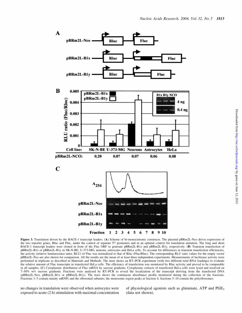
In order to investigate the effect of the BACE-1 transcriptleader on cap-dependent translation, we prepared a series ofbi-monocistronic plasmids (Fig. 3A). These plasmids containtwo independent transcriptional units under the control of twoT7 promoters. The ®rst cassette generates a transcriptcontaining the Rluc reporter gene, which was used tonormalize for differences in transfection ef®ciency. Thesecond cassette generates an mRNA with the Fluc genepreceded by either the long (pBRm2L-B1x) or the short(pBRm2L-B1y) BACE-1 transcript leader. These plasmidswere transiently transfected by the MVA-T7pol system inthe following cells: SK-N-BE (human neuroblastoma);
U-373-MG (human astrocytoma); primary hippocampalneurons and cortical astrocytes from newborn rat; HeLa(human adenocarcinoma). Translation was quanti®ed as theratio of the activities of the two reporter genes (Fluc/Rluc).Both the long and the short BACE-1 transcript leaders wereless active (one to two orders of magnitude) than the emptyvector pBRm2L-Nco in supporting translation initiation(Fig. 3B) [for quanti®cation, see also De Pietri Tonelli et al.(37)]. The possibility that changes in transcript stability mightaccount for such a difference was ruled out by evaluating thelevels of Fluc transcripts by RT±PCR (Fig. 3B, inset).Interestingly, the short form of BACE-1 leader, which lacksthree out of four uAUGs, was 2±8-fold more active than thelong form. To better evaluate the translation ef®ciency, weexamined by polysome analysis the cytoplasmic distributionof Fluc mRNA in transfected cells. The result in Figure 3Cshows that the control mRNA (from pBRm2L-Nco) wasmostly present in the polysomal fractions of the gradient. Incontrast, the majority of the mRNA from either pBRm2L-B1x
Figure 1. Scheme of the proposed alternative splicing in BACE-1 transcript leader. (A) Alignment of full-length BACE-1 transcript leaders from human (Hs),pig (Ss), rat (Rn) and mouse (Mm) cDNAs. Arrowheads mark proposed donor±acceptor splice sites for the new variant of the BACE-1 transcript leader.Conserved nucleotides are highlighted in grey. Upstream ATGs are written in bold and boxed sequences represent the putative uORFs. (B) Scheme of thealternative splicing within the ®rst exon of human BACE-1. Dotted line and arrows delimit the sequence missing in the shorter variant. Arrowheads highlightthe upstream ATGs, while the black and the striped boxes, the uORF and the ®rst codons of the main ORF, respectively.
Nucleic Acids Research, 2004, Vol. 32, No. 5 1811
by guest on June 12, 2015http://nar.oxfordjournals.org/
Dow
nloaded from

or pBRm2L-B1y was detected in the slower sedimentingregion of the gradient, thereby indicating repression oftranslation.
In order to investigate whether the data obtained bytransfection were affected by variations in mRNA stability,we performed experiments of in vitro translation also moni-toring the mRNA decay rate. Translation driven by eitherBACE-1 leader in a rabbit reticulocyte translation assay wasabout two orders of magnitude lower than control (Fig. 4A).Noticeably, the stability of all radioactively labelled tran-scripts was identical (Fig. 4B).
To understand the relative contribution of the four uATGsin the translation repression, we performed an analysis of thecontext for translation initiation surrounding each uATG (27)with the NetStart program (42). The prediction showed thatthe second uATG, which is positioned in a good Kozakcontext, is recognized by the ribosome with at least the sameef®ciency as the main ATG, while the other three uATGs arenot in good contexts for translation initiation. To test thisprediction, we mutated the second uATG of the long BACE-1transcript leader (pBRm2L-B1x-mut1 plasmid) (Fig. 5A). Thetransfection in SK-N-BE cells revealed that the activity ofthe mutant was ~5-fold higher than the long BACE-1leader (pBRm2L-B1x), but still lower than the short form
(pBRm2L-B1y). Then, we mutated sequentially the otheruATGs as described in Figure 5A, generating pBRm2L-B1x-mut2 and pBRm2L-B1x-mut3. The effect of these mutationswas additive in overcoming the translational hurdle (Fig. 5B).
According to this result, the second uATG exerts aprominent repression on translation. This might be accom-plished by recruiting the ribosomal machinery for an abortivecycle of translation. To test this hypothesis, we generated thebi-monocistronic plasmid pBRm2L-B1x-uORF1, in whichthe second reporter gene (Fluc) is positioned immediately afterthe second uATG. As predicted, transfection in SK-N-BE andU-373-MG cells con®rmed that this uATG is ef®cientlyrecognized by the translation machinery (data not shown).
To see whether translational control of BACE-1 hasrelevance in physiopathological conditions, we used theactivated astrocyte model. Astrocytes pretreated for 24 hwith IL-1b (10 ng/ml) and TNF-a (30 ng/ml) or nothing weretransfected for 4 h with bi-monocistronic plasmids using theMVA-T7pol system. In this way differences between expres-sions of the reporter genes can be attributed to variations intranslation ef®ciency. Interestingly, Fluc translation driven byBACE-1 transcript leader, either long or short, was 4-foldhigher in activated astrocytes, while translation of the emptyconstruct pBRm2L-Nco was unaffected (Fig. 6). In contrast,
Figure 2. RNase protection assay for the BACE-1 transcript leader. (A) Scheme for the synthesis of the riboprobe for BACE-1. The plasmidpGEMD-BACE1-5¢x with the full-length BACE-1 transcript leader was linearized with SpeI and used as template for the synthesis of the 32P-labelledantisense riboprobe performed with the T7 RNA polymerase (probe length 484 nt). (B) Scheme of RNase protection assay. The protected fragments derivingfrom the full-length and the short BACE-1 leaders are 435 and 240 nt, respectively. The short variant produces an additional fragment of 26 nt that remainsundetectable. (C) Result of RNase protection. 15 mg of total RNA from either human exocrine pancreas (Pc) or brain (Br) were incubated with 55 000 c.p.m.of 32P-labelled riboprobe. The longer fragment of 435 nt is visible below the undigested probe, while the shorter fragment of 240 nt is present in the samplefrom pancreas only. Size markers of 316 and 240 nt were prepared as described in Materials and Methods.
1812 Nucleic Acids Research, 2004, Vol. 32, No. 5
by guest on June 12, 2015http://nar.oxfordjournals.org/
Dow
nloaded from
no changes in translation were observed when astrocytes wereexposed to acute (2 h) stimulation with maximal concentration
of physiological agonists such as glutamate, ATP and PGE2
(data not shown).
Figure 3. Translation driven by the BACE-1 transcript leaders. (A) Scheme of bi-monocistronic constructs. The plasmid pBRm2L-Nco drives expression ofthe two reporter genes, Rluc and Fluc, under the control of separate T7 promoters and in an optimal context for translation initiation. The long and shortBACE-1 transcript leaders were cloned in front of the Fluc ORF to generate pBRm2L-B1x and pBRm2L-B1y, respectively. (B) Transient transfection ofpBRm2L-B1x or pBRm2L-B1y in SK-N-BE, U-373-MG, neurons, astrocytes and HeLa cells. To account for differences in transient transfection ef®ciencies,the activity (relative luminescence units, RLU) of Fluc was normalized to that of Rluc (Fluc/Rluc). The corresponding RLU ratio values for the empty vectorpBRm2L-Nco are also shown for comparison. All the results are the mean of at least three independent experiments. Measurements of luciferase activity wereperformed in triplicate as described in Materials and Methods. The inset shows an RT±PCR experiment (with two different total RNA loadings) to evaluatethe relative amount of Fluc transcripts in transfected HeLa cells. The ef®ciency of transfection was monitored by Rluc activity and proved to be comparablein all samples. (C) Cytoplasmic distribution of Fluc mRNA by sucrose gradients. Cytoplasmic extracts of transfected HeLa cells were lysed and resolved on7±50% w/v sucrose gradients. Fractions were analysed by RT±PCR to reveal the localization of the transcript deriving from the transfected DNA(pBRm2L-Nco, pBRm2L-B1x or pBRm2L-B1y). The trace shows the continuous absorbance pro®le monitored during the collection of the fractions.Fractions 1±3 contain mainly mRNPs and the ribosomal subunits; the monosome region peaks at fraction 4; fractions 5±10 contain the polyribosomes.
Nucleic Acids Research, 2004, Vol. 32, No. 5 1813
by guest on June 12, 2015http://nar.oxfordjournals.org/
Dow
nloaded from

DISCUSSION
The BACE-1 transcript leader is long and highly conservedamong various mammalian species, two features that are
strongly suggestive of a translational control of proteinexpression. By working with reporter genes, we demonstratethat BACE-1 transcript leader acts as a `silencer' of transla-tion. Since we had previously reported that the level of a toxicprotein, called Scamper, is maintained at a low level by anIRES (37), we explored whether this was also the case forBACE-1. The use of the dicistronic approach, an experimentalstrategy conventionally used to assess the presence of anIRES, did not provide con®rmation for the presence of such amechanism in the BACE-1 transcript. Rather, we obtainedevidence that the inhibition of translation is, at least partially,due to the presence of uATGs. It is known that uATGs caninhibit translation in several ways. When they are recognizedby the translation machinery, a futile cycle can occur, so thatonly the ribosomes skipping the uATG (leaky-scanning) canreach the main ORF. Accordingly, the level of inhibition isdirectly related to the context for translation initiation: thebetter an uATG is recognized, the higher is the resultinginhibition (27). Even in conditions of optimal uATG
Figure 4. In vitro translation driven by the BACE-1 transcript leader. (A) Activity of Fluc translated under the control of the long (B1x) or the short (B1y)BACE-1 transcript leaders. The cRNA produced from the empty vector pZac-Luc+ gives the 100% value. (B) Decay of the corresponding cRNAs during thetranslation reactions. cRNA from pZac-Luc+ is the control and radioactive input is considered to be 100%.
Figure 5. Effect of upstream ATGs in BACE-1 transcript leader ontranslation ef®ciency. (A) Scheme of the long BACE-1 transcript leader inthe bi-monocistronic plasmid. The four upstream ATGs are marked witharrowheads. The black box represents the upstream ORF. The upstreamATGs were mutated to TTG, generating pBRm2L-B1x-mut1,pBRm2L-B1x-mut2, pBRm2L-B1x-mut3. (B) Transient transfection ofbi-monocistronic plasmids in SK-N-BE cells. Results are normalized to thevalue of the long BACE-1 leader (pBRm2L-B1x).
Figure 6. Translation driven by BACE-1 transcript leader in resting andactivated astrocytes. Astrocytes were transfected in normal culture condi-tions (resting) or after activation with IL-1b and TNF-a (activated).Transfection was performed with bi-monocistronic plasmids containingeither the long (pBRm2L-B1x) or short (pBRm2L-B1y) BACE-1 transcriptleader. The increase in Fluc translation between resting and activated astro-cytes is also reported (fold increase).
1814 Nucleic Acids Research, 2004, Vol. 32, No. 5
by guest on June 12, 2015http://nar.oxfordjournals.org/
Dow
nloaded from

recognition, a small percentage of ribosomes, after translationof the uORF, can continue scanning for start sites (re-initiation), eventually reaching the ATG of the main ORF(43,44). Moreover, some peptides produced by translation of auORF have been reported to interact with the ribosome,further diminishing the ef®ciency of protein translation(45±48). In the case of BACE-1, the second uATG isef®ciently recognized by the ribosome. However, the presenceof another ATG as the last codon before termination in the72 nt uORF made it impossible to discriminate between leakyscanning and a reinitiation mechanism. We cannot provideconclusive evidence whether complete translation of thisuORF occurs and has relevance to the process. Since therecognition of start sites can be modulated, the presence ofuATGs might provide a way to regulate protein expression.For instance, phosphorylation of the eukaryotic initiationfactor eIF2-a is known to decrease the ef®ciency of ATGrecognition with ensuing inhibition of the translation of mosttranscripts (49). However, translation of uATG-containingtranscripts is paradoxically increased because more ribosomesovercome uATGs and become available for the translation ofthe main ORF (50). According to this view, BACE-1translation might increase in conditions that favour eIF2-aphosphorylation.
The discovery that BACE-1 transcript leader can bealternatively spliced provides an additional possibility forthe regulation of BACE-1 expression. In fact, the alternativesplicing we describe here reduces the number of uATGs (from4 to 1) and, as expected, increases the ef®ciency of translationof reporter genes. By RNase protection and RT±PCR we showthat this splicing occurs in exocrine pancreas and neuro-blastoma cells, but not in normal human brain. Therefore,BACE-1 expression is likely to be affected by changes in theef®ciency of this splicing event and this might play a role underdifferent physiological as well as pathological conditions.
Although it has long been assumed that the toxic A-bpeptide is produced by neurons, the possibility that astrocytescan contribute to this process is gaining momentum (51,52).Noticeably, in astrocytes from either AD patients or theTg2576 transgenic mouse model, BACE-1 protein expressionwas found only around amyloid plaques, where reactiveastrocytes are likely to be present (17,51). The current view isthat astrocytes do not express BACE-1Ðand thus do notproduce A-bÐunless they are brought to a state of chronicactivation or they are put in culture (51). Nothing is knownabout the switch to promote BACE-1 expression.Transcriptional mechanisms are likely to be involved, butwe provide evidence that translational regulation can contrib-ute as well. Our data clearly show that when astrocytes areactivated by chronic exposure to cytokines in culture, amarked reduction in the inhibition of translation driven by theBACE-1 transcript leader is recorded. This mechanism isindependent of the splicing event described above, since it wasobserved with both splice variants when transient transfectionof reporter genes was performed with the RNA-T7pol vacciniasystem, i.e. under conditions that bypass nuclear processing ofthe transcripts. Interestingly, translation driven by the BACE-1 transcript leader in activated astrocytes reaches an ef®ciencythat is similar to that obtained in neurons. This increase hasbeen observed during a process of chronic activation, whichalso occurs in vivo, while acute activation with physiological
stimuli failed to reproduce the effect. In our conditions, themost likely explanation of this effect resides in a variation intranslation ef®ciency. We cannot exclude that variations intranscript stability may contribute. However, this clearlyshows that after astrocyte activation a non-transcriptionaleffect on BACE-1 protein production operates.
In conclusion, we provide evidence that BACE-1 expres-sion is inhibited at the translational level by the presence ofuATGs. Furthermore, we report that splicing of the transcriptleader can change the number of uATGs that are present,thereby providing an additional mechanism for the control.Finally, we show that a release of the inhibition of translationoccurs in activated astrocytes, potentially contributing to A-bproduction. Therefore, even though it is known that BACE-1can also act on substrates other than APP (53), our ®ndingsprovide a possible explanation of why in brain tissue of ADpatients an increase in BACE-1 protein or activity does notrequire an increase in BACE-1 mRNA or in the ratio betweena- and b-secretase mRNAs (21,22,23), and why changes inBACE-1 mRNA have never been observed around amyloidplaques in transgenic Tg2576 mice (16).
ACKNOWLEDGEMENTS
We thank S.Durante for excellent technical help, L.Sala forhelp with polysome analysis, D.Dunlap for critical reading ofthe manuscript and M.Losa and F.Bertuzzi for the gift ofhuman temporal cortex and exocrine pancreas, respectively.We also thank G.Sutter and the Institute of MolecularVirology of the GSFÐForschungszentrum fuer Umwelt undGesundheit GmbH for the MVA-T7pol virus. This work wascarried out within the framework of the Italian Ministry ofResearch Centre of Excellence in Physiopathology of CellDifferentiation. Financial support was also from theArmenise-Harvard Foundation, the Italian Telethon (grantE.0888 to FG), the CNR target project in Biotechnology (toFG), and grants from the EU Commission (DECG projectCLG3-CT-2001-02004 to FG; APOPIS project LSHM-CT-2003-503330 to DZ).
REFERENCES
1. Vassar,R., Bennett,B.D., Babu-Khan,S., Kahn,S., Mendiaz,E.A.,Denis,P., Teplow,D.B., Ross,S., Amarante,P., Loeloff,R. et al. (1999)Beta-secretase cleavage of Alzheimer's amyloid precursor protein by thetransmembrane aspartic protease BACE. Science, 286, 735±741.
2. Yan,R., Bienkowski,M.J., Shuck,M.E., Miao,H., Tory,M.C.,Pauley,A.M., Brashier,J.R., Stratman,N.C., Mathews,W.R., Buhl,A.E.et al. (1999) Membrane-anchored aspartyl protease with Alzheimer'sdisease beta-secretase activity. Nature, 402, 533±537.
3. Sinha,S., Anderson,J.P., Barbour,R., Basi,G.S., Caccavello,R., Davis,D.,Doan,M., Dovey,H.F., Frigon,N., Hong,J. et al. (1999) Puri®cation andcloning of amyloid precursor protein beta-secretase from human brain.Nature, 402, 537±540.
4. Hussain,I., Powell,D., Howlett,D.R., Tew,D.G., Meek,T.D., Chapman,C.,Gloger,I.S., Murphy,K.E., Southan,C.D., Ryan,D.M. et al. (1999)Identi®cation of a novel aspartic protease (Asp 2) as beta-secretase. Mol.Cell. Neurosci., 14, 419±427.
5. Lin,X., Koelsch,G., Wu,S., Downs,D., Dashti,A. and Tang,J. (2000)Human aspartic protease memapsin 2 cleaves the beta-secretase site ofbeta-amyloid precursor protein. Proc. Natl Acad. Sci. USA, 97,1456±1460.
6. Yu,G., Nishimura,M., Arawaka,S., Levitan,D., Zhang,L., Tandon,A.,Song,Y.Q., Rogaeva,E., Chen,F., Kawarai,T. et al. (2000) Nicastrin
Nucleic Acids Research, 2004, Vol. 32, No. 5 1815
by guest on June 12, 2015http://nar.oxfordjournals.org/
Dow
nloaded from

modulates presenilin mediated notch/glp-1signal transduction and bAPPprocessing. Nature, 407, 48±54
7. Esler,W.P., Kimberly,W.T., Ostaszewski,B.L., Ye,W., Diehl,T.S.,Selkoe,D.J. and Wolfe,M.S. (2002) Activity-dependent isolation of thepresenilin-gamma-secretase complex reveals nicastrin and a gammasubstrate. Proc. Natl Acad. Sci. USA, 99, 2720±2725.
8. Farmery,M.R., Tjernberg,L.O., Pursglove,S.E., Bergman,A., Winblad,B.and Naslund,J. (2003) Partial puri®cation and characterization of g-secretase from post-mortem human brain. J. Biol. Chem., 278,24277±24284.
9. Selkoe,D.J. (2001) Alzheimer's disease: genes, proteins, and therapy.Physiol. Rev., 81, 741±766
10. Bodendorf,U., Danner,S., Fischer,F., Stefani,M., Sturchler-Pierrat,C.,Wiederhold,K.H., Staufenbiel,M. and Paganetti,P. (2002) Expression ofhuman b-secretase in the mouse brain increases the steady-state level ofb-amyloid. J. Neurochem., 80, 799±806.
11. Holsinger,R.M., McLean,C.A., Beyreuther,K., Masters,C.L. and Evin,G.(2002) Increased expression of the amyloid precursor beta-secretase inAlzheimer's disease. Ann. Neurol., 51, 783±786.
12. Tyler,S.J., Dawbarn,D., Wilcock,G.K. and Allen,S.J. (2002) alpha- andbeta-secretase: profound changes in Alzheimer's disease. Biochem.Biophys. Res. Commun., 299, 373±376.
13. Fukumoto,H., Cheung,B.S., Hyman,B.T. and Irizarry,M.C. (2002)Beta-secretase protein and activity are increased in the neocortex inAlzheimer disease. Arch. Neurol., 59, 1381±1389.
14. Yang,L.B., Lindholm,K., Yan,R., Citron,M., Xia,W., Yang,X.L.,Beach,T., Sue,L., Wong,P., Price,D. et al. (2003) Elevated beta-secretaseexpression and enzymatic activity detected in sporadic Alzheimerdisease. Nat. Med., 9, 3±4.
15. Lange-Dohna,C., Zeitschel,U., Gaunitz,F., Perez-Polo,J.R., Bigl,V. andRossner,S. (2003) Cloning and expression of the rat BACE1 promoter.J. Neurosci. Res., 73, 73±80.
16. Bigl,M., Apelt,J., Luschekina,E.A., Lange-Dohna,C., Rossner,S. andSchliebs,R. (2000) Expression of b-secretase mRNA in transgenicTg2576 mouse brain with Alzheimer plaque pathology. Neurosci. Lett.,292, 107±110.
17. Rossner,S., Apelt,J., Schliebs,R., Perez-Polo,J.R. and Bigl,V. (2001)Neuronal and glial beta-secretase (BACE) protein expression intransgenic Tg2576 mice with amyloid plaque pathology. J. Neurosci.Res., 64, 437±446.
18. Bodendorf,U., Fischer,F., Bodian,D., Multhaup,G. and Paganetti,P.(2001) A splice variant of beta-secretase de®cient in the amyloidogenicprocessing of the amyloid precursor protein. J. Biol. Chem., 276,12019±12023.
19. Ehehalt,R., Michel,B., De Pietri Tonelli,D., Zacchetti,D., Simons,K. andKeller,P. (2002) Splice variants of the beta-site APP-cleaving enzymeBACE1 in human brain and pancreas. Biochem. Biophys. Res. Commun.293, 30±37.
20. Huse,J.T., Byant,D., Yang,Y., Pijak,D.S., D'Souza,I., Lah,J.J., Lee,V.M.,Doms,R.W. and Cook,D.G. (2003) Endoproteolysis of beta-secretase(beta-site amyloid precursor protein-cleaving enzyme) within its catalyticdomain. A potential mechanism for regulation. J. Biol. Chem., 278,17141±17149.
21. Marcinkiewicz,M. and Seidah,N.G. (2000) Coordinated expression ofbeta-amyloid precursor protein and the putative beta-secretase BACE andalpha-secretase ADAM10 in mouse and human brain. J. Neurochem., 75,2133±2143.
22. Gatta,L.B., Albertini,A., Ravid,R. and Finazzi,D. (2002) Levels ofbeta-secretase BACE and alpha-secretase ADAM10 mRNAs inAlzheimer hippocampus. Neuroreport, 13, 2031±2033.
23. Preece,P., Virley,D.J., Costandi,M., Coombes,R., Moss,S.J.,Mudge,A.W., Jazin,E. and Cairns,N.J. (2003) beta-Secretase (BACE)and GSK-3 mRNA levels in Alzheimer's disease. Brain Res. Mol. BrainRes., 116, 155±158.
24. Hellen,C.U. and Sarnow,P. (2001) Internal ribosome entry site ineukaryotic mRNA molecules. Genes Dev., 15, 1593±1612.
25. Vagner,S., Galy,B. and Pyronnet,S. (2001) Irresistible IRES: attractingthe translation machinery to internal ribosome entry sites. EMBO Rep., 2,893±898.
26. Hentze,M.W. (1994) Translational control by iron-responsive elements.Adv. Exp. Med. Biol., 356, 119±126.
27. Kozak,M. (1999) Initiation of translation in prokaryotes and eukaryotes.Gene, 234, 187±208.
28. Morris,D.R. and Geballe,A.P. (2000) Upstream open reading frames asregulators of mRNA translation. Mol. Cell. Biol., 20, 8635±8642.
29. Ghilardi,N., Wiestner,A. and Skoda,R.C. (1998) Thrombopoietinproduction is inhibited by a translational mechanism. Blood, 92,4023±4030.
30. Meijer,H.A., Dictus,W.J., Keuning,E.D. and Thomas,A.A. (2000)Translational control of the Xenopus laevis connexin-41 5¢-untranslatedregion by three upstream open reading frames. J. Biol. Chem., 275,30787±30793.
31. Lee,J., Park,E.H., Couture,G., Harvey,I., Garneau,P. and Pelletier,J.(2002) An upstream open reading frame impedes translation of thehuntingtin gene. Nucleic Acids Res., 30, 5110±5119.
32. Gaba,A., Wang,Z., Krishnamoorthy,T., Hinnebusch,A.G. and Sachs,M.S.(2001) Physical evidence for distinct mechanisms of translational controlby upstream open reading frames. EMBO J., 20, 6453±6463.
33. Harding,H.P., Novoa,I., Zhang,Y., Zeng,H., Wek,R., Schapira,M. andRon,D. (2000) Regulated translation initiation controls stress-inducedgene expression in mammalian cells. Mol. Cell, 6, 1099±1108.
34. Schluter,G., Boinska,D. and Nieman-Seyde,S.C. (2000) Evidence fortranslational repression of the SOCS-1 major open reading frame by anupstream open reading frame. Biochem. Biophys. Res. Commun., 268,255±261.
35. Holzmann,K., Ambrosch,I., Elbling,L., Micksche,M. and Berger,W.(2001) A small upstream open reading frame causes inhibition of humanmajor vault protein expression from a ubiquitous mRNA splice variant.FEBS Lett., 494, 99±104.
36. Guyot,B., Arnaud,S., Phothirath,P., Rigal,D. and Mouchiroud,G. (2002)Upstream open reading frames regulate translation of Mona/Gads adaptermRNA in the megakaryocytic lineage. Platelets, 13, 459±464.
37. DePietriTonelli,D., Mihailovich,M., Schnurbus,R., Pesole,G.,Grohovaz,F. and Zacchetti,D. (2003) Translational control of Scamperexpression via a cell-speci®c internal ribosome entry site. Nucleic AcidsRes., 31, 2508±2513.
38. Fisher,C.L. and Pei,G.K. (1997) Modi®cation of a PCR-basedsite-directed mutagenesis method. Biotechniques, 23, 570±571.
39. McCarthy,K.D. and De Vellis,J. (1980) Preparation of separate astroglialand oligodendroglial cell cultures from rat cerebral tissue. J. Cell Biol.,85, 890±902.
40. Ryan,T.A. and Smith,S.J. (1995) Vesicle pool mobilization during actionpotential ®ring at hippocampal synapses. Neuron, 14, 983±989.
41. Sutter,G., Ohlmann,M. and Er¯e,V. (1995) Non replicating vacciniavirus vector ef®ciently expresses bacteriophage T7 RNA polymerase.FEBS Lett., 371, 9±12.
42. Pedersen,A.G. and Nielsen,H. (1997) Neural network prediction oftranslation initiation sites in eukaryotes: perspectives for EST andgenome analysis. ISMB, 5, 226±233.
43. Child,S.J., Miller,M.K. and Geballe,A.P. (1999) Translational control byan upstream open reading frame in the HER-2/neu transcript. J. Biol.Chem., 274, 24335±24341.
44. Grif®n,E., Re,A., Hamel,N., Fu,C., Bush,H., McCaffrey,T. andAsch,A.S. (2001) A link between diabetes and atherosclerosis: Glucoseregulates expression of CD36 at the level of translation. Nat. Med. 7,840±846.
45. Fang,P., Wang,Z. and Sachs,M.S. (2000) Evolutionarily conservedfeatures of the arginine attenuator peptide provide the necessaryrequirements for its function in translational regulation. J. Biol. Chem.,275, 26710±26719.
46. Jousse,C., Bruhat,A., Carraro,V., Urano,F., Ferrara,M., Ron,D. andFafournoux,P. (2001) Inhibition of CHOP translation by a peptideencoded by an open reading frame localized in the chop 5¢UTR. NucleicAcids Res., 29, 4341±4351.
47. Gong,F. and Yanofsky,C. (2002) Instruction of translating ribosome bynascent peptide. Science, 297, 1864±1867.
48. Raney,A., Law,G.L., Mize,G.J. and Morris D.R. (2002) Regulatedtranslation termination at the upstream open reading frame inS-adenosylmethionine decarboxylase mRNA. J. Biol. Chem., 277,5988±5994.
49. Harding,H.P., Calfon,M., Urano,F., Novoa,I. and Ron,D. (2002)Transcriptional and translational control in the Mammalian unfoldedprotein response. Annu. Rev. Cell. Dev. Biol., 18, 575±599.
50. Dever,T.E. (2002) Gene-speci®c regulation by general translationfactors. Cell, 108, 545±556.
51. Hartlage-Rubsamen,M., Zeitschel,U., Apelt,J., Gartner,U., Franke,H.,Stahl,T., Gunther,A., Schliebs,R., Penkowa,M., Bigl,V. et al. (2003)
1816 Nucleic Acids Research, 2004, Vol. 32, No. 5
by guest on June 12, 2015http://nar.oxfordjournals.org/
Dow
nloaded from

Astrocytic expression of the Alzheimer's disease beta-secretase(BACE1) is stimulus-dependent. Glia, 41, 169±179.
52. Lesne,S., Docagne,F., Gabriel,C., Liot,G., Lahiri,D.K., Buee,L.,Plawinski,L., Delacourte,A., MacKenzie,E.T., Buisson,A. et al. (2003)Transforming growth factor-beta 1 potentiates amyloid-beta generationin astrocytes and in transgenic mice. J. Biol. Chem., 278, 18408±18418.
53. Kitazume,S., Tachida,Y., Oka,R., Shirotani,K., Saido,T.C. andHashimoto,Y. (2001) Alzheimer's beta-secretase, beta-site amyloidprecursor protein-cleaving enzyme, is responsible for cleavage secretionof a Golgi-resident sialyltransferase. Proc. Natl Acad. Sci. USA, 98,13554±13559.
Nucleic Acids Research, 2004, Vol. 32, No. 5 1817
by guest on June 12, 2015http://nar.oxfordjournals.org/
Dow
nloaded from