Neuronal muscarinic responses: role of protein
9
0892-6638181002-2575101.50. © FASEB 2575 Neuronal muscarinic responses: role of protein kinase C ESAM E. ELFAKAHANY,* BRADLEY E. ALGER,’ WI. S. LAI, THOMAS A. PITLER,’ PAUL F. WORLEY,t AND JAY M. BARABAN1 *Deparfracnt of Pharmacology and Toxicology, University of Maryland School of Pharrnacy, Baltimore, Maryland 21201, USA; ‘Department of Physiology, University of Maryland School of Medicine, Baltimore, Maryland 21201, USA; tDepartments of Neuroscience, Psychiatry, and Behavioral Sciences, and of Neurology, ]ohns Hopkins University School of Medicine, Baltimore, Maryland 21205, USA ABSTRACT Advances in understanding the phosphoinositide cycle have helped unravel the chain of events initiated by muscarinic receptor stimulation. Hydrolysis of mem- brane phosphoinositides generates both diacylglycerol, an activator of protein kinase C, and inositol phos- phates. In the nervous system, muscarinic receptors elicit a wide range of electrophysiological responses. Recent studieshave made progress inidentifyingwhich of these neuronal muscarinic actions are mediated by activation of protein kinase C. Paradoxically, protein kinaseC alsoexertsa stronginhibitoryinfluenceon muscarinic responses. This complex set of actions sug- gests that in addition to mediating certain muscarinic responses, protein kinase C also blocks signal trans- duction as part of a feedback mechanism. - EL- FAKAHANY, E. E.; ALGER, B. E.; LA!, W. S.; PITLER, T A.; WORLEY, P. F.; BARABAN, J. M. Neuronal mus- carinic responses: role of protein kinase C. FASEBJ 2: 2575-2583; 1988. Key Words: acetyic/toline phosphoinositides . second mes- sengers - phorbol esters muscarinic receptors THE ASSOCIATION OF MUSCARINIC receptors with multiple second messenger systems has provided a framework for understanding the broad range of muscarinic re- sponses in different tissues. Biochemical studies have linked muscarinic receptors to stimulation of cGMP production and phosphoinositide (P1)1 hydrolysis, as well as to inhibition of CAMP accumulation (1). More recently, electrophysiological studies have provided compelling evidence for direct regulation of potassium channels by muscarinic receptors via a GTP-binding protein (2). In view of the wide repertoire of signaling pathways accessible to muscarinic receptors, recent studies have focused on clarifying the roles of each of these pathways in mediating and regulating muscarinic actions. Although activation of P1 metabolism by muscarinic receptors has been known since the 1950’s, the recent breakthrough in our understanding of the P1 second messenger system has stimulated interest in its role in muscarinic signal transduction. Receptor-mediated hydrolysis of membrane phosphoinositides generates both inositol 1,4,5-trisphosphate (1P3), which releases calcium from intracellular stores, and diacylglycerol. Nishizuka’s proposal that endogenous diacylglycerol generated from P1 hydrolysis can activate protein kinase C (PKC) (3) has focused attention on PKC’s actions. As predicted by this hypothesis, phorbol esters that bind to and substitute for diacyiglycerol in acti- vating PKC (4) mimic a wide variety of muscarinic responses. Paradoxically, however, phorbol ester- induced inhibition of muscarinic actions has also been commonly observed, raising the possibility that PKC might also be involved in desensitization of muscarinic responses. In this article,we review progress made in recent years toward understanding the role of PKC in muscarinic signal transduction. Analogous effects of PKC on other receptor-mediated responses suggest that the findings generated by studies of muscarinic re- sponses are relevant to transduction of a wide variety of other neurotransmitter and hormone signals. Protein kinase C: activation and localization PKC is a calcium- and phospholipid-dependent kinase present in particularly high concentrations in the brain. Since diacylglycerol activates PKC, this membrane lipid serves as a second messenger linking P1 turnover to PKC. PKC is found in both soluble and membrane- bound forms; however, its activity is dependent on its association with cellmembrane phospholipids. Accord- ingly, it has been proposed that translocation of PKC from the cytoplasm to the cell membrane, which is also stimulated by calcium and diacylglycerol, is an essen- tial step in its activation. ‘Abbreviations P1, phosphoinositide; PKC, protein kinase C; PDBu, phorbol 12,13-dibutyrate; EPSP, excitatory postsynaptic potential; G,, inhibitory GTP-binding regulatory protein; NMS, N-methylscopolamine; 1P3, inositol l,4,5-trisphosphate; GABA, -y-aminobutyric acid.
-
Upload
independent -
Category
Documents
-
view
0 -
download
0
Transcript of Neuronal muscarinic responses: role of protein

0892-6638181002-2575101.50. © FASEB 2575
Neuronal muscarinic responses: role of protein
kinase CESAM E. ELFAKAHANY,* BRADLEY E. ALGER,’ WI. S. LAI, THOMAS A. PITLER,’PAUL F. WORLEY,t AND JAY M. BARABAN1*Deparfracnt of Pharmacology and Toxicology, University of Maryland School of Pharrnacy, Baltimore, Maryland 21201,
USA; ‘Department of Physiology, University of Maryland School of Medicine, Baltimore, Maryland 21201, USA;tDepartments of Neuroscience, Psychiatry, and Behavioral Sciences, and of Neurology, ]ohns Hopkins University
School of Medicine, Baltimore, Maryland 21205, USA
ABSTRACT
Advances in understanding the phosphoinositide cycle
have helped unravel the chain of events initiated bymuscarinic receptor stimulation. Hydrolysis of mem-
brane phosphoinositides generates both diacylglycerol,an activator of protein kinase C, and inositol phos-phates. In the nervous system, muscarinic receptorselicit a wide range of electrophysiological responses.Recent studieshave made progress in identifyingwhich
of these neuronal muscarinic actions are mediated by
activation of protein kinase C. Paradoxically, proteinkinase C alsoexertsa strong inhibitoryinfluenceonmuscarinic responses. This complex set of actions sug-gests that in addition to mediating certain muscarinicresponses, protein kinase C also blocks signal trans-
duction as part of a feedback mechanism. - EL-
FAKAHANY, E. E.; ALGER, B. E.; LA!, W. S.; PITLER,
T A.; WORLEY, P. F.; BARABAN, J. M. Neuronal mus-
carinic responses: role of protein kinase C. FASEBJ 2:2575-2583; 1988.
Key Words: acetyic/toline phosphoinositides . second mes-sengers - phorbol esters muscarinic receptors
THE ASSOCIATION OF MUSCARINIC receptors with multiple
second messenger systems has provided a frameworkfor understanding the broad range of muscarinic re-sponses in different tissues. Biochemical studies havelinked muscarinic receptors to stimulation of cGMPproduction and phosphoinositide (P1)1 hydrolysis, aswell as to inhibition of CAMP accumulation (1). Morerecently, electrophysiological studies have providedcompelling evidence for direct regulation of potassiumchannels by muscarinic receptors via a GTP-bindingprotein (2). In view of the wide repertoire of signalingpathways accessible to muscarinic receptors, recentstudies have focused on clarifying the roles of each ofthese pathways in mediating and regulating muscarinicactions.
Although activation of P1 metabolism by muscarinicreceptors has been known since the 1950’s, the recent
breakthrough in our understanding of the P1 secondmessenger system has stimulated interest in its role
in muscarinic signal transduction. Receptor-mediatedhydrolysis of membrane phosphoinositides generatesboth inositol 1,4,5-trisphosphate (1P3), which releasescalcium from intracellular stores, and diacylglycerol.Nishizuka’s proposal that endogenous diacylglycerolgenerated from P1 hydrolysis can activate proteinkinase C (PKC) (3) has focused attention on PKC’sactions. As predicted by this hypothesis, phorbol estersthat bind to and substitute for diacyiglycerol in acti-vating PKC (4) mimic a wide variety of muscarinicresponses. Paradoxically, however, phorbol ester-induced inhibition of muscarinic actions has also beencommonly observed, raising the possibility that PKCmight also be involved in desensitization of muscarinicresponses. In this article,we review progress made in
recent years toward understanding the role of PKC inmuscarinic signal transduction. Analogous effects ofPKC on other receptor-mediated responses suggest thatthe findings generated by studies of muscarinic re-sponses are relevant to transduction of a wide variety ofother neurotransmitter and hormone signals.
Protein kinase C: activation and localization
PKC is a calcium- and phospholipid-dependent kinasepresent in particularly high concentrations in the brain.Since diacylglycerol activates PKC, this membrane lipidserves as a second messenger linking P1 turnover toPKC. PKC is found in both soluble and membrane-bound forms; however, its activity is dependent on its
association with cellmembrane phospholipids. Accord-ingly, it has been proposed that translocation of PKCfrom the cytoplasm to the cell membrane, which is alsostimulated by calcium and diacylglycerol, is an essen-tial step in its activation.
‘Abbreviations P1, phosphoinositide; PKC, protein kinase C;
PDBu, phorbol 12,13-dibutyrate; EPSP, excitatory postsynapticpotential; G,, inhibitory GTP-binding regulatory protein; NMS,N-methylscopolamine; 1P3, inositol l,4,5-trisphosphate; GABA,-y-aminobutyric acid.

2576 EL-FAKAHANY ET AL.
The availability of phorbol esters that bind to andactivate protein kinase C (4) has facilitated investiga-tion of the role of PKC in muscarinic responses. Thesedrugs are active when applied extracellularly, makingthem particularly useful in assessing the effects of PKC.
Phorbol esters contain two ester or acyl linkage sites atadjacent carbons 12 and 13, which are thought to cor-respond to the two fatty acid side chains of diacyl-glycerol. Substitution at the 12 and 13 positions of thephorbol nucleus has generated an extensive series ofanalogs. In general, increasing the length of these side
chains increases potency in activating PKC, e.g.,
diacetate < dibutyrate < didecanoate. The unsubsti-tuted parent compound phorbol is inactive. Sincephorbol ester analogs vary widely in their potencies foractivating PKC, checking the rank order of potencies ofseveral phorbol esters can be helpful in assessing theirspecificity for PKC.
The potent phorbol ester phorbol 12,13-dibutyrate(PDBu) has been widely used as a radioligand for
studying PKC. Autoradiographic studies using thisligand have demonstrated PKC’s heterogeneous locali-
zation in the brain (5) (Fig. 1). Very high levels arepresent in cerebellum, cerebral cortex, hippocampus,
Figure 1.Autoradiographiclocalizationof PKC and IP, receptorinrathippocampus. Coronal ratbrainsectionswere processedforauto-radiographywith either[‘H]PDBu (panel A) or [3HIIP3 (panel B) to localize PKC or 1P3 receptor, respectively. Autoradiograms demon-strate high levels of these components of the P1 system in the CAl region of the hippocampus (CAl). Lower levels are present in otherstructures shown, such as cortex (C), dentate gyrus (D), thalamus (T), hypothalamus (H), and amygdala (A).

A CONTROL
NEURONAL MUSCARINIC RESPONSES 2577
and substantia nigra. Within these structures, PKC isdiscretely localized. For example, in the cerebellumPKC is present predominantly in the molecular layer inPurkinje celldendrites. Heavy staining with [3H]PDBu
in the substantia nigra is greatly decreased after ipsi-lateral striatal lesions, which indicates that PKC in thenigra is present in terminals of striatonigral afferents.Accordingly, PKC in the brain can be found in bothdendrites and terminals, which suggests it might affectsynaptic transmission both pre- and postsynaptically.
Cholinergic actions in the hippocampus
The septohippocampal cholinergic pathway has beenutilized extensively to analyze cholinergic mechanismsin the brain (6).This prominent cholinergicinnervationmarkedly exciteshippocampal pyramidal neurons viaactivation of muscarinic receptors. Biochemical studieshave demonstrated strong stimulation of the PT system
by muscarinic agonists in the hippocampus (7). Inaddition, autoradiographic and biochemical studies
have demonstrated the presence of high levelsof PKC
in this region, where itcomprises approximately 0.5%of the membrane protein. [3H]PDBu autoradiographyafter selective excitotoxin lesions indicates that PKC islocated in intrinsic neurons of the hippocampus (5).However, the presence of substantial residual [3H]PDBu
binding after destruction of intrinsic neurons suggeststhat PKC is present in extrinsic afferents as well.
Muscarinic agonists exert multiple effects on ioniccurrents in hippocampal pyramidal neurons (6). Theirexcitatory action reflects the blockade of several distinctinhibitory potassium currents, including the M currentand a calcium-activated potassium current. Apart from
activation of the P1 system, muscarinic agonists also
elevate cGMP and inhibit cAMP. Therefore, one focusof current research has been on understanding the roleof individual second messenger systems in elicitingthese electrophysiological responses.
The M current, which derives its name from its sen-sitivity to muscarinic agonists, is an inhibitory potas-sium current that normally limits cellular response todepolarizing stimuli. By blocking this braking influ-
B 25OnM PDA
fl-Figure 2. Phorbol ester blockade of accommodation. Top tracingsin panels A and B are recordings of membrane potential obtained
during intracellular recording from CAl hippocampal pyramidalneurons. Bottom tracings record intracellular current injections. Atupward deflection in current tracing, depolarizing current is in-jected for 2 s. In panel A, this stimulus elicits a brief train of actionpotentials followed by a pause and a single spike. After applicationof 250 nM phorbol diacetate (PDA), which blocks the calcium-activated potassium conductance, the same current stimulus elicitsaction potentials throughout the depolarizing stimulus.
ence, muscarinic agents enhance excitability of these
neurons.Calcium-activated potassium currents provide another
mechanism for containing excitatory activity. Depolari-zation induced by current injection or by application ofan excitatory transmitter, such as glutamate, allows cal-cium entry through voltage-dependent calcium channels.The elevated calcium in turn activates potassium currentsthat can either frankly hyperpolarize the membrane,thus inhibiting the cell,or produce a slowing of actionpotential firing frequency (Fig. 2) by antagonizing aprolonged depolarizing stimulus. By inhibiting both Mcurrent and a calcium-activated potassium current,muscarinic agonists sharply increase the number of
spikes elicited during a depolarizing stimulus.
Phorbol ester actions in hippocampal neurons
To assess the involvement of PKC in mediating these
muscarinic actions, the effects of phorbol esters havebeen examined on the electrophysiological responses ofhippocampal pyramidal neurons (8, 9). In these neurons,phorbol esters mimic the ability of muscarinic agents toblock the slow hyperpolarization mediated by a calcium-activated potassium conductance. By contrast, the Mcurrent is not blocked by phorbol esters.
At present, it is unclear whether PKC’s blockade ofthe slow calcium-activated potassium potential resultsfrom decreased calcium influx or an inhibitory actiondistal to calcium entry. However, since PKC inhibitsvoltage-dependent calcium currents in a variety of neu-
rons, as well as hippocampal pyramidal neurons (10),its effect may be exerted at the level of calcium influx.It is interesting that the calcium-activated potassiumconductance is also inhibited by the cAMP system (11)
after activation of /3 adrenoceptors. Accordingly, thisconductance, which plays a prominent role in regulat-
ing repetitive firing, is a common target for both PKCand the CAMP system. Such monodirectional control,i.e., similar regulation by the cAMP and PKC systems,has been observed in several other tissues (3).
The discrepancy between the effects of muscarinicagonists and phorbol esters on the M current demon-strates that PKC does not mediate all muscarinicactions. Conversely, phorbol esters exert effects notshared by muscarinic agonists. PKC activation blocksa voltage-dependent chloride conductance not affectedby carbamylcholine (carbachol) (12). The similar effectsof phorbol esters and muscarinic agonists in blockingthe slow calcium-activated potassium conductance sug-gest that the P1 system may mediate this cholinergic
action via PKC. Clearly, however, one cannot concludethat PKC mediates particular muscarinic actions solelyon the basis of such mimickry. By contrast, a broaderrange of evidence supports a role for the PI-PKC systemin mediating another muscarinic response describedbelow.
Muscarinic block of adenosine
In hippocampal pyramidal neurons, phorbol estersmarkedly decrease the inhibitory action mediated by
adonosin. baclofon4Opm 4Opm
CONTROL
2578 EL-FAKAHANY ET AL.
several G protein-linked receptors, such as 7-amino-
butyric acid (GABAB), adenosine A1, and 5HT1Areceptors (13, 14) (Fig. 3). Cholinergic agonists alsoblock these G protein-linked neurotransmitter actionsas does stimulation of cholinergic afferents to thehippocampus. Further evidence that this cholinergic
response is mediated by the PI-PKC system comes
from a comparison of the muscarinic pharmacology
observed in activating the system and in producingblockade of adenosine. Muscarinic agonists varygreatly in their ability to elicit PT turnover. Further-more, as demonstrated by Fisher and Bartus (7), thepharmacology of muscarinic PT stimulation variesamong brain regions. For example, the efficacy of somemuscarinic partial agonists, such as oxotremorine, isweaker in the hippocampus than in other brain regions.This distinctive pharmacology parallels that observedin muscarinic blockade of adenosine, providing furthersupport that muscarinic stimulation of P1 is involved.
Another useful tool in identifying muscarinic re-sponses mediated by P1 is the atypical muscarinic an-tagonist pirenzepine. As opposed to other classicalmuscarinic antagonists that recognize one populationof muscarinic receptor binding sites, pirenzepine dis-tinguishes among two populations of muscarinic recep-tor subtypes, designated as high- (M,) and low-affinity(M2) pirenzepine sites. In the hippocampus, low con-centrations of pirenzepine block P1 turnover (7, 15),which indicates that high-affinity pirenzepine sites arelinked to P1 turnover in this region.
These biochemical studies predict that those mus-carinic actions mediated by PT would be blocked by
wash
pdS
lpm PDA h.f\ v\[ i\
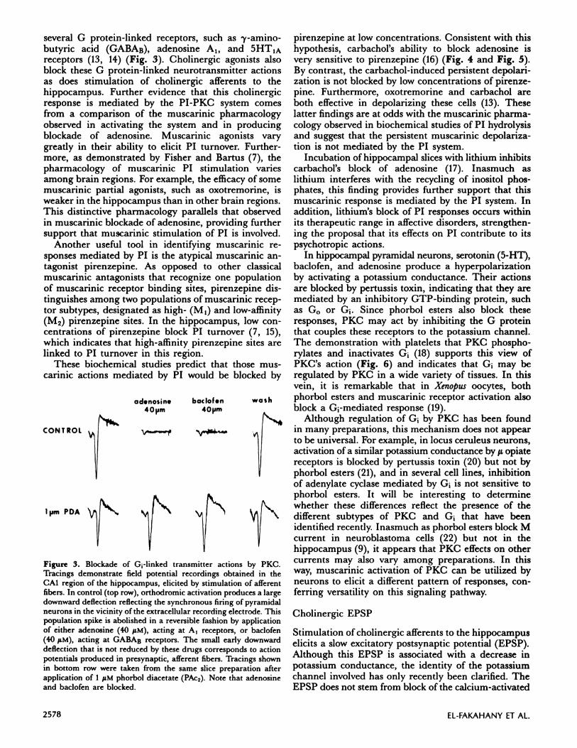
Figure 3. Blockade of G1-linked transmitteractions by PKC.Tracings demonstrate field potential recordings obtained in theCAl region of the hippocampus, elicited by stimulation of afferentfibers. In control (top row), orthodromic activation produces a largedownward deflection reflecting the synchronous firing of pyramidalneurons in the vicinity of the extracellular recording electrode. Thispopulation spike is abolished in a reversible fashion by applicationof either adenosine (40 SM), acting at A, receptors, or baclofen(40 LM), acting at GABAB receptors. The small early downwarddeflectionthatisnot reduced by these drugs corresponds to actionpotentials produced in presynaptic, afferent fibers. Tracings shownin bottom row were taken from the same slice preparation afterapplication of 1 iM phorbol diacetate (PAc2). Note that adenosineand baclofen are blocked.
pirenzepine at low concentrations. Consistent with thishypothesis, carbachol’s ability to block adenosine is
very sensitive to pirenzepine (16) (Fig. 4 and Fig. 5).By contrast, the carbachol-induced persistent depolari-
zation is not blocked by low concentrations of pirenze-
pine. Furthermore, oxotremorine and carbachol areboth effective in depolarizing these cells (13). Theselatter findings are at odds with the muscarinic pharma-cology observed in biochemical studies of P1 hydrolysis
and suggest that the persistent muscarinic depolariza-
tion is not mediated by the P1 system.
Incubation of hippocampal slices with lithium inhibits
carbachol’s block of adenosine (17). Inasmuch aslithium interferes with the recycling of inositol phos-
phates, this finding provides further support that thismuscarinic response is mediated by the PT system. In
addition, lithium’s block of P1 responses occurs withinits therapeutic range in affective disorders, strengthen-
ing the proposal that its effects on P1 contribute to its
psychotropic actions.In hippocampal pyramidal neurons, serotonin (5-HT),
baclofen, and adenosine produce a hyperpolarization
by activating a potassium conductance. Their actionsare blocked by pertussis toxin, indicating that they aremediated by an inhibitory GTP-binding protein, suchas G0 or G. Since phorbol esters also block theseresponses, PKC may act by inhibiting the G proteinthat couples these receptors to the potassium channel.The demonstration with platelets that PKC phospho-rylates and inactivates G (18) supports this view ofPKC’s action (Fig. 6) and indicates that G may beregulated by PKC in a wide variety of tissues. In thisvein, it is remarkable that in Xenopu.s oocytes, both
phorbol esters and muscarinic receptor activation alsoblock a G-mediated response (19).
Although regulation of G1 by PKC has been found
in many preparations, this mechanism does not appear
to be universal. For example, in locus ceruleus neurons,activation of a similar potassium conductance by s opiate
receptors is blocked by pertussis toxin (20) but not by
phorbol esters (21), and in several cell lines, inhibitionof adenylate cyclase mediated by G1 is not sensitive tophorbol esters. It will be interesting to determinewhether these differences reflect the presence of thedifferent subtypes of PKC and G1 that have beenidentified recently. Inasmuch as phorbol esters block Mcurrent in neuroblastoma cells (22) but not in thehippocampus (9), it appears that PKC effects on othercurrents may also vary among preparations. In thisway, muscarinic activation of PKC can be utilized byneurons to elicit a different pattern of responses, con-ferring versatility on this signaling pathway.
Cholinergic EPSP
Stimulation of cholinergic afferents to the hippocampuselicits a slow excitatory postsynaptic potential (EPSP).
Although this EPSP is associated with a decrease in
potassium conductance, the identity of the potassium
channel involved has only recently been clarified. TheEPSP does not stem from block of the calcium-activated

ACARB 5OpN
CARB 50 pM
...411Th%... S
V
.
I 10 my
Adsnoslns Application I 250 pA
3 mm
-I--Jo 100I-
0C.) #{149}
70
‘U
600a.UI‘U
40
30
z 20‘U
10
100
15
0
25
0
0 10 30 1000
NEURONAL MUSCARINIC RESPONSES 2579
BPRZ 1 )iM________PRZ bOnN
V
11I11Il1lI”IIIIIIIfliMIIIIIIIIIIDDIDDDIDIIIIHDJJJllhil]llIllluhll]iIjllMJIlIl111jllhilllllllllllhiIlllillhi
Figure 4. Pirenzepine antagonizes the ability of carbachol to block adenosine response. A) Manual voltage-clamp of an adenosine response
(indicated by filled circles) was obtained in control conditions. The upward deflection on the current trace (I) represents the currentinduced by adenosine, 100 M, applied for 15 s. The membrane was then left unclamped while carbachol (50 M) perfusion was begun.Carbachol depolarized the membrane, as shown in the upper trace (voltage, or V). After 20 mm in carbachol, the cell was again voltage-
clamped at the control membrane potential while adenosine was briefly applied. As shown by the diminished deflection on the currenttrace, the adenosine response was nearly abolished. At that point perfusion was switched to a saline solution containing 50 sM carbacholplus 1 sM pirenzepine (PRZ). Within 15 mm the adenosine-induced current was restored to control values. All traces in A are from the
same CAl cell.B) If present before the application of carbachol, PRZ (100 nM) prevented its effect on the adenosine response. As in A,adenosine responses are indicated by filled circles. After recording the adenosine-induced current in saline solution containing 100 nM
PRZ, perfusion with 50 M carbachol plus PRZ was started. The adenosine currents in the middle and right-hand columns wererecorded 20 and 45 mm later. All records in B were taken from the same cell, which was different from the one shown in A. All calibrationsapply to both parts A and B. Note the current traces in the presence of carbachol plus PRZ are below the control level. This reflects theinward current, which is caused by carbachol itself and is not blocked by PRZ.
potassium current or M current. Instead, it is mediatedby closing a leak potassium conductance (23). The cho-linergic EPSP is blocked by low concentrations of pirenze-pine, which suggests that it may be mediated by the PTsystem (24). At present, it is unclear whether the leakpotassium conductance is the same as the transmitter-activated potassium conductance, stimulated by inhibi-tory transmitters, such as 5-HT and adenosine. How-ever, since adenosine is tonically released in the hippo-campal slice preparation, it could be responsible for theleak conductance. If this were so, then blockade of theleak conductance underlying the cholinergic EPSPcould be mediated by PKC as well.
PKC: feedback inhibition of muscarinic responses
Apart from mimicking some cholinergic responses aspredicted by the link between PT and PKC, PKC alsoinhibits cholinergic responses in many tissues. For ex-ample, smooth muscle contraction elicited by muscarinicagonists is reversed by phorbol esters (25) (Fig. 7), anddiacylglycerol production correlates with inhibition ofcarbachol-induced secretion in pancreatic acini (26). Inhippocampal neurons, the cholinergic EPSP and thedepolarizing action of carbachol are blocked by phorbolesters (9). These effects indicate that PKC can dramati-cally limit muscarinic responses, and fit well with bio-chemical investigations summarized below demonstratingthat PKC blocks muscarinic activation of second-messenger systems.
PKC blockade of cGMP and P1 responses
Phorbol esters suppress muscarinic receptor-mediatedincreases in cGMP synthesis (27, 28) (Fig. 8b). This
inhibitory effect appears to involve a block in couplingreceptor activation to stimulation of guanylate cyclase,since phorbol esters do not affect the elevation of cGMPinduced by other activators such as sodium azide or acalcium ionophore (28). The ability of H-7, a PKC in-hibitor, to attenuate this action of phorbol esters helpsassure that it reflects PKC activation. Furthermore,phorbol esters demonstrate a significantly lowered
PRZ (nU) + 50 pM CARBACIIOL
Figure 5. Dose-response histogram showing the antagonism of the
carbachol block of adenosine by PRZ. Bars represent means ± SEM
of the magnitude of the adenosine-induced current in 3-8 experi-ments per dose of PRZ (experiments performed as illustrated in
Fig. 1). In the absence of PRZ, carbachol caused a 75% reduction
in the adenosine response. The vertical axis on the right-hand sideexpresses the degree of inhibition of the carbachol block caused byPRZ. The IC,0 for the PRZ inhibition is between 10 and 30 nMAt 1 M, PRZ entirely abolished the effect of carbachol on
adenosine.

PDBU
100 nM
,j_Y_I2040 80 160 80 80
OXOTREMORINE (nM) BRADYKININ (nM)
20
4de05.
2580 EL-FAKAHANY ET AL.
Figure 6. Schematic model of interaction between PI-PKC system and inhibitoryresponsesmediated by G proteins.Stimulationofphos-pholipase C by acetylcholine (ACh) at muscarinic receptors is mediated by an as-yet-unidentified G protein. Hydrolysis of phos-phatidylinositol 4,5-bisphosphate (PIP,) generates diacylglycerol (DAG) and inositol trisphosphate (IP,). Stimulation of PKC by DAGresults in the inactivation of the G protein coupling the adenosine receptor to potassium ion channels, thought to be G, or a closely
related 0 protein.
potency in blocking cGMP responses after PKC hasbeen down-regulated by prolonged activation (29).
In addition to blocking the muscarinic cGMP re-sponse, phorbol esters also block muscarinic agonist-induced stimulation of P1 turnover in the brain (30),and many neuronal preparations in cell culture, e.g.,mouse neuroblastoma N1E-115 cells (28, 29) (Fig. 8a),human neuroblastoma SH-SY5Y cells (31), and humanastrocytoma cells (32).
Effects of PKC on inhibition of cAMP
In contrast to the consistent inhibitory effects of PKCon P1 and cGMP responses, PKC appears to have awide variety of effects on the adenylate cyclase system.When phorbol esters exert stimulatory effects onneurotransmitter-induced CAMP accumulation (e.g.,Fig. 8C), these actions appear to reflect both a directstimulatory effect on the adenylate cyclase enzyme itself,
PDBU 25 nM
100 200 400
OXOTREMORINE (nM) HISTAMINE
DPB
00 nM2_-r P5 10 20200 200 200 200
___________0.5g
5mm
Figure 7. Phorbol ester blockade of neurotransmitter-induced contractions of guinea pig ileum. Phorbol dibutyrate, (PBt,, 100 nM) didnot contract the guinea pig ileum strip (top left). However, within 2 mm of bath application it completely blocked contractile effects of
oxotremorine (80 nM). After a brief wash this action of PBt, was completely reversed. The bottom left panel illustrates a similar effectof l2-deoxyphorbol l3-isobutyrate, another active phorbol ester (DPB; 200 nM). DPB rapidly and reversibly blocked oxotremorine’saction. In the top right panel, a contraction was elicited by increasing doses of histamine. PBt, potently reversed this contraction.Propranolol (2 tM) (P)did not alterPBt2’sinhibition,but addition of another 400 nM of histamine overcame PBt,’s blockade. The bottom
right panel illustrates the contractile action of bradykinin. Atropine (1 tM) (A) did not block bradykinin’s effect but PBt, (100 nM) exerted
a powerful inhibition that was partially reversed by 20 nM bradykinin.

(B)0)
0,
00
Eo.20
0
‘po 15
C0
0) 10
E
UU<a.I
(A)
P.B CBC
501
401
301
0)
0,(.)
E0.
V
C.,
b
Va,E0
U-a-
U
U
LJS 20’
0)
a)()
E0.50
V
C.,
40
. 10U>.
020
C,’
101
(C)
BPP
+ +CBC CBC
Control PMA (100 nM)
1Control PMA (100 nM)
B CBC
Control PMA (100 nM)
BPP
Figure 8. Effects of phorbol-l2-myristate-l3-acetate (PMA) on carbachol-mediated increases in [‘HIIP accumulation and [‘HIcGMP
synthesis, and inhibition of PGE1-induced [‘H]cAMP formation. A) N1E-115 cells were preincubated with 100 nM PMA for 60 mm at37#{176}C,then 1 mM carbachol (CBC) was added to initiate [‘H]IP accumulation, and the reaction was stopped after30 mm. LiCI (10mM)was present throughout the whole incubation period. B) [‘H]cGMP formation was induced by the addition of 1 mM CBC to the cellsuspension; the reaction was terminated 30 s later. C) 3 M PGE, (P), or PGE, (3 M) plus CBC (1 mM) were added to the cellstomeasure [‘HIcAMP formation, the reaction time for the assay was 10 mm. Data shown are from one representative experiment performedin triplicateand the bars represent± SD. B = basal level measured in the absence of receptor agonists.
NEURONAL MUSCARINIC RESPONSES 2581
which is phosphorylated by PKC, as well as removal ofthe inhibitory influence of transmitters mediated by G1.However, as noted above, PKC effects on G’-linkedprocesses such as transmitter activation of potassiumchannels appear to vary among preparations. Forexample, pretreatment of mouse neuroblastoma cells(clone NIE-115) with phorbol esters actually potentiatesthe relative inhibitory effects of muscarinic agonists onprostaglandin E,-stimulated cAMP synthesis (28) (Fig.
8C). On the other hand, inhibition of CAMP accumula-tion in human astrocytoma 1321N1 cells is very sensitiveto phorbol esters (33). However, in these particularcells, inhibition of cAMP accumulation is not mediatedin a classical manner via G1. Rather, activation ofmuscarinic receptors stimulates a calcium-dependentphosphodiesterase (33), thereby reducing levels ofCAMP. Thus, the antagonistic effects of phorbol estersin this case might result from the attenuation of mus-carinic receptor-mediated formation of inositol tris-phosphate, because the latter produce induces calciummobilization from intracellular stores.
PKC effects on muscarinic receptor binding
The well-documented inhibitory effects of proteinkinase C on several muscarinic receptor-mediatedcellular responses suggest that it acts at a common,early step in signal transduction. Receptor bindingtechniques have therefore been used to assess effects ofPKC on muscarinic receptors. Classical muscarinicreceptor antagonists display a single binding affinity,whereas muscarinic agonists bind to the receptor withmultiple affinities. As indicated previously, atypicalmuscarinic antagonists, such as pirenzepine, differen-
tiate between two major receptor subclasses. For ex-ample, pirenzepine interacts with the M1 and M2receptor subtypes with high and low affinity, respectively
(2). To assess the status of muscarinic receptors on thecell surface, it has been useful to measure muscarinicreceptor binding in intact cells with a hydrophilicligand [e.g., [3H]N-methylscopolamine ([3H]NMS)].The charged nature of this ligand limits its access to theinterior of the cell where it could bind to intracellular(internalized) receptors.
Incubation of human astrocytoma cells (clone 1321N1)with phorbol esters under conditions that suppressmuscarinic receptor-mediated increases in P1 hydrolysisand calcium mobilization does not alter either the max-imal binding capacity or the affinity of [3HJNMS (clas-sical antagonist) measured in intact cells (33). However,in human neuroblastoma SH-SY5Y cells, phorbol estertreatment abolishes the heterogeneity of the agonistbinding sites(31), and in N1E-115 cells it eliminates thepirenzepine high-affinity receptor conformation (34).This receptor subtype mediates agonist-induced in-
creases in both cGMP formation and P1 hydrolysis,while the inhibition of cAMP is thought to be mediatedvia the pirenzepine low-affinity form (1, 2). These find-ings suggest that PKC’s inhibition of PT and cGMPresponses to muscarinic agonists may reflect a loss ofthis high-affinity receptor form, possibly by a directeffect on the receptor or the G proteins involved.
Role of PKC in desensitization of muscarinic responses
Receptor desensitization is a well-established phenome-non whereby preexposure of a receptor to its agonistsdecreases responsiveness of the receptor to further

2582 EL-FAKAHANY ET AL.
stimulation by the agonist. Desensitization of mus-carinic receptors has been studied in several cell culturesystems. For example, preincubation of mouse neuro-blastoma N1E-115 cells with muscarinic agonists dimin-ishes subsequent muscarinic receptor-mediated cGMPformation. This desensitization is rapid, temperature-dependent, and reversible (1). Although the mecha-nisms underlying this reduced receptor sensitivity arenot fully understood, alterations at the level of thereceptor binding sites have been demonstrated. In thisclone, short-term incubations with muscarinic agonistsreduce cell surface receptors without decreasing totalcellular receptor density (35). This brief desensitizingtreatment is also accompanied by a selective loss of thepirenzepine high-affinity M1 receptor subtype (36).Inasmuch as PKC is activated by muscarinic stimulationof the P1 system, and PKC in turn inhibits both cGMPand P1 muscarinic responses, PKC could mediateagonist-induced desensitization. The finding that phor-bol esters also produce a selective loss of the M1muscarinic receptor conformation that mediates thecGMP response is consistent with this hypothesis (34).In addition, incubation of cells with both a calciumionophore and a phorbol ester decreases the concentra-tion of cell surface receptors, mimicking the patternobserved during agonist-induced desensitization (37).
After exposure to muscarinic agonists, muscarinicreceptor-mediated increases in P1 hydrolysis generallydesensitize more slowly than the cGMP response. Forexample, loss of the P1 response in human astrocytoma1321N1 cells parallels the slow rate of down-regulationof total cell receptors observed after exposure to mus-carinic agonists (38). This pattern differs from that seenwith phorbol esters, since they inhibit the muscarinicPT response quite rapidly before inducing a loss inreceptors (32).
Agonist exposure induces increased phosphorylationof the muscarinic receptor in neuronal (39) and cardiac(40) tissue. In primary cultures of rat cerebellum, car-bachol enhances the phosphorylation of six polypep-tides. The rate of phosphorylation of three of these pro-teins parallels that of agonist-induced down-regulationof the receptor. In addition, one of these phosphopro-teins has a molecular weight similar to that of mus-carinic receptors (39). Although it is possible that PKCis involved in some of these phosphorylation events,recent studies of cardiac tissue, which contains M2muscarinic receptors exclusively, indicate that PKC isnot responsible for agonist-induced phosphorylation ofthe muscarinic receptor itself (40).
Summary
The studies reviewed highlight the prominent roleplayed by PKC in mediating and regulating muscarinicresponses in the nervous system. Clearly, much remainsto be clarified with regard to the mechanisms of PKC’sinhibition of muscarinic responses and its role in agonist-
induced desensitization. In the case of muscarinicresponses mimicked by phorbol esters, the availabilityof selective PKC and P1 inhibitors would be a welcome
adjunct to phorbol esters in assessing the involvementof PT and PKC. Although substantial progress has beenmade in identifying PKC’s role in neuronal muscarinic
responses, a complete picture of PI-PKC involvementin muscarinic actions in the brain must await clarifica-tion of the concerted effects of all the products of P1hydrolysis.
We thank Linda Pottillo for secretarial assistance. Supported byNational Institutes of Health grants NS-22010 (B. E. A.), DA-00266(J. M. B.), AG-00256 (P. F. W.), NS-24158 (E. E. E.), AG-07118(E. E. E.), and AG-00344 (E. E. E.), awards from the EpilepsyFoundation of America (B.B. A.),theLucilleP.Markey CharitableTrust (J. M. B.), and the Sloan Foundation (J. M. B.).
REFERENCES
1. MCKINNEY, M.; RICHELSON, E. The coupling of theneuronal muscarinic receptor to responses. Annu. Rev.Pliarmacol. Toxicol. 24: 121-146; 1984.
2. NATHANSON, N. M. Molecular properties of the mus-carinic acetylcholine receptor. Annu. Rev.Neurosci.10:195-236; 1987.
3. Nisrnzut, Y. Studies and perspectives of proteinkinase C. Science 233: 305-312; 1986.
4. BLUMBERG, P. M.; JAKEN, S.; KONIG, B.; SHARKEY,N. A.; LcH, K. L.;JENG, A. Y.; YEH, E. Mecha-nism of action of the phorbol ester tumor promoters:specific receptors for lipophiic ligands. Biochem. P/tar-macol. 33: 933-940; 1984.
5. WORLEY, P. F.; BARABAN, J. M.; SNYDER, S. H.Heterogeneous localization of protein kinase C in ratbrain: autoradiographic analysis of phorbol esterreceptor binding. J. Neurosci. 6: 199=207; 1986.
6. NIc0LL, R. A. The septo-hippocampal projection: amodel cholinergic pathway. TrendsNeuroSci. 8: 533-536; 1985.
7. FISHER, S.; BARTUS, R. T. Regional differences in thecoupling of muscarinic receptors to inositol phospho-lipid hydrolysis in guinea pig brain. J. Neurochem. 45:1085-1095; 1985.
8. BARABAN,J. M.; SNYDER, S. H.; ALGER, B. E. Proteinkinase C regulates ionic conductance in hippocampalpyramidal neurons: electrophysiological effects ofphorbol esters. Proc. Nail. Acad. Sci. USA 82:2538-2542; 1985.
9. MALENKA, R. C.; MADISON, D. V.; ANDRADE, R.;NICOLL, R. A. Phorbol esters mimic some cholinergicactions in hippocampal pyramidal neurons. J. Neurosci.6: 475-480; 1986.
10. DOERNER, D.; PITLER, T. A.; ALGER, B. E. Proteinkinase C activators block specific calcium and potassiumcurrent components in isolated hippocampal neurons.J. Neurosci. In press.
11. MADISON, D. V.; NIc0LL, R. A. Cyclic adenosine3’ ,5’-monophosphate mediates /3-receptor actions ofnoradrenaline in rat hippocampal pyramidal cells.J. Physiol. (London) 372: 245-259; 1986.
12. MADIsoN, D. V.; MALENKA, R. C.; NICOLL, R. A.Phorbol esters block a voltage-sensitive chloride currentin hippocampal pyramidal cells. Nature (London) 321:695-697; 1986.
13. WORLEY, P. F.; BARABAN, J. M.; MCCARREN, M.;SNYDER, S. H.; ALGER, B. E. Cholinergic phos-phatidylinositol modulation of inhibitory, G protein-linked, neurotransmitter actions: electrophysiologicalstudies in rat hippocampus. Proc. Nati. Acad. Sci. USA84: 3467-3471; 1987.

NEURONAL MUSCARINIC RESPONSES 2583
14. ANDRADE, R.; MALENKA, R. C.; NICOLL, R. A.A G protein couples serotonin and GABAB receptorsto the same channels in hippocampus. Science 234:1261-1265; 1986.
15. GIL, D. W.; WOLFE, B. B. Pirenzepine distinguishes be-tween muscarinic receptor-mediated phosphoinositidebreakdown and inhibition of adenylate cyclase.J. Pharmacol. Exp. Ther.232: 608-616; 1985.
16. ALGER, B. E.; MCCARREN, M. Pirenzepine blockscalcium- and P1-turnover-related muscarinic actionsin rat hippocampus. Soc.Neurosci.Abstr.13: 1005;1987.
17. WORLEY, P. F.; HELLER, W. A.; SNYDER, S. H.;BARABAN, J. M. Lithium blocks a phosphoinositidemediated cholinergic response in hippocampal slices.Science In press.
18. KATADA, T.; GILMAN, A. G.; WATANABE, Y.; BAUER,S.; JAKOBS, K. H. Protein kinase C phosphorylates theinhibitory guanine-nucleotide-binding regulatorycomponent and apparently suppresses itsfunction inhormonal inhibition of adenylate cyclase. Eur. J.Biochern. 151: 431-437; 1985.
19. DASCAL, N.; LOTAN, I.; GILLO, B.; LESTER, H. A.;LAss, Y. Acetylcholine and phorbol esters inhibitpotassium currents evoked by adenosine and cAMP inXenopus oocytes. Proc. Nail. Acad. Sci. USA 82:6001-6005; 1985.
20. AGHAJANIAN, G. K.; WANG, Y.-Y. Pertussis toxin blocksthe outward currents evoked by opiate and a2-agonistsin locus coeruleus neurons. BrainRes. 371: 390-394;1986.
21. NORTH, R. A.; WILLIAMS, J. T.; SUPRENANT, A.;CHRISTIE, M. J. Mu and delta receptors belong to afamily of receptors that are coupled to potassium chan-nels. Proc. Nati. Acad. Sci. USA 84: 5487=5491; 1987.
22. HIGASHIDA, H.; BROWN, D. A. Two polyphosphoinositidemetabolites control two potassium currents in a neuronalcell. Nature(London)323: 333-335; 1986.
23. MADISON, D. V.; LANCASTER, B.; NICOLL, R. A.Voltage clamp analysis of cholinergic action in thehippocampus. J. Neurosci. 7: 733-741; 1987.
24. MULLER, V.; MISGELD, U. Slow cholinergic excitationof guinea pig hippocampal neurons is mediated by twomuscarinic receptor subtypes. Neurosci.LetI.67: 107-112; 1986.
25. BARABAN, J. M.; GOULD, R. J.; PEROUTKA, S. J.;SNYDER, S. H. Phorbol ester effects on neurotransmis-sion: interaction with neurotransmitters and calciumin smooth muscle. Proc.Nati. Acad. Sci. USA 82:604-607; 1985.
26. PANDOL, S. J.; SCHOEFFIELD, M. S. 1,2-Diacylglycerol,protein kinase C, and pancreatic enzyme secretion.j Biol.C/tern. 261: 4438-4444; 1986.
27. LAI, W. S.; EL-FAKAHANY, E. E. Phorbol ester-inducedinhibition of cyclic GMP formation mediated by mus-carinic receptors in murine neuroblastoma cells.J. Pharmacol. Exp. Ther. 241: 366-373; 1987.
28. KANBA, S.; KANBA, K. S.; RICHELSON, E. The proteinkinase C activator, 12-O-tetradecanoyl-phorbol-13-acetate (TPA), inhibits muscarinic (Mi) receptor-mediated inositol phosphate release and cyclic GMP
formation in murine neuroblastoma cells (cloneN1E-115). Eur.J. Pharmacol. 125: 155-156; 1986.
29. LAI, W. S.; EL-FAKAHANY, E. E. Regulation of[3Hjphorbol- 12,1 3-dibutyrate binding sites in mouseneuroblastoma cells;simultaneous down-regulation byphorbol esters and desensitization of their inhibition ofmuscarinic receptor function. J. Pharmacol. Exp. Ther.244: 41-50; 1988.
30. JOPE, R. S.; CASEBOLT, T. L.; JOHNSON, G. V. W.Modulation of carbachol-stimulated inositol phospho-lipid hydrolysis in rat cerebral cortex. Neurocliern. Res.12: 693-700; 1987.
31. SERRA, M.; SMITH, T. L.; YAMAMURA, H. I. Phorbolesters alter muscarinic receptor binding and inhibitphosphoinositide breakdown in human neuroblastoma(SH-SY5Y) cells. Bloc/tern. Biophys. Res. Commun. 140:160=166; 1986.
32. ORELLANA, S. A.; SoLSiu, P. A.; BROWN,J. H. Phorbolester inhibits phosphoinositide hydrolysis and calciummobilization in cultured astrocytoma cells. J. Biol.C/tern. 260: 5236-5239; 1985.
33. HARDEN, T. K.; HENG, M. M.; BROWN,J. H. Receptorreserve in the calcium-dependent cyclic AMP responseof astrocytoma cells to muscarinic receptor stimulation;
demonstration by agonist-induced desensitization,receptor inactivation, and phorbol ester treatment.Mol. Pharinacol. 30: 200-206; 1986.
34. LaU, W. S.; EL-FAKAHANY, E. E. A selective effect ofprotein kinase C activation on pirenzepine high-affinitymuscarinic receptors in a neuronal clone. Eur. J.Pharmacol. 129: 201-202; 1986.
35. FEIGENBAUM, P.; EL-FAKAHANY, E. E. Regulation ofmuscarinic cholinergic receptor density in neuro-blastoma cellsby briefexposure to agonist:possibleinvolvement in desensitization of receptor function.j Pharmacol. Exp. Ther.233: 134-140; 1985.
36. CI0FFI, C. L.; EL-FAKAHANY, E. E. Short-term desen-sitization of muscarinic cholinergic receptors in mouseneuroblastoma cells: selective loss of agonist low-affinityand pirenzepine high-affinity binding sites. J. Phannacol.Exp. Ther. 238: 916-923; 1986.
37. LILES, W. C.; HUNTER, D. D.; MEIER, K. E.; NATHAN-SON, N. M. Activation of protein kinase C inducesrapid internalization and subsequent degradation ofmuscarinic acetylcholine receptors in neuroblastoma
cells. J. Biol.C/tern. 261: 5307-5313; 1986.38. MASTERS, S. B.; QUINN, M. T.; BR0wN,J. H. Agonist-
induced desensitization of muscarinic receptor-mediatedcalcium efflux without concomitant desensitization ofphosphoinositide hydrolysis. Mo!. Pharmacol. 27: 325-332; 1985.
39. BURGOYNE, R. D.; PEARCE, B. Muscarinic acetylcholinereceptor regulation and protein phosphorylation inprimary cultures of rat cerebellum. Dev. BrainRes. 2:55-63; 1982.
40. KWATRA, M. M.; LEUNG, E.; Mw’i, A. C.; MCMAHON,K. K.; PTASIENSKI,J.; GREEN, R. D.; HOSEY, M. M.Correlation of agonist-induced phosphorylation ofchick heart muscarinic receptors with receptor desensi-tization.J. Biol. C/tern. 262: 16314-16321; 1987.