Muscarinic receptor family interacting proteins: Role in receptor function
15
Please cite this article in press as: Borroto-Escuela DO, et al. Muscarinic receptor family interacting proteins: Role in receptor function. J Neurosci Methods (2011), doi:10.1016/j.jneumeth.2010.11.025 ARTICLE IN PRESS G Model NSM-5830; No. of Pages 9 Journal of Neuroscience Methods xxx (2010) xxx–xxx Contents lists available at ScienceDirect Journal of Neuroscience Methods journal homepage: www.elsevier.com/locate/jneumeth Muscarinic receptor family interacting proteins: Role in receptor function Dasiel O. Borroto-Escuela a,b,∗ , Patrícia A. Correia c , Wilber Romero-Fernandez a , Manuel Narvaez a , Kjell Fuxe b , Francisco Ciruela d , Pere Garriga a a Centre de Biotecnologia Molecular, Department d’Enginyeria Química, Universitat Politècnica de Catalunya, Colom 1, 08222 Terrassa, Barcelona, Spain b Department of Neuroscience, Karolinska Institutet, Retzius väg 8, 17177 Stockholm, Sweden c W.M. Keck Center for Integrative Neuroscience, Department of Physiology, University of California, San Francisco, USA d Unitat de Farmacologia, Departament Patologia i Terapèutica Experimental, Facultat de Medicina, IDIBELL-Universitat de Barcelona, Barcelona, Spain article info Article history: Received 28 August 2010 Received in revised form 23 November 2010 Accepted 24 November 2010 Keywords: Muscarinic receptors Interacting proteins Tandem affinity purification Signal network G protein coupled receptors abstract G protein-coupled receptors constitute one of the most important families of membrane receptors through which cells respond to extracellular stimuli. Receptors of this superfamily likely function as signal transduction complexes. The identification and analysis of their components provide new insights into a better understanding of these receptors’ function and regulation. We used tandem-affinity purification and mass spectrometry as a systematic approach to characterize multiprotein complexes in the acetyl- choline muscarinic receptor subfamily. To overcome the limitations associated with membrane protein receptor solubilization with detergents, we developed a strategy in which receptors are co-expressed with a cytoplasmic minigene construct, encoding the third intracellular loop and the C-terminal tail tagged to the tandem-affinity-cassette of each receptor subtype. Numerous protein complexes were identified, including many new interactions in various signalling pathways. Systematic identification data set together with protein interactions reported in the literature revealed a high degree of connectiv- ity. These allow the proposal, for the first time, of an outline of the muscarinic interactome as a network of protein complexes and a context for a more reasoned and informed approach to drug discovery and muscarinic receptor subtype specificities. © 2010 Elsevier B.V. All rights reserved. 1. Introduction As GPCR-researchers enter the so-called “Post-Genomic” era, they have begun to embrace the exciting opportunity of inves- tigating protein–protein interaction and multiprotein complex reorganization by means of high-throughput experiments (Eglen et al., 2007; Tilakaratne and Sexton, 2005). However, our knowl- edge of interacting proteins of specific complexes is limited, and it is based on selected biochemical approaches and genetic analy- ses. The only comprehensive protein-interaction studies on GPCRs are based on ex vivo and in vitro systems, such as two-hybrid systems and proteomics analysis of GPCR-associated protein com- plexes using receptor specific antibodies (Bockaert et al., 2004a; Daulat et al., 2009; Gavarini et al., 2004). These approaches are limited by the availability of adequate tools for each GPCR, and need to be integrated with physiological assays, which have Abbreviations: Iloop-TAP, intracellular loop tandem affinity purification; GPCR, G protein coupled receptor; mAChRs, muscarinic acetylcholine receptor family. ∗ Corresponding author at: Department of Neuroscience, Karolinska Institutet, Retzius väg 8, 17177 Stockholm, Sweden. Tel.: +46 8 52487077; fax: +46 8 315721. E-mail addresses: [email protected], [email protected] (D.O. Borroto-Escuela), [email protected] (W. Romero-Fernandez), [email protected] (F. Ciruela), [email protected] (P. Garriga). been fundamental to the understanding of their biological func- tion. Despite these limitations, several interacting proteins of GPCRs have been recently identified (Presland, 2004; Wang and Limbird, 2007). Most of these proteins interact with the C-terminal domain via their PDZ, Src homology 2 (SH2) and SH3, pleckstrin homology, or Ena/VASP homology (EVH) containing domains (Bockaert et al., 2003). It was also observed that the third intracellular loop might be implicated in protein interactions (Bockaert et al., 2004b). A tandem affinity purification (TAP) method has been suc- cessfully used for high throughput identification of soluble and membrane proteins engaged in interacting complexes in bacterial, yeast and mammalian cells (Borroto Escuela et al., 2006; Daulat et al., 2007; Kaiser et al., 2008; Wacker et al., 2008). This strategy allows for fast purification with a high yield of protein complexes under native conditions and overcomes the aforementioned limita- tions (Gingras et al., 2005; Puig et al., 2001). The protein of interest can be expressed in mammalian cells where subcellular localization and post-translational modifications are conserved. In addition, the recruitment of protein complexes may be induced by treating the cells with different pharmacological compounds or under different physiological conditions (Rosas-Acosta et al., 2005). Previous experiments evidenced that co-expression of GPCR intracellular loops (or peptides derived from these loops) could 0165-0270/$ – see front matter © 2010 Elsevier B.V. All rights reserved. doi:10.1016/j.jneumeth.2010.11.025
-
Upload
independent -
Category
Documents
-
view
0 -
download
0
Transcript of Muscarinic receptor family interacting proteins: Role in receptor function

G
N
M
DMa
b
c
d
a
ARR2A
KMITSG
1
ttreeisaspDln
G
R
(f
0d
ARTICLE IN PRESSModel
SM-5830; No. of Pages 9
Journal of Neuroscience Methods xxx (2010) xxx–xxx
Contents lists available at ScienceDirect
Journal of Neuroscience Methods
journa l homepage: www.e lsev ier .com/ locate / jneumeth
uscarinic receptor family interacting proteins: Role in receptor function
asiel O. Borroto-Escuelaa,b,∗, Patrícia A. Correiac, Wilber Romero-Fernandeza,anuel Narvaeza, Kjell Fuxeb, Francisco Ciruelad, Pere Garrigaa
Centre de Biotecnologia Molecular, Department d’Enginyeria Química, Universitat Politècnica de Catalunya, Colom 1, 08222 Terrassa, Barcelona, SpainDepartment of Neuroscience, Karolinska Institutet, Retzius väg 8, 17177 Stockholm, SwedenW.M. Keck Center for Integrative Neuroscience, Department of Physiology, University of California, San Francisco, USAUnitat de Farmacologia, Departament Patologia i Terapèutica Experimental, Facultat de Medicina, IDIBELL-Universitat de Barcelona, Barcelona, Spain
r t i c l e i n f o
rticle history:eceived 28 August 2010eceived in revised form3 November 2010ccepted 24 November 2010
eywords:uscarinic receptors
a b s t r a c t
G protein-coupled receptors constitute one of the most important families of membrane receptorsthrough which cells respond to extracellular stimuli. Receptors of this superfamily likely function as signaltransduction complexes. The identification and analysis of their components provide new insights into abetter understanding of these receptors’ function and regulation. We used tandem-affinity purificationand mass spectrometry as a systematic approach to characterize multiprotein complexes in the acetyl-choline muscarinic receptor subfamily. To overcome the limitations associated with membrane proteinreceptor solubilization with detergents, we developed a strategy in which receptors are co-expressed
nteracting proteinsandem affinity purificationignal networkprotein coupled receptors
with a cytoplasmic minigene construct, encoding the third intracellular loop and the C-terminal tailtagged to the tandem-affinity-cassette of each receptor subtype. Numerous protein complexes wereidentified, including many new interactions in various signalling pathways. Systematic identificationdata set together with protein interactions reported in the literature revealed a high degree of connectiv-ity. These allow the proposal, for the first time, of an outline of the muscarinic interactome as a networkof protein complexes and a context for a more reasoned and informed approach to drug discovery and
ype s
muscarinic receptor subt. Introduction
As GPCR-researchers enter the so-called “Post-Genomic” era,hey have begun to embrace the exciting opportunity of inves-igating protein–protein interaction and multiprotein complexeorganization by means of high-throughput experiments (Eglent al., 2007; Tilakaratne and Sexton, 2005). However, our knowl-dge of interacting proteins of specific complexes is limited, andt is based on selected biochemical approaches and genetic analy-es. The only comprehensive protein-interaction studies on GPCRsre based on ex vivo and in vitro systems, such as two-hybridystems and proteomics analysis of GPCR-associated protein com-
Please cite this article in press as: Borroto-Escuela DO, et al. Muscarinic receMethods (2011), doi:10.1016/j.jneumeth.2010.11.025
lexes using receptor specific antibodies (Bockaert et al., 2004a;aulat et al., 2009; Gavarini et al., 2004). These approaches are
imited by the availability of adequate tools for each GPCR, andeed to be integrated with physiological assays, which have
Abbreviations: Iloop-TAP, intracellular loop tandem affinity purification; GPCR,protein coupled receptor; mAChRs, muscarinic acetylcholine receptor family.∗ Corresponding author at: Department of Neuroscience, Karolinska Institutet,etzius väg 8, 17177 Stockholm, Sweden. Tel.: +46 8 52487077; fax: +46 8 315721.
E-mail addresses: [email protected], [email protected]. Borroto-Escuela), [email protected] (W. Romero-Fernandez),[email protected] (F. Ciruela), [email protected] (P. Garriga).
165-0270/$ – see front matter © 2010 Elsevier B.V. All rights reserved.oi:10.1016/j.jneumeth.2010.11.025
pecificities.© 2010 Elsevier B.V. All rights reserved.
been fundamental to the understanding of their biological func-tion.
Despite these limitations, several interacting proteins of GPCRshave been recently identified (Presland, 2004; Wang and Limbird,2007). Most of these proteins interact with the C-terminal domainvia their PDZ, Src homology 2 (SH2) and SH3, pleckstrin homology,or Ena/VASP homology (EVH) containing domains (Bockaert et al.,2003). It was also observed that the third intracellular loop mightbe implicated in protein interactions (Bockaert et al., 2004b).
A tandem affinity purification (TAP) method has been suc-cessfully used for high throughput identification of soluble andmembrane proteins engaged in interacting complexes in bacterial,yeast and mammalian cells (Borroto Escuela et al., 2006; Daulatet al., 2007; Kaiser et al., 2008; Wacker et al., 2008). This strategyallows for fast purification with a high yield of protein complexesunder native conditions and overcomes the aforementioned limita-tions (Gingras et al., 2005; Puig et al., 2001). The protein of interestcan be expressed in mammalian cells where subcellular localizationand post-translational modifications are conserved. In addition, the
ptor family interacting proteins: Role in receptor function. J Neurosci
recruitment of protein complexes may be induced by treating thecells with different pharmacological compounds or under differentphysiological conditions (Rosas-Acosta et al., 2005).
Previous experiments evidenced that co-expression of GPCRintracellular loops (or peptides derived from these loops) could

ING
N
2 Neuro
mpoTtagniwTr
2
2
abwFC(wMhTcmhfabUotw
2
mrcmIJtMEseN
2
lmcaifls(
ARTICLEModel
SM-5830; No. of Pages 9
D.O. Borroto-Escuela et al. / Journal of
imic the intact receptor, interacting with the same molecularartners (Abdulaev et al., 2000; Morou and Georgoussi, 2005). Inrder to define protein complexes within GPCRs, we modified aAP method suitable for the purification of associated proteins ofhe muscarinic acetylcholine receptor family (mAChRs), chosen asmodel. Briefly, the idea was to co-express a cytoplasmic mini-
ene construct along with the intact receptors in human SK-N-MCeuroblastome cells. After large-scale TAP of the minigene, we
dentified the interacting protein by LC–MS/MS (an approach thate termed intracellular loop tandem affinity purification (Iloop-
AP). Numerous protein complexes were identified, some of themepresentative of various signals pathways.
. Materials and methods
.1. Reagents
Dulbecco’s modified Eagle’s medium, penicillin/streptomycinnd fetal bovine serum were purchased from Invitrogen (Carls-ad, CA, USA). N-[3H]-methylscopolamine ([3H]-NMS, 81 Ci/mmol),ere from Amersham Biosciences (Piscataway, NJ, USA). The
uGene® transfection reagent was purchased from Roche (La Jolla,A, USA). Restriction enzymes were from New England BiolabsBeverly, MA, USA). The cDNAs of the human muscarinic M3Rere provided by T. Bonner (NIH, USA), human muscarinic M2R,2RRluc and M5R were provided by D. Bello (ETH, Switzerland), and
uman AKAP79GFP2 was kindly provided by P. Calvo (SFU, Canada).he InterPlay® mammalian TAP system, pNTAP-B vector was pur-hased from Stratagene (La Jolla, CA, USA). Mouse anti-paxillinonoclonal antibodies were purchased from Calbiochem (Notting-
am, UK). Rabbit anti-GIT1 monoclonal antibodies were purchasedrom Transduction Laboratories. Mouse anti-AKAP79 monoclonalntibodies and rabbit anti-hemagglutinin (HA) polyclonal anti-ody (clone HA.11) was purchased from Covance (Berkeley, CA,SA). Goat anti-mouse and anti-rabbit secondary antibodies werebtained from DAKO (Glostrup, Denmark). All receptor ligands, pro-ease and phosphatase inhibitor cocktails, and all other reagentsere obtained from Sigma–Aldrich (St. Louis, MO, USA).
.2. Plasmid constructs
The constructs presented here were made using standardolecular biology techniques employing PCR and fragment
eplacement strategies. The cDNAs encoding the entire third intra-ellular loop (3ILoop) and the C-terminal tail of the each humanuscarinic receptor subtypes (M1R-M5R) were subcloned into the
nterPlay® mammalian TAP system, pNTAP-B vector (Stratagene, Laolla, CA, USA) resulting in the pTAP-mini-M1/5 vectors. Basically,he fragments encoding the 3Iloop and C-terminal tail of human
3R to M5R receptors were amplified from their cDNAs using thexpand High Fidelity PCR System (Roche, Basel, Switzerland) andubcloned into BamHI/EcoRI sites of the pNTAP-B vector. The cDNAncoding the M3R without its stop codon was subcloned in pRluc-1 (Packard Bioscience, Spain) (Borroto-Escuela et al., 2010).
.3. Cell culture, transfection and confocal microscopy
SK-N-MC neuroblastome cells (American Type Culture Col-ection, USA) were grown in Dulbecco’s modified Eagle’s
edium supplemented with 2 mM l-glutamine, 100 units/ml peni-illin/streptomycin, and 10% (v/v) fetal bovine serum (FBS) at 37 ◦C
Please cite this article in press as: Borroto-Escuela DO, et al. Muscarinic receMethods (2011), doi:10.1016/j.jneumeth.2010.11.025
nd in an atmosphere of 5% CO2. For transfection, cells were platedn 6-well dishes at a concentration of 1 × 10⁶ cells/well or in 75 cm2
asks and cultured overnight before transfection. Cells were tran-iently transfected either using linear PolyEthylenImine reagentPEI) (Polysciences Inc., USA) or FuGene® transfection reagent (La
PRESSscience Methods xxx (2010) xxx–xxx
Jolla, CA, USA). Transiently transfected cells were grown on poly-d-lysine-coated glass coverslips and fixed with 4% paraformaldehydesolution for 20 min followed two washes with PBS containing20 mM glycine (buffer A) to quench the aldehyde groups. Then, afterpermeabilization with buffer A containing 0.2% Triton X-100 for5 min, cells were treated with PBS containing 1% bovine serum albu-min (buffer B). After 1 h at room temperature, cells were labelledwith the indicated primary antibody for 1 h, extensively washed,and stained with the indicated fluorescently labelled secondaryantibody. Samples were mounted with Vectashield immunofluo-rescence medium (Vector Laboratories, UK) and visualized on aLeica TCS-SL confocal microscope. The degrees of receptor/pTAP-mini-M1/5 co-localization are shown as a single z-scan image.The basal colocalization ratios (RC) represent the fluorescent den-sity ratio between the merge receptor/pTAP-mini-M1/5 signaland the corresponding receptor signal. For each group, at least20 cells from three independent transfections were analyzed bytwo independent investigators. The primary antibodies used wereas follows: mouse anti-SBP (streptavidin binding peptide) mon-oclonal antibody (1:500, Santa Cruz Biotechnology) and rabbitanti-M5 polyclonal antibody (1:500, Santa Cruz Biotechnology).The secondary antibodies used were as follows: Alexa Fluor 488-conjugated donkey anti-mouse IgG (1:200; Invitrogen, Eugene, OR),Alexa Fluor 568-conjugated donkey anti-rabbit IgG (1:1000; Invit-rogen).
2.4. Protein complexes purification
All purification steps were conducted at 4 ◦C in the presence of aprotease and phosphatase inhibitor mixture (Roche, CA, USA) andusing the InterplayTM TAP Purification kit following the instruc-tions of the manufactures (Stratagene, USA). Briefly, for large-scaleexperiments, carbachol treated and untreated 1 × 10⁹ SK-N-MCcells were resuspended in lysis buffer and subjected to successiverounds of freeze–thawing (dry ice/−80 ◦C/cold water). The super-natant was recovered after centrifugation at 16,000 × g for 10 minand incubated under rotation for 4 h with washed streptavidin resin(50 �l of resin by 1 ml of supernatant). The resin was colleted bycentrifugation at 1500 × g for 5 min and washed three times with1 ml of streptavidin binding buffer (SBB), resuspended into 100 �lof streptavidin elution buffer (SEB), and incubated for 30 min toelute the protein complexes. The supernatant was collected andmixed with 2 �l of streptavidin supernatant supplement and 400 �lof calmodulin binding buffer (CBB) per 100 �l of supernatant. Itwas then incubated for 2 h under rotation with 25 �l of calmod-ulin resin per 500 �l of the eluted following centrifugation andwashed five times using 1 ml CBB. Retained proteins from this sec-ond purification step were eluted after 30 min of incubation with a100-�l calmodulin elution buffer (CEB). Eluates from three succes-sive purifications were pooled together and concentrated with anAmicon Ultra-4 10 000 MWCO centrifugal filter device (Millipore).A sample buffer was added to the concentrated sample proteins,which were then separated on a 10% SDSPAGE gel and visualizedwith a colloidal Coomassie staining kit (Invitrogen).
2.5. Mass spectrometry and protein identification
Coomassie Blue-stained bands were excised and subjected toin-gel trypsinization as previously described (Drakas et al., 2005).Digestions were carried out overnight at 37 ◦C with sequencinggrade trypsin (Promega, USA). Resulting peptides were extracted
ptor family interacting proteins: Role in receptor function. J Neurosci
under basic and acidic conditions and subjected to LC–MS/MS anal-ysis. Nanoflow LC was performed using an Ultimate HPLC system(LC Packings, San Francisco, USA) at a flow rate of 200 nL/minover Zorbax SB-C18 reverse phase resin (Agilent, Wilmington, USA)packed into 75-�m inner diameter PicoFrit columns (New Objec-

ING
N
Neuro
tBaeqCdttwoomwttc
tsnwtwTa
2i
ivviTds3piHmArtI(Svd
2B
caaatbiptD
ARTICLEModel
SM-5830; No. of Pages 9
D.O. Borroto-Escuela et al. / Journal of
ive, Woburn, USA) using a 0–50% linear acid gradient of solventin 50 min (solvent A was 0.2% formic acid in 5% acetonitrile,
nd solvent B was 0.2% formic acid in 90% acetonitrile). The LCffluent was electrosprayed into the sampling orifice of a QSTARuadrupole-TOF mass spectrometer (ABI/MDS Sciex, Concord, ON,anada), which operated to collect the MS/MS spectra in a data-ependent manner. The MS/MS data was analyzed and matchedo all human protein sequences in the Swiss-Prot database usinghe MASCOTsearch engine (Matrix Sciences, London, UK). Searchesere performed without constraining protein molecular weight
r isoelectric point. Cysteine carbamidomethylation, methioninexidation and one missed trypsin cleavage were set as variableodifications. Identification was considered positive if the proteinas identified with at least one peptide with an ion score greater
han the Mascot significance threshold of 36 (p < 0.05). For the pro-ein with a score close to the threshold value, the identification wasonfirmed by manual interpretation of corresponding MS/MS data.
As a consequence of both isolation methods used to recover pro-ein complexes from concentrated extracts and the sensitive masspectrometry used to identify proteins in each band, we detectedon-specific contaminants. These recurrent background speciesere filtered from the dataset, taking into consideration two cri-
eria: structural components of the ribosome and mitochondrion,hich were detected in many preparations (see Supplementary
able 1), and all proteins that detectably bound to TAP-systemlone.
.6. Co-immunoprecipitation assay of the selected specificnteracting proteins
Several newly identified putative interacting proteins were val-dated by co-immunoprecipitation experiments. Incubated withehicle or carbachol (20 �M) for 10 min, SK-N-MC cells were har-ested at 48 h after transfection and membrane solubilized ince-cold radio immunoprecipitation assay (RIPA) buffer (50 mMris–HCl pH 7.4, 100 mM NaCl, 1% Triton-X100, 0.5% sodiumeoxycholate, 0.2% SDS and 1 mM EDTA) for 30 min on ice. Theolubilized preparation was then centrifuged at 10,000 × g for0 min. The supernatant (1 mg/ml) was processed for immuno-recipitation as described previously (Ciruela et al., 2004). Protein
mmunodetection on membranes was assessed using rabbit anti-A polyclonal antibody (1:2000; Covance), mouse anti-paxillinonoclonal antibody (1:5000; Sigma–Aldrich) and mouse anti-KAP monoclonal antibody (1: 10,000; Novus Biological, Spain),abbit anti-GIT1 monoclonal antibody as primary antibodies; andhen horseradish-peroxidase (HRP)-conjugated goat anti-rabbitgG (1:60,000; Pierce, Rockford, IL, USA) or goat anti-mouse IgG1:2000; Pierce) as secondary antibody and developed using Super-ignal West Pico Chemiluminescent Substrate (Pierce). Bands wereisualized by ECL (Kodak, USA) and then measured by quantitativeensitometry.
.7. Monitoring receptor-interacting protein specificity usingRET2assay
For BRET2 saturation assay, 48 h after transfection, HEK293Tells transiently transfected with constant (1 �g) or increasingmounts (0.25–9 �g) of plasmids encoding for M3RRluc or M2RRluc
nd AKAP79GFP2, were rapidly washed twice in PBS, detached,nd resuspended in the same buffer. Cell suspensions (40 �g pro-ein) were distributed in duplicate into the 96-well microplate
Please cite this article in press as: Borroto-Escuela DO, et al. Muscarinic receMethods (2011), doi:10.1016/j.jneumeth.2010.11.025
lack plates with a transparent bottom (Corning 3651) (Corn-ng, Stockholm, Sweden) for fluorescence measurement or whitelates with a white bottom (Corning 3600) for BRET determina-ion. For BRET2 measurement, coelenterazine-400a also known aseepBlueTMC substrate (VWR, Sweden) was added at a final con-
PRESSscience Methods xxx (2010) xxx–xxx 3
centration of 5 �M, and readings were performed 1 min after usingthe POLARstar Optima plate-reader (BMG Labtechnologies, Offen-burg, Germany) that allows the sequential integration of the signalsdetected with two filter settings [410 nm (with 80 nm bandwidth)and 515 nm (with 30 nm bandwidth)]. The BRET2 ratio is definedas previously described (Borroto-Escuela et al., 2010). Agonist-promoted BRET2 was calculated by subtracting the BRET2 ratioobtained in the absence of agonist addition from the one obtainedin the presence of an agonist. Coelenterazine-h was added simulta-neously with the agonist, followed by BRET2 measurements. In eachexperiments, the specificities of M3RRluc/AKAP79GFP2 interactionswere assessed by comparison with cell expressing alone M3RRluc.
2.8. Data annotation
Proteins in our dataset were annotated using terms from theGene Ontology (GO) project (http://www.geneontology.org). Asubset of terms from the Biological Process and Cellular Compo-nent GO were selected to form a generalized categorization ofmuscarinic receptor interactome cellular localizations and biolog-ical processes. Some related GO terms were collapsed into a singlecategory. For example, “endoplasmic reticulum” and “Golgi appa-ratus” were combined to form the “endoplasmic reticulum/Golgi”category.
3. Results
3.1. Iloop-TAP for purification of muscarinic receptor interactingproteins
The ILoop-TAP strategy for purifying and identifying the mus-carinic receptor associated proteins is illustrated in Fig. 1. We usedas TAP tag support the modified version of the TAP system; com-posed of a calmodulin binding peptide sequence followed by astreptavidin binding peptide. The basic concept of this system isto combine two high-affinity purification steps, similarly to otherstrategies developed previously. The particular aspect about ournovel strategy is the lack of a mild elution using a site-specific pro-tease, assuring protein complex purification with high efficiencyand specificity. In order to use a tandem affinity purification that iseasily compatible with receptor membrane proteins, we decided tobuild the minigene tagged to the TAP tag instead of the WT recep-tor (pTAP-mini-M1/5 vectors). For each receptor subtype, only thelarge third intracellular loop and the C-terminal tail were tagged tothe TAP cassette. As described previously, this resulted in a vector-containing minigene protein that is soluble expressed and keeps itscapacity to bind interacting proteins by mimicking the endo-facialside of the receptor and competing with them.
Each pTAP-mini-M1/5 vectors was co-expressed along witheach receptor subtype in human SK-N-MC neuroblastome cellsfollowing TAP purification procedures (expression level of eachreceptor subtype was keep around 1.23 pmol/mg). Amicon con-centrated eluted proteins were separated by one-dimensional gelelectrophoresis, and proteins were detected either by CoomassieBlue (1 × 10⁹ cell) or by silver staining (1 × 10⁷ cells). Whereas onlyfew bands were visible in non-transfected cells, several specificprotein bands were reproducibly present in each specific-subtypeexpressing cells.
To evaluate the purification efficiency of the receptor com-plexes, we monitored each purification step by immunoblotting. As
ptor family interacting proteins: Role in receptor function. J Neurosci
shown in Fig. 2A, TAP-mini-M5 was bound efficiently on calmodulinresin because there was no visible amount left in the flow-throughafter the binding step. Proteins eluted from the calmodulin resinwere subsequently incubated with immobilized streptavidin resinfor the second purification step. The TAP-tagged minigene M5-

ARTICLE IN PRESSG Model
NSM-5830; No. of Pages 9
4 D.O. Borroto-Escuela et al. / Journal of Neuroscience Methods xxx (2010) xxx–xxx
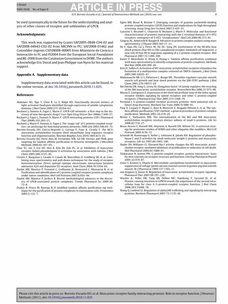
Fig. 1. The ILoop-TAP strategy for identification of muscarinic receptor family interacting proteins. (A) Schematic representation of minigene construction. Stratagene TAPcassette is inserted at the N-terminal of a given 3ILoop and C-terminal tail. (B) Overview of complexes purification and sample processing. (C) and (D) Examples of TAPc ainedt humaf atabasa
crbai
Mmemcoocdc
3
tdsprm
omplexes purified from different receptor subtypes on denaturing protein gels stools used for protein analysis. The MS/MS data was analyzed and matched to allollowing formatting for import into BIND, the Biomolecular Interaction Network Dconnectivity organic distribution network using Cytoscape software.
ontaining complexes were efficiently captured on streptavidinesin as shown in Fig. 2A, lane 5 (flow-through after Streptavidininding), demonstrating that the TAP-mini-M5 can be expressednd purified efficiently with the condition established in our exper-ments.
Also, we checked the subcellular distribution of the pTAP-mini-5 by means of immunocytochemistry experiments and confocalicroscopy analysis. Under these conditions, permeabilized cells
xpressing this construct showed clear cytosolic distribution pTAP-ini-M5 (Fig. 2B). Interestingly, when the pTAP-mini-M5 was
oexpressed together with M5R a partial overlapped distributionf pTAP-mini-M5 and M5R was observed in discrete areas. Thus,ur results show that the pTAP-mini-M5 when transfected inells is heavily synthesized and it might have some degree of co-istribution with the mAChRs. Similar results were observed fromells expressing other receptor subtypes (data not shown).
.2. Sensitivity, specificity and reliability of ILoop-TAP approach
With the aim of detecting proteins that specifically interact withhe intracellular loops of each receptor subtype, we performed
Please cite this article in press as: Borroto-Escuela DO, et al. Muscarinic receMethods (2011), doi:10.1016/j.jneumeth.2010.11.025
ifferential analyses of gel protein patterns obtained with eachpecific TAP tagged-minigene and mock (TAP alone) baits. A com-arison of protein patterns in the gels obtained indicated a selectiveecruitment of different proteins together with several abundantlyitochondrial and ribosomal origin proteins unambiguously iden-
with Coomassie, and fragment spectra of selected peptide ions. (E) Bioinformaticn protein sequences in the Swiss-Prot database using the MASCOT search enginee. The arbitrary molecular interaction network file obtained was then visualized as
tified by LC–MS/MS and present in all lanes (control, M1R to M5R).In addition, we excluded from our list purified heat-shock proteins,as these proteins appeared with higher frequencies in our analysis.However, active role of heat-shock proteins on GPCRs function hasbeen reported (He et al., 2007; Madamanchi et al., 2001).
The systematic purification analysis generated 252 samples formass spectrometry and subsequently identified 142 unique pro-teins (Supplementary materials Fig. 1). Ontology classification ofidentified peptides revealed proteins of various subcellular com-partments and functions, most of them known to have an importantrole in GPCR signalling (Supplementary materials Fig. 2). The major-ity of the peptides identified were from proteins known to belocalized in the plasma membrane or cytosol, followed by proteinslocalized in the reticulum and Golgi apparatus. In addition, identi-fied proteins corresponded to 62% signalling proteins in agreementwith classical function of GPCRs as transducers. On average, 50%of proteins identified were Gq/11, and 30% of those Gi/0, muscarinicreceptor associated proteins. Identification of specific associatedproteins for each receptor subtype from 7 independent purifica-tions resulted in mayor specificities for M4R and M3R subtypes,followed by M2R, M5R and M1R (Supplementary materials Fig. 1).
ptor family interacting proteins: Role in receptor function. J Neurosci
Of the 142 purified associated proteins, approximately 80% pre-sented as specific interacting proteins, showing that the ILoop-TAPmethod is very efficient for the large-scale retrieval and identifi-cation of receptor interacting proteins. Although it is difficult toestimate the percentage of interacting proteins which are cov-

ARTICLE IN PRESSG Model
NSM-5830; No. of Pages 9
D.O. Borroto-Escuela et al. / Journal of Neuroscience Methods xxx (2010) xxx–xxx 5
Fig. 2. (A) Small-scale ILoop-TAP-M5 purification. The M5 muscarinic receptor com-plexes were efficiently isolated using two-step affinity purification as shown byimmunoblotting using anti-CBP antibody. Lane 1, Santa Cruz Biotechnology molec-ular weight; lane 2, without; lane 3, cell lysate; lane 4, unbound fraction afterincubation with streptavidin resin; lane 5, wash with streptavidin binding buffer;lane 6 elution from streptavidin resin; lane 7, unbound fraction after binding tocalmodulin resin. The samples were separated by 15% SDS-PAGE, and the minigenewas detected by western blot using the anti-CBP antibody. (B) Immunofluorescencelocalization of pTAP-mini-M5 and M5R in SK-N-MC cells. Cells were transientlycotransfected with the cDNAs encoding pTAP-mini-M5 (green) and M5R (red) andprocessed for immunocytochemistry using a mouse anti-SBP monoclonal antibodyand a rabbit anti-M5R polyclonal antibody. The bound primary antibodies weredetected using donkey anti-mouse Alexa Fluor-488 (green) and donkey anti-rabbitAlexa Fluor-568 (red). Superimposition of images reveals receptor codistribution inyellow (merge). Microscope observations were performed with a 63× oil immer-sion objective in a Leica TCS-SL confocal microscope (Leica, USA). The degrees ofM5R/IL-TAP-M5 co-localization are shown as a single z-scan image. Co-localizationrmto
etltfmiiwc
3
bpraccbt
t
Fig. 3. (A) Purification of TAP-mini-M1 associated complexes. Approximately1 × 107 SK-N-MC-TAP-mini-M1 cells (with or without carbachol 10 �M treatment)
GIT-1 and paxillin were selected because of their capacity to
atios (RC) revelled that 46 ± 4% of M5R and IL-TAP-M5 protein are co-localizing (forore description see Section 2). Mean ± S.E.M.; 30 pictures. (For interpretation of
he references to color in this figure legend, the reader is referred to the web versionf the article.)
red by our data set, a rough estimation can be made based onhe observation that many identified proteins (at least 42%) areikely to interact with other GPCRs and have been associated withheir function (crude estimate). We also observed that the methodailed to detect some known interactions (for example RGS and GRK
uscarinic receptor scaffold proteins). Proteins which likely partic-pate in transient interaction, low stoichiometric complexes, and/ornteractions which occur only in specific physiological states that
ere either not present or under-represented in our experimentalonditions probably cannot be identified using this method.
.3. Dynamics of protein complexes
Fig. 3A shows small-scale TAP-mini-M1 complex purificationefore and upon agonist stimulation. Comparison of identifiedroteins revealed, in both cases, the presence of distinct bands cor-esponding to different associated proteins. Using the Iloop-TAPpproach, we determined that some associated proteins (likely coreomponents) could be identified and validated independently ofellular state. More dynamic, perhaps regulatory, components may
Please cite this article in press as: Borroto-Escuela DO, et al. Muscarinic receMethods (2011), doi:10.1016/j.jneumeth.2010.11.025
e present differentially depending on cellular pharmacologicalreatment or physiological conditions.
Examples of proteins that were identified as associated pro-eins present before and upon agonist stimulation are �-synuclein,
and SK-N-MC-TAP cells (Mock) were submitted to the ILoop-TAP strategy protocol.Eluates were separated by 15% SDS-PAGE gel and proteins were detected by silverstaining. (B) List of identified proteins.
serine/threonine-protein phosphatase 2A (PP2A) and, surpris-ingly, the Regulatory G protein Signalling 2 (RGS2). RGS proteinshave previously been reported to interact with GPCRs and theirheterotrimeric G-proteins. They were selectively recruited uponagonist stimulation of the plasma membrane by G proteins andcorrespondingly by receptors that activate those G proteins. How-ever, based on our results, it seems that M1 muscarinic receptorskeep their association with specific RGS2 proteins independentlyof receptor activation. We also identified a wide number of pro-teins that were selectively recruited only upon agonist stimulation(Fig. 3B), thus indicating that signalling efficiency/specificity formAChRs is determined in part by dynamics change in multiproteincomplexes reorganization.
3.4. Validation of selected identified proteins
Functional protein–protein interactions were assessed bymeans of biochemical (co-immunoprecipitation) and biophysi-cal (Bioluminescence Resonance Energy Transfer: BRET) methods.Although a comprehensive, step-by-step verification of all proteinsin our data set will be undertaken using complementary meth-ods, we selected and analyzed here only several uncharacterizedmuscarinic-interacting proteins for their ability to form a complexwith muscarinic receptors.
ptor family interacting proteins: Role in receptor function. J Neurosci
interact with each other and their known ability to be involved indiverse cellular processes. AKAP79 has been described as a scaffoldmolecule of the prototypical �2-adrenergic receptor, orchestrat-ing the interactions of various protein kinases (including tyrosine

ARTICLE IN PRESSG Model
NSM-5830; No. of Pages 9
6 D.O. Borroto-Escuela et al. / Journal of Neuroscience Methods xxx (2010) xxx–xxx
Fig. 4. Interaction of the intact muscarinic receptor subtypes with endogenous selected interacting proteins (GIT-1, paxillin, AKAP79). SK-N-MC cells transiently transfectedwith pcDNA3-3xHA-M2, or pcDNA3-3xHA-M3, or pcDNA3-3xHA-M5 were lysed in Nonidet-p40 buffer, and the receptor was immunoprecipitated as described under “Section2”. The membrane transfer was first probed with an anti-GIT1, anti-Paxillin or anti-AKAP79 antibody followed by striping and reprobing with anti-HA monoclonal antibodyt pt.) land
ke
MieGcbeMiiGtcr
cLtmc
iceitwcprFs
o confirm effective receptor immunoprecipitation. The input and supernatant (Suata is representative of two to three independent experiments.
inases), protein phosphatases (e.g. calcineurin) and cytoskeletallements which could enable multivalent signalling.
Lysates of SK-N-MC cells exogenously expressing the 3xHA-2/M3/M5 muscarinic receptor subtype were subjected to
mmunoprecipitation with anti-HA beads and analyzed by West-rn blotting with the anti-GIT-1, anti-paxillin, anti-FAK antibodies.IT-1 and paxillin did not coimmunoprecipitate with M2 mus-arinic receptor subtypes in correspondence with results obtainedy ILoop-TAP approaches (Fig. 4). However, bands corresponding toach interacting protein were observed in cells expressing M3R and5R subtypes. Detection of the Focal Adhesion kinase (FAK) protein
n immunoprecipitated Gq/11 receptor subtypes (data not shown)s in agreement with previous reports, which described paxillin andIT protein as multiplatform components that can actively recruit
yrosin kinases such as FAK protein. This demonstrates dynamicomplex formation around muscarinic receptors by the sequentialecruitment of these scaffold proteins.
A similar set of experiments was performed using immunopre-ipitation and Western blot analysis with the anti-AKAP antibody.ike GIT-1 and paxillin, AKAP copurified with Gq/11-coupled recep-or subtypes. However, it did not coimmunoprecipitate with the M2
uscarinic receptor subtype, a Gi/0-coupled receptor (Fig. 4). Thisonfirms a specific interaction also reported by TAP purification.
As an additional step to address M3R/AKAP79 interactionssues, BRET2 experiments between M3RRluc/AKAP79GFP2 werearried out. The BRET2 assay was performed on HEK293T cells co-xpressing a constant amount of the M3RRluc or M2RRluc whilencreasing the amount of the AKAP79GFP2 plasmids. As a posi-ive control, cells expressing a GFP2-Rluc tandem fusion proteinere used. Stimulation of M3R with a concentration of 100 nM
Please cite this article in press as: Borroto-Escuela DO, et al. Muscarinic receMethods (2011), doi:10.1016/j.jneumeth.2010.11.025
arbachol induced an increase of the maximal BRET2 value com-ared to unstimulated cells, which is consistent with an activeecruitment of the AKAP79GFP to the receptor (Supplementaryig. 3). Interestingly, cells co-expressing 5-M2RRluc/AKAP79GFP2
howed very low and linear bystander BRET signals as the negative
es represent ¼ of the lysate volume used for each immunoprecipitation (IP). The
control (Supplementary Fig. 3). Taken together coimmunoprecip-itation and BRET results, these observations support the notionthat the M3R directly interacts in an agonist-depend manner withAKAP79GFP2 receptors in living cells.
Previous experiments have demonstrated that muscarinicacetylcholine receptor-stimulation suppress M-type potassiumchannel current, inhibition which is mediated by the multivalentAKAP79 protein. This protein is particularly interesting because,in addition to its role in the inhibition of current channels, it alsoassociates with protein kinases, phosphatases and cytoskeletonelements enabling multivalent signalling.
3.5. Network map of muscarinic receptor interactome
It is becoming increasingly clear that GPCRs exist as large com-plexes of proteins involving dimers or oligomers of receptor andassociated proteins that, depending on their variable composi-tion, determine their function. The ILoop-TAP strategy revealed adynamic composition of muscarinic receptor subtype interactome.The dynamics of complex composition are well illustrated by thecellular signalling complexes formed around each subtype. Then,assigning individual proteins to each receptor subtype complex, weinvestigated relationships among them in order to understand theintegration and coordination of the muscarinic receptor signallingprocess. We represented these relationships by linking complexesthat share components (Fig. 5). By plotting all the data of set inter-actions described here, together with the interactions detected byother biochemical methods reported in the literature, we obtaineda comprehensive view of the network of complexes.
Connections in this network not only reflect the physical inter-
ptor family interacting proteins: Role in receptor function. J Neurosci
actions of complexes, but may also represent common regulation,localization, turnover or architecture. All data sets were importedinto the BIND database and exported as an arbitrary molecular net-work. A single large network of muscarinic receptor interactomewas visualized through the use of Cytoscape software.

Please cite this article in press as: Borroto-Escuela DO, et al. Muscarinic receptor family interacting proteins: Role in receptor function. J NeurosciMethods (2011), doi:10.1016/j.jneumeth.2010.11.025
ARTICLE IN PRESSG Model
NSM-5830; No. of Pages 9
D.O. Borroto-Escuela et al. / Journal of Neuroscience Methods xxx (2010) xxx–xxx 7
Fig. 5. The muscarinic receptor family interactome. Graphs were generated automatically by organic algorithm using Cytoscape software. Line thickness represents thenumber of experiments describing a given interaction and illustrates the connection between proteins. Lines are color-coded according to the experiments: ILoop-TAP results(red), reported in the literature (black). Muscarinic receptors subtype’s nodes are highlighted in red. Each protein is labelled using BIND annotation. (For interpretation ofthe references to color in this figure legend, the reader is referred to the web version of the article.)

ING
N
8 Neuro
adtsiaicr
4
tpieem
cmiu1p
stsktGaeautGitIprclfniloaitlerltfa(Rr
ARTICLEModel
SM-5830; No. of Pages 9
D.O. Borroto-Escuela et al. / Journal of
By analyzing this large network containing 400 interactionsmong 100 proteins, as well as several smaller networks, we couldetect some interesting features. First, receptors of similar func-ion tend to cluster together within the network, suggesting thatharing components reflects functional relationships. Second, sim-lar performance can be observed with assessor proteins that sharecommon subcellular localization. Third, the network of interact-
ng nodes reveals sets of interactions that link cellular process andross-connections reflecting the central role of some proteins inegulating other cellular processes.
. Discussion
Several large-scale approaches have been undertaken in ordero assign cellular functions and to identify new GPCRs interactingroteins, and to understand the context in which receptor–protein
nteractions operate. These include co-immunoprecipitationxperiments, yeast two-hybrid screens, proteomic, resonancenergy transfer studies, protein chip and computational in silicoethods (Daulat et al., 2007, 2009).The ILoop-TAP proteomic approach presented here may well
ontribute to the largest analysis of protein–protein interactionap reported in GPCRs. Maximizing sensitivity and reproducibil-
ty over other methods to detect receptor binding partners andsing muscarinic receptor family as a model system, we identified48 different interacting proteins, of which 57 had been detectedreviously as muscarinic receptor-binding partners.
The GO classification of the proteins identified by Iloop-TAPtrategy revealed some interesting data of further considera-ion: the presence of heterotrimeric G proteins in all receptorubtype-associated protein purifications. Consistent with thenown coupling of muscarinic receptors to G proteins, we iden-ified four G�-protein for M1/M3/M5 receptor subtypes (G�-q,�-11, G�-12 and G�-13) and three G�-i isoforms (G�-i1, G�-i2nd G�-i3) for M2R/M4R receptor subtypes, as well as two differ-nt G� isoforms (G�1 and G�4) and three G� isoforms (G�2, G�7nd G�11). This demonstrates that our approach can efficiently besed as a method for large-scale protein–protein interaction detec-ion, not only for the muscarinic receptor family, but also for otherPCRs. In addition we also identified several cytoskeletal proteins,
ncluding those associated with GPCRs reorganization and regula-ion (�- and �-tubulin, filamin A, annexin A2, dynamin 2, clathrin,Q-GAP1/3 and paxillin), some of them existing in a common com-lex with specific receptor subtypes. This suggests that muscariniceceptors selectively bind to different structural components in theell and may have a role in receptor internalisation and the regu-ation of cell spreading, migration, and the attachment at sites ofocal adhesion (van Koppen and Kaiser, 2003). In support of thisotion are recent reports pointing towards different mechanisms of
nternalisation using M2R and M4R receptors depending of the cel-ular type (Reiner and Nathanson, 2008), as well as the active rolesf tubulin and microtubule reorganization in PLD activation medi-ted by Gq/11 muscarinic receptor subtypes (Chae et al., 2005). Morenterestingly, using drugs that stabilized or destabilized micro-ubule organization, it was found that the activation of muscariniceads to modulation of ion currents in cardiac myocyte cells (Unnot al., 1999), functions mediated by the direct interaction of theeceptor and the G protein with the microtubule cytoskeleton. Theast consideration is the identification of several muscarinic recep-or subtypes-specific signalling proteins such as ADP-ribosylation
Please cite this article in press as: Borroto-Escuela DO, et al. Muscarinic receMethods (2011), doi:10.1016/j.jneumeth.2010.11.025
actors, elongation factor 1-A (eEF-1A), oncogenic SET protein, Rac1,nd different isoforms of phospholipase C and the protein kinase C�-1, �-3, � and �, �, � respectively). Interestingly, the small GTPaseac1 has been shown to function downstream of M1R and M3Receptors. In the context of Rac 1 activity but not RhoA activity, M3R
PRESSscience Methods xxx (2010) xxx–xxx
muscarinic receptor-mediated activation of PLC and PKC triggerscell death (Shafer and Williams, 2004). The ADP-ribosylation fac-tors are members of the Arf arm of the Ras superfamily of guanosinetriphosphate (GTP)-binding proteins. Physiologically, Arfs regulatemembrane traffic and the actin cytoskeleton. However, Arf functionlikely involves many additional biochemical activities. Arf activatesphospholipase D and phosphatidylinositol 4-phosphate 5-kinasewith the consequent production of PA and PIP2, respectively. Ithas been shown that vasopressin V2 receptor and M2R muscarinicreceptor processing and trafficking are under ARF-6 control. Also,Arf 1 and 6 mediate PLD1/2 activation by M3R muscarinic recep-tor (Schmidt et al., 1997). eEF-1A and other elongation factors havebeen reported to modulate M4R muscarinic receptor subtype func-tion by direct interaction with the receptor (McClatchy et al., 2006).Several serine/threonine phosphatases participate in the dephos-phorylation of activated GPCRs. Phosphatases of the PP2A and PP2Bsubfamilies have been reported to target GPCRs. M1 receptor sub-types stimulate the formation of a multiprotein complex centredon TRPC6 channels where PP2B plays an active role in the disas-sociation of the muscarinic receptor from the complex (Kim andSaffen, 2005).
The most important outcome of our approach is the possibil-ity to study and identify specific interacting proteins under nativeconditions. Although only one set of experimental parameters wasused here for the evaluation of complex composition, we will, in thefuture, systematically modify experimental parameters (both phar-macological and physiological conditions) to evaluate the impactof a changing environment on complex variability. These studiesshould help to elucidate the dynamics of complex assembly anddisassembly for each receptor subtype. Moreover, it may be a start-ing point in deciphering receptor specifies and a molecular contextfor the choice and evaluation of drug targets.
Another important outcome of our experiments is that, we wereable to construct a muscarinic receptor family network by com-paring the experimental results in the literature with our data-set,allowing us to group interacting proteins into cluster complexes.The result network is a functional description of the muscarinicreceptor interactome at a higher level of organization. Compari-son of our data set with the literature is straightforward, and itis important to keep in mind that our IloopTAP approach unrav-els complexes composition data that, in any case, produces binaryinteractions, just as the results from two hybrid methods. This sup-ports the current view that receptor complex formation is morethan the sum of binary interactions.
However, binary analysis methods are of exceptional value forthe detection of pairwise and transient associations of GPCRs. Thesuccess of our approach in the characterization of receptor com-plexes relies on the conditions used for the assembly and retrievalof the complexes. These include localization and post-translationalmodifications in a manner that closely mimicks normal physiolog-ical conditions. Therefore, because the ILoopTAP method does notprovide information on the orientations of complex components,complex characterization by two-hybrid analysis, or resonanceenergy transfer methods are ideally complementary.
In summary, our ILoopTAP protocol allowed a relatively fastlarge-scale purification of GPCR-associated proteins under nativeconditions by mass spectrometry with low number of non-specificproteins detection. Specific interacting proteins from each recep-tor subtype were identified systematically, representing a majormethodological advance in the identification of GPCR-interactingprotein complexes. In addition, collected data allowed for the first
ptor family interacting proteins: Role in receptor function. J Neurosci
time the construction of an acetylcholine muscarinic receptor net-work (interactome), reflecting receptor complex properties andtendencies. Through the ‘interactome’ concept, we are able to pro-pose signalling roles for interacting partners that had not beenprevious described. This biochemical and theoretical approach can
ING
N
Neuro
by
A
SCIaas
A
t
R
A
B
B
B
B
B
C
C
D
D
D
ARTICLEModel
SM-5830; No. of Pages 9
D.O. Borroto-Escuela et al. / Journal of
e used systematically in the future for the understanding and anal-sis of other classes of receptor and subfamilies of GPCR.
cknowledgments
This work was supported by Grants SAF2005-0848-C04-02 andAF2008-04943-C02-02 from MICINN to PG; SAF2008-01462 andonsolider-Ingenio CSD2R008-00005 from Ministerio de Ciencia e
nnovación to FC and FI2004 from the European Social Foundationnd BE-2006 from the Catalonian Government to DOBE. The authorscknowledge Eric Duval and Jean Philippe van Nyen for his materialupport.
ppendix A. Supplementary data
Supplementary data associated with this article can be found, inhe online version, at doi:10.1016/j.jneumeth.2010.11.025.
eferences
bdulaev NG, Ngo T, Chen R, Lu Z, Ridge KD. Functionally discrete mimics oflight-activated rhodopsin identified through expression of soluble cytoplasmicdomains. J Biol Chem 2000;275:39354–63.
ockaert J, Dumuis A, Fagni L, Marin P. GPCR-GIP networks: a first step in the dis-covery of new therapeutic drugs? Curr Opin Drug Discov Dev 2004a;7:649–57.
ockaert J, Fagni L, Dumuis A, Marin P. GPCR interacting proteins (GIP). PharmacolTher 2004b;103:203–21.
ockaert J, Marin P, Dumuis A, Fagni L. The ‘magic tail’ of G protein-coupled recep-tors: an anchorage for functional protein networks. FEBS Lett 2003;546:65–72.
orroto-Escuela DO, Garcia-Negredo G, Garriga P, Fuxe K, Ciruela F. The M(5)muscarinic acetylcholine receptor third intracellular loop regulates receptorfunction and oligomerization. Biochim Biophys Acta 2010;1803:813–25.
orroto Escuela DO, Perez Alea M, Fernandez WR, Gil DB. Vectors and P64k genetargeting for tandem affinity purification in Neisseria meningitidis. J MicrobiolMethods 2006;65:187–93.
hae YC, Lee S, Lee HY, Heo K, Kim JH, Suh PG, et al. Inhibition of muscarinicreceptor-linked phospholipase D activation by association with tubulin. J BiolChem 2005;280:3723–30.
iruela F, Burgueno J, Casado V, Canals M, Marcellino D, Goldberg SR, et al. Com-bining mass spectrometry and pull-down techniques for the study of receptorheteromerization. Direct epitope–epitope electrostatic interactions betweenadenosine A2A and dopamine D2 receptors. Anal Chem 2004;76:5354–63.
aulat AM, Maurice P, Froment C, Guillaume JL, Broussard C, Monsarrat B, et al.Purification and identification of G protein-coupled receptor protein complexesunder native conditions. Mol Cell Proteom 2007;6:835–44.
Please cite this article in press as: Borroto-Escuela DO, et al. Muscarinic receMethods (2011), doi:10.1016/j.jneumeth.2010.11.025
aulat AM, Maurice P, Jockers R. Recent methodological advances in the discov-ery of GPCR-associated protein complexes. Trends Pharmacol Sci 2009;30:72–8.
rakas R, Prisco M, Baserga R. A modified tandem affinity purification tag tech-nique for the purification of protein complexes in mammalian cells. Proteomics2005;5:132–7.
PRESSscience Methods xxx (2010) xxx–xxx 9
Eglen RM, Bosse R, Reisine T. Emerging concepts of guanine nucleotide-bindingprotein-coupled receptor (GPCR) function and implications for high throughputscreening. Assay Drug Dev Technol 2007;5:425–51.
Gavarini S, Becamel C, Chanrion B, Bockaert J, Marin P. Molecular and functionalcharacterization of proteins interacting with the C-terminal domains of 5-HT2receptors: emergence of 5-HT2 “receptosomes”. Biol Cell 2004;96:373–81.
Gingras AC, Aebersold R, Raught B. Advances in protein complex analysis using massspectrometry. J Physiol 2005;563:11–21.
He F, Qiao ZH, Cai J, Pierce W, He DC, Song ZH. Involvement of the 90-kDa heatshock protein (Hsp-90) in CB2 cannabinoid receptor-mediated cell migration: anew role of Hsp-90 in migration signaling of a G protein-coupled receptor. MolPharmacol 2007;72:1289–300.
Kaiser P, Meierhofer D, Wang X, Huang L. Tandem affinity purification combinedwith mass spectrometry to identify components of protein complexes. MethodsMol Biol 2008;439:309–26.
Kim JY, Saffen D. Activation of M1 muscarinic acetylcholine receptors stimulates theformation of a multiprotein complex centered on TRPC6 channels. J Biol Chem2005;280:32035–47.
Madamanchi NR, Li S, Patterson C, Runge MS. Thrombin regulates vascular smoothmuscle cell growth and heat shock proteins via the JAK-STAT pathway. J BiolChem 2001;276:18915–24.
McClatchy DB, Fang G, Levey AI. Elongation factor 1A family regulates the recyclingof the M4 muscarinic acetylcholine receptor. Neurochem Res 2006;31:975–88.
Morou E, Georgoussi Z. Expression of the third intracellular loop of the delta-opioidreceptor inhibits signaling by opioid receptors and other G protein-coupledreceptors. J Pharmacol Exp Ther 2005;315:1368–79.
Presland J. G-protein-coupled receptor accessory proteins: their potential role infuture drug discovery. Biochem Soc Trans 2004;32:888–91.
Puig O, Caspary F, Rigaut G, Rutz B, Bouveret E, Bragado-Nilsson E, et al. The tan-dem affinity purification (TAP) method: a general procedure of protein complexpurification. Methods 2001;24:218–29.
Reiner C, Nathanson NM. The internalization of the M2 and M4 muscarinicacetylcholine receptors involves distinct subsets of small G-proteins. Life Sci2008;82:718–27.
Rosas-Acosta G, Russell WK, Deyrieux A, Russell DH, Wilson VG. A universal strat-egy for proteomic studies of SUMO and other ubiquitin-like modifiers. Mol CellProteom 2005;4:56–72.
Schmidt M, Rumenapp U, Keller J, Lohmann B, Jakobs KH. Regulation of phospho-lipase C and D activities by small molecular weight G proteins and muscarinicreceptors. Life Sci 1997;60:1093–100.
Shafer SH, Williams CL. Elevated Rac1 activity changes the M3 muscarinic acetyl-choline receptor-mediated inhibition of proliferation to induction of cell death.Mol Pharmacol 2004;65:1080–91.
Tilakaratne N, Sexton PM. G-protein-coupled receptor–protein interactions: basisfor new concepts on receptor structure and function. Clin Exp Pharmacol Physiol2005;32:979–87.
Unno T, Komori S, Ohashi H. Microtubule cytoskeleton involvement in muscarinicsuppression of voltage-gated calcium channel current in guinea-pig ileal smoothmuscle. Br J Pharmacol 1999;127:1703–11.
van Koppen CJ, Kaiser B. Regulation of muscarinic acetylcholine receptor signaling.Pharmacol Ther 2003;98:197–220.
ptor family interacting proteins: Role in receptor function. J Neurosci
Wacker JL, Feller DB, Tang XB, Defino MC, Namkung Y, Lyssand JS, et al.Disease-causing mutation in GPR54 reveals the importance of the second intra-cellular loop for class A G-protein-coupled receptor function. J Biol Chem2008;283:31068–78.
Wang Q, Limbird LE. Regulation of alpha2AR trafficking and signaling by interactingproteins. Biochem Pharmacol 2007;73:1135–45.

SUPPLEMENTARY MATERIALS Borroto-Escuela et al. 2010 Figure 1. (A) Hierarchically clustered dendrograms of specificity interacting protein
identified for each receptor subtypes. Each row represents an identified protein using
Iloop-TAP strategy, and each column represents a particular muscarinic receptor
subtype (M1R to M5R). The specificity of each interacting protein by receptor subtypes
and G protein selectivity was provided by different colour scale. All: means specific
interactions with all five subtypes; Spf: means specific interactions with a subtype
receptor; Gq/11: means specific interactions with all Gq/11 subtypes; Gi/o: means
specific interactions with all Gi/o subtypes and Non: means non-specific interactions.
(B) Number and percentage of sequences coverage for each indentified protein.
Figure 2. Statistic and distribution of protein identified. Numbers outside pie charts
represent the percentages of total proteins. A, classification of protein according to
function; B, subcellular localization; C, number of protein per receptor subtypes and D,
number of specific protein per receptor subtypes.
Figure 3. BRET2 studies of M3R and AKAP79 specific interaction in HEK293T cells.
HEK293T cells were transiently transfected with 1 μg of plasmid encoding M3RRluc or
M2RRluc (donor) and increasing amounts of AKAP79GFP2 plasmid in presence (black
square or circle) or absence (white square or circle) of 100 nM carbachol. Separated
cells were transfected with 2 μg of the positive control plasmid Rluc-GFP2 and in each
experiments, the specificities of M3RRluc/AKAP79GFP2 interactions were assessed by
comparison with cell expressing alone M3RRluc (data not shown). After the addition of
coelenterazine-400a, the BRET2 signal was measured. Cells expressing
M2RRluc/AKAP79GFP2 yielded a background BRET ratio of ∼10% in presence or absence
of 100 nM carbachol, while the cells coexpressing M3RRluc/AKAP79GFP2 yielded
substantially higher BRET2 ratios, indicative of energy transfer and specific and
agonist-sensitive protein-protein interaction. The data represent the mean ± S.E.M.; n =
4 in triplicate. ***: Significantly different compared to M3RRluc/AKAP79GFP2,
M2RRluc/AKAP79GFP2 and M2RRluc/AKAP79GFP2 + Cch (P<0.001), by One-way analysis
of variance (ANOVA). Cch: carbachol.
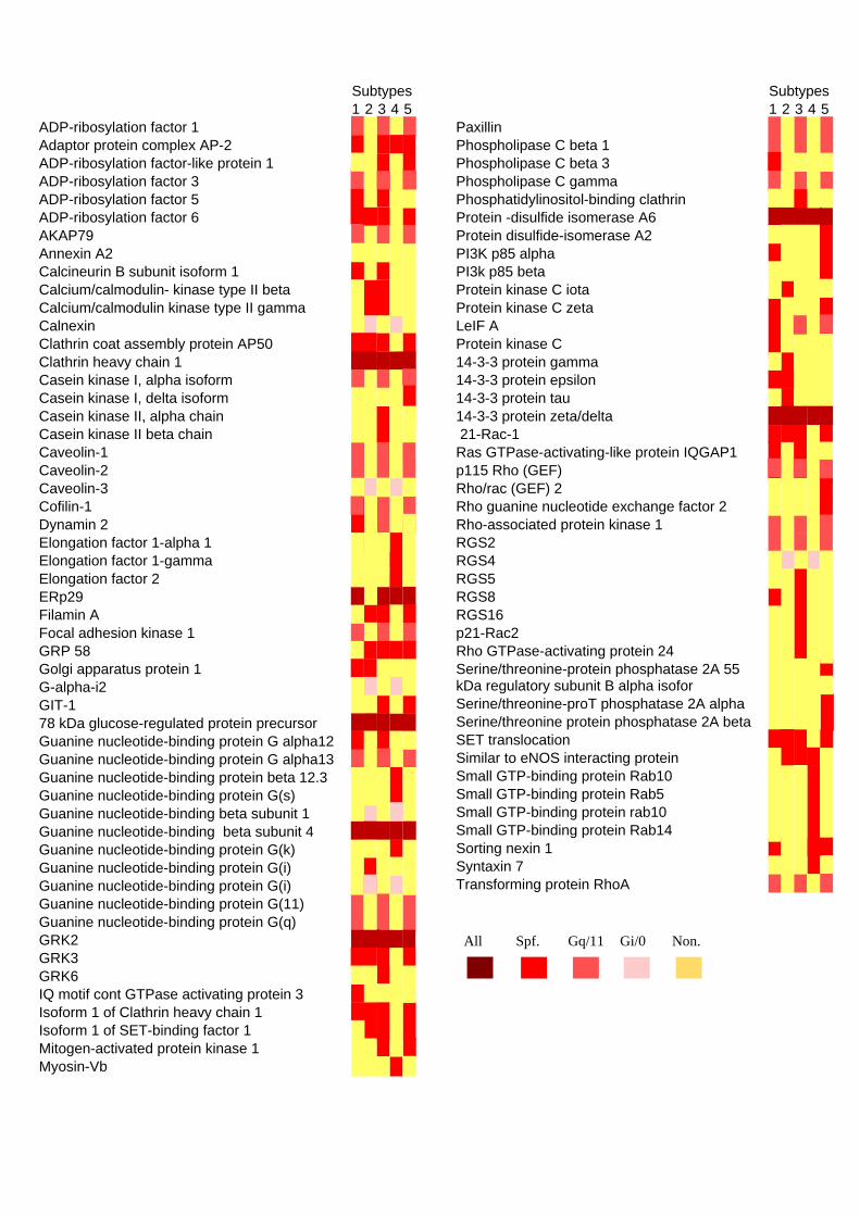
Subtypes 1 2 3 4 5ADP-ribosylation factor 1 Adaptor protein complex AP-2 ADP-ribosylation factor-like protein 1 ADP-ribosylation factor 3 ADP-ribosylation factor 5 ADP-ribosylation factor 6 AKAP79 Annexin A2 Calcineurin B subunit isoform 1 Calcium/calmodulin- kinase type II beta Calcium/calmodulin kinase type II gamma Calnexin Clathrin coat assembly protein AP50 Clathrin heavy chain 1 Casein kinase I, alpha isoform Casein kinase I, delta isoform Casein kinase II, alpha chain Casein kinase II beta chain Caveolin-1 Caveolin-2 Caveolin-3 Cofilin-1 Dynamin 2 Elongation factor 1-alpha 1 Elongation factor 1-gamma Elongation factor 2 ERp29 Filamin A Focal adhesion kinase 1 GRP 58 Golgi apparatus protein 1 G-alpha-i2 GIT-1 78 kDa glucose-regulated protein precursor Guanine nucleotide-binding protein G alpha12 Guanine nucleotide-binding protein G alpha13 Guanine nucleotide-binding protein beta 12.3 Guanine nucleotide-binding protein G(s) Guanine nucleotide-binding beta subunit 1 Guanine nucleotide-binding beta subunit 4 Guanine nucleotide-binding protein G(k) Guanine nucleotide-binding protein G(i) Guanine nucleotide-binding protein G(i) Guanine nucleotide-binding protein G(11) Guanine nucleotide-binding protein G(q) GRK2 GRK3 GRK6 IQ motif cont GTPase activating protein 3 Isoform 1 of Clathrin heavy chain 1 Isoform 1 of SET-binding factor 1 Mitogen-activated protein kinase 1 Myosin-Vb
Subtypes 1 2 3 4 5Paxillin Phospholipase C beta 1 Phospholipase C beta 3 Phospholipase C gamma Phosphatidylinositol-binding clathrin Protein -disulfide isomerase A6 Protein disulfide-isomerase A2 PI3K p85 alpha PI3k p85 beta Protein kinase C iota Protein kinase C zeta LeIF A Protein kinase C 14-3-3 protein gamma 14-3-3 protein epsilon 14-3-3 protein tau 14-3-3 protein zeta/delta 21-Rac-1 Ras GTPase-activating-like protein IQGAP1 p115 Rho (GEF) Rho/rac (GEF) 2 Rho guanine nucleotide exchange factor 2 Rho-associated protein kinase 1 RGS2 RGS4 RGS5 RGS8 RGS16 p21-Rac2 Rho GTPase-activating protein 24 Serine/threonine-protein phosphatase 2A 55 kDa regulatory subunit B alpha isofor
Serine/threonine-proT phosphatase 2A alpha Serine/threonine protein phosphatase 2A beta SET translocation Similar to eNOS interacting protein Small GTP-binding protein Rab10 Small GTP-binding protein Rab5 Small GTP-binding protein rab10 Small GTP-binding protein Rab14 Sorting nexin 1 Syntaxin 7 Transforming protein RhoA
All Spf. Gq/11 Gi/0 Non.

Acc. #
Protein name
Mr (Da)
Number of Peptides
Sequence coverage (%)
P84077 ADP-ribosylation factor 1 20697 3 46 P63010 Adaptor protein complex AP-2 subunit beta 104553 8 13 P40616 ADP-ribosylation factor-like protein 1 20418 1 6 P84077 ADP-ribosylation factor 3 20697 2 19 P84085 ADP-ribosylation factor 5 20530 1 5 P62330 ADP-ribosylation factor 6 20082 4 31 P24588 AKAP79 47088 12 32 P07355 Annexin A2 38604 3 12 P63098 Calcineurin B subunit isoform 1 19300 2 9 Q13554 Calcium/calmodulin-dependent protein kinase type II beta chain 72727 3 5 Q13555 Calcium/calmodulin-dependent protein kinase type II gamma chain 62609 1 1 P27824 Calnexin 67568 1 2 Q96CW1 Clathrin coat assembly protein AP50 49655 3 4 Q00610 Clathrin heavy chain 1 191655 9 5 P48729 Casein kinase I, alpha isoform 38915 5 16 P48730 Casein kinase I, delta isoform 47330 2 3 P68400 Casein kinase II, alpha chain 45114 5 12 P67870 Casein kinase II beta chain 24942 3 9 Q03135 Caveolin-1 20472 5 20 P51636 Caveolin-2 18291 1 3 P56539 Caveolin-3 17259 1 3 P23528 Cofilin-1 18502 2 6 P50570 Dynamin 2 98064 9 10 P68104 Elongation factor 1-alpha 1 50141 24 34 P26641 Elongation factor 1-gamma 50119 4 5 P13639 Elongation factor 2 95338 6 4 P30040 Endoplasmic reticulum protein ERp29 28993 2 7 P21333 filamin A, alpha 280739 65 1 Q05397 Focal adhesion kinase 1 119233 31 18 P30101 GRP 58 56782 10 17 Q92896 Golgi apparatus protein 1 134552 24 2 Q6B6N3 G-alpha-i2 41548 2 3 Q9Y2X7 GRK-interacting protein 1 84341 3 4 P11021 78 kDa glucose-regulated protein 72333 8 8 Q03113 Guanine nucleotide-binding protein G alpha12 44279 2 4 Q14344 Guanine nucleotide-binding protein G alpha13 44050 3 7 P63244 Guanine nucleotide-binding protein beta subunit-like protein 12.3 35077 2 4 P63092 Guanine nucleotide-binding protein G(s), alpha subunit 45665 2 3 P62873 Guanine nucleotide-binding protein G(I)/G(S)/G(T) beta subunit 1 37377 3 7 P08754 Guanine nucleotide-binding protein G(k), alpha subunit (G(i)apha-3) 40532 2 5 P63096 Guanine nucleotide-binding protein G(i), alpha-1 subunit 40361 4 12 P04899 Guanine nucleotide-binding protein G(i), alpha-2 subunit 40451 5 11 P29992 Guanine nucleotide-binding protein G(11), alpha subunit 42123 6 10 P50148 Guanine nucleotide-binding protein G(q), alpha subunit 42142 4 10 P25098 G-protein coupled receptor kinase 2 79574 7 6 P35626 G-protein-coupled receptor kinase 3 79710 3 3 P43250 G protein-coupled receptor kinase GRK6 65991 2 2 Q86VI3 Ras GTPase-activating-like protein IQGAP3 184689 15 1 Q00610 Clathrin heavy chain 1 191615 48 20 Q59EX9 Isoform 1 of SET-binding factor 1 95865 6 6 P28482 Mitogen-activated protein kinase 1 41390 3 8 Q9ULV0 Myosin-Vb 213672 64 20 P49023 Paxillin 64533 8 12 Q9NQ66 Phospholipase C beta 1 138567 7 4

Q01970 Phospholipase C beta 3 138799 5 2 P19174 Phospholipase C gamma 148532 3 1 Q13492 Phosphatidylinositol-binding clathrin assembly protein 70755 3 3 Q15084 Protein -disulfide isomerase A6 48121 3 5 Q13087 Protein -disulfide isomerase A2 58206 3 2 P26450 PI3-kinase subunit p85-alpha 83598 8 6 O00459 PI3-kinase subunit p85-beta 81624 3 2 P41743 Protein kinase C iota 68262 2 2 Q13574 Protein kinase C zeta 124128 2 1 P01563 LeIF A 21550 1 3 Q969G5 Protein kinase C, delta binding protein 27626 2 5 P61981 14-3-3 protein gamma 28303 3 11 P62258 14-3-3 protein epsilon 29174 2 5 P27348 14-3-3 protein tau 27764 4 9 P63104 14-3-3 protein zeta/delta 27745 1 4 P63000 p21-Rac1 21450 5 23 P46940 Ras GTPase-activating-like protein IQGAP1 189252 10 4 Q92888 115 kDa guanine nucleotide exchange factor 102435 8 9 Q14155 Rho guanine nucleotide exchange factor 7 92012 6 7 Q13464 Rho-associated protein kinase 1 158175 13 8 P41220 RGS2 24382 11 31 P49798 RGS4 23256 8 19 O15539 RGS5 20946 4 20 P57771 RGS8 20917 3 11 O15492 RGS16 22768 3 14 p15153 p21-Rac2 21000 2 12 Q8N264 Rho GTPase-activating protein 24 84598 3 5 P63151 Serine/threonine-protein phosphatase 2A 55 kDa regulatory subunit
B alpha isoform 51692 7 14
P67775 Serine/threonine-protein phosphatase 2A catalytic subunit alpha isoform
35594 5 16
P62714 Serine/threonine protein phosphatase 2A, catalytic subunit, beta isoform
35575 4 12
Q5VXV3 SET translocation (myeloid leukemia-associated) 33489 6 17 Q9Y314 eNOS interacting protein 33172 7 21 P61026 Ras-related protein Rab-10 22541 3 14 P61106 Small GTP-binding protein Rab14 23897 4 16 P20339 Small GTP-binding protein Rab5A 23659 3 12 Q13596 Sorting nexin 1 59070 5 8 O15400 Syntaxin 7 29816 7 26 P61586 Transforming protein RhoA 21768 8 37


SUPPLEMENTARY DATAFig. 3 Borroto-Escuela et al.