The Vagal Nerve Stimulates Activation of the Hepatic Progenitor Cell Compartment via Muscarinic...
10
The Vagal Nerve Stimulates Activation of the Hepatic Progenitor Cell Compartment via Muscarinic Acetylcholine Receptor Type 3 David Cassiman,* Louis Libbrecht, † Nicoletta Sinelli, † Valeer Desmet, † Carl Denef,* and Tania Roskams † From the Laboratory of Cell Pharmacology * and the Department of Pathology, † University of Leuven, Leuven, Belgium In the rat the hepatic branch of the nervus vagus stimulates proliferation of hepatocytes after partial hepatectomy and growth of bile duct epithelial cells after bile duct ligation. We studied the effect of he- patic vagotomy on the activation of the hepatic pro- genitor cell compartment in human and rat liver. The number of hepatic progenitor cells and atypical reac- tive ductular cells in transplanted (denervated) hu- man livers with hepatitis was significantly lower than in innervated matched control livers and the number of oval cells in vagotomized rat livers with galac- tosamine hepatitis was significantly lower than in livers of sham-operated rats with galactosamine hep- atitis. The expression of muscarinic acetylcholine re- ceptors (M1-M5 receptor) was studied by immunohis- tochemistry and reverse transcriptase-polymerase chain reaction. In human liver , immunoreactivity for M3 receptor was observed in hepatic progenitor cells , atypical reactive ductules, intermediate hepatocyte- like cells, and bile duct epithelial cells. mRNA for the M1-M3 and the M5 receptor , but not the M4 receptor , was detected in human liver homogenates. In conclu- sion, the hepatic vagus branch stimulates activation of the hepatic progenitor cell compartment in dis- eased liver , most likely through binding of acetylcho- line to the M3 receptor expressed on these cells. These findings may be of clinical importance for pa- tients with a transplant liver. (Am J Pathol 2002, 161:521–530) Hepatic progenitor cells (HPCs) are small epithelial cells that can differentiate toward the hepatocytic and the biliary lineage. 1–6 Because HPCs have an oval nucleus, they are usually referred to as “oval cells” in the rat liver. 7 In the nondiseased liver, HPCs are located in the canals of Hering, which are the smallest ramifications of the biliary tree that connect to the hepatocytes in the peri- portal area. 8 In various human liver diseases and experimental an- imal models that are characterized by damage and loss of hepatocytes and/or bile duct epithelial cells, the num- ber of HPCs strongly increases and atypical reactive ductules and intermediate hepatocyte-like cells ap- pear. 3,9 –12 The former represent differentiation of the HPCs toward bile duct epithelial cells and the latter to- ward hepatocytes. 3,10,12 The increase in number of HPCs and atypical reactive ductules is referred to as “activa- tion” of the HPC or oval cell compartment. 13,14 Studies in experimental models of liver injury have led to the iden- tification of a number of autocrine and paracrine factors that are involved in the regulation of oval cell activation in rat liver. Identified factors include transforming growth factor-, hepatocyte growth factor and its receptor c-met, and the plasminogen activator/plasmin system. 15–17 It remains to be shown whether these or other factors also have a function in the activation of the human HPC com- partment. Hepatic regeneration after partial hepatectomy (PHx) occurs through the proliferation of mature hepatocytes and bile duct epithelial cells 18 and it has previously been shown that this process is suppressed by hepatic branch vagotomy and facilitated by stimulation of the nervus vagus. 19 –21 It is not clear how the nervus vagus influ- ences proliferation of hepatocytes after PHx, but it is not via binding of acetylcholine to nicotinic acetylcholine re- ceptors because these are not expressed in the liv- er. 22–25 Experiments in which bile duct-ligated rats un- derwent cervical vagotomy have revealed that the nervus vagus directly stimulates growth of bile duct epithelial cells via binding of acetylcholine to the muscarinic ace- tylcholine receptor type 3 (M3 receptor). 26 Outside the liver, in vitro binding of acetylcholine or an analog of acetylcholine to the M3 receptor has been shown to stimulate the proliferation of human colon cancer cells, rat astrocytes, human astrocytoma cells, human prostate cancer cells and human oligodendrocyte progeni- tors. 27–30 It is not known whether the nervus vagus also Supported by the Fonds voor Wetenschappelijk Onderzoek-Vlaanderen (“aspirant FWO” grant to D. C.). D. C. and L. L. contributed equally to the article. Accepted for publication April 30, 2002. Address reprint requests to Dr. Louis Libbrecht, Department of Pathol- ogy, University of Leuven, Minderbroedersstraat 12, 3000 Leuven, Bel- gium. E-mail: [email protected]. American Journal of Pathology, Vol. 161, No. 2, August 2002 Copyright © American Society for Investigative Pathology 521
-
Upload
independent -
Category
Documents
-
view
1 -
download
0
Transcript of The Vagal Nerve Stimulates Activation of the Hepatic Progenitor Cell Compartment via Muscarinic...

The Vagal Nerve Stimulates Activation of the HepaticProgenitor Cell Compartment via MuscarinicAcetylcholine Receptor Type 3
David Cassiman,* Louis Libbrecht,†
Nicoletta Sinelli,† Valeer Desmet,† Carl Denef,*and Tania Roskams†
From the Laboratory of Cell Pharmacology * and the Department
of Pathology,† University of Leuven, Leuven, Belgium
In the rat the hepatic branch of the nervus vagusstimulates proliferation of hepatocytes after partialhepatectomy and growth of bile duct epithelial cellsafter bile duct ligation. We studied the effect of he-patic vagotomy on the activation of the hepatic pro-genitor cell compartment in human and rat liver. Thenumber of hepatic progenitor cells and atypical reac-tive ductular cells in transplanted (denervated) hu-man livers with hepatitis was significantly lower thanin innervated matched control livers and the numberof oval cells in vagotomized rat livers with galac-tosamine hepatitis was significantly lower than inlivers of sham-operated rats with galactosamine hep-atitis. The expression of muscarinic acetylcholine re-ceptors (M1-M5 receptor) was studied by immunohis-tochemistry and reverse transcriptase-polymerasechain reaction. In human liver, immunoreactivity forM3 receptor was observed in hepatic progenitor cells,atypical reactive ductules, intermediate hepatocyte-like cells, and bile duct epithelial cells. mRNA for theM1-M3 and the M5 receptor, but not the M4 receptor,was detected in human liver homogenates. In conclu-sion, the hepatic vagus branch stimulates activationof the hepatic progenitor cell compartment in dis-eased liver, most likely through binding of acetylcho-line to the M3 receptor expressed on these cells.These findings may be of clinical importance for pa-tients with a transplant liver. (Am J Pathol 2002,161:521–530)
Hepatic progenitor cells (HPCs) are small epithelial cellsthat can differentiate toward the hepatocytic and thebiliary lineage.1–6 Because HPCs have an oval nucleus,they are usually referred to as “oval cells” in the rat liver.7
In the nondiseased liver, HPCs are located in the canalsof Hering, which are the smallest ramifications of thebiliary tree that connect to the hepatocytes in the peri-portal area.8
In various human liver diseases and experimental an-imal models that are characterized by damage and lossof hepatocytes and/or bile duct epithelial cells, the num-ber of HPCs strongly increases and atypical reactiveductules and intermediate hepatocyte-like cells ap-pear.3,9–12 The former represent differentiation of theHPCs toward bile duct epithelial cells and the latter to-ward hepatocytes.3,10,12 The increase in number of HPCsand atypical reactive ductules is referred to as “activa-tion” of the HPC or oval cell compartment.13,14 Studies inexperimental models of liver injury have led to the iden-tification of a number of autocrine and paracrine factorsthat are involved in the regulation of oval cell activation inrat liver. Identified factors include transforming growthfactor-�, hepatocyte growth factor and its receptor c-met,and the plasminogen activator/plasmin system.15–17 Itremains to be shown whether these or other factors alsohave a function in the activation of the human HPC com-partment.
Hepatic regeneration after partial hepatectomy (PHx)occurs through the proliferation of mature hepatocytesand bile duct epithelial cells18 and it has previously beenshown that this process is suppressed by hepatic branchvagotomy and facilitated by stimulation of the nervusvagus.19–21 It is not clear how the nervus vagus influ-ences proliferation of hepatocytes after PHx, but it is notvia binding of acetylcholine to nicotinic acetylcholine re-ceptors because these are not expressed in the liv-er.22–25 Experiments in which bile duct-ligated rats un-derwent cervical vagotomy have revealed that the nervusvagus directly stimulates growth of bile duct epithelialcells via binding of acetylcholine to the muscarinic ace-tylcholine receptor type 3 (M3 receptor).26 Outside theliver, in vitro binding of acetylcholine or an analog ofacetylcholine to the M3 receptor has been shown tostimulate the proliferation of human colon cancer cells,rat astrocytes, human astrocytoma cells, human prostatecancer cells and human oligodendrocyte progeni-tors.27–30 It is not known whether the nervus vagus also
Supported by the Fonds voor Wetenschappelijk Onderzoek-Vlaanderen(“aspirant FWO” grant to D. C.).
D. C. and L. L. contributed equally to the article.
Accepted for publication April 30, 2002.
Address reprint requests to Dr. Louis Libbrecht, Department of Pathol-ogy, University of Leuven, Minderbroedersstraat 12, 3000 Leuven, Bel-gium. E-mail: [email protected].
American Journal of Pathology, Vol. 161, No. 2, August 2002
Copyright © American Society for Investigative Pathology
521

influences the activation of oval cells, because the stud-ied animal models (bile duct ligation and PHx) do notinvolve oval cell activation.18,31
We have previously shown that the human HPC com-partment has neural/neuroendocrine features, includingthe expression of parathyroid hormone-related peptide,chromogranin-A, neural cell adhesion molecule (NCAM),neurotrophin 4/5 and the neurotrophin receptor tyrosinekinase B.3,32,33 This suggests that neural/neuroendocrinefactors might play a role in the activation of the humanHPC compartment.
In view of these findings, we studied the expression ofthe muscarinic acetylcholine receptors in normal anddiseased human liver and we evaluated the effect ofvagotomy on the HPC compartment in both rat and hu-man liver. This subject is of potential clinical importancebecause a transplanted liver is evidently denervated andreinnervation eventually only occurs in the hilum and thelargest portal tracts.34,35
Materials and Methods
Liver Tissue Specimens
To study the effects of vagotomy on the HPC compart-ment in human liver, we used paraffin-embedded, B5-fixed tissue specimens of denervated and innervatedlivers, all affected by chronic hepatitis. The denervatedliver specimens were biopsies from liver transplant pa-tients suffering from recurrence of HCV hepatitis in thegraft (n � 5) or from hepatitis of unknown origin (n � 5).They showed no signs of rejection. To control establishedconfounding factors such as degree of necro-inflamma-tion, degree of fibrosis, and immunosuppressive treat-ment,11,12,36 the control liver biopsies (innervated) werefrom kidney transplant patients (n � 6) with chronic HBVor HCV hepatitis who were also under immunosuppres-
sive treatment at the time of biopsy and innervated con-trol biopsies and denervated transplant liver biopsieswere matched for necro-inflammatory activity and fibrosis(see Table 1 for patient characteristics) using theScheuer system.37 At the time of biopsy, all 16 patientsshowed a bilirubin level within the normal range. In thisway, all factors that are known to influence activation ofthe HPC compartment11,12,36 were excluded, exceptpresence or absence of the innervation.
To study the effects of vagotomy on oval cell activationin rat liver, we used male Wistar rats with a weight of 300to 350 g. The rats were fed ad libitum and received humancare in accordance with the institution’s ethical guide-lines. Eight rats were subjected to hepatic branch vagot-omy, as previously described,19 and eight rats weresham-operated. Immediately after the operation, all 16rats were intoxicated. Galactosamine intoxication wasinduced by application of galactosamine (Sigma, Stein-heim, Germany) in the abdominal cavity (galactosaminewas diluted to 150 mg/ml in NaCl 0.9% and applied in adose of 500 mg/kg). Liver biopsies, taken at 48 hoursafter intoxication, were snap-frozen in liquid nitrogen-cooled isopentane and stored at �70°C, until further use.The 48-hour time point is the time point of maximal hep-atitis and maximal number of oval cells.38,39
To study the expression of the muscarinic acetylcho-line receptors in normal and diseased human liver, weused 35 liver biopsies that were snap-frozen in liquidnitrogen-cooled isopentane and stored at �70°C. The 35biopsies included 5 biopsies from normal donor livers, 2biopsies of resected focal nodular hyperplasia, 3 biop-sies from the explant liver of patients with submassiveliver cell necrosis, and 25 biopsies of patients withchronic liver disease in septal or cirrhotic stage becauseof HCV (n � 10), alcoholic hepatitis (n � 5), primarybiliary cirrhosis (n � 8), and primary sclerosing cholan-gitis (n � 2).
Table 1. Patient Characteristics
K/L Fibr Infl Duct HPC Duct�HPC Tx IS P D
I K 4 1/1 70.0 10.7 80.7 52 M/T PC/HCV 84L 4 1/1 45.3 8.7 54.0 35 T HCV 29
II K 0 1/1 15.3 2.7 18.0 12 Cs/C/M HBV 120L 0 1/1 14.7 2.0 16.7 14 T/C/M Alcohol *L 0 1/1 18.7 7.0 25.7 12 Cs/Az Cryptogenic *L 0 1/1 12.3 0.7 13.0 12 Cs/C/Az HCV *
III K 0 1/2 18.3 9.3 27.6 178 C/Az HBV 180L 0 1/2 17.3 4.7 22.0 32 Cs/Az HCV *
IV K 1 2/1 51.3 10.7 62.0 122 Cs/C/Az HBV *K 1 2/1 24.7 6.7 31.4 216 C/Az HBV 156K 1 2/1 42.7 11.3 54.0 276 C/M HCV *L 1 2/1 23.7 2.3 26.0 112 Cs/Az PBC *L 1 2/1 13.0 1.3 14.3 19 Cs/M Alcohol *L 1 2/1 20.7 1.0 21.7 10 T/M HCV *L 1 2/1 24.0 6.3 30.3 24 Cs/M HCV 19L 1 2/1 10.0 1.3 11.3 59 Cs/Az PBC *
I-IV, cluster of biopsies, matched for necro-inflammation and fibrosis; K, kidney transplant patient; L, liver transplant patient; Infl, inflammation score;Fibr, fibrosis score; duct, number of atypical reactive ductular cells per field; HPC, number of HPCs per field; duct�HPC, total number of cells in theHPC compartment; Tx, time elapsed (months) after transplantation (� time on immunosuppressive therapy); IS, immunosuppression scheme (M,mycophenolate mofetil; T, tacrolimus; Cs, cyclosporin; C, corticosteroids (methylprednisolon); Az, azathioprin); P, liver pathology diagnosis on biopsyfor kidney transplant patients and on explant liver for liver transplant patients; D, time (months) since diagnosis of hepatitis (since diagnosis ofrecurrence of hepatitis, in case of liver transplantation); *, missing data.
522 Cassiman et alAJP August 2002, Vol. 161, No. 2

Immunohistochemistry
Immunohistochemistry was performed with mouse mono-clonal antibodies against cytokeratin (CK) 7 (dilution1/50; DAKO, Glostrup, Denmark) and NCAM (dilution1/40; Sigma), with OV-6 (dilution 1/200; Dr. S. Sell AlbanyMedical College, Albany, NY) and with goat polyclonalantibodies against the muscarinic acetylcholine receptortypes 1 to 5 (M1-M5 receptor) (dilutions 1/30; Santa CruzBiotechnology, Santa Cruz, CA). The anti-muscarinicacetylcholine receptor antibodies were shown to be spe-cific for the muscarinic acetylcholine receptor subtypeagainst which they were raised and do not cross-reactwith the other subtypes. Specificity of the M3 receptorantibody was further demonstrated in a Western blottingexperiment on mouse brain extract (Santa Cruz Biotech-nology).
After overnight drying and fixation in acetone for 10minutes, 5-�m-thick cryostat sections were incubatedwith the primary antibodies. For the goat polyclonal anti-bodies, the second step consisted of swine anti-goatimmunoglobulins followed by goat peroxidase anti-per-oxidase (both from DAKO). For the monoclonal mouseantibodies, the second and third step consisted of per-oxidase-labeled rabbit anti-mouse and peroxidase-la-beled swine anti-rabbit immunoglobulins (both fromDAKO) for human liver and of peroxidase-labeled goatanti-mouse and peroxidase-labeled swine anti-goat im-munoglobulins (both from Sigma) for OV-6 immunohisto-chemistry on rat liver sections. Secondary and tertiaryantibodies were diluted in phosphate-buffered saline(PBS), pH 7.2, containing 10% normal human serum. Allincubations were performed for 30 minutes at room tem-perature and followed by a wash in three changes of PBSfor 5 minutes.
Four-�m-thick sections were made from the paraffin-embedded human liver biopsies. After deparaffinization,rehydration, and heating in a microwave oven for 10minutes at 750 W, the sections were incubated with theanti-CK7 antibody and subsequently with goat anti-mouse Envision (DAKO). Both incubations were per-formed for 30 minutes at room temperature and followedby a wash in three changes of PBS for 5 minutes.
For all stainings, the reaction product was developedwith the use of 3-amino-9-ethylcarbazole or 3,3-diamino-benzidine tetrahydrochloride and H2O2, 0.01%. The sec-tions were counterstained with hematoxylin. Negativecontrols consisted of omission of the primary antibodyand were consistently negative. Cryostat sections of nor-mal human salivary gland tissue were used as positivecontrol for the M3 receptor.40 In agreement with a previ-ous study,41 the staining showed immunoreactivity in theacini and intercalated and striated ducts.
Double immunostaining for the M3 receptor and CK7was performed on 5-�m-thick human liver cryostat sec-tions using fluorescein isothiocyanate-labeled bovine an-ti-goat immunoglobulins (Santa Cruz Biotechnology) fol-lowed by tetramethylrhodamine isothiocyanate-labeledrabbit anti-mouse immunoglobulins (DAKO). Double im-munostaining was detected using a confocal laser-scan-ning microscope (Zeiss LSM 410, The Netherlands). Con-
trols consisted of omission of the antibody against the M3receptor and/or omission of the antibody against CK7and were consistently negative. Additional controls con-sisted of incubation of the polyclonal goat antibody withthe rabbit anti-mouse secondary antibody and incubationof the monoclonal mouse antibody with the bovine anti-goat secondary antibody. Nonspecific labeling could notbe detected.
Histopathological Evaluation
HPCs were defined as small, singular cells with an ovalnucleus and scanty cytoplasm immunoreactive for CK7and OV-6. Atypical reactive ductules were defined asanastomosing ductules with poorly defined lumina, linedby flattened cells with scanty cytoplasm immunoreactivefor CK7, OV-6, and NCAM. Intermediate hepatocyte-likecells were defined as polygonal cells with morphologicalresemblance to hepatocytes and faint to moderate immu-noreactivity for CK7 and OV-6.3,31 Oval cells were de-fined as OV-6-positive small cells with an oval nucleusand little cytoplasm forming strands at the portal-paren-chymal interface.10
Cell Counting and Statistical Evaluation
The human sections from paraffin-embedded biopsiesand the cryostat rat sections that were subjected tocounting were stained for CK7 and OV-6, respectively.Cell counting was performed using a counter and aneyepiece with a grid, at magnification of �100. Threeportal tracts with an interlobular bile duct were randomlyselected and visualized with the bile duct positioned inthe center of the grid. If more than one bile duct waspresent, the largest was selected. The number of HPCsand cells lining atypical reactive ductules (in humanspecimens, Table 1) or the number of oval cells (in rats)in the field defined by the grid were counted. Only cellswith a clear nucleus were counted. Statistical analysis ofthe human data were done by calculating an exact Pvalue, based on probability. For the rat data, the Mann-Whitney test was used.
Reverse Transcriptase-Polymerase ChainReaction (RT-PCR)
Frozen human liver tissue from explant livers and a nor-mal donor liver was used. Total RNA was extracted andreverse transcribed and PCR was performed as previ-ously described.33 Regions of amplification were chosenin accordance with published regions of amplifica-tion.42,43 Primers used were: 5�-gaaagggcgtgatcgagct-ggc-3� (M1 receptor forward), 5�-cttgtcccagcggcaaag-cagc-3� (M1 receptor reverse), 5�-ctaagcaaacatgcat-cagaattgg-3� (M2 receptor forward), 5�-aaggtgca-caaaaggtgttaatgag-3� (M2 receptor reverse), 5�-ac-ccagctccgagcagatggac-3� (M3 receptor forward), 5�-cctgcaggttgtccgatgaggg-3� (M3 receptor reverse), 5�-tcctcaagagcccactaatgaagc-3� (M4 receptor forward), 5�-
Vagal Stimulation of HPCs 523AJP August 2002, Vol. 161, No. 2

atctggatcttggaccatctggag-3� (M4 receptor reverse), 5�-ggcctataagttccgattggtgg-3� (M5 receptor forward), 5�-tgactgggacacacttgtcacag-3� (M5 receptor reverse).Annealing temperatures were 65°C for M1 and M3, 60°Cfor M2, 62°C for M4, and 57°C for M5 receptor. PCRproducts were calculated to a length of 325 bp for M1receptor (accession no. NM000738.1), 288 bp for M2receptor (accession no. NM000739.1), 209 bp for M3 re-ceptor (accession no. NM000740.1), 298 bp for M4receptor (accession no. NM000741.1), and 293 bp for M5receptor (accession no. NM012125.1), as was confirmedon agarose gel electrophoresis stained with ethidiumbromide. Identification of all amplification products wasachieved by sequencing. All PCR reactions were per-formed on samples that were treated with RNase-freeDNase I (from Life Technologies, Merelbeke, Belgium)before reverse transcription, to exclude amplification ofgenomic DNA. In addition, nonreverse-transcribed sam-ples were run in parallel, to exclude artifacts from ampli-fication of genomic DNA (results not shown). Positivecontrol for M1 and M2 receptor was Sk-N-SH humanneuroblastoma cell line (HTB-11; ATCC, Manassas,VA).44 Positive control for M3, M4, and M5 receptor were
human peripheral blood mononuclear cells,45 isolated bydensity centrifugation as previously described.46
Results
The Number of HPCs and Atypical ReactiveDuctular Cells in Human Transplant Livers(Denervated) Is Lower than in Matched Controls(Innervated)
The probability that our testing hypothesis (the number ofHPCs, atypical reactive ductular cells, and the total num-ber of cells in the HPC compartment in denervated liver issmaller than in a comparable innervated liver) was validand that all but one of the liver transplant cases showeda lower number of HPCs and atypical reactive ductularcells (both separately and added up) than its matchedcontrol liver, as was the case in our study (Table 1), wascalculated to be 0.0078. Thus, the number of HPCs andof atypical reactive ductular cells (both separately andadded up) were significantly lower in liver biopsies of liver
Figure 1. Immunohistochemistry for CK7 (A and B) and OV-6 (C and D). A: Liver biopsy from a kidney transplant patient with chronic hepatitis C (innervated).B: Matched biopsy from a transplant liver with recurrent chronic hepatitis C (denervated). Small arrows, atypical reactive ductular cells; arrowheads, HPCs;large arrows, interlobular bile duct(s). C and D: Liver biopsies of sham-operated (C) and vagotomized (D) galactosamine-intoxicated rats. Small arrows, ovalcells; large arrows, interlobular bile duct; PV, portal vein. Original magnifications: �200 (A, B); �400 (C, D).
524 Cassiman et alAJP August 2002, Vol. 161, No. 2

Figure 2. Immunohistochemical staining for NCAM (A and B), CK7 (C and D), OV-6 (E and F), and the M3 receptor (G and H). A, C, E, and G: Human liver,hepatitis C cirrhosis. B, D, F, and H: Human liver, toxic submassive liver cell necrosis. Large arrows, atypical reactive ductules; arrowheads, HPCs; smallarrows, intermediate hepatocyte-like cells. Original magnifications, �250.
Vagal Stimulation of HPCs 525AJP August 2002, Vol. 161, No. 2

transplant patients (denervated) than in livers of kidneytransplant patients (innervated) (with P � 0.0078) (Figure1, A and B).
The Number of Oval Cells after GalactosamineIntoxication Is Lower in Vagotomized Rat Liversthan in Sham-Operated Rat Livers
The number of oval cells in selectively vagotomized ratlivers was significantly lower than the number in sham-operated rat livers (9.17 � 1.67 and 22.46 � 5.07, re-spectively; P � 0.0008) at 48 hours after intoxication(Figure 1, C and D).
Human HPCs, Atypical Reactive Ductules,Intermediate Hepatocyte-Like Cells, andCholangiocytes Express the M3 Receptor
Single immunohistochemical stainings for CK7, NCAM,and with OV-6 in specimens with primary sclerosingcholangitis, primary biliary cirrhosis, focal nodular hyper-plasia and submassive liver cell necrosis were identicalto what we described before (Figure 2; A to F).3,47 Stain-ing for the M3 receptor revealed the presence of in-tensely staining small cells with a similar morphology andlocalization as the HPCs (CK7�, OV-6�, NCAM�) in thedifferent types of liver disease, indicating that HPCs ex-press the M3 receptor. Atypical reactive ductules (CK7�,OV-6�, NCAM�) were always strongly positive for theM3 receptor. Furthermore, in the same areas where in-termediate hepatocyte-like cells were present, cells withsimilar morphology and faint immunoreactivity for the M3receptor were observed. The number of intermediatehepatocyte-like cells was lower when staining for the M3receptor, than in sections stained for CK7 and OV-6. In alltypes of liver disease, cells lining the interlobular and
septal bile ducts were immunoreactive for the M3 recep-tor. There was no immunoreactivity for the M3 receptor inhepatocytes, sinusoidal cells or any other cell type in anyof the liver disorders studied (Figure 2, G and H).
In normal liver, cholangiocytes lining both large and smallbile ducts were positive for the M3 receptor. Small cellslocated at the portal-parenchymal interface, most likely rep-resenting canals of Hering, were also positive (Figure 3).Other cell types did not show immunoreactivity.
Arterial endothelial cells were immunoreactive for theM1 receptor, both in normal and diseased liver (data notshown). Other cell types showed no immunoreactivity.Immunohistochemistry for the M2, M4 and M5 receptorrevealed no immunoreactivity in normal and diseasedliver. Repeated stainings with undiluted primary antibod-ies against the M2, M4 and M5 receptor also showed nopositivity.
Immunoreactivity could not be obtained with any of thegoat polyclonal antibodies against the different musca-rinic acetylcholine receptor types on frozen liver sectionsfrom galactosamine-intoxicated rats. Additionally, weperformed two immunohistochemical procedures (perox-idase anti-peroxidase and Envision) using rabbit poly-clonal antibodies specific against the different musca-rinic acetylcholine receptor subtypes (Biodesign) indifferent dilutions. On frozen liver sections from both hu-man and galactosamine-intoxicated rats, immunoreactiv-ity could not be obtained with any of these antibodies.The rabbit polyclonal antibody against the M3 receptoralso yielded no immunoreactivity on frozen sections fromnormal human salivary gland (positive control).
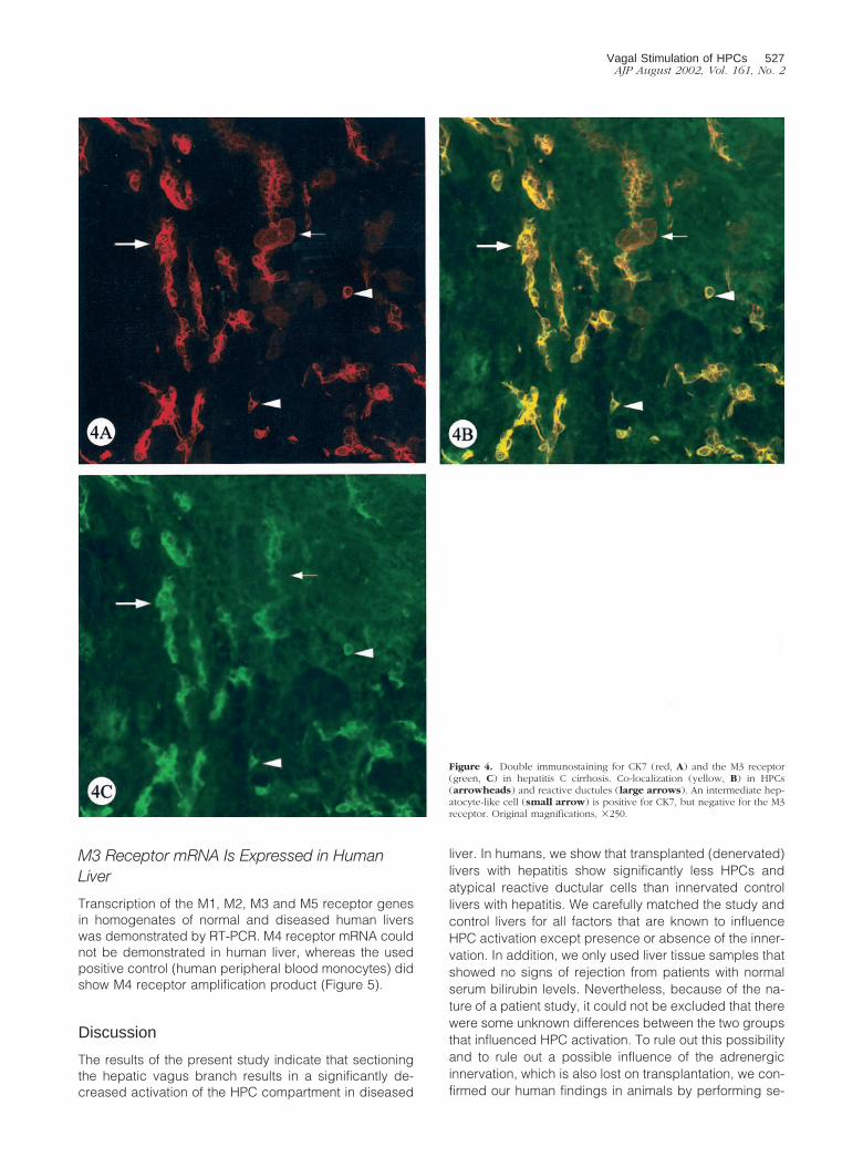
In addition to the results obtained from serial sections,double immunostaining in human liver confirmed that allHPCs and atypical reactive ductules, revealed by CK7staining, expressed the M3 receptor. Less than half of theintermediate hepatocyte-like cells staining for CK7 werealso immunoreactive for the M3 receptor (Figure 4).
Figure 3. Normal donor liver. The interlobular bile duct (arrow) and the canal of Hering (arrowhead) are immunoreactive for CK7 (A) and the M3 receptor(B). PV, portal vein. Original magnifications, �400.
526 Cassiman et alAJP August 2002, Vol. 161, No. 2
M3 Receptor mRNA Is Expressed in HumanLiver
Transcription of the M1, M2, M3 and M5 receptor genesin homogenates of normal and diseased human liverswas demonstrated by RT-PCR. M4 receptor mRNA couldnot be demonstrated in human liver, whereas the usedpositive control (human peripheral blood monocytes) didshow M4 receptor amplification product (Figure 5).
Discussion
The results of the present study indicate that sectioningthe hepatic vagus branch results in a significantly de-creased activation of the HPC compartment in diseased
liver. In humans, we show that transplanted (denervated)livers with hepatitis show significantly less HPCs andatypical reactive ductular cells than innervated controllivers with hepatitis. We carefully matched the study andcontrol livers for all factors that are known to influenceHPC activation except presence or absence of the inner-vation. In addition, we only used liver tissue samples thatshowed no signs of rejection from patients with normalserum bilirubin levels. Nevertheless, because of the na-ture of a patient study, it could not be excluded that therewere some unknown differences between the two groupsthat influenced HPC activation. To rule out this possibilityand to rule out a possible influence of the adrenergicinnervation, which is also lost on transplantation, we con-firmed our human findings in animals by performing se-
Figure 4. Double immunostaining for CK7 (red, A) and the M3 receptor(green, C) in hepatitis C cirrhosis. Co-localization (yellow, B) in HPCs(arrowheads) and reactive ductules (large arrows). An intermediate hep-atocyte-like cell (small arrow) is positive for CK7, but negative for the M3receptor. Original magnifications, �250.
Vagal Stimulation of HPCs 527AJP August 2002, Vol. 161, No. 2

lective hepatic vagotomy in the galactosamine-intoxi-cated rat. Galactosamine intoxication is a well-knownanimal model of liver injury that is associated with HPCactivation,13,48 in contrast to the models that were studiedin combination with hepatic19,21 or cervical26 vagotomy.Selective hepatic vagotomy in combination with galac-tosamine-induced acute hepatitis in rats caused a signif-icant decrease in the number of oval cells compared tosham operation in combination with galactosamine intox-ication.
We also found that HPCs, atypical reactive ductulesand intermediate hepatocyte-like cells in human liver ex-press the M3 receptor, which strongly suggest that thestimulatory effects of the nervus vagus on the HPC com-partment occurs via binding of acetylcholine to the M3receptor present on these cell types. Although M2 andM5 mRNA transcripts were shown to be present in humanliver homogenates, immunohistochemistry could notdemonstrate the presence of the M2 and M5 receptor atthe protein level. Posttranscriptional processes may in-hibit the translation of mRNA into protein.49
Our data show that bile ducts in normal and diseasedhuman liver express the M3 receptor. LeSage and col-leagues26 previously showed the presence of the M3receptor on isolated rat cholangiocytes by Western blot-
ting, but not by in vivo immunohistochemistry. Alvaro andcolleagues50 reported that isolated rat bile duct unitswere immunoreactive when exposed to the M35 anti-body, but not when exposed to an antibody specificagainst the M2 receptor. Because the M35 antibody doesnot discriminate between muscarinic acetylcholine re-ceptor subtypes,51 these authors erroneously concludedthat they demonstrated immunohistochemical expressionof the M3 receptor on isolated rat bile duct units. Usingprimary antibodies from two different sources, we werenot able to demonstrate immunohistochemical expres-sion of the M3 receptor by bile ducts or other cell types inrat liver either, which indicates that there is low abun-dance of protein expression or that the used primaryantibodies are not adequate for immunohistochemistry inrat liver.
Because hepatocytes in normal and diseased liver donot express any of the nicotinic and muscarinic acetyl-choline receptors,22–25 the stimulatory effect of the ner-vus vagus on proliferating hepatocytes after PHx must beindirect. It has been shown that the nervus vagus mod-ulates sinusoidal blood flow52,53 and the present studyreveals that arterial endothelial cells express the M1 re-ceptor. Therefore, it is very well possible that the stimu-latory effect of the nervus vagus on proliferating hepato-cytes after PHx is caused by the modulation of thesinusoidal blood flow.
Interestingly, hepatocytes produce and secrete cho-linesterase and acetylcholinesterase is present at the cellsurface membrane.54–57 The concentration of serumcholinesterase accurately reflects the hepatocyte massand can be used to assess the prognosis of patients withliver cirrhosis.54,58 These observations and the findings ofthe present study have led us to hypothesize the follow-ing model that explains HPC behavior as observed indifferent conditions: in normal liver and after PHx, eachHPC is surrounded by a normal number of hepatocytesthat inhibits the binding of acetylcholine to the M3 recep-tor on the HPC by producing and secreting cholinester-ase. As a consequence, the HPC compartment is not oronly minimally activated in these conditions. When thereis loss and impaired proliferation of hepatocytes (as is thecase in almost all liver diseases), the cholinesterase ac-tivity will decrease proportionally to the topography andseverity of hepatocyte loss. This allows acetylcholine toexert its trophic effects on the HPCs, until hepatocytemass is restored again. In a transplant patient with adiseased liver, the stimulatory effect of the nervus vagusis abolished, which leads to an impaired HPC activation.Intriguingly, an atypical ductular reaction is absent inchronic liver allograft rejection,59 despite the destructionof the bile ducts. The enigmatic paucity of atypical reac-tive ductules in this condition can be explained by ourhypothesis. In our study, signs of rejection were absentfrom all included patient samples. Therefore, this factorcould not have influenced the differences we observedbetween the numbers of HPCs and atypical reactiveductules between the study and control group.
In the hematopoietic system, which is related to theHPC compartment,4,6,60,61 a similar mechanism seems tobe present: acetylcholinesterase, which is synthesized
Figure 5. RT-PCR for the muscarinic acetylcholine receptor subtypes M1 toM5 (A–E). M: 100-bp ladder. Homogenate of a human explant liver ofpatients with primary biliary cirrhosis (lane 1) and toxic submassive liverfailure (lane 2). Lane 3: Homogenate of a healthy human transplant liver,rejected on the basis of macroscopical anatomical defects. Lane 4: Notemplate control. Lane 5: Positive control. Line, 500 bp; arrow, expectedlength of the amplification product.
528 Cassiman et alAJP August 2002, Vol. 161, No. 2

by mature red blood cells,62 enhances apoptosis andreduces proliferation of mouse hematopoietic progenitorcells committed to erythroid and other lineages in vitro.63
These and our findings are two examples of the nonclas-sical, autocrine/paracrine actions of the cholinergic sys-tem.64,65
In conclusion, the hepatic vagus branch stimulatesactivation of the HPC compartment in diseased liver,most likely through binding of acetylcholine to the M3receptor expressed on these cells. These findings maybe of clinical importance for patients with a transplantliver.
References
1. De Vos R, Desmet V: Ultrastructural characteristics of novel epithelialcell types identified in human pathologic liver specimens with chronicductular reaction. Am J Pathol 1992, 140:1441–1450
2. Yasui O, Miura N, Terada K, Kawarada Y, Koyama K, Sugiyama T:Isolation of oval cells from Long-Evans Cinnamon rats and theirtransformation into hepatocytes in vivo in the rat liver. Hepatology1997, 25:329–334
3. Roskams T, De Vos R, Van Eyken P, Myazaki H, Van Damme B,Desmet V: Hepatic OV-6 expression in human liver disease and ratexperiments: evidence for hepatic progenitor cells in man. J Hepatol1998, 29:455–463
4. Theise ND, Nimmakayalu M, Gardner R, Illei PB, Morgan G, Teper-man L, Henegariu O, Krause DS: Liver from bone marrow in humans.Hepatology 2000, 32:11–16
5. Crosby HA, Kelly DA, Strain AJ: Human hepatic stem-like cells iso-lated using c-kit or CD34 can differentiate into biliary epithelium.Gastroenterology 2001, 120:534–544
6. Avital I, Inderbitzin D, Aoki T, Tyan DB, Cohen AH, Ferraresso C,Rozga J, Arnaout WS, Demetriou AA: Isolation, characterization andtransplantation of bone marrow-derived hepatocyte stem cells. Bio-chem Biophys Res Commun 2001, 288:156–164
7. Farber E: Similarities in the sequence of early histological changesinduced in the liver of the rat by ethionine, 2-acetylaminofluorene and3�-methyl-4-dimethylaminoazobenzene. Cancer Res 1956, 16:142–149
8. Theise ND, Saxena R, Portmann BC, Thung SN, Yee H, Chiriboga L,Kumar A, Crawford JM: The canals of Hering and hepatic stem cellsin humans. Hepatology 1999, 30:1425–1433
9. Lai YS, Thung SN, Gerber MA, Chen ML, Schaffner F: Expression ofcytokeratins in normal and diseased livers and in primary liver carci-nomas. Arch Pathol Lab Med 1989, 113:134–138
10. Sell S: Comparison of liver progenitor cells in human atypical ductularreactions with those seen in experimental models of liver injury.Hepatology 1998, 27:317–331
11. Lowes KN, Brennan BA, Yeoh GC, Olynyk JK: Oval cell numbers inhuman chronic liver diseases are directly related to disease severity.Am J Pathol 1999, 154:537–541
12. Libbrecht L, Desmet V, Van Damme B, Roskams T: Deep intralobularextension of human hepatic progenitor cells correlates with paren-chymal inflammation in chronic viral hepatitis: can progenitor cellsmigrate? J Pathol 2000, 192:373–378
13. Dabeva MD, Alpini G, Hurston E, Shafritz DA: Models for hepaticprogenitor cell activation. Proc Soc Exp Biol Med 1993, 204:242–252
14. Bustos M, Sangro B, Alzuguren P, Gil AG, Ruiz J, Beraza N, Qian C,Garcia-Pardo A, Prieto J: Liver damage using suicide genes: a modelfor oval cell activation. Am J Pathol 2000, 157:549–559
15. Evarts RP, Nakatsukasa H, Marsden ER, Hu Z, Thorgeirsson SS:Expression of transforming growth factor-alpha in regenerating liverand during hepatic differentiation. Mol Carcinog 1992, 5:25–31
16. Hu Z, Evarts RP, Fujio K, Marsden ER, Thorgeirsson SS: Expression ofhepatocyte growth factor and c-met genes during hepatic differenti-ation and liver development in the rat. Am J Pathol 1993, 142:1823–1830
17. Bisgaard HC, Santoni Rugiu E, Nagy P, Thorgeirsson SS: Modulation
of the plasminogen activator/plasmin system in rat liver regeneratingby recruitment of oval cells. Lab Invest 1998, 78:237–246
18. Fausto N: Liver regeneration. From laboratory to clinic. Liver Transpl2001, 7:835–844
19. Ohtake M, Sakaguchi T, Yoshida K, Muto T: Hepatic branch vagot-omy can suppress liver regeneration in partially hepatectomized rats.HPB Surg 1993, 6:277–286
20. Kiba T, Tanaka K, Numata K, Hoshino M, Inoue S: Facilitation of liverregeneration after partial hepatectomy by ventromedial hypothalamiclesions in rat. Pflugers Arch 1994, 428:26–29
21. Kiba T, Tanaka K, Inoue S: Lateral hypothalamic lesions facilitatehepatic regeneration after partial hepatectomy in rats. Pflugers Arch1995, 430:666–671
22. Oshima F, Kondo K, Tsubaki T: Binding of iodine 125 alpha-bunga-rotoxin to the thymus of mice. Arch Neurol 1978, 35:31–32
23. Smith MA, Stollberg J, Lindstrom JM, Berg DK: Characterization of acomponent in chick ciliary ganglia that cross-reacts with monoclonalantibodies to muscle and electric organ acetylcholine receptor.J Neurosci 1985, 5:2726–2731
24. MacLennan C, Beeson D, Vincent A, Newsom-Davis J: Human nico-tinic acetylcholine receptor alpha-subunit isoforms: origins and ex-pression. Nucleic Acid Res 1993, 21:5463–5467
25. Flora A, Schulz R, Benfante R, Battaglioli E, Terzano S, Clementi F,Fornasari D: Neuronal and extraneuronal expression and regulationof the human alpha5 nicotinic receptor subunit gene. J Neurochem2000, 75:18–27
26. LeSage G, Alvaro D, Benedetti A, Glaser S, Marucci L, Biaocchi L,Eisel W, Caligiuri A, Phinizy JL, Rodgers R, Francis H, Alpini G:Cholinergic system modulates growth, apoptosis and secretion ofcholangiocytes from bile duct-ligated rats. Gastroenterology 1999,117:191–199
27. Guizzetti M, Costa P, Peters J, Costa LG: Acetylcholine as a mitogen:muscarinic receptor-mediated proliferation of rat astrocytes and hu-man astrocytoma cells. Eur J Pharmacol 1996, 297:265–273
28. Rayford W, Noble MJ, Austenfeld MA, Weigel J, Mebust WK, ShahGV: Muscarinic cholinergic receptors promote growth of human pros-tate cancer cells. Prostate 1997, 30:160–166
29. Frucht H, Jensen RT, Dexter D, Yang WL, Xiao Y: Human coloncancer cell proliferation mediated by the M3 muscarinic cholinergicreceptor. Clin Cancer Res 1999, 5:2523–2539
30. Ragheb F, Molina-Holgado E, Cui QL, Khorchid A, Liu HN, LaroccaJN, Almazan G: Pharmacological and functional characterization ofmuscarinic receptor subtypes in developing oligodendrocytes.J Neurochem 2001, 77:1396–1406
31. Desmet V, Roskams T, Van Eyken P: Ductular reaction in the liver.Pathol Res Pract 1995, 191:513–524
32. Roskams T, Campos RV, Drucker DJ, Desmet VJ: Reactive humanbile ductules express parathyroid hormone-related peptide. Histopa-thology 1993, 23:11–19
33. Cassiman D, Denef C, Desmet V, Roskams T: Human and rat hepaticstellate cells express neutrotrophins and neurotrophin receptors.Hepatology 2001, 33:148–158
34. Boon AP, Hubscher SG, Lee JA, Hines JE, Burt AD: Hepatic reinner-vation following orthotopic liver transplantation in man. J Pathol 1992,167:217–222
35. Dhillon AP, Sankey EA, Wang JH, Wightman AK, Mathur S, BurroughsAK, Scheuer PJ: Immunohistochemical studies on the innervation ofhuman transplanted liver. J Pathol 1992, 167:211–216
36. Nagy P, Kiss A, Schnur J, Thorgeirsson SS: Dexamethasone inhibitsthe proliferation of hepatocytes and oval cells but not bile duct cellsin rat liver. Hepatology 1998, 28:423–429
37. Scheuer PJ: Classification of chronic hepatitis: a need for reassess-ment. J Hepatol 1991, 13:372–374
38. Jonker AM, Dijkhuis FW, Kroese FG, Hardonk MJ, Grond J: Immuno-pathology of acute galactosamine hepatitis in rats. Hepatology 1990,11:622–627
39. Cassiman D, Roskams T, van Pelt J, Libbrecht L, Aertsen P, CrabbeT, Vankelecom H, Denef C: Alpha B-crystallin expression in humanand rat hepatic stellate cells. J Hepatol 2001, 35:200–207
40. Mei L, Roeske WR, Izutsu KT, Yamamura HI: Characterization ofmuscarinic acetylcholine receptors in human labial salivary glands.Eur J Pharmacol 1990, 176:367–370
41. Beroukas D, Goodfellow R, Hiscock J, Jonsson R, Gordon TP, Wa-terman SA: Up-regulation of M3-muscarinic receptors in labial sali-
Vagal Stimulation of HPCs 529AJP August 2002, Vol. 161, No. 2
vary gland acini in primary Sjogren’s syndrome. Lab Invest 2002,82:203–210
42. Yamaguchi O, Shishido K, Tamura K, Ogawa T, Fujimura T, OhtsukaM: Evaluation of mRNAs encoding muscarinic receptor subtypes inhuman detrusor muscle. J Urol 1996, 156:1208–1213
43. Buchli R, Ndoye A, Rodriguez JG, Zia S, Webber RJ, Grando SA:Human skin fibroblasts express m2, m4 and m5 subtypes of musca-rinic acetylcholine receptors. J Cell Biochem 1999, 74:264–277
44. Baumgartner MK, Wei J, Aronstam RS: Retinoic acid-induced differ-entiation of a human neuroblastoma cell line alters muscarinic recep-tor expression. Dev Brain Res 1933, 72:305–308
45. Costa P, Auger C, Traver DJ, Costa LG: Identification of m3, m4 andm5 subtypes of muscarinic receptor mRNA in human blood mononu-clear cells. J Neuroimmunol 1995, 60:45–51
46. Branisteanu DD, Mathieu C, Bouillon R: Synergism between sirolimusand 1,25-dihydroxyvitamin D3 in vitro and in vivo. J Neuroimmunol1997, 60:45–51
47. Roskams T, De Vos R, Desmet V: ‘Undifferentiated progenitor cells’ infocal nodular hyperplasia of the liver. Histopathology 1996, 28:291–299
48. Lemire JM, Shiojiri N, Fausto N: Oval cell proliferation and the originof small hepatocytes in liver injury induced by D-galactosamine. Am JPathol 1991, 139:535–552
49. Kren BT, Steer CJ: Posttranscriptional regulation of gene expressionin liver regeneration: role of mRNA stability. EMBO J 1996, 10:819–828
50. Alvaro D, Alpini G, Jezequel AM, Bassotti C, Francia C, Fraioli F,Romeo R, Marucci L, LeSage G, Glaser SS, Benedetti A: Role andmechanisms of action of acetylcholine in the regulation of rat cholan-giocyte secretory functions. J Clin Invest 1997, 100:1349–1362
51. Carsi-Gabrenas JM, Van der Zee EA, Luiten PG, Potter LT: Non-selectivity of the monoclonal antibody M35 for subtypes of muscarinicacetylcholine receptors. Brain Res Bull 1997, 44:25–31
52. Koo A, Liang IY: Microvascular filling pattern in rat liver sinusoidsduring vagal stimulation. J Physiol (Lond) 1979, 295:191–199
53. Gardemann A, Puschel GP, Jungermann K: Nervous control of livermetabolism and hemodynamics. Eur J Biochem 1992, 207:399–411
54. Brown SS, Kalow W, Whittaker M, Woronick CL: The plasmacholinesterases: a new perspective. Adv Clin Chem 1981, 22:82–83
55. Schuman RF, Brimfield AA, Hunter KW: A micro-method for the de-
tection of butyrylcholinesterase secreted by hepatocytes in vitro.Biosci Rep 1984, 4:149–154
56. Berninsone P, Katz E, Napp M, Azcurra J: Acetylcholinesterase andnonspecific cholinesterase activities in rat liver: subcellular localiza-tion, molecular forms, and some extraction properties. Biochem CellBiol 1989, 67:817–822
57. Perelman A, Brandan E: Different membrane-bound forms of acetyl-cholinesterase are present at the cell surface of hepatocytes. EurJ Biochem 1989, 182:203–207
58. Adler M, Verset D, Bouhdid H, Bourgeois N, Gulbis B, Le Moine O,Van de Stadt J, Gelin M, Thiry P: Prognostic evaluation of patients withparenchymal cirrhosis. Proposal of a new simple score. J Hepatol1997, 26:642–649
59. Demetris A, Adams D, Bellamy C, Blakolmer K, Clouston A, DhillonAP, Fung J, Gouw A, Gustafsson B, Haga H, Harrison D, Hart J,Hubscher S, Jaffe R, Khettry U, Lassman C, Lewin K, Martinez O,Nakazawa Y, Neil D, Pappo O, Parizhskaya M, Randhawa P, Rasoul-Rockenschaub S, Reinholt F, Reynes M, Robert M, Tsamandas A,Wanless I, Wiesner R, Wernerson A, Wrba F, Wyatt J, Yamabe H:Update of the International Banff Schema for Liver Allograft Rejection:working recommendations for the histopathologic staging and report-ing of chronic rejection. An International Panel. Hepatology 2000,31:792–799
60. Petersen BE, Bowen WC, Patrene KD, Mars WM, Sullivan AK, MuraseN, Boggs SS, Greenberger JS, Goff JP: Bone marrow as a potentialsource of hepatic oval cells. Science 1999, 284:1168–1170
61. Alison MR, Poulsom R, Jeffery R, Dhillon AP, Quaglia A, Jacob J,Novelli M, Prentice G, Williamson J, Wright NA: Hepatocytes fromnon-hepatic adult stem cells. Nature 2000, 406:257
62. Alles GA, Hawes RC: Cholinesterase in the blood of man. J Biol Chem1940, 133:375
63. Soreq H, Patinkin D, Lev-Lehman E, Grifman M, Ginzberg D, EcksteinF, Zakut H: Antisense oligonucleotide inhibition of acetylcholinester-ase gene expression induces progenitor cell expansion and sup-presses hematopoietic apoptosis ex vivo. Proc Natl Acad Sci USA1994, 91:7909–7911
64. Wessler I, Kirkpatrick CJ, Racke K: The cholinergic ‘pitfall’: acetyl-choline, a universal cell molecule in biological systems, includinghumans. Clin Exp Pharmacol Physiol 1999, 26:198–205
65. Soreq H, Seidman S: Acetylcholinesterase—new roles for an oldactor. Nat Rev Neurosci 2001, 2:294–302
530 Cassiman et alAJP August 2002, Vol. 161, No. 2

![Mapping muscarinic receptors in human and baboon brain using [N-11C-methyl]-benztropine](https://static.fdokumen.com/doc/165x107/6344f35df474639c9b049d90/mapping-muscarinic-receptors-in-human-and-baboon-brain-using-n-11c-methyl-benztropine.jpg)