Efferent Vagal Nerve Stimulation Attenuates Gut Barrier Injury After Burn: Modulation of Intestinal...
8
ORIGINAL ARTICLE Efferent Vagal Nerve Stimulation Attenuates Gut Barrier Injury After Burn: Modulation of Intestinal Occludin Expression Todd W. Costantini, MD, Vishal Bansal, MD, Carrie Y. Peterson, MD, William H. Loomis, BS, James G. Putnam, BS, Fermin Rankin, BS, Paul Wolf, MD, Brian P. Eliceiri, PhD, Andrew Baird, PhD, and Raul Coimbra, MD, PhD Introduction: Severe injury can cause intestinal permeability through de- creased expression of tight junction proteins, resulting in systemic inflam- mation. Activation of the parasympathetic nervous system after shock through vagal nerve stimulation is known to have potent anti-inflammatory effects; however, its effects on modulating intestinal barrier function are not fully understood. We postulated that vagal nerve stimulation improves intestinal barrier integrity after severe burn through an efferent signaling pathway, and is associated with improved expression and localization of the intestinal tight junction protein occludin. Methods: Male balb/c mice underwent right cervical vagal nerve stimulation for 10 minutes immediately before 30% total body surface area, full- thickness steam burn. In a separate arm, animals underwent abdominal vagotomy at the gastroesophageal junction before vagal nerve stimulation and burn. Intestinal barrier injury was assessed by permeability to 4 kDa FITC-dextran, histology, and changes in occludin expression using immu- noblotting and confocal microscopy. Results: Cervical vagal nerve stimulation decreased burn-induced intestinal permeability to FITC-dextran, returning intestinal permeability to sham levels. Vagal nerve stimulation before burn also improved gut histology and prevented burn-induced changes in occludin protein expression and local- ization. Abdominal vagotomy abrogated the protective effects of cervical vagal nerve stimulation before burn, resulting in gut permeability, histology, and occludin protein expression similar to burn alone. Conclusion: Vagal nerve stimulation performed before injury improves intestinal barrier integrity after severe burn through an efferent signaling pathway and is associated with improved tight junction protein expression. Key Words: Burn, Tight junction, Gut, Intestinal barrier, Permeability, Confocal microscopy. (J Trauma. 2010;68: 1349 –1356) T he intestinal epithelium plays a key role in host defense by maintaining a barrier against harmful bacteria and toxic products present within the gut lumen. Specifically, the intestinal tight junction serves as the regulator of barrier function in an intact intestinal epithelium. 1 Loss of tight junction integrity opens the paracellular space between gut epithelial cells allowing harmful luminal products to access normally protected layers of the intestinal wall. The subse- quent intestinal inflammatory response can further exacerbate the breakdown of tight junction proteins leading to a systemic inflammatory response and serious sequelae. 2 Occludin is an intestinal tight junction protein which contains several transmembrane domains that attach the api- cal portion of adjacent cells forming the tight junction. 3 Increased paracellular permeability has been associated with either decreased occludin protein expression or altered local- ization of occludin away from areas of cell-cell contact. 4 Decreased intestinal occludin expression has been associated with increased intestinal permeability in animal models of severe burn, traumatic brain injury, sepsis, and pancreati- tis. 5–8 Changes in intestinal occludin expression have also been seen clinically in patients with inflammatory bowel disease. 9 Strategies aimed at maintaining normal expression and localization of occludin after injury may be important in preventing intestinal barrier loss. Recent research suggests that the central nervous sys- tem may coordinate an anti-inflammatory response to injury. Tracey et al. 10 have recently described the cholinergic anti- inflammatory pathway, proposing that efferent vagal nerve signaling regulates cytokine production. Vagal nerve stimu- lation has been shown to decrease proinflammatory cytokine levels and also decrease the occurrence of shock in animals given a lethal dose of endotoxin. 11 Signaling from the efferent vagus nerve to the splenic nerve is responsible for the suppression of tumor necrosis factor- seen in endotoxemic animals that underwent vagal nerve stimulation. 12 The ability of vagus nerve stimulation to affect other cell types within the abdomen has yet to be fully elucidated. The gastrointestinal tract is innervated by the enteric nervous system, which contains a complex network of enteric neurons that control many aspects of intestinal function. The central nervous system communicates with the enteric ner- vous system through both efferent and afferent pathways via the vagus nerve. 13 Therefore, stimulation of the vagus nerve may alter intestinal function through modulation of the var- ious cell types within the enteric nervous system; including intestinal epithelial cells, endothelial cells, or enteric glia. 14 Submitted for publication September 12, 2009. Accepted for publication March 9, 2010. Copyright © 2010 by Lippincott Williams & Wilkins From the Division of Trauma, Surgical Critical Care, and Burns, Departments of Surgery (T.W.C., V.B., C.Y.P., W.H.L., J.G.P., F.R., B.P.E., A.B., R.C.) and Pathology (P.W.), University of California-San Diego School of Medicine, San Diego, California. Presented as an oral podium presentation at the 68th Annual Meeting of the American Association for the Surgery of Trauma, October 1–2, 2009, Pittsburgh, Pennsylvania. Address for reprints: Raul Coimbra MD, PhD, FACS, 200 W. Arbor Drive, #8896, San Diego, CA 92103-8896; e-mail: [email protected]. DOI: 10.1097/TA.0b013e3181dccea0 The Journal of TRAUMA ® Injury, Infection, and Critical Care • Volume 68, Number 6, June 2010 1349
-
Upload
independent -
Category
Documents
-
view
7 -
download
0
Transcript of Efferent Vagal Nerve Stimulation Attenuates Gut Barrier Injury After Burn: Modulation of Intestinal...
ORIGINAL ARTICLE
Efferent Vagal Nerve Stimulation Attenuates Gut Barrier InjuryAfter Burn: Modulation of Intestinal Occludin Expression
Todd W. Costantini, MD, Vishal Bansal, MD, Carrie Y. Peterson, MD, William H. Loomis, BS,James G. Putnam, BS, Fermin Rankin, BS, Paul Wolf, MD, Brian P. Eliceiri, PhD, Andrew Baird, PhD,
and Raul Coimbra, MD, PhD
Introduction: Severe injury can cause intestinal permeability through de-creased expression of tight junction proteins, resulting in systemic inflam-mation. Activation of the parasympathetic nervous system after shockthrough vagal nerve stimulation is known to have potent anti-inflammatoryeffects; however, its effects on modulating intestinal barrier function are notfully understood. We postulated that vagal nerve stimulation improvesintestinal barrier integrity after severe burn through an efferent signalingpathway, and is associated with improved expression and localization of theintestinal tight junction protein occludin.Methods: Male balb/c mice underwent right cervical vagal nerve stimulationfor 10 minutes immediately before 30% total body surface area, full-thickness steam burn. In a separate arm, animals underwent abdominalvagotomy at the gastroesophageal junction before vagal nerve stimulationand burn. Intestinal barrier injury was assessed by permeability to 4 kDaFITC-dextran, histology, and changes in occludin expression using immu-noblotting and confocal microscopy.Results: Cervical vagal nerve stimulation decreased burn-induced intestinalpermeability to FITC-dextran, returning intestinal permeability to shamlevels. Vagal nerve stimulation before burn also improved gut histology andprevented burn-induced changes in occludin protein expression and local-ization. Abdominal vagotomy abrogated the protective effects of cervicalvagal nerve stimulation before burn, resulting in gut permeability, histology,and occludin protein expression similar to burn alone.Conclusion: Vagal nerve stimulation performed before injury improvesintestinal barrier integrity after severe burn through an efferent signalingpathway and is associated with improved tight junction protein expression.Key Words: Burn, Tight junction, Gut, Intestinal barrier, Permeability,Confocal microscopy.
(J Trauma. 2010;68: 1349–1356)
The intestinal epithelium plays a key role in host defenseby maintaining a barrier against harmful bacteria and
toxic products present within the gut lumen. Specifically, the
intestinal tight junction serves as the regulator of barrierfunction in an intact intestinal epithelium.1 Loss of tightjunction integrity opens the paracellular space between gutepithelial cells allowing harmful luminal products to accessnormally protected layers of the intestinal wall. The subse-quent intestinal inflammatory response can further exacerbatethe breakdown of tight junction proteins leading to a systemicinflammatory response and serious sequelae.2
Occludin is an intestinal tight junction protein whichcontains several transmembrane domains that attach the api-cal portion of adjacent cells forming the tight junction.3
Increased paracellular permeability has been associated witheither decreased occludin protein expression or altered local-ization of occludin away from areas of cell-cell contact.4
Decreased intestinal occludin expression has been associatedwith increased intestinal permeability in animal models ofsevere burn, traumatic brain injury, sepsis, and pancreati-tis.5–8 Changes in intestinal occludin expression have alsobeen seen clinically in patients with inflammatory boweldisease.9 Strategies aimed at maintaining normal expressionand localization of occludin after injury may be important inpreventing intestinal barrier loss.
Recent research suggests that the central nervous sys-tem may coordinate an anti-inflammatory response to injury.Tracey et al.10 have recently described the cholinergic anti-inflammatory pathway, proposing that efferent vagal nervesignaling regulates cytokine production. Vagal nerve stimu-lation has been shown to decrease proinflammatory cytokinelevels and also decrease the occurrence of shock in animalsgiven a lethal dose of endotoxin.11 Signaling from the efferentvagus nerve to the splenic nerve is responsible for thesuppression of tumor necrosis factor-� seen in endotoxemicanimals that underwent vagal nerve stimulation.12 The abilityof vagus nerve stimulation to affect other cell types within theabdomen has yet to be fully elucidated.
The gastrointestinal tract is innervated by the entericnervous system, which contains a complex network of entericneurons that control many aspects of intestinal function. Thecentral nervous system communicates with the enteric ner-vous system through both efferent and afferent pathways viathe vagus nerve.13 Therefore, stimulation of the vagus nervemay alter intestinal function through modulation of the var-ious cell types within the enteric nervous system; includingintestinal epithelial cells, endothelial cells, or enteric glia.14
Submitted for publication September 12, 2009.Accepted for publication March 9, 2010.Copyright © 2010 by Lippincott Williams & WilkinsFrom the Division of Trauma, Surgical Critical Care, and Burns, Departments of
Surgery (T.W.C., V.B., C.Y.P., W.H.L., J.G.P., F.R., B.P.E., A.B., R.C.) andPathology (P.W.), University of California-San Diego School of Medicine,San Diego, California.
Presented as an oral podium presentation at the 68th Annual Meeting of theAmerican Association for the Surgery of Trauma, October 1–2, 2009,Pittsburgh, Pennsylvania.
Address for reprints: Raul Coimbra MD, PhD, FACS, 200 W. Arbor Drive, #8896,San Diego, CA 92103-8896; e-mail: [email protected].
DOI: 10.1097/TA.0b013e3181dccea0
The Journal of TRAUMA® Injury, Infection, and Critical Care • Volume 68, Number 6, June 2010 1349

Strategies that prevent intestinal barrier breakdownafter injury may be useful in improving outcomes in patientsafter severe trauma and burn. Activation of the parasympa-thetic nervous system after shock through vagal nerve stim-ulation is known to have potent anti-inflammatory effects.The ability of vagal nerve stimulation to improve intestinalbarrier function is not fully understood. We postulated thatvagal nerve stimulation improves intestinal barrier integrityfollowing severe burn through an efferent signaling pathwayand is associated with improved expression and localizationof the intestinal tight junction protein occludin.
MATERIALS AND METHODS
Surgical Abdominal VagotomyMale balb/c mice weighing 24 g to 28 g were obtained
from Jackson Laboratories (Sacramento, CA). Animals wereanesthetized with inhaled isoflurane before the experimentalprotocol. In one cohort of animals, surgical abdominal vagot-omy was performed immediately before vagal nerve stimu-lation and subsequent burn by making a midline laparotomyincision. The gastroesophageal junction was identified andthe dorsal and ventral vagus nerves were visualized on thedistal esophagus using an Olympus SZ61 stereo microscope(Leeds Precision Instruments, Minneapolis, MN). Bothbranches of the vagus nerve were isolated and transected. Theabdomen was then closed using interrupted silk sutures.
Vagal Nerve StimulationA right cervical neck incision was performed and the
right cervical vagus nerve exposed. Stimulation of the rightcervical vagus nerve was performed using a VariStim IIIprobe (Medtronic Xomed, Jacksonville, FL) at 2 mA for 10minutes. After nerve stimulation, the incision was closed withinterrupted silk suture and the animal was immediately sub-jected to burn injury as previously described. Sham animalsunderwent right cervical incision and exposure of the vagusnerve but did not receive electrical stimulation.
Thermal Injury ModelAnimals underwent dorsal fur clipping with an electric
clipper before 30% total body surface area (TBSA) dorsal steamburn for 7 seconds using a template designed to estimate 30%TBSA.15 Following burn, animals received a subcutaneousinjection of normal saline containing buprenorphine (0.05mg/kg) in a non-burned area for fluid resuscitation and paincontrol. Animals were recovered from anesthesia and re-turned to their cage where they were provided food and waterad libitum. All animal experiments were approved by theUniversity of California San Diego Institutional Animal Careand Use Committee.
Intestinal Permeability AssayAn in vivo intestinal permeability assay was performed
to assess intestinal barrier function (n � 3 animals per group).Four hours after injury, animals were anesthetized with in-haled isoflurane. A midline laparotomy incision was per-formed, and a 5-cm segment of distal ileum was isolatedbetween silk ties. A solution of 200 �L containing 4 kDa
FITC-Dextran (25 mg/mL, Sigma, St. Louis, MO) diluted inphosphate buffered saline (PBS) was injected into the lumenof the isolated segment of intestine. The bowel was returnedto the abdominal cavity and the abdomen closed. The animalwas maintained under general anesthesia for 30 minutes, atwhich time systemic blood was drawn by cardiac punctureand placed in heparinized Eppendorf tubes on ice. Plasmawas obtained by centrifuging the blood at 10,000 g for 10minutes at �4°C. Plasma fluorescence was measured in afluorescence spectrophotometer (SpectraMax M5, MolecularDevices, Sunnyvale, CA) and compared with a standardcurve of known concentrations of FITC-Dextran diluted inmouse plasma.
Tissue HarvestAnimals were sacrificed 4 hours after injury and tissues
harvested. Segments of distal small intestine were removedand immediately snap frozen in liquid nitrogen before storageat �80°C for analysis. Segments of distal small intestinewere also harvested and preserved in 10% buffered formalin(Richard Allan Scientific, Pittsburgh, PA) or optimal cuttingtechnique embedding media (OCT, Sakura Finetek, Torrance,CA) for histologic evaluation.
Histologic EvaluationSegments of distal ileum were preserved in 10% buff-
ered formalin, embedded in paraffin, and sectioned. Intestinalsections were stained with Hematoxylin and Eosin (Surgi-path, Richmond, IL). Sections were viewed using an Olym-pus IX70 light microscope (Melville, NY) and viewed withQ-imaging software (Surrey, BC, Canada). Sections werereviewed by a pathologist (Paul Wolf, MD) blinded to theexperimental groups.
Occludin ExpressionDistal small intestine harvested from animals at 4 hours
after burn (n � 4–5 animals per group) were homogenized inice-cold tissue protein extraction reagent, containing 1%protease and 1% phosphatase inhibitors (Pierce Biotechnol-ogy, Rockford, IL). The homogenized tissue was centrifugedat 10,000 g for 5 minutes, the supernatants collected, and theprotein concentration of each sample measured using thebicinchoninic protein assay (Pierce). Protein was suspendedin sodium dodecyl sulfate sample buffer and boiled for5 minutes. Proteins were separated using sodium dodecylsulfate-polyacrylamide gel electrophoresis using 8% to 16%Tris-glycine gradient gels (Invitrogen, Carlsbad, CA), thentransferred to nitrocellulose membranes (Invitrogen). Mem-branes were blocked with 5% bovine serum albumin (BSA,Sigma) in Tris-buffered saline/Tween 20 for 1 hour. Mem-branes were then incubated overnight at 4°C in primaryantibody for occludin (Zymed, Carlsbad, CA) prepared in a1:500 concentration in 5% BSA/Tween 20. The membraneswere then washed and incubated with a horseradish peroxidase-linked anti-rabbit IgG secondary antibody (Cell Signaling)before application of the Pierce Supersignal West PicoChemiluminescent Kit for antibody detection. Luminescencewas detected using the Xenogen IVIS Lumina (Caliper LifeScience, Hopkinton, MA) imaging system. Mean pixel den-
Costantini et al. The Journal of TRAUMA® Injury, Infection, and Critical Care • Volume 68, Number 6, June 2010
© 2010 Lippincott Williams & Wilkins1350

sity of each sample was estimated using UN-SCAN-IT GelDigitizing software (Silk Scientific, Orem, UT). The relativeband density of each band was calculated by dividing thepixel density of each sample by the mean pixel density of thesham samples.
Confocal MicroscopySegments of distal ileum (n � 3 animals per group)
were embedded in optimal cutting technique media andstored at �80°C. Sections of intestine were cut 10 �m thickusing a Reichert-Jung Cryocut 1800 at �20°C (ReichertMicroscopes, Depew, NY). Sections were fixed onto glassslides with 3.7% paraformaldehyde (Electron MicroscopySeries, Hatfield, PA) for 10 minutes, washed with PBS, thenpermeabilized with 0.01% Triton X-100 (Sigma) for 1minute. Sections were washed once again in PBS beforeblocking for 1 hour in 3% BSA, Sigma. The sections werethen incubated overnight in the occludin antibody. Sectionswere then treated with the secondary antibody Alexa Fluor488 (Invitrogen) in 1% BSA for 1 hour. Prolong Fade(Invitrogen) was added upon placement of cover slips. Im-ages were viewed using the Olympus Fluoview laser scan-ning confocal microscope with exposure matched settings(Advanced Software v1.6, Olympus) at 60� magnification.
Statistical AnalysisValues are expressed as the mean � the SEM of n
samples where n represents the number of animals in eachexperimental group. The statistical significance betweengroups was determined using analysis of variance with Bon-ferroni correction. Statistical analysis was performed usingSPSS Statistics software version 11.5 (SPSS, Chicago, IL). Ap value � 0.05 was considered statistically significant.
RESULTS
Vagal Nerve Stimulation Attenuates IntestinalBarrier Injury
The physiologic effects of vagal nerve stimulation onintestinal barrier function following severe burn were as-sessed using an in vivo intestinal permeability assay to 4 kDaFITC-Dextran (Fig. 1). Severe burn injury alters gut barrierfunction resulting in a significant increase in permeability toFITC-Dextran. Performing cervical vagal nerve stimulationbefore 30% TBSA burn attenuated burn-induced intestinalpermeability. There was no significant difference betweensham and burned animals that underwent vagal nerve stimu-lation. Another group of animals underwent abdominal va-gotomy at the gastroesophageal junction before cervical vagalnerve stimulation to confirm that the protective effects weredue to efferent signaling down the vagus nerve. Intestinalpermeability was elevated in burned animals that underwentabdominal vagotomy before vagal nerve stimulation, withsystemic concentration of FITC-Dextran similar to animalssubjected to burn alone.
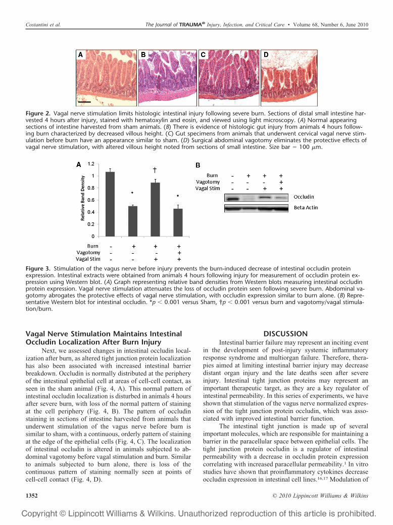
We correlated the finding of the in vivo intestinalpermeability assay by assessing changes in gut histologyfrom sections harvested 4 hours after injury (Fig. 2). Vagalnerve stimulation improves the histologic appearance of the
small intestine following burn, with improved villous heightcompared with animals that were subjected to burn alone.The protective effects of vagal nerve stimulation are dimin-ished in specimens obtained from animals that underwentabdominal vagotomy before vagal stimulation and subse-quent burn injury. This once again suggests that the vagalnerve stimulation decreases intestinal barrier injury throughan efferent pathway from the cervical vagus nerve into theenteric nervous system.
Stimulation of the Vagus Nerve Before BurnImproves Intestinal Occludin Expression
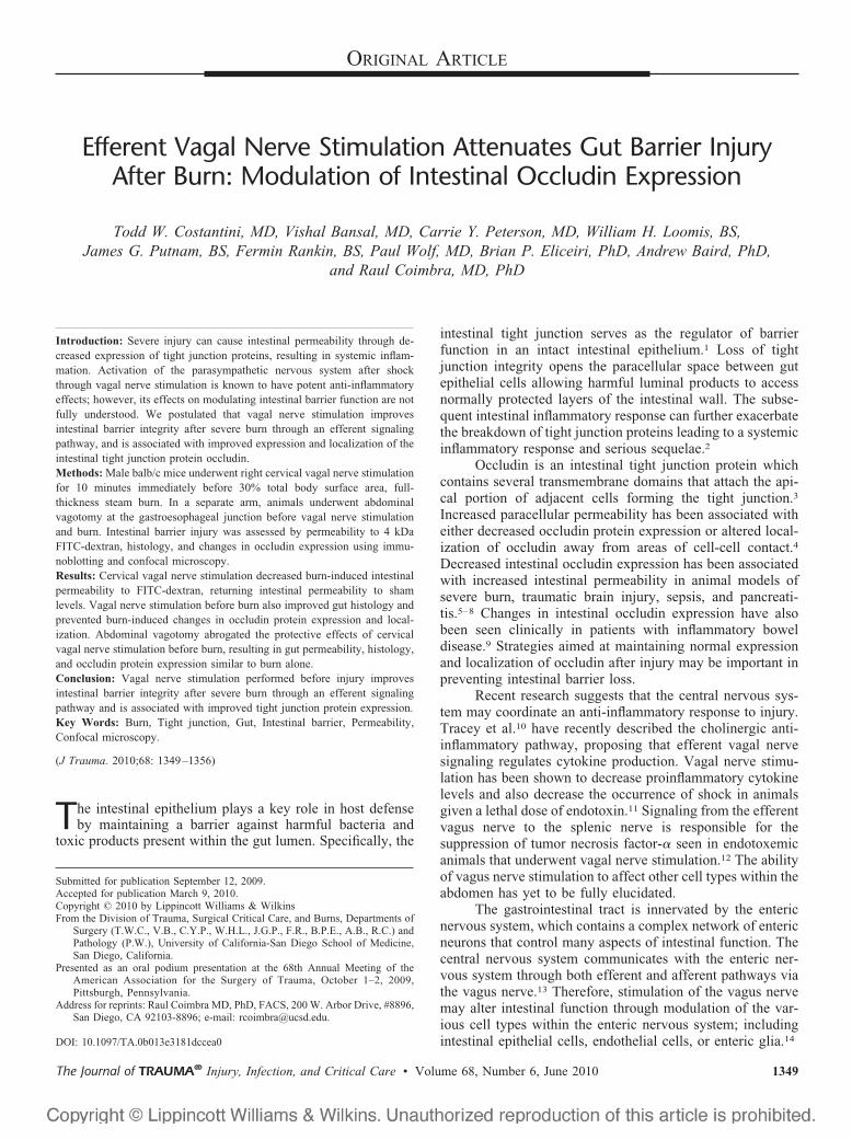
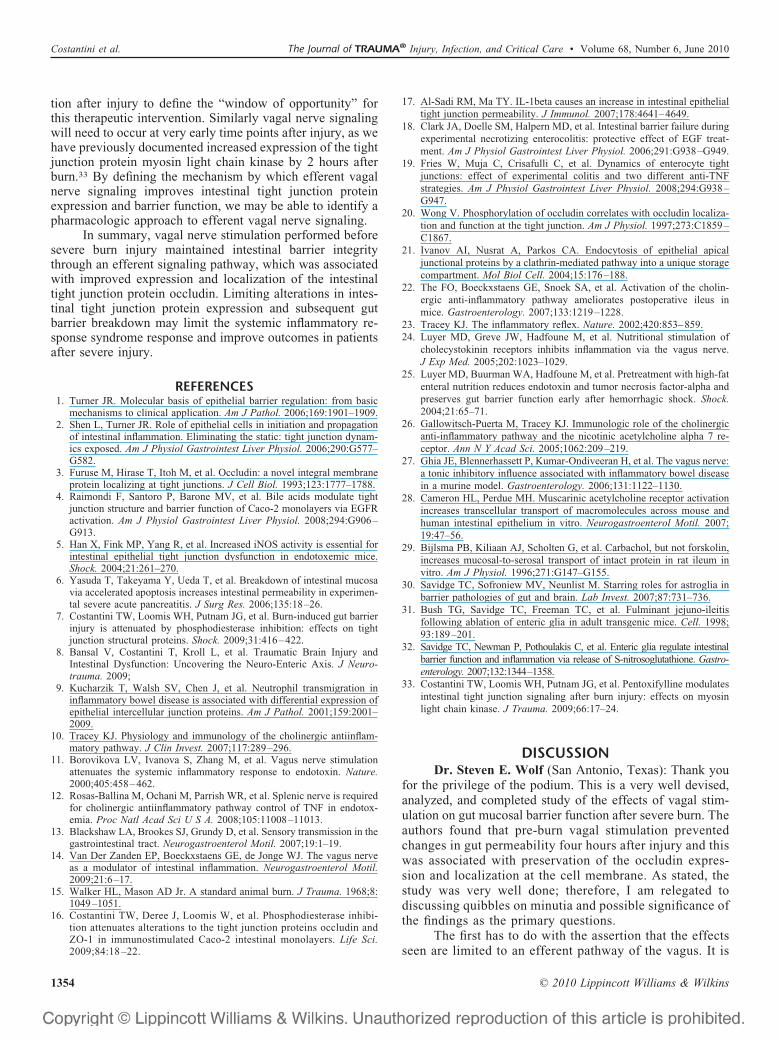
We correlated the changes in intestinal barrier functionwith changes in occludin protein expression from intestinaltissue harvested 4 hours after injury (Fig. 3). There was asignificant decrease in occludin protein expression from in-testinal tissue harvested from animals following 30% TBSAburn. Vagal nerve stimulation before injury prevented theburn-induced decrease in intestinal occludin protein expres-sion. There was no difference in occludin expression betweensham and animals undergoing vagal nerve stimulation beforesevere burn. Intestinal occludin protein levels were signifi-cantly decreased in animals that underwent surgical abdom-inal vagotomy before cervical vagal nerve stimulation andburn.
Figure 1. Vagal nerve stimulation attenuates burn-inducedintestinal permeability. In vivo intestinal permeability to 4kilodalton FITC-Dextran measured 4 hours after injury. Se-vere burn injury significantly increased gut permeability. Ani-mals that underwent right cervical vagal nerve stimulationimmediately before burn decreased intestinal permeability tosham levels. Performing an abdominal vagotomy abrogatedthe protective effects of vagal nerve stimulation, suggestingthat stimulating the vagus nerve improves intestinal barrierfunction through efferent signaling down the vagus nerve.*p � 0.01 versus Sham, †p � 0.01 versus burn, **p � 0.05versus vagotomy/vagal stimulation/burn.
The Journal of TRAUMA® Injury, Infection, and Critical Care • Volume 68, Number 6, June 2010 Vagal Stimulation Modulates Occludin Expression
© 2010 Lippincott Williams & Wilkins 1351
Vagal Nerve Stimulation Maintains IntestinalOccludin Localization After Burn Injury
Next, we assessed changes in intestinal occludin local-ization after burn, as altered tight junction protein localizationhas also been associated with increased intestinal barrierbreakdown. Occludin is normally distributed at the peripheryof the intestinal epithelial cell at areas of cell-cell contact, asseen in the sham animal (Fig. 4, A). This normal pattern ofintestinal occludin localization is disturbed in animals 4 hoursafter severe burn, with loss of the normal pattern of stainingat the cell periphery (Fig. 4, B). The pattern of occludinstaining in sections of intestine harvested from animals thatunderwent stimulation of the vagus nerve before burn issimilar to sham, with a continuous, orderly pattern of stainingat the edge of the epithelial cells (Fig. 4, C). The localizationof intestinal occludin is altered in animals subjected to ab-dominal vagotomy before vagal stimulation and burn. Similarto animals subjected to burn alone, there is loss of thecontinuous pattern of staining normally seen at points ofcell-cell contact (Fig. 4, D).
DISCUSSIONIntestinal barrier failure may represent an inciting event
in the development of post-injury systemic inflammatoryresponse syndrome and multiorgan failure. Therefore, thera-pies aimed at limiting intestinal barrier injury may decreasedistant organ injury and the late deaths seen after severeinjury. Intestinal tight junction proteins may represent animportant therapeutic target, as they are a key regulator ofintestinal permeability. In this series of experiments, we haveshown that stimulation of the vagus nerve normalized expres-sion of the tight junction protein occludin, which was asso-ciated with improved intestinal barrier function.
The intestinal tight junction is made up of severalimportant molecules, which are responsible for maintaining abarrier in the paracellular space between epithelial cells. Thetight junction protein occludin is a regulator of intestinalpermeability with a decrease in occludin protein expressioncorrelating with increased paracellular permeability.1 In vitrostudies have shown that proinflammatory cytokines decreaseoccludin expression in intestinal cell lines.16,17 Modulation of
Figure 2. Vagal nerve stimulation limits histologic intestinal injury following severe burn. Sections of distal small intestine har-vested 4 hours after injury, stained with hematoxylin and eosin, and viewed using light microscopy. (A) Normal appearingsections of intestine harvested from sham animals. (B) There is evidence of histologic gut injury from animals 4 hours follow-ing burn characterized by decreased villous height. (C) Gut specimens from animals that underwent cervical vagal nerve stim-ulation before burn have an appearance similar to sham. (D) Surgical abdominal vagotomy eliminates the protective effects ofvagal nerve stimulation, with altered villous height noted from sections of small intestine. Size bar � 100 �m.
Figure 3. Stimulation of the vagus nerve before injury prevents the burn-induced decrease of intestinal occludin proteinexpression. Intestinal extracts were obtained from animals 4 hours following injury for measurement of occludin protein ex-pression using Western blot. (A) Graph representing relative band densities from Western blots measuring intestinal occludinprotein expression. Vagal nerve stimulation attenuates the loss of occludin protein seen following severe burn. Abdominal va-gotomy abrogates the protective effects of vagal nerve stimulation, with occludin expression similar to burn alone. (B) Repre-sentative Western blot for intestinal occludin. *p � 0.001 versus Sham, †p � 0.001 versus burn and vagotomy/vagal stimula-tion/burn.
Costantini et al. The Journal of TRAUMA® Injury, Infection, and Critical Care • Volume 68, Number 6, June 2010
© 2010 Lippincott Williams & Wilkins1352

intestinal occludin expression has been implicated as a causeof inflammatory bowel disease and necrotizing enterocoli-tis.18,19 Recently, we have shown that severe burn injuryresults in increased intestinal permeability which is associ-ated with both decreased occludin protein expression andaltered occludin localization using confocal microscopy.7
Regulation of occludin expression and its localizationto the apical epithelial cell at the tight junction has been thesubject of considerable investigation. The phosphorylationstate of occludin has been shown to be important, with themore highly phosphorylated form localizing to the tightjunction in an in vitro study.20 Endocytosis of tight junctionproteins has also been implicated as a mechanism by whichbarrier function is disrupted. A study by Ivanov et al.21
suggests that the entire apical tight junction complex mayundergo endocytosis at early time points after stress. Thismay account for the changes in occludin protein expressionseen after burn injury in this study. However, the mechanismby which occludin expression and localization is improved inthis study is unclear.
The vagus nerve innervates the enteric nervous systemand alters numerous gastrointestinal functions including peri-stalsis and digestion.22 Recent research also suggests thatefferent vagal nerve signaling may have important immuno-modulatory properties and specifically serves as a modulatorof intestinal inflammation.14,23 Luyer et al.24 has previouslyshown that efferent vagal nerve signaling stimulated byadministration of high fat enteral nutrition attenuated hemor-rhagic shock-induced intestinal permeability. The protectiveeffects of high fat enteral nutrition on gut barrier integritywere abrogated in vagotomized animals. Subsequent studieshave shown that efferent vagal nerve signaling after injurycan increase expression of the tight junction protein zonulaoccludens protein-1.25
The mechanism by which efferent vagal nerve stimulationis able to improve intestinal barrier integrity and tight junctionprotein expression is unknown. Prior studies detailing the im-munomodulatory effects of vagal nerve signaling have fo-cused on the nicotinic Acetylcholine receptor. Specifically,the alpha 7 subunit of the nicotinic Acetylcholine receptor isknown to be essential in preventing pro-inflammatory cyto-kine release.26 Ghia et al.27 has shown that nicotinic cholin-ergic signaling attenuates intestinal inflammation in a modelof colitis. The role of cholinergic signaling in maintainingtight junction integrity is unclear. Previous studies haveshown that cholinergic agonists can increase paracellularpermeability in rat ileum, whereas another study in mousesmall intestine demonstrated that cholinergic signaling didnot affect paracellular permeability.28,29
Enteric glia cells may also play a role in modulating gutbarrier integrity and intestinal tight junction protein expres-sion. We have recently discovered that vagal nerve stimula-tion increase activation of enteric glia cells in the distal smallintestine after severe burn injury (unpublished data). Entericglia cells are similar to astrocytes of the central nervous systemand play an important role in maintaining barrier integrity.30
Ablation of enteric glia cells in transgenic animals is fatal within19 days due to hemorrhagic necrosis of the gut.31 Enteric gliacells release factors such as S-nitrosoglutathione, which hasbarrier inducing properties and has been shown to increasetight junction protein expression in vitro and in an in vivomodel of inflammatory bowel disease.32 Studies investigatingthe role of vagal nerve stimulation on enteric glia cell acti-vation and secretion of S-nitrosoglutathione are currentlyongoing in our laboratory.
In this study, we performed stimulation of the vagusnerve immediately before severe burn injury. Further studiesare needed to investigate the effects of vagal nerve stimula-
Figure 4. Vagal nerve stimulation limits the altered localization of intestinal occludin after severe burn injury. Sections of smallintestine harvested 4 hours following injury stained for occludin (green) and viewed using a confocal microscope. Representa-tive images from each group are seen in the top row, with a magnification of the inset area marked by the white box seenbelow. (A) Normal distribution of occludin seen in the sham animal with staining localizing to the periphery of the cell. (B)Altered localization of occludin seen from animals 4 hours following burn. There is loss of occludin staining at areas of cell-cellcontact. (C) Localization of intestinal occludin is improved in animals that underwent vagal nerve stimulation before burn,with continuous staining seen at the periphery of the intestinal epithelial cells. (D) Surgical abdominal vagotomy eliminatesthe protective effects of vagal nerve stimulation. The appearance of occludin localization is similar to animals subjected toburn alone, with a discontinuous pattern of occludin staining. Size bar � 30 �m.
The Journal of TRAUMA® Injury, Infection, and Critical Care • Volume 68, Number 6, June 2010 Vagal Stimulation Modulates Occludin Expression
© 2010 Lippincott Williams & Wilkins 1353
tion after injury to define the “window of opportunity” forthis therapeutic intervention. Similarly vagal nerve signalingwill need to occur at very early time points after injury, as wehave previously documented increased expression of the tightjunction protein myosin light chain kinase by 2 hours afterburn.33 By defining the mechanism by which efferent vagalnerve signaling improves intestinal tight junction proteinexpression and barrier function, we may be able to identify apharmacologic approach to efferent vagal nerve signaling.
In summary, vagal nerve stimulation performed beforesevere burn injury maintained intestinal barrier integritythrough an efferent signaling pathway, which was associatedwith improved expression and localization of the intestinaltight junction protein occludin. Limiting alterations in intes-tinal tight junction protein expression and subsequent gutbarrier breakdown may limit the systemic inflammatory re-sponse syndrome response and improve outcomes in patientsafter severe injury.
REFERENCES1. Turner JR. Molecular basis of epithelial barrier regulation: from basic
mechanisms to clinical application. Am J Pathol. 2006;169:1901–1909.2. Shen L, Turner JR. Role of epithelial cells in initiation and propagation
of intestinal inflammation. Eliminating the static: tight junction dynam-ics exposed. Am J Physiol Gastrointest Liver Physiol. 2006;290:G577–G582.
3. Furuse M, Hirase T, Itoh M, et al. Occludin: a novel integral membraneprotein localizing at tight junctions. J Cell Biol. 1993;123:1777–1788.
4. Raimondi F, Santoro P, Barone MV, et al. Bile acids modulate tightjunction structure and barrier function of Caco-2 monolayers via EGFRactivation. Am J Physiol Gastrointest Liver Physiol. 2008;294:G906–G913.
5. Han X, Fink MP, Yang R, et al. Increased iNOS activity is essential forintestinal epithelial tight junction dysfunction in endotoxemic mice.Shock. 2004;21:261–270.
6. Yasuda T, Takeyama Y, Ueda T, et al. Breakdown of intestinal mucosavia accelerated apoptosis increases intestinal permeability in experimen-tal severe acute pancreatitis. J Surg Res. 2006;135:18–26.
7. Costantini TW, Loomis WH, Putnam JG, et al. Burn-induced gut barrierinjury is attenuated by phosphodiesterase inhibition: effects on tightjunction structural proteins. Shock. 2009;31:416–422.
8. Bansal V, Costantini T, Kroll L, et al. Traumatic Brain Injury andIntestinal Dysfunction: Uncovering the Neuro-Enteric Axis. J Neuro-trauma. 2009;
9. Kucharzik T, Walsh SV, Chen J, et al. Neutrophil transmigration ininflammatory bowel disease is associated with differential expression ofepithelial intercellular junction proteins. Am J Pathol. 2001;159:2001–2009.
10. Tracey KJ. Physiology and immunology of the cholinergic antiinflam-matory pathway. J Clin Invest. 2007;117:289–296.
11. Borovikova LV, Ivanova S, Zhang M, et al. Vagus nerve stimulationattenuates the systemic inflammatory response to endotoxin. Nature.2000;405:458–462.
12. Rosas-Ballina M, Ochani M, Parrish WR, et al. Splenic nerve is requiredfor cholinergic antiinflammatory pathway control of TNF in endotox-emia. Proc Natl Acad Sci U S A. 2008;105:11008–11013.
13. Blackshaw LA, Brookes SJ, Grundy D, et al. Sensory transmission in thegastrointestinal tract. Neurogastroenterol Motil. 2007;19:1–19.
14. Van Der Zanden EP, Boeckxstaens GE, de Jonge WJ. The vagus nerveas a modulator of intestinal inflammation. Neurogastroenterol Motil.2009;21:6–17.
15. Walker HL, Mason AD Jr. A standard animal burn. J Trauma. 1968;8:1049–1051.
16. Costantini TW, Deree J, Loomis W, et al. Phosphodiesterase inhibi-tion attenuates alterations to the tight junction proteins occludin andZO-1 in immunostimulated Caco-2 intestinal monolayers. Life Sci.2009;84:18 –22.
17. Al-Sadi RM, Ma TY. IL-1beta causes an increase in intestinal epithelialtight junction permeability. J Immunol. 2007;178:4641–4649.
18. Clark JA, Doelle SM, Halpern MD, et al. Intestinal barrier failure duringexperimental necrotizing enterocolitis: protective effect of EGF treat-ment. Am J Physiol Gastrointest Liver Physiol. 2006;291:G938–G949.
19. Fries W, Muja C, Crisafulli C, et al. Dynamics of enterocyte tightjunctions: effect of experimental colitis and two different anti-TNFstrategies. Am J Physiol Gastrointest Liver Physiol. 2008;294:G938–G947.
20. Wong V. Phosphorylation of occludin correlates with occludin localiza-tion and function at the tight junction. Am J Physiol. 1997;273:C1859–C1867.
21. Ivanov AI, Nusrat A, Parkos CA. Endocytosis of epithelial apicaljunctional proteins by a clathrin-mediated pathway into a unique storagecompartment. Mol Biol Cell. 2004;15:176–188.
22. The FO, Boeckxstaens GE, Snoek SA, et al. Activation of the cholin-ergic anti-inflammatory pathway ameliorates postoperative ileus inmice. Gastroenterology. 2007;133:1219–1228.
23. Tracey KJ. The inflammatory reflex. Nature. 2002;420:853–859.24. Luyer MD, Greve JW, Hadfoune M, et al. Nutritional stimulation of
cholecystokinin receptors inhibits inflammation via the vagus nerve.J Exp Med. 2005;202:1023–1029.
25. Luyer MD, Buurman WA, Hadfoune M, et al. Pretreatment with high-fatenteral nutrition reduces endotoxin and tumor necrosis factor-alpha andpreserves gut barrier function early after hemorrhagic shock. Shock.2004;21:65–71.
26. Gallowitsch-Puerta M, Tracey KJ. Immunologic role of the cholinergicanti-inflammatory pathway and the nicotinic acetylcholine alpha 7 re-ceptor. Ann N Y Acad Sci. 2005;1062:209–219.
27. Ghia JE, Blennerhassett P, Kumar-Ondiveeran H, et al. The vagus nerve:a tonic inhibitory influence associated with inflammatory bowel diseasein a murine model. Gastroenterology. 2006;131:1122–1130.
28. Cameron HL, Perdue MH. Muscarinic acetylcholine receptor activationincreases transcellular transport of macromolecules across mouse andhuman intestinal epithelium in vitro. Neurogastroenterol Motil. 2007;19:47–56.
29. Bijlsma PB, Kiliaan AJ, Scholten G, et al. Carbachol, but not forskolin,increases mucosal-to-serosal transport of intact protein in rat ileum invitro. Am J Physiol. 1996;271:G147–G155.
30. Savidge TC, Sofroniew MV, Neunlist M. Starring roles for astroglia inbarrier pathologies of gut and brain. Lab Invest. 2007;87:731–736.
31. Bush TG, Savidge TC, Freeman TC, et al. Fulminant jejuno-ileitisfollowing ablation of enteric glia in adult transgenic mice. Cell. 1998;93:189–201.
32. Savidge TC, Newman P, Pothoulakis C, et al. Enteric glia regulate intestinalbarrier function and inflammation via release of S-nitrosoglutathione. Gastro-enterology. 2007;132:1344–1358.
33. Costantini TW, Loomis WH, Putnam JG, et al. Pentoxifylline modulatesintestinal tight junction signaling after burn injury: effects on myosinlight chain kinase. J Trauma. 2009;66:17–24.
DISCUSSIONDr. Steven E. Wolf (San Antonio, Texas): Thank you
for the privilege of the podium. This is a very well devised,analyzed, and completed study of the effects of vagal stim-ulation on gut mucosal barrier function after severe burn. Theauthors found that pre-burn vagal stimulation preventedchanges in gut permeability four hours after injury and thiswas associated with preservation of the occludin expres-sion and localization at the cell membrane. As stated, thestudy was very well done; therefore, I am relegated todiscussing quibbles on minutia and possible significance ofthe findings as the primary questions.
The first has to do with the assertion that the effectsseen are limited to an efferent pathway of the vagus. It is
Costantini et al. The Journal of TRAUMA® Injury, Infection, and Critical Care • Volume 68, Number 6, June 2010
© 2010 Lippincott Williams & Wilkins1354
conceivable that stimulation of the vagus induces a subse-quent afferent reflex that feeds back on the original stim-ulus, particularly given the timing of your experimentalprotocol. As we know, the nervous system is quite com-plex and thus to attribute your effect only to an efferentsignal might be just a bit short-sighted. It is clearly anefferent signal at the start, to be sure, from your data. Butthe overall response is not likely to be so simple. So I’dappreciate your comments in that regard.
For the histologic measurements you showed repre-sentative sections but no quantitation was attempted. Doyou plan to further characterize these changes in thefuture?
Lastly, in regards to the significance of the findings,what is the clinical import and where will you go fromhere? So here is your softball, is there some non-invasivedevice that can replicate your experimental protocol inpeople? What effects would you expect to see? And alongthose lines, has anyone measured vagal activity after injuryin patients to demonstrate a relative lack of activity thatcan then be addressed with extra neural means?
Finally, do you plan to perform similar experimentsafter injury – and you just said you did, so, good luck withthese studies.
As stated, this was an excellent study and you and yourgroup are to be congratulated. This really is good workadding to a line of study from folks examining the role of thevagus nerve on other things such as depression and thingsalong those lines. I see we’re picking up the ball now withinjury as well.
Now – you’ve focused just on looking at permeabilityof the gut membrane. That’s a good start but I would lookforward to seeing other things as well such as cytokineexpression and others.
We know there are groups working in this field suchas Kevin Tracy and others, so I’m really looking forward toseeing more work done after the results of your experiment.Thanks.
Dr. Todd W. Costantini (San Diego, California): Thankyou very much, Doctor Wolf, for your kind comments. To firstaddress the question of whether we think that actions of vagalnerve stimulation are limited to an efferent pathway. I com-pletely agree that the afferent information received by the brainmay play an important role.
We do not underestimate the importance of thebrain in this process. The brain certainly has a verysignificant control over inflammation and over vagal nervestimulation. There is definitely a pathway that is alsoafferent.
I do think that ultimately to affect gut function, thatsignal from the brain at some point has to reach the entericnervous system. And that’s why we’re interested in perform-ing this vagotomy, because really the pathway between thetwo is the vagus nerve.
So while there may be some changes in cytokineexpression that are related to efferent signaling to the CNS, Ireally think that efferent signaling via the vagus nerve is an
important aspect when we look at modulation of gut functionthrough the enteric nervous system.
Next, you asked if we had any quantitative scoring ofhistologic injury in from our speciments. We have actuallygone on to score those segments.
We had a pathologist evaluate all of the segments wehave obtained and scored the extent of gut injury in eachsection. And we did, in fact, see a significant protective effectof vagal nerve stimulation.
And, finally, you asked about the clinical endpoint andwhere we will go from here. Obviously the clinical endpoint forpatients is to try to come up with a pharmacologic vagal nervestimulant or a parasympathetic agonist that can be given afterinjury with the goal of preventing gut injury and SIRS.
Vagal nerve stimulation could have significant af-fects not only as we look at gut barrier function, but asothers have shown, has a significant effect on attenuatingcytokine production after injury. Interestingly, high fatdiets have also been shown to increase parasympatheticvagal signaling. This raises the possibility that composi-tion of enteral feedings may affect parasympathetic sig-naling and have an effect on the systemic inflammatoryresponse to injury.
Changes in parasympathetic tone can be measured ina clinical setting. Others have looked at heart rate vari-ability in the ICU as a marker for parasympathetic output.So that’s a very interesting way we can move this into theclinical setting.
We’re also actually very interested in studying waysto assess gut barrier injury and intestinal permeability in anin vivo fashion. And we’ve done some work looking atways to use fluorescence to study changes in gut barrierfunction.
Dr. David B. Hoyt (Irvine, California): Todd, very,very nice work. If I understand what you’re saying, youstimulate the nerve in the neck and that leads to an increasein proliferation of occludin between cells. Is that a correctstatement of your work?
Dr. Todd W. Costantini (San Diego, California): Ithink it prevents, it either prevents the loss of the occludin orquickly restores the occludin, correct.
Dr. David B. Hoyt (Irvine, California): So, you’redoing this four hours before the actual burn injury and are yousaying that the net effect, then, you think of this vagal activityis to actually increase protein expression on the surface ofenterosites?
Dr. Todd W. Costantini (San Diego, California): Ithink that the vagal nerve stimulation is activating theseenteric glia cells which then secrete various factors which allhave barrier forming properties.
So they increase proliferation of epithelial cells, theyincrease expression of these tight junction proteins. So I thinkby activating these glial cells we’re releasing these factorsand causing occludin protein expression.
Dr. David B. Hoyt (Irvine, California): So that leadsme, then, to my question, I guess. Do you think this will workin a post-treatment model?
The Journal of TRAUMA® Injury, Infection, and Critical Care • Volume 68, Number 6, June 2010 Vagal Stimulation Modulates Occludin Expression
© 2010 Lippincott Williams & Wilkins 1355

Dr. Todd W. Costantini (San Diego, California): Ido think so. I think it will work. I think, you know, basedon some of the studies we’ve done looking at occludin andother molecules, specifically myosin light chain kinase it isa very small window that we’re able to sort of effect themodulation of these tight junction proteins.
As we see, myosin light chain kinase really increasesby two hours following burn and really sort of kick starts thisprocess of tight junction breakdown. So I do think there is awindow there but I think it’s a very small window.
Dr. Saman Arbabi (Seattle, Washington): Great studyand very well done. My question is were you surprised to seesuch immediate affect after burn? I mean you can see it aftertwo hours and probably even earlier.
Do you think this is related to actually – to me it soundslike this may be related to hormonal or neuronal function
rather than cytokine expression because it’s too early forcytokine expression.
So did you do any studies to see the opposite group ofnerve functions as in sympathetic system, blocking sympa-thetic system may avoid this, or did you look at any betablockade effect?
Dr. Todd W. Costantini (San Diego, California): Wehave not but that’s actually a very interesting idea.
Certainly the interplay between sympathetic inputand parasympathetic input can really alter the expres-sion of the proteins and changes in the micro environ-ment to the gut so things like pain control and thingslike that.
To limit sympathetic input and beta blockers, as youmentioned, could have important implications in modulatingthe intestinal response here. Thanks a lot.
Costantini et al. The Journal of TRAUMA® Injury, Infection, and Critical Care • Volume 68, Number 6, June 2010
© 2010 Lippincott Williams & Wilkins1356