Tight junction proteins ZO-1, ZO-2, and occludin along isolated renal tubules1
17
Kidney International, Vol. 57 (2000), 2386-2402 Tight junction proteins ZO- 1, 20-2, and occludin along isolated renal tubules 1 LORENZA GONZALEZ-MARISCAL, MARIA C. NAMORADO, DOLORES MARTIN, JOSE LUNA, LOURDES ALARCON, SOCORRO ISLAS, LAURA VALENCIA, PABLO MURIEL, LISSETTE PONCE, and JOSE L. REYES ' Department of Physiology, Biophysics and Neurosciences, and Department of Pharmacology, Center for Research and Advanced Studies (CZNVESTAV), Mexico City, Mexico Tight junction proteins ZO- 1, 20-2, and occludin along iso- lated renal tubules. Background. Tight junctions play a critical role in tubular function. In mammalian kidney, the transepithelial electrical resistance and the complexity of the tight junction increase from the proximal to the collecting tubule. The differential expres- sion of three tight junction proteins, 20-1,ZO-2, and occludin, along isolated rabbit renal tubules is examined in this article. Methods. Microdissected rabbit renal tubules were pro- cessed for immunofluorescence detection of ZO-1,ZO-2, and occludin. The quantitation of these proteins was done by West- em blot determinations in Percoll isolated tubules. Results. ZO-1 stained cell boundaries independently of the identity of the tubule. However, the amount found in distal segments was significantly higher than that expressed in proxi- mal regions. 20-2 in the proximal region was found diffusely distributed in the cytoplasm, with faint staining at cell borders, while a clear signal at cell perimeters was detectable from the Henle's loop to collecting tubules. Nuclear staining of 20-2 was found along the whole nephron. The presence of occludin at the proximal region was faint and discontinuous, while its expression in the more distant portions was conspicuous. The quantity of 20-2 and occludin present at the distal region was significantly higher compared with the proximal segment. Conclusions. The distribution of ZO- 1, 20-2, and occludin follows the increaze in junction complexity encountered in renal tubules. The amount of the three proteins found in proxi- mal and distal segments is significantly higher in the latter. One of the most important properties of epithelial cells is the formation of a paracellular barrier for ions, proteins, and other molecules. This barrier formed by the tight junction (TJ) plays an'important role in normal ' See Editorial by Brown, p. 2652 I Key words: MAGUK, transepithelial electrical resistance, paracellular ion barrier, cell polarity, nephron, confocal microscopy. I ' Received for publication February 11, 1999 1 and in revised form December 11, 1999 Accepted for publication January 11, 2000 .f O 2000 by the International Society of Nephrology physiology, as it allows the development and function of distinct fluid compartments in the organism and is also crucial in pathological situations such as inflamma- tion and edema [I]. Besides its function as a barrier to diffusion across the paracellular route [2], the TJ also has a role as a fence that maintains cell polarity between the apical and baso- lateral plasma membranes [3]. Physiological studies have shown that the TJ barrier is not absolute, and permeability varies over several or- ders of magnitude among different epithelia. In fact, the assembly and-degree of sealing of the TJ are sensitive to many factors such as temperature [4], calcium concen- tration [5, 61, the presence of certain cations in the sur- rounding media [7], the sealing capacity of the neigh- boring cells [8], and the modification of the content of certain membrane lipids [9]. Growth factors, kinases, and second messengers also seem to exert effects on TJs [lo, 111; however, the precise target of their actions still remains obscure. A major obsthcle for understanding the regulation of paracellular permeability had been the lack of knowledge about the molecular structure of the TJ. Fortunately, in recent years, several TJ proteins have been identified. Some of them are integral membrane proteins named occludin [12], claudins [13], and junc- tional adhesion molecules (JAM) [14]. Several others are peripherally localized cytoplasmic proteins, including ZO-1 [15], 20-2 [16], 20-3 [17], cingulin [18], 7H6 anti- gen [19], and symplekyn [20]. To evaluate whether the presence of several TJ pro- teins is related to the degree of tightness of the epithelia, we selected a system in which the transepithelial electri- cal resistance (TER) and number of TJ strands observed in freeze fractured replicas significantly varied from one segment to another (Table 1) [31]. Therefore, in this study, we examined the expression and subcellular distri- bution of the TJ proteins ZO-1,ZO-2, and occludin along the different tubular segments of the mammalian kidney.
-
Upload
independent -
Category
Documents
-
view
0 -
download
0
Transcript of Tight junction proteins ZO-1, ZO-2, and occludin along isolated renal tubules1

Kidney International, Vol. 57 (2000), 2386-2402
Tight junction proteins ZO-1, 2 0 - 2 , and occludin along isolated renal tubules1
LORENZA GONZALEZ-MARISCAL, MARIA C. NAMORADO, DOLORES MARTIN, JOSE LUNA, LOURDES ALARCON, SOCORRO ISLAS, LAURA VALENCIA, PABLO MURIEL, LISSETTE PONCE, and JOSE L. REYES '
Department of Physiology, Biophysics and Neurosciences, and Department of Pharmacology, Center for Research and Advanced Studies (CZNVESTAV), Mexico City, Mexico
Tight junction proteins ZO-1, 20-2, and occludin along iso- lated renal tubules.
Background. Tight junctions play a critical role in tubular function. In mammalian kidney, the transepithelial electrical resistance and the complexity of the tight junction increase from the proximal to the collecting tubule. The differential expres- sion of three tight junction proteins, 20-1,ZO-2, and occludin, along isolated rabbit renal tubules is examined in this article.
Methods. Microdissected rabbit renal tubules were pro- cessed for immunofluorescence detection of ZO-1,ZO-2, and occludin. The quantitation of these proteins was done by West- em blot determinations in Percoll isolated tubules.
Results. ZO-1 stained cell boundaries independently of the identity of the tubule. However, the amount found in distal segments was significantly higher than that expressed in proxi- mal regions. 20-2 in the proximal region was found diffusely distributed in the cytoplasm, with faint staining at cell borders, while a clear signal at cell perimeters was detectable from the Henle's loop to collecting tubules. Nuclear staining of 20-2 was found along the whole nephron. The presence of occludin at the proximal region was faint and discontinuous, while its expression in the more distant portions was conspicuous. The quantity of 20-2 and occludin present at the distal region was significantly higher compared with the proximal segment.
Conclusions. The distribution of ZO-1, 20-2, and occludin follows the increaze in junction complexity encountered in renal tubules. The amount of the three proteins found in proxi- mal and distal segments is significantly higher in the latter.
One of the most important properties of epithelial cells is the formation of a paracellular barrier for ions, proteins, and other molecules. This barrier formed by the tight junction (TJ) plays an'important role in normal
' See Editorial by Brown, p. 2652
I Key words: MAGUK, transepithelial electrical resistance, paracellular ion barrier, cell polarity, nephron, confocal microscopy.
I ' Received for publication February 11, 1999
1 and in revised form December 11, 1999 Accepted for publication January 11, 2000
.f O 2000 by the International Society of Nephrology
physiology, as it allows the development and function of distinct fluid compartments in the organism and is also crucial in pathological situations such as inflamma- tion and edema [I].
Besides its function as a barrier to diffusion across the paracellular route [2], the TJ also has a role as a fence that maintains cell polarity between the apical and baso- lateral plasma membranes [3].
Physiological studies have shown that the TJ barrier is not absolute, and permeability varies over several or- ders of magnitude among different epithelia. In fact, the assembly and-degree of sealing of the TJ are sensitive to many factors such as temperature [4], calcium concen- tration [5, 61, the presence of certain cations in the sur- rounding media [7], the sealing capacity of the neigh- boring cells [8], and the modification of the content of certain membrane lipids [9]. Growth factors, kinases, and second messengers also seem to exert effects on TJs [lo, 111; however, the precise target of their actions still remains obscure. A major obsthcle for understanding the regulation of paracellular permeability had been the lack of knowledge about the molecular structure of the TJ. Fortunately, in recent years, several TJ proteins have been identified. Some of them are integral membrane proteins named occludin [12], claudins [13], and junc- tional adhesion molecules (JAM) [14]. Several others are peripherally localized cytoplasmic proteins, including ZO-1 [15], 20-2 [16], 2 0 - 3 [17], cingulin [18], 7H6 anti- gen [19], and symplekyn [20].
To evaluate whether the presence of several TJ pro- teins is related to the degree of tightness of the epithelia, we selected a system in which the transepithelial electri- cal resistance (TER) and number of TJ strands observed in freeze fractured replicas significantly varied from one segment to another (Table 1) [31]. Therefore, in this study, we examined the expression and subcellular distri- bution of the TJ proteins ZO-1,ZO-2, and occludin along the different tubular segments of the mammalian kidney.
PIVCSB5
Pencil

Gonzalez-Mariscal et al: 2 0 - 1 , 2 0 - 2 , and occludin 2387
Table 1. Transepithelial electrical resistance along different mammalian renal tubules
Tubule Resistance
R . cm2 Animal Reference
Proximal 6 7
5-8 Thick ascending limb of Henle 25
11 34
Distal 600 150
Collecting 200 867
2000
Dog Rat Rat Rabbit Mouse Rabbit Dog Rat Rabbit Rabbit Hamster
METHODS Animals
New Zealand male white rabbits (2.0 to 2.5 kg) were maintained on Diet Science (5321) and water ad libitum in our animal house (temperature 22 to 24OC, 50 to 55% humidity).
Rabbit kidney frozen sections
Animals were sacrificed with sodium pentobarbital (30 mgkg body wt, intravenously), and kidneys were removed and washed with phosphate-buffered saline (PBS). Cubes (0.5 cmlside) were cut and immediately immersed for two minutes in 2-methylbutane (Aldrich M3,263-1; Milwaukee, WI, USA), which was previously cooled in liquid nitrogen. The cubes were then transferred for 10 minutes to liquid nitrogen. Eight micrometer sections were cut in an IEC Minotome cryostat (International Equipment Co., Needham Heights, MA, USA). For im- munofluorescence, the sections were fixed for 20 min- utes in 70% ethanol at -20"C, washed with PBS, and quenched for unspecific staining with 0.2% IgG free al- bumin (Research Organics 1331-A, Cleveland, Ohio, USA) in PBS for 20 minutes at 4°C. The immunofluores- cence protocol proceeded as described later in this ar- ticle.
Isolation of rabbit renal tubules
Rabbit tubules were isolated as previously described [32]. In brief, animals were sacrificed with sodium pento- barbital, and kidneys were rapidly removed and immersed at 4°C in dissection solution of the following composition (mmol/L): 115 NaC1,25 NaHC03, 3.5 KC1,1.25 KH2P04, 1.25 CaC12, 1.25 MgS04, 5.5 dextrose, 1.0 sodium citrate, 1.0 sodium lactate, 1.12 L-alanine, 0.9 glycine, 1 g% bo- vine serum albumin (Sigma Co., St. Louis, MO, USA). One- to 2-mm slices from the isolated kidneys were cut along corticomedullary axis and placed in a Petri dish in the solution described previously in this article. Slices were maintained at 4"C, pH 7.4, and gassed with 0 2 / C 0 2
(95 %/5 %) during dissection. Tubules were dissected and identified in a stereoscopic microscope ( ~ 2 0 ) . For indi- rect immunofluorescence, the isolated renal tubules were washed twice with PBS and fixed for 30 minutes in etha- nol (95%) and for 1 minute in acetone at room tempera- ture. After rinsing in PBS for 10 minutes, the tubules were incubated for 20 minutes at 4OC, with PBS con- taining 0.5 % IgG-free bovine serum albumin.
Immunofluorescence The frozen sections and isolated tubules were incu-
bated overnight with one of the following rabbit poly- clonal antibodies: ZO-1 (Zymed 61-7300; San Francisco, CA, USA; dilution 1:100, 10 pg/mL); 2 0 - 2 (Zymed 71- 1400; dilution 1:25,40 pg/mL); occludin (Zymed 71-1500; 1:100,10 pg/mL), and with the mouse monoclonal against cytokeratin no. 8 (1:10, 5 pg/mL; Boehringer 1238-817; Mannheim, Germany). The tubules were washed three times with PBS and incubated for one hour with a FITC- conjugated goat antirabbit IgG (Zymed 65-6111; 1:100, 6 pg/mL) or with antimouse IgG TRITC conjugate de- veloped in goat (Sigma T-5393; 1:100,7.5 pg/mL). After three more washes with PBS, the tubules were trans- ferred to glass cover slips and mounted with the antifade reagent Fluorogard (Bio-Rad 170-3140; Hercules, CA, USA). The fluorescence of the tubules was examined using a confocal microscope (MRC-600; Bio-Rad) with a krypton argon laser. The images collected had an optical thickness of 1 micron for proximal, distal, and collecting tubules. For Henle's loop, because of its narrowness, 0.5 micron optical sections were performed. The images shown represent a projection of the sections made for each tubule.
Quantitation of immunofluorescence intensity in renal tubules
In the confocal microscope, we collected a number of optical slices (1.0 pm step) sufficient to scan the whole thickness of each tubule, and projections of these data were obtained. We measured the average of the specific fluorescence intensity of each projection, selecting only the area occupied by the tubule. These values were then corrected by tubular unit area (100 pm2 of tubule). For each antibody (ZO-1,ZO-2, and occludin), the arbitrary value of 100 was assigned to the highest fluorescence intensity value obtained, and all of the other data were normalized accordingly.
Isolation and purification of nuclei from rabbit kidneys
Intact nuclei were isolated from rabbit kidneys ac- cording to the method described by Spector, Goldman, and Leinwand [33]. Briefly, kidneys were removed from sacrificed rabbits, minced in a lysis buffer [0.25 mol/L sucrose, 10 mmol/L Tris-HC1, pH 7.4, 10 mmol/L NaCl,

2388 Gonzalez-Mariscal et al: 20-1, 20-2, and occludin
3 mmol/L MgC12, 1 mmoYL dithiothreitol (Dm) , 0.5 mmol/L phenylmethylsulfonyl fluoride (PMSF), and 10 pWmL of the protease inhibitor cocktail CompleteTM], and placed in a tissue grinder. After 35 strokes, the effi- ciency of the cellular lysis was checked y adding 10 mg/mL Hoechst 33258 dye (Molecular Pro "b es, Eugene, OR, USA) to a lysate sample and observed on a fluores- cent microscope. After the kidney homogenate was passed through four layers of cheesecloth, an equal vol- ume of lysate was placed in centrifuge tubes on the top of a concent~ated sucrose solution (2 moYL sucrose, 10 mmoYL Tris-HC1, pH 7.4, 10 mmol1L NaC1, 3 mmolL MgC12, 1 mmol/L D lT , 0.5 mmoYL PMSF, and Com- plete'"). The tubes were placed in a SW 38 rotor and run at 23,000 r.p.m. at 4OC for 30 minutes. The pellets obtained were further purified according to Greenberg, Greene, and Zif [34]. Briefly, they were twice resus- pended in detergent containing buffer (10 mmoYL Tris- HC1, pH 7.4, 10 mmoYL NaCl, 3 mmoYL MgC12, and 0.5% NP-40) and centrifuged at 4000 r.p.m. The pellets were finally solubilized with RadioImmuno Precipitation Assay (RIPA) lysis buffer [40 mmoYL Tris-HC1, pH 7.6, 150 mmoYL NaC1,2 mmoYL ethylenediaminetetraacetic acid (EDTA), 10% glycerol, 1% Triton X-100, 0.5% sodium deoxycholate, and 0.2% SDS].
Percoll gradient method for isolation of rabbit tubules Renal proximal and distal tubule isolation. Isolation
of renal tubules was performed according to the Percoll gradient technique reported by Vinay, Gougox, and Lemieux [35]. In brief, one male New Zealand rabbit was anesthetized with sodium pentobarbital (30 mg/kg body wt). The abdominal cavity was opened, and the kidneys were rapidly removed and placed in 30 mL of ice-cold Krebs-bicarbonate solution (KB), pH 7.4, with an osmolarity of 290 mOsm/kg H 2 0 and gassed with 95% 02/5% C02. The composition of KB was (mmol/L): NaCl 110, NaHC03 25, KC1 3, CaC12 1.12, MgS04 0.7, KH2P04 2, sodium acetate 10, glucose 5.5, alanine 5, and bovine albumin (0.5 g1L). After decapsulation, the kidneys were halved, and the medulla was carefully dis- sected. The resultant pieces of cortex were placedin 20 mL of ice-cold KB and sliced. The slices were pooled in 30 mL of ice-cold KB, washed three times with 30 mL
I of the same solution, and resuspended in 10 mL of KB containing 0.15 g/100 mL of collagenase (obtained from Clostridium histolyticum, type 11; Sigma Co.) and 0.5 mL I of lor o ( wtlvol) bovine serum albumin (fraction V). The
i slices in collagenase were then transferred into a 250 mL siliconized Erlenmeyer flask. After the tissue slices were
' gassed for two minutes with 95% 02/5% C02, the flask / was closed with a rubber stopper and placed in a 40
i r.p.m. shaking bath, at 37OC for 45 minutes. At the end of the digestion procedure, approximately 30 mL of ice- cold KB with the protease inhibitor cocktail CompleteTM
(Boehringer Mannheim) and PMSF (20 pg/mL) were added, and suspension was gently shaken to disperse the tissue fragments. The entire suspension was filtered through a tea strainer to remove the collagen fibers. The tissue suspension was then gently centrifuged. The supernatant was discarded, and the tissue was rapidly resuspended in 30 mL of ice-cold KB with 1 X Com- plete" and PMSF (20 pg/mL). This washing procedure was repeated three times. After the last washing, the tissue pellet was resuspended in 5% bovine serum albu- min with 1 X Complete" and PMSF (20 pg/mL) for five minutes (4°C) and spun again at 75 r.p.m. for one minute, and the supernatant was discarded. The tissue was then resuspended in the Percoll solution as described later in this article.
Separation of nephron segments by a Percoll gradient. Percoll solution (100%) was freshly prepared, and salts were added to obtain a final solution with an osmolality of 300 mOsm/kg HzO at pH 7.4, with the following com- position (mmoYL): HC03 25, sodium 145, chloride 114, potassium 4.9, phosphate 1.2, and calcium 1.1. To this Percoll solution, we added 1 X CompleteTM and PMSF (20 pg1mL). This solution was gassed with a 95% 02/5% C02 mixture at 20°C and chilled to 4OC before utilization. The tissue pellet was resuspended in 120 mL of ice-cold Percoll and was divided into four 30 mL aliquots; they were then placed in four 50 mL polycarbonate centrifuge tubes that were spun for 30 minutes at 4OC and 15,000 r.p.m. on a B120 Beckman centrifuge equipped with a fixed-angle rotor head at 12,200 X g. This procedure established a gradient of density in which the tissue sepa- rated into four distinct bands. The F4 band was signifi- cantly enriched in proximal tubules, while the F1 band was markedly enriched in distal tubules, as assessed by microscopic observation of these tubular populations. Bands F2 and F3 had a mixture of nephron segments that included glomeruli, proximal, and distal segments. Rabbit proximal and distal tubules that were separated in bands F4 and F1, respectively, were washed twice with PBS and Complete".
Alkaline phosphatase activity measurement. Alkaline phosphatase activity has been proposed as an enzyme marker of proximal tubules [36,37]; therefore, its activity was measured to confirm an adequate isolation of proxi- mal and distal tubule populations with the Percoll gradient procedure. Care was taken to perform the measurements on tissue homogenates with similar protein concentra- tions for each fraction. Bands were carefully separated. Each fraction was then resuspended in ice-cold KB and washed three times using a substrate reagent solution con- taining 0.25 mL glycine buffer 0.1 mom, MgC12 1 mom, and 0.25 mL of 2-amino-2-methylpropanol-1 p-nitro- phenylphosphate 8 mmoYL (pH 10.5; Sigma Co.). The reaction was stopped with NaOH (0.02 N) after incuba- tion for one hour at 37OC, and the color was developed
PIVCSB5
Pencil
PIVCSB5
Pencil
Gonzalez-Mariscal et al: ZO-I, 2 0 - 2 , and occludin 2389
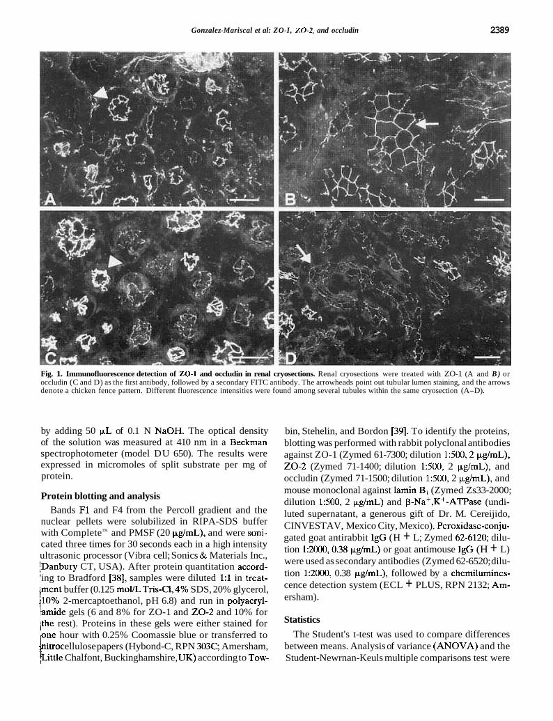
Fig. 1. Immunofluorescence detection of 20-1 and occludin in renal cryosections. Renal cryosections were treated with ZO-1 (A and B ) or occludin (C and D) as the first antibody, followed by a secondary FITC antibody. The arrowheads point out tubular lumen staining, and the arrows denote a chicken fence pattern. Different fluorescence intensities were found among several tubules within the same cryosection (A-D).
by adding 50 p,L of 0.1 N NaOH. The optical density of the solution was measured at 410 nm in a Beckman spectrophotometer (model DU 650). The results were expressed in micromoles of split substrate per mg of protein.
Protein blotting and analysis Bands F1 and F4 from the Percoll gradient and the
nuclear pellets were solubilized in RIPA-SDS buffer with CompleteTM and PMSF (20 pg/mL), and were soni- cated three times for 30 seconds each in a high intensity ultrasonic processor (Vibra cell; Sonics & Materials Inc., lDanbury CT, USA). After protein quantitation accord- 'ing to Bradford [38], samples were diluted 1:l in treat- Iment buffer (0.125 moVL Tris-C1,4% SDS, 20% glycerol, {lo% 2-mercaptoethanol, pH 6.8) and run in polyacryl- iamide gels (6 and 8% for ZO-1 and 2 0 - 2 and 10% for /the rest). Proteins in these gels were either stained for ,one hour with 0.25% Coomassie blue or transferred to ' itrocellulose papers (Hybond-C, RPN 303C: Amersham, t i t t l e Chalfont, Buckinghamshire, UK) according to Tow-
bin, Stehelin, and Bordon [39]. To identify the proteins, blotting was performed with rabbit polyclonal antibodies against ZO-1 (Zymed 61-7300; dilution 1:500,2 p,g/mL), 2 0 - 2 (Zymed 71-1400; dilution 1500, 2 p,g/mL), and occludin (Zymed 71-1500; dilution 1:500, 2 p,g/mL), and mouse monoclonal against lamin B1 (Zymed Zs33-2000; dilution 1500, 2 p,g/mL) and P-Naf ,K+-ATPase (undi- luted supernatant, a generous gift of Dr. M. Cereijido, CINVESTAV, Mexico City, Mexico). Peroxidase-conju- gated goat antirabbit IgG (H + L; Zymed 62-6120; dilu- tion 1:2000,0.38 p,g/mL) or goat antimouse IgG (H + L) were used as secondary antibodies (Zymed 62-6520; dilu- tion 1:2000, 0.38 yg/mL), followed by a chemilumines- cence detection system (ECL + PLUS, RPN 2132; Am- ersham).
Statistics
The Student's t-test was used to compare differences between means. Analysis of variance (ANOVA) and the Student-Newrnan-Keuls multiple comparisons test were
PIVCSB5
Pencil
Gonzalez-Mariscal et al: ZO-1, 2 0 - 2 , and occludin
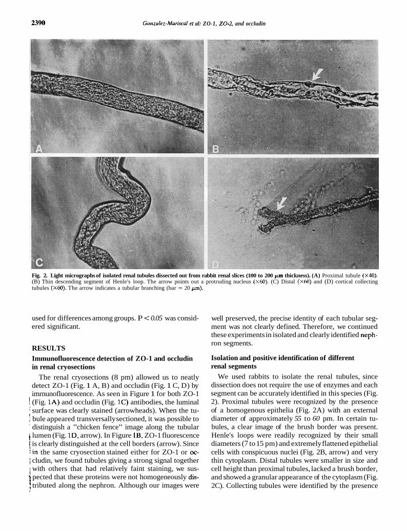
Fig. 2. Light micrographs of isolated renal tubules dissected out from rabbit renal slices (100 to 200 pm thickness). (A) Proximal tubule (X40). (B) Thin descending segment of Henle's loop. The arrow points out a protruding nucleus (X60). (C) Distal (X60) and (D) cortical collecting tubules (x60). The arrow indicates a tubular branching (bar = 20 pm).
used for differences among groups. P < 0.05 was consid- ered significant.
RESULTS Immunofluorescence detection of ZO-1 and occludin in renal cryosections
The renal cryosections (8 pm) allowed us to neatly detect ZO-1 (Fig. 1 A, B) and occludin (Fig. 1 C, D) by imrnunofluorescence. As seen in Figure 1 for both ZO-1
1 (Fig. 1A) and occludin (Fig. 1C) antibodies, the luminal / surface was clearly stained (arrowheads). When the tu- ? bule appeared transversally sectioned, it was possible to ' distinguish a "chicken fence" image along the tubular
t lumen (Fig. ID, arrow). In Figure lB, ZO-1 fluorescence is clearly distinguished at the cell borders (arrow). Since
:in the same cryosection stained either for ZO-1 or oc- 1 cludin, we found tubules giving a strong signal together
i with others that had relatively faint staining, we sus- pected that these proteins were not homogeneously dis-
1 tributed along the nephron. Although our images were
well preserved, the precise identity of each tubular seg- ment was not clearly defined. Therefore, we continued these experiments in isolated and clearly identified neph- ron segments.
Isolation and positive identification of different renal segments
We used rabbits to isolate the renal tubules, since dissection does not require the use of enzymes and each segment can be accurately identified in this species (Fig. 2). Proximal tubules were recognized by the presence of a homogenous epithelia (Fig. 2A) with an external diameter of approximately 55 to 60 pm. In certain tu- bules, a clear image of the brush border was present. Henle's loops were readily recognized by their small diameters (7 to 15 pm) and extremely flattened epithelial cells with conspicuous nuclei (Fig. 2B, arrow) and very thin cytoplasm. Distal tubules were smaller in size and cell height than proximal tubules, lacked a brush border, and showed a granular appearance of the cytoplasm (Fig. 2C). Collecting tubules were identified by the presence
PIVCSB5
Pencil
Gonzaiez-Mariscal et al: 20-1, 20-2, and occludin
of the characteristic branching of this segment (Fig. 2D, arrow), their irregular polygonal shape, and their diame- ter (25 to 30 ~ m ) .
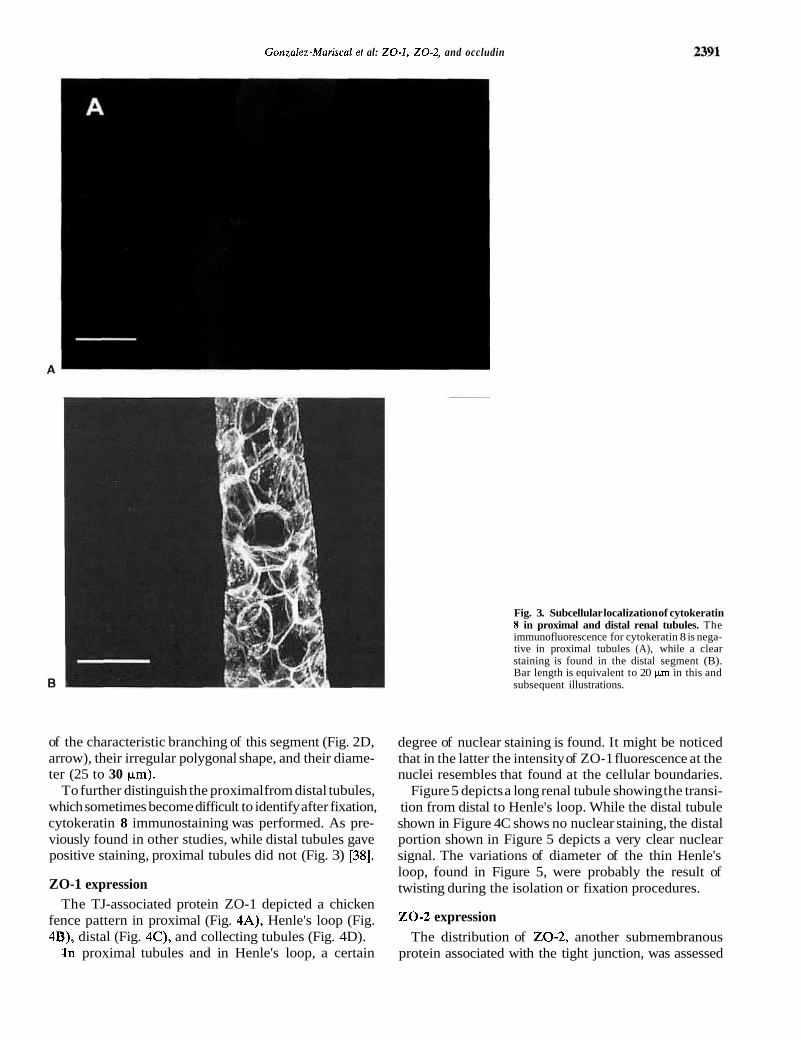
To further distinguish the proximal from distal tubules, which sometimes become difficult to identify after fixation, cytokeratin 8 immunostaining was performed. As pre- viously found in other studies, while distal tubules gave positive staining, proximal tubules did not (Fig. 3) [38].
ZO-1 expression The TJ-associated protein ZO-1 depicted a chicken
fence pattern in proximal (Fig. 4A), Henle's loop (Fig. 4B), distal (Fig. 4C), and collecting tubules (Fig. 4D).
.In proximal tubules and in Henle's loop, a certain
Fig. 3. Subcellular localization of cytokeratin 8 in proximal and distal renal tubules. The immunofluorescence for cytokeratin 8 is nega- tive in proximal tubules (A), while a clear staining is found in the distal segment (B). Bar length is equivalent to 20 pm in this and subsequent illustrations.
degree of nuclear staining is found. It might be noticed that in the latter the intensity of ZO-1 fluorescence at the nuclei resembles that found at the cellular boundaries.
Figure 5 depicts a long renal tubule showing the transi- tion from distal to Henle's loop. While the distal tubule shown in Figure 4C shows no nuclear staining, the distal portion shown in Figure 5 depicts a very clear nuclear signal. The variations of diameter of the thin Henle's loop, found in Figure 5, were probably the result of twisting during the isolation or fixation procedures.
20-2 expression The distribution of 20-2, another submembranous
protein associated with the tight junction, was assessed
PIVCSB5
Rectangle

2392 Gonzalez-Mariscal et al: ZO-1, 20-2, and occludin
Fig. 4. Localization of ZO-1 in renal tubules. Tubules were stained with rabbit anti-ZO-1 polyclonal antibodies. In proximal (A) , Henle's (B), distal (C) , and collecting (D) tubules, ZO-1 staining is clearly arranged as a chicken fence, surrounding the tubular lumen. In proximal tubules and in Henle's loop, a certain degree of nuclear staining is found (A and B, arrowheads). In Henle's loop (B), the intensity of ZO-1 staining is similar both at the cellular borders (arrow) and in cell nuclei (arrowhead). In distal and collecting tubules (C and D), ZO-1 staining is significantly reduced in cell nuclei.
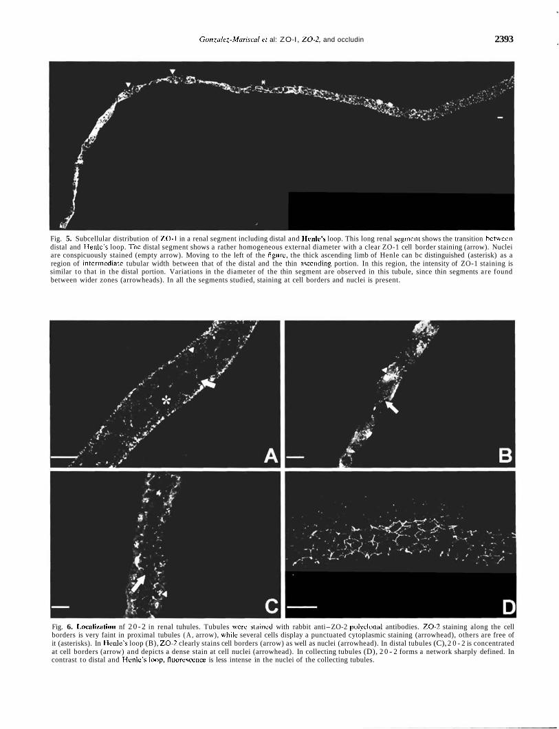
in the different renal segments. 20 -2 in proximal tubules gave a punctuate staining in the cytoplasm (Fig. 6A, arrowheads) that made the appreciation of labeling at the cell boundaries difficult (Fig. 6A, arrow). At Henle's loop, cell borders were clearly stained with 20-2, and an intense signal was found in almost all of the nuclei observed (Fig. 6B). In the distal segment, 20 -2 was distinctly found at cell borders; nevertheless, its staining was weak, in contrast to the strong fluorescent signal found at the nuclei (Fig. 6C). In the collecting portion, a clear chicken fence pattern was present. Although in Figure 6D almost no nuclear staining was found, in other collecting tubules (data not shown), a certain degree of nuclear staining was still observed.
1 Occludin location and expression The most striking difference in the distribution pattern
I of the TJ proteins along the nephron was found for 1
9 occludin. While in the proximal segment a discrete punc- 1 tuated and discontinuous pattern of occludin along the
cell borders was depicted (Fig. 7A), at the collecting tubule the fluorescent signal was neatly located at the cellular borders (Fig. 7B). Two independent tubular seg- ments, including a descending Henle's loop (arrow in Fig. 8) and the other an ascending one (Fig. 9), showed a clear staining of the cell borders in both the ascending and descending Henle's segments. In Figure 8, proximal segments S2 and S3, as defined from the distance to the Henle's loop [39], showed an increasing definition for occludin staining, beginning at the S2 segment with faint lines depicting the cell borders (single asterisk in Fig. €9, more defined and continuous than those observed in the proximal region shown in Figure 7A, and ending at the S3 segment with a more clearly defined chicken fence pattern (double asterisk in Fig. 8).
In Figure 9, the distal tubule that appears after the ascending Henle's loop shows a clear and strong occludin staining along the cell borders. As expected for a trans- membrane protein, no nuclear staining for occludin was ever found.
PIVCSB5
Pencil
Gonzalez-Mariscal er al: ZO-I, 20 -2 , and occludin 2393
Fig. 5. Subcellular distribution of ZO-1 in a renal segment including distal and Henle's loop. This long renal segment shows the transition betwccn distal and IIenle's loop. Thc distal segment shows a rather homogeneous external diameter with a clear ZO-1 cell border staining (arrow). Nuclei are conspicuously stained (empty arrow). Moving to the left of the figure, the thick ascending limb of Henle can bc distinguished (asterisk) as a region of interrnediatc tubular width between that of the distal and the thin asccnding portion. In this region, the intensity of ZO-1 staining is similar to that in the distal portion. Variations in the diameter of the thin segment are observed in this tubule, since thin segments are found between wider zones (arrowheads). In all the segments studied, staining at cell borders and nuclei is present.
Fig. 6. Imcalization nf 2 0 - 2 in renal tuhules. Tubules wcre staincd with rabbit anti-ZO-2 polyclonal antibodies. 2 0 - 2 staining along the cell borders is very faint in proximal tubules ( A , arrow), whilc several cells display a punctuated cytoplasmic staining (arrowhead), others are free of it (asterisks). In Ilenle's loop (B), 2 0 - 2 clearly stains cell borders (arrow) as well as nuclei (arrowhead). In distal tubules (C), 2 0 - 2 is concentrated at cell borders (arrow) and depicts a dense stain at cell nuclei (arrowhead). In collecting tubules (D), 2 0 - 2 forms a network sharply defined. In contrast to distal and I.lcnlc's Imp, fluorcsccnce is less intense in the nuclei of the collecting tubules.

2394 Gonzalez-Mariscal et al: 20-1, 20-2, and occludin
Fig. 7. Occludin staining in proximal and dis- tal renal tubules. In proximal tubules (A) , occludin shows a discrete punctuated and dis- continuous pattern along the cellular bound- aries (arrow), while in the collecting tubule ( B ) , occludin neatly stains cellular borders.
ZO-1 and 20-2 , but not occludin, are expressed in the nuclei of tubular cells
To assess whether the nuclear staining was real and , not a result of the tubular isolation procedure, we de- , tected the presence of ZO-1, 20-2, and occludin by
Western blot in nuclear extracts obtained from rabbit 4 kidneys through a sucrose gradient. The purity of this ! nuclear extract was proven by its enrichment in the nu-
clear protein lamin B, and its lack of the plasma mem- brane-enriched protein P-Na+,K+-ATPase. While ZO-1 and 20-2 were present in both the total and nuclear extracts, occludin was detectable in only the total extract (Fig. 10). Therefore, our fluorescence observation of ZO-1
and 20-2 at the nucleus of isolated tubules might not be artifactual.
Quantitation of fluorescence intensity values for ZO-1, 20-2 , and occludin
When the average specific fluorescence intensity was determined on each tubular segment, a significant differ- ence in the values was found between proximal and the more distal segments for the three proteins examined. In accordance with the fluorescence observations described previously in this article, ZO-1 displayed a significant value even in the proximal region, whereas'occludin, in full agreement with the fluorescence images described
PIVCSB5
Pencil

Gonzalez-Mariscal et al: ZO-1, 20-2, and occludin 2395
Fig. 8. Occludin staining in S2 and S3 segments of proximal tubule and in Henle's descending loop from a single nephron. Going from the proximal S2 region (asterisk) to S3 (double asterisk) and Henle's descending looo (arrow). a clear increase in occludin exoression at the cell borders is obseGed. ~ o t e that the right extreme (A) continues with the left eitre&e (B).
'
Fie. 9. Ex~ression of occludin at an isolated renal tubule with Henle's ascending limb and the initial distal tubule. Along these nephron segments, - the chicken fence pattern is homogeneous and strong.
previously here, had a scant signal at the proximal region (Fig. 11).
Western blot quantitation of ZO-1, 20-2, and occludin
To quantitate the amount of ZO-1, 20-2, and oc- cludin, we isolated proximal and distal tubule popula- tions by a Percoll gradient method, since microdissection of well-defined nephron segments provides only limited amounts of cell material. This technique provided highly purified suspensions of viable tubular fragments of the iabbit nephron and allowed the measurement of enzy- matic activities that could identify these segments [35].
It also supplied sufficient amount of tubules to quantitate ZO-1, 20-2, and occludin.
Furthermore, to assess the adequacy of the isolation procedure to separate proximal and distal populations of rabbit tubules by the Percoll gradient, we observed those fractions with a phase contrast microscope. Proxi- mal and distal tubular images obtained by light micros- copy are shown in Figure 12. In addition, to verify the correct separation of proximal and distal fractions, we measured the activity of alkaline phosphatase, since it is higher in proximal tubules than in the distal populations [36,37]. As expected, the proximal tubular fraction iso- lated by Percoll presented a higher alkaline phosphatase

Gonzalez-Mariscal et al: ZO-1, 20-2 , and occludin 2396
Blot: P-Na+,K+- ATPase Lamin B,
T N
ZO- 1 Occludin
T N
Fig. 10. 2 0 - 1 and 20-2, but not occludii, are expressed in the nucleus of tubular cells. Extracts from whole tissue lysates and from isolated and purified nuclei were separated by SDS-polyacrylamide gel electrophoresis and immunoblotted with different antibodies. While P-Na+,K+-ATPase, an abundant plasma membrane protein, is undetectable in the nuclear extracts, this fraction is enriched in the nuclear protein lamin B1. The submembranous TJ proteins 20-1 and 20-2 are clearly observed in both the nuclear and total fractions, while the transmembrane protein occludin is undetectable in the nuclear extract. The bracket and arrows indicate the position of the detected proteins.
activity (6.98 pmoVmg protein) than the distal fraction (4.80 pmollmg protein). Using this procedure, the enzy- matic activities were similar to those reported by Vinay (ratio of proxima1:distal for Vinay was 1.33 and this study was 1.45) [35]. These results, therefore, suggest that the isolation protocol was effective to separate tubule popu- lations formed predominantly by proximal segments from those that contained mostly distal tubules.
The Western blot in Figure 13 (A) and the densitomet- ric analyses (Fig. 13B) show that the amount of ZO-1, 20-2, and occludin in the distal tubules was significantly higher than in the proximal segment. Proximal and distal detection for each protein were done on the same blot for comparison purposes. Since ZO-1 signal at the proximal tubule was very weak, the blot needed to be overexposed in order to be detectable, leaving the distal blot with a dark signal. It is noteworthy that while ZO-1 was already distributed as a chicken fence at the proximal region, the amount of this protein in these tubules was significantly lower than that found at the distal segments. This obser- vation confirms the difference in ZO-1 intensity of stain- ing found in renal frozen sections by our group and that of Piepenhagen et a1 [40].
DISCUSSION 1 This study discusses the subcellular distribution of three tight junction (TJ) proteins along different seg-
ments of the mammalian nephron, an epithelia with seg- ments of great variation in TJ permeability. The isolated renal tubules have preserved their characteristic mor- phology after manual dissection from adult rabbits, as previously described by Reyes et a1 [28,32], thus allowing for the adequate identification of the nephron segments. However, since distal tubules are somewhat difficult to recognize, cytokeratin 8 immunostaining was used as a distal tubule marker for further characterization. Our results confirmed that cytokeratin 8 did not stain proxi- mal tubules, while it was a clear marker of the distal segments.
We have studied the distribution of ZO-1 and 20-2, two peripheral membrane proteins associated to the cytoplasmic surface of the TJ that are members of the membrane-associated guanylate kinase (MAGUK) pro- - tein family [42], and occludin, an integral membrane pro- tein found at the TJ, since only the antibodies generated against these TJ proteins are commercially available. In fluorescence images, in fluorescence quantitation experi- ments, and by Western blot detection, we have observed that the distribution of these three proteins along the different renal segments varied. ZO-1 is clearly present along the entire nephron and displays a chicken fence pattern from the beginning of the tubular structures, where it is relatively abundant, according to our fluores- cence intensity data. However, in contrast to occludin and 20-2,ZO-1 determined by Western blot in extracts
PIVCSB5
Pencil

Gonzalez-Mariscal et al: ZO-1, 20-2, and occludin 2397
Proximal Henle Distal CCD
Proximal Henle Distal CCD
C Occludin
1 a
h -I-
Proximal Henle Distal CCD Fig. 11. Fluorescence intensity values of ZO-1, 20-2, and occludin. The average specific fluorescence intensity of each tubular projection was corrected by tubular unit area (100 pn2). For each protein, the arbitrary value of 100 was assigned to the highest fluorescence intensity value obtained, and the rest of the data was normalized accordingly. Mean t SEM are shown. Literals show significant statistical differences among groups. CCD, cortical collecting ducts.
of Percoll isolated proximal tubules was only made evi- 'dent when protease inhibitors were included during the isolation procedure, thus indicating that ZO-1 is signifi- cantly more sensitive than 20 -2 and occludin to prote- ases present in the proximal tubular extracts, which are particularly rich in lysosomal and other proteolytic en- zymes. This might explain why the signal for ZO-1 at the proximal region is lower in the Western blot determi- nation than what could be expected from the fluores- bence quantitation experiments. The fact that the distri-
bution of ZO-1 is not related to the significant differences in resistance found along the nephron strongly suggests that this protein might be necessary but not critical for the development and maintenance of this property of renal epithelia. Furthermore, ZO-1 is also present in sev- eral nonepithelial cell types, including myocardium inter- calated discs, astrocytes, Schwann cells, fibroblasts glioma, sarcoma, and mieloma cell lines [4346]. In them, ZO-1 either colocalizes with cadherins [46] or concentrates in lamellipodia where actin filaments accumulate [45,47].
In certain tissues, a differential expression of ZO-1 isoforms has been observed. In mouse embryo, ZO-1 a- is present from the oocytes stage, while the a+ isoform is detected only from the blastocyte stage onward, when TJs are sealed [48]; in Sertoli cells, the a+ and a- iso- forms are differentially expressed in puberty and adult- hood [49]. In our study, a polyclonal antibody against 20-1 was used that cannot differentiate between the ZO-1 isoforms a- and a+. However, this situation poses no problem, since both isoforms are expressed along the entire tubular structures, with the exception of the glomerular slit diaphragms, where only the isoform a- is present [50].
When frozen renal slices were stained for ZO-1, a clear distinction of the different nephron segments could not be accomplished unless several antibodies specific for each tubular section were used. Therefore, we choose to isolate the different tubular segments by direct dissec- ten. Nevertheless, the frozen sections were very useful, since they allowed us to observe different intensities of staining among the renal segments that were conspicuous and in agreement with the observations by Piepenhagen et a1 [40]. Western blot results confirmed the fact that although ZO-1 is present along the whole tubular struc- ture, it is nevertheless expressed with differential amounts that increase from proximal to distal segments.
20 -2 is a 160 kD peripheral protein that is a member of the MAGUK family [51]. Like ZO-1,ZO-2 is present not only in TJs at epithelial and endothelial cells, but also in adherens junctions of nonepithelial cells such as fibroblasts and cardiac muscle cells [52]. 20-2 coimmu- noprecipitates with 20-1; however, the kinetics of ap- pearance of newly synthesized 20 -2 in anti-ZO-1 immu- noprecipitates are much lower than that of ZO-1 [53]. 20 -2 presents two alternatively spliced regions located at the proline-rich domain, in which the precise role is not yet elucidated [51, 541. In this article, the antibody used against 20 -2 gives a higher background staining than ZO-1 and occludin antibodies. Nevertheless, in the collecting tubule, a clear chicken-fence pattern is de- tected. The fact that in the proximal region 20 -2 gives a patchy pattern with few clear definitions of cell bound- aries suggests that the presence of this protein might be related to the acquisition of mature TJ.
In a previous article, we reported the presence of nu-

2398 Gonzalez-Mariscal et al: 20-1, 20-2, and occludin
Fig. 12. Proximal and distal tubules isolated by a Percoll gradient method. (A) A proximal tubule from rabbit shows the characteristic brush border structure (arrow). (B) A seg- ment of distal nephron segment shows the classic cobblestone pattern with clearly de- fined cell boundaries.
clear sorting signals in several members of the MAGUK family, including ZO-1 and 20-2. While the former has two signals located at the first PDZ and GK domains, 20-2 only presents the first one at the PDZ repeat [55].
i The nuclear fluorescence that we have found for ZO-1 i and 20-2 along the nephron is scattered in all segments j with a more intense and frequent staining in Henle's ' loop. The nuclear presence of ZO-1 was previously ob-
I served by Gottardi et a1 [56], in cultured epithelial cells and canine intestine. It seemed to be inversely related
a to the extent and/or maturity of cell-cell contact. How- ] ever, this might not be the case in the tubular system
since the nephrons studied here come from mature rab- bits, where their cells are firmly joined and integrate a tubular structure. However, it is worth noting that
collecting tubules less frequently present this staining. While Gottardi et a1 have shown that ethanol-fixed cells lose their nuclear ZO-1 staining, our study found that renal tubules maintain their ZO-1 and 20 -2 nuclear labeling even when fixed with ethanol and acetone [56], thus suggesting that perhaps a higher amount of these proteins is present at the nucleus in tubular cells as com- pared with Madin-Darby canine kidney epithelia.
Occludin is an integral protein of the TJ that has been located by freeze-fracture immunolabeling at the junc- tional strands [57, 581. It is present in epithelial and endothelial cells, and absent both at the mRNA and protein level in cells that lack TJs such as fibroblasts [58]. Paradoxically, it has been shown that occludin-deficient embryonic stem cells can differentiate into polarized epi-
PIVCSB5
Pencil

Gonzalez-Mariscal et al: 20-1, 20-2, and occludin
ZO- 1
D
ZO- 1 20-2 Occludin
Occludin
P D
Fig. 13. Western blot determinations of ZO-1, 20-2, and occludin in proximal (P) and distal (D) tubules. One representative experiment (A) and the results of the densitometric analy- ses of the whole set (B) are presented. Sym- bols are: (m) proximal tubules; (0) distal tubules; N = number of observations. The amount of ZO-1.20-2, and occludin expressed in proximal tubules is significantly lower than that found in the distal segments (P < 0.01, P < 0.001, and P < 0.001, respectively).
thelial cells, bearing regular TJ strands, thus suggesting that under certain conditions other integral proteins may fulfill occludin function at the TJ [59]. Recently, such molecules, named claudins, were identified. Claudins are a family of 16 integral membrane proteins that localize at the TJ and significantly participate in the TJ specific obliteration of the intercellular space [60-631.
Nevertheless, several lines of evidence involve oc- cludin in the sealing function of the TJ: ( I ) Expression of COOH-terminally truncated occludin resulted either in an electrically tighter paracellular pathway with a par- adoxically increased paracellular flux [64] or in the case of the Xenopus embryo in the generation of leaky junc-
, tions [65]. (2) Induced expression of occludin increased ' TER [64] with a concomitant increase in the mean num- ber and complexity of the TJ strands [66]. (3) Both extra- cellular occludin loops seem to participate in intercellular adhesion, since a synthetic peptide corresponding to the
-,second extracellular domain of occludin reversibly dis- : rupted TER and increased the paracellular flux of tracers
in kidney epithelial monolayers [67], and a peptide gen- erated from the first extracellular loop inhibited homo- philic occludin adhesion in transfected fibroblasts [68]. (4) The expression level of occludin correlates well with the permeability and number of TJ strands in epithelial and endothelial cells [58,69] and during functional devel- opment of the TJs [70]. (5) After common bile duct ligation, the TER significantly diminishes, and occludin staining is discontinuous but remains localized along the canalicular margins [71]. (6) When occludin is overex- pressed in insect cells, multilamellar structures, positive to antioccludin labeling, accumulate in the cytoplasm and resemble TJ structures [57]. (7) In L fibroblasts lack- ing TJs, claudins form TJ strands and recruit occludin, therefore suggesting that both type of proteins are inte- gral constituents of TJ strands [60].
In line with these observations, we found that the overall distribution of occludin along the nephron agrees with the TER reported in those segments. Thus, at the proximal tubule, a region where TER is low as compared
PIVCSB5
Pencil
PIVCSB5
Pencil

2400 Gonzalez-Mariscal et al: ZO-1, 20-2, and occludin
with other segments of the nephron (Table 1) and the TJ is composed of only one or two strands, a discrete,
unctuated and discontinuous pattern of occludin along the cell borders is depicted. In contrast, in the collecting tubule, a high-resistance epithelium, the occludin fluo- I' jrescent signal is evenly located at the cell borders.
The glomerular epithelium is unique among renal epi- thelia, since the intercellular spaces are open and TJs are replaced by slit diaphragms. Although no TJ fibrils ;are found at this structure by freeze fracture and occludin is hardly detectable, ZO-1 is clearly present [72]. There- fore, the expression of ZO-1 alone does not necessarily indicate the presence of a TJ assembly.
In summary, in tubular renal cells, occludin was exclu- sively present at the cell borders, and its distribution follows the increase in tight junction complexity and function displayed by the nephron, thus suggesting an intimate relationship between its presence and the strength of the tight junction. 20-2 is frequently found at cell nuclei, and in the case of the proximal tubule, it is dispersed in the cytoplasm. From Henle's loop to the collecting segment, an increased location of this protein along the cell borders is clearly observed. It remains to be clarified why 20-2 is so frequently present at the nuclei of mature tubular epithelia. In contrast, ZO-1 gives a clear border pattern unrelated to the sealing capacity of the segments. However, the intensity of the staining increases as it reaches the collecting region, therefore suggesting that the presence of this protein is necessary for the establishment of tight junction, but is not directly related to the sealing capacity of the junction.
ACKNOWLEDGMENTS This study was supported by grants from the National Research
Council of Mexico (CONACYT No. 28083N and 26649M). Dr. Valen- cia is a recipient of a fellowship from the Mexican Minister of Education (SEP). We thank Mr. Jose Juan Rivera and Mr. Ignacio Araoz Martinez for their valuable support in the processing of the photographic ma- terial.
Reprint requests to Lorenza Gonzalez-Mariscal, Ph.D., Department of Physiology, Biophysics and Neuroscience, CINVESTAV, Apartado Postal 14-740, Mexico, D. F. 07000, Mexico. E-mail: [email protected]
REFERENCES CEREIJIDO M: Evolution of the ideas on the tight junction, in Tight Junctions, edited by CEREIJIDO M, Boca Raton, CRC Press, 1992, pp 1-13 CEREUIDO M, GONZALEZ-MARISCAL L, CONTRERAS RG: Tight junc- tion: Barrier between higher organisms and environment. NIPS 472-75, 1989. DRAGSTEN PR, BLUMENTHAL R, HANDLER JS: Membrane asymme- try in epithelia: Is the tight junction a barrier to diffusion in plasma membrane? Nature 94718'122,1981 GONZALEZ-MARISCAL L, CHAVEZ DE RAMIREZ B, CEREIJIDO M: Effect of temperature on the occluding junctions of monolayers of epithelioid cells (MDCK). J Membr Biol79:175-184, 1984 GONZALEZ-MARISCAL L. CHAVEZ DE RAMIREZ B. CEREIJIDO M:
Tight junction formation in cultured epithelial cells (MDCK). J Membr Biol86:113-125,1985 GONZALEZ-MARISCAL L, CONTRERAS RG, BOLIVAR JJ, PONCE A, CHAVEZ DE RAMIREZ B, CEREUIDO M: The role of calcium in tight junction formation between epithelial cells. Am J Physiol 259: C978-C986, 1990 CONTRERAS G, MILLER M, ZAMORA M, GONZALEZ-MARISCAL L, CEREIJIDO M: Interaction of calcium with the plasma membrane of epithelial (MDCK) cells during junction formation. Am J Physiol 263:C313-C318, 1992 GONZALEZ-MARISCAL L, CHAVEZ DE RAMIREZ B, LAZARO A, CEREI- nDo M: Establishment of tight junctions between cells from differ- ent animal species and different sealing capacities. J Membr Biol 110:l-9, 1989 CALDERON V, LAZARO A, CONTRERAS RG, SHOSHANI L, FLORES- MALDONADO C, GONZALEZ-MARISCAL L, ZAMPIGHI G, CEREIJIDO M: Tight junctions and experimental modifications of lipid content. J Membr Biol164:5949,1998 BALDA MS, GONZALEZ-MARISCAL L, CONTRERAS RG, CEREIJIDO M: Assembly and sealing of tight junctions: Possible participation of G-proteins, phospholipase C, protein kinase C and calmodulin. J Membr Biol122193-202, 1991 BALDA S, GONZALEZ-MARISCAL L, MATTER K, CEREIJIDO M: Assem- bly of the tight junction: The role of diacylglycerol. J Cell Biol 123:293-302,1993 FURUSE M, HIRASE T, ITOH M, NAFGAFUCHI A, YONEMURA S, TSU- KITA SA, TSUKITA SH: Occludin: A novel integral membrane pro- tein localizing at tight junctions. J Cell Biol 123:1777-1788, 1993 FURUSE M, FUJITA K, HIIRAGI T, FUJIMOTO K, TSUKITA S: Claudin- 1 and -2: Novel integral membrane proteins localizing at tight junctions with no sequence similarity to occludin. J Cell Biol 141:1539-1550, 1998 MARTIN-PADURA I, LOSTAGLIO S, SCHNEEMANN M, WILLIAMS L, ROMANO M, FRUSCELLA P, PANZERI C, STOPPACCIARO A, RUCO L, VILLA A, SIMMONS D, DEJANA E: Junctional adhesion molecule, a novel member of the immunoglobulin superfamily that distributes at intercellular junctions and modulates monocyte transmigration. J Cell Biol142117-127, 1998 STEVENSON BR, SILICIANO JD, MOOSEKER MS, GOODENOUGH DA: Identification of ZO-1: A high molecular weight polypeptide asso- ciated with the tight junction (zonula occludens) in a variety of epithelia. J Cell Biol 103:755-766, 1986 GUMBINER B, LOWENKOPF T, APATIRA D: Identification of a 160- kDa polypeptide that binds to the tight junction protein ZO-1. Proc Natl Acad Sci USA 88:346&3464,1991 HASKINS J, GU L, WITTCHEN ES, HIBBARD J, STEVENSON BR: 20-3, a novel member of the MAGUK protein family found at the tight junction, interacts with ZO-1 and occludin. J Cell Biol141:199-208, 1998 CITI S, SABANAY H, JAKES R, GEIGER B, KENDRICK-JONES J: Cingulin, a new peripheral component of tight junctions. Nature 33:272-276, 1988 ZHONG Y, SAITOH T, MINASE T, SAWADA N, ENOMOTO K, MORI
M: Monoclonal antibody 7H6 reacts with a novel tight junction- associated protein distinct from ZO-1, cingulin and 20-2. J Cell Biol 120:477-483,1993 KEON BH. SCHAFER S. KUHN C. GRUND C. FRANKE WW: Svm~lekin. a novel type of tight junction plaque protein. J Cell ~ i o l i34:10031 1018,1996 BOULPAEP EL, SEELY JF: Electrophysiology of proximal and distal tubules in the autoperfused dog kidney. Am J Physiol221:1084- 1096,1971 FROMTER E, DIAMOND J: Route of passive ion permeation in epithe- lia. Nature 235:9-13, 1972 HEGEL U, FROMTER E, WICKT: Der elektrische wandwiderstand des proximalen konvolutes der rattenniere. Pfiigers Arch 294:274-290, 1967 BURG M, GREEN N: Function of the thick ascending limb of Henle's loop. Am J Physiol224659468, 1973 HEBERT SC, CULPEPPER RM, ANDREOLI TE: NaCl transport in mouse medullary thick ascending limbs. I. Functional nephron heterogeneity and ADH-stimulated NaCl cotransport. Am J Phys- iol 10:F412-F431.1981
PIVCSB5
Pencil
PIVCSB5
Pencil
PIVCSB5
Pencil
PIVCSB5
Pencil
Gonzalez-Mariscal et al: ZO-1, 20-2, and occludin 2401
26. GREGER R: Cation selectivity of the isolated perfused cortical thick ascending limb of Henle's loop of rabbit kidney. miigers Arch 390:30-37,1981
27. MALNIC G, GIEBISH G: Some electrical properties of distal tubular epithelium in the rat. Am J Physiol223:797-808, 1972
28. REYES JL, ROCH-RAMEL F, BESSEGHIR K: Net sodium and water movements in the newborn rat collecting tubule: Lack of modifica- tions by indomethacin. Biol Neonate 51:212-216, 1987
29. HELMAN SI, GRANTHAM JJ, BURG MB: Effect of vasopressin on electrical resistance of renal cortical collecting tubules. Am J Phys- iol220:1825-1832, 1971
30. RAU WS, FROMTER E: Electrical properties of the medullary ducts of the golden hamster kidney. miigers Arch 351:113-131, 1974
31. CLAUDE P, GOODENOUGH DA: Fracture faces of zonulae occlude- ntes from tight and leaky epithelia. J Cell Biol58:39&400, 1973
32. REYES JL, NAVA E, NAMORADO MC: Receptor-mediated effect of a synthetic thromboxane-analog on cytosolic calcium in isolated proximal tubules. Prostaglandins 44:145-154, 1992
33. SPECTOR DL, GOLDMAN RD, LEINWAND LA: Preparation of nuclei from tissues and suspension cells, in Cells: A Laboratory Manual, Cold Spring Harbor, Cold Spring Harbor Laboratory Press, 1998, pp 43.1-43.6
34. GREENBERG ME, GREENE LA, ZIFF EB: Nerve growth factor induce rapid transient changes in proto-oncogene transcription in PC-12 cells. J Biol Chem 260:14101-14110, 1985
35. VINAY P, GOUGOX A, LEMIEUX G: Isolation of a pure suspension of rat proximal tubules. Am J Physiol241:F403-F411, 1981
36. MAUNSBACH AB: Ultrastructure of the proximal tubule, in Hand- book of Physiology, Renal Physiology, edited by ORLOFF J, BERLI- NER RW, Washington D.C., Am Physiology Society, 1973, pp 31-79
37. REALE E, LUCIANO L: Kritische elektronenmikroskopische studien uber die lokalisation des aktivitat alkalischer phosphatase im haup- tstuck der niere von mausen. Histochemie 8:302-314,1967
38. BRADFORD M: A rapid and sensitive method for the quantitation of microgram quantities of protein utilizing the principles of protein dye-binding. Anal Biochem 72248-254, 1976
39. TOWBIN H, STEHELIN T, BORDON J: Electrophoretic transfer of proteins from polyacrylamide gels to nitrocellulose sheets: Proce- dure and some applications. Proc Natl Acad Sci USA 764350-4356, 1979
40. P~EPENHAGEN PA, PETERS LL, LUX SE, NELSON WJ: Differential expression of Na+K+-ATPase, ankyrin, fodrin and E-cadherin along the kidney nephron. Am J Physiol269:Cl417<1432, 1995
41. TISHER CC, MADSEN KM: Anatomy of the kidney, in The Kidney (vol 1,5th ed), edited by BRENNER BM, Philadelphia, W.B. Saun- ders, 1996, pp 21-34
42. ANDERSON JM: Cell signaling: MAGUK magic. Curr Biol6382- 384, 1996
43. HOWARTH AG, HUGHES MR, STEVENSON BR: Detection of the tight junction-associated protein ZO-1 in astrocytes and other nonepi- thelial cell types. Am J Physiol262:C461-C469, 1992
44. HOWARTH AG, SINGER KL, STEVENSON BR: Analysis of the distribu- tion and phosphorylation state of ZO-1 in MDCK and nonepithe- lial cells. J Membr Biol 137:261-270, 1994
45. HOWARTH AG, STEVENSON BR: Molecular environment of ZO-1 in epithelia and non-epithelial cells. Cell Motil Cytoskeleton 31:323- 332, 1995
46. ITOH M, NAGAFUCHI A, YONEMURA S, KITANI-YASUDA T, TSUKITA
S, TSUKITA S: The 220-kD protein colocalizing with cadherins in non-epithelial cells is identical to 20-1, a tight junction-associated protein in epithelial cells: cDNA cloning and immunoelectron mi- croscopy. J Cell Biol 121:491-502, 1993
47. YONEMURA S, ITOH M, NAGAFUCHI A, TSUKITA S: Cell to cell ad- herens junction formation and actin filament organization: Similari- ties and differences between non-polarized fibroblasts and polar- ized epithelial cells. J Cell Sci 108:127-142, 1995
48. SHETH B, FESENKO I, COLLINS JE, MORAN B, WILD A, ANDERSON
JM, FLEMING TP: Tight junction assembling during mouse blasto- cyte formation is regulated by late expression of 20-1 a+ isoform. Development 1242027-2037, 1997
49. PELLETIER RM, OKAWARA Y, VITALE ML, ANDERSON JM: Differen- tial distribution of the tight junction associated protein ZO-1 iso-
forms a+ and a- in guinea pig Sertoli cells: A possible association with F-actin and G-actin. Biol Reprod 57:367-376, 1997
50. KURIHARA H, ANDERSON JM, FARQUHAR MG: Diversity among tight junctions in rat kidney: Glomerular slit diaphragms and endothelial junctions express only one isoform of the tight junction protein 20-1. Proc Natl Acad Sci USA 897075-7079, 1992
51. BEATCH M, JESAITIS LA, GALLIN WJ, GOODENOUGH DA, STEVENSON BR: The tight junction protein 20-2 contains three PDZ (PSD- 95IDiscs-large11 XO-1) domains and an alternatively splice region. J Biol Chem 271:25723-25726, 1996
52. ITOH M, MORITA K, TSUKITA S: Characterization of 2 0 - 2 as a MAGUK family member associated with tight as well as adherent junctions with a binding affinity to occludin and a catenin. J Biol Chem 2745981-5986, 1999
53. JESAITIS LA, GOODENOUGH DA: Molecular characterization and tissue distribution of 20-2, a tight junction protein homologous to 2 0 - 1 and the Drosophila discs-large tumor suppressor protein. J Cell Biol 124:949-961, 1994
54. COLLINS JR, RIZZOLO L: Protein binding domains of the tight junction protein 20-2, are highly conserved between avian and mammalian species. Biochem Biophys Res Commun 252617-622, 1998
55. GONZALEZ-MARISCAL L, ISLAS S, CONTRERAS R, GARCIA-VILLEGAS
MR, BETANZOS A, VEGA J, DIAZ QUIRONES A, MARTIN OROZCO
N, ORTIZ NAVARRETE V, CEREIJIDO M, VALDES J: Molecular charac- terization of the tight junction protein ZO-1 in MDCK cells. Exp Cell Res 248:97-109, 1999
56. GOTTARDI CJ, ARPIN M, FANNING AS, LOUVARD D: The junction- associated protein, zonula occludens-1, localizes to the nucleus before the maturation and during the remodeling of cell-cell con- tacts Proc Natl Acad Sci USA 93:10779-10784, 1996
57. FURUSE M, FUJIMOTO K, SATO N, HIRASE T, TSUKITA S, TSUKITA S: Overexpression of occludin, a tight junction-associated integral membrane protein, induces the formation of intracellular multila- mellar bodies bearing tight junction-like structures. J Cell Sci 109:429-435, 1996
58. SAITOU M, ANDO-AKATSUKA Y, ITOH M, FURUSE M, INAZAWA J, FUJIMOTO K, TSUKITA S: Mammalian occludin in epithelial cells: Its expression and subcellular distribution. Eur J Cell Biol73:222-231, 1997
59. SAITOU M, FUJIMOTO K, DOI Y, ITOH M, FUIIMOTO T, FURUSE M, TAKANO H, NODA T, TSUKITA S: Occludin-deficient embryonic stem cells can differentiate into polarized epithelial cells bearing tight junctions. J Cell Biol 141:397-408, 1998
60. FURUSE M, SASAKI H, FUJIMOTO K, TSUKITA S: A single gene prod- uct, claudin-1 or -2, reconstitutes tight junction strands and recruits occludin in fibroblasts. J Cell Biol143:391-401, 1998
61. MORITA K, FURUSE M, FUJIMOTO K, TSUKITA S: Claudin multigene family encoding four-transmembrane domain protein components of tight junction strands. Proc Natl Acad Sci USA 96511-516,1999
62. MORITA K, SASAKI H, FUJIMOTO K, FURUSE M, TSUKITA S: Claudin- 11IOSP-based tight junctions of myelin sheaths in brain and Sertoli cells in testis. J Cell Biol 145x579-588, 1999
63. KUBOTA K, FURUSE M, SASAKI S, SONODA N, FUJITA K, NAGAFUCHI
A, TSUKITA S: Caz+-independent cell-adhesion activity of claudins, a family of integral membrane proteins localized at tight junctions. Curr Biol9:1035-1038, 1999
64. BALDA MS, WHITNEY JA, FLORES C, GONZALEZ S, CEREIJIDO M, MATTER K: Functional dissociation of paracellular permeability and transepithelial electrical resistance and disruption of the apical- basolateral intramembrane diffusion barrier by expression of a mutant tight junction membrane protein. J Cell Bio1134:1031-1049, 1996
65. CHEN Y, MERZDORF C, PAUL DL, GOODENOUGH DA: COOH termi- nus of occludin is required for tight junction barrier function in early Xenopus embryos. J Cell Biol138:891-899, 1997
66. MCCARTHY KM, SKARE IB, STANKEWICH MC, FURUSE M, TSUKITA
S, ROGERS RA, LYNCH RD, SCHNEEBERGER EE: Occludin is a func- tional component of the tight junction. J Cell Sci 109:2287-2298, 1996
67. WONG V, GUMBINER BM: A synthetic peptide corresponding to the extracellular domain of occludin perturbs the tight junction permeability barrier. J Cell Biol 136399-409, 1997

2402 Gonzalez-Mariscal et al: ZO-1, 20-2, and occludin
68. VAN ITALLIE CM, ANDERSON JM: Occludin confers adhesiveness when expressed in fibroblasts. J Cell Sci 110:1113-1121, 1997
69. HIRASE T, STADDON JM, SAITOU M, ANDO-AKATSUKA Y, ITOH M, FURUSE M, FUJIMOTO K, TSUKITA S, RUBIN L: Occludin as a possible determinant of tight junction permeability in endothelial cells. J Cell Sci 110:1603-1613, 1997
70. KAWASAKI K, HAYASHI Y, NISHIDA Y, MIKI A, ITOH H: Develop- mental expression of the tight junction protein, occludin, in the
gastrointestinal tract of the chick embryo. Histochem Cell Biol109: 19-24, 1998
71. FALLON MB, BRECHER AR, BALDA MS, MAITER K, ANDERSON
JM: Altered hepatic localization and expression of occludin after common bile duct ligation. Am J Physiol269:C1057-(21062, 1995
72. SCHNABEL E, ANDERSON JM, FARQUHAR MG: The tight junction protein ZO-1 is concentrated along slit diaphragm of the glomeru- lar epithelium. J Cell Biol 111:1255-1263, 1990