Recessive Mutations in the Gene Encoding the Tight Junction Protein Occludin Cause Band-like...
11
REPORT Recessive Mutations in the Gene Encoding the Tight Junction Protein Occludin Cause Band-like Calcification with Simplified Gyration and Polymicrogyria Mary C. O’Driscoll, 1 Sarah B. Daly, 1 Jill E. Urquhart, 1 Graeme C.M. Black, 1 Daniela T. Pilz, 2 Knut Brockmann, 3 Meriel McEntagart, 4 Ghada Abdel-Salam, 5 Maha Zaki, 5 Nicole I. Wolf, 6,7 Roger L. Ladda, 8 Susan Sell, 8 Stefano D’Arrigo, 9 Waney Squier, 10 William B. Dobyns, 11 John H. Livingston, 12 and Yanick J. Crow 1, * Band-like calcification with simplified gyration and polymicrogyria (BLC-PMG) is a rare autosomal-recessive neurological disorder showing highly characteristic clinical and neuroradiological features. Affected individuals demonstrate early-onset seizures, severe microcephaly, and developmental arrest with bilateral, symmetrical polymicrogyria (PMG) and a band of gray matter calcification on brain imaging; as such, the disorder can be considered as a ‘‘pseudo-TORCH’’ syndrome. By using autozygosity mapping and copy number analysis we identified intragenic deletions and mutations in OCLN in nine patients from six families with BLC-PMG. The OCLN gene encodes occludin, an integral component of tight junctions. Neuropathological analysis of an affected individual showed similarity to the mouse model of occludin deficiency with calcification predominantly associated with blood vessels. Both intracranial calcification and PMG are heterogeneous in etiology. Neuropathological and clinical studies of PMG have suggested that in utero ischemic or vascular insults may contribute to this common cortical abnormality. Tight junctions are functional in cerebral blood vessels early in fetal development and continue to play a vital role in maintenance of the blood-brain barrier during postnatal life. We provide evidence that the tight junction protein occludin (encoded by the OCLN gene) is involved in the pathogenesis of malformations of cortical development. Band-like calcification with simplified gyration and polymicrogyria (BLC-PMG) is a rare autosomal-recessive neurological condition demonstrating clinical and neuro- radiological features that may be interpreted as sequelae of congenital infection, a so-called pseudo-TORCH syndrome (MIM 251290). We have previously described 12 affected children from 5 families with this disorder. 1–3 Patients experienced early-onset seizures, severe progres- sive microcephaly, and developmental arrest. This patient cohort was collated on the basis of the pattern of gray matter calcification and cortical malformation. CT and MR imaging showed a prominent band of cortical gray matter calcification as well as calcification in the cere- bellum and basal ganglia (Figures 1 and 2). Brain imaging also showed characteristic bilateral, symmetrical, predom- inantly fronto-parietal PMG. Intracranial calcification (ICC) is a finding common to a heterogeneous group of genetic syndromes, as well as a prominent manifestation of intrauterine infection, in particular with congenital cytomegalovirus (CMV). These phenotypes are typically defined, not by the pattern of ICC, but by the presence of other clinical features. The combination of ICC and PMG suggests congenital CMV infection during mid-gesta- tion. 4,5 However, in BLC-PMG, the ICC is seen in a uniform, semicontinuous ribbon or band on CT brain, unlike the patterning typical of CMV infection. PMG is an increasingly recognized and common malformation of cortical development associated with a growing number of syndromes and consistent cytogenetic abnormalities. 6–8 Mutations in several genes have been identified as associated with PMG, 9–19 underlining the heterogeneous etiology of this malformation. The importance of ischemic or vascular insults, occurring at around 5 months of gesta- tion, in the pathogenesis of PMG has been suggested on the basis of animal models, 20–23 twin studies, 24–26 in utero insults, 27 and case studies of affected patients. 28 The site of PMG is most commonly within the territory of the middle cerebral artery, lending further weight to a vascular etiology. 29–31 Here, we report mutations in the OCLN gene (MIM 602876) encoding the tight junction protein occludin in nine patients with BLC-PMG. Occludin is expressed as an integral component of the tight junction in all epithelia as well as endothelia in the brain. 32,33 The Ocln knockout mouse model has a complex phenotype including abnormalities of salivary glands, gastric epithe- lium, bone, testes, and ICC. 34 The human phenotype 1 Genetic Medicine, University of Manchester, Manchester Academic Health Science Centre, Central Manchester Foundation Trust University Hospitals, Manchester, M13 9WL, UK; 2 Department of Medical Genetics, University Hospital of Wales, Cardiff, CF14 4XW, UK; 3 Department of Paediatrics and Paediatric Neurology, Children’s Hospital, Georg August University, Robert-Koch-Str. 40, 37075, Goettingen, Germany; 4 Department of Clinical Genetics, St. George’s Hospital, London, SW17 0RE, UK; 5 Clinical Genetics Department, Human Genetics and Genome Research Division, National Research Centre, Cairo, 12311, Egypt; 6 Paediatric Neurology, University Children’s Hospital, 69120 Heidelberg, Germany; 7 Department of Child Neurology, VU Medical Center, 1007 MB Amsterdam, The Netherlands; 8 Division of Human Genetics, Growth & Development, Department of Pediatrics, Penn State Hershey Children’s Hospital, Hershey, PA 17033, USA; 9 Development Neurology Department, Fondazione IRCCS Istituto Neurologico ‘‘C. Besta,’’ 20133 Milan, Italy; 10 Departments of Neurology and Neuropathology, Radcliffe Infirmary, Oxford, OX3 9DU, UK; 11 Departments of Human Genetics, Neurology and Pediatrics, The University of Chicago, Chicago, IL 60637, USA; 12 Department of Paediatric Neurology, Leeds General Infirmary, Leeds, LS9 7TF, UK *Correspondence: [email protected] DOI 10.1016/j.ajhg.2010.07.012. Ó2010 by The American Society of Human Genetics. All rights reserved. The American Journal of Human Genetics 87, 1–11, August 13, 2010 1 Please cite this article in press as: O’Driscoll et al., Recessive Mutations in the Gene Encoding the Tight Junction Protein Occludin Cause Band-like Calcification with Simplified Gyration and..., The American Journal of Human Genetics (2010), doi:10.1016/j.ajhg.2010.07.012
Transcript of Recessive Mutations in the Gene Encoding the Tight Junction Protein Occludin Cause Band-like...

Please cite this article in press as: O’Driscoll et al., Recessive Mutations in the Gene Encoding the Tight Junction Protein Occludin CauseBand-like Calcification with Simplified Gyration and..., The American Journal of Human Genetics (2010), doi:10.1016/j.ajhg.2010.07.012
REPORT
Recessive Mutations in the Gene Encoding the TightJunction Protein Occludin Cause Band-like Calcificationwith Simplified Gyration and Polymicrogyria
Mary C. O’Driscoll,1 Sarah B. Daly,1 Jill E. Urquhart,1 Graeme C.M. Black,1 Daniela T. Pilz,2
Knut Brockmann,3 Meriel McEntagart,4 Ghada Abdel-Salam,5 Maha Zaki,5 Nicole I. Wolf,6,7
Roger L. Ladda,8 Susan Sell,8 Stefano D’Arrigo,9 Waney Squier,10 William B. Dobyns,11
John H. Livingston,12 and Yanick J. Crow1,*
Band-like calcification with simplified gyration and polymicrogyria (BLC-PMG) is a rare autosomal-recessive neurological disorder
showing highly characteristic clinical and neuroradiological features. Affected individuals demonstrate early-onset seizures, severe
microcephaly, and developmental arrest with bilateral, symmetrical polymicrogyria (PMG) and a band of gray matter calcification on
brain imaging; as such, the disorder can be considered as a ‘‘pseudo-TORCH’’ syndrome. By using autozygosity mapping and copy
number analysis we identified intragenic deletions and mutations in OCLN in nine patients from six families with BLC-PMG. The
OCLN gene encodes occludin, an integral component of tight junctions. Neuropathological analysis of an affected individual showed
similarity to the mouse model of occludin deficiency with calcification predominantly associated with blood vessels. Both intracranial
calcification and PMG are heterogeneous in etiology. Neuropathological and clinical studies of PMG have suggested that in utero
ischemic or vascular insultsmay contribute to this common cortical abnormality. Tight junctions are functional in cerebral blood vessels
early in fetal development and continue to play a vital role in maintenance of the blood-brain barrier during postnatal life. We provide
evidence that the tight junction protein occludin (encoded by the OCLN gene) is involved in the pathogenesis of malformations of
cortical development.
Band-like calcification with simplified gyration and
polymicrogyria (BLC-PMG) is a rare autosomal-recessive
neurological condition demonstrating clinical and neuro-
radiological features that may be interpreted as sequelae
of congenital infection, a so-called pseudo-TORCH
syndrome (MIM 251290). We have previously described
12 affected children from 5 families with this disorder.1–3
Patients experienced early-onset seizures, severe progres-
sive microcephaly, and developmental arrest. This patient
cohort was collated on the basis of the pattern of gray
matter calcification and cortical malformation. CT and
MR imaging showed a prominent band of cortical gray
matter calcification as well as calcification in the cere-
bellum and basal ganglia (Figures 1 and 2). Brain imaging
also showed characteristic bilateral, symmetrical, predom-
inantly fronto-parietal PMG. Intracranial calcification
(ICC) is a finding common to a heterogeneous group of
genetic syndromes, as well as a prominent manifestation
of intrauterine infection, in particular with congenital
cytomegalovirus (CMV). These phenotypes are typically
defined, not by the pattern of ICC, but by the presence
of other clinical features. The combination of ICC and
PMG suggests congenital CMV infection during mid-gesta-
1Genetic Medicine, University of Manchester, Manchester Academic Health S
Manchester, M13 9WL, UK; 2Department of Medical Genetics, University H
Paediatric Neurology, Children’s Hospital, Georg August University, Robert-Ko
St. George’s Hospital, London, SW17 0RE, UK; 5Clinical Genetics Department,
Cairo, 12311, Egypt; 6Paediatric Neurology, University Children’s Hospital, 6
Center, 1007 MB Amsterdam, The Netherlands; 8Division of Human Genetic
Children’s Hospital, Hershey, PA 17033, USA; 9Development Neurology Dep
Italy; 10Departments of Neurology and Neuropathology, Radcliffe Infirmary, O
Pediatrics, The University of Chicago, Chicago, IL 60637, USA; 12Department
*Correspondence: [email protected]
DOI 10.1016/j.ajhg.2010.07.012. �2010 by The American Society of Human
The
tion.4,5 However, in BLC-PMG, the ICC is seen in
a uniform, semicontinuous ribbon or band on CT brain,
unlike the patterning typical of CMV infection. PMG is
an increasingly recognized and common malformation
of cortical development associated with a growing number
of syndromes and consistent cytogenetic abnormalities.6–8
Mutations in several genes have been identified as
associated with PMG,9–19 underlining the heterogeneous
etiology of this malformation. The importance of ischemic
or vascular insults, occurring at around 5 months of gesta-
tion, in the pathogenesis of PMG has been suggested on
the basis of animal models,20–23 twin studies,24–26 in utero
insults,27 and case studies of affected patients.28 The site of
PMG is most commonly within the territory of the middle
cerebral artery, lending further weight to a vascular
etiology.29–31 Here, we report mutations in the OCLN
gene (MIM 602876) encoding the tight junction protein
occludin in nine patients with BLC-PMG. Occludin is
expressed as an integral component of the tight junction
in all epithelia as well as endothelia in the brain.32,33 The
Ocln knockout mouse model has a complex phenotype
including abnormalities of salivary glands, gastric epithe-
lium, bone, testes, and ICC.34 The human phenotype
cience Centre, Central Manchester Foundation Trust University Hospitals,
ospital of Wales, Cardiff, CF14 4XW, UK; 3Department of Paediatrics and
ch-Str. 40, 37075, Goettingen, Germany; 4Department of Clinical Genetics,
Human Genetics and Genome Research Division, National Research Centre,
9120 Heidelberg, Germany; 7Department of Child Neurology, VU Medical
s, Growth & Development, Department of Pediatrics, Penn State Hershey
artment, Fondazione IRCCS Istituto Neurologico ‘‘C. Besta,’’ 20133 Milan,
xford, OX3 9DU, UK; 11Departments of Human Genetics, Neurology and
of Paediatric Neurology, Leeds General Infirmary, Leeds, LS9 7TF, UK
Genetics. All rights reserved.
American Journal of Human Genetics 87, 1–11, August 13, 2010 1

Figure 2. SelectedMRI Images from a Single Affected Individualwith BLC-PMGSerial MRI images age 2 weeks (A–C) and 9 months (D–F) fromF085a1 with an age-appropriate control (G–I) showing progressivecerebral atrophy. T1 (A, D) and T2 (B, E) weighted axial images alsoshow the severe reduction in cerebral volume, deep cortical (blackarrowheads) and basal ganglia (arrow) calcification, and bilateralfronto-parietal PMG (white arrowheads) seen in other affectedindividuals.
Figure 1. Selected MRI Images from Four Affected Individualswith BLC-PMG(A–C) F085a2 age 7 months.(D–G) F351 age 3 months.(H–K) F386a1 age 4 days.(L–O) F386a2 age 4 days.(P–S) An age-appropriate control.T1 (D, H, L) and T2 (A, E, I, M) weighted axial and T1/T2 coronal(B, F, J, N) and T1 sagittal (C, G, K, O) images show severereduction in cerebral volume, simplified gyration, and bilateralfronto-parietal PMG (white arrowheads). A band of abnormalsignal in both hemipheres in all images (black arrowheads) repre-sents calcification in the deep cortical gray matter. Coronal andsagittal images (B, F, J, N) show calcification in the basal ganglia(arrows) that was also evident in the cerebellum and pons. Notice-able occipital scalp rugae are evident in F386a1 and F386a2.
Please cite this article in press as: O’Driscoll et al., Recessive Mutations in the Gene Encoding the Tight Junction Protein Occludin CauseBand-like Calcification with Simplified Gyration and..., The American Journal of Human Genetics (2010), doi:10.1016/j.ajhg.2010.07.012
reported here is confined to the brain, suggesting, as in the
mouse model, functional redundancy of occludin in other
tissue types. We postulate that absence of occludin in the
developing brain33 and subsequent abnormal blood-brain
barrier (BBB) function35 results in cortical malformation.
Affected individuals were recruited into our ongoing
study of patients with ICC. Further patients were ascer-
tained on the basis of highly concordant clinical and
neuroradiological phenotypes. Written informed consent
was obtained for all participants and the study has full
ethical approval from the Leeds Multi-center Research
Ethics Committee (Reference number 07/Q1206/7). Ten
affected individuals from six families with the typical
BLC-PMG phenotype are described (Table 1 and Table S1
available online). Four families, all from the Middle East,
2 The American Journal of Human Genetics 87, 1–11, August 13, 201
were consanguineous. In two other families, originating
from the UK and from Mexico, the parents were not
known to be related. The clinical details of affected individ-
uals from families F275,3 F312,1 and F3751 have been
previously reported. Information on the clinical pheno-
type for families F085, F351, and F386 are available in Table
S1 including the sibling from F386 in whom genetic
testing was not performed. In brief, affected individuals
were severely microcephalic, developed seizures within
4 months of birth, and demonstrated minimal develop-
mental progress and a spastic tetraparesis. Birth occipito-
frontal circumference (OFC) ranged from þ1 SD to �3 SD
with early and sustained progression (�2.5 SD to �8 SD
on review) in all patients in whom follow-up information
was available1,3 (Table S1). CSF analysis performed on
affected individuals from families F085, F312, F351,
F375, and F386 was normal except for raised protein levels
(Table 2). CSF interferon alpha levels were measured in
a single patient and were found to be normal. One patient
(F351) had mild hepatomegaly but testing for congenital
infections was normal at birth. Further testing at 4 months
showed CMV IgG in serum and positive CMV PCR in urine
but not in blood or CSF. Her mother had raised serumCMV
IgG. As previously reported,1 both siblings from F375 had
positive CMV serology, indicative of postnatal infection in
the elder sibling, but all remaining patients had negative
neonatal CMV serology (Table S1). Mutation testing for
the four known genes causing Aicardi-Goutieres syndrome
0

Table 1. Details of Families with BLC-PMG and OCLN Mutations Described in This Report
Family Ancestry Tested Nucleotide Alteration Exon Amino Acid AlterationParentalConsanguinity
F085 Turkish 2A 46,XY, arr 5q13.2(68,839,890x2, 68,840,602-68,844,536x0, 68,852,777x2)
3 (& 4?) p.Lys18_Glu243?deletion of transmembrane domains
yes
F275 Egyptian 1A 46,XY, arr 5q13.2(68,839,890x2, 68,840,602-68,844,536x0, 68,852,777x2)
3 (& 4?) p.Lys18_Glu243?deletion of transmembrane domains
yes
F312 British 2A c.512 dupA 3 p.Tyr171X no
M, F c.656T>C 3 p.Phe219Ser no
F351 Saudi 1A, M, F c.1037þ5G>A intron 5-6 alteration of donor splice site yes
F375 Turkish 2A, M, F c.171_193 del ATGGACCTCTCCTCCAGGAGTG 3 p.Trp58PhefsX9deletion of first 8 amino acidsof conserved Marvel domain
yes
F386 Mexican 1A, M, F c.51-?_730-?del 3 p.Lys18_Glu243?deletion of transmembrane domains
no
Nomenclature according to current ISCN70 and HGVS standards.Abbreviations: A, affected; M, mother; F, father.
Please cite this article in press as: O’Driscoll et al., Recessive Mutations in the Gene Encoding the Tight Junction Protein Occludin CauseBand-like Calcification with Simplified Gyration and..., The American Journal of Human Genetics (2010), doi:10.1016/j.ajhg.2010.07.012
(MIM 225750) was negative in F351. No health concerns
were reported in any of the parents.
The CT and MRI scans of affected individuals were
reviewed for the presence of cortical, basal ganglia, and
brainstem calcification and for cortical abnormality.
Images from families F275, F312, and F375 were presented
in the original case reports.1,3 Selected images of both
siblings in F085, the affected child in F351, and two
affected siblings in F386 are shown in Figures 1 and 2.
All demonstrated bilateral symmetrical PMG in a perisyl-
vian and temporal distribution with severe loss of cerebral
volume, simplified gyration, and wide sylvian fissures.
Calcification was present bilaterally in the deep cortical
gray matter with variable calcification in the pons,
thalami, and globus pallidus. Serial scans at 2 weeks and
Table 2. Results of Cerebrospinal Fluid Analysis for Five Children with
Patient F085a1 (1) F085a1 (2) F085a1 (3)
Age 1 month 3 months 4 months
White cell count 9/mL (0–5/mL) 4/mL (0–5/mL) 2/mL (0-5/mL)
Protein 1021 mg/L(100–200 mg/L)
824 mg/L(100–200 mg/L)
666 mg/L(100–200 mg/L)
Glucose 3 mmol/L(2.2–3.9 mmol/L)
2.7 mmol/L(2.2–3.9 mmol/L)
-
Lactate 1.1 mmol/L(<2.1 mmol/L)
1.3 mmol/L(<2.1 mmol/L)
1.5 mmol/L(<2.1 mmol/L)
CSF IFNa normal - -
Serum IFNa normal - -
CSF IgG - - 26.6 mg/L
Oligoclonal bands - - negative
Normal values for each measurement are in parentheses. Cerebrospinal fluid (CSprotein in F085a1 showed a reduction in protein concentration over a 3 month penoglobulin G.
The
9 months of age in a single affected individual from F085
show evidence of progressive atrophy (Figure 2).
Postmortem analysis of a single patient with BLC-PMG
(F312a1) showed widespread gliosis, white matter loss,
calcification, and perisylvian predominant PMG within
the cerebral cortex as previously described.1 In light of
the molecular findings, neuropathological samples from
this patient were re-evaluated to determine the pattern of
calcification (Figure 3). Mineral deposition, presumed to
be calcification, was most prominent in the deep cerebral
and cerebellar cortex, frequently surrounding small blood
vessels. Calcification was noted in cells in close apposition
to endothelia, whichmay represent pericytes or astrocytes.
Unlike endothelial cells, brain-derived pericytes do not
express themarker CD31 (PECAM-1).36 Figure 3 also shows
BLC-PMG Including Three Measurements from F085a1
F085a2 F312a1 F375a1 F386a1
6 months 1 week unknown 3 days
1/mL (0-5/mL) 1/mL (0-5/mL) normal(value not available)
2/mL (0-5/mL)
631 mg/L(100–200 mg/L)
60 mg/L(40–400 mg/L)
700 mg/L(100–200 mg/L)
1260 mg/L(200–600 mg/L)
64 mg/dL(40–70 mg/dL)
- - 79 mg/dL(50–80 mg/dL)
1.0 mmol/L(<2.1 mmol/L)
2.2 mmol/L(<2.1 mmol/L)
- -
- - - -
- - - -
37.8 mg/L - - -
negative - - -
F) protein levels were raised in 4 of 5 individuals. Serial measurement of CSFriod. All other measurements were normal. IFNa, interferon alpha; IgG, immu-
American Journal of Human Genetics 87, 1–11, August 13, 2010 3

Figure 3. Sections from the Cerebral Cortex of Patient F312a1with BLC-PMG(A–C) H&E-stained sections. Low-power section (A) showinglinear bands of calcification surrounding blood vessels and athigher power (B) showing calcification adjacent to (arrow), butnot within, vessel walls (arrowheads). Rim (C) of calcification(arrows) surrounding two blood vessels in cross-section.(D) Low-power view of section showing fused gyri indicative ofPMG.(E) CD31 stained section showing endothelium (arrowheads)staining adjacent to large region of calcification (arrow).(F–I) H&E- (F, H, I) and CD31- (G) stained sections of cerebellumfrom F312a1.(F) Calcification (arrowheads) surrounding two vessels of differentsizes (arrows).(G) Apparently intact endothelium (brown) within a ring of calci-fication (arrows).(H) Calcification in a cell (arrow) in close apposition to a normalblood vessel (arrowheads).(I) ‘‘Stag-horn’’ appearance suggesting calcification in a Purkinjecell (arrow).
Please cite this article in press as: O’Driscoll et al., Recessive Mutations in the Gene Encoding the Tight Junction Protein Occludin CauseBand-like Calcification with Simplified Gyration and..., The American Journal of Human Genetics (2010), doi:10.1016/j.ajhg.2010.07.012
sections from the cerebral cortex and cerebellum stained
with CD31. These demonstrate endothelia adjacent to,
but not incorporated within, areas of calcification. Blood
vessels without associated calcification were also seen. No
evidence of neuronal overmigration was apparent and
the pial surface was intact. Widespread gliosis and centri-
lobular sclerosis was also evident in the cerebellum, but
the pattern of calcification differed from that seen in the
cortex. Calcification surrounded blood vessels of varying
sizes and was commonly globular rather than laminar in
appearance. It was present in other cells closely allied to
blood vessels, which may represent pericytes. Calcification
was also present in Purkinje cells.
Genomic DNA was extracted from lymphocytes from
affected individuals, parents, and siblings by standard
techniques. A genome-wide SNP microarray by means of
the Affymetrix Human SNP Array 6.0 (Affymetrix, High
Wycombe, Buckinghamshire, UK) was performed in six
affected individuals according to the manufacturer’s
instructions. Copy number analysis was performed with
Chromosome Analysis Suite (ChAS; Affymetrix). Regions
4 The American Journal of Human Genetics 87, 1–11, August 13, 201
of homozygosity were identified via AutoSNPa software37
with NCBI build 36 (hg18). Mutation analysis was per-
formed by direct sequencing of purified genomic PCR
products. Primers were designed for individual exons
and intron boundaries of OCLN with Primer3Plus and
the reference sequence NM_002358.2. PCR was performed
on genomic DNA with Abgene ReddyMix PCR Mastermix
and sequencing was performed with BigDye terminator
cycle sequencer system v3.1 (primer sequences available
in Table S2 and experimental conditions available on
request). All eight coding exons and flanking intronic
boundaries of OCLN were screened in affected individuals
and the relevant exons were sequenced in both parents
where available.
A 6.5 Mb region of shared homozygosity was identified
in six affected individuals from consanguineous families
(F085, F275, F351, and F375) on chromosome 5q13 (Fig-
ure 4). The region contained 1317 SNPs and was flanked
by SNPs rs1423233 (position 67,827,934) and rs7711157
(position 74,275,250). The BLC-PMG critical interval
contained approximately 65 genes.
Detailed analysis of SNP and copy number probes within
this region identified a deletion of three contiguous copy
number probes: CN1139669 (position 68,840,602),
CN1139670 (position 68,844,101), and CN303606 (posi-
tion 68,844,536) in both affected individuals from family
F085 (Figure 5). The probes were located within intron
2–3 and intron 3–4 of the OCLN gene. Probes on either
side of this putative intragenic deletion (CN1139668 and
CN1139671) were present on both alleles, as were 10 other
probes (one SNP and nine copy number probes) mapping
within the OCLN gene. A duplication of exons 6–9 of
OCLN is located approximately 1.5 Mb downstream on
5q13. No SNP or copy number probes are annotated within
this region.
Mutations in OCLN were identified in nine individuals
from six families (Table 1 and Figures 5 and 6). Homozy-
gous deletions of copy number probes within exons 3
and 4 of OCLN were detected by microarray analysis in
three affected individuals from two consanguineous fami-
lies (F085 and F275). These two families are not known to
be related, one originating from Egypt and the other from
Turkey. A homozygous deletion of exon 3 was found in a
single affected individual from a nonconsanguineous
native Mexican family via PCR and sequencing (F386).
A full-length PCR product corresponding to exon 3 was
obtained from DNA from both parents. DNA from his
affected sibling was not available for analysis. A homozy-
gous 22 base pair frameshift deletion of exon 3
(c.171_193 delATGGACCTCTCCTCCAGGAGTG) was
found in two affected individuals from F375, a consanguin-
eous Turkish family. Both parents from F375 were hetero-
zygous for the same deletion found in their children.
Compound heterozygous mutations (c.512 dupA and
c.656T>C) in exon 3 were identified in two affected
siblings from a nonconsanguineous British family (F312).
The paternally inherited lesion c.512 dupA leads to the
0
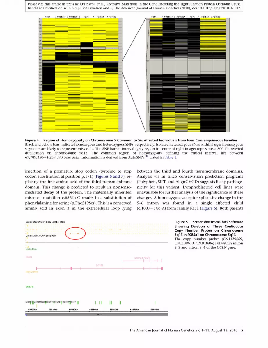
Figure 4. Region of Homozygosity on Chromosome 5 Common to Six Affected Individuals from Four Consanguineous FamiliesBlack and yellow bars indicate homozygous and heterozygous SNPs, respectively. Isolated heterozygous SNPs within larger homozygoussegments are likely to represent miss-calls. The SNP-barren interval (gray region in center of right image) represents a 500 kb invertedduplication on chromosome 5q13. The common region of homozygosity defining the critical interval lies between67,789,350-74,259,390 base pairs. Information is derived from AutoSNPa.36 Listed in Table 1.
Please cite this article in press as: O’Driscoll et al., Recessive Mutations in the Gene Encoding the Tight Junction Protein Occludin CauseBand-like Calcification with Simplified Gyration and..., The American Journal of Human Genetics (2010), doi:10.1016/j.ajhg.2010.07.012
insertion of a premature stop codon (tyrosine to stop
codon substitution at position p.171) (Figures 6 and 7), re-
placing the first amino acid of the third transmembrane
domain. This change is predicted to result in nonsense-
mediated decay of the protein. The maternally inherited
missense mutation c.656T>C results in a substitution of
phenylalanine for serine (p.Phe219Ser). This is a conserved
amino acid in exon 3 in the extracellular loop lying
The
between the third and fourth transmembrane domains.
Analysis via in silico conservation prediction programs
(Polyphen, SIFT, and AlignGVGD) suggests likely pathoge-
nicity for this variant. Lymphoblastoid cell lines were
unavailable for further analysis of the significance of these
changes. A homozygous acceptor splice site change in the
5–6 intron was found in a single affected child
(c.1037þ5G>A) from family F351 (Figure 6). Both parents
Figure 5. Screenshot fromChASSoftwareShowing Deletion of Three ContiguousCopy Number Probes on Chromosome5q13 in F085a1 on Chromosome 5q13The copy number probes (CN1139669,CN1139670, CN303606) fall within intron2–3 and intron 3–4 of the OCLN gene.
American Journal of Human Genetics 87, 1–11, August 13, 2010 5

Figure 6. Demonstration of Four OCLN VariantsTop: Sequence chromatograms of affected individuals. Middle: Sequence chromatograms of control sample with wild-type allele.Bottom: Sequence chromatograms of parent with heterozygous mutation. Mutations are highlighted (arrow).(A) Chromatogram of c.512 dupA in F312.(B) Chromatogram of c.656T>C in F312.(C) Chromatogram of splice site variant in F351.(D) Chromatogram of 22 base pair deletion in F375.
Please cite this article in press as: O’Driscoll et al., Recessive Mutations in the Gene Encoding the Tight Junction Protein Occludin CauseBand-like Calcification with Simplified Gyration and..., The American Journal of Human Genetics (2010), doi:10.1016/j.ajhg.2010.07.012
were heterozygous and an unaffected sibling was homo-
zygous for the wild-type allele at the same nucleotide.
By means of analysis software (Alamut, MaxEntScan,
Drosophila BDG, Hbond, SROOGLE), this change was pre-
dicted to result in a severe splicing mutation leading to
exon skipping. None of the above mutations were identi-
fied in 220 control chromosomes.
Occludin is highly conserved in vertebrates and contains
two known functional domains, a Marvel domain and an
occludin/ELL domain. A multiple alignment of occludin
orthologs highlighting the mutations identified in the
affected individuals and the position of theMarvel domain
and all four transmembrane domains is shown in Figure S1
(generated by MUSCLE version 3.638 on NCBI). In four
families (F085, F275, F375, and F386), intragenic deletion
of exon 3 and/or exon 4 is predicted to result in removal
of some or all of the Marvel domain in the absence of
nonsense-mediated decay. Both mutations found in
affected individuals in F312 are located within the Marvel
domain and alter conserved amino acids.
We have shown that mutations in the OCLN gene cause
BLC-PMG, a severe neurodevelopmental disorder demon-
strating highly characteristic clinical and neuroradiolog-
ical features. The OCLN gene in humans maps to two
regions on 5q13.2, a full-length gene containing nine
exons and a pseudogene (LOC647859) approximately
6 The American Journal of Human Genetics 87, 1–11, August 13, 201
1.5 Mb away containing a copy of exons 6–9 within a
500 kb inverted duplication (Figure 4).
Full-length occludin is a ~65 kDa protein with four
transmembrane (TM) domains and two cytoplasmic
domains.32 Marvel is a four transmembrane (TM) domain
that has functional importance in proteins involved in
membrane apposition (reviewed by Sanchez-Pulido et al.,
2002).39 The tight junction Marvel protein family includes
two other proteins, tricellulin and MarvelD3,40,41 which
may contribute to noncerebral TJ function in patients
with BLC-PMG. All three proteins have similar but
nonredundant roles in epithelial barrier formation and
maintenance.40,41 The Marvel domain itself is highly
conserved (Figure S1), being present in related Caenorhab-
ditis elegans and Drosophila melanogaster proteins (no
OCLN ortholog has been identified in these organisms).
Several splicing variants of occludin have been identified
including forms lacking the Marvel domain encoded by
exons 3 and 4.42–46 Proteins lacking the Marvel domain
are located in the cytoplasm rather than the cell membrane
and are therefore not incorporated into TJs.44,47,48 Five of
six families reported here have deletions or mutations
affecting the Marvel domain (Table 1 and Figure S1), and
therefore we propose that these mutations will result in a
protein product that fails to locate to the cell membrane
and hence to TJs.
0

Figure 7. Occludin In Situ Hybridization Expression in Adult Mouse Brain(A–C) Sagittal images from C57BL/6J mice at 55 days. Occludin is expressed throughout the brain, particularly in the cerebral cortex andcerebellum (B). Magnified images (boxed) of cerebral cortex (A) and cerebellum (C) showing marked occludin expression in layers 2 and3 of the cerebral cortex and in the Purkinje cell layer in the cerebellum (arrows). Images from the Allen Mouse Brain Atlas, Section ID79394328_12.(E–H)Whole-brain sagittal images (E, G) and highermagnification of sections of cerebral cortex (F, H). Claudin 5 expression appears to beconcentrated in cerebral blood vessels (E, F). Tubb2b is expressed throughout the brain and concentrated in the cerebral cortex andcerebellum (G, H) in a similar distribution to occludin (B, A). Images from the Allen Mouse Brain Atlas, Section IDs 79360268_14 (E,F) and 69854163_14 (G, H).
Please cite this article in press as: O’Driscoll et al., Recessive Mutations in the Gene Encoding the Tight Junction Protein Occludin CauseBand-like Calcification with Simplified Gyration and..., The American Journal of Human Genetics (2010), doi:10.1016/j.ajhg.2010.07.012
TJs are multiprotein cell-cell adhesion complexes in
epithelia and endothelia that form a barrier to free paracel-
lular transport between tissue compartments.49 Despite
a wealth of research on tight junctions in different organs,
the function of occludin has proven difficult to define. In
vitro studies have shown that functional epithelial TJs
can form in the absence of occludin. Other TJ proteins,
primarily the claudins, are the major structural compo-
nents of TJs to which occludin later becomes incorpo-
rated.34,48,50,51 Occludin is thought to be involved in the
regulation of TJ function or intracellular signaling rather
than TJ assembly per se.52 The phenotype of the Ocln
knockout mouse34 demonstrates postnatal growth retarda-
tion, chronic gastritis, thinning of compact bone,
abnormal salivary striatal duct, and atrophy of the seminif-
erous ducts of the testes with sparing of the Sertoli cells.
Examination showed no evidence of a systemic abnor-
mality of calcium metabolism on blood and urine testing,
and tight junction morphology was unaffected in intes-
tinal, kidney, and liver epithelia. Of note, the brain of
occludin-deficient mice showed progressive cerebellar
and basal ganglia calcification, particularly along the walls
of capillaries and postcapillary venules. Measurement of
BBB permeability was not reported. These mice were viable
at birth and, although homozygous mutant males were
unable to mate and females unable to suckle, they showed
The
none of the severe neurodevelopmental aspects (seizures,
developmental arrest) of the human phenotype we report
here. Conversely, in the patients we describe there was no
evidence of the extracerebral abnormalities seen in the
Ocln knockout mouse. Occludin expression in wild-type
adult mouse is prominent in layers 2/3 of the cerebral
cortex and in Purkinje cells of the cerebellum (Figure 7),
reflecting the pattern of brain calcification in patients
with BLC-PMG (Figures 1 and 2). This compares to claudin
5, the major claudin subtype in cerebral TJs, which, unlike
occludin, appears to be confined to distribution along
blood vessels (Figure 7).
Occludin is ubiquitously expressed in epithelial TJs
throughout the body. In the developing human brain,
occludin is expressed early in neuroepithelium, but expres-
sion is lost during the transition from epithelia to neuronal
cell types.53,54 Occludin, along with other components of
the TJ such as claudin-5 and JAM-1, are highly expressed
in neurovascular TJs during angiogenesis and in mature
cerebral (but not noncerebral) endothelia that form the
BBB.33,55 Prior to this report a cerebral phenotype has
been reported in knockout models of only two TJ proteins
(claudin-5 and claudin-11),56,57 neither of which includes
cerebral malformation. The mouse model of claudin-5
has a size-specific defect in TJ permeability in the BBB
but shows no other alterations in structure or function,
American Journal of Human Genetics 87, 1–11, August 13, 2010 7

Please cite this article in press as: O’Driscoll et al., Recessive Mutations in the Gene Encoding the Tight Junction Protein Occludin CauseBand-like Calcification with Simplified Gyration and..., The American Journal of Human Genetics (2010), doi:10.1016/j.ajhg.2010.07.012
leaving the early postnatal mortality of these mice unex-
plained. Occludin has a putative role in the regulation of
paracellular transport. Paracellular transport of macromol-
ecules across the BBB is blocked by TJs, whereas transmi-
gration of leucocytes via TJs is permitted (reviewed in
Abbott et al., 2010).58 Leucocytes have been shown to
cause neuronal injury in response to inflammatory
stimuli59 and therefore a BBB lacking functional occludin
may increase the vulnerability of the brain to immune
cell-mediated tissue damage. The window of PMG develop-
ment suggests that such an insult occurs by, or during,
a critical interval in fetal development.
The calcification in BLC-PMG is found adjacent to endo-
thelium comprising small blood vessels (Figure 3) in a
pattern highly similar to that reported in Ocln null mice
(Figure 7 in Saitou et al., 2000).34 Pericytes are multipoten-
tial cells present in almost all tissues and organ systems,
selectively associated with the microvasculature. Together
with astrocytes they encapsulate cerebral microvessels
and are separated from them by a basal lamina. Pericytes
are reported to have roles in both normal fetal cerebral
angiogenesis60 and ectopic vascular calcification.61 Peri-
cytes have the ability to transform into osteoblasts and
are thus an obvious candidate cell type for aberrant
mineral deposition in these patients. The cortical malfor-
mations in BLC-PMG may reflect disordered signaling
between cells of the neurovascular unit as well as increased
permeability of the BBB.
Mutations in a number of genes encoding components
or regulators of the microtubule network have been
reported in patients with PMG and pachygyria.19,62–65
The cerebral distribution of the expression of one of these
proteins, Tubb2b, is similar to occludin (Figure 7), but,
unlike PMG associated with mutations in TUBB2B64
(MIM 610031), no neuronal overmigration through the
pial membrane was seen in brain tissue from a patient
with BLC-PMG. Few studies have examined the relation-
ship between microtubules and tight junction complexes.
Elements of the microtubular complex mediate assembly
and disassembly of the apical junctional complex (adhe-
rens and tight junctions) in epithelia,66,67 but this process
has not been described in endothelia. Targeting and main-
tenance of occludin at the cell membrane is dependent on
both microtubules and the actin cytoskeleton.68 More
recently, involvement of occludin in organization and
orientation of themicrotubular network and actin cytoske-
letion during cell migration has been demonstrated in
epithelia.69 This process has not been explored during
neuronal migration.
The neurodevelopmental phenotype in human we
report, and to a lesser extent that seen in the Ocln null
mouse, reflects the functional importance of occludin in
the developing brain, and its redundancy in other tissues.
The BLC-PMG phenotype may be mediated by more than
one cell type involved in the neurovascular unit and impli-
cates TJs and BBB dysfunction in malformations of cortical
development. Finally, we note that several of the patients
8 The American Journal of Human Genetics 87, 1–11, August 13, 201
included in this report were originally referred to us as
possible cases of Aicardi-Goutieres syndrome, after the
identification of intracranial calcification on brain
imaging. This report demonstrates the utility of the classi-
fication of neurological diseases on the basis of the pattern
and distribution of intracranial calcification, taken in
clinical context and association with other neuroimaging
features, as a tool to gene discovery.
Supplemental Data
Supplemental Data include one figure and three tables and can be
found with this article online at http://www.cell.com/AJHG/.
Acknowledgments
We sincerely thank the families for their involvement in this work.
Y.J.C. acknowledges the Manchester NIHR Biomedical Research
Centre and the Manchester Academic Health Sciences Centre.
M.C.O’D. is funded by the Manchester NIHR Biomedical Research
Centre and the Wellcome Trust. We are grateful to Dr. E. Hellen in
the Manchester National Genetics Reference Laboratory at the
University of Manchester and Dr. Diana Baralle at the University
of Southampton for their assistance with mutation analysis.
Received: April 18, 2010
Revised: June 29, 2010
Accepted: July 8, 2010
Published online: August 19, 2010
Web Resources
The URLs for data presented herein are as follows:
Alamut, http://www.interactive-biosoftware.com/alamut.html
AlignGVGD, http://agvgd.iarc.fr/agvgd_input.php
Allen Mouse Brain Atlas, http://mouse.brain-map.org/
AutoSNPa, http://dna.leeds.ac.uk/autosnpa
Chromas software, http://www.technelysium.com.au/chromas.
html
Chromosome Analysis Suite, http://www.affymetrix.com/
Drosophila BDG, http://www.fruitfly.org/seq_tools/splice.html
Hbond, http://www.uni-duesseldorf.de/rna/html/hbond_score.php
Human Genome Variation Society, http://www.hgvs.org/
mutnomen/
MaxEntScan, http://genes.mit.edu/burgelab/maxent/Xmaxentscan_
scoreseq.html
National Center for Biotechnology Information, http://www.ncbi.
nlm.nih.gov/
Online Mendelian Inheritance in Man (OMIM), http://www.ncbi.
nlm.nih.gov/Omim/
PolyPhen, http://genetics.bwh.harvard.edu/pph/
Primer3Plus, http://www.bioinformatics.nl/cgi-bin/primer3plus/
primer3plus.cgi
SIFT, http://blocks.fhcrc.org/sift/SIFT.html
SROOGLE, http://sroogle.tau.ac.il/
University of California, Santa Cruz (UCSC) Genome Browser,
http://www.genome.ucsc.edu
References
1. Briggs, T.A., Wolf, N.I., D’Arrigo, S., Ebinger, F., Harting, I.,
Dobyns, W.B., Livingston, J.H., Rice, G.I., Crooks, D.,
0

Please cite this article in press as: O’Driscoll et al., Recessive Mutations in the Gene Encoding the Tight Junction Protein Occludin CauseBand-like Calcification with Simplified Gyration and..., The American Journal of Human Genetics (2010), doi:10.1016/j.ajhg.2010.07.012
Rowland-Hill, C.A., et al. (2008). Band-like intracranial calcifi-
cation with simplified gyration and polymicrogyria: A distinct
‘‘pseudo-TORCH’’ phenotype. Am. J. Med. Genet. A. 146A,
3173–3180.
2. Abdel-Salam, G.M., Zaki, M.S., Saleem, S.N., and Gaber, K.R.
(2008). Microcephaly, malformation of brain development
and intracranial calcification in sibs: Pseudo-TORCH or
a new syndrome. Am. J. Med. Genet. A. 146A, 2929–2936.
3. Abdel-Salam, G.M., and Zaki, M.S. (2009). Band-like intracra-
nial calcification (BIC), microcephaly and malformation of
brain development: A distinctive form of congenital infection
like syndromes. Am. J. Med. Genet. A. 149A, 1565–1568.
4. Boppana, S.B., Fowler, K.B., Vaid, Y., Hedlund, G., Stagno, S.,
Britt, W.J., and Pass, R.F. (1997). Neuroradiographic findings
in the newborn period and long-term outcome in children
with symptomatic congenital cytomegalovirus infection.
Pediatrics 99, 409–414.
5. Noyola, D.E., Demmler, G.J., Nelson, C.T., Griesser, C.,
Williamson, W.D., Atkins, J.T., Rozelle, J., Turcich, M.,
Llorente, A.M., Sellers-Vinson, S., et al; Houston Congenital
CMV Longitudinal Study Group. (2001). Early predictors of
neurodevelopmental outcome in symptomatic congenital
cytomegalovirus infection. J. Pediatr. 138, 325–331.
6. Jansen, A., and Andermann, E. (2005). Genetics of the polymi-
crogyria syndromes. J. Med. Genet. 42, 369–378.
7. Dobyns, W.B., Mirzaa, G., Christian, S.L., Petras, K., Roseberry,
J., Clark, G.D., Curry, C.J., McDonald-McGinn, D., Medne, L.,
Zackai, E., et al. (2008). Consistent chromosome abnormalities
identify novel polymicrogyria loci in 1p36.3, 2p16.1-p23.1,
4q21.21-q22.1, 6q26-q27, and 21q2. Am. J. Med. Genet. A.
146A, 1637–1654.
8. Robin, N.H., Taylor, C.J., McDonald-McGinn, D.M., Zackai,
E.H., Bingham, P., Collins, K.J., Earl, D., Gill, D., Granata, T.,
Guerrini, R., et al. (2006). Polymicrogyria and deletion
22q11.2 syndrome: Window to the etiology of a common
cortical malformation. Am. J. Med. Genet. A. 140, 2416–2425.
9. Roll, P., Rudolf, G., Pereira, S., Royer, B., Scheffer, I.E., Massa-
crier, A., Valenti, M.P., Roeckel-Trevisiol, N., Jamali, S., Beclin,
C., et al. (2006). SRPX2 mutations in disorders of language
cortex and cognition. Hum. Mol. Genet. 15, 1195–1207.
10. Baala, L., Briault, S., Etchevers, H.C., Laumonnier, F., Natiq, A.,
Amiel, J., Boddaert, N., Picard, C., Sbiti, A., Asermouh, A., et al.
(2007). Homozygous silencing of T-box transcription factor
EOMES leads to microcephaly with polymicrogyria and
corpus callosum agenesis. Nat. Genet. 39, 454–456.
11. Passemard, S., Titomanlio, L., Elmaleh, M., Afenjar, A., Ales-
sandri, J.L., Andria, G., de Villemeur, T.B., Boespflug-Tanguy,
O., Burglen, L., Del Giudice, E., et al. (2009). Expanding the
clinical and neuroradiologic phenotype of primary micro-
cephaly due to ASPM mutations. Neurology 73, 962–969.
12. Valente, E.M., Marsh, S.E., Castori, M., Dixon-Salazar, T.,
Bertini, E., Al-Gazali, L., Messer, J., Barbot, C., Woods, C.G.,
Boltshauser, E., et al. (2005). Distinguishing the four genetic
causes of Jouberts syndrome-related disorders. Ann. Neurol.
57, 513–519.
13. Mitchell, T.N., Free, S.L., Williamson, K.A., Stevens, J.M.,
Churchill, A.J., Hanson, I.M., Shorvon, S.D., Moore, A.T.,
van Heyningen, V., and Sisodiya, S.M. (2003). Polymicrogyria
and absence of pineal gland due to PAX6mutation. Ann. Neu-
rol. 53, 658–663.
14. Brooks, A.S., Bertoli-Avella, A.M., Burzynski, G.M., Breedveld,
G.J., Osinga, J., Boven, L.G., Hurst, J.A., Mancini, G.M.,
The
Lequin, M.H., de Coo, R.F., et al. (2005). Homozygous
nonsense mutations in KIAA1279 are associated with malfor-
mations of the central and enteric nervous systems. Am. J.
Hum. Genet. 77, 120–126.
15. Keren, B., Suzuki, O.T., Gerard-Blanluet, M., Bremond-Gignac,
D., Elmaleh,M., Titomanlio, L., Delezoide, A.L., Passos-Bueno,
M.R., and Verloes, A. (2007). CNS malformations in Knobloch
syndrome with splice mutation in COL18A1 gene. Am. J.
Med. Genet. A. 143A, 1514–1518.
16. Graham, J.M. Jr., Hennekam, R., Dobyns,W.B., Roeder, E., and
Busch, D. (2004). MICRO syndrome: An entity distinct from
COFS syndrome. Am. J. Med. Genet. A. 128A, 235–245.
17. Hevner, R.F. (2005). The cerebral cortex malformation in
thanatophoric dysplasia: Neuropathology and pathogenesis.
Acta Neuropathol. 110, 208–221.
18. Geerdink, N., Rotteveel, J.J., Lammens, M., Sistermans, E.A.,
Heikens, G.T., Gabreels, F.J., Mullaart, R.A., and Hamel, B.C.
(2002). MECP2 mutation in a boy with severe neonatal
encephalopathy: Clinical, neuropathological and molecular
findings. Neuropediatrics 33, 33–36.
19. Abdollahi, M.R., Morrison, E., Sirey, T., Molnar, Z., Hayward,
B.E., Carr, I.M., Springell, K., Woods, C.G., Ahmed, M.,
Hattingh, L., et al. (2009). Mutation of the variant alpha-
tubulin TUBA8 results in polymicrogyria with optic nerve
hypoplasia. Am. J. Hum. Genet. 85, 737–744.
20. Dvorak, K., and Feit, J. (1977). Migration of neuroblasts
through partial necrosis of the cerebral cortex in newborn
rats-contribution to the problems of morphological develop-
ment and developmental period of cerebral microgyria. Histo-
logical and autoradiographical study. Acta Neuropathol. 38,
203–212.
21. Ferrer, I. (1984). A Golgi analysis of unlayered polymicrogyria.
Acta Neuropathol. 65, 69–76.
22. Ferrer, I., and Catala, I. (1991). Unlayered polymicrogyria:
Structural and developmental aspects. Anat. Embryol. (Berl.)
184, 517–528.
23. Suzuki, M., and Choi, B.H. (1991). Repair and reconstruction
of the cortical plate following closed cryogenic injury to the
neonatal rat cerebrum. Acta Neuropathol. 82, 93–101.
24. Barth, P.G., and van der Harten, J.J. (1985). Parabiotic twin
syndrome with topical isocortical disruption and gastroschi-
sis. Acta Neuropathol. 67, 345–349.
25. Graff-Radford, N.R., Bosch, E.P., Stears, J.C., and Tranel, D.
(1986). Developmental Foix-Chavany-Marie syndrome in
identical twins. Ann. Neurol. 20, 632–635.
26. Van Bogaert, P., Donner, C., David, P., Rodesch, F., Avni, E.F.,
and Szliwowski, H.B. (1996). Congenital bilateral perisylvian
syndrome in a monozygotic twin with intra-uterine death of
the co-twin. Dev. Med. Child Neurol. 38, 166–170.
27. Richman, D.P., Stewart, R.M., and Caviness, V.S. Jr. (1974).
Cerebral microgyria in a 27-week fetus: An architectonic
and topographic analysis. J. Neuropathol. Exp. Neurol. 33,
374–384.
28. Williams, R.S., Ferrante, R.J., and Caviness, V.S. Jr. (1976). The
cellular pathology of microgyria. A Golgi analysis. Acta
Neuropathol. 36, 269–283.
29. Reutens, D.C., Berkovic, S.F., Kalnins, R.M., McKelvie, P.,
Saling, M.M., and Fabinyi, G.C. (1993). Localised neuronal
migration disorder and intractable epilepsy: A prenatal
vascular aetiology. J. Neurol. Neurosurg. Psychiatry 56,
314–316.
American Journal of Human Genetics 87, 1–11, August 13, 2010 9

Please cite this article in press as: O’Driscoll et al., Recessive Mutations in the Gene Encoding the Tight Junction Protein Occludin CauseBand-like Calcification with Simplified Gyration and..., The American Journal of Human Genetics (2010), doi:10.1016/j.ajhg.2010.07.012
30. Raybaud, C., and Di Rocco, C. (2007). Brain malformation in
syndromic craniosynostoses, a primary disorder of white
matter: A review. Childs Nerv. Syst. 23, 1379–1388.
31. McBride, M.C., and Kemper, T.L. (1982). Pathogenesis of
four-layered microgyric cortex in man. Acta Neuropathol.
57, 93–98.
32. Furuse, M., Hirase, T., Itoh, M., Nagafuchi, A., Yonemura, S.,
Tsukita, S., and Tsukita, S. (1993). Occludin: A novel integral
membrane protein localizing at tight junctions. J. Cell Biol.
123, 1777–1788.
33. Virgintino, D., Errede, M., Robertson, D., Capobianco, C.,
Girolamo, F., Vimercati, A., Bertossi, M., and Roncali, L.
(2004). Immunolocalization of tight junction proteins in the
adult and developing human brain. Histochem. Cell Biol.
122, 51–59.
34. Saitou, M., Furuse, M., Sasaki, H., Schulzke, J.D., Fromm, M.,
Takano, H., Noda, T., and Tsukita, S. (2000). Complex pheno-
type of mice lacking occludin, a component of tight junction
strands. Mol. Biol. Cell 11, 4131–4142.
35. Hirase, T., Staddon, J.M., Saitou, M., Ando-Akatsuka, Y., Itoh,
M., Furuse, M., Fujimoto, K., Tsukita, S., and Rubin, L.L.
(1997). Occludin as a possible determinant of tight junction
permeability in endothelial cells. J. Cell Sci. 110, 1603–1613.
36. Shimizu, F., Sano, Y., Maeda, T., Abe, M.A., Nakayama, H.,
Takahashi, R., Ueda, M., Ohtsuki, S., Terasaki, T., Obinata,
M., and Kanda, T. (2008). Peripheral nerve pericytes origi-
nating from the blood-nerve barrier expresses tight junctional
molecules and transporters as barrier-forming cells. J. Cell.
Physiol. 217, 388–399.
37. Carr, I.M., Flintoff, K.J., Taylor, G.R., Markham, A.F., and
Bonthron, D.T. (2006). Interactive visual analysis of SNP
data for rapid autozygosity mapping in consanguineous
families. Hum. Mutat. 27, 1041–1046.
38. Edgar, R.C. (2004). MUSCLE: Multiple sequence alignment
with high accuracy and high throughput. Nucleic Acids Res.
32, 1792–1797.
39. Sanchez-Pulido, L., Martın-Belmonte, F., Valencia, A., and
Alonso, M.A. (2002). MARVEL: A conserved domain involved
in membrane apposition events. Trends Biochem. Sci. 27,
599–601.
40. Steed, E., Rodrigues, N.T., Balda, M.S., and Matter, K. (2009).
Identification of MarvelD3 as a tight junction-associated
transmembrane protein of the occludin family. BMC Cell
Biol. 10, 95.
41. Raleigh, D.R., Marchiando, A.M., Zhang, Y., Shen, L., Sasaki,
H., Wang, Y., Long, M., and Turner, J.R. (2010). Tight junc-
tion-associated MARVEL proteins marveld3, tricellulin, and
occludin have distinct but overlapping functions. Mol. Biol.
Cell 21, 1200–1213.
42. Ghassemifar, M.R., Sheth, B., Papenbrock, T., Leese, H.J.,
Houghton, F.D., and Fleming, T.P. (2002). Occludin TM4(-):
An isoform of the tight junction protein present in primates
lacking the fourth transmembrane domain. J. Cell Sci. 115,
3171–3180.
43. Gu, J.M., Lim, S.O., Park, Y.M., and Jung, G. (2008). A novel
splice variant of occludin deleted in exon 9 and its role in
cell apoptosis and invasion. FEBS J. 275, 3145–3156.
44. Kohaar, I., Ploss, A., Korol, E., Mu, K., Schoggins, J.W., O’Brien,
T.R., Rice, C.M., and Prokunina-Olsson, L. (2010). Splicing
diversity of the human OCLN gene and its biological signifi-
cance for hepatitis C virus entry. J. Virol. 84, 6987–6994.
10 The American Journal of Human Genetics 87, 1–11, August 13, 20
45. Mankertz, J., Waller, J.S., Hillenbrand, B., Tavalali, S., Florian,
P., Schoneberg, T., Fromm,M., and Schulzke, J.D. (2002). Gene
expression of the tight junction protein occludin includes
differential splicing and alternative promoter usage. Biochem.
Biophys. Res. Commun. 298, 657–666.
46. Muresan, Z., Paul, D.L., and Goodenough, D.A. (2000).
Occludin 1B, a variant of the tight junction protein occludin.
Mol. Biol. Cell 11, 627–634.
47. Chen, Y., Merzdorf, C., Paul, D.L., and Goodenough, D.A.
(1997). COOH terminus of occludin is required for tight junc-
tion barrier function in early Xenopus embryos. J. Cell Biol.
138, 891–899.
48. Saitou,M., Fujimoto, K., Doi, Y., Itoh, M., Fujimoto, T., Furuse,
M., Takano, H., Noda, T., and Tsukita, S. (1998). Occludin-
deficient embryonic stem cells can differentiate into polarized
epithelial cells bearing tight junctions. J. Cell Biol. 141,
397–408.
49. Forster, C. (2008). Tight junctions and the modulation of
barrier function in disease. Histochem. Cell Biol. 130, 55–70.
50. Furuse, M., Sasaki, H., Fujimoto, K., and Tsukita, S. (1998).
A single gene product, claudin-1 or -2, reconstitutes tight
junction strands and recruits occludin in fibroblasts. J. Cell
Biol. 143, 391–401.
51. Balda, M.S., Whitney, J.A., Flores, C., Gonzalez, S., Cereijido,
M., and Matter, K. (1996). Functional dissociation of paracel-
lular permeability and transepithelial electrical resistance
and disruption of the apical-basolateral intramembrane diffu-
sion barrier by expression of a mutant tight junction
membrane protein. J. Cell Biol. 134, 1031–1049.
52. Rao, R. (2009). Occludin phosphorylation in regulation of
epithelial tight junctions. Ann. N Y Acad. Sci. 1165, 62–68.
53. Aaku-Saraste, E., Hellwig, A., and Huttner, W.B. (1996). Loss of
occludin and functional tight junctions, but not ZO-1, during
neural tube closure—remodeling of the neuroepithelium prior
to neurogenesis. Dev. Biol. 180, 664–679.
54. Hay, E.D. (1995). An overview of epithelio-mesenchymal
transformation. Acta Anat. (Basel) 154, 8–20.
55. Ballabh, P., Hu, F., Kumarasiri, M., Braun, A., and Nedergaard,
M. (2005). Development of tight junction molecules in blood
vessels of germinal matrix, cerebral cortex, and white matter.
Pediatr. Res. 58, 791–798.
56. Gow, A., Southwood, C.M., Li, J.S., Pariali, M., Riordan, G.P.,
Brodie, S.E., Danias, J., Bronstein, J.M., Kachar, B., and
Lazzarini, R.A. (1999). CNS myelin and sertoli cell tight
junction strands are absent in Osp/claudin-11 null mice.
Cell 99, 649–659.
57. Nitta, T., Hata, M., Gotoh, S., Seo, Y., Sasaki, H., Hashimoto,
N., Furuse, M., and Tsukita, S. (2003). Size-selective loosening
of the blood-brain barrier in claudin-5-deficient mice. J. Cell
Biol. 161, 653–660.
58. Abbott, N.J., Patabendige, A.A., Dolman, D.E., Yusof, S.R., and
Begley, D.J. (2010). Structure and function of the blood-brain
barrier. Neurobiol. Dis. 37, 13–25.
59. Scholz, M., Cinatl, J., Schadel-Hopfner, M., and Windolf, J.
(2007). Neutrophils and the blood-brain barrier dysfunction
after trauma. Med. Res. Rev. 27, 401–416.
60. Virgintino, D., Girolamo, F., Errede, M., Capobianco, C.,
Robertson, D., Stallcup, W.B., Perris, R., and Roncali, L.
(2007). An intimate interplay between precocious, migrating
pericytes and endothelial cells governs human fetal brain
angiogenesis. Angiogenesis 10, 35–45.
10

Please cite this article in press as: O’Driscoll et al., Recessive Mutations in the Gene Encoding the Tight Junction Protein Occludin CauseBand-like Calcification with Simplified Gyration and..., The American Journal of Human Genetics (2010), doi:10.1016/j.ajhg.2010.07.012
61. Collett, G.D., and Canfield, A.E. (2005). Angiogenesis and
pericytes in the initiation of ectopic calcification. Circ. Res.
96, 930–938.
62. des Portes, V., Pinard, J.M., Billuart, P., Vinet, M.C., Koulakoff,
A., Carrie, A., Gelot, A., Dupuis, E., Motte, J., Berwald-Netter,
Y., et al. (1998). A novel CNS gene required for neuronal
migration and involved in X-linked subcortical laminar heter-
otopia and lissencephaly syndrome. Cell 92, 51–61.
63. Gleeson, J.G., Allen, K.M., Fox, J.W., Lamperti, E.D., Berkovic,
S., Scheffer, I., Cooper, E.C., Dobyns, W.B., Minnerath, S.R.,
Ross, M.E., and Walsh, C.A. (1998). Doublecortin, a brain-
specific gene mutated in human X-linked lissencephaly and
double cortex syndrome, encodes a putative signaling protein.
Cell 92, 63–72.
64. Jaglin, X.H., Poirier, K., Saillour, Y., Buhler, E., Tian, G.,
Bahi-Buisson, N., Fallet-Bianco, C., Phan-Dinh-Tuy, F., Kong,
X.P., Bomont, P., et al. (2009). Mutations in the beta-tubulin
gene TUBB2B result in asymmetrical polymicrogyria. Nat.
Genet. 41, 746–752.
65. Keays, D.A., Tian, G., Poirier, K., Huang, G.J., Siebold, C.,
Cleak, J., Oliver, P.L., Fray, M., Harvey, R.J., Molnar, Z., et al.
The A
(2007). Mutations in alpha-tubulin cause abnormal neuronal
migration in mice and lissencephaly in humans. Cell 128,
45–57.
66. Ivanov, A.I., McCall, I.C., Babbin, B., Samarin, S.N., Nusrat, A.,
and Parkos, C.A. (2006). Microtubules regulate disassembly of
epithelial apical junctions. BMC Cell Biol. 7, 12.
67. Shultz, T., Shmuel, M., Hyman, T., and Altschuler, Y. (2008).
Beta-tubulin cofactor D and ARL2 take part in apical junc-
tional complex disassembly and abrogate epithelial structure.
FASEB J. 22, 168–182.
68. Subramanian, V.S., Marchant, J.S., Ye, D., Ma, T.Y., and Said,
H.M. (2007). Tight junction targeting and intracellular
trafficking of occludin in polarized epithelial cells. Am. J.
Physiol. Cell Physiol. 293, C1717–C1726.
69. Du, D., Xu, F., Yu, L., Zhang, C., Lu, X., Yuan, H., Huang, Q.,
Zhang, F., Bao, H., Jia, L., et al. (2010). The tight junction
protein, occludin, regulates the directional migration of
epithelial cells. Dev. Cell 18, 52–63.
70. Shaffer, L.G., Slovak, M.L., and Campbell, L.J. (2009). An
International System for Human Cytogenetic Nomenclature
(Basel: S Karger).
merican Journal of Human Genetics 87, 1–11, August 13, 2010 11