Photosynthesis and calcification in the articulated coralline ...

●
bi●
●
MLvP
pta(crt●
acIpwnapifsoec●
tplg
SA
SCAG
Ue
0d
Opacification of Hydrophilic Acrylic Intraocular LensAttributable to Calcification: Investigation
on Mechanism
SOTIRIOS P. GARTAGANIS, DIMITRA G. KANELLOPOULOU, EPHIGENIA K. MELA, VASSILIKI S. PANTELI,
AND PETROS G. KOUTSOUKOSn1r
OrtIgritOlaeplttseIoh
cmcceHfach
W
PURPOSE: To identify the nature and to investigate theiochemical mechanisms leading to late opacification ofmplanted hydrophilic acrylic intraocular lenses (IOLs). DESIGN: Retrospective laboratory investigation. METHODS: SETTING: Department of Ophthalmology,edical School, Department of Chemical Engineering,aboratory of Inorganic and Analytical Chemistry, Uni-ersity of Patras and FORTH-ICEHT, Greece. STUDY
OPULATION: Thirty IOLs were explanted one to 12 yearsostimplantation attributable to gradual opacification ofhe lens material. OBSERVATION PROCEDURES: Materialsnalysis was done using scanning electron microscopySEM) equipped with a microanalysis probe (EDS),onfocal microscopy, x-ray diffraction (XRD), and Fou-ier transform infrared (FTIR) for the identification ofhe substances involved in the opacified lenses. RESULTS: SEM investigation showed plate-like as wells prismatic nanoparticle deposits of calcium phosphaterystallites on the surface and in the interior of opacifiedOLs. The plate-like deposits exhibited morphology andarticle size typical for octacalcium phosphate (OCP),hile the respective characteristics of the prismaticanocrystals were typical of hydroxyapatite (HAP). EDSnalysis confirmed the chemical composition of the de-osits. Aqueous humor analysis showed that the humors supersaturated with respect to both OCP and HAP,avoring the formation of the thermodynamically moretable HAP, while the formation and kinetic stabilizationf other transient phases is also very likely. In vitroxperiments using polyacrylic materials confirmed thelinical findings. CONCLUSIONS: Hydrophilic acrylic IOLs’ opacifica-ion may be attributed to the deposition of calciumhosphate crystallites. HAP is the predominant crystal-ine phase of these crystallites. Surface hydroxylroups of the polyacrylic materials facilitate surface
ee accompanying Editorial on page 341.ccepted for publication Apr 22, 2008.From the Department of Ophthalmology (S.P.G., E.K.M., V.S.P.),
chool of Medicine, University of Patras; and the Department ofhemical Engineering (D.G.K., P.G.K.), Laboratory of Inorganic andnalytical Chemistry, University of Patras and FORTH-ICEHT, Patras,reece.Inquiries to Sotirios P. Gartaganis, Department of Ophthalmology,
wniversity of Patras Medical School, 26500 Rion, Patras 26504, Greece;
-mail: [email protected]
© 2008 BY ELSEVIER INC. A002-9394/08/$34.00oi:10.1016/j.ajo.2008.04.032
ucleation and growth. (Am J Ophthalmol 2008;46:395– 403. © 2008 by Elsevier Inc. All rightseserved.)
VER THE PAST 20 YEARS, PHACOEMULSIFICATION
has gained in popularity and is now the mostcommon surgical procedure for removing cata-
acts. This small-incision cataract surgery has establishedhe implantation of foldable intraocular lenses (IOLs).ntraocular foldable lens are made of hydrophilic (hydro-el) or hydrophobic materials (e.g., silicone). Continuousefinements in the manufacturing of IOLs have greatlymproved the results of implant surgery while minimizinghe incidence of complications leading to IOL removal.pacification of IOL is an unusual complication that has
ed to progressive visual worsening and has been includedmong the indications leading to posterior chamber IOLxplantation. During the last few years, a lot of researchresentations, publications, and abstracts have shown aate opacification of hydrophilic acrylic IOLs attributableo calcium deposition.1– 4 Several studies have also shownhe opacification to occur intraoperatively during cataracturgery with implantation of silicone IOLs,5–7 or in thearly postoperative period after implantation of hydrogelOLs.8 –10 Dystrophic calcification has been described toccur on the surface, or within the substance of someydrophilic acrylic lenses.1,4,11
From the data presented in the literature so far, it may beoncluded that the formation of calcium phosphates is theost common cause of IOL opacification. The formation of
alcium phosphates seems to depend both on the material ofonstruction of the IOLs and on the local chemical micro-nvironment of the aqueous humor in contact with the IOL.owever, very little is known concerning the mechanism of
ormation of the mineral deposits. The present contributionims at identifying the causes and the underlying physico-hemical mechanisms of the late opacification of implantedydrophilic acrylic IOLs.
METHODS
E STUDIED RETROSPECTIVELY 29 PATIENTS (30 EYES)
ho had been referred to the Department of Ophthal-
LL RIGHTS RESERVED. 395

mGrohga
tstsfcVihfittanArcos
c(dro
●
fePCT1c
isemgs
dVteI
nxNI
●
cerswtAssCcIuihuctCm5
●
awlp
Fi
3
ology, Regional Hospital of University of Patras (Rio-reece) between October 2000 and April 2006,
equiring IOL replacement because of late-onset post-perative hydrophilic acrylic IOL opacification. All eyesad undergone uncomplicated phacoemulsification sur-ery with IOL implantation in the capsular bag, throughsmall incision.All patients underwent a standard complete eye examina-
ion, including visual acuity (VA) measurement, tonometry,lit-lamp biomicroscopy, and dilated fundus examina-ion. In addition, complete systemic examination anderologic and blood analytic examination were per-ormed. All IOLs were explanted as a result of opacifi-ation of the lens optic, associated with severe loss ofA. In general, the patients sought ophthalmic exam-
nation after cataract surgery for poor vision, when VAad dropped to values ranging from 20/200 to countingngers. Explantation was performed under local anes-hesia in all cases. The opacified implant was removedhrough a 6-mm scleral incision and it was replaced byhydrophobic implant. In all cases, the lens capsule wasoted to be adherent to the anterior IOL optic surface.
25-gauge needle was used to create a new capsulo-hexis, and then with the aid of viscoelastic material theapsular bag was inflated and the implant was dialed outf the capsular bag. Surgeries were performed by theame surgeon (S.P.G.).
The explanted lenses were characterized with physico-hemical methods including scanning electron microscopySEM) chemical microanalysis of the composition of theeposits electron dispersion spectrometry (EDS) and Fou-ier transform infrared (FTIR) spectroscopic investigationf the opacified areas of the lenses.
MORPHOLOGIC ANALYSIS EVALUATION: Twenty-fouroldable Hydroview model H60M (Bausch & Lomb, Roch-ster, New York, USA), two SC60B-OUV (MDR Inc,rescott, Arizona, USA), 2 MemoryLens (Ciba Vision Inc,hicago, Illinos, USA), and two Aqua-Sense (OII Inc,ierra Verde, Florida, USA) IOLs were explanted one to2 years postimplantation, attributable to gradual opacifi-ation of the lens material.
After removal from the eye, all the explanted IOLs weremmediately placed in empty sterile vials and were pre-erved in the freezer (4 C) for further analyses. Duringxplantation procedure, care was taken to avoid anyanipulation of the IOL optics with forceps or other
rasping instruments. All lenses were examined by theame investigator (P.G.K.).
The morphology of the surface of the lenses and theeposits formed were examined using SEM (LEO SUPRAP 35; Carl Zeiss, Oberkochen, Germany). The composi-
ion of the deposits was identified by microanalysis withnergy dispersive spectrometry (JEOL 5200 with Oxford
SIS microanalysis unit [Oxford, United Kingdom]). The rAMERICAN JOURNAL OF96
ature of the crystalline solid deposits was investigated by-ray diffraction (XRD, Philips 1830/40; Eindhoven, Theetherlands) analysis and FTIR spectrometry (Brookhaven
nstruments Corp, Holtsville, NY, USA).
AQUEOUS HUMOR ANALYSIS: Aqueous humor wasollected and chemically analyzed in five cases ofxplanted calcified IOLs. Volumes of aqueous humoranging between 20 and 50 �L were analyzed. Standardolutions were prepared for all analytes. The samplesere diluted 1:50 and the anions were analyzed using
he IONPAC AG 16 4-mm guard column with IONPACG 16 4-mm analytical column. The detection was one by
uppressed conductivity using the anion self-regeneratinguppressor ASRS Ultra II 4 mm (Dionex Corp, Sunnyvale,alifornia, USA). For the cations the cation separation
olumn IONPAC CG12A 4-mm guard column withONPAC CS12A 4-mm analytical column (Dionex) wassed. The analysis temperature was at 25 C and thenjection volume 25 �L. For the anion analysis, sodiumydroxide and for the cations, sulfuric acid solutions weresed as eluents. The chemical analysis was done by ionhromatography (Dionex D-500), atomic absorption spec-rometry (AAnalyst 300; Perkin Elmer Corp, Norwalk,onnecticut, USA), and inductively coupled plasma withass spectrometric detector (Perkin Elmer ICP-MS ELAN
000).
EXPERIMENTAL SIMULATION: In addition to the ex-mination of the explanted IOLs, in vitro experimentsere conducted, using poly-hydroxyl-ethyl-methacry-
ate (PHEMA) and poly-methyl-methacrylate (PMMA)olymers, suspended in solutions supersaturated with
IGURE 1. Slit-lamp appearance of an opacified Aqua-Sensentraocular lens (IOL). A dusty haze covers its anterior surface.
espect to calcium phosphate. The experiments were
OPHTHALMOLOGY SEPTEMBER 2008
dctasp
W
lT
Ips
tpdocHtbV
FI
FoTcl
Fe�
FeH
V
one at constant temperature 37 C, sodium chlorideoncentration 0.15 M; initial levels of calcium concen-ration were in the range of 6 to 9 � 10�4 M, pH 7.40nd at conditions of constant supersaturation in order toimulate satisfactorily the in vivo conditions of theatient’s eye.
RESULTS
E PERFORMED 30 IOL EXPLANTATIONS ATTRIBUTABLE TO
ate opacification from 29 patients: 13 men and 16 women.
IGURE 2. Slit-lamp appearance of an opacified HydroviewOL. Imprints created by forceps can be observed.
IGURE 3. Scanning electron microscopy (SEM) micrographsf the anterior external surface of opacified Aqua-Sense IOL.he surface texture is characterized by the presence of lumps
ontaining salts outgrowing from the interior of the polymericens material.
he mean age was 71.5 years (range, 58 to 80 years). The a
OPACIFICATION OF INTOL. 146, NO. 3
OLs were explanted 12 to 144 months (mean, 45 months)ostoperatively. The interval between the initial cataracturgery and the onset of the lens opacification was obscure.
Twenty patients had systemic hypertension, five hadype II diabetes and systemic hypertension, and four of theatients neither had any disease nor had used any systemicrugs. One of the patients was found to have bilateral IOLpacification. Another patient underwent phacoemulsifi-ation in both eyes, on separate week interval, withydroview IOL in-the-bag implantation. In this patient
he IOL in the right eye was removed eight years laterecause of opacification, while the left eye had correctedA of 20/20. On slit-lamp examination, the clinical
IGURE 4. SEM image of the deposits in the direction anteriorxternal surface towards the interior of the Aqua-Sense IOL 30M from the surface.
IGURE 5. SEM image shows deposits on the IOL’s anteriorxternal surface near a scratch on the anterior surface of aydroview lens.
ppearance of the IOL in the left eye did not show any
RAOCULAR LENSES 397

stp
(bifaopiowaFpt
●
A
F
acbcsfmhoc
cp
Fsc
Fos
Fcs
FIs
3
igns of surface opacification. The substances used duringhe surgery were unknown to us, as all operations wereerformed in other hospitals.Slit-lamp examination revealed a dusty haze in all IOLs
Figure 1), and in some cases two parallel clear lines onoth sides of the IOL’s optic center were recognized asmprints created by the implantation forceps during theolding process (Figure 2). There was no flare or cells in thenterior chamber. The haze prevented a clear examinationf the posterior capsule, but in 18 cases, a Nd:YAG laserosterior capsulotomy had been attempted without anymprovement of the VA, as revealed by the case historiesf the patients. Intraocular pressure (IOP) was measuredith a Goldmann applanation tonometer. All patients hadn IOP less than 21 mm Hg, with no medical therapy.undus examination by indirect ophthalmoscopy was im-ossible because of the dusty haze on the IOL, but a mild
IGURE 6. Energy-dispersive spectrometric analysis of IOLshown in Figures 4 and 5, exhibiting peaks corresponding toalcium and phosphorus.
IGURE 7. SEM image of the Hydroview lens shows plate-likectacalcium phosphate deposits formed on the anterior externalurface of calcified IOL.
o moderate uniform red reflex was produced. 5
AMERICAN JOURNAL OF98
SCANNING ELECTRON MICROSCOPY EQUIPPED WITH
MICROANALYSIS PROBE, X-RAY DIFFRACTION, AND
OURIER TRANSFORM INFRARED SPECTROMETRY: Ex-mination of opacified IOLs showed a surface textureharacterized by lumps (Figure 3). This morphology haseen reported in the literature as being characteristic ofalcified IOLs.12,13 Closer examination of the lumps on theurface of the IOLs showed that they contained salts,ormed more likely in the interior of the IOLs, approxi-ately 30 �m from the surface (Figure 4). Deposits were,owever, observed in some instances on the surface ofpacified IOLs, mainly on or near linear forceps marksaused to the IOLs accidentally (Figure 5).
Equipped with a microanalysis probe showed that the depositsonsisted of calcium and phosphorus. The molar calcium-to-hosphorus ratio determined by standardless analysis was near
IGURE 8. SEM image of the Hydroview lens shows prismaticrystals of hydroxyapatite formed on the anterior externalurface of calcified IOL.
IGURE 9. Powder x-ray diffraction (XRD) of a specimen ofOL with calcific deposits formed on the anterior externalurface.
:3; that is, the ratio corresponding to hydroxyapatite
OPHTHALMOLOGY SEPTEMBER 2008

[ImoitocecioficF
●
ctmTdtccs
wte
oruTasSMtstp�SbwlatOtsohsf
●
O
Fse
V
Ca5(PO4)3OH] (HAP) (Figure 6). In only two calcifiedOLs, platy crystals were observed on the surface. Theorphology of these crystals showed clearly that they were
ctacalcium phosphate [Ca8H2(PO4)65H2O] (OCP). OCPs a transient calcium phosphate phase hydrolyzing to thehermodynamically most stable HAP (Figure 7). This typef crystal has been formed within in vitro experiments atonstant solution supersaturation.14 The rest of the IOLsxamined showed that the deposits in the opacified areasonsisted exclusively of prismatic HAP nanocrystals form-ng on their surface. In the interior of the calcified IOLsnly the thermodynamically most stable HAP was identi-ed (Figure 8). The presence of OCP and HAP wasonfirmed with a combination of XRD (Figure 9) andTIR spectroscopic measurements (Figure 10).
AQUEOUS HUMOR ION CHROMATOGRAPHY: Thehemical composition of the aqueous humor, with respecto the inorganic components, was measured by ion chro-atography and the results obtained are summarized in theable. From these measurements it was possible to make aetailed chemical speciation analysis and to calculate thehermodynamic driving force for the formation of thealcium phosphate salts. The driving force is the change inhemical potential, ��, for going from the supersaturatedolutions to equilibrium:
�� � �s � �� [1]
here subscripts s and � denote the supersaturated solu-ions and equilibrium, respectively. For HAP and OCPquation 1 may be written as:
��HAP � �kT
9ln
(Ca2�)s5(PO4
3�)s3(OH�)s
(Ca2�)�5 (PO4
3�)�3 (OH�)
�
IGURE 10. Fourier transform infrared (FTIR) spectrumhowing the presence of calcium phosphate on the anteriorxternal surface of opacified IOL.
� kTlnSHAP [2] d
OPACIFICATION OF INTOL. 146, NO. 3
��OCP � �kT
16ln
(Ca2�)s8(PO4
3�)s6(H�)s
2
(Ca2�)�8 (PO4
3�)�6 (H�)�
2 � kTlnSOCP
[3]
In equations 2 and 3, parentheses denote the activitiesf the respective ions and S is the supersaturation withespect to the corresponding salt. Apparently, for supersat-rated solutions lnS � 0, and for undersaturated lnS � 0.he speciation calculations were done taking into accountll possible equilibria using the MINEQL� software ver-ion 4.0. (MINEQL � A Chemical Equilibrium Modelingystem; Environmental Research Software Inc, Hallowell,aine, USA).15 Considering the chemical composition of
he aqueous humor, as obtained from the analytical datahown in the Table, at 37 C and pH 7.4, the supersatura-ion of the aqueous humor with respect to calcium phos-hates was: Solution 1, lnSHAP 8,29 and lnSOCP 0,22; Solution 2, lnSHAP 8,94 and lnSOCP 0,38;
olution 3, lnSHAP 8,22 and lnSOCP �0,33. As maye seen, the normal lenses examined were supersaturatedith respect to both OCP and HAP while the opacified
ens was supersaturated only with respect to HAP. This isnticipated since opacification resulted in de-supersatura-ion of the aqueous humor with respect to the transientCP. According to the thermodynamic calculations,
herefore, the formation of the transient OCP phasehould not be precluded from the deposits found in IOLpacification events. Calculations done in other aqueousumor solutions (total of five solutions) yielded veryimilar results. The results shown in the Table are typicalor the aqueous humor of opacified lenses.
EXPERIMENTAL SIMULATION OF THE CALCIFICATION
F INTRAOCULAR LENS: A series of experiments were
TABLE. Composition of the Aqueous Humor (InorganicComponents), Measured by Ion Chromatography
(pH 7.40, 37 C)
Ion
Concentration of
Aqueous Humor
in Normal Eyes
(mM)a
Concentration of
Aqueous Humor
in Eyes With
Clear IOLs (mM)
Concentration of
Aqueous Humor
in Eyes With
Opacified IOLs
(mM)
Na� 142.0 153.4 139.3
K� 4.0 4.3 3.7
Ca2� 1.2 1.4 1.2
Mg2� 1.0 0.8 0.7
Cl� 131.0 112.0 157.8
HCO3� 20.0 24.7 5.4
H2PO4� 0.6 0.8 0.5
aAdapted in part from Riley (1983) and Berman (1991). From
Harding JJ (ed). Biochemistry of the Eye. London: Champan and
Hall; 1997.
one in order to investigate the relationship between the
RAOCULAR LENSES 399
afisasjasmsm
ustfTppc
dHsorfmH
Fas�
opt
Fe
FmFtovacFessv1asfiscbC
4
queous humor composition and the materials employedor the construction of IOLs. These experiments were donen batch reactors, kept at 37 C, with a circulating thermo-tat. Supersaturated solutions were prepared by mixingppropriate volumes of standard calcium chloride andodium hydrogen phosphate solutions. The pH was ad-usted to 7.40 by standard sodium hydroxide solutionsnd the ionic strength was adjusted to 0.15 M withodium chloride. Weighted amounts of powdered poly-eric materials were suspended in the supersaturated
olutions and the calcification process was monitored by
2 3 4 5 6 7 8 9 100,0
5.0x10⁻⁷
1.0x10⁻⁶
1.5x10⁻⁶
2.0x10⁻⁶
2.5x10⁻⁶
3.0x10⁻⁶
3.5x10⁻⁶
4.0x10⁻⁶
Rate on HAP Rate on PMMA Rate on HEMA
Rate
of P
recip
itatio
n / m
ol-1m
in-1m
-2
Supersaturation
IGURE 11. Plot of the rate of crystal growth of hydroxy-patite on various substrates, as a function of the solutionupersaturation (37 C, pH 7.40, 0.15 M NaCl; ‘ HEMA,HAP, ● PMMA). As may be seen, the dependence of the rate
f hydroxyapatite crystal growth on the driving force for therecipitation is parabolic, suggesting a surface diffusion–con-rolled process.
20 25 30 35 404500
5000
5500
6000
Inte
nsity
/ Arb
. Uni
ts
2θ0
002
211
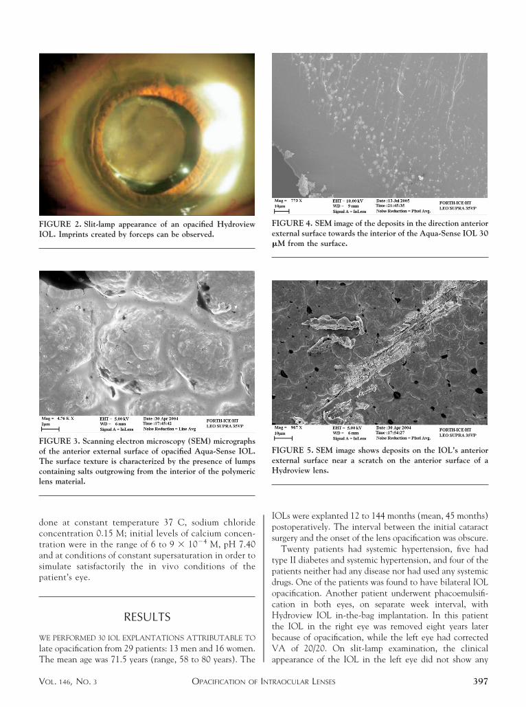
IGURE 12. Powder XRD spectrum of opacified IOL inxperimental simulation.
easurements of the hydrogen ion activity, which was r
AMERICAN JOURNAL OF00
sed to keep the supersaturation of the solutions con-tant, past the onset of precipitation.16,17 The simula-ion experiments showed that similar calcium phosphateormations developed on PMMA and PHEMA substrates.he crystal formation on the polymeric substrates occurredast the lapse of induction times, which were inverselyroportional to the solution supersaturation with respect toalcium phosphates.
The kinetics plots of the rate of HAP growth on threeifferent substrates, namely HAP crystals and PMMA andEMA powders, suspended in supersaturated solutions, are
hown in Figure 11. The parabolic dependence of the ratesf crystal growth as a function of the solution supersatu-ation suggested a surface diffusion–controlled mechanismor the overgrowth of HAP on the substrates tested. Asay be seen from the plots in Figure 11, the formation ofAP on PMMA was slower in comparison with the
4000 3500 3000 2500 2000 1500 1000 5000.5
0.6
0.7
0.8
0.9
1.0
1.1
1.2
PO4
Tran
smitt
ance
/ %
Wavenumber/cm-1
Opacified IOL HEMA PMMA
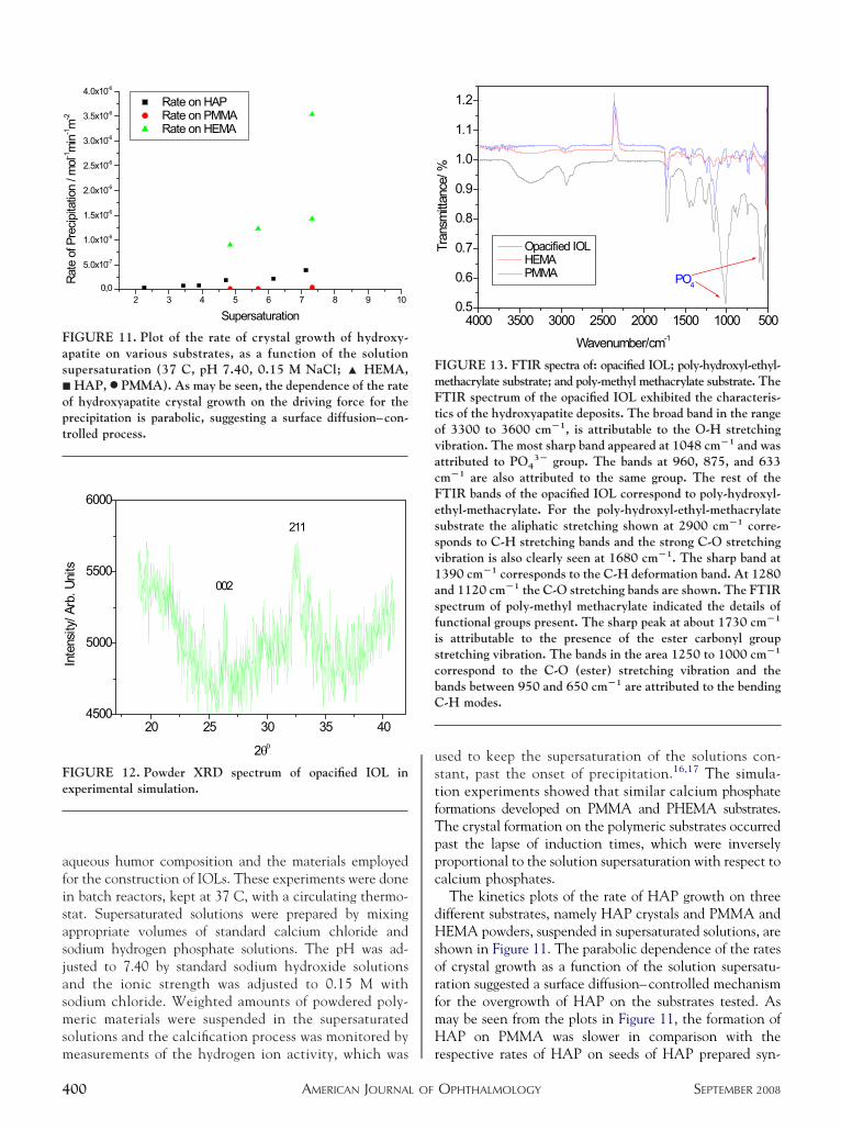
IGURE 13. FTIR spectra of: opacified IOL; poly-hydroxyl-ethyl-ethacrylate substrate; and poly-methyl methacrylate substrate. TheTIR spectrum of the opacified IOL exhibited the characteris-ics of the hydroxyapatite deposits. The broad band in the rangef 3300 to 3600 cm�1, is attributable to the O-H stretchingibration. The most sharp band appeared at 1048 cm�1 and wasttributed to PO4
3� group. The bands at 960, 875, and 633m�1 are also attributed to the same group. The rest of theTIR bands of the opacified IOL correspond to poly-hydroxyl-thyl-methacrylate. For the poly-hydroxyl-ethyl-methacrylateubstrate the aliphatic stretching shown at 2900 cm�1 corre-ponds to C-H stretching bands and the strong C-O stretchingibration is also clearly seen at 1680 cm�1. The sharp band at390 cm�1 corresponds to the C-H deformation band. At 1280nd 1120 cm�1 the C-O stretching bands are shown. The FTIRpectrum of poly-methyl methacrylate indicated the details ofunctional groups present. The sharp peak at about 1730 cm�1
s attributable to the presence of the ester carbonyl grouptretching vibration. The bands in the area 1250 to 1000 cm�1
orrespond to the C-O (ester) stretching vibration and theands between 950 and 650 cm�1 are attributed to the bending-H modes.
espective rates of HAP on seeds of HAP prepared syn-
OPHTHALMOLOGY SEPTEMBER 2008

tmsm
trmuTsrfersotvaarPibcc1sgiscat
pa
P
ctntdyoat
ep
ckcfpItccfpaetmccctnnfAapcr
perst
cflcopcdtnpttsdI
tI
V
hetically (order of magnitude 10�8 � 10�7 mol HAP m�2
in�1). The rates of HAP formation on PHEMA wereignificantly faster (order of magnitude 10�6 mol HAP
�2 min�1).Typical XRD and FTIR spectra used for the identifica-
ion of the materials are shown in Figures 12 and 13,espectively. The XRD spectrum obtained exhibited theost characteristic 002, 211 reflections, thus confirming
nambiguously the formation of HAP in the lens matrix.he increased noise was a result of the small size of the
pecimen and it was not possible to distinguish moreeflections of the mineral deposits. An additional reasonor the noise may be that the mineral phase was mostlymbedded in the polymer matrix, which in the two-thetaange investigated is amorphous to x-ray. The FTIRpectrum of the opacified IOL exhibited the characteristicsf the HAP deposits. The broad band in the range of 3300o 3600 cm�1 is attributable to the O-H stretchingibration. The most sharp band appeared at 1048 cm�1
nd was attributed to PO43� group. The bands at 960, 875,
nd 633 cm�1 were also attributed to the same group. Theest of the FTIR bands of the opacified IOL correspond toHEMA. For the PHEMA substrate the aliphatic stretch-
ng shown at 2900 cm�1 correspond to C-H stretchingands and the strong C-O stretching vibration is alsolearly seen at 1680 cm�1. The sharp band at 1390 cm�1
orresponds to the C-H deformation band. At 1280 and120 cm�1 the C-O stretching bands are shown. The FTIRpectrum of PMMA indicated the details of functionalroups present. The sharp peak band at about 1730 cm�1
s attributable to the presence of the ester carbonyl grouptretching vibration. The bands in the area 1250 to 1000m�1 correspond to the C-O (ester) stretching vibrationnd the bands between 950 and 650 cm�1 are attributed tohe bending C-H modes.
It should be noted that in these cases the substraterovides the necessary active growth sites for the prolifer-tion of the nuclei formed heterogeneously.
DISCUSSION
REVIOUS STUDIES HAVE SHOWN THE DEPOSITION OF CAL-
ium phosphate crystallites on the surface,18,19 as well as inhe interior of hydrophilic acrylic IOLs.20,21 The exami-ation of the opacified IOLs in the present study showedhat HAP was the predominant form of the crystallineeposits responsible for the opacification. Chemical anal-sis of the aqueous humor collected from patients withpacified IOLs and thermodynamic calculations involvingll related chemical species corroborated the findings forhe mineral deposited in the IOLs examined.
Various mechanisms explaining the formation of min-ral deposits have been suggested.1,2 Yet, the underlying
athogenetic mechanisms of the delayed postoperative tOPACIFICATION OF INTOL. 146, NO. 3
alcification of the hydrophilic acrylic lens remain un-nown and a broad spectrum of biological, pharmacologi-al, technological, and/or certain topical environmentalactors might be involved. It has been found that theresence of calcium was involved in crystallization on theOL surface.7 In the cases investigated, it has been shownhat the propensity for the development of calcium-ontaining precipitates was significantly stronger for sili-on IOLs in comparison with IOLs made of PMMA. Theormation of calcific deposits containing both calcium andhosphorus in the interior of IOLs implanted intramuscularlynd subcutaneously has been reported and differences in thextent of calcification has been attributed to the chemistry ofhe microenvironment of the implants.22 Detailed in vitroodel studies have shown that it is possible to precipitate
alcium phosphates at the humor/acrylic lens interface be-ause the former is a solution supersaturated with respect toalcium phosphate phases.14,23 According to these inves-igations the mineral salts may be formed by heterogeneousucleation, a process affected not only by the thermody-amic driving force in the aqueous phase but also from the
oreign surfaces and impurities in contact with the humor.lthough the deposits found in IOLs have been identified
s hydroxyapatite (Ca5(PO4)3OH, HAP),10,12,24–26 theresence of transient calcium phosphates such as octacal-ium phosphate (Ca8H2(PO4)65H2O, OCP) has also beeneported.27
In thermodynamic terms, the formation of calcific de-osits on the surface or in the interior of IOLs may bexplained by the fact that the aqueous humor is supersatu-ated with respect to different phases of calcium phosphatealts, which in the order of decreasing solubility are OCP,ricalcium phosphate (Ca3(PO4)2, -TCP), and HAP.
The aqueous humor was modeled as a supersaturatedalcium phosphate solution from which HAP may beormed either directly or through the transformation ofess-stable transient calcium phosphate phases. The cal-ium content of the normal humor is low, about half thatf the serum. The concentration of calcium bound to lensroteins, however, is high. Phosphorous and calcium in-rease in cataractous lenses, the latter up to 4.4 mmol/kg ofry weight. This is twice the normal amount.10 Weherefore presume that in some cases the calcium origi-ated from residual cataractous lens material may predis-ose the formation of calcium deposits. A possible factorhat may predispose to the formation of calcium deposits ishe inadequate cortex cleaning from the capsular bag, inome cases. It is interesting to note that the salts wereeposited both in the interior and on the surface of theOLs, on or near linear forceps marks.
From the mechanistic point of view, on what concernshe formation of calcium phosphate on the surface of theOLs, it may be suggested that the acrylate polymers,
hrough surface complexation with calcium ions, mayRAOCULAR LENSES 401

pnCIpoorkanvstepalvw2oppbc
spvc
aefimttfrHpcdc
Moffpsfissfieic
eksfSotcrrribPr
cfttmicifcta
T((a(n
m
4
rovide the active sites needed for the initiation ofucleation and further growth of the mineral phase.28
oncerning the formations found in the interior of theOLs it may be suggested that diffusion of calcium andhosphate ions, possibly through the swelling mechanismf the polymers, may contribute to the local developmentf the supersaturation needed for the formation of theespective crystals.29 The measurement results for theinetics formation of HAP to polymeric substrates, whichre used for the IOLs, showed that these materials favorucleation and calcium phosphate salt formation even atery low supersaturations. The greater tendency towardalt formation shown with PHEMA may be attributed tohe presence of surface hydroxyl groups. These groups,ither through ionization or through incorporation ofhosphate groups, may also facilitate surface nucleationnd growth of crystal formation.28,30,31 Not all hydrophilicenses are the same. One of their main differences is theariable water content. Hydroview lenses contain 18%ater, Memory lenses 20%, SC60B 28%, and AquaSense5%.4,21 Higher extent of hydration leads to higher degreef ionization of the polymeric functional groups, thusromoting calcification through the formation of com-lexes with ionized calcium. These surface complexes areelieved to serve as active sites for the growth of mineralalcium salts.28
The present study is a contribution to a better under-tanding of the pathogenetic mechanism leading to hydro-hilic acrylic IOL calcification. Model experiments initro are currently in progress for the validation of thelinical findings of the opacified IOLs.
It is interesting that there are reports in the literaturessociating the formation of IOL deposits with the pres-nce of silicon in the packaging materials.14,32,33 In ourndings we did not detect the presence of Si in theicroanalysis of the calcified IOLs, as may be seen in the
ypical spectrum shown in Figure 6. Actually, it is nothe silicon that is the problem. Silicon (in the elementalorm or bound with chemical bonds to other elements) isather inactive, as the authors admit in their study.33
owever, in the oxide form, which is hydrophilic, it isossible that the hydrated silicate surface may inducealcification as it possesses hydroxyl groups that may beissociated. Werner and associates, in their study, are not
lear about the provenance of the calcification products.33 fAMERICAN JOURNAL OF02
oreover, the findings that the deposits consisted of layersf fatty acids might considered to be responsible foravoring a local decrease in pH, which in turn favored theormation of the less basic (more acidic) octacalciumhosphate. Without precluding the importance of silicatepecies as active centers for the initiation of the calci-cation process, it should be noted that if this were theole cause for opacification, it should be limited to theurface. In any case, it seems very likely that the keyactor in the development of crystalline phosphate saltss related to the conditions of local supersaturationither in the vicinity of the surface of the IOL or in theirnterior, where it is developed by diffusion of thealcium and phosphate ions.
In the simulation experiments, powdered materials weremployed in order to be able to assess quantitatively theinetics of the calcification process, which in most cases isurface controlled.28,30 Maximization of the available sur-ace area is expected, therefore, to accelerate the process.everal preliminary experiments were conducted inrder to decide on the most suitable amount of substrateo be used for the most accurate monitoring of therystal growth process, which is accompanied by protonelease in the solution. It is interesting to note that theesults obtained are in agreement with experimentseported on animals. It has been reported that PHEMAmplants showed calcification starting at six weeks andecoming very heavy after six months.34 Moreover,HEMA implanted in rabbit cornea has shown ratherapid calcification.35
In conclusion, late postoperative IOL opacificationauses a severe loss of VA. The mechanism is not yetully understandable, but it does seem to be related tohe supersaturation of the aqueous humor with respecto calcium phosphates. In addition, the nature of theaterials used for the construction of the IOLs is of
mportance, as they may promote the formation ofalcium phosphates, either on the surface or in thenterior. A possible factor that may predispose to theormation of calcium deposits is the inadequate cortexleaning from the capsular bag. Nevertheless, the onlyherapeutic approach in such patients is IOL exchange,
technically difficult procedure that should be per-
ormed by experienced implant surgeons.HE AUTHORS INDICATE NO FINANCIAL SUPPORT OR FINANCIAL CONFLICT OF INTEREST. INVOLVED IN DESIGN OF STUDYS.P.G., P.G.K.); conduct of study (S.P.G., P.G.K., D.G.K.); collection of the data (S.P.G., D.G.K., E.K.M.); management and analysis of the dataP.G.K., S.P.G.); interpretation of the data (P.G.K., S.P.G.); preparation of the manuscript (S.P.G., P.G.K., E.K.M., D.G.K., V.S.P.); and review andpproval of the manuscript (P.G.K., S.P.G.). The study was approved by the ethical committee of the Regional Hospital of University of PatrasRio-Patras). In accordance with the Declaration of Helsinki, informed consent was obtained from all participants after explanation of the purpose andature of the investigation.
The authors wish to express thanks to Dr Eva Valsami-Jones of the Natural History Museum, London, United Kingdom for the XRD–confocalicroscopy measurements.
OPHTHALMOLOGY SEPTEMBER 2008

1
1
1
1
1
1
1
1
1
1
2
2
2
2
2
2
2
2
2
2
3
3
3
3
3
3
V
REFERENCES
1. Werner L, Apple DJ, Escobar-Gomez M, et al. Postoperativedeposition of calcium on the surfaces of a hydrogel intraoc-ular lens. Ophthalmology 2000;107:2179–2185.
2. Werner L, Apple DJ, Kaskaloglu M, Pandey SK. Denseopacification of the optical component of a hydrophilicacrylic intraocular lens: a clinicopathological analysis of 9explanted lenses. J Cataract Refract Surg 2001;27:1485–1492.
3. Frohn A, Dick HB, Augustin AJ, Grus FH. Late opacificationof the foldable hydrophilic acrylic lens SC60B-OUV. Oph-thalmology 2001;108:1999–2004.
4. Izak AM, Werner L, Pandey SK, Apple DJ. Calcification ofmodern foldable hydrogel intraocular lens designs. Eye 2003;17:393–406.
5. Jensen MK, Crandall AS, Mamalis N, Olson RJ. Crystalli-zation on the intraocular lens surfaces associated with the useof Healon GV. Arch Ophthalmol 1994;112:1037–1042.
6. Olson RJ. New cases of crystalline deposits on intraocularlenses not related to any specific viscoelastic (Letter). ArchOphthalmol 1995;113:1229.
7. Olson RJ, Caldwell KD, Crandall AS, Jensen MK, HuangSC. Intraoperative crystallization on the intraocular lenssurface. Am J Ophthalmol 1998;126:177–184.
8. Amon M, Menapace R. Cellular invasion on hydrogel andpoly(methyl methacrylate) implants. An in vivo study. JCataract Refract Surg 1991;17:774–779.
9. Amon M, Menapace R. In vivo observation of surfaceprecipitates of 200 consecutive hydrogel intraocular lenses.Ophthalmologica 1992;204:13–18.
0. Bucher PJM, Büchi ER, Daiker BC. Dystrophic calcificationof an implanted hydroxyethylmethacrylate intraocular lens.Arch Ophthalmol 1995;113:1431–1435.
1. Pandey SK, Werner L, Apple DJ, Kaskaloglu MM. Hydro-philic acrylic intraocular lens optic and haptics opacificationin a diabetic patient: bilateral case report and clinicopatho-logical correlation. Ophthalmology 2002;109:2042–2051.
2. Shek TW, Wong A, Yau B, Yu AK. Opacification of artificialintraocular lens: an electron microscopic study. UltrastructPathol 2001;25:281–283.
3. Neuhann IM, Werner L, Izak AM, et al. Late postoperativeopacification of a hydrophilic acrylic (hydrogel) intraocularlens: A clinicopathological analysis of 106 explants. Oph-thalmology 2004;111:2094–2101.
4. Guan X, Tang R, Nancollas GH. The potential calcificationof octacalcium phosphate on intraocular lens surfaces.J Biomed Mater Res 2004;71:488–496.
5. Spanos N, Misirlis DY, Kanellopoulou DG, Koutsoukos PG.Seeded growth of hydroxyapatite in simulated body fluid. JMater Sci 2006;41:1805–1812.
6. Koutsoukos PG, Amjad Z, Tomson MB, Nancollas GH.Crystallization of calcium phosphates. A constant composi-tion study. J Amer Chem Soc 1980;102:1553–1557.
7. Mavrilas D, Kapolos J, Koutsoukos PG, Dougenis D. Screen-ing biomaterials with a new in vitro method for potentialcalcification: Porcine aortic valves and bovine pericardium. JMater Sci Mater Med 2004;15:699–704.
8. Hatou S, Inoue M, Kurosaka D, Hida YR, Shinoda K, Oguchi
Y. Evaluation of calcification of a hydrogel intraocular lensOPACIFICATION OF INTOL. 146, NO. 3
by optical coherence tomography. J Cataract Refract Surg2004;30:1590–1592.
9. Van Geluwe I, Foets B, Van Ginderdeuren R, Zeyen T.Precipitation of calcium salts on a hydrophilic acrylic in-traocular lens after a vitreous hemorrhage: case report andhistopathologic correlation. J Cataract Refract Surg 2007;33:1328–1331.
0. Yong JL, Lertsumitkul S, Killingsworth MC, Filipic M.Calcification of intraocular hydrogel lens: evidence of dys-trophic calcification. Clin Experiment Ophthalmol 2004;32:492–500.
1. Tehrani M, Mamalis N, Wallin T, et al. Late postoperativeopacification of Memory Lens hydrophilic acrylic intraocularlenses: case series and review. J Cataract Refract Surg2004;30:115–122.
2. Buchen SY, Cunanan CM, Gwon A, Weinschenk JI III, GruberL, Knight PM. Assessing intraocular lens calcification in ananimal model. J Cataract Refract Surg 2001;27:1473–1484.
3. Wu W, Guan X, Tang R, et al. Calcification of intraocularimplant lens surfaces. Langmuir 2004;20:1356–1361.
4. Werner L, Kollarits CR, Mamalis N, Olson R. Surfacecalcification of a 3-piece silicone intraocular lens in a patientwith asteroid hyalosis: a clinicopathologic case report. Oph-thalmology 2005;112:447–452.
5. Wackernagel W, Ettinger K, Weitgasser U, et al. Opacifica-tion of a silicone intraocular lens caused by calcium depositson the optic. J Cataract Refract Surg 2004;30:517–520.
6. Fernando GT, Crayford BB. Visually significant calcificationof hydrogel intraocular lenses necessitating explantation.Clin Exper Ophthalmol 2000;28:280–286.
7. Lin S-Y, Chen K-H, Li M-J, Cheng W-T, Wang S-L.Evidence of octacalcium phosphate and type-B carbonatedapatites deposited on the surface of explanted acrylic hydro-gel intraocular lenses. J Biomed Mater Res B Appl Biomater2004;70:203–208.
8. Dalas E, Kallitsis JK, Koutsoukos PG. Crystallization ofhydroxyapatite on polymers. Langmuir 1991;7:1822–1826.
9. Apple DJ, Werner L. Complications of cataract and refrac-tive surgery: a clinicopathological documentation. Trans AmOphthalmol Soc 2001;99:95–107.
0. Koutsoukos PG. Growth of calcium phosphates on differentsubstrates: epitaxial considerations. In: Amjad Z, editor. Cal-cium Phosphates in Biological and Industrial Systems. Boston,Massachusetts: Kluwer Academic Publishers, 1998:41–66.
1. Chirila TV, Zainuddin, Hill DJ, Whittaker AK, Kemp A.Effect of phosphate functional groups on the calcificationcapacity of acrylic hydrogels. Acta Biomater 2007;3:95–102.
2. Dorey MW, Brownstein S, Hill VE, et al. Proposed patho-genesis for the delayed postoperative opacification of thehydroview hydrogel intraocular lens. Am J Ophthalmol2003;135:591–598.
3. Werner L, Hunter B, Stevens S, Chew JJL, Mamalis N.Role of silicon contamination on calcification of hydro-philic acrylic intraocular lenses. Am J Ophthalmol 2006;141:35– 43.
4. Imai Y. Effect of age on calcification of poly(hydroxyethylmethacrylate) in animals. Artif Organs 1985;9:255–258.
5. Vijayasekaran S, Chirila TV, Robertson TA, et al. Calcificationof poly(2-hydroxyethyl methacrylate) hydrogel sponges im-planted in the rabbit cornea: a 3-month study. J Biomater Sci
Polym Ed 2000;11:599–615.RAOCULAR LENSES 403

SriHAf
4
Biosketch
otirios P. Gartaganis is an Professor of Ophthalmology, Chairman, Medical School, University of Patras, Greece. Heeceived his MD in 1976 and PhD in 1981 from Athens National and Capodistrian University. Dr Gartaganis researchesn cataracts, pseudoexfoliation, and retinal diseases. He worked at the Retinal Service of the Manchester Royal Eyeospital in 1991 and at the Massachusetts Eye and Ear Infirmary in 1999. Dr Gartaganis is a member of the Americancademy of Ophthalmology, the Association for Research in Vision and Ophthalmology, and the European Association
or Vision and Eye Research.
AMERICAN JOURNAL OF OPHTHALMOLOGY03.e1 SEPTEMBER 2008
P1(Bc
V
Biosketch
etros G. Koutsoukos is an Professor of the Department of Chemical Engineering of the University of Patras, Greece, since989. He is a director of the Laboratory of Inorganic and Analytical Chemistry. He received the Ptichion of ChemistryBSc) from the University of Patras in 1972, MBA from the Athens School of Economics in 1974, PhD from SUNY atuffalo in 1980, and Ifigesia (eq. DSc), from the University of Patras in 1984. Dr Koutsoukos researches in therystallization of inorganic salts with emphasis in biomineralization. He has published over 170 publications.
OPACIFICATION OF INTRAOCULAR LENSESOL. 146, NO. 3 403.e2
Copyright © 2022 FDOKUMEN