Antigenic and biological characterization of avian paramyxovirus type 1 isolates from pigeons
R
L
La
b
c
1
aabvatrpcttdi
sT
0d
Behavioural Brain Research 193 (2008) 69–78
Contents lists available at ScienceDirect
Behavioural Brain Research
journa l homepage: www.e lsev ier .com/ locate /bbr
esearch report
imits of intraocular and interocular transfer in pigeons
aura Jimenez Ortegaa,c,∗, Katrin Stoppaa, Onur Gunturkuna, Nikolaus F. Trojea,b
Ruhr-Universitat-Bochum, GermanyQueen’s University, Kingston, Ontario, CanadaUniversidad Complutense de Madrid, Spain
as twfieldhereae howon brated aansfeterale betwterpraces, in afugalw fieition,nocul
a r t i c l e i n f o
Article history:Received 10 January 2008Received in revised form 23 April 2008Accepted 26 April 2008Available online 3 May 2008
Keywords:Interocular transferIntraocular transferVisual asymmetriesAvian visual perceptionInterhemispheric transfer
a b s t r a c t
The retina of the pigeon hular field and the yellowthe tectofugal pathway, wpresent study we examinintegrated within the pigeon one of the two keys loctwo feeders. Intraocular trfrom the frontal to the laperceived among the edgof performance that we inThere were virtually no trofugal pathway, althoughthalamofugal to the tectomation between the yellotransfer was found. In addvisual field than in the mo
of the thalamofugal system in t. Introduction
Rock pigeons (Columbia livia) and many other birds have later-lised eyes with only relatively small binocular overlap (about 25◦)nd a large monocular visual field extending about 120◦ laterally tooth sides. The pigeon’s eyes are specialised for frontal binocularision at near distances during pecking, and for panoramic visiont far distances [3,10]. These two visual functions are mediated bywo different retinal areas: a binocular dorso-temporal oil dropleded field and a monocular oil dropled yellow field. The red field isointing into the lower frontal visual field, while the yellow field isovering the remaining visual field: upper frontal visual field andhe lateral one [32]. In a natural situation a bird might perceivehe same visual stimulus in different parts of the visual field atifferent times. The goal of inter- and intraocular transfer exper-
ments is to clarify the way in which information is transmitted
∗ Corresponding author at: Universidad Complutense de Madrid, Ciudad Univer-itaria, Facultad de Odontologıa, D. Psicobiologıa, 28040 Madrid, Spain.el.: +34 65 022 28 09; fax: +34 91 387 75 48.
E-mail address: [email protected] (L.J. Ortega).
166-4328/$ – see front matter © 2008 Elsevier B.V. All rights reserved.oi:10.1016/j.bbr.2008.04.022
o areas of enhanced vision: the red field looking into the frontal binoc-projecting into the lateral monocular field. The entire retina projects tos the monocular areas mainly project to the thalamofugal pathway. In the
the information received in different retinal areas and hemispheres isain. The pigeons’ task was to discriminate between two shapes by peckingt one end of an experimental alley, while walking back and forth betweenr between the red and the yellow field was tested by moving the stimulusvisual field in consecutive steps and vice versa. When the stimuli wereeen the red and the yellow field, the pigeons showed a drastic decrease
et to result from a switch from the tectofugal to the thalamofugal system.of intraocular transfer of information from the tectofugal to the thalam-second experiment a weak intraocular transfer of information from thesystem was observed. In a third experiment, interocular transfer of infor-lds of the two eyes was tested. In eight out of nine birds, no interocularpigeons showed more difficulties to learn the task in the monocular rightar left visual field, suggesting the existence of an asymmetric organizationhe pigeon brain.
© 2008 Elsevier B.V. All rights reserved.
between the two eyes and between the lateral and the frontal visual
field.Two independent visual systems have been described in theavian brain. The tectofugal pathway that processes visual infor-mation proceeding from the entire retina, and the thalamofugalpathway which receives visual input from the yellow field. Thetectofugal pathway is in large parts homologous to the extra-geniculocortical pathway in mammals [2,21,14,31]. The visual inputascends from the retina to the contralateral optic tectum (OT),which projects bilaterally to the entopallium (E) via the thalamicnucleus rotundus (Rt). The thalamofugal pathway can be consid-ered homologeous to the mammalian geniculo-cortical pathway.In pigeons, but not in chickens, this pathway mainly receives visualinput from the yellow visual field [36,19], which is transmitted tothe contralateral thalamic nucleus geniculatus lateralis, pars dor-salis (GLd). The GLd projects bilaterally to the telencephalic visualwulst [41,21,14].
1.1. Intraocular transfer
The first attempt to train birds in the lateral visual field in orderto test intraocular transfer of information was done by Nye [34]
l Brain
70 L.J. Ortega et al. / Behaviourawho trained six pigeons in different discrimination tasks. The birdslearned the task successfully if the stimuli were presented behindthe pecking keys in the frontal visual field. In contrast, when thestimuli were located at 90◦ to each side of the axis of the beak, thepigeons did not learn the task. Nye concluded that “pigeons do notpossess the neural capability required to learn to use informationcontained in laterally located stimuli to directly control peckingbehaviour”. In a previous experiment, Levine [25] had found a dropin the discrimination performance when the stimuli were shiftedfrom a subrostral (in a plane below the pigeon’s head) to an antero-rostral position (in front of the pigeon head). Mallin and Delius[26] trained head-fixed pigeons to discriminate two coloured lightsusing jaw movements as an operant response. They found a poorintraocular discrimination transfer when the stimuli were shiftedfrom frontal to lateral position and vice versa. However, the transferfrom lateral to frontal position was slightly better (around 10%) thanthe reverse performance. Remy and Emmerton [36] described theexistence of information transfer from the lateral to frontal visualfield and a lack of information transfer from the frontal to the lat-eral visual field, in a light detection task, also using jaw movementas a measure. Roberts et al. [38] confirmed those results in unre-strained pigeons employing a symbolic delayed matching to sampletask. The observed lack of transfer was interpreted as an absenceof communication between the thalamofugal and tectofugal path-ways.
In the present study we test intraocular transfer of informationin free walking pigeons by presenting two patterns either in thefrontal visual field or in the lateral visual field.
1.2. Interocular transfer
According to Levine [23], the earliest interocular transfer exper-iment was done in 1917 by Wolfgang Kohler. Patching one of thetwo eyes, he observed that chickens showed interocular transferbetween the two eyes while discriminating two sheets of grey paperdiffering in brightness, which were located horizontally at groundlevel. However, in an experiment in which the stimuli were dis-played on a vertical screen above ground level, interocular transferwas not observed [1]. Two hypotheses were proposed to explainthese contradictory results: the “sensorimotor integration” hypoth-esis [46,45] and the “retinal locus” hypothesis [23–25,10,26].
The “sensorimotor integration” hypothesis proposes thatpigeons may transfer information depending on whether theresponse key and the visual stimulus have the same or differ-
ent spatial locations [37]. To test the “sensorimotor integration”hypothesis, pigeons were trained in three spatial tasks employingtwo pecking keys arranged either vertically or horizontally [46]. Nomatter whether the keys were arranged horizontally or vertically,if response key and stimulus were located in the same pecking key,there was a perfect interocular transfer of information. However, ifresponse key and stimulus were located in different keys (for exam-ple: the stimulus was presented in the lower key and the pigeonshad to peck in the upper key) the pigeons did not show interoculartransfer.The “retinal locus” hypothesis proposes that interocular transferoccurs when the stimuli are presented in the dorso-temporal partof the retina (red field), but not when the stimuli are presentedin the other parts of the retina (yellow field) [23–25,10,26]. Levine[23–25] conducted a set of experiments using a jumping stand. Thebirds were placed into a rotating perch, which forced them to jumponto one of two platforms according to the presented stimuli. If theanimals jumped to the incorrect platform, it collapsed and the ani-mal dropped into a net. The correct platform remained safe untilthe bird was returned to its cage. They found that when the stim-uli were presented horizontally in a plane below the pigeon’s head
Research 193 (2008) 69–78
(subrostral), interocular transfer between the two eyes occurred,but if the stimuli were presented vertically in front of the pigeon’shead (anterostral) there was an absence of transfer. Catania [5]challenged these observations by training pigeons in brightness,colour and pattern discrimination tasks to peck on a key locatedin front of the pigeon head. The stimuli were projected either onthe frontal key or on one of two lateral screens. Pigeons showedinterocular transfer of information in both conditions. Catania pro-posed that the lack of interocular transfer in Levine’s experimentscould be explained by the amount of training and by the occlusionof the eye due to changes in the pigeon’s posture. However a set ofexperiments replicating Levine’s jumping stand [10] concluded thatthe absence of transfer was a genuine phenomenon which did notdepend on postural habit, amount of training and task complex-ity. Furthermore, birds trained binocularly in the jumping standoften gave evidences of learning with only one eye when testedmonocularly. Mallin and Delius [26] conducted an experiment withhead-fixed pigeons using jaw movements as an operant in a colourdiscrimination task. They presented two coloured lights at differ-ent locations on the retina. Birds showed interocular transfer ofinformation when the discrimination task was monocularly pre-sented inside the red field and a lack of interocular transfer whenthe stimulus was monocularly presented within the yellow field.
According to Remy and Watanabe [37], the “retinal locus” and“sensorimotor integration” hypotheses may not be contradictory.Retinal locus may be crucial when a task does not require sensori-motor integration. Up to now, none of the proposed hypotheses iscapable of explaining all experimental results on interocular trans-fer. Some experimental findings suggest that other characteristicsof the task, such as biological relevance, type of reinforcement,or trained response, may affect interocular transfer. For instance,transfer of information was absent in heat reinforcement, but itwas present in the same task when using food reinforcement [9].Moreover, interocular transfer in pigeons was found in colour dis-crimination, but it was not observed in the motor response requiredto show the colour discrimination [43].
Pigeons show an asymmetric information transfer betweenhemispheres. Nottelmann et al. [33] observed transfer of memoriesfrom the left eye/right hemisphere to the right eye/left hemisphere,but not vice versa. However, Skiba et al. [42] observed a faster shiftof learned colour cues from the right to the left eye than vice versa.Most probably, cerebral asymmetries and interocular transfer ofinformation are interrelated phenomena in birds. Interhemisphericinteractions may be an important component for understanding
visual asymmetries [15,35,22].Interhemispheric information transfer in the avian and thehuman brain seems to be a complex phenomenon. Many ques-tions remain unanswered and none of the proposed theories canexplain all experimental results. The restricted experimental condi-tions of many investigations result in a very low ecological validity.Very often, the experimental conditions prevented the birds fromwalking freely while solving the task (examples can be found inRefs. [26,38,36,34]). Therefore, visual mechanisms that may play animportant role in ecologically valid conditions like optic flow dueto locomotion and head movements have not been considered. Inaddition, there are few experiments investigating interocular trans-fer between the yellow visual fields. This is probably due to thedifficulties in training pigeons to solve visual tasks in the lateralvisual field [37].
In the present study, intraocular transfer and interocular transferwere investigated in free walking pigeons. In the first experimentwe tested intraocular transfer of information from the red to theyellow field. That is, if pigeons learn a visual task using their frontalbinocular field, can they perform the same task using their lateralvisual field of the same eye? In the second one, interocular transfer
l Brain
L.J. Ortega et al. / Behaviouraof information between the yellow fields of both eyes was investi-gated. That is, if pigeons learn a visual task within the yellow visualfield of one eye, are able to perform the task with the other eye? Inaddition, we also examined whether pigeons were able to learn apattern recognition task directly in the yellow visual field. Finallywe tested intraocular transfer from the yellow fields to the red fieldin the few birds that succeeded in this later task.
2. General methods
2.1. Subjects
Ten rock pigeons (C. livia), males and females, aged between 3and 7 years obtained from the aviary of the Biopsychology Depart-ment of Ruhr-University in Bochum were initially trained in apattern recognition task. During the experiments, they were keptin individual cages on a 12-h light–dark cycle. They had ad libitumaccess to drinking water. Food was restricted to keep their weightat 85% of their free-feeding weight.
Nine of the ten pigeons successfully completed the initial train-ing (see below) and were used in Experiment 1. Eight of them were
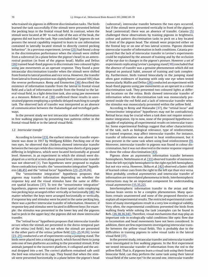
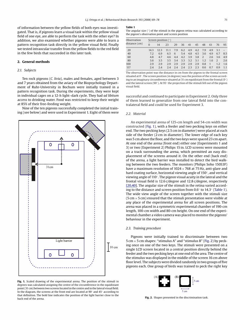
Fig. 1. Scaled drawing of the experimental arena. The position of the stimuli indegrees was calculated assigning the centre of the circumference in the equidistantpoint (31 cm) between two screens located in the centre and in the lateral visual field.In the diagram, the screens at the front end are located at 90◦ and 45◦ according tothat definition. The bold line indicates the position of the light barrier close to theback end of the arena.
Research 193 (2008) 69–78 71
Table 1The angular size (◦) of the stimuli in the pigeon retina was calculated according tothe pigeon’s observation point and screen position
Observationdistance (cm)
Screen position (◦)
0 14 23 29 36 41 45 48 61 76 95
20 14.3 12.3 11.1 7.9 6.2 4.9 4.2 7.9 4.9 3.1 –40 7.2 6.9 6.5 6 5.4 4.8 4.5 3.6 4.9 6.7 8.560 4.8 4.7 4.6 4.4 4.2 3.9 3.8 2 2.6 3.6 4.980 3.6 3.5 3.5 3.4 3.3 3.2 3.1 1.2 1.6 2 2.6
100 2.9 2.9 2.9 2.9 2.9 2.9 2.9 0.8 1 1.2 1.6117 2.4 2.4 2.4 2.4 2.4 2.3 2.3 0.6 0.7 0.9 1.1
The observation point was the distance in cm from the pigeon to the frontal screensituated at 0◦ . The screen position (in degrees) was the position of the screen accord-ing to an imaginary circumference situated at 31 cm equidistant from the frontal (0◦)and the lateral screen (90◦). At 95◦ the projection of the stimuli fell out of the pigeonvisual field.
successful and continued to participate in Experiment 2. Only threeof them learned to generalize from one lateral field into the con-tralateral field and could be used for Experiment 3.
2.2. Material
An experimental arena of 125-cm length and 54-cm width wasconstructed (Fig. 1), with a feeder and two pecking keys on eitherend. The two pecking keys (2.5 cm in diameter) were placed at eachside of the feeder (2 cm in diameter). The lower edge of each keywas 5 cm above the floor, and the two keys were spaced 23 cm apart.At one end of the arena (front end) either one (Experiments 1 and3) or two (Experiment 2) Philips 15 in. LCD screens were mountedon a track surrounding the arena, which permitted an easy dis-placement of the screens around it. On the other end (back end)of the arena, a light barrier was installed to detect the bird walk-ing between the two feeders. The monitors (Philips Soho 150S3F)have a maximum resolution of 1024 × 768 at 75 Hz, anti-glare andhard coating surface, horizontal viewing angle of 150◦, and verticalviewing angle of 110◦. The pigeon visual acuity in the lateral and the
frontal visual field is 12.6 c/degree and 12.8 c/degree, respectively[20,40]. The angular size of the stimuli in the retina varied accord-ing to the distance and screen position from 0.6◦ to 14.3◦ (Table 1).The wide view angle of the screen together with the stimuli size(5 cm × 5 cm) ensured that the stimuli presentation were visible atany place of the experimental arena for all screen positions. Thearena was placed in a symmetric experimental chamber of 190-cmlength, 100-cm width and 80-cm height. On one end of the experi-mental chamber a video camera was placed to monitor the pigeons’behaviour in the experiment.2.3. Training procedure
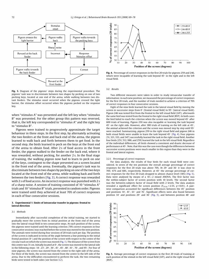
Pigeons were initially trained to discriminate between two5 cm × 5 cm shapes: “stimulus A” and “stimulus B” (Fig. 2) by peck-ing once on one of the two keys. The stimuli were presented on asingle LCD screen located in a central position directly behind thefeeder and the two pecking keys at one end of the area. The centre ofthe stimulus was displayed in the middle of the screen 16 cm abovefloor level. The subjects were divided randomly in two groups of fivepigeons each. One group of birds was trained to peck the right key
Fig. 2. Shapes presented in the discrimination task.
72 L.J. Ortega et al. / Behavioural Brain
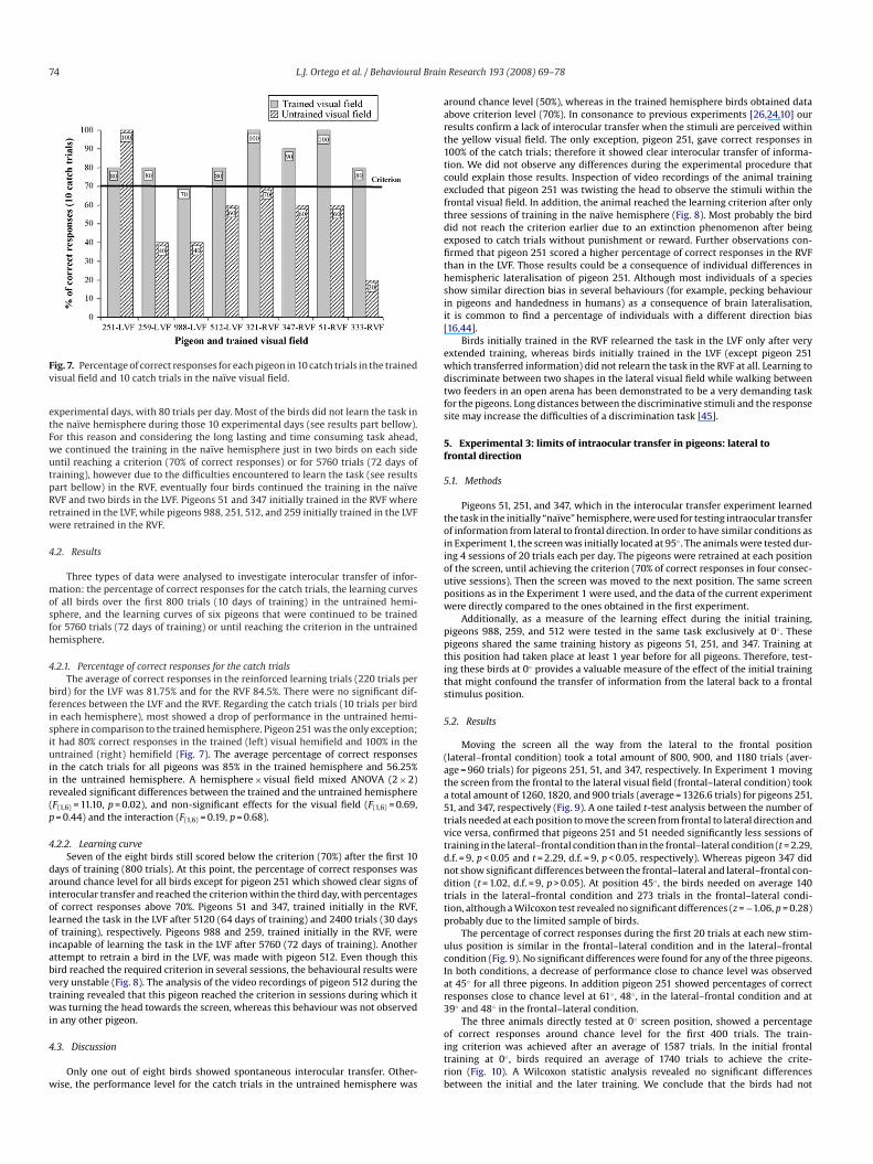
600 trials of learning. Pigeon 259 was also incapable or learning the task beyond45◦ on the right side. However, after 380 trials of training on the left side at 45◦ ,the animal achieved the criterion and the screen was moved step by step until 95◦
were reached. Summarizing, pigeon 259 in the right visual field and pigeon 246 inboth visual fields were unable to learn the task beyond 45◦ (Fig. 4). Four pigeons(51, 321, 333, and 347) successfully learned the task in the right visual field. Anotherfour birds (251, 512, 988, and 259) learned the task in the left visual field. Regardlessof the individual differences, all birds showed a consistent and drastic decrease ofperformance at 45◦ . Note, that this was the case even though the differences betweensuccessive screen positions were much smaller in this region than they were in thefrontal and lateral regions.
3.2.1. Percentage of correct responsesFor data analysis, the results of four birds for each visual field were con-
sidered. In seven of the ten positions the overall average percentage of correctresponses for the first 20 trials was above 70%. At 29◦ and 36◦ it was close to70%: 67% and 68%, respectively. However, at 45◦ the average percentage of cor-rect responses for the first 20 trials dropped to almost chance level (58%) (Fig. 5).Data were analysed by means of a 10 × 2 mixed ANOVA. The first factor wasthe within-subject factor of screen position with 10 levels. The second factor
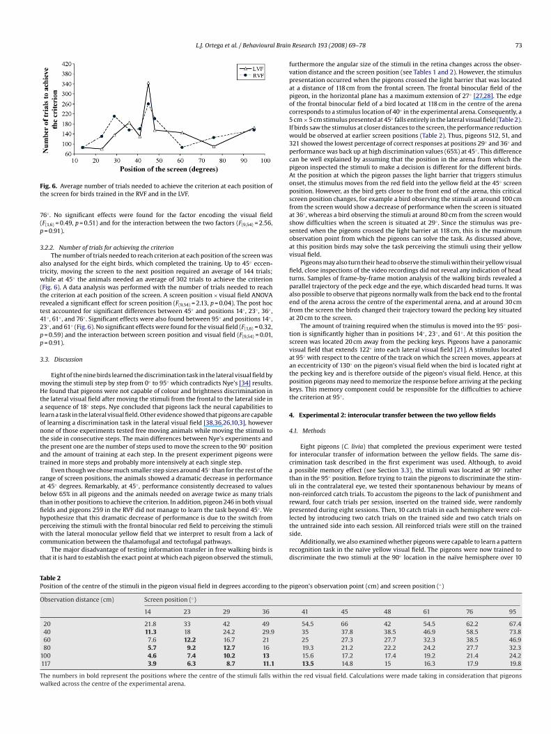
Fig. 3. Diagram of the pigeons’ steps during the experimental procedure. Thepigeons’ task was to discriminate between two shapes by pecking on one of twopecking keys, located at one end of the arena, while walking between two dis-tant feeders. The stimulus onset occurred when the pigeons crossed the lightbarrier, the stimulus offset occurred when the pigeons pecked on the responsekey.
when “stimulus A” was presented and the left key when “stimulusB” was presented. For the other group this pattern was reversed,that is, the left key corresponded to “stimulus A” and the right keyto “stimulus B”.
Pigeons were trained to progressively approximate the targetbehaviour in three steps. In the first step, by alternately activatingthe two feeders at the front and back end of the arena, the pigeonlearned to walk back and forth between them to get food. In thesecond step, the birds learned to peck on the keys at the front endof the arena to obtain food. After 2 s of food access in the frontfeeder, the pigeon walked to the feeder on the back end, where itwas rewarded, without pecking, for another 2 s. In the final stageof training, the walking pigeon now had to learn to peck on oneof the keys, contingent to the shape presented on a screen locatedin the front end of the arena. Eventually, the pigeon’s task was todiscriminate between two shapes by pecking on one of the two keyslocated at the front end of the arena, while walking back and forthbetween the two feeders (Fig. 3). A correct response was rewardedwith 2 s of food access. An incorrect response was punished with 2 s
of a sharp noise. A session of training consisted of 10 “stimulus A”trials and 10 “stimulus B” trials, presented in random order. Pigeonswere trained until they achieved at least 70% of correct responsesin each of four consecutive sessions.3. Experimental 1: limits of intraocular transfer in pigeons: frontal tolateral direction
3.1. Methods
Immediately after successful completion of the initial training, we started togradually move the screen from its initial position at the front end of the arenainto the lateral visual field in ten consecutive steps. At each position of the screen,the pigeons were trained until the learning criterion (70% correct responses in fourconsecutive sessions) was reached before the screen was moved to the next position.The animals were tested during four sessions of 20 trials each per day. The positionof the screen is indicated in terms of the angle between a line pointing to the initialfrontal position (0◦) and the position of the screen with respect to the centre of thecircular track on which the screen was moved (Fig. 1). The distance of the screen fromthis centre was 31 cm. Initially located at 0◦ , the screen was moved to the lateral sidein the following steps: 14◦ , 23◦ , 29◦ , 36◦ , 41◦ , 45◦ , 48◦ , 61◦ , 76◦ , and 95◦ . For four ofnine pigeons, the screen was moved from the centre to the right side of the arena.For three of the birds, the screen was moved from the centre to the left side of thearena. Due to the difficulties encountered to perform the task, the two remainingbirds were tested in both sides (see Section 3.2).
Research 193 (2008) 69–78
Fig. 4. Percentage of correct responses in the first 20 trials for pigeons 259 and 246,which were incapable of learning the task beyond 45◦ in the right and in the leftvisual field.
3.2. Results
Two different measures were taken in order to study intraocular transfer ofinformation: in each new position, we measured the percentage of correct responsesfor the first 20 trials, and the number of trials needed to achieve a criterion of 70%of correct responses in four consecutive sessions.
Eight of the nine birds learned the task in the lateral visual field by moving thescreen in successive steps from 0◦ (frontal visual field) to 95◦ (lateral visual field).Pigeon 246 was tested first from the frontal to the left visual field (LVF); afterwardsthe same bird was tested from the frontal to the right visual field (RVF). In both casesthe bird failed to reach the criterion when the screen was moved beyond 45◦ after
was the between-subjects factor of visual field with 2 levels. The data analysisrevealed a significant effect for screen position (F(9,54) = 2.55, p = 0.01). A pair-wise comparison accounted for significant differences between the 45◦ positionand positions 14◦ , 41◦ , 61◦ and 76◦ . Significant effects were also found betweenposition 14◦ and positions 29◦ and 36◦ (Fig. 5), and between position 48◦ and
Fig. 5. Average percentage of correct responses in the first 20 trials of training ateach position of the stimuli in the left visual field (LVF), and in the right visual field(RVF).
l Brain
at 95 with respect to the centre of the track on which the screen moves, appears at
L.J. Ortega et al. / Behavioura
Fig. 6. Average number of trials needed to achieve the criterion at each position ofthe screen for birds trained in the RVF and in the LVF.
76◦ . No significant effects were found for the factor encoding the visual field(F(1,6) = 0.49, p = 0.51) and for the interaction between the two factors (F(9,54) = 2.56,p = 0.91).
3.2.2. Number of trials for achieving the criterionThe number of trials needed to reach criterion at each position of the screen was
also analysed for the eight birds, which completed the training. Up to 45◦ eccen-tricity, moving the screen to the next position required an average of 144 trials;while at 45◦ the animals needed an average of 302 trials to achieve the criterion(Fig. 6). A data analysis was performed with the number of trials needed to reachthe criterion at each position of the screen. A screen position × visual field ANOVArevealed a significant effect for screen position (F(9,54) = 2.13, p = 0.04). The post hoctest accounted for significant differences between 45◦ and positions 14◦ , 23◦ , 36◦ ,41◦ , 61◦ , and 76◦ . Significant effects were also found between 95◦ and positions 14◦ ,23◦ , and 61◦ (Fig. 6). No significant effects were found for the visual field (F(1,6) = 0.32,p = 0.59) and the interaction between screen position and visual field (F(9,54) = 0.01,p = 0.91).
3.3. Discussion
Eight of the nine birds learned the discrimination task in the lateral visual field bymoving the stimuli step by step from 0◦ to 95◦ which contradicts Nye’s [34] results.He found that pigeons were not capable of colour and brightness discrimination inthe lateral visual field after moving the stimuli from the frontal to the lateral side ina sequence of 18◦ steps. Nye concluded that pigeons lack the neural capabilities tolearn a task in the lateral visual field. Other evidence showed that pigeons are capableof learning a discrimination task in the lateral visual field [38,36,26,10,3], howevernone of those experiments tested free moving animals while moving the stimuli tothe side in consecutive steps. The main differences between Nye’s experiments andthe present one are the number of steps used to move the screen to the 90◦ positionand the amount of training at each step. In the present experiment pigeons weretrained in more steps and probably more intensively at each single step.
Even though we chose much smaller step sizes around 45◦ than for the rest of therange of screen positions, the animals showed a dramatic decrease in performanceat 45◦ degrees. Remarkably, at 45◦ , performance consistently decreased to valuesbelow 65% in all pigeons and the animals needed on average twice as many trialsthan in other positions to achieve the criterion. In addition, pigeon 246 in both visualfields and pigeons 259 in the RVF did not manage to learn the task beyond 45◦ . Wehypothesize that this dramatic decrease of performance is due to the switch fromperceiving the stimuli with the frontal binocular red field to perceiving the stimuliwith the lateral monocular yellow field that we interpret to result from a lack ofcommunication between the thalamofugal and tectofugal pathways.
The major disadvantage of testing information transfer in free walking birds isthat it is hard to establish the exact point at which each pigeon observed the stimuli,
Table 2Position of the centre of the stimuli in the pigeon visual field in degrees according to the
Observation distance (cm) Screen position (◦)
14 23 29 36
20 21.8 33 42 4940 11.3 18 24.2 29.960 7.6 12.2 16.7 2180 5.7 9.2 12.7 16
100 4.6 7.4 10.2 13117 3.9 6.3 8.7 11.1
The numbers in bold represent the positions where the centre of the stimuli falls withinwalked across the centre of the experimental arena.
Research 193 (2008) 69–78 73
furthermore the angular size of the stimuli in the retina changes across the obser-vation distance and the screen position (see Tables 1 and 2). However, the stimuluspresentation occurred when the pigeons crossed the light barrier that was locatedat a distance of 118 cm from the frontal screen. The frontal binocular field of thepigeon, in the horizontal plane has a maximum extension of 27◦ [27,28]. The edgeof the frontal binocular field of a bird located at 118 cm in the centre of the arenacorresponds to a stimulus location of 40◦ in the experimental arena. Consequently, a5 cm × 5 cm stimulus presented at 45◦ falls entirely in the lateral visual field (Table 2).If birds saw the stimulus at closer distances to the screen, the performance reductionwould be observed at earlier screen positions (Table 2). Thus, pigeons 512, 51, and321 showed the lowest percentage of correct responses at positions 29◦ and 36◦ andperformance was back up at high discrimination values (65%) at 45◦ . This differencecan be well explained by assuming that the position in the arena from which thepigeon inspected the stimuli to make a decision is different for the different birds.At the position at which the pigeon passes the light barrier that triggers stimulusonset, the stimulus moves from the red field into the yellow field at the 45◦ screenposition. However, as the bird gets closer to the front end of the arena, this criticalscreen position changes, for example a bird observing the stimuli at around 100 cmfrom the screen would show a decrease of performance when the screen is situatedat 36◦ , whereas a bird observing the stimuli at around 80 cm from the screen wouldshow difficulties when the screen is situated at 29◦ . Since the stimulus was pre-sented when the pigeons crossed the light barrier at 118 cm, this is the maximumobservation point from which the pigeons can solve the task. As discussed above,at this position birds may solve the task perceiving the stimuli using their yellowvisual field.
Pigeons may also turn their head to observe the stimuli within their yellow visualfield, close inspections of the video recordings did not reveal any indication of headturns. Samples of frame-by-frame motion analysis of the walking birds revealed aparallel trajectory of the peck edge and the eye, which discarded head turns. It wasalso possible to observe that pigeons normally walk from the back end to the frontalend of the arena across the centre of the experimental arena, and at around 30 cmfrom the screen the birds changed their trajectory toward the pecking key situatedat 20 cm to the screen.
The amount of training required when the stimulus is moved into the 95◦ posi-tion is significantly higher than in positions 14◦ , 23◦ , and 61◦ . At this position thescreen was located 20 cm away from the pecking keys. Pigeons have a panoramicvisual field that extends 122◦ into each lateral visual field [21]. A stimulus located
◦
an eccentricity of 130◦ on the pigeon’s visual field when the bird is located right atthe pecking key and is therefore outside of the pigeon’s visual field. Hence, at thisposition pigeons may need to memorize the response before arriving at the peckingkeys. This memory component could be responsible for the difficulties to achievethe criterion at 95◦ .
4. Experimental 2: interocular transfer between the two yellow fields
4.1. Methods
Eight pigeons (C. livia) that completed the previous experiment were testedfor interocular transfer of information between the yellow fields. The same dis-crimination task described in the first experiment was used. Although, to avoida possible memory effect (see Section 3.3), the stimuli was located at 90◦ ratherthan in the 95◦ position. Before trying to train the pigeons to discriminate the stim-uli in the contralateral eye, we tested their spontanenous behaviour by means ofnon-reinforced catch trials. To accustom the pigeons to the lack of punishment andreward, four catch trials per session, inserted on the trained side, were randomlypresented during eight sessions. Then, 10 catch trials in each hemisphere were col-lected by introducing two catch trials on the trained side and two catch trials onthe untrained side into each session. All reinforced trials were still on the trainedside.
Additionally, we also examined whether pigeons were capable to learn a patternrecognition task in the naıve yellow visual field. The pigeons were now trained todiscriminate the two stimuli at the 90◦ location in the naıve hemisphere over 10
pigeon’s observation point (cm) and screen position (◦)
41 45 48 61 76 95
54.5 66 42 54.5 62.2 67.435 37.8 38.5 46.9 58.5 73.825 27.3 27.7 32.3 38.5 46.919.3 21.2 22.2 24.2 27.7 32.315.6 17.2 17.4 19.2 21.4 24.213.5 14.8 15 16.3 17.9 19.8
the red visual field. Calculations were made taking in consideration that pigeons
74 L.J. Ortega et al. / Behavioural Brain
Fig. 7. Percentage of correct responses for each pigeon in 10 catch trials in the trainedvisual field and 10 catch trials in the naıve visual field.
experimental days, with 80 trials per day. Most of the birds did not learn the task inthe naıve hemisphere during those 10 experimental days (see results part bellow).For this reason and considering the long lasting and time consuming task ahead,we continued the training in the naıve hemisphere just in two birds on each sideuntil reaching a criterion (70% of correct responses) or for 5760 trials (72 days oftraining), however due to the difficulties encountered to learn the task (see resultspart bellow) in the RVF, eventually four birds continued the training in the naıveRVF and two birds in the LVF. Pigeons 51 and 347 initially trained in the RVF whereretrained in the LVF, while pigeons 988, 251, 512, and 259 initially trained in the LVFwere retrained in the RVF.
4.2. Results
Three types of data were analysed to investigate interocular transfer of infor-mation: the percentage of correct responses for the catch trials, the learning curvesof all birds over the first 800 trials (10 days of training) in the untrained hemi-sphere, and the learning curves of six pigeons that were continued to be trainedfor 5760 trials (72 days of training) or until reaching the criterion in the untrainedhemisphere.
4.2.1. Percentage of correct responses for the catch trialsThe average of correct responses in the reinforced learning trials (220 trials per
bird) for the LVF was 81.75% and for the RVF 84.5%. There were no significant dif-ferences between the LVF and the RVF. Regarding the catch trials (10 trials per birdin each hemisphere), most showed a drop of performance in the untrained hemi-sphere in comparison to the trained hemisphere. Pigeon 251 was the only exception;it had 80% correct responses in the trained (left) visual hemifield and 100% in theuntrained (right) hemifield (Fig. 7). The average percentage of correct responsesin the catch trials for all pigeons was 85% in the trained hemisphere and 56.25%
in the untrained hemisphere. A hemisphere × visual field mixed ANOVA (2 × 2)revealed significant differences between the trained and the untrained hemisphere(F(1,6) = 11.10, p = 0.02), and non-significant effects for the visual field (F(1,6) = 0.69,p = 0.44) and the interaction (F(1,6) = 0.19, p = 0.68).4.2.2. Learning curveSeven of the eight birds still scored below the criterion (70%) after the first 10
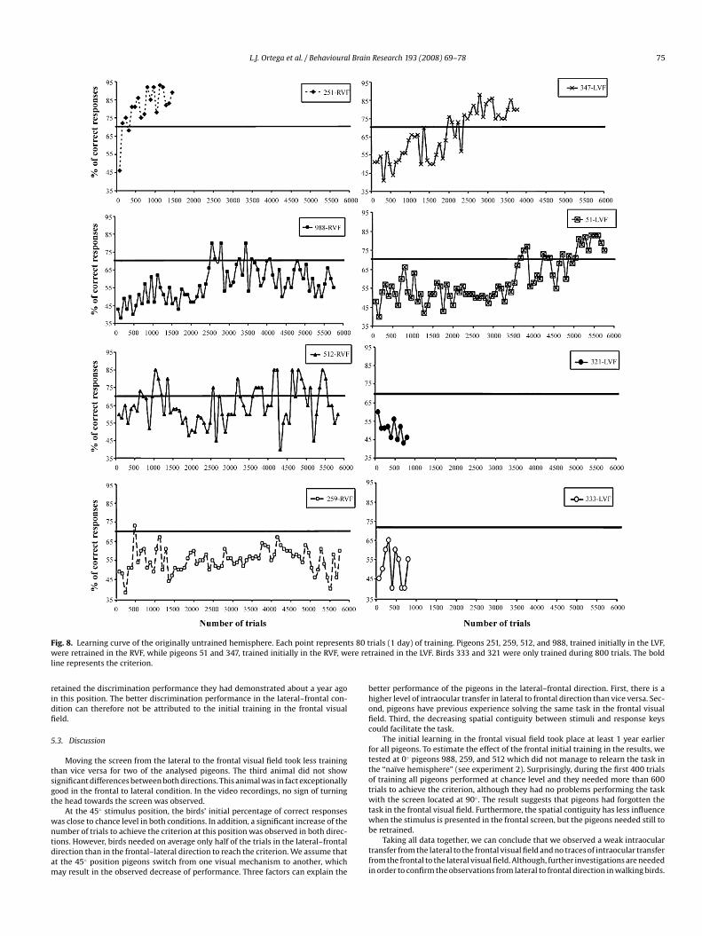
days of training (800 trials). At this point, the percentage of correct responses wasaround chance level for all birds except for pigeon 251 which showed clear signs ofinterocular transfer and reached the criterion within the third day, with percentagesof correct responses above 70%. Pigeons 51 and 347, trained initially in the RVF,learned the task in the LVF after 5120 (64 days of training) and 2400 trials (30 daysof training), respectively. Pigeons 988 and 259, trained initially in the RVF, wereincapable of learning the task in the LVF after 5760 (72 days of training). Anotherattempt to retrain a bird in the LVF, was made with pigeon 512. Even though thisbird reached the required criterion in several sessions, the behavioural results werevery unstable (Fig. 8). The analysis of the video recordings of pigeon 512 during thetraining revealed that this pigeon reached the criterion in sessions during which itwas turning the head towards the screen, whereas this behaviour was not observedin any other pigeon.
4.3. Discussion
Only one out of eight birds showed spontaneous interocular transfer. Other-wise, the performance level for the catch trials in the untrained hemisphere was
Research 193 (2008) 69–78
around chance level (50%), whereas in the trained hemisphere birds obtained dataabove criterion level (70%). In consonance to previous experiments [26,24,10] ourresults confirm a lack of interocular transfer when the stimuli are perceived withinthe yellow visual field. The only exception, pigeon 251, gave correct responses in100% of the catch trials; therefore it showed clear interocular transfer of informa-tion. We did not observe any differences during the experimental procedure thatcould explain those results. Inspection of video recordings of the animal trainingexcluded that pigeon 251 was twisting the head to observe the stimuli within thefrontal visual field. In addition, the animal reached the learning criterion after onlythree sessions of training in the naıve hemisphere (Fig. 8). Most probably the birddid not reach the criterion earlier due to an extinction phenomenon after beingexposed to catch trials without punishment or reward. Further observations con-firmed that pigeon 251 scored a higher percentage of correct responses in the RVFthan in the LVF. Those results could be a consequence of individual differences inhemispheric lateralisation of pigeon 251. Although most individuals of a speciesshow similar direction bias in several behaviours (for example, pecking behaviourin pigeons and handedness in humans) as a consequence of brain lateralisation,it is common to find a percentage of individuals with a different direction bias[16,44].
Birds initially trained in the RVF relearned the task in the LVF only after veryextended training, whereas birds initially trained in the LVF (except pigeon 251which transferred information) did not relearn the task in the RVF at all. Learning todiscriminate between two shapes in the lateral visual field while walking betweentwo feeders in an open arena has been demonstrated to be a very demanding taskfor the pigeons. Long distances between the discriminative stimuli and the responsesite may increase the difficulties of a discrimination task [45].
5. Experimental 3: limits of intraocular transfer in pigeons: lateral tofrontal direction
5.1. Methods
Pigeons 51, 251, and 347, which in the interocular transfer experiment learnedthe task in the initially “naıve” hemisphere, were used for testing intraocular transferof information from lateral to frontal direction. In order to have similar conditions asin Experiment 1, the screen was initially located at 95◦ . The animals were tested dur-ing 4 sessions of 20 trials each per day. The pigeons were retrained at each positionof the screen, until achieving the criterion (70% of correct responses in four consec-utive sessions). Then the screen was moved to the next position. The same screenpositions as in the Experiment 1 were used, and the data of the current experimentwere directly compared to the ones obtained in the first experiment.
Additionally, as a measure of the learning effect during the initial training,pigeons 988, 259, and 512 were tested in the same task exclusively at 0◦ . Thesepigeons shared the same training history as pigeons 51, 251, and 347. Training atthis position had taken place at least 1 year before for all pigeons. Therefore, test-ing these birds at 0◦ provides a valuable measure of the effect of the initial trainingthat might confound the transfer of information from the lateral back to a frontalstimulus position.
5.2. Results
Moving the screen all the way from the lateral to the frontal position(lateral–frontal condition) took a total amount of 800, 900, and 1180 trials (aver-
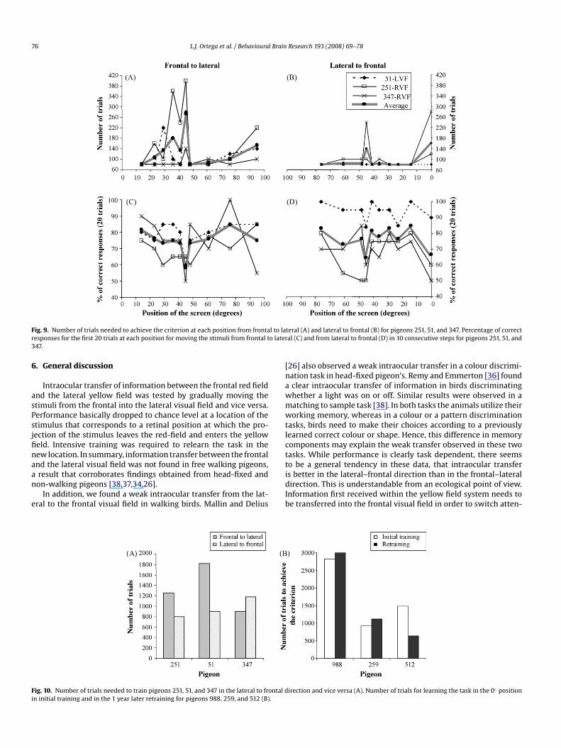
age = 960 trials) for pigeons 251, 51, and 347, respectively. In Experiment 1 movingthe screen from the frontal to the lateral visual field (frontal–lateral condition) tooka total amount of 1260, 1820, and 900 trials (average = 1326.6 trials) for pigeons 251,51, and 347, respectively (Fig. 9). A one tailed t-test analysis between the number oftrials needed at each position to move the screen from frontal to lateral direction andvice versa, confirmed that pigeons 251 and 51 needed significantly less sessions oftraining in the lateral–frontal condition than in the frontal–lateral condition (t = 2.29,d.f. = 9, p < 0.05 and t = 2.29, d.f. = 9, p < 0.05, respectively). Whereas pigeon 347 didnot show significant differences between the frontal–lateral and lateral–frontal con-dition (t = 1.02, d.f. = 9, p > 0.05). At position 45◦ , the birds needed on average 140trials in the lateral–frontal condition and 273 trials in the frontal–lateral condi-tion, although a Wilcoxon test revealed no significant differences (z = −1.06, p = 0.28)probably due to the limited sample of birds.The percentage of correct responses during the first 20 trials at each new stim-ulus position is similar in the frontal–lateral condition and in the lateral–frontalcondition (Fig. 9). No significant differences were found for any of the three pigeons.In both conditions, a decrease of performance close to chance level was observedat 45◦ for all three pigeons. In addition pigeon 251 showed percentages of correctresponses close to chance level at 61◦ , 48◦ , in the lateral–frontal condition and at39◦ and 48◦ in the frontal–lateral condition.
The three animals directly tested at 0◦ screen position, showed a percentageof correct responses around chance level for the first 400 trials. The train-ing criterion was achieved after an average of 1587 trials. In the initial frontaltraining at 0◦ , birds required an average of 1740 trials to achieve the crite-rion (Fig. 10). A Wilcoxon statistic analysis revealed no significant differencesbetween the initial and the later training. We conclude that the birds had not
L.J. Ortega et al. / Behavioural Brain Research 193 (2008) 69–78 75
Fig. 8. Learning curve of the originally untrained hemisphere. Each point represents 80 twere retrained in the RVF, while pigeons 51 and 347, trained initially in the RVF, were retline represents the criterion.
retained the discrimination performance they had demonstrated about a year agoin this position. The better discrimination performance in the lateral–frontal con-dition can therefore not be attributed to the initial training in the frontal visualfield.
5.3. Discussion
Moving the screen from the lateral to the frontal visual field took less trainingthan vice versa for two of the analysed pigeons. The third animal did not showsignificant differences between both directions. This animal was in fact exceptionallygood in the frontal to lateral condition. In the video recordings, no sign of turningthe head towards the screen was observed.
At the 45◦ stimulus position, the birds’ initial percentage of correct responseswas close to chance level in both conditions. In addition, a significant increase of thenumber of trials to achieve the criterion at this position was observed in both direc-tions. However, birds needed on average only half of the trials in the lateral–frontaldirection than in the frontal–lateral direction to reach the criterion. We assume thatat the 45◦ position pigeons switch from one visual mechanism to another, whichmay result in the observed decrease of performance. Three factors can explain the
rials (1 day) of training. Pigeons 251, 259, 512, and 988, trained initially in the LVF,rained in the LVF. Birds 333 and 321 were only trained during 800 trials. The bold
better performance of the pigeons in the lateral–frontal direction. First, there is ahigher level of intraocular transfer in lateral to frontal direction than vice versa. Sec-ond, pigeons have previous experience solving the same task in the frontal visualfield. Third, the decreasing spatial contiguity between stimuli and response keyscould facilitate the task.
The initial learning in the frontal visual field took place at least 1 year earlierfor all pigeons. To estimate the effect of the frontal initial training in the results, wetested at 0◦ pigeons 988, 259, and 512 which did not manage to relearn the task inthe “naıve hemisphere” (see experiment 2). Surprisingly, during the first 400 trialsof training all pigeons performed at chance level and they needed more than 600trials to achieve the criterion, although they had no problems performing the taskwith the screen located at 90◦ . The result suggests that pigeons had forgotten thetask in the frontal visual field. Furthermore, the spatial contiguity has less influencewhen the stimulus is presented in the frontal screen, but the pigeons needed still tobe retrained.
Taking all data together, we can conclude that we observed a weak intraoculartransfer from the lateral to the frontal visual field and no traces of intraocular transferfrom the frontal to the lateral visual field. Although, further investigations are neededin order to confirm the observations from lateral to frontal direction in walking birds.
76 L.J. Ortega et al. / Behavioural Brain Research 193 (2008) 69–78
Fig. 9. Number of trials needed to achieve the criterion at each position from frontal to laresponses for the first 20 trials at each position for moving the stimuli from frontal to late347.
6. General discussion
Intraocular transfer of information between the frontal red fieldand the lateral yellow field was tested by gradually moving thestimuli from the frontal into the lateral visual field and vice versa.Performance basically dropped to chance level at a location of thestimulus that corresponds to a retinal position at which the pro-jection of the stimulus leaves the red-field and enters the yellowfield. Intensive training was required to relearn the task in thenew location. In summary, information transfer between the frontaland the lateral visual field was not found in free walking pigeons,a result that corroborates findings obtained from head-fixed andnon-walking pigeons [38,37,34,26].
In addition, we found a weak intraocular transfer from the lat-eral to the frontal visual field in walking birds. Mallin and Delius
Fig. 10. Number of trials needed to train pigeons 251, 51, and 347 in the lateral to frontal din initial training and in the 1 year later retraining for pigeons 988, 259, and 512 (B).
teral (A) and lateral to frontal (B) for pigeons 251, 51, and 347. Percentage of correctral (C) and from lateral to frontal (D) in 10 consecutive steps for pigeons 251, 51, and
[26] also observed a weak intraocular transfer in a colour discrimi-nation task in head-fixed pigeon’s. Remy and Emmerton [36] founda clear intraocular transfer of information in birds discriminatingwhether a light was on or off. Similar results were observed in amatching to sample task [38]. In both tasks the animals utilize theirworking memory, whereas in a colour or a pattern discriminationtasks, birds need to make their choices according to a previouslylearned correct colour or shape. Hence, this difference in memorycomponents may explain the weak transfer observed in these twotasks. While performance is clearly task dependent, there seemsto be a general tendency in these data, that intraocular transferis better in the lateral–frontal direction than in the frontal–lateraldirection. This is understandable from an ecological point of view.Information first received within the yellow field system needs tobe transferred into the frontal visual field in order to switch atten-
irection and vice versa (A). Number of trials for learning the task in the 0◦ position
l Brain
[
[
L.J. Ortega et al. / Behavioura
tion to interesting or important environmental stimuli into an areaof the visual field in which they can be manipulated (e.g. wherefood can be picked up). Also, note that our experimental design didrequire a certain degree of information transfer between the yellowand the red field, because in our task birds perceived the patternwithin the yellow field but directed the pecking response to a pointin the red field.
Birds have been observed fixating objects within the lateralvisual field [6]. These fixation movements may be required to focusthe stimuli with the fovea centralis, an area of high ganglion celldensity within the yellow visual field. Indeed, the monocular acu-ity in the lateral field of pigeons is about the same in the binocularfrontal field [20,17]. In the experimental task as soon as the birdslearned the task in the lateral visual field, they did not show difficul-ties to transfer information from one position to another within thelateral visual field, but transfer from frontal to lateral visual fieldswas lacking.
We also investigated interocular transfer of informationbetween the two yellow fields in our free walking pigeons. Pigeonsinitially trained in the LVF were tested in the RVF, and vice versa.In eight of the nine pigeons, no traces of interocular transfer ofinformation were observed. Only one pigeon was capable of per-forming the task spontaneously in the untrained visual field. Ourresults confirm, in a more ecological situation, previous experimen-tal findings in which a lack of interocular transfer was found whenthe stimuli were perceived within the yellow visual field [10,23,26].However, several studies demonstrate the existence of interoculartransfer of information within the red visual field [10,26,23–25].Due to the almost complete decussation of the optic nerve inpigeons [47], interhemispheric transfer of information received inthe red visual field is required for local coarse stereopsis [30,29].By contrast transfer between the lateral visual fields might not bea requirement for the birds. The lateral visual field serves visionat longer distances, monitoring predators and cospecifics [11,8], aswell as to detect food at farther distances than 10 cm [3]. In natu-ral conditions an alert response can be activated without transfer ofvisual information. The trigger of the motor response after process-ing a possible alert signal in one of the two hemispheres could beenough for the bird to exhibit the appropriate behaviour in a givensituation. In the present experiment, birds exposed to catch trialsin the untrained hemisphere exhibited pecking behaviour, but theywere not able to discriminate between the two stimuli. Whereas ifthe stimulus was not presented, the birds remained static until anew stimulus appeared.
In pigeons, a number of studies have demonstrated a righteye/left hemisphere dominance for object recognition in the frontalbinocular field [18,12,13]. In contrast, we observed no such asym-metry. In the intraocular transfer experiment, two animals trainedin the right visual field never learned the task beyond 45◦. Fur-thermore, in the interocular transfer experiments (excluding a birdthat was capable of transfer) after long lasting training, birds ini-tially trained in the RVF relearned the task in the LVF, whereas birdsinitially trained in the LVF (except the animal capable of informa-tion transfer) did not relearn the task in the RVF. A left hemispheredominance of the thalamofugal visual pathway was observed ina pattern discrimination task in an open arena [4] social recogni-tion in chicks, and novel stimuli detection [3,44,7,39]. A lateralisedbrain may allow dual attention to short distance tasks like feed-ing (using the right eye/left hemisphere system) and long distancetasks like vigilance for predators (left eye/right hemisphere system)[39].
In summary, information transfer was not observed from thefrontal to the lateral visual field of the same eye. Interocular transferbetween the yellow fields of the two eyes was absent. A weak trans-fer from the lateral to the frontal visual field of the same eye was
[
[
[
[
[
[
[
[
[
[
[
[
Research 193 (2008) 69–78 77
observed. At first glance, it is surprising to find that the two hemi-spheres of the pigeon’s brain work in many aspects as independentbrains, however a closer look reveals that they are perfectly adaptedanimals capable of solving environmental demands minimizingbrain complexity. Pigeons show interocular transfer of informationwithin the red visual field of the eyes that serves local stereop-sis, but no interocular transfer of information between the yellowfields. Information transfer from the lateral to the frontal visual fieldof the same eye may be useful to fixate relevant stimuli within thered visual field, but intraocular transfer from the frontal to the lat-eral visual field has no obvious function and may not provide anyadvantage.
Acknowledgements
This work was supported by the International Graduate Schoolof Neuroscience, and grants from the Deutsche Forschungsgemein-schaft to NFT and OG, as well as grants from Natural Science andEngineering Research Council and Canada Fund for Innovation toNT. We thank Antigoni Marioli for helping with the initial trainingof the pigeons, and the workshop of the Psychology Department ofthe Ruhr-Universitat-Bochum and Tobias Otto for constructing theexperimental arena and programming its software.
References
[1] Beritov IS, Chichinadse. Localisation of visual perception in the pigeon bulletinde biologie et de médicine expérimentale 1935;11:103–4.
[2] Bischof HJ, Watanabe S. On the structure and function of the tectofugal visualpathway in laterally eyed birds. Eur J Morphol 1997;35:246–54.
[3] Bloch S, Martinoya C. In: Erwert JP, et al., editors. Advances in vertebrate neu-roethology. New York: Plehum Press; 1982. p. 359–68.
[4] Budzynski CA, Bingman VP. Participation of the thalamofugal visual pathwayin a coarse pattern discrimination task in an open arena. Behav Brain Res2004;153:543–56.
[5] Catania AC. Interocular transfer of discriminations in pigeon. J Exp Anal Behav1965;47:147–55.
[6] Dawkins MS. What are birds looking at? Head movements and eye use in chick-ens. Anim Behav 2002:63.
[7] Evans CS, Evans L, Marier P. On the meaning of alarm calls: functional referencesin ten avian vocal system. Anim Behav 1993;46:23–8.
[8] Fernandez-Juricic E, Erichsen JT, Kacelnik A. Visual perception and social forag-ing in birds. Trends Ecol Evol 2004;19:25–31 [Personal edition].
[9] Gaston KE. Interocular transfer of pattern discrimination learning in chicks.Brain Res 1984;310:213–21.
10] Goodale MA, Graves JA. In: Ingle DJ, et al., editors. Advances in analysis of visualbehaviour. London: MIT Press; 1982. p. 211–40.
11] Green PR, Davies MNO, Thorpe PH. Head-bobbing and head orientation duringlanding flights of pigeons. J Comp Physiol 1994;174:249–56.
12] Güntürkün O. Lateralization of visually controlled behavior in pigeons. PhysiolBehav 1985;34:575–7.
13] Güntürkün O. Morphological asymmetries of the tectum opticum in the pigeon.Exp Brain Res 1997;116:561–6.
14] Güntürkün O. In: Davidson KHRJ, editor. The asymmetrical brain. London: MITPress; 2003. p. 4–36.
15] Güntürkün O, Bohringer PG. Lateralization reversal after intertectal commis-surotomy in the pigeon. Brain Res 1987;408:1–5.
16] Güntürkün O, Diekamp B, Manns M, Nottelmann F, Prior H, Schwarz A, etal. Asymmetry pays: visual lateralization improves discrimination success inpigeons. Curr Biol 2000;10:1079–81.
17] Güntürkün O, Hahmann U. Visual acuity and hemispheric asymmetries inpigeons. Behav Brain Res 1994;60:171–5.
18] Güntürkün O, Kesch S. Visual lateralization during feeding in pigeons. BehavNeurosci 1987;101:433–5.
19] Güntürkün O, Miceli D, Watanabe M. In: Zeigler HP, Bischof HJ, editors. Vision,brain and behavior in birds. Cambridge, MA: MIT Press; 1993. p. 115–35.
20] Hahmann U, Güntürkün O. The visual acuity for the lateral visual field of thepigeon (Columba livia). Vision Res 1993;33:1659–64.
21] Jarvis ED, Güntürkün O, Bruce L, Csillag A, Karten H, Kuenzel W, et al. Avianbrains and a new understanding of vertebrate brain evolution. Nat Rev Neurosci2005;6:151–9.
22] Keysers C, Diekamp B, Güntürkün O. Evidence for physiological asymmetriesin the intertectal connections of the pigeon (Columba livia) and their potentialrole in brain lateralisation. Brain Res 2000;852:406–13.
23] Levine J. Studies in the interrelations of central nervous structures in binocularvision. I. The lack of bilateral transfer of visual discriminative habits acquiredmonocularly by pigeons. J Genet Psychol 1945;67:105–29.
l Brain
[
[
[
[
[
[
[
[
[
[
[
[
[
78 L.J. Ortega et al. / Behavioura
24] Levine J. Studies in the interrelations of central nervous structures in binocularvision. II. The conditions under which interocular transfer of discriminatives
habits takes place in pigeon. J Genet Psychol 1945;67:131–42.25] Levine J. Studies in the interrelations of central nervous structures in vision.III. Localization of the memory trace as evidenced by the lack of inter- andintraocular habit transfer in the pigeon. J Genet Psychol 1952;81:19–27.
26] Mallin HD, Delius J. Inter- and intraoccular transfer of colour discriminationswith mandibulation as an operant in the head-fixed pigeon. Behav Anal Lett1983;3:297–309.
27] Martin GR. Visual fields and their functions in birds. J Ornithol 2007;148(Suppl.2):547–62 [december de 2007].
28] Martin GR, Young SR. The retinal binocular field of the pigeon (Columba livia:English racing homer). Vision Res 1983;23:911–5.
29] McFadden SA. In: Zeigler HP, Bischof H-J, editors. Vision, brain and behavior inbirds. Cambridge, MA: The MIT Press; 1993. p. 245–63.
30] McFadden SA, Wild JM. Binocular depth perception in the pigeon Columba livia.J Exp Anal Behav 1986;45:149–60.
31] Miceli D, Reperant J, Medina M, Volle M, Rio JP. Distribution of ganglion cells inthe pigeon retina labeled via retrograde transneuronal transport of the fluores-cent dye rhodamine beta-isothiocyanate from the telencephalic visual. WulstBrain Res 2006;1098:94–105.
32] Nalbach HO, Wolf-Oberhollenzer F, Remy M. In: Zeigler HP, Bischof HJ, editors.Vision, brain and behavior in birds. Cambridge, MA: MIT Press; 1993. p. 25–46.
33] Nottelmann F, Wohlschlager A, Güntürkün O. Unihemispheric memory inpigeons-knowledge, the left hemisphere is reluctant to share. Behav Brain Res2002;133:309–15.
34] Nye PW. On the functional differences between frontal and lateral visual fieldsof the pigeon. Vision Res 1973;13:559–74.
35] Parsons CH, Rogers LJ. Role of the tectal and posterior commissures in lateral-ization of the avian brain. Behav Brain Res 1993;54:153–64.
[
[
[
[
[
[
[
[
[
[
[
Research 193 (2008) 69–78
36] Remy M, Emmerton J. Directional dependence of intraocular transfer of stimu-lus detection in pigeons (Columba livia). Behav Neurosci 1991;105:647–52.
37] Remy M, Watanabe S. Two eyes and one world: studies of interocular andintraocular transfer in birds. In: Zeigler HP, Bischof H-J, editors. Vision, brain,and behaviour in birds. Cambridge, MA: The MIT Press; 1993. p. 330–50.
38] Roberts WA, Phelps MT, Macuda T, Brodbeck DR, Russ T. Intraocular transferand simultaneous processing of stimuli presented in different visual fields ofthe pigeon. Behav Neurosci 1996;110:290–9.
39] Rogers LJ. Evolution of hemispheric specialization: advantages and disadvan-tages. Brain Lang 2000;73:236–53.
40] Rounsley KJ, McFadden SA. Limits of visual acuity in the frontal field of the rockpigeon (Columba livia). Perception 2005;34:983–93.
41] Shimizu T, Cox K, Karten HJ. Intratelencephalic projections of the visual wulstin pigeons (Columba livia). J Comp Neurol 1995;359:551–72.
42] Skiba M, Diekamp B, Prior H, Güntürkün O. Lateralized interhemispheric trans-fer of color cues: evidence for dynamic coding principles of visual lateralizationin pigeons. Brain Lang 2000;73:254–73.
43] Stevens JV, Kirsch WR. Interocular transfer in pigeons of color discriminationbut not motor response training. Anim Learn Behav 1980;8:17–21.
44] Vallortigara G. The evolutionary psychology of left and right: costs and benefitsof lateralization. Dev Psychobiol 2006;48:418–27.
45] Watanabe S. Interocular transfer of learning in the pigeon: visuo-motor inte-gration and separation of discriminanda and manipulanda. Behav Brain Res1986;19:227–32.
46] Watanabe S, Hodos W, Bessette BB, Shimizu T. Interocular transfer in parallelvisual pathways in pigeons. Brain Behav Evol 1986;29:184–95.
47] Weidner C, Reperant J, Miceli D, Haby M, Rio JP. An anatomical studyof ipsilateral retinal projections in the quail using radioautographic,horseradish peroxidase, fluorescence and degeneration techniques. Brain Res1985;340:99–108.
Copyright © 2022 FDOKUMEN