A Differential Phase-Modulated Interferometer with Rotational ...
Upload
khangminh22Category
view
1download
0
RESEARCH ARTICLE
Rotational stability of modified toric
intraocular lens
Ryoko Osawa1, Tetsuro OshikaID2*, Masahiko Sano1, Takuma Yuguchi1,
Tadayoshi Kaiya1
1 Kaiya Eye Clinic, Hamamatsu, Shizuoka, Japan, 2 Department of Ophthalmology, Faculty of Medicine,
University of Tsukuba, Ibaraki, Japan
Abstract
We evaluated the rotational stability of a new toric intraocular lens (IOL), HOYA XY-1 toric
IOL that is an improved version of HOYA 355 toric IOL, with longer overall length (13.0 mm
vs. 12.5 mm), shortened unfolding time, and texture processing of the surface of haptics.
Data from 193 eyes of 165 patients (76.4 ± 8.3 years old) with preoperative corneal astigma-
tism exceeding 0.75 diopters who had undergone phacoemulsification and toric IOL implan-
tation were collected and analyzed. Corneal astigmatism, refractive astigmatism, and
uncorrected (UDVA) and corrected distance visual acuity (CDVA) were evaluated before
and 1 day, 1 week, and 1 month after surgery. The degree of IOL decentration, IOL tilt, and
toric axis misalignment was assessed at 1 day and 1 month postoperatively. Fifty eyes
received AcrySof toric IOL, 51 eyes TECNIS toric IOL, 46 eyes HOYA 355 toric IOL, and 46
eyes HOYA XY-1 toric IOL. The amount of axis misalignment from the intended axis was
significantly different among IOLs (p = 0.004, one-way ANOVA), and HOYA XY-1 showed
significantly less amount of axis misalignment than TECNIS (p = 0.020, Tukey’s multiple
comparison) and HOYA 355 (p = 0.010). The proportion of eyes that showed axis misalign-
ment <10˚ at 1 month postoperatively was significantly higher with HOYA XY-1 toric IOL
than with other toric IOLs (χ2 test, p = 0.020). HOYA XY-1 toric IOL, the modified version of
HOYA 355 toric IOL, showed excellent rotational stability in comparison with other models
of toric IOLs.
Introduction
Reduction of preexisting astigmatism at the time of cataract surgery using a toric intraocular
lens (IOL) has become increasingly popular and is now regarded as the integral part of modern
cataract surgery by many surgeons. For the success of toric IOL implantation, placement accu-
racy and rotational stability are the key factors. Placement accuracy of a toric IOL during sur-
gery has been significantly improved by the advent of digital image guidance systems [1–3].
On the other hand, postoperative rotational stability varies across different models of toric
IOLs [4–7]. A previous study demonstrated that the incidence of repositioning surgery to cor-
rect misalignment of toric IOLs was significantly higher with HOYA 355 toric IOL (HOYA,
PLOS ONE
PLOS ONE | https://doi.org/10.1371/journal.pone.0247844 March 1, 2021 1 / 10
a1111111111
a1111111111
a1111111111
a1111111111
a1111111111
OPEN ACCESS
Citation: Osawa R, Oshika T, Sano M, Yuguchi T,
Kaiya T (2021) Rotational stability of modified toric
intraocular lens. PLoS ONE 16(3): e0247844.
https://doi.org/10.1371/journal.pone.0247844
Editor: Ahmed Awadein, Faculty of Medicine, Cairo
University, EGYPT
Received: December 18, 2020
Accepted: February 13, 2021
Published: March 1, 2021
Copyright: © 2021 Osawa et al. This is an open
access article distributed under the terms of the
Creative Commons Attribution License, which
permits unrestricted use, distribution, and
reproduction in any medium, provided the original
author and source are credited.
Data Availability Statement: The data underlying
the results presented in the study are available
from figshare, https://doi.org/10.6084/m9.figshare.
13724857.v1
Funding: The authors received no specific funding
for this work.
Competing interests: Tetsuro Oshika has received
research grants and speaker honorarium from
Alcon, HOYA, and Johnson & Johnson Vision.
Other authors have declared that no competing
interests exist. This does not alter our adherence to
PLOS ONE policies on sharing data and materials.

Tokyo, Japan) than with AcrySof toric IOL (Alcon Laboratories, Inc., Fort Worth, TX) [6].
Another clinical study reported relatively large degree of lens rotation associated with implan-
tation of HOYA 355 toric IOL [8].
Recently, HOYA modified its 355 toric IOL to be a new version, HOYA XY-1 toric IOL, by
increasing the overall length, reducing the unfolding time, and introducing texture processing
to the surface of haptics. These modifications are likely to affect the rotational stability of toric
IOLs, but there has been no study to prove that hypothesis. We conducted the current study to
compare the surgical outcomes of new HOYA toric IOLs with those of other models of toric
IOLs.
Patients and methods
Patients
Data from 193 eyes of 165 patients who had been treated with phacoemulsification and
implantation of a toric IOL from July 2017 to February 2020 were retrospectively collected.
They had corneal astigmatism of 0.75 diopter (D) or more, and were targeted emmetropic.
None of the eyes had ocular or systemic diseases that could affect the surgical outcomes includ-
ing visual acuity. Eyes were excluded if there were any intraoperative complications that affect
IOL stability. After surgery, the patients were followed up for at least 1 month. An informed
consent in written form was obtained from each patient. The study adhered to the tenets of the
Declaration of Helsinki, and the institutional review board of Kaiya Eye Clinic approved the
study protocol.
Intraocular lenses and surgery
Four models of toric IOLs were used, including AcrySof toric IOL, TECNIS toric IOL (John-
son & Johnson Vision Care, Inc., Santa Ana, CA), HOYA 355 toric IOL, and HOYA XY-1
toric IOL.
HOYA XY-1 is an improved version of HOYA 355 toric IOL, with the overall length 0.5
mm longer (13.0 mm vs. 12.5 mm), shortened unfolding time by changing the material (man-
ufacturer’s internal data), and texture processing of the surface of haptics against smooth sur-
face with the old model. The optic is 6.0 mm in diameter and has toricity on the posterior
surface with asphericity on the anterior surface. The lens is made of hydrophobic acrylic
material.
A single surgeon (TK) operated on all cases with standard phacoemulsification and IOL
implantation through a 2.4-mm wound. Anterior capsulorhexis of approximately 5.0 mm in
diameter was created and the IOL was implanted into the capsular bag using an injector. The
VERION Image-Guided System (Alcon), which consists of a measurement module and digital
marker, was employed to conduct digital marking for axis alignment of toric IOLs. Using the
measurement module, a high-resolution color reference image of preoperative patient’s eye
was captured, which was transferred to the digital marker. Based on multiple reference points
of the conjunctiva and limbus, a digital overlay of the imported preoperative image and live-
surgery image were created. The eye-tracking navigation system suppressed the influence of
cyclotorsion and eye movements, and the targeted placement axis of a toric IOL is accurately
projected in the right ocular of the surgeon’s microscope.
Examinations
Preoperatively, axial length and corneal curvature were measured with IOLMaster 700 (Carl
Zeiss, Germany) and IOL power was calculated using the SRK/T formula. The emmetropia
PLOS ONE Rotational stability of modified toric intraocular lens
PLOS ONE | https://doi.org/10.1371/journal.pone.0247844 March 1, 2021 2 / 10

was the target in all of the eyes. The IOL cylinder power and alignment axis were determined
using the designated manufacturer’s online calculator programs.
Corneal astigmatism, manifest astigmatism, and uncorrected (UDVA) and corrected dis-
tance visual acuity (CDVA) were measured before and 1 day, 1 week, and 1 month after sur-
gery. Using the swept-source anterior segment optical coherence tomography (AS-OCT,
CASIA, Tomey Corp., Nagoya, Japan), IOL decentration, IOL tilt, and toric axis misalignment
was measured at 1 day and 1 month postoperatively [9–11]. The toric IOL analysis tool
equipped with the AS-OCT shows an image of the anterior segment with an overlapped green
linear marker that can be rotated on a fulcrum automatically centered on the corneal apex
[11]. The linear marker is aligned parallel to the line connecting the marking dots of the toric
IOL, and the direction of this alignment is expressed in angle degrees. The corneal topography
obtained from the same scan is shown together with the power of steeper and flatter meridians,
their axes, as well as the amount of the resulting topographic cylinder. The rotation of the toric
IOL from the intended position can be expressed by the difference in degrees between the
topographic axis and the value calculated for the linear marker [11]. The examiners were
blinded to the type of IOL that the eye had received.
Statistical analysis
Numerical data are expressed as mean ± standard deviation. Statistical comparisons among
multiple groups were performed using the one-way analysis of variance (ANOVA) followed by
the Tukey’s multiple comparison. The categorical data were compared among groups with the
χ2 test. Statistical analysis was conducted with SPSS Statistics for Windows software (version
26, IBM Corp., Armonk, NY, USA). A p-value of less than 0.05 was considered statistically
significant.
Results
Preoperative characteristics of patients are shown in Table 1. Fifty eyes received AcrySof toric
IOL, 51 eyes TECNIS toric IOL, 46 eyes HOYA 355 toric IOL, and 46 eyes HOYA XY-1 toric
IOL. No significant differences were found in the baseline characteristics of patients among
the groups.
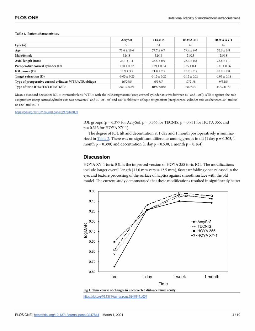
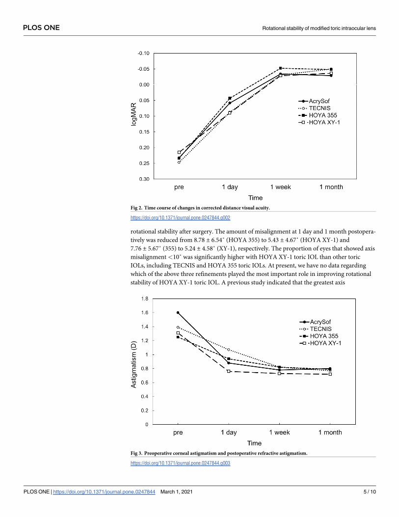
Postoperatively, there was no significant difference in UDVA (Fig 1), CDVA (Fig 2), and
residual refractive astigmatism (Fig 3) among the four toric IOLs. There were no intraopera-
tive and postoperative complications relevant to the use of toric IOLs.
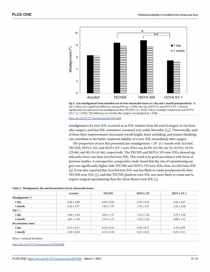
The amount of axis misalignment from the intended axis is shown in Fig 4 and Table 2. At
1 day postoperatively, there was a significant difference among IOLs (p = 0.004, one-way
ANOVA), and HOYA XY-1 showed significantly less amount of axis misalignment than TEC-
NIS (p = 0.020, Tukey’s multiple comparison) and HOYA 355 (p = 0.010). The difference at 1
month after surgery was marginal (p = 0.08), with HOYA XY-1 showing the smallest amount
of misalignment. The proportion of eyes that presented axis misalignment <10˚ at 1 month
with AcrySof, TECNIS, HOYA 355, and HOYA XY-1 toric IOLs was 82.0% (41/50), 64.7%
(33/51), 54.3% (25/46), and 89.1% (41/46), respectively; HOYA XY-1 toric IOL had signifi-
cantly higher rate than other toric IOLs (χ2 test, p = 0.020). In all groups, more than half of
eyes showed counterclockwise rotation of toric IOLs at 1 month (Fig 5). The ratio did not dif-
fer significantly among groups (p = 0.456). The repositioning surgery to correct axis misalign-
ment was not carried out in all groups.
There was no statistically significant correlation between axial length and the among of mis-
alignment at 1 month in all cases (p = 0.977). The correlation was not significant in individual
PLOS ONE Rotational stability of modified toric intraocular lens
PLOS ONE | https://doi.org/10.1371/journal.pone.0247844 March 1, 2021 3 / 10
IOL groups (p = 0.377 for AcrySof, p = 0.366 for TECNIS, p = 0.731 for HOYA 355, and
p = 0.313 for HOYA XY-1).
The degree of IOL tilt and decentration at 1 day and 1 month postoperatively is summa-
rized in Table 2. There was no significant difference among groups in tilt (1 day p = 0.305, 1
month p = 0.390) and decentration (1 day p = 0.530, 1 month p = 0.164).
Discussion
HOYA XY-1 toric IOL is the improved version of HOYA 355 toric IOL. The modifications
include longer overall length (13.0 mm versus 12.5 mm), faster unfolding once released in the
eye, and texture processing of the surface of haptics against smooth surface with the old
model. The current study demonstrated that these modifications resulted in significantly better
Table 1. Patient characteristics.
AcrySof TECNIS HOYA 355 HOYA XY-1
Eyes (n) 50 51 46 46
Age 71.6 ± 10.6 77.7 ± 6.7 79.4 ± 6.0 76.0 ± 6.8
Male/female 32/18 32/19 21/25 28/18
Axial length (mm) 24.1 ± 1.4 23.5 ± 0.9 23.3 ± 0.8 23.6 ± 1.1
Preoperative corneal cylinder (D) 1.60 ± 0.67 1.39 ± 0.54 1.25 ± 0.41 1.31 ± 0.56
IOL power (D) 18.9 ± 3.7 21.0 ± 2.5 20.2 ± 2.5 20.9 ± 2.8
Target refraction (D) -0.05 ± 0.23 -0.15 ± 0.22 -0.15 ± 0.24 -0.03 ± 0.18
Type of preoperative corneal cylinder: WTR/ATR/oblique 16/29/5 6/38/7 17/21/8 9/32/5
Type of toric IOLs: T3/T4/T5/T6/T7 29/10/8/2/1 40/8/3/0/0 39/7/0/0 34/7/4/1/0
Mean ± standard deviation; IOL = intraocular lens; WTR = with-the-rule astigmatism (steep corneal cylinder axis was between 60˚ and 120˚); ATR = against-the-rule
astigmatism (steep corneal cylinder axis was between 0˚ and 30˚ or 150˚ and 180˚); oblique = oblique astigmatism (steep corneal cylinder axis was between 30˚ and 60˚
or 120˚ and 150˚).
https://doi.org/10.1371/journal.pone.0247844.t001
Fig 1. Time course of changes in uncorrected distance visual acuity.
https://doi.org/10.1371/journal.pone.0247844.g001
PLOS ONE Rotational stability of modified toric intraocular lens
PLOS ONE | https://doi.org/10.1371/journal.pone.0247844 March 1, 2021 4 / 10
rotational stability after surgery. The amount of misalignment at 1 day and 1 month postopera-
tively was reduced from 8.78 ± 6.54˚ (HOYA 355) to 5.43 ± 4.67˚ (HOYA XY-1) and
7.76 ± 5.67˚ (355) to 5.24 ± 4.58˚ (XY-1), respectively. The proportion of eyes that showed axis
misalignment <10˚ was significantly higher with HOYA XY-1 toric IOL than other toric
IOLs, including TECNIS and HOYA 355 toric IOLs. At present, we have no data regarding
which of the above three refinements played the most important role in improving rotational
stability of HOYA XY-1 toric IOL. A previous study indicated that the greatest axis
Fig 2. Time course of changes in corrected distance visual acuity.
https://doi.org/10.1371/journal.pone.0247844.g002
Fig 3. Preoperative corneal astigmatism and postoperative refractive astigmatism.
https://doi.org/10.1371/journal.pone.0247844.g003
PLOS ONE Rotational stability of modified toric intraocular lens
PLOS ONE | https://doi.org/10.1371/journal.pone.0247844 March 1, 2021 5 / 10
misalignment of a toric IOL occurred as an IOL rotation from the end of surgery to one hour
after surgery, and that IOL orientation remained very stable thereafter [12]. Theoretically, each
of those three improvements (increased overall length, faster unfolding, and texture finishing)
can contribute to the better rotational stability of a toric IOL immediately after surgery.
The proportion of eyes that presented axis misalignment <10˚ at 1 month with AcrySof,
TECNIS, HOYA 355, and HOYA XY-1 toric IOLs was 82.0% (41/50), 64.7% (33/51), 54.3%
(25/46), and 89.1% (41/46), respectively. The TECNIS and HOYA 355 toric IOLs showed sig-
nificantly lower rate than AcrySof toric IOL. This result is in good accordance with those of
previous studies. A retrospective comparative study found that the rate of repositioning sur-
gery was significantly higher with TECNIS and HOYA 355 toric IOLs than AcrySof toric IOL
[6]. It was also reported that AcrySof toric IOL was less likely to rotate postoperatively than
TECNIS toric IOL [5], and that TECNIS platform toric IOL was more likely to rotate and to
require surgical repositioning than the Alcon Restor toric IOL [7].
Fig 4. Axis misalignment from intended axis of toric intraocular lenses at 1 day and 1 month postoperatively. At
day 1, there was a significant difference among IOLs (p = 0.004, one-way ANOVA), and HOYA XY-1 showed
significantly less amount of axis misalignment than TECNIS (�p = 0.020, Tukey’s multiple comparison) and HOYA
355 (��p = 0.010). The difference at 1 month after surgery was marginal (p = 0.08).
https://doi.org/10.1371/journal.pone.0247844.g004
Table 2. Misalignment, tilt, and decentration of toric intraocular lenses.
AcrySof TECNIS HOYA 355 HOYA XY-1
Misalignment (˚)
1 day 6.30 ± 4.80 8.38 ± 6.84 8.78 ± 6.54 5.43 ± 4.67
1 month 6.26 ± 4.77 7.83 ± 7.07 7.76 ± 5.67 5.24 ± 4.58
Tilt (˚)
1 day 4.60 ± 1.64 4.84 ± 1.17 5.12 ± 1.24 4.79 ± 1.28
1 month 4.81 ± 1.49 5.18 ± 1.21 5.12 ± 1.24 4.80 ± 1.11
Decentration (mm)
1 day 0.21 ± 0.11 0.18 ± 0.12 0.18 ± 0.11 0.16 ±0.09
1 month 0.30 ± 0.60 0.16 ± 0.10 0.21 ± 0.15 0.18 ± 0.11
Mean ± standard deviation.
https://doi.org/10.1371/journal.pone.0247844.t002
PLOS ONE Rotational stability of modified toric intraocular lens
PLOS ONE | https://doi.org/10.1371/journal.pone.0247844 March 1, 2021 6 / 10

Regarding the direction of axis misalignment of toric IOLs, a previous study reported that
AcrySof toric IOLs presented clockwise rotation in 76.2% and anti-clockwise rotation in 23.8%
[13]. Another study, on the other hand, found no trend for either clockwise or counterclock-
wise rotation with AcrySof toric IOLs [14]. By analyzing the on-line database, it was shown
that TECNIS toric IOL was more likely to be rotated in a counterclockwise direction, but no
such bias was found with AcrySof toric IOL [4]. In the present study, anti-clockwise misdirec-
tion was more frequent than clockwise misdirection with all four toric IOLs, but no significant
differences were observed in the incidence between groups.
In this study, the amount of IOL tilt and decentration of four toric IOLs was approximately
5˚ and 0.2 mm, respectively, and there were no significant differences among IOLs. The influ-
ence of IOL decentration and tilt on visual function varies depending on the design of IOLs
[15]. For toric IOLs, decentration and tilt can result in less predictable correction of astigma-
tism [16]. An experimental study using a ray tracing model demonstrated that the alignment
and toricity as well as orientation of tilt caused over-correction or under-correction of astig-
matism by toric IOLs [16]. A review analysis of studies about IOL decentration and tilt demon-
strated that 2–3˚ tilt and 0.2–0.3 mm decentration are common and practically unnoticed for
IOLs with any design [17]. A model eye investigation indicated that more than 5˚ tilt and
more than 1 mm IOL decentration can be visually significant, inducing oblique astigmatism
[18]. According to optical simulations with consideration of various visual aspects such as cor-
neal aberrations and pupil function, decentration of 0.5 mm or greater could be a source of
considerable visual symptoms [19]. The tilt of a 28 D aspheric IOL by 5˚ and 10˚ was calculated
to induce astigmatism of 0.14 D and 0.56 D, respectively [16]. In the literature, an average of
approximately 5˚ tilt and 0.2 mm decentration of IOLs implanted in the capsular bag have
been reported [20–24]. Judging from these previous studies, the degree of tilt and decentration
found with the toric IOLs used in our study is within an acceptable range, and its impacts on
postoperative visual functions seem minimal.
There are several limitations to this study. First, because this was a retrospective study, ran-
dom assignment of eyes to one of four toric IOLs was not conducted. Second, the part of study
Fig 5. Ratio of counterclockwise and clockwise rotation of toric intraocular lenses at 1 month postoperatively.
https://doi.org/10.1371/journal.pone.0247844.g005
PLOS ONE Rotational stability of modified toric intraocular lens
PLOS ONE | https://doi.org/10.1371/journal.pone.0247844 March 1, 2021 7 / 10

using HOYA 355 and XY-1 IOLs was not parallel, but sequential. This was because HOYA 355
toric IOL was pulled out of the market on the launch of HOYA XY-1 toric IOL, making the
use of both IOLs at the same time impossible. Third, the follow-up period of patients was 1
month, and longer clinical outcomes were not assessed. The orientation of toric IOLs, how-
ever, is highly stable after 1 day postoperatively [12], and thus we suppose that results at 1
month postoperatively are able to extrapolate the longer-term rotational stability of toric IOLs.
Third, the amount of postoperative misalignment found in the current study was somewhat
greater than those previously reported [25]. The degree of misalignment, however, does vary
significantly among studies, indicating that simple comparison of data between different stud-
ies with various background conditions of patients and surgical techniques would be difficult
[26].
Conclusions
We retrospectively assessed the surgical outcomes of four toric IOLs. HOYA XY-1 toric IOL,
the improved version of HOYA 355, showed excellent rotational stability in comparison with
other models of toric IOLs.
Author Contributions
Conceptualization: Ryoko Osawa, Tetsuro Oshika.
Data curation: Ryoko Osawa, Masahiko Sano, Takuma Yuguchi, Tadayoshi Kaiya.
Formal analysis: Ryoko Osawa, Tetsuro Oshika.
Funding acquisition: Tadayoshi Kaiya.
Investigation: Ryoko Osawa, Tetsuro Oshika, Masahiko Sano.
Methodology: Tetsuro Oshika.
Project administration: Tetsuro Oshika, Tadayoshi Kaiya.
Supervision: Tetsuro Oshika.
Validation: Ryoko Osawa, Tetsuro Oshika.
Writing – original draft: Tetsuro Oshika.
Writing – review & editing: Ryoko Osawa, Masahiko Sano, Takuma Yuguchi, Tadayoshi
Kaiya.
References1. Abdel Hamid Elhofi Hany Ahmed Helaly. Comparison between digital and manual marking for toric intra-
ocular lenses: A randomized trial. Medicine (Baltimore). 2015; 94: e1618. https://doi.org/10.1097/MD.
0000000000001618 PMID: 26402830
2. Titiyal JS, Kaur M, Jose CP, Falera R, Kinkar A, Bageshwar LM. Comparative evaluation of toric intraoc-
ular lens alignment and visual quality with image-guided surgery and conventional three-step manual
marking. Clin Ophthalmol. 2018; 12: 747–753. https://doi.org/10.2147/OPTH.S164175 PMID:
29731603
3. Panagiotopoulou EK, Ntonti P, Gkika M, Konstantinidis A, Perente I, Dardabounis D, et al. Image-
guided lens extraction surgery: a systematic review. Int J Ophthalmol. 2019; 12: 135–151. https://doi.
org/10.18240/ijo.2019.01.21 PMID: 30662853
4. Potvin R, Kramer BA, Hardten DR, Berdahl JP. Toric intraocular lens orientation and residual refractive
astigmatism: an analysis. Clin Ophthalmol. 2016; 10: 1829–1836. https://doi.org/10.2147/OPTH.
S114118 PMID: 27703323
PLOS ONE Rotational stability of modified toric intraocular lens
PLOS ONE | https://doi.org/10.1371/journal.pone.0247844 March 1, 2021 8 / 10
5. Lee BS, Chang DF. Comparison of the rotational stability of two toric intraocular lenses in 1273 conse-
cutive eyes. Ophthalmology. 2018; 125: 1325–1331. https://doi.org/10.1016/j.ophtha.2018.02.012
PMID: 29544960
6. Oshika T, Fujita Y, Hirota A, Inamura M, Inoue Y, Miyata K, et al. Comparison of incidence of reposition-
ing surgery to correct misalignment with three toric intraocular lenses. Eur J Ophthalmol. 2020; 30:
680–684. https://doi.org/10.1177/1120672119834469 PMID: 30841757
7. Lee BS, Onishi AC, Chang DF. Comparison of rotational stability and repositioning rates of two presbyo-
pia-correcting and two monofocal toric intraocular lenses. J Cataract Refract Surg. 2020 Nov 19. https://
doi.org/10.1097/j.jcrs.0000000000000497 Epub ahead of print. PMID: 33181626.
8. Bissen-Miyajima H, Negishi K, Hieda O, Kinoshita S. Microincision hydrophobic acrylic aspheric toric
intraocular lens for astigmatism and cataract correction. J Refract Surg. 2015; 31: 358–364. https://doi.
org/10.3928/1081597X-20150521-01 PMID: 26046701
9. Kimura S, Morizane Y, Shiode Y, Hirano M, Doi S, Toshima S, et al. Assessment of tilt and decentration
of crystalline lens and intraocular lens relative to the corneal topographic axis using anterior segment
optical coherence tomography. PLoS ONE. 2017; 12: e0184066. https://doi.org/10.1371/journal.pone.
0184066 PMID: 28863141
10. Sato T, Shibata S, Yoshida M, Hayashi K. Short-term dynamics after single- and three-piece acrylic
intraocular lens implantation: a swept-source anterior segment optical coherence tomography study.
Sci Rep. 2018; 8: 10230. https://doi.org/10.1038/s41598-018-28609-1 PMID: 29980770
11. Lucisano A, Ferrise M, Balestrieri M, Busin M, Scorcia V. Evaluation of postoperative toric intraocular
lens alignment with anterior segment optical coherence tomography. J Cataract Refract Surg. 2017; 43:
1007–1009. https://doi.org/10.1016/j.jcrs.2017.05.025 PMID: 28917397
12. Inoue Y, Takehara H, Oshika T. Axis misalignment of toric intraocular lens: Placement error and postop-
erative rotation. Ophthalmology. 2017; 124: 1424–1425. https://doi.org/10.1016/j.ophtha.2017.05.025
PMID: 28647201
13. Shah GD, Praveen MR, Vasavada AR, Vasavada VA, Rampal G, Shastry LR. Rotational stability of a
toric intraocular lens: influence of axial length and alignment in the capsular bag. J Cataract Refract
Surg. 2012; 38: 54–59. https://doi.org/10.1016/j.jcrs.2011.08.028 PMID: 22055077
14. Zuberbuhler B, Signer T, Gale R, Haefliger E. Rotational stability of the AcrySof SA60TT toric intraocu-
lar lenses: a cohort study. BMC Ophthalmol. 2008; 8: 8. https://doi.org/10.1186/1471-2415-8-8 PMID:
18460196
15. Ashena Z, Maqsood S, Ahmed SN, Nanavaty MA. Effect of intraocular lens tilt and decentration on
visual acuity, dysphotopsia and wavefront aberrations. Vision (Basel). 2020; 4: 41. https://doi.org/10.
3390/vision4030041 PMID: 32937750
16. Weikert MP, Golla A, Wang L. Astigmatism induced by intraocular lens tilt evaluated via ray trac-
ing. J Cataract Refract Surg. 2018; 44: 745–749. https://doi.org/10.1016/j.jcrs.2018.04.035
PMID: 29861054
17. Ale J.B. Intraocular lens tilt and decentration: A concern for contemporary IOL designs. Nepal J
Ophthalmol. 2011; 3: 68–77. https://doi.org/10.3126/nepjoph.v3i1.4281 PMID: 21505548
18. Korynta J, Bok J, Cendelin J, Michalova K. Computer modeling of visual impairment caused by intraocu-
lar lens misalignment. J. Cataract Refract Surg. 1999; 25: 100–105. https://doi.org/10.1016/s0886-
3350(99)80019-4 PMID: 9888085
19. Lawu T, Mukai K, Matsushima H, Senoo T. Effects of decentration and tilt on the optical performance of
6 aspheric intraocular lens designs in a model eye. J. Cataract Refract Surg. 2019; 45: 662–668. https://
doi.org/10.1016/j.jcrs.2018.10.049 PMID: 30876781
20. Kimura S, Morizane Y, Shiode Y, Hirano M, Doi S, Toshima S, et al. Assessment of tilt and decentration
of crystalline lens and intraocular lens relative to the corneal topographic axis using anterior segment
optical coherence tomography. PLoS ONE. 2017; 12: e0184066. https://doi.org/10.1371/journal.pone.
0184066 PMID: 28863141
21. Sato T. Shibata S, Yoshida M, Hayashi K. Short-term dynamics after single- and three-piece acrylic
intraocular lens implantation: A swept-source anterior segment optical coherence tomography study.
Sci Rep. 2018; 8: 10230. https://doi.org/10.1038/s41598-018-28609-1 PMID: 29980770
22. Hirnschall N, Buehren T, Bajramovic F, Trost M, Teuber T, Findl O. Prediction of postoperative intraocu-
lar lens tilt using swept-source optical coherence tomography. J Cataract Refract Surg. 2017; 43: 732–
736. https://doi.org/10.1016/j.jcrs.2017.01.026 PMID: 28732605
23. Wang L, Guimaraes de Souza R, Weikert MP, Koch DD. Evaluation of crystalline lens and intraocular
lens tilt using a swept-source optical coherence tomography biometer. J Cataract Refract Surg. 2019;
45: 35–40. https://doi.org/10.1016/j.jcrs.2018.08.025 PMID: 30309775
PLOS ONE Rotational stability of modified toric intraocular lens
PLOS ONE | https://doi.org/10.1371/journal.pone.0247844 March 1, 2021 9 / 10

24. Chen X, Gu X, Wang W, Xiao W, Jin G, Wang L, et al. Characteristics and factors associated with intra-
ocular lens tilt and decentration after cataract surgery. J Cataract Refract Surg. 2020; 46:1126–1131.
https://doi.org/10.1097/j.jcrs.0000000000000219 PMID: 32352251
25. Zhou F, Jiang W, Lin Z, Li X, Li J, Lin H, et al. Comparative meta-analysis of toric intraocular lens align-
ment accuracy in cataract patients: Image-guided system versus manual marking. J Cataract Refract
Surg. 2019; 45: 1340–1345. https://doi.org/10.1016/j.jcrs.2019.03.030 PMID: 31470944
26. Visser N, Bauer NJ, Nuijts RM. Toric intraocular lenses: historical overview, patient selection, IOL calcu-
lation, surgical techniques, clinical outcomes, and complications. J Cataract Refract Surg. 2013; 39:
624–37. https://doi.org/10.1016/j.jcrs.2013.02.020 PMID: 23522584
PLOS ONE Rotational stability of modified toric intraocular lens
PLOS ONE | https://doi.org/10.1371/journal.pone.0247844 March 1, 2021 10 / 10
Copyright © 2022 FDOKUMEN