Introduction: Sociolinguistics and tourism – mobilities, markets, multilingualism
Upload
khangminh22Category
view
3download
0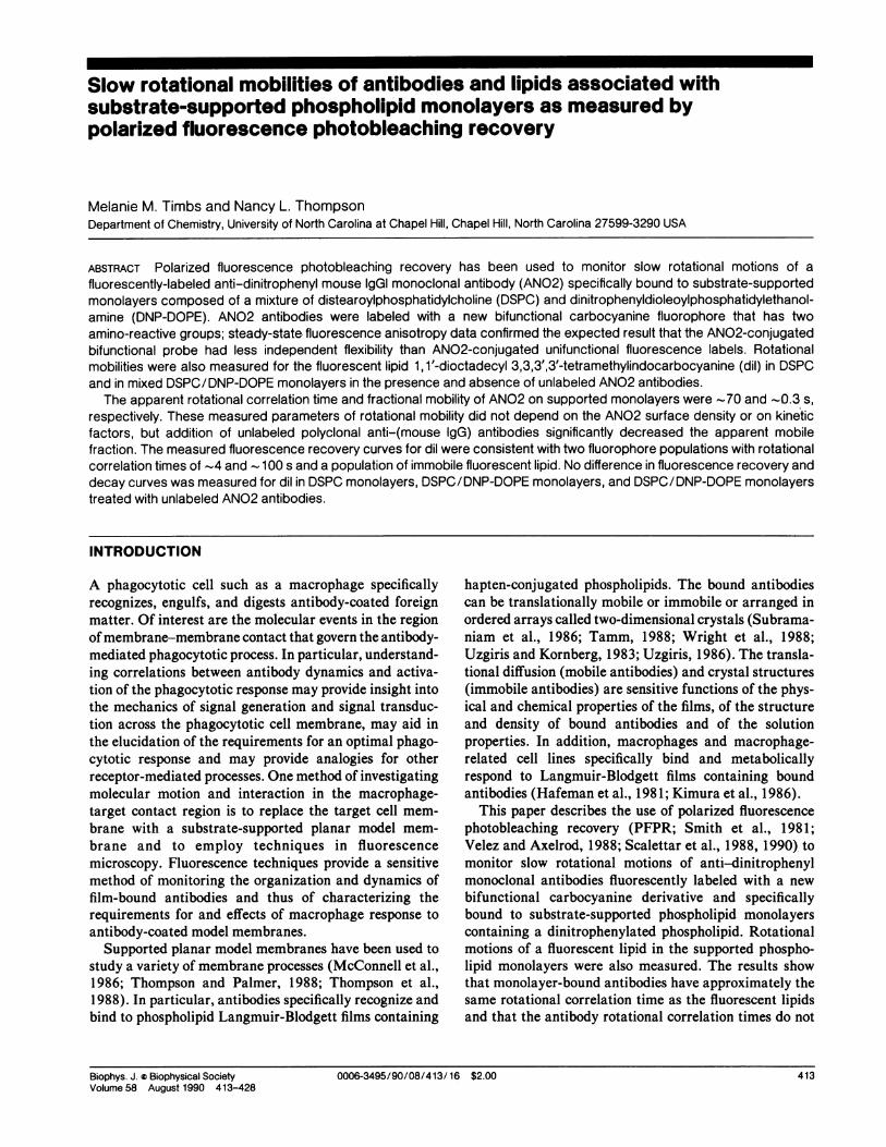
Slow rotational mobilities of antibodies and lipids associated withsubstrate-supported phospholipid monolayers as measured bypolarized fluorescence photobleaching recovery
Melanie M. Timbs and Nancy L. ThompsonDepartment of Chemistry, University of North Carolina at Chapel Hill, Chapel Hill, North Carolina 27599-3290 USA
ABSTRACT Polarized fluorescence photobleaching recovery has been used to monitor slow rotational motions of a
fluorescently-labeled anti-dinitrophenyl mouse IgGI monoclonal antibody (AN02) specifically bound to substrate-supportedmonolayers composed of a mixture of distearoylphosphatidylcholine (DSPC) and dinitrophenyidioleoylphosphatidylethanol-amine (DNP-DOPE). AN02 antibodies were labeled with a new bifunctional carbocyanine fluorophore that has twoamino-reactive groups; steady-state fluorescence anisotropy data confirmed the expected result that the AN02-conjugatedbifunctional probe had less independent flexibility than AN02-conjugated unifunctional fluorescence labels. Rotationalmobilities were also measured for the fluorescent lipid 1, 1'-dioctadecyl 3,3,3',3'-tetramethylindocarbocyanine (dil) in DSPCand in mixed DSPC/DNP-DOPE monolayers in the presence and absence of unlabeled AN02 antibodies.
The apparent rotational correlation time and fractional mobility of AN02 on supported monolayers were -70 and -0.3 s,respectively. These measured parameters of rotational mobility did not depend on the AN02 surface density or on kineticfactors, but addition of unlabeled polyclonal anti-(mouse IgG) antibodies significantly decreased the apparent mobilefraction. The measured fluorescence recovery curves for dil were consistent with two fluorophore populations with rotationalcorrelation times of -4 and - 100 s and a population of immobile fluorescent lipid. No difference in fluorescence recovery anddecay curves was measured for dil in DSPC monolayers, DSPC/DNP-DOPE monolayers, and DSPC/DNP-DOPE monolayerstreated with unlabeled AN02 antibodies.
INTRODUCTION
A phagocytotic cell such as a macrophage specificallyrecognizes, engulfs, and digests antibody-coated foreignmatter. Of interest are the molecular events in the regionofmembrane-membrane contact that govern the antibody-mediated phagocytotic process. In particular, understand-ing correlations between antibody dynamics and activa-tion of the phagocytotic response may provide insight intothe mechanics of signal generation and signal transduc-tion across the phagocytotic cell membrane, may aid inthe elucidation of the requirements for an optimal phago-cytotic response and may provide analogies for otherreceptor-mediated processes. One method of investigatingmolecular motion and interaction in the macrophage-target contact region is to replace the target cell mem-brane with a substrate-supported planar model mem-
brane and to employ techniques in fluorescencemicroscopy. Fluorescence techniques provide a sensitivemethod of monitoring the organization and dynamics offilm-bound antibodies and thus of characterizing therequirements for and effects of macrophage response toantibody-coated model membranes.
Supported planar model membranes have been used tostudy a variety of membrane processes (McConnell et al.,1986; Thompson and Palmer, 1988; Thompson et al.,1988). In particular, antibodies specifically recognize andbind to phospholipid Langmuir-Blodgett films containing
hapten-conjugated phospholipids. The bound antibodiescan be translationally mobile or immobile or arranged inordered arrays called two-dimensional crystals (Subrama-niam et al., 1986; Tamm, 1988; Wright et al., 1988;Uzgiris and Kornberg, 1983; Uzgiris, 1986). The transla-tional diffusion (mobile antibodies) and crystal structures(immobile antibodies) are sensitive functions of the phys-ical and chemical properties of the films, of the structureand density of bound antibodies and of the solutionproperties. In addition, macrophages and macrophage-related cell lines specifically bind and metabolicallyrespond to Langmuir-Blodgett films containing boundantibodies (Hafeman et al., 1981; Kimura et al., 1986).
This paper describes the use of polarized fluorescencephotobleaching recovery (PFPR; Smith et al., 1981;Velez and Axelrod, 1988; Scalettar et al., 1988, 1990) tomonitor slow rotational motions of anti-dinitrophenylmonoclonal antibodies fluorescently labeled with a new
bifunctional carbocyanine derivative and specificallybound to substrate-supported phospholipid monolayerscontaining a dinitrophenylated phospholipid. Rotationalmotions of a fluorescent lipid in the supported phospho-lipid monolayers were also measured. The results showthat monolayer-bound antibodies have approximately thesame rotational correlation time as the fluorescent lipidsand that the antibody rotational correlation times do not
Biophys. J. Biophysical SocietyVolume 58 August 1990 413-428
0006-3495/90/08/413/16 $2.00 4130006-3495/90/08/413/16 $2.00 413

dramatically depend on the antibody surface density or onkinetic factors. Analysis of the PFPR data employsexperimental measurements of the orientation distribu-tion of fluorophore absorption dipoles made with polar-ized evanescent illumination (Thompson et al., 1984) anda generalization of the theoretical basis of PFPR (Velezand Axelrod, 1988) that includes multiple fluorophorepopulations.
AFI(t) and AFL(t). Using Eq. 2 with c = 0 in Eq. 3 yields
2 (b/a) e-4D,3 - (b/a) e-4D' (4)
A convenient method of measuring the values of a, b, andD is to obtain D and the ratio b/a from the anisotropyr(t) and constant a from the average fractional bleach:
a = 1/2 [AF(0) + AF_L(0)]. (5)
THEORETICAL BACKGROUND
Rotational mobilitiesIn PFPR, fluorescent molecules are photochemicallybleached with a pulse of linearly polarized light. Thefluorescence intensities monitored with linearly-polarizedlight oriented parallel, F1(t), or perpendicular, FL(t), tothe bleach polarization increase and decrease, respec-tively, with time. The rate and shape of the fluorescencerecovery and decay provide information about the rota-tional mobility of the fluorescent molecules relative to thedirection of light propagation. A theoretical basis forPFPR has been developed by a number of groups (Smithet al., 1981; Wegener, 1984; Wegener and Rigler, 1984;Dale, 1985, 1987; Velez and Axelrod, 1988).The relationship between the postbleach fluorescence
intensities FL, (t) and the prebleach fluorescence intensityF(-) is described by the normalized postbleach changein fluorescence intensities
AFj, (t) = [F(-) - FL,(t)]/F(-).
The relationship of constants a, b, and D to FL,(t) andr(t) are illustrated in Fig. 1. At long times, F1(oo) =
Fj(oo) and r(oo) = 0.
For two-dimensional samples in which different popula-tions of fluorophores rotationally diffuse with coefficientsDi and in which all populations are bleached with equalefficiency, a straightforward generalization of Eqs. 1-5shows that, for R fluorophore types of relative abundancefi,)
R
AFIl,(t) = ZfJ[ai ± bie-4Dit _ Cie- 16DhIti=l
i
.4-J
LL
wllJzw
U,
wcc0
Li.(1)
For azimuthally-symmetric, two-dimensional samples inwhich all fluorophores are equivalently oriented and fullyrotationally mobile about the normal to the sample duringthe observed time,
AJF, (t) = a ± be-4Dt - ce- 16Dt
L.
I(2)
where a, b, and c are positive constants that depend in a
known manner on the bleaching depth and on the orienta-tion and flexibility of the absorption and emission dipolesand D is the rotational diffusion coefficient (Velez andAxelrod, 1988). For all but very deep bleaching, theconstant c is small and is therefore assumed equal to zero.The normalized fluorescence recoveries and decays, AFI(t)and AFL(t), thus contain three independent and measur-able constants (a, b, and D).The anisotropy r(t), defined as
4)
LL
wLLzw
cnw
0
lJ
--Ij-
-
- 11 W0.4
IC-
_ Z o
0.
-~~~
-I
04Dt
I
IIeX o. 14
E_
Iz o
(6)
a
|b
3
3
a If' If bi
3
4Dt
[AFI (t) - AF_ (t)]=[AFI(t) + 2AF_±(t)I
is less sensitive to postbleach fluorescence changes that donot arise from rotational mobility than the functions
FIGURE 1 Theoretical shape of fluorescence recovery F1(t), decayF1(t), and anisotropy r(t) for PFPR. Curves are calculated using (top)Eqs. 1, 2, and 4 with a = 0.3, b = 0.15, c = 0, and F(-) = 1; and(bottom) Eqs. 1, 2, and 7 with a = 0.3, R = 2,f1b1 = f2b2 = 0.075,c =O, DI = O, D2 = D, and F(-) = 1.
414 Biophysical Journal Volume 58 August 1990414 Biophysical Journal Volume 58 August 1990
where a1 and bi depend on the orientation distribution ofthe i th population and the bleaching depth and ci t 0.Eqs. 3 and 6 imply that
R
2 (f bi/a) e-4Di'r(t) = (7)
3 - E3 (fb,/a)e 4ai-i
where
R
a = Zfiai (8)i-I
equals the fractional bleach as defined in Eq. 5. Data for asample with two fluorophore populations contain fiveindependent and measurable parameters (a, f1 bI, f2b2, Dland D2) provided the time decays due to D2 and D, can beresolved and are in the time range of measurement anddata for a sample with three populations contain seven
independent parameters. The constantsfi and bi appear as
a product and cannot be independently measured. How-ever, if all populations have identical or similar values ofbi = b, then the fractional abundance of the ith popula-tion can be estimated from the measured values offibi as
fi fibi/[fibi. (9)
Fig. 1 illustrates how F1(t), Fj(t), and r(t) depend on a,
2.0
1.5
i 1.0
0.5
2.0
1.5
LU
i2 1.0
if
0.5
0 15 30 45 60
WOBBLE ANGLE
f,bj,f2b2, and D2 for a sample with both mobile (D2 = D)and immobile (D, = 0) fluorophore populations. At longtimes, FI(oo) F1(oo) and r(oo) 0.As shown by Velez and Axelrod (1988), a and b are
functions of the following variables: a constant propor-tional to the bleaching intensity, bleaching duration, andmolar absorptivity that describes the depth of bleaching(B); the average tilt angle of absorption dipoles from theplane of the sample (e); the semiangle of a cone in whichthe absorption dipoles wobble on a time scale faster thanthat of fluorescence bleaching (,B); and the angle betweenthe fluorophore absorption and emission dipole moments(x). For diI in model membranes, both e and x arebetween 0 and 300 (Badley et al., 1973; Yguerabide andStryer, 1971; Axelrod, 1979). For weak to moderate levelsof bleaching (B < 2), low to moderate wobbling ampli-tudes ((3 < 600), and e = x = 00, constant a ranges from0 to 0.6 and constant b ranges from 0 to 0.3 (Fig. 2 A).Given a wobble angle and a bleach depth B, the valuesof the fractional bleach a and recovery b are slightly lowerfor e = x = 300 than for e = X = 00 (Fig. 2 B). Fig. 2 Cdepicts the B - curves for a two-dimensional sample ofrandomly-oriented fluorophores. For known values of e
and x and measured values of a and b, the intersection ofthe lines of constant a and b in the B - (3plane determinesthe values of B and (3. If more than one fluorophorepopulation is present, then the values of a; and bi dependon B, X, and the parameters of the orientation distributionof the ith population Ei and (,i.
15 30 45
WOBBLE ANGLE
3.0
X 2.0
g 1.0
60
WOBBLE ANGLE a3
Tib msnRttoa oiiiso nioisadLpd
A 0.25, 0.20, 0.15 1 0 100
/ /
I I
I I
~~/
///
/
--/ /0._ -- ~
~/ .05///I
_/_-- / /
_ ~~~~~~/_ ~~/
_ _-
0.20/ O.1S1 0.10' 0.051/ I I
/ I I/ I I I
_ / I
/ I I/ /I
/ / I
_ //
/ 0.1
_ ~~~~/
= ~~~~~/
C-
_~~ ~~-_0__
l
FIGURE 2 Theoretical values of constants a and b. These curves show the values of a ( ) and b (---, . * *) as a function of the bleaching parameterB and the wobble angle ft. The tilt angle e and the angle between the absorption and emission dipoles x equal (A) 00 and (B) 300. In C, angle e israndomly distributed and angle x equals 00 (---) or 300 (...*). Plots A and B were generated according to Eqs. 21 and 22 of Velez and Axelrod (1988)with 4,, = 0 and 42 = (; plot C was obtained by numerical integration of Eqs. 21 and 22 over all e.
I -
Timbs and Thompson Rotational Mobilities of Antibodies and Lipids 415

Absorption dipole orientationdistributionsThe orientation distribution of absorption dipoles dependson angles e and ,3 and can be independently characterizedby fluorescence microscopy as described previously(Thompson et al., 1984). In this technique, the fluores-cence is measured as a function of the polarization of theexciting light with a high-aperture objective that collectsnearly one-half of the emitted light. If the exciting lightpropagates normal to the membrane (e.g., with epi-illumination) then, in principle, information about azi-muthally asymmetric distributions can be obtained. How-ever, measurements over large areas of supportedphospholipid monolayers should be exactly or approxi-mately azimuthally symmetric. If the exciting light is theevanescent field produced by a light beam that is totallyinternally reflected at the substrate/solution interface,then rotating the polarization of the incident beam rotatesthe polarization of the evanescent field primarily betweendirections perpendicular and parallel to the interface andinformation about the tilt angle e and the fast wobbleangle d is obtained.
For azimuthally symmetric samples, the fluorescenceexcited with evanescent illumination and normalized to a
maximum value of one is given by (Thompson et al.,1984)
J(+) = 1 + C(cos2L - cos24AO), (10)
where ,6 is the angle that the beam polarization makeswith the incidence plane, 4/0 is the angle at which I(#I) is a
maximum (O or 900), and C is a measured constant(positive or negative, respectively). The constant C de-pends on the following parameters: (i) the polarization ofthe evanescent field for different incident beam polariza-tions, which depends on the angle of incidence of thetotally internally reflected beam (a) and the relativerefractive index of the solution/glass interface (n), (ii) a
constant that describes the relative fluorescence collectionefficiency for emission dipoles oriented perpendicular or
parallel to the ootical axis of the objective (I - y), and(iii) the orientation distribution of fluorophore absorptiondipoles. If the orientation distribution of absorption di-poles N(O) is written as an expansion in Legendre polyno-mials Pi (cosO) with order parameters si, so that
co
N(O) = Esi P (cosO) ( 11)i=l
N(6) sinG dO = 1,all space
(12)
then C depends only on the order parameters S2 and S4
where -1/2 < 52 < land -(3/7) s< 4 C 1 .
When the fluorescence collection parameter y equals 0,the measured constant C depends only on S2 and not on s4
(Thompson et al., 1984). As shown in Fig. 3 A, thedependence of C on S2 is strong and does not changesignificantly within the possible range of values for theincidence angle a (640 = sin-n < a < 900) or the rela-tive refractive index n (ia0.9). Thus, fairly accuratedetermination of S2 is possible in the simplest case where
.o0
I1.
cu
U)
urLii
a:
cc0
CC0
0.5
0.0
-0.5 0.0 1
MEASURED PARAMETER C
cu
01
cc
c:LI-
ccw0.Cc0
0.5
-0.50.5 1.0
y - 0.2
'1- 0.2
c1.0
0.8
0.6
0.4
0.0 _- -
0.0
I-0.4
0.0 0.5 1.0
ORDER PARAMETER s4
Biophysical Journal Volume 58 August 1990
A
'y = 0
II /
FIGURE 3 Fluorescence-detected absorption dichroism constant C. (A) When the fluorescence collection parameter Y equals 0, constant C is amonotonic function of s2 and depends only weakly on the relative refractive index n and the incidence angle a; curves are for a = 700, n = 0.88 ( );a = 700, n = 0.92 (... ); a = 850, n = 0.88 (-.-); and a = 850, n = 0.92 (---). (B) When the fluorescence collection parameter y equals 0.2,constant C depends only weakly on S4; the plot is for a = 700 and n = 0.89. Plots were generated according to Eqs. 18 and 19 of Thompson et al.(1984).
0,11
Il
416 Biophysical Journal Volume 58 August 1990

y = 0. In practice, however, the parameter y 0 becausefluorescence is not collected over a full 27r steradians andbecause the substrate affects the angular distribution offluorescence emitted from fluorophores with differentorientations; previous work has shown that the value of y
for the conditions used in this study is -0.1 (Burghardtand Thompson, 1984). Nonzero y introduce S4 depen-dence in C, but as shown in Fig. 3 B the dependence isweak for small y (= 0.2), and the s2 value that corre-
sponds to a given measured value of C is approximatelyequal to the value that would be obtained from Fig. 3 Awhen,y = 0.
For stationary dipoles uniformly oriented at a singlepolar angle 900 - e, Eqs. 11 and 12 imply that
S2 = ('/2)(3 sin2 E - 1), (13)
and a given measured value of s2 specifies the tilt angle e.
A more general treatment is required if the absorptiondipoles rapidly fluctuate about their average orientationwithin a characteristic angle ,B (Badley et al., 1973). Asshown in Appendix A for a model in which dipoles are
found within a cone of semiangle centered at tilt angle e,
the relationship between s2, f, and ,B is given by
S2 = ('/4)(3 sin2 e - 1) cos#j (1 + cos#). (14)
In this case, a given measured value of s2 is consistentwith a range of pairs of e and ,B. Other models for thefunctional form of the orientation distribution of absorp-tion dipoles may also be appropriate, but this model hasbeen chosen for consistency with the theoretical treat-ment of PFPR developed by Velez and Axelrod (1988).
Steady-state anisotropies offluorescent probes on antibodies insolutionInformation about rapid motions of fluorophores conju-gated to antibodies can be obtained by measuring steady-state anisotropies in solution. A simple expression for theanisotropy A of a spherical fluorophore that is attached toa spherical macromolecule and experiences rotationalmotion independent of the macromolecular motion is(Leach, 1969)
A(T/i) +0 )Aq(51 + (Cp Tln) +1 + (CpTi) + (Cf T/l)' (1)
where AO is the limiting anisotropy, q is a constant relatedto the amount of independent fluorophore motion, T is theabsolute temperature, v is the viscosity,
kr krCP= 6V 9 Cf = 6V' (16)
k is Boltzmann's constant, r is the fluorescence lifetime,Vp is the hydrodynamic volume of an antibody, and Vf isthe molecular volume that corresponds to the correlationtime of independent fluorophore rotational flexibility.Although a number of possible phenomena such asantibody segmental flexibility and the nonspherical shapeof both the antibody and probe are not included in Eq. 15,the values of Cf/ Cp and q provide an indication of thedegree of independent fluorophore rotational mobility.
MATERIALS AND METHODS
AntibodiesMouse and sheep polyclonal IgG (Sigma Chemical Co., St. Louis, MO)and goat anti-(mouse IgG) (Jackson ImmunoResearch Labs, WestGrove, PA) were dialyzed against phosphate-buffered saline (PBS, 0.05M sodium phosphate, 0.15 M sodium chloride, 0.01% sodium azide, pH7.4) and passed through a 0.22-,Mm filter. The mouse IgGi anti-dinitrophenyl monoclonal antibody ANO2 (Balakrishnan et al., 1982)was purified from cell culture supernatants by protein A affinitychromatography (Anglister et al., 1984; Wright et al., 1988), dialyzedagainst PBS, and stored at - 150C.ANO2 antibodies were labeled with the new bifunctional, amine-
reactive, tetramethylindocarbocyanine fluorophore CY3.18 (a generousgift of Alan Waggoner and Ratan Majumdar of Carnegie MellonUniversity; Wagner et al., 1990; C-ANO2) by treating 0.1-0.3 mg/mlANO2 in 0.1 M NaHCO3/Na2CO3 pH 9.5 with a 12-fold molar excessof dye in 10-20 ul anhydrous dimethylformamide for 1 h at roomtemperature. ANO2 antibodies were labeled with fluorescein iso-thiocyanate (Molecular Probes, Inc., Eugene, OR; F-ANO2) andtetramethylrhodamine isothiocyanate (Molecular Probes, Inc.; R-ANO2)according to published procedures (Mishell and Shiigi, 1980). Fluores-cently-labeled ANO2 was purified by Sephadex G50-80 chromatogra-phy in PBS, dialysis against PBS, 0.22 Mm filtration, and sedimentationat 100,000 g for 1-2 h. Labeled antibodies were stored at 40C and usedwithin 3 wk.ANO2 concentrations and the molar ratios of fluorophore to ANO2
(F:A) were determined spectrophotometrically using the followingestimated absorptivities and measured ratios of fluorophore absorptivi-ties at 280 nm to those at their absorption maxima:' ANO2, 210,000M-' cm ' (280 nm); carbocyanine fluorophore, 130,000 M-' cm ' (550nm), 0.13 (280 nm/550 nm); fluorescein, 75,000 M-'cm-' (494 nm),0.25 (280 nm/494 nm); tetramethylrhodamine, 75,000 M-'cm-' (550nm), 0.36 (280 nm/550 nm). The spectrophotometrically-determinedfluorophore-to-antibody ratios ranged from one to three except as noted.Sodium dodecyl sulfate polyacrylamide gel electrophoresis (SDS-PAGE) of labeled and unlabeled ANO2 was carried out according tostandard procedures. In some experiments (as noted), 0.1 ml of 0.1mg/ml ANO2 in PBS was first treated with a 100- to 800-fold molarexcess of disuccinimidylsuberate (Pierce Chemical Co., Rockford, IL)in 10 Ml dimethylsulfoxide.
'Labeled ANO2 concentrations were also determined by the method ofLowry et al. (Peterson, 1979) using unlabeled ANO2 as a standard.These concentrations were 10-30% higher than the spectrophotometri-cally-determined concentrations, and the discrepancy could not beaccounted for by chemical effects of fluorophores on the Lowry assay orby uncertainties in the fluorophore absorptivities. Concentrations aregiven as spectrophotometric estimates throughout.
T_mbsandThompsonRotationalMobilitiesofAntibodiesan s .4..7. . . .Timbs and Thompson Rotational Mobilities of Antibodies and Lipids 417

The specificity of unlabeled ANO2 and C-ANO2 for dinitrophenyl(DNP) was confirmed by monitoring the quenching of the fluorescence(SLM 8000C, Inc., Urbana, IL) of ANO2 in PBS (10-30 Mg/ml) byDNP-glycine (Sigma Chemical Co.) with excitation and emissionwavelengths equal to 280 and 334 nm, respectively. The independentrotational mobility of ANO2-conjugated fluorophores was examined bymeasuring the steady-state fluorescence anisotropies as a function ofviscosity in PBS/glycerol (10-50 Ag/ml) with excitation and emissionwavelengths as follows: R-ANO2 and C-ANO2, 550 and 565 nm;
F-ANO2, 494 and 520 nm.
Supported phospholipid monolayersAll water was purified to exceed standards for type I reagent gradewater. Distearoylphosphatidylcholine (DSPC; Sigma Chemical Co.),dinitrophenyldioleoylphosphatidylethanolamine (DNP-DOPE; AvantiPolar Lipids, Birmingham, AL) and 1,1'-dioctadecyl-3,3,3',3'-tetrameth-ylindocarbocyanine perchlorate (diI; Molecular Probes, Inc., Eugene,OR) were judged to be pure by thin layer chromatography. Monolayersof DSPC, DSPC:diI (99.5:0.5, mol/mol), DSPC:DNP-DOPE:dil (70:29.5:0.5, mol/mol/mol) or DSPC:DNP-DOPE (70:30, mol/mol) were
spread from hexane:ethanol (9:1) at 100 A2/molecule on the air/waterinterface of a Joyce-Loebl Langmuir Trough (model 4, Vickers Instru-ments, Inc., Malden, MA) at room temperature (24-280C). Themonolayers were compressed at 1-2 A2/molecule per min to a finalsurface pressure of 35 dyn/cm. Single phospholipid monolayers were
deposited on glass microscope slides treated with octadecyltrichlorosi-lane (Aldrich Chemical Co., Milwaukee, IL) by vertical dipping at 5.4mm/min (von Tscharner and McConnell, 1981; Thompson et al., 1984;Wright et al., 1988).
For measurements in the absence of antibodies, substrate-supportedmonolayers were transferred to a sample chamber in PBS. For measure-
ments in the presence of antibodies, monolayers were washed with 1 mlPBS, treated with sheep IgG in PBS (200 Ml, 0.1 mg/ml) for 2 min,treated with one of the following procedures, and then washed with 3 mlPBS: (i) ANO2 or C-ANO2 (200 ,l, 20 Mg/ml, and 10 min unlessotherwise indicated); (ii) ANO2 (100 Ml, 0.86 mg/ml, 5 min) followedby a mixture of C-ANO2 (20 Ag/ml) and ANO2 (0.64 mg/ml) (200 gl,10 min); or (iii) a mixture of C-ANO2 (20 ,ug/ml) and DNP-glycine(1.3 mM) (200 Ml, 10 min). Some samples were further treated withunlabeled, polyclonal anti-(mouse IgG) (200 Ml, 0.1 mg/ml, 10 min)and washed with 1 ml PBS. All samples were transferred in PBS to a
sample holder suitable for microscopy and brought to 180C with a
thermoelectrically-cooled microscope stage (Wright et al., 1988).
Polarized fluorescencephotobleaching recovery
The fluorescence microscope was constructed from an inverted opticalmicroscope (Zeiss IM-35), the 514.5-nm line of an argon ion laser(Coherent Innova 90-3), a single-photon counting photomultiplier(RCA 31034A), and an IBM PC AT microcomputer, as previouslydescribed (Fig. 4; Palmer and Thompson, 1989; Poglitsch and Thomp-son, 1989). The apparatus was configured to measure rotational mobili-ties according to previous designs (Smith et al., 1981; Velez andAxelrod, 1988). The laser beam was split into bleaching and observationbeams; the bleaching beam passed through a polarization rotator (LexelCorp., Palo Alto, CA). Both beams were passed through a beamexpander (Edmund Scientific Co., Barrington, NJ), reflected by a
dichroic mirror and focused through an objective (Nikon, 40x, 0.55NA, air) to illuminate a Gaussian-shaped area of radius -25,um (diI) or
-38 MAm (C-ANO2). Fluorescence was collected through a linearpolarizer aligned with the observation beam polarization and an image
3 S
PR
BS
FIGURE 4 Optical apparatus. Fluorescence microscope: /, mirror; BS,90%/10% beam splitter; S, computer-controlled mechanical shutter;NF, neutral density filter; PR, polarization rotator; P, hemicylindricalfused silica prism; BE, beam expander; RR, Rhonchi ruling; QWP,quarter wave plate; DM, dichroic mirror; BF, barrier filter; PL, linearpolarizer; IPA, image plane aperture.
plane aperture that restricted fluorescence collection to the illuminatedarea. The relative intensity of the bleaching beam when parallel or
perpendicular to the monitoring beam was determined to equal 1.032 ±
0.004 by measuring the fluorescence of a solution of CY3. 18 inPBS/ethanol (19:1) with the analyzing polarizer removed. The polariza-tion of the exciting light in the sample plane was estimated by measuringthe intensity of the laser beam passing through a polarizer placed abovethe objective and aligned parallel or perpendicular to the beam polariza-tion with a photodiode; the average intensity ratio was 100 ± 15.
Fluorescence photobleaching parameters were as follows: observationand bleaching laser powers, 3-30 uW and < 0.4 W, respectively;bleaching pulse duration, 50 ms (diI) to 2 s (C-ANO2); depth ofbleaching, 15-35%. The temperature rise during the bleach pulse was
estimated (Axelrod, 1977) to equal -1°C assuming that the carbocya-nine absorptivity and quantum efficiency were 130,000 M-'cm-' and0.5, respectively, and that the bleaching beam was attenuated in powerby a factor of two by the optical elements between the laser aperture andthe sample.
Fluorescence recovery curves were monitored for bleaching polariza-tions parallel and perpendicular to the observation polarization. The setsof curves F1(t) and F_(t) for a given sample type and data set were
normalized to prebleach fluorescence intensities of one and then aver-
aged. The averages of F1(t) and FL(t) were then used to calculate thefluorescence changes AFI(t) and AF1(t) according to Eq. 1 and theanisotropy r(t) and the value of constant a were calculated according toEqs. 3 and 5. Values for the parameters f1bj/a and Di were obtainedfrom iterative Gauss-Newton fits of r(t) to Eq. 7 with DI = 0.
41 41pyia8JunlVlm 5 uut19Volume 58 August 1990418 Biophysical Journal

Depolarized fluorescencephotobleaching recovery
To assess the potential contribution of phenomena other than rotationalmobility to the observed fluorescence recovery and decay under linearlypolarized conditions, fluorescence recovery curves were obtained usingcircularly polarized bleaching and observation beams. The analyzingpolarizer was removed and the laser beam was circularly polarized by a
quarter wave plate (Dale, 1985; Melles-Griot, Irvine, CA) below theobjective; the ratio of the laser intensities at the sample measuredthrough a linear polarizer aligned parallel or perpendicular to the initialbeam polarization was 0.96 ± 0.01. Apparent lateral diffusion coeffi-cients were measured with fluorescence pattern photobleaching recovery(Smith and McConnell, 1978; Wright et al., 1988) with circularlypolarized light and a spatial periodicity of 8 ,um. Other experimentalparameters were as listed above.
Fluorescence-detected absorptiondichroismTo measure the absorption dichroism as a function of the polar anglefrom the substrate normal, the laser beam was passed through thepolarization rotator and was totally internally reflected at the monolayer/solution interface through a hemicylindrical fused silica prism (HarrickScientific Corp., Ossining, NY) at an incidence angle of a .70°.Evanescently excited fluorescence was collected through a high-aperturemicroscope objective (Nikon, 60x, 1.4 N.A.), a 550-nm cutoff filter, andan image plane aperture that restricted fluorescence collection to theelliptically illuminated area (-30 x 100 ,um). To measure the absorp-tion dichroism as a function of the azimuthal angle about the substratenormal, the laser beam was passed through the polarization rotator andwas then reflected by the dichroic mirror and focused on the samplethrough a microscope objective (Zeiss, 40x, 0.75 N.A.). Fluorescencewas collected through the dichroic filter, a barrier filter, and the imageplane aperture. To quantitatively analyze the experimentally obtainedI(4,), the data were normalized so that I(4,) for a sample in which it wasassumed that the fluorophore absorption dipoles were randomly oriented(C-ANO2 antibodies adsorbed to glass) had a value of C equal to thetheoretical value for a = 70°, n = 0.89, and y = 0.1 (C = 0.16). Thenormalized curves I(O) were fit to Eq. 10 with a Gauss-Newton routineto obtain values for C.
fluorophores and that no detectable unreacted fluorophorewas present. Comparison of fluorescently-labeled AN02with AN02 cross-linked by disuccinimidylsuberate on
overloaded gels and stained with silver indicated that thebifunctional carbocyanine label but not fluorescein or
tetramethylrhodamine isothiocyanate cross-linked a smallfraction of the AN02 polypeptides and that at least someof the cross-links were intermolecular. The quantity ofCY3.18 -induced antibody aggregates was higher whenthe labeling reaction was carried out at higher AN02 andfluorophore concentrations (F:A 2 5). The amount ofcross-linked C-AN02 that was present was judged to beinsignificant for the preparations used in the experimentsreported herein (1 < F:A < 3).Quenching of ultraviolet-excited antibody fluorescence
by DNP-glycine was significantly greater for AN02 andC-AN02 than for polyclonal mouse IgG. Solution associ-ation constants were obtained from these data using an
iterative procedure that determines the free DNP-glycineconcentration, corrects for collisional quenching usingdata for polyclonal mouse IgG, and finds the best fit to thefunctional form that describes a simple bimolecularreaction (Pisarchick, M. L., and N. L. Thompson, submit-ted for publication). The analysis yielded a DNP-glycineassociation constant of 1.8 ± 0.5 x 10⁶ M-1 for unlabeledAN02, which equals one previously reported measure-
ment (Anglister et al., 1984) and is within a factor of twoof another reported measurement (Leahy et al., 1986).The measured equilibrium constant for C-AN02 was
1.7 ± 0.1 x 10⁶ M-1, which is equivalent to the value forAN02 and indicates that the presence of covalently-bound bifunctional carbocyanine fluorophore did notsignificantly alter AN02 hapten recognition. Heavily-labeled C-AN02 (F:A 2 5; not used in subsequent exper-
iments) showed a slightly decreased affinity for DNP-glycine.
RESULTS
CY3.18-labeled AN02 antibodiesAN02 antibodies were labeled with the bifunctionalfluorophore CY3.18 because a probe attached at twopositions rather than one should experience less indepen-dent flexibility and therefore more accurately report therotational motions of the antibody to which it is bound.Fluorescently-labeled (C-ANO2, R-AN02, and F-AN02) and unlabeled (AN02) antibodies were analyzedwith SDS-PAGE under reducing and nonreducing condi-tions and stained with Coomassie blue. All types ofAN02 antibodies ran as bands with appropriate apparentmolecular weights. Observation of unstained gels underultraviolet illumination indicated that both the heavy andlight chains were fluorescently labeled for all three
Mobility of AN02-conjugatedfluorescent probesTo examine the relative flexibility of antibody-boundfluorophores, steady-state anisotropies were measured as
a function of viscosity. As shown in Fig. 5, the anisotro-pies of R-ANO2 and F-ANO2 were dependent on theviscosity, whereas the C-ANO2 anisotropy was not. Thedifferences between the steady-state anisotropies for solu-tions of the lowest and highest viscosities were muchlarger for R-ANO2 (0.128 ± 0.005) and F-ANO2(0.092 ± 0.013) than for C-ANO2 (0.009 ± 0.003) (Ta-ble 1).The data in Fig. 5 were fit to the functional form of Eq.
15 with 1/A as the ordinate and T/I7 as the abscissa; T =296°K; X ranging from 0.01 to 0.28 Poise; Cf and q as free
Timbs and Thompson Rotational Mobilities of Antibodies and Lipids 419
Timbs and Thompson Rotational Mobilities of Antibodies and Lipids 419

-c 7.0
rn
FIGURE 5 Steady-state anisotropy as a function of viscosity. Shown arethe measured anisotropies for (*) C-ANO2, (-) R-ANO2, and (m)F-ANO2 and the best fits to Eq. 15 with A. = 0.4 and with q and Cf asfree parameters (-) and with AO, q, and Cf as free parameters (---).
parameters; Vp = 2.3 x 10- 19 cm3/molecule; r = 3.65 ns
(fluorescein), r = 2.34 ns (tetramethylrhodamine), or
r = 1 ns (carbocyanine) (Chen and Scott, 1985; Packardand Wolf, 1985); and AO = 0.4 (the theoretical maximumwhich corresponds to aligned absorption and emissiondipoles moments). In this analysis, the ratio Cf/ Cp andthe parameter q were large for both F-ANO2 andR-ANO2, suggesting that fluorophore mobility contrib-uted significantly to the observed anisotropies, but theviscosity-independent C-ANO2 data could not be well fit.The data in Fig. 5 were also fit to Eq. 15 with AO,
Cf/ Cp, and q as free parameters. The C-ANO2 data were
best fit with AO = 0.27 (corresponding to an interdipoleangle of 280) and either a small q or a small Cf/ Cp ratio(e.g., q = 0.05; Cf/Cp ' 50 or q = 0.5; Cf/Cp '10). The
values of q and Cf/ Cp were still higher for F-ANO2 andR-ANO2 than the maximum estimated values forC-ANO2; the values of A0 (0.22-0.30) corresponded toabsorption and emission dipole moments oriented at24-330 (Table 1).The data in Fig. 5 and Table 1 suggest that the
bifunctional carbocyanine probe had less rotational mobil-ity than the unifunctional probes. This lower apparentmobility of antibody-bound carbocyanine in the nanosec-
ond time range means that, if all other factors are
equivalent, this probe is a better choice for measurementsof antibody rotational mobility. However, probe flexibilitywith any characteristic time that is faster than thebleaching duration will affect PFPR data and probemobility with a characteristic time in the microsecond-millisecond time range would not be detected by measure-
ments of the dependence of steady-state anisotropies on
viscosity. The data also imply that ANO2-conjugatedtetramethylrhodamine has less flexibility than fluoresceinin the nanosecond-microsecond time range, suggestingincreased noncovalent interactions between the tetrameth-ylrhodamine probes and ANO2 antibodies to which theyare attached.
Supported phospholipid monolayersThe fluorescence arising from diI in supported DSPCmonolayers was nonuniformly distributed with opticallyresolved features of size 1-5 ,um. The fluorescence of diI inDSPC/DNP-DOPE monolayers was also nonuniform,but the features were usually of a finer texture. The visualdetection of spatially nonuniform diI fluorescence sug-
gests the presence of coexistent solidlike and fluidlikedomains similar to those previously observed in dipal-mitoylphosphatidylcholine monolayers at the air/water
TABLE 1 Steady-state anisotropies for fluorophores conjugated to AN02 antibodies
Fluorophore Carbocyanine Tetramethylrhodamine Fluorescein
Anisotropies:A (PBS) 0.261 ± 0.002 0.158 ± 0.001 0.122 ± 0.013A (PBS/glycerol) 0.270 ± 0.002 0.286 ± 0.005 0.214 ± 0.002
Two free parameters (AO = 0.4)q 0.72 0.68Cf/CP 850 3100
Three free parametersAO 0.27 0.30 0.22q 0.05* 0.88 0.56Cf/CP <50* 190 310
Steady-state anisotropies for ANO2-conjugated fluorophores in the least (PBS) and most (28% PBS/72% glycerol) viscous solutions are shown at thetop. A., q, and Cf /Cp are from the best fits of the data in Fig. 5 to Eq. 15 with fixed parameters as described in the text. *C-ANO2 data were equallywell fit with larger q values and smaller Cf/Cp ratios (e.g., q = 0.5 and Cf/Cp <10).
420 Biophysical Journal Volume 58 August 1990420 Biophysical Journal Volume 58 August 1990
interface (McConnell et al., 1984) and on alkylatedsubstrates (Seul et al., 1985). The fluorescence measuredover large areas (-2,000 ,im2) was uniform, with a typicalstandard deviation of 5-10% for 5-10 measurements on a
single monolayer.DSPC/DNP-DOPE supported monolayers containing
bound C-ANO2 usually appeared uniformly fluorescentwith a fine granular texture. However, distinct snowflake-shaped nonfluorescent regions of size 5-15,m containingfour to six main branches with multiple protrusions fromeach major branch were occasionally observed. Thesepatterns, which may have been areas depleted of lipid,depleted of DNP-DOPE or containing sequestered DNP,were not readily reproducible. The presence of thesenonfluorescent regions had no effect on the measuredrotational mobility or the overall uniformity of the mea-
sured fluorescence over large areas. Typical standarddeviations of 5-10 measurements of fluorescence intensityover large areas (-2,000 ,um2) were -5% for DNP-DOPE/DSPC supported monolayers treated with C-ANO2.
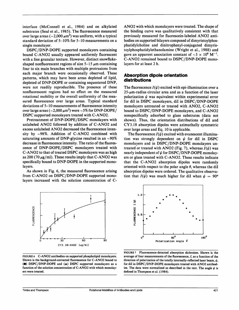
Pretreatment of DNP-DOPE/DSPC monolayers withunlabeled ANO2 followed by addition of C-ANO2 andexcess unlabeled ANO2 decreased the fluorescence inten-sity by -98%. Addition of C-ANO2 combined withsaturating amounts of DNP-glycine resulted in an -90%decrease in fluorescence intensity. The ratio of the fluores-cence of DNP-DOPE/DSPC monolayers treated withC-ANO2 to that of treated DSPC monolayers was as highas 200 (70 ,tg/ml). These results imply that C-ANO2 was
specifically bound to DNP-DOPE in the supported mono-
layers.As shown in Fig. 6, the measured fluorescence arising
from C-ANO2 on DSPC/DNP-DOPE supported mono-
layers increased with the solution concentration of C-
w
z
w
w
0
LL
ANO2 with which monolayers were treated. The shape ofthe binding curve was qualitatively consistent with thatpreviously measured for fluorescein-labeled ANO2 anti-bodies on supported bilayers composed of dimyristoylphos-phatidylcholine and dinitrophenyl-conjugated dimyris-toylphosphatidylethanolamine (Wright et al., 1988) andgave an apparent association constant of -3 x 10⁶ M-l.C-ANO2 remained bound to DSPC/DNP-DOPE mono-
layers for at least 2 h.
Absorption dipole orientationdistributionsThe fluorescence I(/') excited with epi-illumination over a
25-,um-radius circular area and as a function of the laserpolarization ,6 was equivalent within experimental error
for diI in DSPC monolayers, diI in DSPC/DNP-DOPEmonolayers untreated or treated with ANO2, C-ANO2bound to DSPC/DNP-DOPE monolayers, and C-ANO2nonspecifically adsorbed to glass substrates (data notshown). Thus, the orientation distributions of diI andCY3.18 absorption dipoles were azimuthally symmetricover large areas and Eq. 10 is applicable.The fluorescence I(0) excited with evanescent illumina-
tion was strongly dependent on for diI in DSPCmonolayers and in DSPC/DNP-DOPE monolayers un-
treated or treated with ANO2 (Fig. 7), whereas I(+) was
nearly independent of , for DSPC/DNP-DOPE monolay-ers or glass treated with C-ANO2. These results indicatethat the C-ANO2 absorption dipoles were randomlyoriented with respect to the polar angle 0, whereas the diIabsorption dipoles were ordered. The qualitative observa-tion that I(,6) was much higher for diI when ,6 = 900
i .o
wC-)
zw
C.)wcc0
IL.
0.5
00 900Polarization Angle 4'
180°
CY3. 18-AN02 (mg/ml)
FIGURE 7 Fluorescence-detected absorption dichroism. Shown is theaverage of four measurements of the fluorescence, I, as a function of thedirection of polarization of the totally internally-reflected laser beam, 4,for diI in DSPC/DNP-DOPE monolayers treated with AN02 antibod-ies. The data were normalized as described in the text. The angle 4# isdefined in Thompson et al. (1984).
Rotational Mobilities of Antibodies and Lipids 421
a a
U Ua a
* a
a a
*U
U U
I
FIGURE 6 C-ANO2 antibodies on supported phospholipid monolayers.Shown is the background-corrected fluorescence for C-AN02 bound to(U) DSPC/DNP-DOPE and (A) DSPC supported monolayers as afunction of the solution concentration of C-AN02 with which monolay-ers were treated.
Timbs and Thompson Rotational Mobilities of Antibodies and Lipids 421

TABLE 2 Fractional fluorescence changes for dil and C-AN02 associated with supported monolayers
Polarization dil/DSPC/DNP-DOPE DSPC/DNP-DOPE(bleach) diI/DSPC diI/DSPC/DNP-DOPE and ANO2 and C-ANO2
A. Circular 0.009 ± 0.003 0.007 ± 0.007 0.016 ± 0.016 0.002 ± 0.001(no)
B. Circular 0.002 ± 0.001 0.002 ± 0.002 0.008 ± 0.005 0.012 ± 0.005(yes)
C. Linear 0.035 ± 0.003 0.022 ± 0.011 0.034 ± 0.003 0.042 ± 0.012(11)
D. Linear -0.038 ± 0.002 -0.048 ± 0.004 -0.043 ± 0.004 0.007 ± 0.006(1~)
Average values and standard errors in the mean were calculated from experimental curves as [F(oo) - F(-)]/F(-) for A, and [F(OO) - F(0)]/F (-) for B-D, where F (-) is the prebleach fluorescence, F (0) is the fluorescence immediately after the bleach pluse, F (X) - F (5 min) for diI andF (oo) - F (2 min) for C-ANO2.
suggests that the absorption transition dipole momentswere approximately parallel to the substrate, consistentwith other studies of the orientation of diI or relatedcompounds in model membranes (Axelrod, 1979; Badleyet al., 1973).The values of the constant C obtained from I(i/) for
evanescent illumination were -0.55 ± 0.01 for diI inDSPC monolayers and -0.57 ± 0.01 for diI in DSPC/DNP-DOPE monolayers untreated or treated with unla-beled ANO2 antibodies. These values of C correspond toS2 = -0.29 ± 0.03 and s2 = -0.30 ± 0.03 (Fig. 3 A).The nonzero magnitude of s2 argues strongly against thepresence of a large number of substrate-absorbed vesiclesor other nonplanar structures in which diI could laterallymove, giving rise to fluorescence recovery under polarizedconditions that was not due to rotational mobility aboutthe normal to the monolayer (e.g., Smith and McConnell,1981). For stationary dipoles uniformly oriented at a
single polar angle 90 - E, the measured value of s2
corresponds to e = 220 ± 20 (Eq. 13). For dipoles that are
centered at angle c and undergo fast wobbling in a cone ofsemiangle ,B, the measured value of s2 is consistent with a
smaller mean value of e; e.g., for A3 = 200, e = 200 20,for A = 300, e = 18° ± 20, and for , = 400, e = 120 40(Eq. 14). For the measured value of s2, the maximumwobble angle, which occurs for e = 00, is ,B = 460 ± 40.
Depolarized fluorescencephotobleaching recovery
The extent to which postbleach fluorescence changesmeasured with linearly polarized light might arise fromphenomena other than rotational mobility of the lipids orantibodies was assessed using circularly polarized light.In the absence of a bleach pulse, the fluorescence intensityilluminated over a large area was occasionally observed toslowly increase (Table 2 A). Similar effects have previ-ously been observed by others and have been attributed to
photochemical instabilities (e.g., Smith et al., 1981). Themeasured fluorescence for this optical configuration alsoincreased slightly after photobleaching, but not at a ratesignificantly greater than that observed in the absence ofbleaching (Table 2 B). The fractional postbleach fluores-cence changes monitored with linearly polarized lightwere significant, of comparable magnitudes and positive(or negative) for parallel (or perpendicular) bleachingpolarizations (Table 2, C and D). These results stronglysuggest that a significant fraction of the postbleachfluorescence changes measured with linearly polarizedlight arose from rotational motion of the fluorophore.
Because the most likely source of fluorescence recoveryother than rotational mobility is lateral mobility, appar-ent lateral diffusion coefficients and fractional mobilitieswere measured with fluorescence pattern photobleachingrecovery (Smith and McConnell, 1978) using circularlypolarized light and a spatial periodicity of 8 ,m (Table 3).The C-ANO2 and diI lateral difiusion coefficients did notsignificantly differ, which suggests that the antibodies donot undergo significant lateral translation across thesesolidlike surfaces by detachment of one antigen bindingsite followed by subsequent attachment at a differentposition. The measured translational diffusion coefficientsand fractional mobilities2 together with the known func-tional form for fluorescence recovery due to lateraldiffusion in a plane through Gaussian-shaped illumina-tion (Axelrod et al., 1976) imply that the fractionalfluorescence recovery due to lateral mobility that wouldbe observed under conditions used for measurement ofrotational mobility (25 ,um radius and 5 min for diI; 38,um radius and 2 min for C-ANO2) is small (Table 3).
2Without monitoring fluorescence recoveries for much longer times, theexperiment cannot distinguish between higher apparent diffusion coeffi-cients with lower fractional mobilities and lower apparent diffusioncoefficients with higher fractional mobilities. Results are reported forthe maximum diffusion coefficient and minimum fractional mobility.
422 Biophysical Journal Volume 58 August 1990422 Biophysical Journal Volume 58 August 1990

TABLE 3 Apparent lateral diffusion coefficients and mobile fractions measured by pattern photobleaching with circularlypolarized light
ApparentMobile fractional
Sample DIat fraction recovery
10-1) cm2/sdiI/DSPC 3 ± 2 0.06 ± 0.03 <2 x 10⁻⁴diI/DSPC/DNP-DOPE 20 ± 10 0.12 ± 0.03 <2 x 10⁻³dil/DSPC/DNP-DOPE and ANO2 8 ± 2 0.27 ± 0.09 <2 x 10⁻³DSPC/DNP-DOPE and C-ANO2 4 ± 2 0.35 ± 0.02 <2 x 10⁻⁴
Apparent lateral diffusion coefficients and fractional mobilities and standard errors in the mean were measured using pattern photobleaching withcircularly polarized light and a spatial periodicity of 8 Am (see footnote 2). Apparent fractional recoveries were calculated assuming aGaussian-shaped illumination (Axelrod et al., 1976) and refer to the expected recovery [F(oo) - F(O)]/F(-) due to lateral mobility under conditionsused for rotational mobility (25 ,um radius, 5 min for diI; 38 Am radius and 2 min for C-ANO2).
Polarized fluorescencephotobleaching recovery of dilThe postbleach fluorescence intensities F1(t) and FL(t)for diI in supported monolayers increased and decreasedfor bleaching beam polarization directions parallel andperpendicular to the observation beam polarization, respec-tively (Fig. 8) and the anisotropies r(t) decayed with time(Fig. 9 A) as theoretically predicted (Eqs. 1-9). Theanisotropy data could not be well fit with the functionalform for a single rotationally diffusing fluorophore (Eq. 4)and were thus fit to the functional form shown in Eq. 7with D, = 0 and R = 2, 3, and 4. The mean values of theF-statistic that compares the best fits for two components(one mobile and one immobile) and for three components(two mobile and one immobile) were much larger than the
La.
wC.)zwu)llJc:0
-J11
0.7 L
0 i 2 3
Time (min)
FIGURE 8 PFPR curves F1(t) and FL(t) for diI in DSPC monolayers.The postbleach fluorescence intensity decays when the bleaching beamis polarized perpendicular to the observation beam and recovers whenthe bleaching beam is polarized parallel to the observation beam. Shownis the average of three parallel and four perpendicular curves obtainedfrom different positions on a single monolayer.
critical value of 3.0 (Wright et al., 1988) indicatingstatistical significance in the increased accuracy of the fit.The mean values of the F-statistic that compares the bestfits for three and four components were <3.0. Theparameters of the best fits of r(t) to the three-componentfunction are shown in Table 4; additional constantscalculated from the best-fit parameters are shown inTable 5. The very slow rotational correlation times(5-100 s) are consistent with previous measurements ofdiI rotational mobility in solid-phase DSPC multibilayersand liposomes (Smith et al., 1981) and solid-phase DPPClipid vesicles (Johnson and Garland, 1983).The total fluorescence change relative to the prebleach
fluorescence was 5-10% and the relative magnitudes ofeach component (fibi/a) were approximately equal sothat each component corresponded to only a small post-bleach fluorescence change. Thus, the possibilities thatthe nonmonophasic behavior arose from an unidentifiedproperty of the sample or experimental apparatus oroverly simple theoretical assumptions leading to the formof Eq. 7 cannot be completely ruled out. The assumptionsof uniform (not Gaussian-shaped) illumination and low-aperture microscope optics are only approximately cor-rect, but the assumption that the constant c is of negligiblemagnitude should be valid (see below). For a singlerotationally diffusing fluorophore, the measured differ-ence in the intensity of the bleaching beam when orientedparallel or perpendicular to the monitoring beam (seeMaterials and Methods) would give rise to an r(oo) 0.01which is much less than the measured value of r(oo) = 0.13(Table 5).The nonmonophasic experimental anisotropies could
arise from restricted rotational motion, three or moredistinct diI environments, or the presence of small diIoligomers and/or mixed diI/phospholipid aggregates.The second possibility is consistent with the visual obser-vation of inhomogeneities in the spatial distribution of diIand with previous demonstrations of coexistent phases of
Timbs and Thompson Rotational Mobilities of Antibodies and Lipids
IA.'J.A.4L.
II
I-
Timbs and Thompson Rotational Mobilities of Antibodies and Lipids 423

0.4 0
4-J
C-L
0
cc
H0
U,
z
0
Time (min)
4-
L.
a.0
cc
0
zn
zcc
0 1.4
Time (min)
FIGURE 9 PFPR anisotropies r(t). (A) The anisotropy r(t) decays with time for diI in DSPC monolayers. The solid line is the best fit to Eq. 7 withR = 3 and D, = 0; parameter values aref,bl/a = 0.14, f2b2/a = 0.15, f3b3/a = 0.17, D2 = 2.7 x 10⁻³ s', and D3 = 45 x 10-3 s'1. (B) Theanisotropy r(t) decays for C-ANO2 on DSPC/DNP-DOPE monolayers. The solid line is the best fit,to Eq. 7 withR = 2 and D, = 0; parameter valuesaref,b,/a = 0.15, f2b2/a 0.05, and D2 = 9.4 x I0-3 s-'. Insets show the residuals of the theoretical fits.
different effective viscosities in both liposomes (Wu andMcConnell, 1975; Shimshick and McConnell, 1973;Johnson and Garland, 1983) and phospholipid monolay-ers (Seul et al., 1985; McConnell et al., 1984) of pure andmixed lipid compositions. However, the extent to whichdiI partitions between fluidlike and solidlike phases maydepend on a number of factors (Ethier et al., 1983;Packard and Wolf, 1985). The third possibility agrees
with previous reports of the formation of diI aggregates inmodel membranes (Ethier et al., 1983; Klausner andWolf, 1980).None of the measured parameters for the rotational
motion of diI in DSPC monolayers and DSPC/DNP-DOPE monolayers untreated and treated with unlabeledANO2 antibodies were statistically different according totwo-tailed t-tests with a 95% confidence limit (Table 4).This result is at first surprising in that the fractional
lateral mobility of diI increased with the presence ofDNP-DOPE and bound ANO2 (Table 3), suggestingeither that the DSPC/DNP-DOPE films are more fluid-like or that a larger fraction of the diI resides in fluidlikeregions in DSPC/DNP-DOPE monolayers. However, themultiplicity of possible physical explanations for themeasured nonmonophasic anisotropies together with thelimitations of the statistical accuracy of the data probablyaccount for the insensitivity of the PFPR data to thepresence of DNP-DOPE and bound, unlabeled ANO2.
If the three components reflect distinct diI populationsand if the values of bi, which depend on the orientationand flexibility of the absorption dipole moments of the ithpopulation, are equivalent, then bT m bi . 0.10 (Table 5).This value together with the measured value of a = 0.25implies that the bleaching parameter B - 0.7 and wobbleangle 400 for e = X = 00 (Fig. 2 A) and that B > 0.8
TABLE 4 Measured characteristics of dil rotational mobility In supported monolayers
DSPC/DNP-DOPEDSPC DSPC/DNP-DOPE and ANO2
AF1(0) 0.32 ± 0.04 0.34 ± 0.03 0.33 ± 0.04AF1(0) 0.15 ± 0.03 0.18 ± 0.03 0.15 ± 0.04f,b,/a 0.17 ± 0.04 0.19 ± 0.05 0.19 ± 0.09f2b2/a 0.11 ± 0.01 0.08 ± 0.02 0.09 ± 0.02f3b3/a 0.17 ± 0.04 0.09 ± 0.03 0.11 ± 0.01D2 (10-3 s-') 3.1 ± 0.6 2.1 ± 0.3 2.6 ± 0.4D3(10-3S-') 46 ± 9 56 ± 19 71 ± 14F-statistic 70 31 35
Shown are the average values and standard errors in the mean of the independent parameters measured from PFPR curves for diI in supportedphospholipid monolayers.
424 Biophysical Journal Volume 58 August 1990
.sw.'.-''. .- *-.._'""'''7
HI I I II
0.2
I
424 Biophysical Journal Volume 58 August 1990
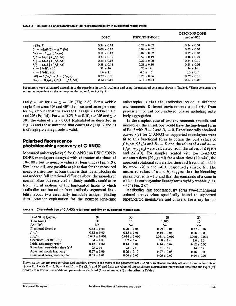
TABLE 5 Calculated characteristics of dil rotational mobility In supported monolayers
DSPC/DNP-DOPEDSPC DSPC/DNP-DOPE and ANO2
a (Eq. 5) 0.24 ± 0.03 0.26 ± 0.02 0.24 ± 0.03bT = V/2[AF(0) -AF(0)] 0.09 ± 0.03 0.08 ± 0.02 0.09 ± 0.03*bc = aE=.I (fJbi/a) 0.11 ± 0.02 0.09 ± 0.02 0.09 ± 0.02*fC = (a/b T) (fib, /a) 0.37 ± 0.12 0.52 ± 0.19 0.48 ± 0.27*f = (a/b T) (f2b2/a) 0.25 ± 0.05 0.22 ± 0.06 0.24 ± 0.10*fe = (a/b T) (f3b3/a) 0.38 ± 0.11 0.26 ± 0.10 0.28 ± 0.08r2= 1/(4D2) (s) 81 ± 16 120 ± 19 96 ± 14'r= 1/(4D3) (s) 5.4 ± 1.1 4.5 ± 1.5 3.5 ± 0.7r(0) = 2(bT/a)/[3 - (bT/a)] 0.29 ± 0.10 0.23 ± 0.06 0.29 ± 0.10r(oo) = 2(f1b1/a)/[3 - (f1bl/a)] 0.12 ± 0.03 0.13 ± 0.04 0.13 ± 0.06
Parameters were calculated according to the equations in the first column and using the measured constants shown in Table 4. *These constants areestimates dependent on the assumption that b, = b2 = b3 (Eq. 9).
and , :: 300 for e = x = 300 (Fig. 2 B). For a wobbleangle ,B between 300 and 400, the measured order parame-ter, S2, implies that the average tilt angle e is between 100and 200 (Eq. 14). For a = 0.25, b = 0.10, <300 andx <
300, the value of c is -0.001 (calculated as described inFig. 2) and the assumption that constant c (Eqs. 2 and 6)is of negligible magnitude is valid.
Polarized fluorescencephotobleaching recovery of C-AN02Measured anisotropies r(t) for C-ANO2 on DSPC/DNP-DOPE monolayers decayed with characteristic times of10-100 s but to nonzero values at long times (Fig. 9 B).Similar to diI, one possible explanation for the measurednonzero anisotropy at long times is that the antibodies donot undergo full rotational diffusion about the monolayernormal. Slow but restricted antibody mobility could arisefrom lateral motions of the haptenated lipids to whichantibodies are bound or from antibody segmental flexi-bility about two essentially immobile antigen bindingsites. Another explanation for the nonzero long-time
anisotropies is that the antibodies reside in differentenvironments. Different environments could arise frompreexistent or antibody-induced phases including anti-body aggregation.
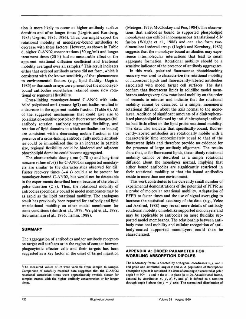
In the simplest case of two environments (mobile andimmobile), the anisotropy would have the functional formof Eq. 7 with R = 2 and DI = 0. Experimentally obtainedcurves r(t) for C-ANO2 on supported monolayers were
fit to this functional form to obtain the best values off1b1/a,f2b2/a and D2 = D and the values of a and bT =(f1bl + f2 b2) were calculated from the values of AF1 (0)and AFL(O). For samples treated with low C-ANO2concentrations (20 ,ug/ml) for a short time (10 min), theapparent rotational correlation time and fractional mobil-ity were -70 s and ~0.3, respectively (Table 6). Themeasured values of a and bT suggest that the bleachingparameter, B, is - 1.8 and that the semiangle of a cone inwhich the carbocyanine fluorophores rapidly wobble, A3, is,450 (Fig. 2 C).Antibodies can spontaneously form two-dimensional
ordered arrays when specifically bound to supportedphospholipid monolayers and bilayers; the array forma-
TABLE 6 Characteristics of C-AN02 rotational mobility on supported monolayers
[C-ANO2] (Mg/ml) 20 50 20 20Time (min) 10 10 1,200 10Anti-IgG No No No YesFractional bleach a 0.33 ± 0.05 0.20 ± 0.06 0.29 ± 0.04 0.27 ± 0.04f,bl/a 0.12 ± 0.03 0.15 ± 0.06 0.14 ± 0.04 0.16 ± 0.03f2b2/a 0.045 ± 0.006 0.054 ± 0.010 0.051 ± 0.010 0.010 ± 0.005Coefficient D (o-3 s-') 3.4 ± 0.8 2.7 ± 0.6 4.9 ± 2.4 3.0 ± 2.3Initial anisotropy r(0)* 0.12 ± 0.02 0.14 ± 0.01 0.14 ± 0.04 0.12 ± 0.03Rotational correlation time (s)* 73 ± 16 92 ± 22 51 ± 25 84 ± 65Apparent mobile fractionf2t 0.27 ± 0.06 0.26 ± 0.10 0.27 ± 0.08 0.06 ± 0.03Fractional decay/recovery bTt 0.05 ± 0.01 0.04 ± 0.03 0.06 ± 0.02 0.04 ± 0.01
Shown at the top are average values and standard errors in the mean of the parameters of C-ANO2 rotational mobility obtained from the best fits ofr(t) to Eq. 7 -with R = 2, DI = 0 and D2 = D (fbi/a and D) and from the values of the postbleach fluorescence intensities at time zero and Eq. 5 (a).Shown at the bottom are additional parameters calculated (*) or estimated (t) as described in Table 5.
Timbs and Thompson Rotational Mobilities of Antibodies and LipidsTimbs and Thompson 425Rotational Mobilities of Antibodies and Lipids
tion is more likely to occur at higher antibody surfacedensities and after longer times (Uzgiris and Kornberg,1983; Uzgiris, 1985, 1986). Thus, one might expect therotational mobility of monolayer-bound antibodies todecrease with these factors. However, as shown in Table6, higher C-AN02 concentrations (50,ug/ml) and longertreatment times (20 h) had no measurable effect on theapparent rotational diffusion coefficient and fractionalmobility averaged over all samples.3 This result indicateseither that ordered antibody arrays did not form, which isconsistent with the known sensitivity of that phenomenonto environmental factors (e.g., lipid fluidity; Uzgiris,1985) or that such arrays were present but the monolayer-bound antibodies nonetheless retained some slow rota-tional or segmental flexibility.
Cross-linking monolayer-bound C-AN02 with unla-beled polyclonal anti-(mouse IgG) antibodies resulted ina decrease in the apparent mobile fraction (Table 6). Allof the suggested mechanisms that could give rise topolarization-sensitive postbleach fluorescence changes (fullantibody rotation, antibody segmental flexibility, androtation of lipid domains to which antibodies are bound)are consistent with a decreasing mobile fraction in thepresence of a cross-linking antibody: fully mobile antibod-ies could be immobilized due to an increase in particlesize, regional flexibility could be hindered and adjacentphospholipid domains could become aggregated.The characteristic decay time ('70 s) and long-time
nonzero values of r(t) for C-AN02 on supported monolay-ers are similar to the characteristics observed for diI.Faster recovery times (-4 s) could also be present formonolayer-bound C-AN02, but would not be detectablein the experiments described herein because of the bleachpulse duration (2 s). Thus, the rotational mobility ofantibodies specifically bound to model membranes may beas rapid as the lipid rotational mobility. The analogousresult has previously been reported for antibody and lipidtranslational mobility on other model membranes forsome conditions (Smith et al., 1979; Wright et al., 1988;Subramaniam et al., 1986; Tamm, 1988).
SUMMARY
The aggregation of antibodies and/or antibody receptorson target cell surfaces or in the region of contact betweenphagocytotic effector cells and their targets has beensuggested as a key factor in the onset of target ingestion
3The measured values of D were variable from sample to sample.Comparison of carefully matched data suggested that the C-AN02rotational correlation times were approximately twofold slower forsamples treated with the higher antibody concentration or for longertimes.
(Metzger, 1979; McCloskey and Poo, 1984). The observa-tions that antibodies bound to supported phospholipidmonolayers can exhibit inhomogeneous translational dif-fusion (Wright et al., 1988) and can also form two-dimensional ordered arrays (Uzgiris and Kornberg, 1983)suggests that the monolayer-bound antibodies may expe-
rience intermolecular interactions that lead to smallaggregate formation. Rotational mobility should be a
sensitive indicator of the presence of antibody aggregates.In this work, polarized fluorescence photobleaching
recovery was used to characterize the rotational mobilityof fluorescent lipids and fluorescently-labeled antibodiesassociated with model target cell surfaces. The dataconfirm that fluorescent lipids in solidlike model mem-
branes undergo very slow rotational mobility on the orderof seconds to minutes and indicate that the rotationalmobility cannot be described as a simple, monomericrotational diffusion about the axis normal to the mono-
layer. Addition of significant amounts of a dinitropheny-lated phospholipid followed by anti-dinitrophenyl antibod-ies had little effect on the lipid probe rotational mobility.The data also indicate that specifically-bound, fluores-cently-labeled antibodies are rotationally mobile with a
characteristic time approximately equal to that of thefluorescent lipids and therefore provide no evidence forthe presence of large antibody oligomers. The resultsshow that, as for fluorescent lipids, the antibody rotationalmobility cannot be described as a simple rotationaldiffusion about the monolayer normal, implying thateither bound antibodies are significantly restricted intheir rotational mobility or that the bound antibodiesreside in more than one environment.
This work contributes to the currently small number ofexperimental demonstrations of the potential of PFPR as
a probe of molecular rotational mobility. Adaptation ofPFPR to faster times and the use of signal averaging toincrease the statistical accuracy of the data (e.g., Velezand Axelrod, 1988) may reveal more details of antibodyrotational mobility on solidlike supported monolayers andmay be applicable to antibodies on more fluidlike sup-
ported model membranes. The relationship between anti-body rotational mobility and cellular recognition of anti-body-coated supported monolayers could then becharacterized.
APPENDIX A: ORDER PARAMETER FORWOBBLING ABSORPTION DIPOLES
The laboratory frame is denoted by orthogonal coordinates x, y, and zand polar and azimuthal angles 0 and 0. A population of fluorophoreabsorption dipoles is contained in a cone of semiangle # centered at polarangle 6 = 900 - sand in the x - z plane (4t = 0). An additional frame,denoted by coordinates x', y', z', 0', and 0', is defined as a rotationthrough angle 6 about the y = y' axis. The normalized distribution of
426 Biophysical Journal Volume 58 August 1990
426 Biophysical Journal Volume 58 August 1990
absorption dipoles in the primed frame is given by
N(_') J[2ir(l -cos #)I-' 0 < '< # and 0 < 0' < 2wr (Al)N(O')-l 0 otherwise. (l
Then,
S2 - 1/2 [3(Cos2e) - 1] (A2)
(cos2 0) = [2X(1 - cos)]-' f2 Cos2 sin dOd+' (A3)
cos = z'cos5-x' sin5 (A4)z' = cost', x' = sinO'coso'. (A5)
Substituting Eq. A5 into Eqs. A4, A3, and A2 yields Eq. 14.
We thank Daniel Axelrod of the University of Michigan for numeroushelpful conversations, Alan Waggoner and Ratan Majumdar of Carn-egie-Mellon, University for the carbocyanine dye CY3.18 with whichANO2 antibodies were labeled, Harden McConnell of Stanford Univer-sity for ANO2 hybridoma cells, and Arthur Palmer of the ScrippsInstitute and Mary Lee Pisarchick and Claudia Poglitsch of theUniversity of North Carolina for their contributions to various biochem-ical and optical aspects of this work.
Support was provided by National Institutes of Health Grant GM-37145, National Science Foundation Presidential Young InvestigatorAward DCB-8552986, and a University of North Carolina JuniorFaculty Development Award.
Receivedfor publication 7 November 1989 and infinalform 29March 1990.
REFERENCES
Anglister, J., T. Frey, and H. M. McConnell. 1984. Magnetic resonanceof a monoclonal anti-spin label antibody. Biochemistry. 23:1138-1142.
Axelrod, D. 1977. Cell surface heating during fluorescence photobleach-ing recovery experiments. Biophys. J. 18:129-131.
Axelrod, D. 1979. Carbocyanine dye orientation in red cell membranestudied by microscopic fluorescence polarization. Biophys. J. 26:557-574.
Axelrod, D., D. E. Koppel, J. Schlessinger, E. Elson, and W. W. Webb.1976. Mobility measurement by analysis of fluorescence photobleach-ing recovery kinetics. Biophys. J. 16:1055-1069.
Badley, R. A., W. G. Martin, and H. Schneider. 1973. Dynamicbehavior of fluorescent probes in lipid bilayer model membranes.Biochemistry. 12:268-275.
Balakrishnan, K. F., F. J. Hsu, D. G. Hafeman, and H. M. McConnell.1982. Monoclonal antibodies to a nitroxide lipid hapten. Biochim.Biophys. Acta. 721:30-38.
Burghardt, T. P., and N. L. Thompson. 1984. Effect of planar dielectricinterfaces on fluorescence emission and detection. Biophys. J. 46:729-737.
Chen, R. F., and C. H. Scott. 1985. Atlas of fluorescence spectra andlifetimes of dyes attached to protein. Anal. Lett. 18:393-421.
Dale, R. E. 1985. Interpretation of fluorescence photobleaching recov-ery experiments on oriented cell membranes. FEBS (Fed. Eur.Biophys. Soc.) Lett., 192:255-258.
Dale, R. E. 1987. Depolarized fluorescence photobleaching recovery.Eur. Biophys. J. 14:179-193.
Ethier, M. F., D. E. Wolf, and D. L. Melchior. 1983. Calorimetricinvestigation of the phase partitioning of the fluorescent carbocyanineprobes in phosphatidylcholine bilayers. Biochemistry. 22:1178-1182.
Hafeman, D. G., V. von Tscharner, and H. M. McConnell. 1981.Specific antibody-dependent interactions between macrophages andlipid haptens in planar lipid monolayers. Proc. Natl. Acad. Sci. USA.78:4552-4556.
Johnson, P., and P. B. Garland. 1983. Carbocyanine dyes used asfluorescent triplet probes for measuring slow rotational diffusion oflipids in membranes. FEBS (Fed. Eur. Biophys. Soc.) Lett., 153:391-394.
Kimura, K., M. Nakanishi, M. Ueda, J. Ueno, H. Nariuchi, S. Furukawa,and T. Yasuda. 1986. The effect of immunoglobulin GI structure onmacrophage binding to supported planar lipid monolayers. Immunol-ogy. 59:235-238.
Klausner, R. D., and D. E. Wolf. 1980. Selectivity of fluorescent lipidanalogues for lipid domains. Biochemistry. 19:6199-6203.
Leach, S. J. 1969. Physical Principles and Techniques of ProteinChemistry. Academic Press, Inc., New York. 184-185.
Leahy, D. J., G. S. Rule, M. M. Whittaker, and H. M. McConnell.1986. Sequences of 12 monoclonal anti-dinitrophenyl spin-labelantibodies for NMR studies. Proc. Nat!. Acad. Sci. USA. 85:3661-3665.
McCloskey, M., and M. M. Poo. 1984. Protein diffusion in cellmembranes: some biological implications. Int. Rev. Cytol. 87:19-81.
McConnell, H. M., L. K. Tamm, and R. M. Weis. 1984. Periodicstructures in lipid monolayer phase transitions. Proc. Natl. Acad. Sci.USA. 81:3249-3253.
McConnell, H. M., T. H. Watts, R. M. Weis, and A. A. Brian. 1986.Supported planar membranes in studies of cell-cell recognition in theimmune system. Biochim. Biophys. Acta. 864:95-106.
Metzger, H. 1979. Early molecular events in antigen-antibody cellactivation. Ann. Rev. Pharmacol. Toxicol. 19:427-445.
Mishell, B. B., and S. M. Shiigi. 1980. Selected Methods in CellularImmunology. W. H. Freeman & Co., San Francisco. 295.
Packard, B. S., and D. E. Wolf. 1985. Fluorescence lifetimes ofcarbocyanine lipid analogues in phospholipid bilayers. Biochemistry.24:5176-5181.
Palmer, A. G., and N. L. Thompson. 1989. High-order fluorescencefluctuation analysis of model protein clusters. Proc. Natl. Acad. Sci.USA. 86:6148-6152.
Peterson, G. L. 1979. Review of the Folin phenol protein quantitationmethod of Lowry, Rosebrough, Farr and Randall. Anal. Biochem.100:201-220.
Poglitsch, C. L., and N. L. Thompson. 1990. Interaction of antibodieswith Fc receptors in substrate-supported planar membranes measuredby total internal reflection fluorescence microscopy. Biochemistry.29:248-254.
Scalettar, B. A., P. R. Selvin, D. Axelrod, J. E. Hearst, and M. P. Klein.1988. A fluorescence photobleaching study of the microsecond reori-entational motions of DNA. Biophys. J. 53:215-226.
Scalettar, B. A., P. R. Selvin, D. Axelrod, M. P. Klein, and J. E. Hearst.1990. A polarized photobleaching study of DNA reorientation inagarose gels. Biochemistry. 29:4790-4798.
Seul, M., S. Subramaniam, and H.M. McConnell. 1985. Mono- andbilayers of phospholipids at interfaces: interlayer coupling and phasestability. J. Phys. Chem. 89:3592-3595.
Shimshick, E. J., and H. M. McConnell. 1973. Lateral phase separationin phospholipid membranes. Biochemistry. 12:2351-2360.
Timbs and Thompson Rotational Mobilities of Antibodies and Lipids 427

Smith, B. A., and H. M. McConnell. 1978. Determination of molecularmotion in membranes using periodic pattern photobleaching. Proc.Natl. Acad. Sci. USA. 75:2759-2763.
Smith, L. M., and H. M. McConnell. 1981. Pattern photobleaching offluorescent phospholipid vesicles using polarized laser light. Biophys.J. 33:139-146.
Smith, L. M., J. W. Parce, B. A. Smith, and H. M. McConnell. 1979.Antibodies bound to lipid haptens in model membranes diffuse asrapidly as the lipids themselves. Proc. Natl. Acad. Sci. USA.76:4177-4179.
Smith, L. M., R. M. Weis, and H. M. McConnell. 1981. Measurementof rotational motion in membranes using fluorescence recovery afterphotobleaching. Biophys. J. 36:73-9 1.
Subramaniam, S., M. Seul, and H. M. McConnell. 1986. Lateraldiffusion of specific antibodies bound to lipid monolayers on alkylatedsubstrates. Proc. Natl. Acad. Sci. USA. 83:1169-1173.
Tamm, L. K. 1988. Lateral diffusion and fluorescence microscopestudies of a monoclonal antibody specifically bound to supportedphospholipid bilayers. Biochemistry. 27:1450-1457.
Thompson, N. L., and A. G. Palmer. 1988. Model cell membranes onplanar substrates. Comments Mol. Cell. Biophys. 5:39-56.
Thompson, N. L., H. M. McConnell, and T. P. Burghardt. 1984. Orderin supported phospholipid monolayers detected by the dichroism offluorescence excited with polarized evanescent illumination. Biophys.J. 46:739-747.
Thompson, N. L., A. G. Palmer, L. L. Wright, and P. E. Scarborough.1988. Fluorescence techniques for supported planar model mem-branes. Comments Mol. Cell. Biophys. 5:109-13 1.
Uzgiris, E. E. 1985. Antibody crystallization on phospholipid films:dynamics and the effects of antibody conformation. J. Cell. Biochem.29:239-251.
Uzgiris, E. E. 1986. Supported phospholipid bilayers for two-dimensional protein crystallization. Biochem. Biophys. Res. Com-mun. 134:819-826.
Uzgiris, E. E., and R. D. Kornberg. 1983. Two-dimensional crystalliza-tion technique for imaging macromolecules, with application toantigen-antibody-complement complexes. Nature (Lond.). 301:125-129.
Velez, M., and D. Axelrod. 1988. Polarized fluorescence photobleachingrecovery for measuring rotational diffusion in solutions and mem-branes. Biophys. J. 53:575-591.
von Tscharner, V., and H. M. McConnell. 1981. Physical properties oflipid monolayers on alkylated planar glass surfaces. Biophys. J.36:421-427.
Wagner, M., L. Ernst, R. Majumdar, S. Majumdar, J. Chao, and A.Waggoner. 1990. Applications of new-cyanine labeling reagents inflow cytometry. 14th International Meeting of the Society forAnalytical Cytology, Asheville, NC. Abstract No. 459.
Wegener, W. A. 1984. Fluorescence recovery spectroscopy as a probe ofslow rotational motions. Biophys. J. 46:795-803.
Wegener, W. A., and R. Rigler. 1984. Separation of translational androtational contributions in solution studies using fluorescence photo-bleaching recorvery. Biophys. J. 46:787-793.
Wright, L. L., A. G. Palmer, and N. L. Thompson. 1988. Inhomoge-neous translational diffusion of monoclonal antibodies on phospho-lipid Langmuir-Blodgett films. Biophys. J. 54:463-470.
Wu, S. H., and H. M. McConnell. 1975. Phase separations in phospho-lipid membranes. Biochemistry. 14:847-854.
Yguerabide, J., and L. Stryer. 1971. Fluorescence spectroscopy of anoriented model membrane. Proc. Natl. Acad. Sci. USA. 68:1217-1221.
428 Biophysical Journal Volume 58 August 1990
Copyright © 2022 FDOKUMEN