Marine Lipids 2017 - MDPI
170
Marine Lipids 2017 Rosário Domingues, Ricardo Calado and Pedro Domingues www.mdpi.com/journal/marinedrugs Edited by Printed Edition of the Special Issue Published in Marine Drugs marine drugs Books MDPI
-
Upload
khangminh22 -
Category
Documents
-
view
3 -
download
0
Transcript of Marine Lipids 2017 - MDPI

Marine Lipids 2017
Rosário Domingues, Ricardo Calado and Pedro Domingues
www.mdpi.com/journal/marinedrugs
Edited by
Printed Edition of the Special Issue Published in Marine Drugs
marine drugs
Books
MDPI

Marine Lipids 2017 Special Issue Editors Rosário Domingues Ricardo Calado Pedro Domingues MDPI • Basel • Beijing • Wuhan • Barcelona • Belgrade
Books
MDPI

Special Issue Editors Rosário Domingues Ricardo Calado University of Aveiro University of Aveiro Portugal Portugal
Pedro Domingues University of Aveiro Portugal
Editorial Office MDPI AG St. Alban-Anlage 66 Basel, Switzerland
This edition is a reprint of the Special Issue published online in the open access journal Marine Drugs (ISSN 1660-3397) in 2017 (available at: http://www.mdpi.com/journal/marinedrugs/special_issues/marine_lipids_2017).
For citation purposes, cite each article independently as indicated on the article page online and as indicated below:
Lastname, F.M.; Lastname, F.M. Article title. Journal Name Year, Article number, page range.
First Edition 2018
ISBN 978-3-03842-799-5 (Pbk) ISBN 978-3-03842-800-8 (PDF)
Articles in this volume are Open Access and distributed under the Creative Commons Attribution license (CC BY), which allows users to download, copy and build upon published articles even for commercial purposes, as long as the author and publisher are properly credited, which ensures maximum dissemination and a wider impact of our publications. The book taken as a whole is © 2018 MDPI, Basel, Switzerland, distributed under the terms and conditions of the Creative Commons license CC BY-NC-ND (http://creativecommons.org/licenses/by-nc-nd/4.0/).
Books
MDPI

Table of Contents
About the Special Issue Editors . . . . . . . . . . . . . . . . . . . . . . . . . . . . . . . . . . . . . v
Preface to ”Marine Lipids 2017”. . . . . . . . . . . . . . . . . . . . . . . . . . . . . . . . . . . . . vii
Elisabete da Costa, Tnia Melo, Ana S. P. Moreira, Carina Bernardo, Luisa Helguero,
Isabel Ferreira, Maria Teresa Cruz, Andreia M. Rego, Pedro Domingues, Ricardo Calado,
Maria H. Abreu and Maria Rosario Domingues
Valorization of Lipids from Gracilaria sp. through Lipidomics and Decoding ofAntiproliferative and Anti-Inflammatory Activitydoi: 10.3390/md15030062 . . . . . . . . . . . . . . . . . . . . . . . . . . . . . . . . . . . . . . . 1
Gabriel A. Bonaterra, David Driscoll, Hans Schwarzbach and Ralf Kinscherf
Krill Oil-In-Water Emulsion Protects against Lipopolysaccharide-Induced ProinflammatoryActivation of Macrophages In Vitrodoi: 10.3390/md15030074 . . . . . . . . . . . . . . . . . . . . . . . . . . . . . . . . . . . . . . . 18
Oscar Monroig, Rosa de Llanos, Inmaculada Varo, Francisco Hontoria, Douglas R. Tocher,
Sergi Puig and Juan C. Navarro
Biosynthesis of Polyunsaturated Fatty Acids in Octopus vulgaris: Molecular Cloning andFunctional Characterisation of a Stearoyl-CoA Desaturase and an Elongation of Very Long-Chain Fatty Acid 4 Proteindoi: 10.3390/md15030082 . . . . . . . . . . . . . . . . . . . . . . . . . . . . . . . . . . . . . . . 30
Yongping Zhang, Guangling Jiao, Cai Song, Shelly Gu, Richard E. Brown, Junzeng Zhang,
Pingcheng Zhang, Jacques Gagnon, Steven Locke, Roumiana Stefanova, Claude Pelletier,
Yi Zhang and Hongyu Lu
An Extract from Shrimp Processing By-Products Protects SH-SY5Y Cells from NeurotoxicityInduced by Aβ25–35
doi: 10.3390/md15030083 . . . . . . . . . . . . . . . . . . . . . . . . . . . . . . . . . . . . . . . 46
Kai-Min Yang and Po-Yuan Chiang
Variation Quality and Kinetic Parameter of Commercial n-3 PUFA-Rich Oil duringOxidation via Rancimatdoi: 10.3390/md15040097 . . . . . . . . . . . . . . . . . . . . . . . . . . . . . . . . . . . . . . . 65
Lucıa Mendez, Gabriel Dasilva, Nria Taltavull, Marta Romeu and Isabel Medina
Marine Lipids on Cardiovascular Diseases and Other Chronic Diseases Induced by Diet:An Insight Provided by Proteomics and Lipidomicsdoi: 10.3390/md15080258 . . . . . . . . . . . . . . . . . . . . . . . . . . . . . . . . . . . . . . . 76
Phuc Nguyen Thien Le and Andrew P. Desbois
Antibacterial Effect of Eicosapentaenoic Acid against Bacillus cereus and Staphylococcus aureus:Killing Kinetics, Selection for Resistance, and Potential Cellular Targetdoi: 10.3390/md15110334 . . . . . . . . . . . . . . . . . . . . . . . . . . . . . . . . . . . . . . . 105
Sonia A. O. Santos, Stephanie S. Trindade, Catia S. D. Oliveira, Paula Parreira, Daniela Rosa,
Maria F. Duarte, Isabel Ferreira, Maria T. Cruz, Andreia M. Rego, Maria H. Abreu,
Silvia M. Rocha and Armando J. D. Silvestre
Lipophilic Fraction of Cultivated Bifurcaria bifurcata R. Ross: Detailed Composition and InVitro Prospection of Current Challenging Bioactive Propertiesdoi: 10.3390/md15110340 . . . . . . . . . . . . . . . . . . . . . . . . . . . . . . . . . . . . . . . 115
iii
Books
MDPI

Alexander N. Shikov, Into Laakso, Olga N. Pozharitskaya, Tuulikki Seppanen-Laakso,
Anna S. Krishtopina, Marina N. Makarova, Heikki Vuorela and Valery Makarov
Chemical Profiling and Bioactivity of Body Wall Lipids from Strongylocentrotus droebachiensisdoi: 10.3390/md15120365 . . . . . . . . . . . . . . . . . . . . . . . . . . . . . . . . . . . . . . . 132
M. Barra, A. Llanos-Rivera, F. Cruzat, N. Pino-Maureira and R. R. Gonzlez-Saldıa
The Marine Fungi Rhodotorula sp. (Strain CNYC4007) as a Potential Feed Source for FishLarvae Nutritiondoi: 10.3390/md15120369 . . . . . . . . . . . . . . . . . . . . . . . . . . . . . . . . . . . . . . . 143
iv
Books
MDPI

Books
MDPI

Books
MDPI

Books
MDPI

Books
MDPI

marine drugs
Article
Valorization of Lipids from Gracilaria sp. throughLipidomics and Decoding of Antiproliferative andAnti-Inflammatory Activity
Elisabete da Costa 1, Tânia Melo 1, Ana S. P. Moreira 1, Carina Bernardo 2, Luisa Helguero 2,
Isabel Ferreira 3, Maria Teresa Cruz 3, Andreia M. Rego 4, Pedro Domingues 1, Ricardo Calado 5,
Maria H. Abreu 4 and Maria Rosário Domingues 1,*
1 Centro de Espectrometria de Massa, Departamento de Química & QOPNA, Universidade de Aveiro,Campus Universitário de Santiago, 3810-193 Aveiro, Portugal; [email protected] (E.d.C.);[email protected] (T.M.); [email protected] (A.S.P.M.); [email protected] (P.D.)
2 Instituto de Biomedicina (IBIMED), Departamento de Ciências Médicas, Universidade de Aveiro,3810-193 Aveiro, Portugal; [email protected] (C.B.); [email protected] (L.H.)
3 Centro de Neurociências e Biologia Celular (CNC), Universidade de Coimbra, 3004-517 Coimbra &Faculdade de Farmácia, Universidade de Coimbra, 3000-548 Coimbra, Portugal; [email protected] (I.F.);[email protected] (M.T.C.)
4 ALGAplus-Produção e Comercialização de Algas e seus Derivados, Lda., 3830-196 Ílhavo, Portugal;[email protected] (A.M.R.); [email protected] (M.H.A.)
5 Departamento de Biologia & CESAM, Universidade de Aveiro, Campus Universitário de Santiago,3810-193 Aveiro, Portugal; [email protected]
* Correspondence: [email protected]
Academic Editor: Peer B. JacobsonReceived: 11 November 2016; Accepted: 13 February 2017; Published: 2 March 2017
Abstract: The lipidome of the red seaweed Gracilaria sp., cultivated on land-based integratedmultitrophic aquaculture (IMTA) system, was assessed for the first time using hydrophilicinteraction liquid chromatography-mass spectrometry and tandem mass spectrometry (HILIC–MSand MS/MS). One hundred and forty-seven molecular species were identified in the lipidome of theGracilaria genus and distributed between the glycolipids classes monogalactosyl diacylglyceride(MGDG), digalactosyl diacylglyceride (DGDG), sulfoquinovosyl monoacylglyceride (SQMG),sulfoquinovosyl diacylglyceride (SQDG), the phospholipids phosphatidylcholine (PC), lyso-PC,phosphatidylglycerol (PG), lyso-PG, phosphatidylinositol (PI), phosphatidylethanolamine (PE),phosphatic acid (PA), inositolphosphoceramide (IPC), and betaine lipids monoacylglyceryl-and diacylglyceryl-N,N,N-trimethyl homoserine (MGTS and DGTS). Antiproliferative andanti-inflammatory effects promoted by lipid extract of Gracilaria sp. were evaluated by monitoringcell viability in human cancer lines and by using murine macrophages, respectively. The lipidextract decreased cell viability of human T-47D breast cancer cells and of 5637 human bladdercancer cells (estimated half-maximal inhibitory concentration (IC50) of 12.2 μg/mL and 12.9 μg/mL,respectively) and inhibited the production of nitric oxide (NO) evoked by the Toll-like receptor4 agonist lipopolysaccharide (LPS) on the macrophage cell line RAW 264.7 (35% inhibition at aconcentration of 100 μg/mL). These findings contribute to increase the ranking in the value-chain ofGracilaria sp. biomass cultivated under controlled conditions on IMTA systems.
Keywords: glycolipids; phospholipids; betaine lipids; seaweeds; bioactivity; mass spectrometry;hydrophilic interaction liquid chromatography–electrospray ionization–mass spectrometryHILIC–ESI–MS
Mar. Drugs 2017, 15, 62 1 www.mdpi.com/journal/marinedrugs
Books
MDPI
Mar. Drugs 2017, 15, 62
1. Introduction
Red seaweeds within the genus Gracilaria are one of the world’s most cultivated and valuablemarine macrophytes. This group of seaweeds is well adapted to cultivation on land-based integratedmultitrophic aquaculture (IMTA) systems, allowing its sustainable production under controlled andreplicable conditions that provide a secure supply of high-grade seaweed biomass for demandingmarkets (e.g., food, pharmaceuticals) [1–3]. Gracilaria sp. is a source of multiple products, amongwhich lipids, namely polyunsaturated fatty acids (PUFAs) such as arachidonic (20:4(n-6), AA) andeicosapentaenoic (20:5(n-6), EPA) acids, are emerging as valuable components [4]. Fatty acids (FAs) aremainly esterified to polar lipids such as glycolipids, phospholipids, and betaine lipids. Polar lipids arenowadays recognized as an important reservoir of fatty acids with nutritional value, e.g., n-3 FAs [5,6],and they are also considered high-value novel lipids with beneficial health effects such as antitumoral [7,8],antiviral [8,9], antifungal [10], antibacterial [11], and anti-inflammatory [11,12], with potentialapplications in the nutraceutical and pharmaceutical industries [7,8,11]. However, in spite of theirrecognized potential, they are still scarcely studied [12–15]. Some studies reported that polar lipidsisolated from seaweeds can promote growth-inhibiting effects on human hepatocellular carcinomacell lines (HepG2) [16] and thus can act as inhibitors of DNA polymerases with capability to inhibittumor cell proliferation [17]. Moreover, they have been associated with anti-inflammatory propertiesthrough the inhibition of pro-inflammatory cytokines interleukin IL-6 and IL-8 production [15] and/orby the inhibition of nitric oxide (NO) production [18–21]. Lipid-based agents are therefore emergingmolecules in therapeutics aimed to regulate inflammatory pathways or even impair downstreamtumorigenic processes [22–24].
To fully explore the bioactive properties of seaweed lipids and thus contribute to seaweedvalorization, it is fundamental to characterize their structure and understand how it modulatesbioactivity [13,14]. Nowadays, the detailed structural characterization of lipids can be accomplishedby using mass spectrometry (MS) coupled with liquid chromatography (LC). This lipidomic approachhas the advantage of providing a detailed analysis of the lipid profile and affording the identificationand quantification of more than 200 lipid molecular species in one single LC–MS run [25,26]. Thisdetailed information on the specificity of molecular species and corresponding classes of polar lipidscannot be achieved using traditional approaches, typically based on the previous separation of polarlipid classes by thin-layer chromatography (TLC) and silica gel on column chromatography, followedby off-line gas chromatography-mass spectrometry (GC–MS) analysis of FAs [27–33]. MS-basedtechnologies allowed researchers to explore the full lipidomic signature of distinct matrices [34–36].To date, they allowed for the identification of the full lipidome signature of cultivated seaweedsUlva lactuca Linnaeus, 1753 [37], Chondrus crispus Stackhouse, 1797 [26], and Codium tomentosumStackhouse, 1797 [25]. These novel approaches based on specific identification and quantification atthe molecular level using high-throughput analysis are promising tools for bioprospection [3,38,39].
The main goal of the present study was to identify and characterize the polar lipid profileof Gracilaria sp. cultivated under controlled conditions on a land-based integrated multitrophicaquaculture (IMTA) system, using hydrophilic interaction liquid chromatography-electrosprayionization-mass spectrometry (HILIC–ESI–MS). The lipid extract of this red seaweed was alsoscreened for its growth inhibitory effects in human breast and bladder cancer cell lines, as wellas anti-inflammatory effects by inhibiting the production of NO.
2. Results and Discussion
The lipid extract of Gracilaria sp. obtained by chloroform:methanol extraction accounted for about3000 ± 600 mg/kg dry mass (relative standard deviation (RSD) < 20%). The lipid extract was mainlycomposed of glycolipids (1980 ± 148 mg/kg of biomass) and phospholipids (165 ± 53 mg/kg ofbiomass), and the remaining lipid extract corresponded to betaine lipids and others (Table 1).
2
Books
MDPI

Mar. Drugs 2017, 15, 62
Table 1. Composition of lipid extract of Gracilaria sp. (mean and SD of triplicate).
Composition Mean SD
Lipids (mg/kg biomass) 3000 600Glycolipids (mg/kg biomass) 1980 148
Phospholipids (mg/kg biomass) 165 52.7Betaines and others 1 855 -
1 Betaines and others were determined by the difference of lipid content and the sum of content of glycolipidsand phospholipids.
2.1. Polar Lipidome
The profile of Gracilaria polar lipidome was determined by HILIC–ESI–MS and allowed for theidentification of molecular species of glycolipids, phospholipids, and betaine lipids. Overall, thelipidome of Gracilaria sp. comprised 147 molecular species (Figure 1).
Figure 1. Number of molecular species identified by HILIC–ESI–MS, distributed by theclasses of glycolipids: monogalactosyl diacylglyceride (MGDG), digalactosyl diacylglyceride(DGDG), sulfoquinovosyl monoacylglyceride (SQMG), sulfoquinovosyl diacylglyceride (SQDG),phospholipids: phosphatidylcholine (PC) and lyso-PC (LPC), phosphatidylglycerol (PG) andlyso-PG (LPG), phosphatidylinositol (PI), phosphatic acid (PA), phosphatidylethanolamine (PE),inositolphosphoceramide (IPC), and betaine lipids: monoacylglyceryl- and diacylglyceryl-N,N,N-trimethyl homoserine (MGTS and DGTS).
The glycolipids of the classes monogalactosyl diacylglyceride (MGDG) and digalactosyldiacylglyceride (DGDG) were identified in the LC–MS spectra in positive mode as [M + NH4]+ ions [25].Detailed structure of MGDG and DGDG molecular species was accomplished by LC–MS/MS analysisof [M + NH4]+ ions and analysis of ESI–MS/MS of the [M + Na]+ ions after solid phase extraction(SPE) fractionation of lipid extract (fraction 3 rich in glycolipids). Overall, 34 molecular specieswere identified, as described in Table 2. Galactolipids contained nine MGDG molecular species and10 DGDG molecular species (Table 2, Figure S1a,b). The most abundant MGDG molecular specieswere found at m/z 774.3 and 796.3 [M + NH4]+, corresponding to MGDG (18:1/16:0) and to MGDG(20:4/16:0), with a minor contribution from MGDG (18:2/18:2), respectively. Other MGDG molecularspecies identified contained in their composition 14-, 16-, and 18-carbon saturated fatty acids (SFAs)and monounsaturated fatty acids (MUFAs) and 18:2, 20:4, and 20:5 polyunsaturated fatty acyl (PUFAs)moieties (Table 2). Regarding DGDGs, the most abundant molecular species were identified as[M + NH4]+ ions at m/z 936.3, corresponding to DGDG (18:1/16:0), followed by DGDG (20:4/16:0)with minor contribution of DGDG (18:2/18:2) at m/z 958.2. Moreover, other DGDG molecular specieswere identified containing 14-, 16-, 18-, and 20-carbon fatty acids (FAs) such as 20:4 and 20:5 PUFAs.
Concerning sulfolipids, 12 sulfoquinovosyl diacylglycerides (SQDGs) and three sulfoquinovosylmonoacylglycerides (SQMGs) were identified as negative [M − H]− ions. The most abundantspecies were attributed to SQDG (14:0/16:0), SQDG (16:0/16:0) and SQDG (16:0/20:4), observedas [M − H]− ions at m/z 765.5, 793.5, and 841.6, respectively. The fatty acyl signature of SQDGs
3
Books
MDPI

Mar. Drugs 2017, 15, 62
included 14-, 16-, 18-carbon SFAs and MUFAs and 18- and 20-carbon PUFAs (Table 2, Figure S1c).Three SQMGs were identified as SQMG (14:0), SQMG (16:0), and SQMG (16:1). SQMGs were neverbefore reported in the lipidome of seaweeds from the genus Gracilaria. Glycolipids have already beenidentified for members of the Rhodophyta (red seaweeds), namely in the genus Gracilaria [21,30,40–42].However, the majority of published works only identified a few species of glycolipids, either by usingoffline TLC–MS [30,40,42,43] or selected solvent extraction and MS analysis [15,20,29]. More recently,a detailed profile of Chondrus crispus was reported using LC–MS and MS/MS [26].
Table 2. Identification of MGDG and DGDG molecular species observed by HILIC–ESI–MS,as [M + NH4]+ ions and SQDG and SQMG molecular species observed as [M − H]− ions 2.
[M + NH4]+ Lipid Species Fatty Acyl Chains
m/z (C:N)
Monogalactosyl diacylglyceride (MGDG)
746.3 MGDG (32:1) 16:1/16:0 and 14:0/18:1748.3 MGDG (32:0) 16:0/16:0 and 14:0/18:0774.3 MGDG (34:1) 18:1/16:0776.3 MGDG (34:0) 18:0/16:0794.3 MGDG (36:5) 20:5/16:0796.3 MGDG (36:4) 20:4/16:0 and 18:2/18:2
Digalactosyl diacylglyceride (DGDG)
908.3 DGDG (32:1) 16:1/16:0 and 14:0/18:1910.3 DGDG (32:0) 16:0/16:0 and 14:0/18:0934.3 DGDG (34:2) 18:2/16:0 and 18:1/16:1936.3 DGDG (34:1) 18:1/16:0956.3 DGDG (36:5) 20:5/16:0958.3 DGDG (36:4) 20:4/16:0 and 18:2/18:2
[M − H]− Lipid Species Fatty Acyl Chains
Sulfoquinovosyl diacylglyceride (SQDG)
763.6 SQDG (30:1) 14:0/16:1765.6 SQDG (30:0) 14:0/16:0791.6 SQDG (32:1) 16:1/16:0 and 14:0/18:2793.6 SQDG (32:0) 16:0/16:0 and 14:0/18:0813.6 SQDG (34:4) 18:4/16:0817.6 SQDG (34:2) 18:2/16:0819.6 SQDG (34:1) 18:1/16:0839.6 SQDG (36:5) 20:5/16:0841.6 SQDG (36:4) 20:4/16:0857.6 SQDG (36:4-OH) 20:4-OH/16:0
Sulfoquinovosyl monoacylglyceride (SQMG)
527.4 SQMG (14:0)553.4 SQMG (16:1)555.4 SQMG (16:0)
2 The assignment of the fatty acyl composition of molecular species was made according to the interpretationof the corresponding MS/MS spectra. Bold m/z values correspond to the most abundant species detectedin the LC–MS spectrum; C means the number of carbon atoms; N represents double bonds in the fatty acylchains; MGDG: monogalactosyl diacylglyceride; DGDG: digalactosyl diacylglyceride; SQMG: sulfoquinovosylmonoacylglyceride; SQDG: sulfoquinovosyl diacylglyceride; and HILIC–ESI–MS: hydrophilic interaction liquidchromatography–electrospray ionization–mass spectrometry.
Gracilaria sp. lipidome included 87 molecular species of phospholipids (PLs) within eightclasses, namely phosphatidylglycerol (PG) and lyso-PG (LPG), phosphatidylcholine (PC) and lyso-PC(LPC), phosphatidylethanolamine (PE), phosphatidylinositol (PI), inositolphosphoceramide (IPC) andphosphatidic acid (PA). PC is a main component of extraplastidial membranes, while PG is found inchloroplastic membranes [5].
The PL classes PG, lyso-PG, PA, PI, and IPC were identified as negative [M − H]− ions, while PCand lyso-PC were identified as negative [M + CH3COO]− ions. PC, LPC and PE were also identified
4
Books
MDPI

Mar. Drugs 2017, 15, 62
as positive [M + H]+ ions. The identity of all molecular species identified (Table 3) was confirmedby LC–MS/MS, as described in the literature [25,26]. About 39 PCs were identified by LC–MS(Figure S2a). The most abundant ions were observed at m/z 760.6 and at m/z 782.6, respectivelyattributed to PC (16:0/18:1) and to PC (16:0/20:4) with a minor contribution of PC (18:2/18:2). OtherPC molecular species were identified and contained 14- to 22-carbon fatty acids. Lyso-PC consistedof eight molecular species (Table 3, Figure S2b) and the most abundant was LPC (20:4), observed atm/z 544.4. All molecular species identified are described in Table 3. Thirteen PGs and four lyso-PGsspecies were identified by LC–MS as [M – H]− ions (Table 3, Figure S2c,d). The most abundant ion wasobserved at m/z 769.4, mainly corresponding to PG (16:0/20:4), with a minor contribution from PG(18:2/18:2). The prominent lyso-PG at m/z 483.3 was LPG (16:0). PI species were observed as [M − H]−
ions at m/z 833.5 and 835.5 and attributed to PI (16:1/18:1) and PI (16:0/18:1), respectively. Eight PAswere identified (Table 3, Figure S2e), with the most abundant species identified as PA (20:4/20:4) atm/z 743.3, while the other PA molecular species were esterified to 16:0, 18:1, 18:2, 18:3, 20:3, 20:4, and20:5 FAs. PEs contained eight molecular species, identified as [M + H]+ (Table 3, Figure S2f). Themost abundant ion was observed at m/z 716.4 and identified as PE (16:1/16:1) and PE (16:0/18:2).The phospholipids from Gracilaria sp. hold PCs and LPCs, PGs, LPGs, PIs, PEs, and PAs, alreadyreported for the lipidome of other Rhodophyta [20,26,31,32]. Fatty acids esterified in the PLs includedsaturated and unsaturated 16-, 18-, and 20-carbon FAs, and PCs and PAs were the only PLs classes thatincluded 20:3(n-6) FA. The 20:3(n-6) FA is usually a minor component of the whole pool of FAs in redseaweeds [31,32] but is an important intermediate compound in the biosynthesis of 20:4(n-6) FA.
Table 3. Identification of phospholipid molecular species observed by HILIC–ESI–MS, as [M + H]+
ions for PC, LPC, and PE and as [M − H]− ions for PG, LPG, PI, PA, and IPC 2.
[M + H]+ Lipid Species Fatty Acyls Chain
m/z (C:N)
Phosphatidylcholine (PC)
732.6 PC (32:1) 16:0/16:1 and 14:0/18:1734.6 PC (32:0) 16:0/16:0 and 14:0/18:0754.6 PC (34:4) 14:0/20:4 and 16:2/18:2756.6 PC (34:3) 16:0/18:3 and 14:0/20:3758.6 PC (34:2) 16:0/18:2 and 16:2/18:1760.6 PC (34:1) 16:0/18:1762.6 PC (34:0) 16:0/18:0780.6 PC (36:5) 16:0/20:5 and 18:2/18:3782.6 PC (36:4) 16:0/20:4 and 18:2/18:2784.6 PC (36:3) 16:0/20:3 and 18:1/18:2786.6 PC (36:2) 18:0/18:2 and 18:1/18:1788.6 PC (36:1) 18:0/18:1798.5 PC (37:3) 16:0/21:3 and 18:1/19:2804.5 PC (38:7) 18:3/20:4 and 18:2/20:5806.5 PC (38:6) 18:2/20:4 and 18:1/20:5808.5 PC (38:5) 18:1/20:4 and 18:2/20:3810.5 PC (38:4) 18:1/20:3 and 16:0/22:4812.5 PC (38:3) 18:0/20:3 and 18:1/20:2814.5 PC (38:2) 16:0/22:2 and 18:1/20:1818.5 PC (38:0) 18:0/20:0 and 16:0/22:0840.4 PC (40:3) 18:1/22:2844.4 PC (40:1) 18:1/22:0
Lyso-phosphatidylcholine (LPC)
494.4 LPC (16:1)496.4 LPC (16:0)518.4 LPC (18:3)520.4 LPC (18:2)522.4 LPC (18:1)524.4 LPC (18:0)542.4 LPC (20:5)544.4 LPC (20:4)
5
Books
MDPI

Mar. Drugs 2017, 15, 62
Table 3. Cont.
[M + H]+ Lipid Species Fatty Acyls Chain
m/z (C:N)
Phosphatidyletanolamine (PE)
716.4 PE (34:2) 16:1/18:1 and 16:0/18:2718.3 PE (34:1) 16:1/18:0 and 16:0/18:1740.4 PE (34:0) 16:0/18:0742.4 PE (36:3) 18:1/18:2744.4 PE (36:2) 18:1/18:1746.3 PE (36:1) 18:0/18:1
[M − H]− Lipid Species Fatty Acyl Chains
Phosphatidylglycerol (PG)
717.4 PG (32:2) 16:1/16:1 and 16:0/16:2719.4 PG (32:1) 16:0/16:1721.4 PG (32:0) 16:0/16:0741.4 PG (34:4) 16:0/18:4743.5 PG (34:3) 16:0/18:3745.5 PG (34:2) 16:1/18:1747.5 PG (34:1) 16:0/18:1 and 16:1/18:0767.5 PG (36:5) 16:0/20:5769.4 PG (36:4) 16:0/20:4 and 18:2/18:2773.5 PG (36:2) 18:1/18:1
Lyso-phosphatidylglycerol (LPG)
481.3 LPG (16:1)483.3 LPG (16:0)509.3 LPG (18:1)531.3 LPG (20:4)
Phosphatidylinositol (PI)
833.5 PI (34:2) 16:1/18:1835.5 PI (34:1) 16:0/18:1
Phosphatidic acid (PA)
693.4 PA (36:5) 16:0/20:5695.4 PA (36:4) 16:0/20:4717.4 PA (38:7) 18:3/20:4719.4 PA (38:6) 18:2/20:4721.4 PA (38:5) 18:1/20:4741.3 PA (40:9) 20:4/20:5743.3 PA (40:8) 20:4/20:4745.3 PA (40:7) 20:3/20:4
Inositolphosphoceramide (IPC)
810.5 IPC (t35:0) t18:0/17:0908.6 IPC (d42:0) d18:0/24:0920.6 IPC (t42:2) t18:1/24:1922.6 IPC (t42:1) t18:0/24:1924.6 IPC (t42:0) t18:0/24:0
2 The assignment of the fatty acyl composition of molecular species was made according to the interpretationof the corresponding MS/MS spectra. Bold m/z values correspond to the most abundant species detectedin the LC–MS spectrum; C means the number of carbon atoms; N represents double bonds in the fatty acylchains; PC: phosphatidylcholine; LPC: lyso-PC; PG: phosphatidylglycerol; LPG: lyso-PG; PI: phosphatidylinositol;PA: phosphatic acid; PE: phosphatidylethanolamine; and IPC: inositolphosphoceramide.
Five molecular species were assigned as IPCs and the most abundant ones were IPC (t18:0/17:0),observed at m/z 810.5, and IPC (t18:1/24:1), observed at m/z 920.6 (Figure S3a). LC–MS/MS spectrumof the [M − H]− ions of IPCs, as exemplified for IPC at m/z 920.6 in Figure S3b, showed the typicalfragmentation pathways of IPCs such as the losses of 162 Da and 180 Da, due to the fragmentationpathways that lead to the elimination of inositol, the product ion at m/z 538.3 resulting from the loss offatty acyl chains, and the product ion at m/z 259.0 that corresponded to an inositol monophosphateanion. IPCs were identified in lipid extract of Gracilaria sp. and are considered an important biomarker
6
Books
MDPI

Mar. Drugs 2017, 15, 62
of Rhodophyta taxonomy, in accordance with what was already reported for Chondrus crispus using alipidomic approach [26]. IPCs are required to maintain membrane properties such as viscosity andelectrical charge and participate in the control of enzymatic activity or act as membrane anchors forsome proteins [44].
Twenty-one DGTS and five MGTS molecular species were identified by LC–MS and MS/MS as[M + H]+ ions (Table 4, Figure S4a,b, respectively). The most abundant DGTS species were found at m/z710.7, corresponding to DGTS (16:0/16:1), with a minor contribution from DGTS (14:0/18:1) species,followed by DGTS (18:1/18:1) observed at m/z 764.8. Overall, DGTSs combine distinctive molecularspecies bearing different combinations of FAs, ranging between 14- and 20-carbon FAs, as reported onTable 4. MGTSs comprised MGTS (14:0), MGTS (16:1), MGTS (16:0), MGTS (18:2), and MGTS (18:1)species, identified at m/z 446.5, 472.5, 474.5, 498.6, and 500.6, respectively.
Table 4. Identification of DGTS and MGTS molecular species observed by HILIC–ESI–MS as [M + H]+ ions 2.
[M + H]+ Lipid Species Fatty Acyls Chain
m/z (C:N)
Diacylglyceryl trimethyl homoserine (DGTS)
656.7 DGTS (28:0) 14:0/14:0682.7 DGTS (30:1) 14:0/16:1684.8 DGTS (30:0) 14:0/16:0708.7 DGTS (32:2) 16:1/16:1 and 14:0/18:2710.7 DGTS (32:1) 16:0/16:1 and 14:0/18:1712.7 DGTS (32:0) 16:0/16:0 and 14:0/18:0732.7 DGTS (34:4) 16:2/18:2 and 14:0/20:4734.7 DGTS (34:3) 16:1/18:2736.7 DGTS (34:2) 16:0/18:2 and 16:1/18:1738.7 DGTS (34:1) 16:0/18:1 and 16:1/18:0740.7 DGTS (34:0) 16:0/18:0 and 14:0/20:0760.6 DGTS (36:4) 16:0/20:4764.8 DGTS (36:2) 18:1/18:1766.8 DGTS (36:1) 18:0/18:1
Monoacylglyceryl trimethyl homoserine (MGTS)
446.5 MGTS (14:0)472.5 MGTS (16:1)474.5 MGTS (16:0)498.6 MGTS (18:2)500.6 MGTS (18:1)
2 The assignment of the fatty acyl composition of molecular species was made according to the interpretationof the corresponding MS/MS spectra. Bold m/z values correspond to the most abundant species detected in theLC–MS spectrum; C means the number of carbon atoms; N represents double bonds in the fatty acyl chains;MGTS: monoacylglyceryl-N,N,N-trimethyl homoserine; and DGTS: diacylglyceryl-N,N,N-trimethyl homoserine.
Betaine lipids are components of extraplastidial membranes [45]. They are naturally occurringlipids not found in higher plants, but are widely distributed in algae [45,46]. Betaines are a class of acylglycerolipids that have a quaternary amine alcohol ether-linked to a diacylglycerol moiety, lackingin phosphorous. Interestingly, DGTSs are described herein for the first time in the lipidome of genusGracilaria and MGTS species were not reported before in the lipidome of red seaweeds. This may be dueto the lack of sensitivity of most reported analytical tools based on TLC and GC–MS approaches, sincethe co-elution of betaines and PC by TLC approaches could have prevented their discrimination [47].Only recently, through the use of MS-based tools, betaine lipids were identified in the lipidome of redseaweed Chondrus crispus [26] and green seaweed Codium tomentosum [25].
2.2. Fatty Acid Profile
The fatty acid profile of the lipid extract was characterized by GC–MS analysis of fatty acid methylesters (FAMEs). The profile of fatty acids included 14:0, 16:0, 18:0, 18:1(n-9), 18:2(n-6), 20:4(n-6), and20:5(n-3), among which 16:0 (48.5% ± 1.1%), 18:1(n-9) (14.4% ± 0.38%), and 20:4(n-6) (13.6% ± 0.46%)
7
Books
MDPI

Mar. Drugs 2017, 15, 62
were the most abundant (Figure 2). Overall, SFAs accounted for 57.5% of the total FAs identified,followed by MUFAs (18.3%) and PUFAs (18.4%). The fatty acids identified by GC–MS were alsoreported to be esterified to polar lipids of Gracilaria sp. by LC–MS and MS/MS analysis.
Figure 2. Fatty acid profile of lipids from Gracilaria sp. determined by GC–MS analysis of fatty acidmethyl esters (FAMEs). Mean ± SD (%) of triplicate, traces < 0.1% not shown.
The n-6/n-3 ratio determined for our Gracilaria sp. sample was 3.6. The World Health Organization(WHO) recommends an optimal balance intake of n-6 PUFAs and n-3 PUFAs to prevent chronic diseasesand that this balance should be maintained with an adequate daily dosage of n-6 PUFAs (5%–8% ofdaily energy intake) and n-3 PUFAs (1%–2% of daily energy intake) [48]. With this recommendationin mind, it is possible to estimate that a suitable n-6/n-3 ratio is less than 5. Also, some authorsreported that a ratio of n-6/n-3 less than 4 is adequate in the prevention of several diseases suchas cardiovascular [49], autoimmune [50], and inflammatory diseases [50,51], and cancer [49,50].These findings support the use of Gracilaria sp. for human consumption.
2.3. Bioactivity of Lipid Extract of Gracilaria sp.
The bioactivity of Gracilaria sp. lipid extract was assessed, specifically its antiproliferative effect intwo human cancer cell lines (breast cancer—T-47D and bladder cancer—5637) and its anti-inflammatoryeffect in a mouse leukemic monocyte macrophage cell line (RAW 264.7) stimulated with LPS.
2.3.1. Activity of Lipid Extract on Human Cancer Cell Viability
The growth inhibitory effect induced by the lipid extract on cancer cells is shown in Figure 3.A lipid extract of Gracilaria sp. reduced cell viability in both cell lines in a dose-dependent mannerat concentration range of 10 to 20 μg/mL (p < 0.001), with a calculated half-maximal inhibitoryconcentration (IC50) of 12.2 μg/mL and 12.9 μg/mL for T-47D (Figure 3A) and 5637 (Figure 3B) cancercells, respectively.
The anti-tumor effects of polar lipids were previously reported as affecting angiogenesis andsolid tumor growth via inhibition of replicative DNA polymerase activities [22,52]. Extracts rich inglycolipids isolated from distinct seaweeds inhibited the growth of a human hepatocellular carcinomacell line (HepG2) (IC50 of 126 μg/mL) [16] and were found to induce apoptosis of human coloncarcinoma Caco-2 cells when associated with sodium butyrate [53]. Otherwise, SQDG isolatedfrom Gigartina tenella Harvey, 1860, accepted as Chondracanthus tenellus (Harvey) Hommersand,1993, inhibited DNA polymerase α, DNA polymerase β, and HIV-reverse transcriptase type 1 ordownregulated Tie2 gene expression in tumors [17,54]. It has been hypothesized that the biologicalproperties of glycolipids such as SQDG are closely related to the sugar moiety and the presence ofPUFA chains.
8
Books
MDPI

Mar. Drugs 2017, 15, 62
Figure 3. Effect of lipid extracts of Gracilaria sp. on T-47D breast (A) and 5637 bladder (B) cancer celllines, after 96 h incubation. Results are shown as mean ± SD of three independent determinations(*** p < 0.001, compared to control). OD: optical density; a.u.: arbitrary units.
2.3.2. Activity of the Lipid Extract on Nitric Oxide Production
The anti-inflammatory activity of the lipid extract of Gracilaria sp. was assessed based on its abilityto inhibit nitric oxide (NO) production in RAW 264.7 macrophages stimulated with LPS. For a range ofconcentrations between 25 and 100 μg/mL, the lipid extract did not compromise the cellular viabilityof macrophages (Figure 4A). The extract showed a dose-dependent NO inhibition of 35% attained atthe concentration of 100 μg/mL (Figure 4B). Therefore, the concentration exhibiting anti-inflammatoryactivity also presented a safety profile to macrophages (Figure 4A). Meanwhile, at lower concentrations(≤ 50 μg/mL), the extract had no significant inhibitory effect.
Figure 4. Cell viability and anti-inflammatory activity of Gracilaria sp. lipid extract. (A) Assessmentof metabolically active cells was performed using a resazurin bioassay. Results are expressed as apercentage of resazurin reduction relative to the control (Ctrl); (B) Anti-inflammatory activity wasmeasured as inhibition of NO production, quantified by the Griess assay. Nitrite concentration wasdetermined from a sodium nitrite standard curve and the results are expressed as concentration (μM)of nitrite in a culture medium. Each value represents the mean ± SD from at least three independentexperiments (** p < 0.01 compared to Ctrl; # p < 0.05, ### p < 0.001 compared to ethanol (EtOH, vehicle)plus lipopolysaccharide (LPS)).
Previous works have reported that polar lipid may be beneficial for inflammatory diseases [11,20,55,56].Accordingly, polar lipids isolated from red algae have demonstrated strong anti-inflammatory activity,even higher when compared with pure 20:5(n-3) FA isolated from the same extracts [43], suggesting thatthe polar lipid itself may contribute to the anti-inflammatory activity. In the cases of Chondrus crispus
9
Books
MDPI

Mar. Drugs 2017, 15, 62
and Palmaria palmata (Linnaeus) Weber & Mohr, 1805, the polar lipids such as glycolipids andphospholipids showed NO inhibitory activity through downregulation of inducible nitric oxidesynthase (iNOS) [20,21,57]. Moreover, extracts rich in glycolipids bearing high proportions of PUFA,isolated from the red seaweeds Palmaria palmata, Porphyra dioica J. Brodie & L. M. Irvine, 1997, andChondrus crispus, downregulated LPS-induced pro-inflammatory responses in human macrophagesthrough the inhibition of IL-6 and IL-8 production, thus inferring their potential anti-inflammatoryactivity [15]. Therefore, as for other red seaweeds, the lipid extract from Gracilaria sp. proved to haveeffective anti-inflammatory activity.
Lipid extract from Gracilaria sp. showed antiproliferative and anti-inflammatory activity. However,it was not possible to determine exactly which lipid components are responsible for these bioactivities.Even in the literature, the majority of studies have also addressed biological activities of lipid extractsrather than pure lipid molecules, which hampers the determination of a relationship between structureand bioactivity. This is due to the fact that the isolation of a pure lipid molecule is very difficult andeven pure lipid standards are not available for several lipid classes. Some authors have put some effortinto this issue, scarcely addressed for the lipid extracts from seaweed, and isolated enriched extracts insome classes of lipids to further test their bioactivity. Ohta et al. [17] reported that SQDG (20:5/16:0),isolated from red seaweed Gigartina tenella, was a potent inhibitor of eukaryotic DNA polymerases [17].Tsai et al. also reported that enriched extract with SQDG isolated from red seaweeds, with highlevels of PUFAs such as 20:4(n-6) FA and 20:5(n-3) FA, inhibited the growth of human hepatocellularcarcinoma cell line (HepG2), rather than enriched extracts with MGDG or DGDG [16]. This researchgroup has also showed that the sulfolipids isolated from seaweed exhibited higher inhibitory effectthan sulfolipids isolated from spinach, previously reported as inhibitors of DNA polymerases andof the proliferation of human cervix carcinoma (HeLa) [22]. The aforementioned SQDG-enrichedextracts displayed strong inhibitory effects and contained SQDG (20:5/16:0) [17] or contained SQDGassembling PUFAs [16], which are also found in the extract of Gracilaria sp. analyzed within this work.Thus, SQDG (18:2/16:0), SQDG (18:4/16:0), SQDG (20:4/16:0), and SQDG (20:5/16:0), identified in theextract of Gracilaria sp., can contribute to the observed antiproliferative effects.
In what concern anti-inflammatory activities, Banskota et al. reported that the extracts rich inMGDG and DGDG isolated from red seaweed Chondrus crispus inhibited NO production throughdownregulation of iNOS [21]. The enriched extract contained MGDG (20:5/20:5), MGDG (20:5/20:4),MGDG (18:4/16:0), MGDG (20:4/16:0), and MGDG (20:5/16:0) and the respective DGDG analogues.Interestingly, the majority of these molecular species were also found in the extract of Gracilaria sp.analyzed in the present work. Moreover, the same group of researchers isolated MGDG, DGDG,SQDG, PC, and PG molecular species from the lipid extract of Palmaria palmata and all the polar lipidsshowed NO inhibitory activity [20]. The isolated polar lipids identified were MGDG (20:5/20:5),MGDG (20:5/16:0), DGDG (20:5/20:5), DGDG (20:5/14:0), DGDG (20:5/16:0), SQDG (20:5/14:0), PG(20:5/16:0), PG (20:5/16:1) and PC (20:5/20:5). All the molecular species contained 20:5(n-3) FA, andshowed higher activity than the free FA 20:5(n-3), suggesting that the entire polar lipid structure(e.g., sulfolipid, phospholipid, or galactolipids) is essential for the extension of NO inhibition. Asidefrom the PC, the reported glycolipids and PG were also found in the lipidome of Gracilaria sp. Thus,the presence of these glycolipids and PGs in the lipid extract of Gracilaria sp. can contribute to theobserved anti-inflammatory properties.
The presence of several polar lipids with recognized bioactive polar lipids in Gracilaria sp. can berelated to the bioactivity observed in this work. However, more studies are needed to understand thestructural/bioactivity relation of seaweed polar lipids, which deserve to be explored.
10
Books
MDPI

Mar. Drugs 2017, 15, 62
3. Experimental Section
3.1. Biomass
Dried samples (25 ◦C, up to 12% moisture content) of Gracilaria sp. (G. vermiculophylla or G. gracilis,pending confirmation by DNA barcode analysis) (harvested in August 2014) were provided byALGAplus Ltd. (production site located at Ria de Aveiro, mainland Portugal, 40◦36’43” N, 8◦40’43” W).The biomass is continuously produced by clonal propagation (asexual reproduction strategy) and thushas lower variability than would be expected from wild harvested biomass.
3.2. Reagents
HPLC grade chloroform and methanol were purchased from Fisher Scientific Ltd. (Loughborough,UK). All other reagents were purchased from major commercial sources. Milli-Q water (Synergy,Millipore Corporation, Billerica, MA, USA), RPMI 1640 media from PAA (Pasching, Austria),Phenol-red-free RPMI 1640 medium, penicillin–streptomycin, TrypLE express, fetal bovine serum(FBS), and Presto Blue from Gibco Technologies (Invitrogen Life Sciences, Paisley, UK) were used.
3.3. Lipid Extraction Procedure
A mixture of chloroform/methanol (1:2, v/v) was added to 250 mg of dry weight seaweed.The mixture was transferred to a glass tube with a Teflon-lined screw cap and, after the addition of3.75 mL of solvent mixture, it was homogenized by vortexing for 2 min and then incubated in ice onan orbital shaker for 2 h 30 min. The mixture was centrifuged at 2000 rpm for 10 min and the organicphase collected. The biomass residue was re-extracted twice with 1.5 mL of solvent mixture and2.3 mL of water was added to the total collected organic phase to induce phase separation. Followingthis procedure, samples were centrifuged for 10 min at 1500 rpm, and the organic (lower) phase wascollected in a new tube. Three biological replicates were performed, with extractions and analysestaking place on different days. Lipid extracts were dried under a stream of nitrogen gas and the lipidcontent was estimated as (%) of dry weight. Lipid extracts were stored at −20 ◦C prior to analysisby LC–MS.
3.4. Quantification of Glycolipids and Phospholipids
Glycolipid quantification was achieved by calculating the hexose content (% glucose) through theorcinol colorimetric method (CyberLipids, [58]). The amount of sugar was read from a calibration curveprepared by performing the reaction on known amounts of glucose (up to 40 μg, from an aqueoussolution containing 5 mg/mL of sugar). Phospholipids were quantified by a molybdovanadate methodfor the simultaneous assay of orthophosphate and some organic phosphates, as described by Bartlettand Lewis, and routinely performed in the authors’ laboratory [26,59,60]. Absorbance of standards andsamples was measured on a microplate UV-Vis spectrophotometer (Multiskan GO, Thermo Scientific,Hudson, NH, USA).
3.5. Fractionation of Lipid Extract
Isolation of polar lipids from pigments was performed using a modification of Pacetti’smethod [61]. A sample of lipid extract (1 mg) was dissolved in 300 μL of chloroform and transferred toa Supelclean™ LC–Si SPE Tube (bed wt. 500 mg, volume 3 mL cartridges; SUPELCO, Sigma–Aldrich,St. Louis, MO, USA), followed by sequential elution with 4 mL of chloroform, 3 mL of ether diethylether:acetic acid (98:2), 5 mL of acetone:methanol (9:1 v/v), and 4 mL of methanol. Fractions 1 and 2,corresponding to neutral lipids and pigments, were discarded. Fractions 3 and 4, rich in glycolipidsand in phospholipids plus betaines, respectively, were recovered, separated, dried under nitrogen, andstored at −20 ◦C prior to analysis by ESI–MS.
11
Books
MDPI

Mar. Drugs 2017, 15, 62
3.6. Hydrophilic Interaction Liquid Chromatography–Electrospray Ionization–MassSpectrometry (HILIC–ESI–MS)
Lipid extracts were analyzed by hydrophilic interaction liquid chromatography (HILIC) on aWaters Alliance 2690 HPLC system (Waters Corp., Milford, MA, USA) coupled to a Finnigan LXQelectrospray linear ion trap mass spectrometer (Thermo Fisher, San Jose, CA, USA). Mobile phaseA consisted of 25% water, 50% acetonitrile, and 25% methanol, with 1 mM ammonium acetate, andmobile phase B consisted of 60% acetonitrile and 40% methanol with 1 mM ammonium acetate.Lipid extracts (12.5 μg) were diluted in mobile phase B (100 μL) and 10 μL of the reaction mixture wereintroduced into an Ascentis Si HPLC Pore column (15 cm × 1.0 mm, 3 μm; Sigma–Aldrich, St. Louis,MO, USA). The solvent gradient, flow rate through column and conditions used for acquisition of fullscan LC–MS spectra and LC–MS/MS spectra in both positive and negative ion modes were the sameas previously described [25,26]. The identification of molecular species of polar lipids was based onthe assignment of the molecular ions observed in LC–MS spectra. Only ions observed in the LC–MSspectra with a relative abundance >2% were considered for identification. All analyses were performedin analytical triplicate.
3.7. Electrospray–Mass Spectrometry (ESI–MS) Conditions
Fractions 3 and 4 recovered from lipid extract were analyzed by ESI–MS on a Q-Tof 2 quadrupoletime of flight mass spectrometer (Micromass, Manchester, UK) operating in positive mode.Each sample, diluted in 195 μL of methanol, was introduced through direct infusion with the followingelectrospray conditions: flow rate of 10 mL/min, voltage applied to the needle at 3 kV, a cone voltageat 30 V, source temperature of 80 ◦C, and solvation temperature of 150 ◦C [62]. The resolution was setto about 9000 FWHM (full width at half maximum). Tandem mass spectra (MS/MS) were acquiredby collision induced dissociation (CID), using argon as the collision gas (pressure measured as thesetting in the collision cell 3.0 × 10⁵ Torr). The collision energy was between 30 and 60 eV. Both MSand MS/MS spectra were recorded for 1 min. Data acquisition was carried out with a MassLynx4.0 data system.
3.8. Fatty Acid Analysis by Gas Chromatography-Mass Spectrometry (GC–MS)
Fatty acid methyl esters (FAMEs) were prepared from lipid extracts using a methanolic solutionof potassium hydroxide (2.0 M) according to the methodology previously described [26]. Volumesof 2.0 μL of the hexane solution containing FAMEs were analyzed by gas chromatography-massspectrometry (GC–MS) on an Agilent Technologies 6890 N Network (Santa Clara, CA, USA) equippedwith a DB-FFAP column with the following specifications: 60 m long, 0.25 mm internal diameter,and 0.25 μm film thickness (J & W Scientific, Folsom, CA, USA). The GC equipment was connected toan Agilent 5973 Network Mass Selective Detector operating with an electron impact mode at 70 eV andscanning the range m/z 40–500 in a 1 s cycle in a full scan mode acquisition. The oven temperature wasprogrammed from an initial temperature of 80 ◦C, a linear increase to 155 ◦C at 15 ◦C/min, followedby linear increase at 8 ◦C/min to 210 ◦C, then at 30 ◦C/min to 250 ◦C, standing at 250 ◦C for 18 min.The injector and detector temperatures were 220 and 280 ◦C, respectively. Helium was used as thecarrier gas at a flow rate of 0.5 mL/min. The identification of each FA was performed by mass spectrumcomparison with those in the Wiley 275 library and confirmed by its interpretation and comparisonwith the literature. The relative amounts of FAs were calculated by the percent area method withproper normalization, considering the sum of all areas of the identified FAs.
3.9. Cell Viability Assay on T-47D and 5637 Tumor Cell Lines
The antiproliferative activity of lipid extracts was examined by the effect of Gracilaria sp. lipidextracts on the T-47D human breast cancer and urinary bladder cancer cell lines’ metabolism usingthe Prestoblue colorimetric assay (Invitrogen Life Sciences, Paisley, UK). Tumor cells were cultivated
12
Books
MDPI

Mar. Drugs 2017, 15, 62
in Dulbecco’s Modified Eagle Medium (DMEM-F12, Invitrogen Life Technologies, Paisley, UK) with10% fetal bovine serum (FBS; Gold, PAA) and 5 mg/L 1% penicillin/steptomicin (Invitrogen) in ahumidified incubator at 37 ◦C under an atmosphere of 5% CO2. Cell were plated on 96-well platesand allowed to attach for 24 h, 100 μL of cell suspension (1–2 × 10⁴ cell/mL in complete medium)were used. Following this step, 200 μL of the treatment solution in a range of 25–100 μg/mL wereapplied to the culture. The lipid extract was dissolved in DMSO and diluted to a final concentrationof 0.1% DMSO in a phenol-red free RPMI 1640 medium supplemented with 2% charcoal treatedFBS (DCC), 1% glutamate, and 1% PEST. The same concentration of DMSO was used in untreatedcontrols [63]. The treatment medium was changed 48 h later, and was removed from each cell after48 h for viability assay using PrestoBlue Absorbance measured at 570 nm and 600 nm at 1, 2, 3, 4,and 5 h on a plate reader, which gave a linear absorbance range. Experiments were carried out inquadruplicate and three independent experiments were carried out for each cell line.
3.10. Anti-Inflammatory Activity on Nitrite Production in RAW 264.7 Cells
Test solutions of Gracilaria sp. lipid extracts (25 mg/mL) were prepared in ethanol and storedat –20 ◦C until used. Serial dilutions of tested solutions with culture medium were prepared andsterilized by filtration immediately before in vitro assays. Ethanol concentrations ranged from 0.1% to0.8% (v/v).
RAW 264.7, a mouse leukemic monocyte macrophage cell line from American Type CultureCollection (ATCC TIB-71), was supplied by Otília Vieira (Centro de Neurociências e Biologia Celular,Universidade de Coimbra, Coimbra, Portugal) and cultured in Dulbecco’s Modified Eagle Medium(Invitrogen Life Technologies, Paisley, UK) supplemented with 10% non-inactivated fetal bovine serum,100 U/mL penicillin, and 100 μg/mL streptomycin at 37 ◦C in a humidified atmosphere of 95% airand 5% CO2. During the experiments, cells were monitored through microscope observation to detectany morphological change. Assessment of metabolically active cells was performed using a resazurinbioassay [64]. Briefly, cell duplicates were plated at a density of 0.1 × 10⁶/well, in a 96-well plateand allowed to stabilize overnight. Following this period, cells were either maintained in a culturemedium (control) or pre-incubated with various concentrations of Gracilaria sp. lipid extracts or itsvehicle for 1 h, and later activated with 50 ng/mL LPS for 24 h. After the treatments, resazurin solution(50 μM in culture medium) was added to each well and incubated at 37 ◦C for 1 h, in a humidifiedatmosphere of 95% air and 5% CO2. As viable cells are able to reduce resazurin (a non-fluorescent bluedye) into resorufin (pink and fluorescent), their number correlates with the magnitude of dye reduction.Quantification of resofurin was performed on a Biotek Synergy HT (BioTek Instruments, Winooski, VT,USA) plate reader at 570 nm, with a reference wavelength of 620 nm. The production of nitric oxidewas measured by the accumulation of nitrite in the culture supernatants, using a colorimetric reactionwith the Griess reagent [65]. Briefly, 170 μL of culture supernatants were diluted with equal volumesof the Griess reagent [0.1% (w/v) N-(1-naphthyl)-ethylenediamine dihydrochloride and 1% (w/v)sulphanilamide containing 5% (w/v) H3PO4] and maintained for 30 min in the dark. The absorbanceat 550 nm was measured on a Biotek Synergy HT plate reader. Culture medium was used as a blankand nitrite concentration (μM) was determined from a regression analysis using serial dilutions ofsodium nitrite as standard. Experiments were carried out at least three times.
3.11. Statistical Analysis
Antiproliferative and anti-inflammatory bioassays were measured in quadruplicate and in threedifferent and independent experiments. Results were expressed as mean ± SD. One-way analysisof variance (ANOVA) followed by Dunnett’s multiple comparison tests was used to compare thetreatment group to a single control group, after checking for assumptions. Statistical differences werecalculated and represented with the following symbols of significance level ** p < 0.01, *** p < 0.001,# p < 0.05, ### p < 0.001. Statistical analysis was performed using GraphPad Prism 5.0 for Windows(GraphPad Software, San Diego, CA, USA).
13
Books
MDPI

Mar. Drugs 2017, 15, 62
4. Conclusions
The comprehensive elucidation of the Gracilaria sp. lipidome has been successfully accomplishedfor the first time. Liquid chromatography–mass spectrometry–based approach afforded the identification of147 molecular species of polar lipids, distributed between the glycolipids, phospholipids, and betainelipids classes. It was possible to identify novel sulfolipids (SQMG) and betaine lipids, among whichDGTS were identified for the first time on the genus Gracilaria and MGTS within the Rhodophyta.Lipid extracts (~80% polar lipids) from Gracilaria sp. cultivated on land-based IMTA were screened forbioactivity and collectively shown to be a natural source of bioactive lipids with antiproliferative andanti-inflammatory activities. The presence of these bioactive polar lipids in Gracilaria sp. promotesits consumption as a functional food for the prevention of various diseases. Seaweeds’ land-basedculture using IMTA is a sustainable solution towards the production of large volumes of biomassdisplaying replicable bioactive properties. The higher degree of production conditions control enabledby land-based IMTA, versus open-water large-scale culture, allows for the production of higher valueproducts with better positioning in value-chains supplying high-end markets.
Supplementary Materials: LC–MS spectra information are available online at www.mdpi.com/1660-3397/15/3/62/s1.
Acknowledgments: The authors are grateful to ALGAplus—Produção e Comércio de algas e seus derivados,Lda. for supplying the seaweed samples. Thanks are due to the Fundação para a Ciência e a Tecnologia(FCT, Portugal), European Union, QREN, POPH, FEDER, and COMPETE for funding QOPNA research unit(UID/QUI/00062/2013), CESAM (UID/AMB/50017/2013) and the strategic project UID/NEU/04539/2013. Wealso thank RNEM (REDE/1504/REM/2005) for the Portuguese Mass Spectrometry Network. Elisabete da Costa(SFRH/BD/52499/2014), Tânia Melo (SFRH/BD/84691/2012), and Isabel Ferreira (SFRH/BD/110717/2015)are grateful to FCT for their grants. Luisa Helguero´s contribution to this work was financed by national fundsthrough FCT within the project UID/BIM/04501/2013 granted to Institute for Biomedicine. This work is acontribution of the Marine Lipidomics Laboratory.
Author Contributions: Elisabete da Costa conceived and designed the experiments, prepared samples, performedextraction protocols, acquisition and data analyses by GC–MS and HILIC–MS/MS, antiproliferative bioassays,and wrote the paper; Tânia Melo supervised acquisition of data by HILIC–MS/MS and the analyses of data;Ana S. P. Moreira supervised the acquisition of data by GC–MS and the analyses of data and participated onantiproliferative bioassays. Carina Bernardo cultivated human breast cancer and urinary bladder cancer celllines and supervised the antiproliferative bioassay and the analysis of data; Luisa Helguero coordinated theantiproliferative bioassay experiments, analyzed the data, and co-wrote the paper; Isabel Ferreira conceived anddesigned the anti-inflammatory bioassay experiment, supervised the analysis of data, and co-wrote the paper;Maria Teresa Cruz coordinated the anti-inflammatory bioassay experiments, analyzed the data, and co-wrote thepaper; Andreia M. Rego cultivated and prepared the seaweed samples; Pedro Domingues optimized the LC–MSconditions used; Ricardo Calado supervised the statistical analyses and co-wrote the paper; Maria H. Abreucoordinated the experimental design, provided algal samples, and co-wrote the paper; and Maria RosárioDomingues coordinated all the experiments and data analyses and co-wrote the paper.
Conflicts of Interest: The authors declare no conflict of interest.
References
1. Abreu, M.H.; Pereira, R.; Sassi, J.-F. Chapter 12: Marine algae and the global food industry. In Marine Algae:Biodiversity, Taxonomy, Environmental Assessment, and Biotechnology; Pereira, L., Magalhaes, J., Eds.; CRC Press:Boca Raton, FL, USA, 2014.
2. Abreu, M.H.; Pereira, R.; Yarish, C.; Buschmann, A.H.; Sousa-Pinto, I. IMTA with Gracilaria vermiculophylla:Productivity and nutrient removal performance of the seaweed in a land-based pilot scale system. Aquaculture2011, 312, 77–87. [CrossRef]
3. Leal, M.C.; Rocha, R.J.M.; Rosa, R.; Calado, R. Aquaculture of marine non-food organisms: What, why andhow? Rev. Aquac. 2016, 1–24. [CrossRef]
4. Francavilla, M.; Franchi, M.; Monteleone, M.; Caroppo, C. The red seaweed Gracilaria gracilis as a multiproducts source. Mar. Drugs 2013, 11, 3754–3776. [CrossRef] [PubMed]
5. Guschina, I.A.; Harwood, J.L. Lipids and lipid metabolism in eukaryotic algae. Prog. Lipid Res. 2006, 45,160–186. [CrossRef] [PubMed]
14
Books
MDPI

Mar. Drugs 2017, 15, 62
6. Van Ginneken, V.J.T.; Helsper, J.P.F.G.; de Visser, W.; van Keulen, H.; Brandenburg, W.A. Polyunsaturatedfatty acids in various macroalgal species from North Atlantic and tropical seas. Lipids Health Dis. 2011,10, 104. [CrossRef] [PubMed]
7. Stengel, D.B.; Connan, S.; Popper, Z.A. Algal chemodiversity and bioactivity: Sources of natural variabilityand implications for commercial application. Biotechnol. Adv. 2011, 29, 483–501. [CrossRef] [PubMed]
8. Mohamed, S.; Hashim, S.N.; Rahman, H.A. Seaweeds: A sustainable functional food for complementary andalternative therapy. Trends Food Sci. Technol. 2012, 23, 83–96. [CrossRef]
9. Plouguerné, E.; De Souza, L.M.; Sassaki, G.L.; Cavalcanti, J.F.; Romanos, M.T.V.; da Gama, B.A.P.; Pereira, R.C.;Barreto-Bergter, E.; Villela Romanos, M.T.; da Gama, B.A.P.; et al. Antiviral sulfoquinovosyldiacylglycerols(SQDGs) from the Brazilian brown seaweed Sargassum vulgare. Mar. Drugs 2013, 11, 4628–4640.
10. Mattos, B.B.; Romanos, M.T.V.; de Souza, L.M.; Sassaki, G.; Barreto-Bergter, E. Glycolipids from macroalgae:Potential biomolecules for marine biotechnology? Rev. Bras. Farmacogn. 2011, 21, 244–247. [CrossRef]
11. Plouguerné, E.; da Gama, B.A.P.; Pereira, R.C.; Barreto-Bergter, E. Glycolipids from seaweeds and theirpotential biotechnological applications. Front. Cell. Infect. Microbiol. 2014, 4, 1–3. [CrossRef] [PubMed]
12. Küllenberg, D.; Taylor, L.A.; Schneider, M.; Massing, U. Health effects of dietary phospholipids.Lipids Health Dis. 2012, 11, 1–16. [CrossRef] [PubMed]
13. Da Costa, E.; Silva, J.; Mendonça, S.; Abreu, M.; Domingues, M. Lipidomic approaches towards decipheringglycolipids from microalgae as a reservoir of bioactive lipids. Mar. Drugs 2016, 14, 101. [CrossRef] [PubMed]
14. Maciel, E.; Leal, M.C.; Lillebø, A.I.; Domingues, P.; Domingues, M.R.; Calado, R. Bioprospecting of marinemacrophytes using MS-based lipidomics as a new approach. Mar. Drugs 2016, 14, 49. [CrossRef] [PubMed]
15. Robertson, R.C.; Guihéneuf, F.; Bahar, B.; Schmid, M.; Stengel, D.B.; Fitzgerald, G.F.; Ross, R.P.; Stanton, C.The Anti-Inflammatory effect of algae-derived lipid extracts on lipopolysaccharide (LPS)-stimulated humanTHP-1 macrophages. Mar. Drugs 2015, 13, 5402–5424. [CrossRef] [PubMed]
16. Tsai, C.; Pan, B.S. Identification of sulfoglycolipid bioactivities and characteristic fatty acids of marinemacroalgae. JAFC 2012, 60, 8404–8410. [CrossRef] [PubMed]
17. Ohta, K.; Mizushina, Y.; Hirata, N.; Takemura, M.; Sugawara, F.; Matsukage, A.; Yoshida, S.; Sakaguchi, K.Sulfoquinovosyldiacylglycerol, KM043, a new potent inhibitor of eukaryotic DNA polymerases andHIV-reverse transcriptase type 1 from a marine red alga, Gigartina tenella. Chem. Pharm. Bull. 1998,46, 684–686. [CrossRef] [PubMed]
18. Bergé, J.P.; Debiton, E.; Dumay, J.; Durand, P.; Barthomeuf, C. In vitro anti-inflammatory and anti-proliferativeactivity of sulfolipids from the red alga Porphyridium cruentum. J. Agric. Food Chem. 2002, 50, 6227–6232.[CrossRef] [PubMed]
19. Lopes, G.; Daletos, G.; Proksch, P.; Andrade, P.B.; Valentão, P. Anti-inflammatory potential of monogalactosyldiacylglycerols and a monoacylglycerol from the edible brown seaweed Fucus spiralis Linnaeus. Mar. Drugs2014, 12, 1406–1418. [CrossRef] [PubMed]
20. Banskota, A.H.; Stefanova, R.; Sperker, S.; Lall, S.P.; Craigie, J.S.; Hafting, J.T.; Critchley, A.T. Polar lipidsfrom the marine macroalgae Palmaria palmata inhibit lipopolysaccharide-induced nitric oxide production inRAW264.7 macrophage cells. Phytochemistry 2014, 101, 101–108. [CrossRef] [PubMed]
21. Banskota, A.H.; Stefanova, R.; Sperker, S.; Lall, S.; Craigie, J.S.; Hafting, J.T. Lipids isolated from the cultivatedred alga Chondrus crispus inhibit nitric oxide production. J. Appl. Phycol. 2014, 26, 1565–1571. [CrossRef]
22. Maeda, N.; Kokai, Y.; Ohtani, S.; Sahara, H.; Hada, T.; Ishimaru, C.; Kuriyama, I.; Yonezawa, Y.; Iijima, H.;Yoshida, H.; et al. Anti-tumor effects of the glycolipids fraction from spinach which inhibited DNApolymerase activity. Nutr. Cancer 2007, 57, 216–223. [CrossRef] [PubMed]
23. Mayer, A.M.S.; Rodríguez, A.D.; Berlinck, R.G.S.; Fusetani, N. Marine pharmacology in 2007–2008: Marinecompounds with antibacterial, anticoagulant, antifungal, anti-inflammatory, antimalarial, antiprotozoal,antituberculosis, and antiviral activities; affecting the immune and nervous system, and other miscellaneousmec. Comp. Biochem. Physiol. C Toxicol. Pharmacol. 2011, 153, 191–222. [PubMed]
24. Pereira, H.; Barreira, L.L.; Figueiredo, F.; Custódio, L.L.; Vizetto-Duarte, C.; Polo, C.; Rešek, E.; Engelen, A.;Varela, J.J. Polyunsaturated fatty acids of marine macroalgae: Potential for nutritional and pharmaceuticalapplications. Mar. Drugs 2012, 10, 1920–1935. [CrossRef] [PubMed]
25. Da Costa, E.; Melo, T.; Moreira, A.S.P.; Alves, E.; Domingues, P.; Calado, R.; Abreu, M.H.M.H.;Domingues, M.R. Decoding bioactive polar lipid profile of the macroalgae Codium tomentosum from asustainable IMTA system using a lipidomic approach. Algal Res. 2015, 12, 388–397. [CrossRef]
15
Books
MDPI

Mar. Drugs 2017, 15, 62
26. Melo, T.; Alves, E.; Azevedo, V.V.; Martins, A.S.; Neves, B.; Domingues, P.; Calado, R.; Abreu, M.H.;Domingues, M.R. Lipidomics as a new approach for the bioprospecting of marine macroalgae-Unravelingthe polar lipid and fatty acid composition of Chondrus crispus. Algal Res. 2015, 8, 181–191. [CrossRef]
27. Dembitsky, V.M.; Rezanková, H.; Rezanka, T.; Hanuš, L.O. Variability of the fatty acids of the marine greenalgae belonging to the genus Codium. Biochem. Syst. Ecol. 2003, 31, 1125–1145. [CrossRef]
28. Khotimchenko, S.Y.V.; Vaskovsky, Y.E.; Titlyanova, T.V.; Vaskovsky, V.E.; Titlyanova, T.V. Fatty acids ofmarine algae from the Pacific Coast of North California. Bot. Mar. 2002, 45, 17–22. [CrossRef]
29. Ragonese, C.; Tedone, L.; Beccaria, M.; Torre, G.; Cichello, F.; Cacciola, F.; Dugo, P.; Mondello, L.Characterisation of lipid fraction of marine macroalgae by means of chromatography techniques coupled tomass spectrometry. Food Chem. 2014, 145, 932–940. [CrossRef] [PubMed]
30. Khotimchenko, S.V. Distribution of glyceroglycolipids in marine algae and grasses. Chem. Nat. Compd. 2002,38, 186–191. [CrossRef]
31. Khotimchenko, S.V. Lipids from marine alga Gracilaria verrucosa. Chem. Nat. Compd. 2005, 41, 230–232.[CrossRef]
32. Sanina, N.M.; Goncharova, S.N.; Kostetsky, E.Y. Fatty acid composition of individual polar lipid classes frommarine macrophytes. Phytochemistry 2004, 65, 721–730. [CrossRef] [PubMed]
33. Kendel, M.; Couzinet-Barnathan, G.; Mossion, A.; Viau, M.; Fleurence, J.; Barnathan, G.; Wielgosz-Collin, G.Seasonal composition of lipids, fatty acids, and sterols in the edible red alga Grateloupia turuturu.J. Appl. Phycol. 2013, 25, 425–432. [CrossRef]
34. He, H.; Rodgers, R.P.; Marshall, A.G.; Hsu, C.S. Algae polar lipids characterized by online liquidchromatography coupled with hybrid linear quadrupole ion trap/fourier transform ion cyclotron resonancemass spectrometry. Energy Fuels 2011, 25, 4770–4775. [CrossRef]
35. Naumann, I.; Darsow, K.H.; Walter, C.; Lange, H.A.; Buchholz, R. Identification of sulfoglycolipids fromthe alga Porphyridium purpureum by matrix-assisted laser desorption/ionisation quadrupole ion traptime-of-flight mass spectrometry. Rappid Commun. Mass Spectrom. 2007, 21, 3185–3192. [CrossRef] [PubMed]
36. Okazaki, Y.; Kamide, Y.; Hirai, M.Y.; Saito, K. Plant lipidomicss based on hydrophilic interaction chromatographycoupled to ion trap time-of flight mass spectrometry. Metabolomics. 2011, 9, 121–131. [CrossRef] [PubMed]
37. Kumari, P.; Kumar, M.; Reddy, C.R.K.; Jha, B. Nitrate and phosphate regimes induced lipidomic andbiochemical changes in the intertidal macroalga Ulva lactuca (Ulvophyceae, Chlorophyta). Plant Cell Physiol.2014, 55, 52–63. [CrossRef] [PubMed]
38. Popendorf, K.J.; Fredricks, H.F.; Van Mooy, B.A.S. Molecular ion-independent quantification of polarglycerolipid classes in marine plankton using triple quadrupole MS. Lipids 2013, 48, 185–195. [CrossRef][PubMed]
39. Leal, M.C.; Munro, M.H.G.; Blunt, J.W.; Puga, J.; Jesus, B.; Calado, R.; Rosa, R.; Madeira, C. Biogeographyand biodiscovery hotspots of macroalgal marine natural products. Nat. Prod. Rep. 2013, 30, 1380–1390.[CrossRef] [PubMed]
40. Pettitt, T.R.; Harwood, J.L. Alterations in lipid metabolism caused by illumination of the marine red algaeChondrus crispus and Polysiphonia lanosa. Phytochemistry 1989, 28, 3295–3300. [CrossRef]
41. Trevor, R.; Pettitt, A.; Jones, L.; Harwood, J.L. Lipids of the marine red algae, Chondrus crispus andPolysiphonia lanosa. Phytochemistry 1989, 28, 399–405. [CrossRef]
42. Harwood, J.L. Lipid metabolism in the red marine algae Chondrus Crispus and Polysinphonza Lanosa asmodified by temperature. Phytochemistry 1989, 28, 1–6.
43. De Souza, L.M.; Sassaki, G.L.; Romanos, M.T.V.; Barreto-Bergter, E. Structural characterization and anti-HSV-1and HSV-2 activity of glycolipids from the marine algae Osmundaria obtusiloba isolated from SoutheasternBrazilian coast. Mar. Drugs 2012, 10, 918–931. [CrossRef] [PubMed]
44. Khotimchenko, S.V.; Vaskovsky, V.E. An inositol-containing sphingolipid from the red alga Gracilaria verrucosa.Russ. J. Bioorg. Chem. 2004, 30, 168–171. [CrossRef]
45. Sato, N. Betaine Lipids. Bot. Mag. 1992, 1, 185–197. [CrossRef]46. Dembitsky, V.M. Betaine ether-linked glycerolipids. Prog. Lipid Res. 1996, 35, 1–51. [CrossRef]47. Khotimchenko, S.V. Variations in lipid composition among different developmental stages of
Gracilaria verrucosa (Rhodophyta). Bot. Mar. 2006, 49, 34–38. [CrossRef]
16
Books
MDPI

Mar. Drugs 2017, 15, 62
48. Diet, Nutrition and the Prevention of Chronic Diseases; WHO Technical Report Series 916; World HealthOrganization: Geneva, Switzerland, 2003; Available online: http://www.who.int/dietphysicalactivity/publications/trs916/en/ (accessed on 10 January 2017).
49. Simopoulos, A. The importance of the omega-6/omega-3 fatty acid ratio in cardiovascular disease and otherchronic diseases. Exp. Biol. Med. 2008, 233, 674–688. [CrossRef] [PubMed]
50. Simopoulos, A. The importance of the ratio of omega-6/omega-3 essential fatty acids. Biomed. Pharmacother.2002, 56, 365–379. [CrossRef]
51. Husted, K.S.; Bouzinova, E.V. The importance of n-6/n-3 fatty acids ratio in the major depressive disorder.Medicina 2016, 52, 139–147. [CrossRef] [PubMed]
52. Murray, M.; Hraiki, A.; Bebawy, M.; Pazderka, C.; Rawling, T. Anti-tumor activities of lipids and lipidanalogues and their development as potential anticancer drugs. Pharmacol. Ther. 2015, 150, 109–128.[CrossRef] [PubMed]
53. Hossain, Z.; Kurihara, H.; Hosokawa, M.; Takahashi, K. Growth inhibition and induction of differentiationand apoptosis mediated by sodium butyrate in Caco-2 cells with algal glycolipids. In Vitro Cell. Dev.Biol. Anim. 2005, 41, 154–159. [CrossRef] [PubMed]
54. Zhang, J.; Li, C.; Yu, G.; Guan, H. Total synthesis and structure-activity relationship of glycoglycerolipidsfrom marine organisms. Mar. Drugs 2014, 12, 3634–3659. [CrossRef] [PubMed]
55. Burri, L.; Hoem, N.; Banni, S.; Berge, K. Marine omega-3 phospholipids: Metabolism and biological activities.Int. J. Mol. Sci. 2012, 13, 15401–15419. [CrossRef] [PubMed]
56. D’Arrigo, P.; Servi, S. Synthesis of lysophospholipids. Molecules 2010, 15, 1354–1377. [CrossRef] [PubMed]57. Banskota, A.H.; Gallant, P.; Stefanova, R.; Melanson, R.; O’Leary, S.J.B. Monogalactosyldiacylglycerols,
potent nitric oxide inhibitors from the marine microalga Tetraselmis chui. Nat. Prod. Res. 2012, 27, 37–41.58. Koch, A.K.; Kappeli, O.; Fiechter, A.; Reiser, J. Hydrocarbon assimilation and biosurfactant production in
Pseudomonas aeruginosa mutants. J. Bacteriol. 1991, 173, 4212–4219. [CrossRef] [PubMed]59. Dória, M.L.; Cotrim, Z.; Macedo, B.; Simões, C.; Domingues, P.; Helguero, L.; Domingues, M.R.
Lipidomic approach to identify patterns in phospholipid profiles and define class differences in mammaryepithelial and breast cancer cells. Breast Cancer Res. Treat. 2012, 133, 635–648. [CrossRef] [PubMed]
60. Bartlett, E.M.; Lewis, D.H. Spectrophotometric determination of phosphate esters in the presence and absenceof orthophosphate. Anal. Biochem. 1970, 36, 159–167. [CrossRef]
61. Pacetti, D.; Boselli, E.; Lucci, P.; Frega, N.G. Simultaneous analysis of glycolipids and phospholids molecularspecies in avocado (Persea americana Mill) fruit. J. Chromatogr. A 2007, 1150, 241–251. [CrossRef] [PubMed]
62. Melo, T.; Silva, E.M.P.; Simões, C.; Domingues, P.; Domingues, M.R.M. Photooxidation of glycated andnon-glycated phosphatidylethanolamines monitored by mass spectrometry. J. Mass Spectrom. 2013, 48, 68–78.[CrossRef] [PubMed]
63. Dória, M.L.; Cotrim, C.Z.; Simões, C.; Macedo, B.; Domingues, P.; Domingues, M.R.; Helguero, L.A.Lipidomic analysis of phospholipids from human mammary epithelial and breast cancer cell lines.J. Cell. Physiol. 2013, 228, 457–468. [CrossRef] [PubMed]
64. O’Brien, J.; Wilson, I.; Orton, T.; Pognan, F. Investigation of the Alamar Blue (resazurin) fluorescent dye forthe assessment of mammalian cell cytotoxicity. Eur. J. Biochem. 2000, 267, 5421–5426. [CrossRef] [PubMed]
65. Green, L.C.; Wagner, D.A.; Glogowski, J.; Skipper, P.L.; Wishnok, J.S.; Tannenbaum, S.R. Analysis of nitrate,nitrite, and 15N nitrate in biological fluids. Anal. Biochem. 1982, 126, 131–138. [CrossRef]
© 2017 by the authors. Licensee MDPI, Basel, Switzerland. This article is an open accessarticle distributed under the terms and conditions of the Creative Commons Attribution(CC BY) license (http://creativecommons.org/licenses/by/4.0/).
17
Books
MDPI

marine drugs
Article
Krill Oil-In-Water Emulsion Protects againstLipopolysaccharide-Induced ProinflammatoryActivation of Macrophages In Vitro
Gabriel A. Bonaterra 1,*, David Driscoll 2,3, Hans Schwarzbach 1 and Ralf Kinscherf 1
1 Department of Medical Cell Biology, Philipps-University Marburg, Robert-Koch-Straße 8,35032 Marburg, Germany; [email protected] (H.S.);[email protected] (R.K.)
2 Stable Solutions LLC, Easton Industrial Park, 19 Norfolk Avenue, South Easton, MA 02375, USA;[email protected]
3 Department of Medicine, University of Massachusetts Medical School, Worcester, MA 01655, USA* Correspondence: [email protected]; Tel.: +49-6421-286-4097; Fax: +49-6421-286-8983
Academic Editor: Sylvia UrbanReceived: 24 October 2016; Accepted: 10 March 2017; Published: 15 March 2017
Abstract: Background: Parenteral nutrition is often a mandatory therapeutic strategy for casesof septicemia. Likewise, therapeutic application of anti-oxidants, anti-inflammatory therapy, andendotoxin lowering, by removal or inactivation, might be beneficial to ameliorate the systemicinflammatory response during the acute phases of critical illness. Concerning anti-inflammatoryproperties in this setting, omega-3 fatty acids of marine origin have been frequently described.This study investigated the anti-inflammatory and LPS-inactivating properties of krill oil(KO)-in-water emulsion in human macrophages in vitro. Materials and Methods: DifferentiatedTHP-1 macrophages were activated using specific ultrapure-LPS that binds only on the toll-likereceptor 4 (TLR4) in order to determine the inhibitory properties of the KO emulsion on theLPS-binding capacity, and the subsequent release of TNF-α. Results: KO emulsion inhibited themacrophage binding of LPS to the TLR4 by 50% (at 12.5 μg/mL) and 75% (at 25 μg/mL), whereas,at 50 μg/mL, completely abolished the LPS binding. Moreover, KO (12.5 μg/mL, 25 μg/mL,or 50 μg/mL) also inhibited (30%, 40%, or 75%, respectively) the TNF-α release after activationwith 0.01 μg/mL LPS in comparison with LPS treatment alone. Conclusion: KO emulsion influencesthe LPS-induced pro-inflammatory activation of macrophages, possibly due to inactivation of theLPS binding capacity.
Keywords: krill oil-in-water emulsion; omega-3 fatty acids; phospholipids; LPS; cytokines;septic shock
1. Introduction
Sepsis and septic shock due to Gram-negative pathogens are responsible for significant morbidityand mortality in human populations [1]. LPS binding to phagocytic cells stimulates the synthesis andrelease of cytokines, such as TNF-α, IL-1β, and IL-6 [2]. Cytokine secretion is an important componentof host defense, but when overstimulation occurs, excessive cytokine secretion may lead to the systemicsigns and symptoms of sepsis [3]. Exogenous or endogenous stimulation of biological factors thatmodulate the extent of binding of LPS to monocytes and macrophages may play a pivotal role indetermining the outcome of endotoxin exposure [1]. In this context, serum factors that bind LPS mayprevent macrophage activation [4]. In vitro and in vivo, HDL binds LPS and neutralizes it and theLPS-induced cytokine response is attenuated [5,6]. However, the phospholipid (PL) content, rather than
Mar. Drugs 2017, 15, 74 18 www.mdpi.com/journal/marinedrugs
Books
MDPI

Mar. Drugs 2017, 15, 74
the cholesterol content, correlates with the effectiveness of LPS neutralization [7]. Additionally,circulating levels of HDL are reduced in sepsis/septic shock, and this reduction is positively correlatedwith the severity of the illness [8], and decreased LDL levels (≤70 mg/dL) were associated withincreased risks of sepsis [9]. In an optimal way, the substance used to neutralize the endotoxin effectduring sepsis should be anti-oxidative, anti-inflammatory, and with endotoxin-binding capacity. In thiscontext, omega-3 fatty acids (n-3 fatty acids) decrease the production of inflammatory eicosanoids,cytokines, reactive oxygen species (ROS) and adhesion molecules [10]. The key link between PUFAsand inflammation is that eicosanoids, which are among the primary mediators and regulators ofinflammation during acute metabolic stress, are generated from 20-carbon PUFAs [10]. The threetypes of omega-3 fatty acids involved in human physiology are α-eicosapentaenoic acid (EPA) anddocosahexaenoic acid (DHA), both of which are usually found in marine fish oils and linolenic acid(ALA), commonly found in plant oils. With respect to the precursor fatty acid ALA, in human it haspoor bioconversion to the essential omega-3 fatty acids EPA and DHA and, therefore, it is an unreliablesource for these bioactive fatty acids [11]. In this context, fish oil dietary supplements play a role ofincreasing the strategic importance in meeting daily requirements of essential nutrients [12].
Applications of intravenous lipid emulsions containing fish oil reduce the length of stay inhospital [13], as well as antibiotic use and mortality [14]. Moreover, fish oil with parenteral nutritionprovided to septic intensive care patients increases plasma EPA, modifies inflammatory cytokine,improves gas exchange [15], and may exert profound influence on the status of immunocompetenceand inflammation [16,17] The anti-inflammatory properties of marine omega-3 fatty acids have alreadybeen described [18,19]. In addition to triglycerides, marine n-3 fatty acids are also available in otherforms, such as in crude krill oil (KO), which provides EPA and DHA, mainly in the form of PLs, andas ethyl esters of pharmaceutical grade, highly-concentrated preparations [18]. In this context, mostrecently, KO, which contains a significant portion of its n-3 LC-PUFA in PLs, is also increasingly foundon the market, and is promoted as being of “higher efficacy” [12,20]. Additionally, most recently a newproduct category, derived from Antarctic krill (Euphausia superba Dana), has been brought onto theomega-3 market, characterized by a greater ease of absorption due to higher PL content [12,20].
KO comes from sustainable fisheries and is nearly at the beginning of a food chain, compared withfish sources that are more affected by environmental pollutants [12,20]. In addition, a higher fractionof omega-3 LC-PUFA is associated with PLs in KO, compared to triacylglycerol in fish oils, and thisproperty may improve gastrointestinal absorption and bioavailability of omega-3 LC-PUFA [21]. KOcontains PUFAs, including the bioactive EPA and DHA, (up to 35% w/w of the fatty acids profile),with up to 95% w/w PLs and up to 45% triglycerides [22]. According to these characteristics, wehypothesize that an injectable KO emulsion might in vitro exert anti-inflammatory properties fromthe presence of omega-3 fatty acids, and also bind endotoxin, thereby inhibiting LPS mediated effects,i.e., LPS is less able to stimulate and activate macrophages to release pro-inflammatory cytokines.
2. Results
2.1. Effect of KO Emulsion or LPS on the Viability of Differentiated Human THP-1 Macrophages
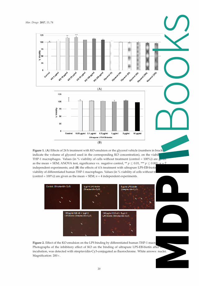
As shown in Figure 1A, we found that, after 24 h, treatment with 5–250 μg/mL KO did notdisplay any cytotoxicity. Glycerol used as the vehicle was not cytotoxic (Figure 1A). Incubation ofdifferentiated human THP-1 macrophages for 4 h with LPS did not show cytotoxicity (Figure 1B).
2.2. Effect of KO Emulsion on the LPS Binding
We used two binding assays to evaluate the interaction of LPS with macrophage-TLR4 and theinhibitory effect of KO. As shown in Figure 2, macrophages incubated 24 h with 1 μg or 5 μg/mLLPS-EB-biotin displays positive binding, detected by fluorescence (Figure 2), compared with controlswithout LPS.
19
Books
MDPI
Mar. Drugs 2017, 15, 74
(A)
(B)
Figure 1. (A) Effects of 24 h treatment with KO emulsion or the glycerol vehicle (numbers in bracketsindicate the volume of glycerol used in the corresponding KO concentration), on the viability ofTHP-1 macrophages. Values (in % viability of cells without treatment (control = 100%)) are givenas the mean + SEM; ANOVA test, significance vs. negative control, ** p ≤ 0.01, *** p ≤ 0.001; n = 7independent experiments; and (B) the effects of 4 h treatment with ultrapure LPS-EB-biotin on theviability of differentiated human THP-1 macrophages. Values (in % viability of cells without treatment(control = 100%)) are given as the mean + SEM; n = 4 independent experiments.
Figure 2. Effect of the KO emulsion on the LPS binding by differentiated human THP-1 macrophages.Photographs of the inhibitory effect of KO on the binding of ultrapure LPS-EB-biotin after 24 hincubation, was detected with streptavidin-Cy3-conjugated as fluorochrome. White arrows: nuclei.Magnification: 200×.
20
Books
MDPI

Mar. Drugs 2017, 15, 74
LPS (1 μg/mL) pre-incubated with KO (100 μg/mL) inhibited the binding, but not when usingLPS at a concentration of 5 μg/mL (Figure 2). The LPS binding was increased at 0.1 μg/mL (6.5%),1 μg/mL (20.3%, p ≤ 0.05), and 5 μg/mL (100%, p ≤ 0.05) when compared with the negative control(Figure 3).
Figure 3. Effect of the KO emulsion on the LPS binding on macrophage-TLR4. Ultrapure LPS-EB-biotinbinding assay was performed by using PMA-differentiated THP-1 macrophages after 3 h incubation,including streptavidin-HRP-OPD system for detection and colorimetric quantification. Values(binding relative to negative control (value = 1) without LPS-EB-biotin) are given as the mean + SEM;ANOVA test, significance vs. negative control without LPS, * p ≤ 0.05; n = 4 independent experiments.
After co-incubation of 0.1 μg/mL LPS-EB with KO, the macrophages were treated for 3 h withdifferent concentrations of LPS + KO: at 12.5 μg/mL LPS + KO, LPS-binding decreased by 50%(not significant); at 25 μg/mL LPS + KO LPS binding significantly decreased by 75% (p ≤ 0.05) and,at 50 μg/mL LPS + KO, it was abolished (p ≤ 0.01), as shown in Figure 4.
Figure 4. Ultrapure LPS-EB-biotin binding assay on macrophage-TLR4. LPS-EB-biotin (0.1 μg/mL) wasco-incubated overnight with different concentrations of KO emulsion or LPS antagonist (LPS-RS AN),added to differentiated THP-1 macrophages (3 h). LPS binding was spectrophotometrically quantifiedusing streptavidin-HRP-OPD system. Values (in % binding relative to positive control (100% binding)with LPS-EB-biotin) are given as the mean + SEM; ANOVA test, significance vs. positive control(=LPS-EB-Biotin), * p ≤ 0.05, ** p ≤ 0.01; n = 6 independent experiments.
21
Books
MDPI

Mar. Drugs 2017, 15, 74
As the LPS-binding control, 25 μg/mL nLDL showed a similar effect as KO at 12.5 μg/mL,however, this was not significant. LPS-RS-AN antagonist significantly (p ≤ 0.01) prevented the bindingof LPS-biotin and, thus, mimicked the effect of KO (Figure 4). A total and significant inhibition ofthe binding was obtained when 1 μg/mL LPS-EB was co-incubated with 6.25, 12.5 μg/mL (p ≤ 0.01),25 μg/mL (p ≤ 0.01), or 50 μg/mL (p ≤ 0.001) KO when compared with the control. Furthermore, LDLshowed a similar inhibition pattern as KO. Additionally, the LPS-RS antagonist completely inhibitedthe LPS binding on the TLR4 (Figure 5). Moreover, we have performed this experiment using 5 μg/mLLPS-EB-biotin, however, without a statistically significant difference between LPS/KO co-incubationand LPS alone (data not shown).
Figure 5. Ultrapure LPS-EB-biotin binding assay on macrophage-TLR4. LPS-EB-biotin (1 μg/mL)was co-incubated overnight together with different concentrations of KO emulsion or 100 μg/mLLPS antagonist (LPS-RS AN) added to differentiated human THP-1 macrophages (3 h). LPS bindingwas spectrophotometrically quantified using streptavidin-HRP-OPD system. Values (in % bindingrelative to positive control (100% binding) with LPS-EB-biotin) are given as the mean + SEM; ANOVAtest, significance vs. positive control (=LPS-EB-Biotin), * p ≤ 0.05, ** p ≤ 0.01, *** p ≤ 0.001; n = 6independent experiments using four wells per treatment and experiment.
2.3. Effect of KO Emulsion on TNF-α Release of Differentiated Human THP-1 Macrophages Stimulatedwith LPS-EB
The TNF-α release in cell supernatants of differentiated human THP-1 macrophages treated withLPS-EB (pre-incubated with KO) was markedly reduced when 0.01 μg/mL or 0.1 μg/mL LPS wasused. A significant inhibition of the TNF-α release was obtained after incubation of 0.01 μg/mL LPSwith 12.5 μg/mL (−30%, p ≤ 0.01), 25 μg/mL (−40%, p ≤ 0.001) or 50 μg/mL (−75%, p ≤ 0.001) KOin comparison with the control LPS (Figure 6A).
Treatment with 0.1 μg/mL LPS pre-incubated with 25 μg/mL or 50 μg/mL KO showed a 50%(p ≤ 0.05) or 60% (p ≤ 0.01) inhibition in comparison with control LPS (Figure 6B). Treatment with KOalone had no effect on the TNF-α production (Figure 6B). Incubation with antagonist LPS-RS abolishedthe TNF-α release (Figure 6B).
22
Books
MDPI

Mar. Drugs 2017, 15, 74
(A)
(B)
Figure 6. Analyses (by ELISA) of the inhibitory effect of KO emulsion on the TNF-α release by LPS-EBdifferentiated human THP-1 macrophages. (A) LPS-EB 0.01 μg/mL, or (B) 0.1 μg/mL was co-incubatedovernight together with different concentrations of KO or 10 μg/mL LPS antagonist (LPS-RS-AN) andafterwards for 3 h with the differentiated THP-1 macrophages. Values (in %TNF-α release relative topositive (LPS-EB) control (=100% release)) are given as the mean + SEM; ANOVA test, significance vs.positive control (=LPS-EB), * p ≤ 0.05, ** p ≤ 0.01, *** p ≤ 0.001; n = 3–5 independent experiments.
3. Discussion
Sepsis has been defined as a systemic immune activation in patients with infection, characterizedby low rates of survival [23]. Bacterial endotoxemia is considered one of the major causes of sepsis,resulting from the release of LPS by microorganisms, e.g., within the colon, that translocate acrossa compromised intestinal wall [17,24]. In this context, we performed in vitro experiments to test theefficacy of a KO emulsion against endotoxin-triggered pro-inflammatory effects.
Death rates in patients caused by sepsis reach about 30%–80% [25], especially in oncologicpatients [26]. Sepsis-associated organ failure and death result from an overwhelming inflammatoryimmune response that culminates in a generalized autodestructive process [27] and development ofmulti-organ dysfunction syndrome, or MODS [28]. Under normal physiological conditions, in whichROS levels are controlled by endogenous anti-oxidant systems, sepsis induces an imbalance betweenpro- and antioxidant systems, which leads to oxidative stress [29]. Moreover, long-chain n-3 PUFAsdecrease the production of inflammatory mediators (eicosanoids, cytokines, and ROS) [22,30]. Onceincorporated into cell membranes, a key step to exert cytoprotection, n-3-FAs dramatically modulatethe body’s response to inflammation, oxidative stress, ischemia, and immune function throughseveral downstream bioactive mediators, (e.g., cytokines, prostaglandins, thromboxanes, leukotrienes,resolvins, protectins, etc.) [22]. The combination of “cytoprotective excipients” [31], together withnutritional, anti-oxidant, and anti-inflammatory properties, make the omega-3 therapy a successfulcandidate for the management of sepsis in patients under intensive care. By using EPA or fish oil,
23
Books
MDPI

Mar. Drugs 2017, 15, 74
inhibition of endotoxin-induced TNF-α production by monocytes [32], may exert effects on both,the generation of inflammatory mediators, and on the resolution of inflammatory processes [15].These observations suggest direct effects of long-chain n-3 PUFA on inflammatory gene expressionvia inhibition of activation of the transcription factor NF-κB [33]. Several studies in healthy humanvolunteers involving supplementation of the diet with fish oil have demonstrated decreased productionof TNF-α, IL-1β, and IL-6 after endotoxin-stimulation of monocytes or mononuclear cells [34,35].The benefits of fish oil in animal models of experimental endotoxemia have been clearly demonstratedwhen dietary fish oil or fish oil infused intravenously enhances the survival of guinea pigs afterintraperitoneal endotoxin injection [36]. In this context, strategies to attenuate the immune responseand prevent organ failure could help patients with sepsis or septic shock. Given that ultrapure LPS-EBspecifically binds to TLR4 and the LPS antagonist LPS-RS, leading to an almost complete suppressionof the TNF-α release, confirms the specific binding of LPS-EB to TLR4. The same effect observed withKO, can be explained by an inactivation of LPS during the co-incubation with KO or an inhibitionof the binding to TLR4. Nevertheless, the present study clearly demonstrates that KO inhibited theproduction of TNF-α after stimulation with LPS. Available data have confirmed an effective reductionin the LPS level in the patients’ blood after this procedure [37], and could effectively eliminate a widerange of the factors as LPS, cytokines, etc., from peripheral blood.
Our findings suggest that KO has properties to inactivate and bind LPS, leading to the inhibitionof activation of macrophages. In this context, we found a reduction of LPS binding capacity ondifferentiated human THP-1 macrophages after 3 h treatment with LPS that has been co-incubated(24 h) with 100 μg/mL KO (Figure 2), by reduction of the fluorescence intensity at a concentration of1 μg/mL LPS (Figure 2). These data support studies showing less LPS internalization into murinemacrophages after 24 h treatment, when LPS was pre-exposed to high-density lipoprotein (HDL) [4].In agreement with these studies, we used human LDL as binding control and we found that LDLreduced the binding of LPS on macrophages (Figures 4 and 5). Moreover, KO significantly reducedthe LPS-binding on macrophages over a range of LPS concentrations when both had been previouslyco-incubated and, afterwards, were applied to the macrophages. Human serum or LDL inactivateendotoxins and inhibit the IL-1β release in LPS-activated monocytes [38]. Consequently, this observedKO characteristic may be a sign of a possible LPS adsorption, inactivation, or hiding of the bindingsites. Using the LPS antagonist Rhodobacter sphaeroides, which binds to the TLR4 but does not induceTLR4 signaling, we can confirm the specificity of the activation (Figures 4 and 5). After co-incubation(24 h) of LPS and KO (12.5, 25 or 50 μg/mL), LPS lost its pro-inflammatory capacity and inhibited theTNF-α release. KO alone or LPS antagonist did not affect the TNF-α production, and confirmed thatKO has beneficial, and not detrimental, effects. Therefore, our results impressively indicate that KOcan efficiently prevent macrophages from being activated by LPS, including suppression of negativeconsequences. like the release of pro-inflammatory cytokines, such as TNF-α.
The effects we have observed with a KO emulsion are likely the result of two main mechanismsof action. First, the phopsholipids present in KO bind and neutralize LPS endotoxin. This effecthas been shown previously in a study of normal human volunteers receiving an intravenousdose of Escherichia coli endotoxin during a 6-h PL infusion, and attenuation of the clinical andlaboratory responses were directly related to PL levels in the bloodstream [39]. However, whenthis formulation was tested in the critical care setting in patients with severe Gram-negative sepsis, thehigh dose arm (1350 mg/kg by continuous infusion) had to be stopped because of the increasedincidence of life-threatening significant adverse events and obvious futilty to show a survivaladvantage [40]. The PL emulsion used in this investigation consisted of 92.5% soy-based, PL, and 7.5%soy triglycerides. Although this formulation contained PLs capable of binding endotoxin, the dosingwas probably excessive. Additionally, the fatty acid profile of soybean oil-derived triacylglycerols andphosphoglycerides mainly contain pro-inflammatory omega-6 fatty acids (i.e., linoleic acid). In contrast,the KO-based PL emulsion in the present study contained both the necessary PL to bind the endotoxin,
24
Books
MDPI

Mar. Drugs 2017, 15, 74
plus the anti-inflammatory omega-3 fatty acids to modulate eicosanoid metabolism, and they clearlyexhibit positive synergistic effects.
Finally, the KO emulsion used in this study was a crude formulation made from an unrefinednatural source that is widely found in oral supplements and, thus, would be unsuitable forintravenous administration. Therefore, our study provided an indication of a proof-of-concept withthis phospholipid-omega-3 combination in a cell culture model. We clearly recognize that for such aproduct to be a potentially viable injectable emulsion in the clinical setting, the crude KO would needto undergo refinement steps (similar to that applied to fish oil) [41] to concentrate the PL in amountsand levels of purity similar to currently available and widely used egg PL which, for example, containat least 80% phosphatides (versus ~40%, and about equal amounts of triglycerides). In addition,since the triglycerides present in crude KO are essentially devoid of omega-3 fatty acids, the idealinjectable product would also include fish oil triglcyerides enriched with omega-3 fatty acids formaximal therapeutic efficacy.
We conclude that KO emulsion inhibits the LPS-binding on macrophage-TLR4 and, thus, theTNF-α release induced by LPS in vitro. The addition of omega-3 fatty acids potentiates the therapeuticactions by reducing the intensity of the systemic inflammatory response. These properties may bebeneficial for patients under intensive care with septicemia.
4. Materials and Methods
4.1. Krill Oil-In-Water Emulsion
Three separate batches of a 5% KO-in-water emulsion were aseptically prepared in the laboratoryand sterilized prior to use. This was done to be sure the emulsions could be successfully made usinga crude source of KO, since a pharmaceutical-quality grade, suitable for parenteral administration,does not exist. The mean droplet size of all the emulsions was approximately 190 nm and, thus,they were considered pharmaceutically equivalent. The composition of the final emulsions are shownin Table 1.
Table 1. Krill oil-in-water emulsion.
Ingredient Amount/1000 mL
Glycerol 25.0 gKrill Oil * 50.0 g
Sterile Water for Injection 1000 mLpH 6.89 ± 0.06
* Virgin Krill Oil, LLC, Braintree, MA, USA, Lot #1107091, Key Ingredient Totals (w/w): n-3-FAs = 26%; PLs = 41.2%.
4.2. Cells and Culture Conditions
The in vitro experiments were performed using the THP-1 (human acute monocytic leukemia)cell line (DSMZ GmbH, Braunschweig, Germany), cultured in 90% RPMI-1640 (PAA GmbH,Cölbe, Germany), 10% FBS (PAA GmbH); 100 U/mL penicillin; and 0.1 mg/mL streptomycin (PAAGmbH). All experiments were carried out in medium with 10% FBS.
4.3. Determination of LPS and KO Emulsion Cytotoxicity
THP-1 (5 × 10⁴) cells were seeded in 96-well plates (BD Falcon™, Becton Dickinson GmbH,Heidelberg, Germany). After differentiation with 0.1 μg/mL phorbol-12-myristate-13-acetate (PMA)Sigma-Aldrich, St. Louis, MO, USA), the medium was changed and the macrophages treated with0.05 μg/mL-10 μg/mL ultrapure biotinylated lipopolysaccharide (LPS-EB) from E. coli O111:B4(Cayla-InvivoGen Europe, Toulouse, France), which is recognized only by toll-like receptor-4 (TLR4);KO 5 μg/mL to 250 μg/mL, or glycerol. After 24 h KO or 4 h LPS treatment, viability was assessedusing PrestoBlue™ reagent (Invitrogen-Life Technologies GmbH, Darmstadt, Germany). After 1 h the
25
Books
MDPI

Mar. Drugs 2017, 15, 74
optical density (OD) was measured at 570 nm/600 nm with a SUNRISE ELISA-reader (Tecan Salzburg,Austria). Results are expressed as the % of viability/survival (OD570 nm/600 nm of samples ×100/OD570 nm/600 nm of control without substances).
4.4. Determination of the LPS Binding on Differentiated THP-1 Macrophages, Effect of the Treatment with KOEmulsion, Detected by Fluorescence Microscopy
THP-1 (5 × 10⁴) cells were seeded in 100 μL medium/well in 96-well plates (BD Falcon™).After eight days of differentiation into macrophages using 0.1 μg/mL PMA, the medium was changedand the macrophages were treated with 1 μg/mL or 5 μg/mL ultrapure LPS-EB-biotin alone orwith KO. After 3 h the cells were fixed with 1% paraformaldehyde (PFA/PBS) for 20 min andincubated afterwards with streptavidin-Cy3 (Dianova, Hamburg, Germany). Digitalized imageswere obtained using an inverted microscope Eclipse-TS100 (Nikon GmbH, Düsseldorf, Germany) andan AxioCamMRc/AxioVision digital imaging system (Carl Zeiss GmbH, Jena, Germany).
4.5. Determination of the LPS Binding on Differentiated THP-1 Macrophages Detected by Spectrophotometry
THP-1 (5 × 10⁴) cells were seeded as described above and treated with 0.1 μg/mL,1 μg/mL, or 5 μg/mL ultrapure LPS-EB-biotin. After 3 h the macrophages were fixed with1% PFA/PBS for 20 min, then incubated with streptavidin-biotinylated horseradish peroxidase(HRP-streptavidin-biotin) complex Amersham (GE Healthcare Europe GmbH, Freiburg, Germany).Subsequently, the macrophages were incubated with 50 μL peroxidase substrate Sigma Fast™ (OPD)(Sigma-Aldrich, St. Louis, MO, USA) for 30 min at RT. The reaction was stopped with 25 μL 3NHCl, and the absorbance was measured at 490 nm/655 nm. Afterwards, the cells were stained withcrystal violet solution (0.04% paraformaldehyde crystal violet in 4% (v/v)). The OPD absorbance wasnormalized against the crystal violet absorbance measured at 595 nm/660 nm.
4.6. Inhibitory Effects of the KO Emulsion on the LPS Binding Capacity
PMA-differentiated THP-1 macrophages were treated for 3 h with the LPS-EB-biotin and KOmixture (both previously incubated together at 4 ◦C overnight), afterwards the cells were fixedwith 1% PFA-PBS for 20 min, washed with PBS, incubated with streptavidin-biotin-complex-HRP(Amersham, GE Healthcare), afterwards with OPD and measured as described above. The positivecontrol was performed by incubation of macrophages with ultrapure LPS-EB-biotin alone and thenegative controls incubated only with medium or with KO. In humans, low-density lipoproteins (LDLs)may bind LPS and inactivate it; we used native LDL (nLDL, HoelzelDiagnostika GmbH, Cologne,Germany) as a control to compare the adsorption properties of KO [38]. The antagonist LPS fromRhodobactersphaeroides (LPS-RS) was used as control of the TLR4 specific binding by competitiveinhibition at 100-fold excess of the agonist LPS-EB.
4.7. Effects of the KO Emulsion on TNF-α Release
The release of TNF-α was determined using ELISA. Therefore, 5 × 10⁵ THP-1 cells wereseeded in 24-well plates (BD Falcon™). After five days of PMA-induced differentiation (0.1 μg/mL),the macrophages were treated 3 h with LPS-EB and KO (previously incubated as described). After thetreatment, the culture medium was harvested and centrifuged at 500× g (5 min). The cells werehomogenized in RIPA buffer (Cell Signaling Technology, Inc., Danvers, MA, USA) for proteinquantification using the bicinchoninic acid assay (Thermo Fisher Scientific, Bonn, Germany). HumanTNF-α was determined in the supernatant using the DuoSet-ELISA kit (R&D Systems Europe,Ltd., Abingdon, UK) according to the manufacturer’s instructions; 96-well NUNC MaxiSorp™(Thermo Fisher Scientific) were used. The amount of TNF-α was normalized with the protein content.
26
Books
MDPI

Mar. Drugs 2017, 15, 74
4.8. Statistical Analyses
The SigmaPlot®-12 software (Systat Software GmbH, Erkrath, Germany) was used to carry outstatistical analyses by one-way analysis of variance test (ANOVA) using Dunnett’s method appropriatefor multiple comparisons versus the control group. Data are shown as mean + SEM.
Acknowledgments: The authors thank Andrea Cordes for the excellent technical assistance, as well as Ellen Essenand Gabriella Stauch for preparation of the manuscript.
Author Contributions: G.A. Bonaterra, D. Driscoll and R. Kinscherf contributed to the conception and designof the research. G.A. Bonaterra contributed to the acquisition, analysis, interpretation of the data, and draftedthe manuscript. H. Schwarzbach analyzed the data and image processing. D. Driscoll and R. Kinscherf criticallyrevised the manuscript, agreed to be fully accountable for ensuring the integrity and accuracy of the work,and read and approved the final manuscript.
Conflicts of Interest: D. Driscoll has been awarded a patent, assigned to Stable Solutions LLC, entitled:Therapeutic application of parenteral krill oil. US 8,895,074 B2. Financial disclosure: This work was supported byB. Braun Melsungen AG, Germany. The founding sponsors had no role in the design of the study; in the collection,analyses, or interpretation of data; in the writing of the manuscript, and in the decision to publish the results
References
1. Bochsler, P.N.; Maddux, J.M.; Neilsen, N.R.; Slauson, D.O. Differential binding of bacterial lipopolysaccharideto bovine peripheral-blood leukocytes. Inflammation 1993, 17, 47–56. [CrossRef] [PubMed]
2. Adams, J.L.; Czuprynski, C.J. Bacterial lipopolysaccharide induces release of tumor necrosis factor-alphafrom bovine peripheral blood monocytes and alveolar macrophages in vitro. J. Leukoc. Biol. 1990, 48, 549–556.[PubMed]
3. Sprague, A.H.; Khalil, R.A. Inflammatory cytokines in vascular dysfunction and vasculardisease.Biochem. Pharmacol. 2009, 78, 539–552. [CrossRef] [PubMed]
4. Cavaillon, J.M.; Fitting, C.; Haeffner-Cavaillon, N.; Kirsch, S.J.; Warren, H.S. Cytokine response by monocytesand macrophages to free and lipoprotein-bound lipopolysaccharide. Infect. Immun. 1990, 58, 2375–2382.[PubMed]
5. Emancipator, K.; Csako, G.; Elin, R.J. In vitro inactivation of bacterial endotoxin by human lipoproteins andapolipoproteins. Infect. Immunol. 1992, 60, 596–601.
6. Eggesbo, J.B.; Hjermann, I.; Hostmark, A.T.; Kierulf, P. LPS induced release of IL-1 beta, IL-6, IL-8 andTNF-alpha in EDTA or heparin anticoagulated whole blood from persons with high or low levels of serumHDL. Cytokine 1996, 8, 152–160. [CrossRef] [PubMed]
7. Baumberger, C.; Ulevitch, R.J.; Dayer, J.M. Modulation of endotoxic activity oflipopolysaccharide byhigh-density lipoprotein. Pathobiology 1991, 59, 378–383. [CrossRef] [PubMed]
8. Wu, A.; Hinds, C.J.; Thiemermann, C. High-density lipoproteins in sepsis and septic shock: Metabolism,actions, and therapeutic applications. Shock 2004, 21, 210–221. [CrossRef] [PubMed]
9. Shor, R.; Wainstein, J.; Oz, D.; Boaz, M.; Matas, Z.; Fux, A.; Halabe, A. Low serum LDL cholesterol levels andthe risk of fever, sepsis, and malignancy. Ann. Clin. Lab. Sci. 2007, 37, 343–348. [PubMed]
10. Calder, P.C. n-3 polyunsaturated fatty acids, inflammation, and inflammatory diseases. Am. J. Clin. Nutr.2006, 83 (Suppl. 6), 1505S–1519S. [PubMed]
11. Arterburn, L.M.; Hall, E.B.; Ojen, H. Distribution, interconversion and dose response of n-3 fatty acids inhumans. Am. J. Clin. Nutr. 2006, 83 (Suppl. 6), 1467S–1476S. [PubMed]
12. Nash, S.M.B.; Schlabach, M.; Nichols, P.D. A nutritional-toxicological assessment of antarctic krill oil versusfish oil dietary supplements. Nutrients 2014, 6, 3382–3402. [CrossRef] [PubMed]
13. Wichmann, M.W.; Thul, P.; Czarnetski, H.D.; Morlion, B.J.; Kemen, M.; Jauch, K.W. Evaluation of clinicalsafety and beneficial effects of a fish oil containing lipid emulsion (Lipoplus, MLF541): Data from aprospective, randomized, multicenter trial. Crit. Care Med. 2007, 35, 700–706. [CrossRef] [PubMed]
14. Heller, A.R.; Rössler, S.; Litz, R.J.; Stehr, S.N.; Heller, S.C.; Koch, R.; Koch, T. Omega-3 fatty acids improvediagnosis-related clinical outcome. Crit. Care Med. 2006, 34, 972–979. [CrossRef] [PubMed]
15. Barbosa, V.M.; Miles, E.A.; Calhau, C.; Lafuente, E.; Calder, P.C. Effects of a fish oil containing lipidemulsion on plasma phospholipid fatty acids, inflammatory markers, and clinical outcomes in septicpatients: A randomized, controlled clinical trial. Crit. Care 2010, 14, R5. [CrossRef] [PubMed]
27
Books
MDPI

Mar. Drugs 2017, 15, 74
16. Mayer, K.; Fegbeutel, C.; Hattar, K.; Sibelius, U.; Krämer, H.J.; Heuer, K.U.; Temmesfeld-Wollbrück, B.;Gokorsch, S.; Grimminger, F.; Seeger, W. Omega-3 vs. omega-6 lipid emulsions exert differential influenceon neutrophils in septic shock patients: Impact on plasma fatty acids and lipid mediator generation.Intensive Care Med. 2003, 29, 1472–1481. [CrossRef] [PubMed]
17. Tsutsumi, R.; Horikawa, Y.T.; Kume, K.; Tanaka, K.; Kasai, A.; Kadota, T.; Tsutsumi, Y.M. Peptide-BasedFormulas With ω-3 Fatty Acids Are Protective in LPS-Mediated Sepsis. JPEN J. Parenter. Enter. Nutr. 2015,39, 552–561. [CrossRef] [PubMed]
18. Calder, P.C. Marine omega-3 fatty acids and inflammatory processes: Effects, mechanisms and clinicalrelevance. Biochim. Biophys. Acta 2015, 1851, 469–484. [CrossRef] [PubMed]
19. Ramsvik, M.S.; Bjørndal, B.; Bruheim, I.; Bohov, P.; Berge, R.K. A Phospholipid-Protein Complex fromKrill with Antioxidative and Immunomodulating Properties Reduced Plasma Triacylglycerol and HepaticLipogenesis in Rats. Mar. Drugs 2015, 13, 4375–4397. [CrossRef] [PubMed]
20. Ghasemifard, S.; Turchini, G.M.; Sinclair, A.J. Omega-3 long chain fatty acid “bioavailability”: A review ofevidence and methodological considerations. Prog. Lipid Res. 2014, 56C, 92–108. [CrossRef] [PubMed]
21. Cansell, M.; Nacka, F.; Combe, N. Marine lipid-based liposomes increase in vivo FA bioavailability. Lipids2003, 38, 551–559. [CrossRef] [PubMed]
22. Bistrian, B.R. Clinical aspects of essential fatty acid metabolism: Jonathan Rhoads Lecture. JPEN J. Parenter.Enter. Nutr. 2003, 27, 168–175. [CrossRef] [PubMed]
23. Wohlmuth, C.; Dünser, M.W.; Wurzinger, B.; Deutinger, M.; Ulmer, H.; Torgersen, C.; Schmittinger, C.A.;Grander, W.; Hasibeder, W.R. Early fish oil supplementation and organ failure in patients with septic shockfrom abdominal infections: A propensitymatchedcohort study. JPEN J. Parenter. Enter. Nutr. 2010, 34,431–437. [CrossRef] [PubMed]
24. Su, G.L. Lipopolysaccharides in liver injury: Molecular mechanisms of Kupffer cell activation. Am. J. Physiol.Gastrointest. Liver Physiol. 2002, 283, G256–G265. [CrossRef] [PubMed]
25. Yegenaga, I.; Hoste, E.; Van Biesen, W.; Vanholder, R.; Benoit, D.; Kantarci, G.; Dhondt, A.; Colardyn, F.;Lameire, N. Clinical characteristics of patients developing ARF due to sepsis/systemic inflammatoryresponse syndrome: Results of a prospective study. Am. J. Kidney Dis. 2004, 43, 817–824. [CrossRef][PubMed]
26. Angus, D.C.; Linde-Zwirble, W.T.; Lidicker, J.; Clermont, G.; Carcillo, J.; Pinsky, M.R. Epidemiology of severesepsis in the United States: Analysis of incidence, outcome, and associated costs of care. Crit. Care Med. 2001,29, 1303–1310. [CrossRef] [PubMed]
27. Annane, D.; Belissant, E.; Cavaillon, J.M. Septic shock. Lancet 2005, 365, 63–78. [CrossRef]28. Thomas, S.; Balasubramanian, K.A. Role of intestine in postsurgical complications: Involvement of free
radicals. Free Radic. Biol. Med. 2004, 36, 745–756. [CrossRef] [PubMed]29. Coquerel, D.; Kušíková, E.; Mulder, P.; Coëffier, M.; Renet, S.; Dechelotte, P.; Richard, V.; Thuillez, C.;
Tamion, F. Omega-3 polyunsaturated fatty acids delay the progression of endotoxic shock-inducedmyocardial dysfunction. Inflammation 2013, 36, 932–940. [CrossRef] [PubMed]
30. Yates, C.M.; Calder, P.C.; Ed Rainger, G. Pharmacology and therapeutics of omega-3 polyunsaturated fattyacids in chronic inflammatory disease. Pharmacol. Ther. 2014, 14, 272–782. [CrossRef] [PubMed]
31. Bonaterra, G.A.; Wakenhut, F.; Röthlein, D.; Wolf, M.; Bistrian, B.R.; Driscoll, D.; Kinscherf, R. Cytoprotectionby omega-3 fatty acids as a therapeutic drug vehicle when combined with nephrotoxic drugs in anintravenous emulsion: Effects on intraglomerular mesangial cells. Toxicol. Rep. 2014, 1, 843–857. [CrossRef]
32. Zhao, Y.; Joshi-Barve, S.; Barve, S.; Chen, L.H. Eicosapentaenoic acid prevents LPS induced TNF-α expressionby preventing NF-κB activation. J. Am. Coll. Nutr. 2004, 23, 71–78. [CrossRef] [PubMed]
33. Calder, P.C. Use of fish oil in parenteral nutrition: Rationale and reality. Proc. Nutr. Soc. 2006, 65, 264–277.[CrossRef] [PubMed]
34. Trebble, T.; Arden, N.K.; Stroud, M.A.; Wootton, S.A.; Burdge, G.C.; Miles, E.A.; Ballinger, A.B.;Thompson, R.L.; Calder, P.C. Inhibition of tumour necrosis factor-α and interleukin-6 production bymononuclear cells following dietary fish-oil supplementation in healthy men and response to antioxidantco-supplementation. Br. J. Nutr. 2003, 90, 405–412. [CrossRef] [PubMed]
35. Wallace, F.A.; Miles, E.A.; Calder, P.C. Comparison of the effects of linseed oil and different doses of fish oilon mononuclear cell function in healthy human subjects. Br. J. Nutr. 2003, 89, 679–689. [CrossRef] [PubMed]
28
Books
MDPI
Mar. Drugs 2017, 15, 74
36. Mascioli, E.A.; Leader, L.; Flores, E.; Trimbo, S.; Bistrian, B.; Blackburn, G. Enhanced survival to endotoxin inguinea pigs fed iv fish oil emulsion. Lipids 1988, 23, 623–625. [CrossRef] [PubMed]
37. Kulabukhov, V.V. Use of an endotoxin adsorber in the treatment of severe abdominal sepsis.Acta Anaesthesiol. Scand. 2008, 52, 1024–1025. [CrossRef] [PubMed]
38. Weinstock, C.; Ullrich, H.; Hohe, R.; Berg, A.; Baumstark, M.W.; Frey, I.; Northoff, H.; Flegel, W.A. Lowdensity lipoproteins inhibit endotoxin activation of monocytes. Arterioscler. Thromb. 1992, 12, 341–347.[CrossRef] [PubMed]
39. Gordon, B.R.; Parker, T.S.; Levine, D.M.; Feuerbach, F.; Saal, S.D.; Sloan, B.J.; Chu, C.; Stenzel, K.H.;Parrillo, J.E.; Rubin, A.L. Neutralization of endotoxin by a phospholipid emulsion in healthy volunteers.J. Infect. Dis. 2006, 191, 1515–1522. [CrossRef] [PubMed]
40. Dellenger, R.P.; Tomayko, J.F.; Angus, D.C.; Opal, S.; Cupo, M.A.; McDermott, S.; Ducher, A.; Calandra, T.;Cohen, J. Lipid Infusion and Patient Outcomes in Sepsis (LIPOS) Investigators. Efficacy and safety ofa phospholipid emulsion (GR270773) in gram-negative severe sepsis: Results of a phase II multicenter,randomized, placebo-controlled, dose finding trial. Crit. Care 2009, 37, 2929–2938. [CrossRef] [PubMed]
41. European Food Safety Authority (EFSA). Panel on Biological Hazards. Scientific opinión on fish oil forhuman consumption. EFSA J. 2010, 8, 1874.
© 2017 by the authors. Licensee MDPI, Basel, Switzerland. This article is an open accessarticle distributed under the terms and conditions of the Creative Commons Attribution(CC BY) license (http://creativecommons.org/licenses/by/4.0/).
29
Books
MDPI

marine drugs
Article
Biosynthesis of Polyunsaturated Fatty Acids inOctopus vulgaris: Molecular Cloning and FunctionalCharacterisation of a Stearoyl-CoA Desaturase andan Elongation of Very Long-Chain Fatty Acid4 Protein
Óscar Monroig 1,*, Rosa de Llanos 2,3, Inmaculada Varó 4, Francisco Hontoria 4,
Douglas R. Tocher 1, Sergi Puig 3 and Juan C. Navarro 4
1 Institute of Aquaculture, Faculty of Natural Sciences, University of Stirling, Stirling FK9 4LA, Scotland, UK;[email protected]
2 School of Applied Sciences, Edinburgh Napier University, Edinburgh EH11 4BN, Scotland, UK;[email protected]
3 Instituto de Agroquímica y Tecnología de Alimentos (IATA-CSIC), Paterna, Valencia 46980, Spain;[email protected]
4 Instituto de Acuicultura Torre de la Sal (IATS-CSIC), Ribera de Cabanes, Castellón 12595, Spain;[email protected] (I.V.); [email protected] (F.H.); [email protected] (J.C.N.)
* Correspondence: [email protected]; Tel.: +44-1786-467892
Academic Editors: Rosário Domingues, Ricardo Calado and Pedro DominguesReceived: 14 February 2017; Accepted: 16 March 2017; Published: 21 March 2017
Abstract: Polyunsaturated fatty acids (PUFAs) have been acknowledged as essential nutrientsfor cephalopods but the specific PUFAs that satisfy the physiological requirements are unknown.To expand our previous investigations on characterisation of desaturases and elongases involvedin the biosynthesis of PUFAs and hence determine the dietary PUFA requirements in cephalopods,this study aimed to investigate the roles that a stearoyl-CoA desaturase (Scd) and an elongationof very long-chain fatty acid 4 (Elovl4) protein play in the biosynthesis of essential fatty acids(FAs). Our results confirmed the Octopus vulgaris Scd is a Δ9 desaturase with relatively high affinitytowards saturated FAs with ≥ C18 chain lengths. Scd was unable to desaturate 20:1n-15 (Δ520:1)suggesting that its role in the biosynthesis of non-methylene interrupted FAs (NMI FAs) is limited tothe introduction of the first unsaturation at Δ9 position. Interestingly, the previously characterised Δ5fatty acyl desaturase was indeed able to convert 20:1n-9 (Δ1120:1) to Δ5,1120:2, an NMI FA previouslydetected in octopus nephridium. Additionally, Elovl4 was able to mediate the production of 24:5n-3and thus can contribute to docosahexaenoic acid (DHA) biosynthesis through the Sprecher pathway.Moreover, the octopus Elovl4 was confirmed to play a key role in the biosynthesis of very long-chain(>C24) PUFAs.
Keywords: biosynthesis; elongation of very long-chain fatty acids 4 protein; non-methylene-interrupted fatty acids; polyunsaturated fatty acids; Octopus vulgaris; stearoyl-CoA desaturase
1. Introduction
Cephalopods have been regarded as promising candidates for the diversification of marineaquaculture due to their great commercial interest [1]. Despite significant progress made over thelast decade, culture of cephalopod species with pelagic paralarval stages like the common octopusOctopus vulgaris is still challenging due to the massive mortalities occurring upon the settlementphase [2]. The specific factors causing such mortalities of paralarvae remain unclear, although it has
Mar. Drugs 2017, 15, 82 30 www.mdpi.com/journal/marinedrugs
Books
MDPI

Mar. Drugs 2017, 15, 82
become increasingly obvious that nutritional issues associated with inadequate supply of essentialnutrients such as lipids are crucial to ensure normal growth and development of O. vulgaris paralarvaeand ultimately improve their viability [3].
Previous investigations postulated that polyunsaturated fatty acids (PUFAs) are essential nutrientsfor the common octopus [4,5]. However, the specific PUFAs that satisfy the physiological requirementswere not determined, partly due to the difficulties in running nutritional trials on octopus paralarvae.In order to provide insights to the endogenous capability for PUFA biosynthesis in O. vulgaris,we have recently conducted a series of studies aiming to identify and characterise the functionof genes encoding enzymes that mediate the conversions in the PUFA biosynthetic pathways.First, we identified a fatty acyl desaturase (Fad) cDNA sequence with homology to the vertebrateFads family [6], enzymes that participate in long-chain (C20–24) PUFA (LC-PUFA) biosyntheticpathways [7–9]. The expression of the common octopus Fad in yeast demonstrated that the enzymewas a Δ5 desaturase (Δ5 Fad) with high efficiency towards both saturated and polyunsaturated fattyacid (FA) substrates [6]. Thus, the O. vulgaris Δ5 Fad was able to desaturate the yeast endogenoussaturated FAs 16:0 and 18:0 to the corresponding monoenes 16:1n-11 (Δ516:1) and 18:1n-13 (Δ518:1),respectively. Furthermore, the O. vulgaris Δ5 Fad efficiently desaturated the PUFA 20:4n-3 and 20:3n-6to the Δ5 desaturation products eicosapentaenoic acid (EPA, 20:5n-3) and arachidonic acid (ARA,20:4n-6), respectively (Figure 1).
A second study provided further evidence of the existence of an active PUFA biosynthetic systemin the common octopus [10]. Thus, a cDNA encoding a protein with high homology to an elongationof very long-chain fatty acids (Elovl) protein was isolated [10]. Phylogenetic analysis comparing theamino acid (aa) sequence of the O. vulgaris Elovl with other elongases from molluscs and vertebratesclearly showed that the common octopus Elovl, as well as other putative elongases from molluscs,was grouped as a basal cluster of the vertebrate Elovl2 and Elovl5 families [10]. Consequently,such an elongase has been termed “Elovl5/2” [11] or “Elovl2/5” [12,13]. Regarding its function,the common octopus Elovl2/5 exhibited substrate specificities resembling those of vertebrate Elovl5but not Elovl2, as it efficiently elongated C18–20 PUFAs [10] but had no activity towards C22 substrates.This was hypothesised as one of the reasons accounting for the inability of cephalopods to biosynthesisedocosahexaenoic acid (DHA; 22:6n-3) (Figure 1) [10,11] through the so-called “Sprecher pathway”requiring the production of 24:5n-3 as an intermediate in the pathway [14]. Among alternativeElovl-like enzymes with a role in the biosynthesis of LC-PUFAs such as DHA in the common octopus,the Elovl4 elongase is an interesting candidate, as studies on teleost orthologues have shown thatElovl4 can efficiently catalyse the elongation of 22:5n-3 to 24:5n-3 [15–19], and recent studies havefurther demonstrated a similar elongation ability in molluscs [12].
The above studies on O. vulgaris [6,10], as well as those on homologous genes from the commoncuttlefish Sepia officinalis [20], have enabled us to predict the biosynthetic pathways of PUFAs incephalopods (Figure 1). Beyond the biosynthesis of standard PUFAs, i.e., FAs whose double bonds arealways separated by a methylene group (-CH2-) [9], one can predict that some pathways involvingthe Δ5 Fad and Elovl2/5 lead to the production of so-called “non-methylene-interrupted FAs”(NMI FAs), a particular type of PUFA that had been previously reported in other molluscan classes(bivalves, gastropods), as well in sponges, echinoderms and other phyla [21–23]. Analyses performedin wild-caught specimens of O. vulgaris confirmed that the polar lipid fractions of nephridium,male gonad, eye and caecum contained NMI FAs identified as Δ5,1120:2, Δ7,1320:2, Δ5,11,1420:3 andΔ7,1322:2 [10]. From the unsaturation pattern of these compounds, it became clear that, in addition toΔ5 Fad, a further desaturase with Δ9 activity was likely involved in the NMI FA biosynthetic pathwaysaccounting for the Δ5,9 unsaturation patterns typically found among these compounds [22,23].The stearoyl-CoA desaturase (Scd), an enzyme that is expressed in virtually all living organisms [24],has Δ9 desaturation capability and thus appears to play a role in NMI FA biosynthesis [7].
Our overall aim is to characterise the biosynthetic pathways of PUFAs including NMI FAs incephalopods. Using the common octopus O. vulgaris as model species, we herein isolated two cDNAs,
31
Books
MDPI

Mar. Drugs 2017, 15, 82
namely Scd and Elovl4 sequences, and characterised their functions by heterologous expression inyeast. In order to establish the mechanisms accounting for biosynthesis of Δ5,9 dienes (NMI FA) wefurther investigated the roles that the herein characterised Scd and the previously reported Δ5 Fad [6]play within these pathways.
Figure 1. Model of biosynthetic pathways of polyunsaturated fatty acids in cephalopods.Enzymatic activities shown in the diagram are predicted from heterologous expression in yeast(Saccharomyces cerevisiae) of fatty acyl desaturases (red arrows) and elongation of very long-chainfatty acid (Elovl) proteins (green arrows) from Octopus vulgaris [6,10] and Sepia officinalis [20].Dotted arrows indicate reactions that have not yet been demonstrated prior to the present study.β-ox, partial β-oxidation.
2. Results
2.1. Octopus vulgaris Scd Sequence
The O. vulgaris Scd-like cDNA consisted of a 981-bp open reading frame (ORF) encoding a putativeprotein of 326 amino acids (aa) with a predicted molecular weight of 37.9 kDa. Its sequence wasdeposited in the GenBank database with the accession number JX310655. In common with other Scdproteins, the O. vulgaris putative Scd possessed three histidine boxes (HXXXH, HXXHH and HXXHH),four membrane-spanning regions rich in hydrophobic aa, and lacked the cytochrome b5 domaincharacteristic of Fads (Figure 2).
The deduced aa sequence from the common octopus Scd was 50.8%–53.4% identical to Scdsequences from vertebrates including Homo sapiens (NP_005054.3), Gallus gallus (NP_990221.1)and Xenopus laevis (NP_001087809.1), and 40.7% and 43.9% identical to Scd from the nematodeCaenorhabditis elegans FAT-5 (NP_507482.1) and FAT-6 (NP_001255595.1), respectively. When comparedto mollusc Scd-like protein sequences, the O. vulgaris Scd showed relatively high identity scoreswith orthologues from Octopus bimaculoides (99.0%) (XP_014788510.1), Crassostrea gigas (63.0%)(XP_011452904.1) and Lottia gigantea (60.7%). Importantly, identities between the newly cloned Scd andseveral Fads desaturases including the Δ5-like desaturase identified in O. vulgaris [6] were below 16.0%.
32
Books
MDPI

Mar. Drugs 2017, 15, 82
Figure 2. ClustalW amino acid alignment comparing the Octopus vulgaris stearoyl-CoAdesaturase (Scd) with homologous sequences from the Octopus bimaculoides (XP_014788514.1),the Caenorabditis elegans FAT-5 (NP_507482.1) and the C. elegans FAT-6 (NP_001255595.1), and a portionof the Saccharomyces cerevisiae Ole1p sequence. Identical residues are shaded black and similar residues(using ClustalW2 default parameters) are shaded grey. The three histidine boxes (HXXXH, HXXHH andQXXHH) are highlighted with grey squares. Four (I–IV) trans-membrane domains predicted by Wattsand Browse [25] are underlined with dashed lines.
2.2. Octopus Elovl4-Like Sequence
The Elovl4-like cDNA consisted of an ORF of 930 bp whose deduced protein had 309 aawith a predicted molecular weight of 37.9 kDa (deposited in GenBank database with accessionnumber KJ590963). From analogy to vertebrate orthologues [15–18], five putative transmembranedomains containing hydrophobic aa stretches can be predicted (Figure 3). The common octopusputative Elovl4 contained the histidine dideoxy-binding motif HXXHH, and the putative endoplasmicreticulum (ER) retrieval signal with a histidine (H) and lysine (K) residues at the carboxyl terminus,HXKXX (Figure 3) [26].
Comparison of the deduced aa sequence of the O. vulgaris Elovl4 with other orthologues revealedhigh identity with the O. bimaculoides Elovl4 (93.5%) (XP_014784234.1), with remarkable lower identityscores obtained when compared with Elovl4 sequences from L. gigantea (57.9%) (XP_009051096.1),C. gigas (41.6%) (XP_011450778.1), the sea squirt Ciona intestinalis (48.9%) (AAV67802.1) [27],H. sapiens (50.8%) (NP_073563.1), G. gallus (49.1%) (NP_001184238.1) and Anolis carolinensis (47.6%)(XP_003215742.1). Identity scores of 36.6% and 36.2% were obtained by comparing the O. vulgarisElovl4 with the previously characterised Elovl2/5 from O. vulgaris (AFM93779.1) [10] and S. officinalis(AKE92956.1) [20], respectively.
33
Books
MDPI

Mar. Drugs 2017, 15, 82
Figure 3. ClustalW amino acid alignment of the Octopus vulgaris Elovl4 with homologous sequencesfrom Octopus bimaculoides (XP_014784234.1), Lottia gigantea (XP_009051096.1), Ciona intestinalis(AAV67802.1) and Homo sapiens (NP_073563.1). Identical residues are shaded black and similar residues(using ClustalW default parameters) are shaded grey. Indicated are the conserved histidine box motifHXXHH, five (I–V) putative membrane-spanning domains, and the putative endoplasmic reticulum(ER) retrieval signal.
2.3. Functional Characterisation of the Octopus Scd
Yeast Saccharomyces cerevisiae cells lacking the OLE1 gene are unable to synthesiseΔ9-monounsaturated FAs including palmitoleic acid (16:1n-7) and oleic acid (18:1n-9), which areessential for growth [28]. To address whether the O. vulgaris Scd was able to complement yeast OLE1function, an S. cerevisiae ole1Δ mutant strain (L8-14C) was transformed with a plasmid expressingthe octopus Scd (p416OLE1-Scd) under the control of yeast OLE1 promoter. The ole1Δ mutant yeastwere also transformed with a plasmid expressing the S. cerevisiae OLE1 (Δ9 desaturase) under thecontrol of its own promoter (p416OLE1-OLE1) as a positive control, and with empty vector (pRS416)as a negative control. All three yeast transformants were able to grow in media supplemented with atleast one supplemented FA (16:1n-7 and/or 18:1n-9) (Figure 4a–c). However, only yeast cells expressingeither the S. cerevisiae OLE1 or the O. vulgaris Scd grew on medium that was not supplemented withmonounsaturated FAs (Figure 4d). These results indicated that the common octopus Scd complementedthe function of the yeast OLE1 desaturase.
Figure 4. Complementation of the Saccharomyces cerevisiae ole1Δ mutant strain L8-14C with theOctopus vulgaris stearoyl-CoA desaturase (Scd) coding region. Yeast were transformed with the pRS416empty vector (negative control), p416OLE1-OLE1 (expressing the S. cerevisiae OLE1 under the controlof its own promoter) and p416OLE1-Scd (expressing the O. vulgaris Scd under the control of S. cerevisiaeOLE1 promoter) and grown in SC medium lacking uracil (SC-ura) supplemented with 16:1n-7 and18:1n-9 (a), 16:1n-7 (b) or 18:1n-9 (c), or in the absence of these fatty acids (d).
34
Books
MDPI

Mar. Drugs 2017, 15, 82
To analyse in more detail the complemention of yeast ole1Δ mutants by the O. vulgaris Scd,we determined the FA composition of ole1Δ yeast cells transformed with either p416OLE1-OLE1 (i.e.,expressing the S. cerevisiae OLE1) or p416OLE1-Scd (i.e., expressing the O. vulgaris Scd), and grown inliquid medium with no exogenously supplemented FAs. Yeast expressing the O. vulgaris Scd grewnotably slower compared to controls and formed clumps as previously described [29]. Moreover,the p416OLE1-Scd yeast contained significantly (p ≤ 0.05) less 16:1n-7 and more 16:0 than control yeastexpressing the S. cerevisiae OLE1, suggesting a low affinity of the O. vulgaris Scd towards 16:0. On thecontrary, contents of 18:1n-9 showed no statistical differences between yeast cells expressing Scd orOLE1, indicative of the octopus Scd participating in the biosynthesis of 18:1n-9 similar to S. cerevisiaeOle1p. The distinctive substrate specificities of the O. vulgaris Scd were further emphasised by thehigher 18:1n-9/16:1n-7 ratio of yeast complemented with the O. vulgaris Scd compared to that of OLE1control yeast (Table 1).
The substrate specificities of the O. vulgaris Scd were further investigated throughan overexpression assay using the S. cerevisiae strain InvSc1 (Invitrogen, Paisley, UK), possessing theendogenous Ole1p. Comparison of the FA profiles between the control yeast (i.e., transformed with theempty pYES2) and yeast expressing the O. vulgaris Scd (i.e., transformed with pYES2-Scd) confirmeda role of the common octopus Scd in the biosynthesis of monounsaturated FAs (Table 2). Thus,significant (p ≤ 0.05) increases in the contents of 18:1n-9 (oleic acid) in pYES2-Scd yeast compared tocontrol yeast were observed. Parallel decreased levels of the saturated FA precursor 18:0 were detectedin pYES2-Scd yeast (p ≤ 0.05). Additionally, other monoenes corresponding to 20:1n-11 and 22:1n-13were detected in total lipids of InvSc1 yeast expressing the O. vulgaris Scd. These results confirmed thatthe O. vulgaris Scd is a Δ9 desaturase with activity towards saturated FAs with chain-lengths ≥ C18.In contrast, shorter FAs (≤C16) did not appear to be adequate substrates for the octopus Scd and,for instance, the content of 16:1n-7 in yeast expressing the common octopus Scd was significantly lowerthan that of control yeast (p ≤ 0.05) (Table 2).
Table 1. Fatty acid (FA) composition of the S. cerevisiae strain L8-14C transformed with theyeast-endogenous OLE1 (SC OLE1) or stearoyl-CoA desaturase from O. vulgaris (OV Scd). Results areexpressed as an area percentage of total fatty acids (FAs) found in transformed yeast. Different lettersfor each FA or ratio indicate significant differences among treatments (t-test, p ≤ 0.05).
Fatty Acid SC OLE1 OV Scd
16:0 23.5 ± 3.6 b 59.3 ± 4.9 a
16:1n-9 0.5 ± 0.4 0.7 ± 0.716:1n-7 35.0 ± 2.2 a 4.5 ± 1.0 b
18:0 8.0 ± 1.7 5.6 ± 1.318:1n-9 31.8 ± 2.5 29.8 ± 6.018:1n-7 1.1 ± 0.2 a 0.0 ± 0.0 b
18:1n-9/16:1n-7 0.9 ± 0.1 b 6.8 ± 2.0 a
Table 2. Fatty acid (FA) composition of the S. cerevisiae InvSc1 transformed with either empty pYES2vector (Control) or the common octopus Scd open reading frame (ORF). Different letters for each FAindicate significant differences among treatments (t-test, p ≤ 0.05).
Fatty Acid Control OV Scd
14:0 1.6 ± 0.2 a 1.0 ± 0.1 b
14:1n-5 0.5 ± 0.1 0.7 ± 0.116:0 25.9 ± 0.3 a 22.2 ± 1.2 b
16:1n-7 37.7 ± 0.8 a 28.6 ± 3.3 b
18:0 8.5 ± 0.3 a 4.4 ± 0.9 b
18:1n-9 24.1 ± 0.7 b 40.9 ± 3.6 a
18:1n-7 1.1 ± 0.1 1.1 ± 0.220:0 0.1 ± 0.0 0.1 ± 0.0
20:1n-11 N.D. b 0.4 ± 0.2 a
22:0 0.1 ± 0.0 0.1 ± 0.022:1n-13 N.D. b 0.1 ± 0.0 a
35
Books
MDPI

Mar. Drugs 2017, 15, 82
In order to establish the biosynthetic pathways of NMI FAs with Δ5,9 unsaturation patterns(or their derivatives such as Δ5,11) found in the common octopus lipids [10], InvSc1 yeast transformedwith pYES2-Scd were grown in the presence of the monoene 20:1n-15 (Δ520:1), while InvSc1 yeastexpressing the Δ5 Fad (i.e., transformed with pYES2-Fad) [6] were grown in the presence of 20:1n-9(Δ1120:1) (Figure 5). Our results showed that the O. vulgaris Scd was unable to desaturate the substrateΔ520:1 (20:1n-15) to Δ5,920:2 (Figure 5a), although yeast transformed with pYES2-Fad were able toproduce Δ5,1120:2 (2.7% ± 0.2% conversion), thus confirming activity as Δ5 desaturase on Δ1120:1)(Figure 5b).
(a) (b)
Figure 5. Biosynthesis of non-methylene interrupted fatty acids (FAs) in cephalopods: (a) Gaschromatography (GC) trace of yeast Saccharomyces cerevisiae expressing the Octopus vulgaris Scd andgrown in the presence of 20:1n-15 (Δ520:1); (b) GC trace of yeast S. cerevisiae expressing the previouslycharacterised O. vulgaris Δ5 Fad [6] and grown in the presence of 20:1n-9 (Δ1120:1). Peaks 1-5 representS. cerevisiae endogenous FAs, namely 16:0 (1), 16:1 isomers (2), 18:0 (3), 18:1n-9 (4) and 18:1n-7 (5).Peaks derived from exogenously added substrates (*) and the desaturation product Δ5,1120:2 (b) areindicated accordingly.
2.4. Functional Characterisation of the Octopus Elovl4
The role of the O. vulgaris Elovl4 in the biosynthesis of very long-chain (>C24) PUFAs (VLC-PUFAs)was investigated in yeast S. cerevisiae (strain InvSc1) expressing the Elovl4 and grown in the presence ofone of either C18 (18:3n-3, 18:2n-6, 18:4n-3 and 18:3n-6), C20 (20:5n-3 and 20:4n-6), C22 (22:5n-3, 22:4n-6,22:6n-3) or C24 (24:5n-3) PUFA substrates (Table 3). Gas Chromatography-Mass Spectrometry (GC–MS)analyses confirmed that the control yeast did not have the ability to elongate PUFAs, consistent with thepreviously reported lack of a PUFA elongase in S. cerevisiae strain InvSc1 [30]. However, the commonoctopus Elovl4 conferred the yeast the ability to elongate PUFAs to the corresponding elongatedpolyenoic products of both n-3 and n-6 series (Table 3). With the exception of DHA (22:6n-3),the addition of C22 and C24 PUFA substrates resulted in the production of polyenes of C32 products oreven C34 when 24:5n-3 was used as substrate (Table 3). Indeed, the endogenous production of PUFAswith chain lengths ≥ C26 in yeast supplemented with exogenously supplemented C22 and C24 PUFAsallowed us to estimate the Elovl4 efficiency (as % conversion) towards potential VLC-PUFA substratesthat are not commercially available. The results showed that the highest % conversions (often over88.0%) were consistently detected on C28, C30 and C32 substrates (Table 3). It is noteworthy that theoctopus Elovl4 was able to convert the exogenously added 20:5n-3 and 22:5n-3 to 24:5n-3, the substratefor DHA biosynthesis via the Sprecher pathway [14], although it showed relative low elongase activitytowards DHA itself, which was marginally elongated (0.7%) to 24:6n-3.
36
Books
MDPI

Mar. Drugs 2017, 15, 82
Table 3. Role of the Octopus vulgaris Elovl4 in the biosynthesis of very long-chain (>C24)polyunsaturated fatty acids (FAs). Conversions were calculated for each stepwise elongationaccording to the formula (areas of first product and longer chain products/(areas of all productswith longer chain than substrate + substrate area)) × 100. The substrate FA varies as indicated in eachstep-wise elongation.
FA Substrate Product % Conversion Elongation
18:3n-320:3n-3 2.8 C18→2222:3n-3 3.0 C20→22
18:2n-620:2n-6 1.1 C18→2222:2n-6 26.2 C20→22
18:4n-3 20:4n-3 1.0 C18→20
18:3n-6 20:3n-6 0.8 C18→20
20:5n-322:5n-3 1.9 C20→2424:5n-3 8.1 C22→24
20:4n-622:4n-6 1.0 C20→2424:4n-6 6.2 C22→24
22:5n-3
24:5n-3 4.8 C22→3226:5n-3 37.9 C24→3228:5n-3 95.0 C26→3230:5n-3 93.5 C28→3232:5n-3 51.9 C30→32
22:4n-6
24:4n-6 3.6 C22→3226:4n-6 44.5 C24→3228:4n-6 94.1 C26→3230:4n-6 91.5 C28→3232:4n-6 46.3 C30→32
22:6n-3 24:6n-3 0.7 C22→24
24:5n-3
26:5n-3 1.4 C24→3428:5n-3 88.8 C26→3430:5n-3 88.6 C28→3432:5n-3 63.2 C30→3434:5n-3 17.3 C32→34
3. Discussion
Fish and seafood are the primary sources of omega-3 long-chain (C20–24) PUFAs for humans [31]and this partly explains the considerable interest in elucidating the PUFA biosynthetic pathways inaquatic and marine species, particularly in farmed fish for which current trends in feed formulationare impacting the nutritional quality for human consumers [32]. Previous investigations onO. vulgaris [6,10,33] and S. officinalis [20] revealed that cephalopods possess active desaturase andelongase enzymes involved in the biosynthesis of PUFAs including NMI FAs. Using the commonoctopus O. vulgaris as model species, the present study aimed to expand our knowledge of roles thatfurther desaturases and elongases could have on PUFA biosynthesis in cephalopods. Consequently,we characterised a stearoyl-CoA desaturase (Scd) with a putative role in the biosynthesis of NMI FAs,and an Elovl4 elongase that, in addition to its participation in the biosynthesis of VLC-PUFAs [15–19,34],could potentially catalyse the elongation of 22:5n-3 to 24:5n-3 required for DHA biosynthesis throughthe Sprecher pathway [14].
The Scd, present in virtually all living organisms [24], is an enzyme with Δ9 desaturase activity.Consistently, the newly-cloned O. vulgaris Scd cDNA was confirmed to encode a Δ9 desaturase that wasable to operate on a range of saturated FA substrates with different chain lengths, particularly ≥ C18.These functions are similar to those described for two homologous sequences found in the nematode
37
Books
MDPI

Mar. Drugs 2017, 15, 82
C. elegans, namely FAT-6 and FAT-7, which can efficiently desaturate 18:0 to 18:1n-9 but have loweractivity towards 16:0 [25]. Interestingly, FAT-5, another Scd-like sequence existing in C. elegans,efficiently desaturated 16:0 to 16:1n-7 but had nearly undetectable activity on 18:0, and it is thusregarded as a palmitoyl-CoA-specific desaturase [25]. The functional characterisation of the O. vulgarisScd and its particularly high substrate affinity towards 18:0 is consistent with 18:1n-9 typicallyappearing several folds above the levels of shorter monoenes such as 16:1n-7 in lipids across theMollusca phylum [21]. Beyond its role in biosynthesis of monounsaturated FA, occurrence of NMI FAswith Δ5,9 unsaturation patterns in lipids from molluscs [22] suggested a role of Scd in the biosynthesisof this particular type of PUFA. Our results revealed that the O. vulgaris Scd was not able to introducea second double bond on a Δ5 monoene such as 20:1n-15 (Δ520:1). While the possibility that cephalopodScd can introduce Δ9 desaturations on other monoenes cannot be ruled out, these results stronglysuggest that the role of Scd in the biosynthesis of NMI FAs with Δ5,9 unsaturation patterns is limitedto the insertion of the first unsaturation at Δ9 position on saturated FAs. Indeed, the O. vulgaris Δ5Fad, previously characterised as Δ5 desaturase [6], was able to introduce a Δ5 unsaturation on 20:1n-9(Δ1120:1) producing the NMI FA Δ5,1120:2. Overall, these results allow us to hypothesise that theinsertion of double bonds required for NMI FA biosynthesis is initiated with a Δ9 desaturation byScd and a subsequent Δ5 desaturation catalysed by Δ5 Fad. Compared to other molluscan classes,the occurrence of NMI FAs in cephalopods appears to be rather limited and hence the contributionof endogenous production of NMI FAs vs. dietary input is difficult to establish. Nevertheless,the mechanism of Δ5,1120:2 biosynthesis postulated herein aligns well with the occurrence of Δ5,1120:2in the polar lipids of nephridia of adult O. vulgaris [10], suggesting that NMI FA biosynthesis is possiblein cephalopods and likely other molluscs.
In addition to desaturases, certain Elovl enzymes play crucial roles in the biosynthesis of essentialPUFAs such as EPA, ARA and DHA [9,11,26]. Whereas the previously characterised cephalopodElovl2/5 demonstrated an ability to efficiently elongate C18 and C20 PUFA substrates [6,20], lack ofelongase capability towards 22:5n-3 was hypothesised as a major limiting factor for DHA biosynthesisthrough the Sprecher pathway [6,20]. Functional characterisation of the O. vulgaris Elovl4 demonstratedthe ability of this enzyme to contribute potentially to DHA biosynthesis by producing 24:5n-3,which is the substrate for Δ6 desaturation and chain-shortening to produce DHA in the Sprecherpathway [14]. Such an ability of Elovl4 has been reported in fish orthologues [15–19]. Despite theelongation capability of O. vulgaris herein revealed, our overall observations on gene repertoire andfunction [6,10,20,33] strongly suggest that DHA is still an essential FA for cephalopods. The limitedability to biosynthesise DHA is related, rather than to an issue with elongase activity, to the apparentabsence of key desaturation abilities (Δ6 or Δ4) required for DHA biosynthesis according to the twoaerobic pathways known in vertebrates (Figure 1). In addition to the abovementioned Δ6 desaturationrequired in the Sprecher pathway [14], an alternative pathway described in some teleost fish involvesa Δ4 desaturation from 22:5n-3 to produce DHA directly (Figure 1) [9]. Molecular evidence providedby the recently published Octopus bimaculoides genome project [35] suggests that cephalopods possessone single Fad-like desaturase in their genome and this is likely to be a Δ5 desaturase since all theFad-like desaturase functionally characterised not only from cephalopods such as O. vulgaris andS. officinalis [6,10] but also from bivalves [36] and gastropods [37], have Δ5 desaturase activities.In non-cephalopod molluscs, further Fad-like desaturases exist [38] but, with the exception of the Δ8desaturase found in the scallop Chlamys nobilis [39], their functions have not been determined andtherefore it is yet not possible to predict whether Δ6 and/or Δ4 desaturase activities exist to enableDHA biosynthesis in those species.
The O. vulgaris Elovl4 is also involved in the biosynthesis of VLC-PUFAs as it was able to elongateexogenously supplemented PUFAs ranging from C18–24 to PUFA products with chain lengths of ≥C26.Such conversions are largely consistent with those previously exhibited by human [34] and fish [15–19]Elovl4 proteins. Surprisingly, no elongation products beyond C24 were reported in the functionalcharacterisation of the bivalve C. nobilis Elovl4 [39]. The elongation activities of the O. vulgaris Elovl4
38
Books
MDPI

Mar. Drugs 2017, 15, 82
estimated in the yeast expression system strongly suggested that this enzyme is particularly efficienttowards C26–30 substrates, for which the highest % conversions were observed. Similarly, the humanELOVL4 had PUFAs of ≥C26 as preferred substrates for elongation, although the human ELOVL4was able to produce up to C38 PUFA elongation products in mammalian cell lines [34]. Due totechnical challenges in their analysis and relatively low abundance, the functions of VLC-PUFAsare not fully understood [40]. Nevertheless, the structural features of VLC-PUFAs combining thosefrom saturated FAs at one end and those from PUFAs at the other allow unique membrane lipidconformations in photoreceptors and spermatozoa, thus suggesting important roles of VLC-PUFAsin vision and reproduction of vertebrates [40–43]. Investigations of VLC-PUFAs in cephalopodsand, therefore, the herein reported activities of the O. vulgaris Elovl4 cannot be correlated with thepresence of VLC-PUFAs in vivo. Interestingly, some very long-chain NMI FAs have been reportedin nudibranchs [44,45] but, unfortunately, the potential role of Elovl4 in the biosynthesis of suchcompounds was not determined. Interestingly, unpublished data on tissue distribution analysis of thecommon octopus Elovl4 mRNA indicated that gonads and, to a lesser extent eye, were also major sitesfor VLC-PUFA biosynthesis in cephalopods.
In conclusion, the present study demonstrated that the common octopus O. vulgaris possessesan Scd with high affinity for saturated FA substrates with chain lengths of 18 carbons or longer.Its inability to introduce a double bond into Δ5 monoene strongly suggested that the role of theScd in NMI FA biosynthesis was restricted to the introduction of the first double bond at the Δ9position, whereas the O. vulgaris Δ5 Fad can introduce the second unsaturation at the Δ5 positionaccording to the herein confirmed ability to convert 20:1n-9 to Δ5,1120:2, an NMI FA previously reportedin nephridia of O. vulgaris adult specimens. Beyond desaturases, we could also demonstrate thatO. vulgaris possesses an Elovl4 responsible for the biosynthesis of VLC-PUFAs.
4. Materials and Methods
4.1. Tissue Samples
Tissue samples including brain, nerve, muscle, heart, hepatopancreas, gill, caecum, eye,nephridium and gonads were obtained from the dissection of two (male and female) common octopusadult specimens as previously described [6,10]. Briefly, two (male and female) wild O. vulgaris adults(~1.5 kg) were maintained in seawater tanks at the facilities of the Instituto de Acuicultura Torrede la Sal; before they were cold, they were anesthetised and sacrificed by direct brain puncture.After collection, samples were immediately frozen at −80 ◦C until further analysis. Total RNA wasextracted from octopus tissues using TriReagent® (Sigma-Aldrich, Alcobendas, Spain) accordingto manufacturer’s instructions. Two μg of total RNA tissue samples were used for synthesis offirst strand cDNA using M-MLV reverse transcriptase (Promega, Southampton, UK) primed withrandom hexamers.
4.2. Molecular Cloning of the Scd and Elovl4 cDNA Sequences
The full-length sequences of the Scd and Elovl4 cDNA were obtained as follows. The deducedaa sequences of Scd proteins from Homo sapiens (NP_005054.3), Gallus gallus (NP_990221.1),Anolis carolinensis (XP_003226591.1), Caenorhabditis elegans (FAT-7) (NP_504814.1), Acheta domesticus(AAK25796.1) and Pediculus humanus (XP_002424386.1) were aligned using BioEdit v5.0.6 (Tom Hall,Department of Microbiology, North Carolina State University, Raleigh, NC, USA). Conserved regionswere used for in silico searches of mollusc expressed sequence tags (EST) using The National Center forBiotechnology Information (NCBI) tblastn tool (http://www.ncbi.nlm.nih.gov/). Processed Scd-likeExpressed Sequence Tags (ESTs) from the molluscs Lottia gigantea (FC767047.1 and FC644199.1),Aplysia californica (FF074235.1, FF076278.1, EB307164.1, EB253812.1 and EB281044.1), Limnaea stagnalis(ES580199.1 and ES579822.1), Crassostrea gigas (CU997931.1 and FP006991.1), Euprymna scolopes(DW280836.1, DW269578.1 and DW273589.1) and Ruditapes decussatus (AM870139.1), were aligned
39
Books
MDPI

Mar. Drugs 2017, 15, 82
(Bioedit) and conserved regions used for the design of the degenerate primers UNID9F(5′-ATCACAGCTGGWGCTCAYCG-3′) and UNID9R (5′-TGGCATTGTGWGGGTCWGCATC-3′).
To clone the first fragment of the octopus Elovl4, blastn searches of mollusc ESTs wereperformed using the so-called “transcript 2” from L. gigantea that was previously identifiedby [6] (gi|Lotgi1|178149|) as query. Thus, additional Elovl4-like consensus sequences derivedfrom ESTs from A. californica (EB285681.1, GD233360.1, EB316848.1, GD212825.1, EB325217.1,EB345626.1, EB345430.1), Saccostrea kagaki (AB375033.1) and C. gigas (HS215834.1, AM866458.1)were obtained and aligned with L. gigantea Elovl4-like to design the degenerate primers UNIE4F(5′-GCCAAGGCATTRTGGTGGTT-3′) and UNIE4R (5′-GTSAGRTATCKYTTCCACCA-3′).
Polymerase chain reactions (PCR) were performed with the GoTaq® Green Master Mix (Promega)and using a mixture of cDNAs from brain, nerve and hepatopancreas as template. The PCR consistedof an initial denaturing step at 95 ◦C for 2 min, followed by 35 cycles of denaturation at 95 ◦C for30 s, annealing at 50 ◦C for 30 s, extension at 72 ◦C for 40 s, followed by a final extension at 72 ◦C for5 min. PCR products of approximately 180 bp (Scd) and 290 bp (Elovl4) were obtained and thereafterconfirmed as positive by sequencing (DNA Sequencing Service, IBMCP-UPV, Valencia, Spain).Full-length cDNA of the octopus Scd and Elovl4 were completed by 5′ and 3′ rapid amplification ofcDNA ends (RACE) PCR (FirstChoice RLM-RACE kit, Ambion, Applied Biosystems, Warrington, UK)with gene-specific primers shown in Table S1.
4.3. Sequence Analysis
The deduced aa sequences of the O. vulgaris Scd and Elovl4 ORF were compared to correspondingorthologues from other invertebrate and vertebrate species and sequence identity scores werecalculated using the EMBOSS Needle Pairwise Sequence Alignment tool (http://www.ebi.ac.uk/Tools/psa/emboss_needle/). Deduced aa sequence alignments were carried out using the built-inClustalW tool (BioEdit v7.0.9, Tom Hall, Department of Microbiology, North Carolina State University,Raleigh, NC, USA).
4.4. Functional Characterisation of Octopus Scd: Complementation Assay of the S. cerevisiae ole1Δ MutantStrain L8-14C
The open reading frame (ORF) of the common octopus Scd was amplified from a mixtureof cDNAs (brain, nerve and hepatopancreas) using the high fidelity Pfu Turbo DNA polymerase(Promega) and the primers OVD9VF and OVD9VR, containing restriction sites HindIII and XhoI,respectively (underlined in Table S1). PCR conditions consisted of an initial denaturing step at 95 ◦Cfor 2 min, followed by 32 cycles of denaturation at 95 ◦C for 30 s, annealing at 58 ◦C for 30 s, extension at72 ◦C for 2.5 min, followed by a final extension at 72 ◦C for 5 min. After restriction of the PCR product,the O. vulgaris Scd ORF was cloned into the yeast expression vector p416TEF (a centromeric plasmidwith a URA3 selectable marker) to produce the construct p416TEF-Scd, in which octopus Scd was underthe control of the yeast TEF1 promoter. Subsequently, the promoter region of the S. cerevisiae OLE1Δ9-fatty acid desaturase gene amplified from yeast genomic DNA with the primers SCPromOLE1F andSCPromOLE1R containing SacI and HindIII sites, respectively, was cloned upstream of the O. vulgarisScd, replacing the TEF1 promoter and producing the construct p416OLE1-Scd, in which octopusScd is expressed under the control of the yeast OLE1 promoter. As a positive control, the promoterand ORF of S. cerevisiae OLE1 were amplified by PCR from yeast genomic DNA using the primersSCPromOLE1F and SCOLE1R that contained restriction sites for SacI and XhoI, respectively (Table S1).To obtain the p416OLE1-OLE1 plasmid, the OLE1 PCR fragment was cloned into p416TEF SacI andXhoI restriction sites, allowing TEF1 promoter replacement by OLE1 promoter and ORF.
To address the common octopus Scd function we used S. cerevisiae L8-14C (MATa ole1Δ::LEU2,leu2-3,112, trp1-1, ura3-52, his4; kindly donated by Dr. Charles E. Martin), a strain whose OLE1 gene hasbeen deleted. Therefore, L8-14C cells lack the only yeast Δ9-fatty acid desaturase and require the supplyof monounsaturated FAs including 0.5 mM palmitoleic (16:1n-7) and 0.5 mM oleic (18:1n-9) acids in the
40
Books
MDPI

Mar. Drugs 2017, 15, 82
medium for growth. To address whether the octopus Scd complemented the growth defect displayedby L8-14C cells in media lacking FAs, yeast mutants transformed with pRS416 (empty vector asnegative control), p416OLE1-OLE1 (positive control) or p416OLE1-Scd plasmids were grown in liquidSC medium lacking uracil (SC-ura) with supplemented FAs until exponential phase, and then assayedfor growth on plates in 10-fold serial dilution drops starting at OD600 = 0.1. Plates were incubated for3 days at 30 ◦C and photographed. To determine FA composition, four L8-14C colonies from yeasttransformed with either p416OLE1-OLE1 or p416OLE1-Scd were grown in 10 mL of yeast extractpeptone dextrose (YPD) broth lacking 18:1n-9 and 16:1n-7. After incubation at 30 ◦C for 48 h, a 5 mLaliquot of yeast culture was pelleted (1000 g for 2 min), washed twice with ddH2O, homogenised inchloroform/methanol (2:1, v/v) containing 0.01% butylated hydroxy toluene (BHT) [30,46], and keptat −20 ◦C until further analysis.
4.5. Functional Characterisation of Octopus Scd: Overexpression in the S. cerevisiae Strain InvSc1
Primers containing HindIII (forward) and XhoI (reverse) restriction sites (underlined in Table S1)OVD9VF and OVD9VR (Scd) were used to amplify the ORF of the O. vulgaris Scd, using the highfidelity Pfu Turbo DNA polymerase (Promega). Further cloning into the yeast expression vector pYES2(Invitrogen), which contains a GAL1 promoter that is inducible by galactose and has URA3 as selectivemarker, was achieved after ligation of restricted ORF amplicons and plasmid pYES2 to produce theconstruct pYES2-Scd. The recombinant plasmids pYES2-Scd or pYES2 empty (negative control) weretransformed into S. cerevisiae competent cells InvSc1 (S.c. EasyComp Transformation Kit, Invitrogen).Yeast were grown in SC-ura for 3 days.
One single yeast colony containing the pYES2-Scd or pYES2 was grown overnight at 30 ◦C in5 mL of liquid SC-ura. Cell cultures were then used to inoculate 10 mL of fresh SC-ura for a finalOD600 of 0.4. Four replicates for each construct (pYES2-Scd or pYES2) were run. Cells were grownat 30 ◦C for 5 h before the expression of the transgene was induced by the addition of galactoseto 2% (w/v) [46]. After 48 h of galactose induction, yeast samples were collected, washed andhomogenised in chloroform/methanol (2:1, v/v) containing 0.01% BHT. Samples were kept at −20 ◦Cuntil further analysis.
4.6. Role of the Octopus Scd and Δ5 Fad in the Biosynthesis of Non-Methylene-Interrupted FAs
In order to establish the role of Scd and the previously characterised Δ5 Fad [6] in the biosyntheticpathways of Δ5,9 (or Δ5,11) non-methylene interrupted FAs (NMI FAs) we conducted the followingexperiment. First, yeast InvSc1 transformed with pYES2-Scd, i.e., expressing the octopus Scd,were grown in the presence of exogenously added 5-eicosenoic acid (20:1n-15 or Δ520:1). Second,yeast InvSc1 transformed with pYES2-Fad, i.e., expressing the octopus Fads that was previouslyreported to exhibit Δ5-desaturase [6], were grown in the presence of 11-eicosenoic acid (20:1n-9or Δ1120:1). Final concentration of exogenously-added substrates (Δ520:1 or Δ1120:1) was 0.75 mM.Culture conditions and yeast sample collection were performed as described above. Yeast transformedwith empty pYES2 were also grown in presence of Δ520:1 and Δ1120:1 as control treatments.
4.7. Functional Characterisation of Octopus Elovl4: Expression in the S. cerevisiae Strain InvSc1
The octopus Elovl4 was functionally characterised by heterologous expression in yeast S. cerevisiae(strain InvSc1, Invitrogen). Similarly, as described above for the Scd overexpression experiment,the octopus Elovl4 ORF was amplified with primers OVE4VF and OVE4VR containing restrictionsites for HindIII and XhoI, respectively (Table S1) which allowed its cloning into pYES2 to produce theconstruct pYES2-Elovl4. Yeast InvSc1 transformed with pYES2-Elovl4 were grown in SC-ura plates.One colony was subsequently grown in SC-ura broth to produce a bulk culture that allowed us toestablish subcultures of OD600 0.4 in Erlenmeyer flasks that contained 5 mL of SC-ura broth and werein some cases supplemented with potential PUFA substrates for fatty acyl elongases. In order to assessthe ability of the octopus Elovl4 to elongate PUFA substrates, yeast transformed with pYES2-Elovl4
41
Books
MDPI

Mar. Drugs 2017, 15, 82
were grown in the presence of one of the following PUFA substrates: 18:3n-3, 18:2n-6, 18:4n-3, 18:3n-6,20:5n-3, 20:4n-6, 22:5n-3, 22:4n-6, 22:6n-3 and 24:5n-3. The FA substrates were added to the yeastcultures at final concentrations of 0.5 (C18), 0.75 (C20), 1.0 (C22) and 1.2 (C24) mM to compensate fordecreased efficiency of uptake with increased chain length [47].
To test the ability of the octopus Elovl4 to elongate saturated FAs, yeast transformed withpYES2-Elovl4 or pYES2 (negative control) were grown in the absence of exogenously added substratesand the capability of the Elovl4 to elongate saturated FAs was estimated by comparing the saturatedFA profiles of both transformants. Three different replicates for each treatment were established.
4.8. Fatty Acid Analysis by GC-MS
Lipid extracts [48] from the transgenic yeast were utilised for preparing fatty acid methyl esters(FAME) as previously described [6,10]. Briefly, FAME were identified and quantified using an Agilent6850 Gas Chromatograph coupled to a 5975 series Mass Selective Detector (MSD, Agilent Technologies,Santa Clara, CA, USA). The activity of the newly cloned octopus Scd was estimated by comparingthe FA profiles (expressed as % of total FAs) of control yeast with those from yeast transformedwith p416OLE1-Scd (complementation experiment) or pYES2-Scd (overexpression experiment).The efficiency of the O. vulgaris Δ5 Fad to desaturate Δ1120:1 into the NMI FAs Δ5,1120:2 was calculatedas % conversion according to the formula: (area of product/(area of product + area of substrate)) × 100.The double bond positions in FA products derived from conversion by transgenic yeast towards Δ520:1and Δ1120:1 was confirmed by preparing FA picolinyl ester derivatives according to the methodologydescribed by [49] and modified according to [10]. Finally, the ability of the O. vulgaris Elovl4 to elongatethe exogenously-added PUFA substrates (18:3n-3, 18:2n-6, 18:4n-3, 18:3n-6, 20:5n-3, 20:4n-6, 22:5n-3,22:4n-6, 22:6n-3 and 24:5n-3) was calculated by the step-wise proportion of substrate FAs convertedto elongated product as (areas of first product and longer chain products/(areas of all products withlonger chain than substrate + substrate area)) × 100 [50].
4.9. Statistical Analyses
For the functional characterisation experiments (complementation and overexpression) of theoctopus Scd, FA analyses from yeast samples were expressed as mean values ± standard deviation(n = 4). Similarly, the assay aiming to determine the ability of the octopus Elovl4 for elongationsaturated FA was run in replicates (n = 3) and FA contents expressed as mean values ± standarddeviation. Homogeneity of variances was checked by Barlett’s test. Comparison of FA profiles fromcontrol and yeast expressing the O. vulgaris Scd (complementation and overexpression experiments) orElovl4 were compared with a Student’s t-test. Comparisons of the means with p values less or equalthan 0.05 were considered significantly different. All the statistical analyses were carried out using theSPSS statistical package (SPSS Inc., Chicago, IL, USA).
4.10. Materials
All PUFA substrates were purchased from Nu-Chek Prep, Inc. (Elysian, MN, USA), except stearidonicacid (18:4n-3) from Sigma-Aldrich (Alcobendas, Spain) and tetracosapentaenoic acid (24:5n-3) fromLarodan (Larodan Fine Chemicals AB, Malmö, Sweden). All chemicals used to prepare the S. cerevisiaemedia were from Sigma-Aldrich, except for the bacteriological agar obtained from Oxoid Ltd.(Hants, UK).
42
Books
MDPI

Mar. Drugs 2017, 15, 82
Supplementary Materials: The following are available online at www.mdpi.com/1660-3397/15/3/82/s1,Table S1: Primer sequences used in the present study.
Acknowledgments: This research and OM were supported by a Marie Curie Reintegration Grant within the 7thEuropean Community Framework Programme (PERG08-GA-2010-276916, LONGFA), with additional supportfrom “Ministerio de Ciencia e Innovación” through the OCTOPHYS Project (AGL-2010-22120-C03-02) and a Juande la Cierva postdoctoral contract to OM. Further support was obtained from the Generalitat Valenciana throughgrants within the PROMETEO (2010/006), “Grupos Emergentes” (GV/2013/123) programes, and AGL2011-29099and BIO2014-56298-P grants from the Spanish Ministry of Economy and Competitiveness. R.D.L. was supportedby a postdoctoral JAE-Doc contract from the Spanish Research Council (CSIC) and the European Social Fund.
Author Contributions: O.M., S.P. and J.C.N. conceived and designed the experiments; O.M., R.d.L. andF.H. performed the experiments; O.M., R.d.L. and I.V. analysed the data; I.V., S.P. and J.C.N. contributedreagents/materials/analysis tools; O.M., D.R.T. and J.C.N. wrote the paper.
Conflicts of Interest: The authors declare no conflict of interest. The founding sponsors had no role in the designof the study; in the collection, analyses, or interpretation of data; in the writing of the manuscript, and in thedecision to publish the results.
References
1. Iglesias, J.; Fuentes, L.; Villanueva, R. Cephalopod Culture; Springer: Dordrecht, The Netherlands, 2014; p. 494.2. Iglesias, J.; Sánchez, F.J.; Bersano, J.G.F.; Carrasco, J.F.; Dhont, J.; Fuentes, L.; Linares, F.; Muñoz, J.L.;
Okumura, S.; Roo, J.; et al. Rearing of Octopus vulgaris paralarvae: Present status, bottlenecks and trends.Aquaculture 2007, 266, 1–15. [CrossRef]
3. Navarro, J.C.; Monroig, Ó.; Sykes, A.V. Nutrition as a key factor for cephalopod aquaculture. In CephalopodCulture; Iglesias, J., Fuentes, L., Villanueva, R., Eds.; Springer: Dordrecht, The Netherlands, 2014; pp. 77–96.
4. Navarro, J.C.; Villanueva, R. Lipid and fatty acid composition of early stages of cephalopods: An approachto their lipid requirements. Aquaculture 2000, 183, 161–177. [CrossRef]
5. Navarro, J.C.; Villanueva, R. The fatty acid composition of Octopus vulgaris paralarvae reared with live andinert food: Deviation from their natural fatty acid profile. Aquaculture 2003, 219, 613–631. [CrossRef]
6. Monroig, Ó.; Navarro, J.C.; Dick, J.R.; Alemany, F.; Tocher, D.R. Identification of a Δ5-like fatty acyl desaturasefrom the cephalopod Octopus vulgaris (Cuvier 1797) involved in the biosynthesis of essential fatty acids.Mar. Biotechnol. 2012, 14, 411–422. [CrossRef] [PubMed]
7. Guillou, H.; Zadravec, D.; Martin, P.G.P.; Jacobsson, A. The key roles of elongases and desaturases inmammalian fatty acid metabolism: Insights from transgenic mice. Prog. Lipid Res. 2010, 49, 186–199.[CrossRef] [PubMed]
8. Monroig, Ó.; Navarro, J.C.; Tocher, D.R. Long-chain polyunsaturated fatty acids in fish: Recent advances ondesaturases and elongases involved in their biosynthesis. In Proceedings of the XI International Symposiumon Aquaculture Nutrition; Cruz-Suarez, L.E., Ricque-Marie, D., Tapia-Salazar, M., Nieto-López, M.G.,Villarreal-Cavazos, D.A., Gamboa-Delgado, J., Hernández-Hernández, L.H., Eds.; Universidad Autónomade Nuevo León: Monterrey, Mexico, 2011; pp. 257–282.
9. Castro, L.F.C.; Tocher, D.R.; Monroig, O. Long-chain polyunsaturated fatty acid biosynthesis in chordates:Insights into the evolution of Fads and Elovl gene repertoire. Prog. Lipid Res. 2016, 62, 25–40. [CrossRef][PubMed]
10. Monroig, Ó.; Guinot, D.; Hontoria, F.; Tocher, D.R.; Navarro, J.C. Biosynthesis of essential fatty acids inOctopus vulgaris (Cuvier, 1797): Molecular cloning, functional characterisation and tissue distribution ofa fatty acyl elongase. Aquaculture 2012, 360–361, 45–53. [CrossRef]
11. Monroig, Ó.; Tocher, D.R.; Navarro, J.C. Biosynthesis of polyunsaturated fatty acids in marine invertebrates:Recent advances in molecular mechanisms. Mar. Drugs 2013, 11, 3998–4018. [CrossRef] [PubMed]
12. Liu, H.; Zheng, H.; Wang, S.; Wang, Y.; Li, S.; Liu, W.; Zhang, G. Cloning and functional characterization ofa polyunsaturated fatty acid elongase in a marine bivalve noble scallop Chlamys nobilis Reeve. Aquaculture2013, 416, 146–151. [CrossRef]
13. Monroig, Ó.; Lopes-Marques, M.; Navarro, J.C.; Hontoria, F.; Ruivo, R.; Santos, M.M.; Venkatesh, B.;Tocher, D.R.; Castro, L.F.C. Evolutionary wiring of the polyunsaturated fatty acid biosynthetic pathway.Sci. Rep. 2016, 6, 20510. [CrossRef] [PubMed]
14. Sprecher, H. Metabolism of highly unsaturated n-3 and n-6 fatty acids. Biochim. Biophys. Acta 2000, 1486,219–231. [CrossRef]
43
Books
MDPI

Mar. Drugs 2017, 15, 82
15. Monroig, Ó.; Rotllant, J.; Cerdá-Reverter, J.M.; Dick, J.R.; Figueras, A.; Tocher, D.R. Expression and role ofElovl4 elongases in biosynthesis of very long-chain fatty acids during zebrafish Danio rerio early embryonicdevelopment. Biochim. Biophys. Acta 2010, 1801, 1145–1154. [CrossRef] [PubMed]
16. Monroig, Ó.; Webb, K.; Ibarra-Castro, L.; Holt, G.J.; Tocher, D.R. Biosynthesis of long-chain polyunsaturatedfatty acids in marine fish: Characterization of an Elovl4-like elongase from cobia Rachycentron canadum andactivation of the pathway during early life stages. Aquaculture 2011, 312, 145–153. [CrossRef]
17. Carmona-Antoñanzas, G.; Monroig, Ó.; Dick, J.R.; Davie, A.; Tocher, D.R. Biosynthesis of very long-chainfatty acids (C>24) in Atlantic salmon: Cloning, functional characterisation, and tissue distribution of an Elovl4elongase. Comp. Biochem. Physiol. B 2011, 159, 122–129. [CrossRef] [PubMed]
18. Monroig, Ó.; Wang, S.; Zhang, L.; You, C.; Tocher, D.R.; Li, Y. Elongation of long-chain fatty acids in rabbitfishSiganus canaliculatus: Cloning, functional characterisation and tissue distribution of Elovl5- and Elovl4-likeelongases. Aquaculture 2012, 350–353, 63–70. [CrossRef]
19. Li, S.; Monroig, Ó.; Navarro, J.C.; Yuan, Y.; Xu, W.; Mai, K.; Tocher, D.R.; Ai, Q. Molecular Cloning,functional characterization and nutritional regulation by dietary fatty acid profiles of a putative Elovl4 genein orange-spotted grouper Epinephelus coioides. Aquac. Res. 2017, 48, 537–552. [CrossRef]
20. Monroig, Ó.; Hontoria, F.; Varó, I.; Tocher, D.R.; Navarro, J.C. Investigating the essential fatty acidsin the common cuttlefish Sepia officinalis (Mollusca, Cephalopoda): Molecular cloning and functionalcharacterisation of fatty acyl desaturase and elongase. Aquaculture 2016, 450, 38–47. [CrossRef]
21. Joseph, J.D. Lipid composition of marine and estuarine invertebrates. Part II: Mollusca. Prog. Lipid Res. 1982,21, 109–153. [CrossRef]
22. Barnathan, G. Non-methylene-interrupted fatty acids from marine invertebrates: Occurrence,characterization and biological properties. Biochimie 2009, 91, 671–678. [CrossRef] [PubMed]
23. Kornprobst, J.M.; Barnathan, G. Demospongic acids revisited. Mar. Drugs 2010, 8, 2569–2577. [CrossRef][PubMed]
24. Castro, L.F.C.; Wilson, J.M.; Gonçalves, O.; Galante-Oliveira, S.; Rocha, E.; Cunha, I. The evolutionary historyof the stearoyl-CoA desaturase gene family in vertebrates. BMC Evol. 2011, 11, 132. [CrossRef] [PubMed]
25. Watts, J.L.; Browse, J. A palmitoyl-CoA-specific Δ9 fatty acid desaturase from Caenorhabditis elegans.Biochem. Biophys. Res. Commun. 2000, 272, 263–269. [CrossRef] [PubMed]
26. Jakobsson, A.; Westerberg, R.; Jacobsson, A. Fatty acid elongases in mammals: Their regulation and roles inmetabolism. Prog. Lipid Res. 2006, 45, 237–249. [CrossRef] [PubMed]
27. Meyer, A.; Kirsch, H.; Domergue, F.; Abbadi, A.; Sperling, P.; Bauer, J.; Cirpus, P.; Zank, T.K.; Moreau, H.;Roscoe, T.J.; et al. Novel fatty acid elongases and their use for the reconstitution of docosahexaenoic acidbiosynthesis. J. Lipid Res. 2004, 45, 1899–1909. [CrossRef] [PubMed]
28. Stukey, J.; McDonough, V.; Martin, C. Isolation and characterization of OLE1, a gene affecting fatty aciddesaturation from Saccharomyces cerevisiae. J. Biol. Chem. 1989, 264, 16537–16544. [PubMed]
29. Porta, A.; Fortino, V.; Armenante, A.; Maresca, B. Cloning and characterization of a D9-desaturase geneof the Antarctic fish Chionodraco hamatus and Trematomus bernacchii. J. Comp. Physiol. B 2013, 183, 379–392.[CrossRef] [PubMed]
30. Agaba, M.; Tocher, D.R.; Dickson, C.; Dick, J.R.; Teale, A.J. Zebrafish cDNA encoding multifunctional fattyacid elongase involved in production of eicosapentaenoic (20:5n-3) and docosahexaenoic (22:6n-3) acids.Mar. Biotechnol. 2004, 6, 251–261. [CrossRef] [PubMed]
31. Bell, M.V.; Tocher, D.R. Biosynthesis of polyunsaturated fatty acids in aquatic ecosystems: General pathwaysand new directions. In Lipids in Aquatic Ecosystems; Arts, M.T., Brett, M., Kainz, M., Eds.; Springer:New York, NY, USA, 2009; pp. 211–236.
32. Tocher, D.R. Omega-3 long-chain polyunsaturated fatty acids and aquaculture in perspective. Aquaculture2015, 449, 94–107. [CrossRef]
33. Reis, D.B.; Acosta, N.G.; Almansa, E.; Navarro, J.C.; Tocher, D.R.; Monroig, O.; Andrade, J.P.; Sykes, A.V.;Rodríguez, C. In vivo metabolism of unsaturated fatty acids in Octopus vulgaris hatchlings determined byincubation with 14C-labelled fatty acids added directly to seawater as protein complexes. Aquaculture 2014,431, 28–33. [CrossRef]
34. Agbaga, M.-P.; Brush, R.S.; Mandal, M.N.A.; Henry, K.; Elliott, M.H.; Anderson, R.E. Role of Stargardt-3macular dystrophy protein (ELOVL4) in the biosynthesis of very long chain fatty acids. Proc. Natl. Acad.Sci. USA 2008, 105, 12843–12848. [CrossRef] [PubMed]
44
Books
MDPI

Mar. Drugs 2017, 15, 82
35. Albertin, C.B.; Simakov, O.; Mitros, T.; Wang, Z.Y.; Pungor, J.R.; Edsinger-Gonzales, E.; Brenner, S.;Ragsdale, C.W.; Rokhsar, D.S. The octopus genome and the evolution of cephalopod neural andmorphological novelties. Nature 2015, 524, 220–224. [CrossRef] [PubMed]
36. Liu, H.; Guo, Z.; Zheng, H.; Wang, S.; Wang, Y.; Liu, W.; Zhang, G. Functional characterization of a Δ5-likefatty acyl desaturase and its expression during early embryogenesis in the noble scallop Chlamys nobilisReeve. Mol. Biol. Rep. 2014, 41, 7437–7445. [CrossRef] [PubMed]
37. Li, M.; Mai, K.; He, G.; Ai, Q.; Zhang, W.; Xu, W.; Wang, J.; Liufu, Z.; Zhang, Y.; Zhou, H. Characterization ofΔ5 fatty acyl desaturase in abalone Haliotis discus hannai Ino. Aquaculture 2013, 416–417, 48–56. [CrossRef]
38. Surm, J.M.; Prentis, P.J.; Pavasovic, A. Comparative analysis and distribution of Omega-3 LC-PUFAbiosynthesis genes in marine molluscs. PLoS ONE 2015, 10, e0136301. [CrossRef] [PubMed]
39. Liu, H.; Zang, H.; Zheng, H.; Wang, S.; Guo, Z.; Zhang, G. PUFA Biosynthesis pathway in marine scallopChlamys nobilis Reeve. J. Agric. Food Chem. 2014, 62, 12384–12391. [CrossRef] [PubMed]
40. Agbaga, M.-P.; Mandal, M.N.A.; Anderson, R.E. Retinal very long-chain PUFAs: New insights from studieson ELOVL4 protein. J. Lipid Res. 2010, 51, 1624–1642. [CrossRef] [PubMed]
41. McMahon, A.; Jackson, S.N.; Woods, A.S.; Kedzierski, W. A Stargardt disease-3 mutation in the mouseElovl4 gene causes retinal deficiency of C32–C36 acyl phosphatidylcholines. FEBS Lett. 2007, 581, 5459–5463.[CrossRef] [PubMed]
42. Furland, N.E.; Oresti, G.M.; Antollini, S.S.; Venturino, A.; Maldonado, E.N.; Aveldaño, M.I. Very long-chainpolyunsaturated fatty acids are the major acyl groups of sphingomyelins and ceramides in the head ofmammalian spermatozoa. J. Biol. Chem. 2007, 282, 18151–18161. [CrossRef] [PubMed]
43. Zadravec, D.; Tvrdik, P.; Guillou, H.; Haslam, R.; Kobayashi, T.; Napier, J.A.; Capecchi, M.R.; Jacobsson, A.ELOVL2 controls the level of n-6 28:5 and 30:5 fatty acids in testis, a prerequisite for male fertility and spermmaturation in mice. J. Lipid Res. 2011, 52, 245–255. [CrossRef] [PubMed]
44. Zhukova, N.V. Lipid classes and fatty acid composition of the tropical nudibranch mollusks Chromodoris sp.and Phyllidia coelestis. Lipids 2007, 42, 1169–1175. [CrossRef] [PubMed]
45. Zhukova, N.V. Lipids and fatty acids of nudibranch mollusks: Potential sources of bioactive compounds.Mar. Drugs 2014, 12, 4578–4592. [CrossRef] [PubMed]
46. Hastings, N.; Agaba, M.; Tocher, D.R.; Leaver, M.J.; Dick, J.R.; Sargent, J.R.; Teale, A.J. A vertebrate fatty aciddesaturase with Δ5 and Δ6 activities. Proc. Natl. Acad. Sci. USA 2001, 98, 14304–14309. [CrossRef] [PubMed]
47. Lopes-Marques, M.; Ozório, R.; Amaral, R.; Tocher, D.R.; Monroig, Ó.; Castro, L.F.C. Molecular and functionalcharacterization of a fads2 orthologue in the Amazonian teleost, Arapaima gigas. Comp. Biochem. Physiol. B2017, 203, 84–91. [CrossRef] [PubMed]
48. Folch, J.; Lees, N.; Sloane-Stanley, G.H. A simple method for the isolation and purification of total lipidsfrom animal tissues. J. Biol. Chem. 1957, 226, 497–509. [PubMed]
49. Destaillats, F.; Angers, P. One-step methodology for the synthesis of FA picolinyl esters from intact lipids.J. Am. Oil Chem. Soc. 2002, 79, 253–256. [CrossRef]
50. Li, Y.; Monroig, Ó.; Zhang, L.; Wang, S.; Zheng, X.; Dick, J.R.; You, C.; Tocher, D.R. Vertebrate fatty acyldesaturase with Δ4 activity. Proc. Natl. Acad. Sci. USA 2010, 107, 16840–16845. [CrossRef] [PubMed]
© 2017 by the authors. Licensee MDPI, Basel, Switzerland. This article is an open accessarticle distributed under the terms and conditions of the Creative Commons Attribution(CC BY) license (http://creativecommons.org/licenses/by/4.0/).
45
Books
MDPI

marine drugs
Article
An Extract from Shrimp Processing By-ProductsProtects SH-SY5Y Cells from Neurotoxicity Inducedby Aβ25–35
Yongping Zhang 1,2, Guangling Jiao 3,4, Cai Song 1,2,5,*, Shelly Gu 2, Richard E. Brown 2,
Junzeng Zhang 4, Pingcheng Zhang 2, Jacques Gagnon 3, Steven Locke 6, Roumiana Stefanova 4,
Claude Pelletier 3, Yi Zhang 1 and Hongyu Lu 1
1 Research Institute for Marine Drugs and Nutrition, College of Food Science and Technology,Guangdong Ocean University, Zhanjiang 524088, China; [email protected] (Y.Z.);[email protected] (Y.Z.); [email protected] (H.L.)
2 Department of Psychology and Neuroscience, Dalhousie University, Halifax, NS B3H 4R2, Canada;[email protected] (S.G.); [email protected] (R.E.B.); [email protected] (P.Z.)
3 Coastal Zones Research Institute Inc., 232B, avenue de l’Église, Shippagan, NB E8S 1J2, Canada;[email protected] (G.J.); [email protected] (J.G.); [email protected] (C.P.)
4 Aquatic and Crop Resource Development, National Research Council of Canada, 1411 Oxford Street, Halifax,NS B3H 3Z1, Canada; [email protected] (J.Z.); [email protected] (R.S.)
5 Graduate Institute of Neural and Cognitive Sciences, China Medical University Hospital,Taichung 40402, Taiwan
6 Aquatic and Crop Resource Development, National Research Council of Canada, 550 University Avenue,Charlottetown, PE C1A 4P3, Canada; [email protected]
* Correspondence: [email protected]; Tel.: +86-759-239-6047
Academic Editors: Rosário Domingues, Ricardo Calado and Pedro DominguesReceived: 8 December 2016; Accepted: 15 March 2017; Published: 22 March 2017
Abstract: Increased evidence suggests that marine unsaturated fatty acids (FAs) can protectneurons from amyloid-β (Aβ)-induced neurodegeneration. Nuclear magnetic resonance (NMR),high performance liquid chromatography (HPLC) and gas chromatography (GC) assays showedthat the acetone extract 4-2A obtained from shrimp Pandalus borealis industry processing wastescontained 67.19% monounsaturated FAs and 16.84% polyunsaturated FAs. The present studyevaluated the anti-oxidative and anti-inflammatory effects of 4-2A in Aβ25–35-insulted differentiatedSH-SY5Y cells. Cell viability and cytotoxicity were measured by using 3-(4,5-Dimethylthiazol-2-yl)-2,5-diphenyltetrazolium bromide (MTT) and lactate dehydrogenase (LDH) assays. Quantitative PCR andWestern blotting were used to study the expression of neurotrophins, pro-inflammatory cytokinesand apoptosis-related genes. Administration of 20 μM Aβ25–35 significantly reduced SH-SY5Y cellviability, the expression of nerve growth factor (NGF) and its tyrosine kinase TrkA receptor, aswell as the level of glutathione, while increased reactive oxygen species (ROS), nitric oxide, tumornecrosis factor (TNF)-α, brain derived neurotrophic factor (BDNF) and its TrkB receptor. Aβ25–35 alsoincreased the Bax/Bcl-2 ratio and Caspase-3 expression. Treatment with 4-2A significantly attenuatedthe Aβ25–35-induced changes in cell viability, ROS, GSH, NGF, TrkA, TNF-α, the Bax/Bcl-2 ratio andCaspase-3, except for nitric oxide, BDNF and TrKB. In conclusion, 4-2A effectively protected SH-SY5Ycells against Aβ-induced neuronal apoptosis/death by suppressing inflammation and oxidativestress and up-regulating NGF and TrKA expression.
Keywords: Acetone extract from shrimp processing by-product (4-2A); polyunsaturated fatty acids;Aβ25–35; human neuroblastoma cell (SH-SY5Y); neuroprotection; Alzheimer’s disease (AD)
Mar. Drugs 2017, 15, 83 46 www.mdpi.com/journal/marinedrugs
Books
MDPI

Mar. Drugs 2017, 15, 83
1. Introduction
The neurodegenerative process in Alzheimer’s disease (AD) is associated with progressiveaccumulation of intracellular and extracellular neurotoxic amyloid-β (Aβ) oligomers in thebrain [1–3]. Excessive Aβ-deposition may induce AD through oxidative stress and neuroinflammation.Amyloid-beta oligomers can activate microglia in vitro and in vivo [4], resulting in the production andrelease of reactive oxygen species (ROS) and pro-inflammatory cytokines such as tumor necrosis factor(TNF)-α, both of which can cause neural degeneration. Elevated levels of ROS interfere with the actionsof many key molecules including enzymes, membrane lipids and DNA, which leads to cell apoptosis ordeath [5,6]. Increased pro-inflammatory cytokine release may stimulate neurons to produce increasedamounts of Aβ oligomers and cause neuronal dysfunction and apoptosis [7,8]. Another hallmarkof AD is decreased neurogenesis due to the dysfunction in neurotrophic signaling mechanisms [9].In particular, nerve growth factor (NGF) and brain-derived neurotrophic factor (BDNF) and theirreceptors in the brain are disrupted. Reduced BDNF expression in the brain is a common featureof AD and cognitive dysfunction [10]. In addition, Aβ peptides are able to interfere with BDNFsignal transduction pathways involved in neuronal survival and synaptic plasticity, hampering thetransmission of neurotrophic responses [11].
Because the etiology of AD remain unknown, treatments that target AD are ineffective and oftencause severe side-effects [12]. Most neurodegenerative diseases, including AD, are irreversible becausethe failure of neurogenesis and the increase in neuron death occurs before the clinical symptomsappear [13]. Thus, much effort is directed towards the discovery of neural pathways and theirmolecular mechanism that can be targeted by novel therapeutics to prevent AD. Natural substanceswith anti-oxidative and/or anti-inflammatory activity could provide effective treatments for theprevention of AD.
In the past decade, many studies have demonstrated that unsaturated fatty acids of marineorigin, such as omega (n)-3 and n-9 fatty acids, could play a beneficial role in brain functions. Ourprevious studies have highlighted the effectiveness of dietary n-3 polyunsaturated fatty acids (PUFAs)as a potential treatment strategy for affective diseases [14,15]. Recent studies have demonstrated thateicosapentaenoic acid (EPA) and docosahexaenoic acid (DHA) possess neuroprotective propertiesbecause of their anti-oxidant and anti-inflammatory functions [14–16]. There are also data showingneuroprotective potential of monounsaturated fatty acids (MUFAs). For example, oleic acid, whichbelongs to n-9 MUFAs, has been found to modulate mitochondrial dysfunction, insulin resistance andinflammatory signaling [17–19] and act as neurotrophic factors for neurons [20]. Palmitoleic acid, anaturally occurring 16-carbon n-7 MUFA, and one of the most abundant fatty acids in the serum andtissues, was considered to be a lipokine and has been found to benefit some physiological function,such as regulating cell proliferation, and decreasing the expression of pro-inflammatory mediators andadipokines [21–23].
Tchoukanova and Benoit [24] developed a method to recover organic solids and oils frommarine by-products. Using this method, shrimp oil was produced and found to be rich in long-chainunsaturated fatty acids [25]. The solid residue may also contain nutritional and bioactive ingredientsthat can be exploited. In the present study, an acetone extract 4-2A obtained from the solid phase wasfound to be rich in n-3 and n-9 unsaturated fatty acids. Because of the above mentioned neuroprotectivefunction of unsaturated fatty acids, the present study aimed to determine whether the 4-2A extractfrom shrimp by-products would protect neurons from Aβ-induced neurotoxicity via its regulation ofneurotrophic function, anti-inflammatory and anti-oxidative effects. To carry out this experiment, acellular model of AD was set up using Aβ25–35-insulted differentiated SH-SY5Y cells. Following this,the effects of 4-2A on Aβ25–35-induced changes in cell viability, oxidative stress (ROS, NO, and GSH)and neurotrophins (NGF, BDNF and their TrkA and TrkB receptors) were measured. Since TNF-α is akey “pro-neuropathic” cytokine [26] and can activate a pro-apoptotic factor JNK pathway and triggercellular death signaling [27], TNF-α expression was measured to test 4-2A anti-inflammatory effect inthe present study. Then, the ability of 4-2A to regulate the expression of apoptosis-related genes (Bcl-2,
47
Books
MDPI

Mar. Drugs 2017, 15, 83
Bax and Caspase-3) in the model was explored. The experimental design and content is presented inFigure 1.
Figure 1. Experimental design and contents. NMR: Nuclear Magnetic Resonance; HPLC: HighPerformance Liquid Chromatography; GC: Gas Chromatograph. “+”: strengthening; “−”: weakening.
2. Results
2.1. Characterization of Shrimp Extract 4-2A
Reddish and oily sample 4-2A was extracted from the solid residue of shrimp processing wasteusing acetone and yielded 2.55% of original dry mass. Both 1H-NMR and 13C-NMR spectra indicatedthat this shrimp extract was rich in lipids. The detailed assignments of the most common proton andcarbon signals are shown in Figure 2. They have been compiled in the online resources of AOCS LipidLibrary by a wide range of fatty compounds [28]. The spectra ranging from 0 to 6 ppm in the 1H-NMRspectrum covered the proton chemical shifts of most lipids (Figure 2A). To be noted, the signals around2.8 ppm indicated the presence of methylenes between two double bonds –CH=CH-CH2-CH=CH-, atypical feature of polyunsaturated fatty acids. The shifts at 0.93–1.01 ppm suggested the protons ofω-3 terminal methyl groups. The intensity of these signals indicated that 4-2A contained relativelyhigh level of ω-3 PUFAs. The chemical shifts between 3.5 and 4.0 ppm showed the presence of smallamount of monoglycerides.
In the 13C-NMR spectrum (Figure 2B), the chemical shifts at 180.3 ppm indicated the carbons ofC=O groups. The shifts around 130 ppm showed the two olefinic carbon atoms of double bonds.The carbons of the terminal methyl groups of lipids were identified by the chemical shifts at15.3–15.6 ppm. Collectively, the major components of 4-2A were identified by NMR as lipids containinglarge amount of unsaturated fatty acids.
Lipid standards were subjected on HPLC to qualify the separations. As indicated in Figure 3, themajor components of 4-2A are free fatty acids and monoglycerides with elution window from 16 minto 36 min by charged aerosol detector (CAD). Based on the chromatogram, this extract does not containmuch triglycerides and phospholipids. This is consistent with the result obtained from NMR analyses.
In order to understand the fatty acids profiles in this shrimp extract, the total fatty acids werereleased from 4-2A and analyzed on GC instrument. As a result shown in Table 1, the major FAMEprofiles of 4-2A was 18:1n-9 (20.65%), 16:1n-7 (14.75%), 22:1n-7 (11.04%), 20:5n-3 (8.45%), 20:1n-9(7.97%), 18:1n-7 (7.07%), 16:0 (6.56%) and 22:6n-3 (6.54%). Most fatty acids (over 93%) are unsaturated,including 67.19% of monounsaturated fatty acids (MUFAs) and 16.84% of polyunsaturated fatty acids(PUFAs). To be mentioned, 4-2A contains 14.99% of omega-3 fatty acids (20:5n-3, Eicosapentaenoicacid, EPA and 22:6n-3, Docosahexaenoic acid, DHA).
48
Books
MDPI

Mar. Drugs 2017, 15, 83
Figure 2. Characterization of 4-2A by NMR: (A) 1H-NMR spectrum (n ≥ 1); and (B) 13C-NMR spectrum.
Figure 3. Characterization of 4-2A by HPLC with CAD detector.
Table 1. Major fatty acid methylated esters (FAME) profiles of 4-2A by GC analysis.
FAME Intensity (%) Stdev (n = 4, %)
14:0 2.10 0.2516:0 6.56 0.5818:0 0.70 0.08
16:1n-7 14.75 0.2118:1n-5 0.77 0.1618:1n-7 7.07 0.3118:1n-9 20.65 2.6220:1n-7 1.08 0.0320:1n-9 7.97 0.97
20:1n-11 1.95 0.0122:1n-7 11.04 0.2422:1n-9 1.91 0.7818:2n-6 0.86 0.1020:4n-6 0.99 0.12
49
Books
MDPI

Mar. Drugs 2017, 15, 83
Table 1. Cont.
FAME Intensity (%) Stdev (n = 4, %)
20:5n-3 8.45 0.8322:6n-3 6.54 1.02Total 93.39
Saturated 9.36Monounsaturated 67.19Polyunsaturated 16.84
Omega-3 14.99Omega-6 1.85Omega-7 33.94Omega-9 30.53
In brief, the acetone extract 4-2A obtained from shrimp processing wastes was identified byNMR, HPLC and GC to contain mainly unsaturated free fatty acids and monoglycerides, includingω-3 PUFAs.
2.2. Aβ25–35-Induced Decrease in Neuronal Cell Viability Was Ameliorated by 4-2A
The cell morphology of differentiated SH-SY5Y cells is more like the classic neuron-like cells withlong synapse compared to undifferentiated cells (Figure 4). As detected by 3-(4,5-Dimethylthiazol-2-yl)-2,5-diphenyltetrazolium bromide (MTT) method, Aβ25–35 significantly decreased cell viability ofdifferentiated SH-SY5Y cells in a time- and dose-dependent manner, such as at 15 μM for 12 h (p > 0.05),24 h (p < 0.05) and 48 h (p < 0.01); and at 10 μM (p > 0.05), 15 μM (p < 0.05) and 20 μM (p < 0.01) for24 h (Figure 5A). 4-2A treatment (1–20 μg/mL) alone did not exert any significant influence on thesurvival rate of SH-SY5Y cells, though 40 μg/mL of 4-2A slightly decreased the cell viability (p > 0.05)(Figure 5B). However, pretreated with different concentrations of 4-2A markedly attenuated thereduction of cell viability caused by Aβ25–35 in a dose-dependent manner at 1 (p > 0.05), 5 (p < 0.05),and 10–20 μg/mL (all p < 0.01) (Figure 5C). The protective role of 4-2A against Aβ25–35-induced insultsin SH-SY5Y cells was further confirmed by lactate dehydrogenase (LDH) release assay (Figure 5D),which is an index of cell death. Combining the results from above assays, we could safely draw theconclusion that 4-2A could effectively protect the differentiated SH-SY5Y cells from Aβ25–35-inducedcellular damage.
(A)
(C)
(B)
(D)
Figure 4. Cell morphology of undifferentiated SH-SY5Y cells (A); differentiated SH-SY5Y cells (B);differentiated SH-SY5Y cells insulted by Aβ25–35 (C); and differentiated SH-SY5Y cells treated with4-2A and Aβ25–35 (D). Scale bar = 50 μm.
50
Books
MDPI

Mar. Drugs 2017, 15, 83
(A)
(C)
(B)
(D)
Figure 5. Cell viability and Cytotoxicity: (A) Treatment of Aβ25–35 at various doses for 12 h, 24 h and48 h; (B) cells treated with 4-2A at indicated doses for 24 h; (C) changes in cell survival rate after thetreatment with 20 μM Aβ25–35 for 24 h in the absence or presence of 4-2A at indicated doses; and (D)changes in lactate dehydrogenase (LDH) release after the treatment with 20 μM Aβ25–35 for 24 h inthe absence or presence of 4-2A at indicated doses. Data represent mean ± SEM of three separateexperiments (n = 4 in each experiment) and three mean values from independent experiment wereused for statistics. * p < 0.05, ** p < 0.01 vs. control group; # p < 0.05, ## p < 0.01 vs. Aβ25–35 group.
2.3. Treatment with 4-2A Attenuated the Changes in ROS, Nitric Oxide (NO) and Glutathione (GSH) LevelInduced by Aβ25–35
Figure 6A illustrated that Aβ25–35 markedly increased ROS fluorescence when compared tocontrol group (p < 0.01). However, cells pretreated with 4-2A (10 μg/mL) showed a partial decreasein mean fluorescence intensities by about 23% when compared to Aβ25–35-insulted group (p < 0.05).As shown in Figure 6C, a significant increase of about 44.42% in the level of nitrate was observedwhen the cells were treated with Aβ25–35 alone (p < 0.05), while 4-2A pre-treatment could not attenuatethe Aβ25–35-induced change in NO concentration. The results shown in Figure 6C indicate a markedreduction in the GSH content of the Aβ25–35 insulted cells (p < 0.05), and 4-2A pre-treatment couldcompletely restore this reduction (p < 0.01).
2.4. Treatment with 4-2A Attenuated the Aβ25–35-Inducued Increased Expression of Pro-InflammatoryCytokine TNF-α
TNF-α mRNA expression was significantly increased by Aβ25–35 administration as early as 4 h ofincubation (p < 0.05), but the increase in protein expression could not be found until 12 h of incubationwith Aβ25–35 (p < 0.01). Pretreatment with 4-2A alone did not significantly affect TNF-α expressioneither in mRNA or in protein levels, but it could partially but significantly decrease the effect of Aβ25–35
(mRNA expression: p < 0.05 and protein expression p < 0.01, Figure 7).
51
Books
MDPI

Mar. Drugs 2017, 15, 83
(A) (B)
(C)
Figure 6. The effect of 4-2A on the changes of oxidative and anti-oxidative response induced by Aβ25–35.(A) ROS production in fully differentiated SH-SY5Y cells treated with Aβ25–35 20 μM and/or 4-2A10 μM for 24 h. Data represent mean ± SEM percentage of control. (B) NO production and (C) GSHcontent in fully differentiated SH-SY5Y cells treated with Aβ25–35 20 μM and/or 4-2A 10 μM for 24 h.Data represent mean ± SEM of concentration (μM per mL). The statistics were based on data fromsix separate experiments, n = 6 in each experiment. * p < 0.05, ** p < 0.01 vs. control group; # p < 0.05,## p < 0.01 vs. Aβ25–35 group.
(A) (B)
Figure 7. The expression of pro-inflammatory cytokine tumor necrosis factor-alpha (TNF-α). mRNAexpression level (A); and protein expression level (B) of TNF-α, normalized to the corresponding levelexpression of housekeeping gene β-Actin, in differentiated SH-SY5Y cells treated with Aβ25–35 (20 μM)and/or 4-2A (10 μg/mL) for indicated times. The data of both mRNA and protein expression from sixindependent experiments (n = 1 in each experiment) were analyzed and expressed as mean ± SEM.* p < 0.05, ** p < 0.01 vs. control group; # p < 0.05 vs. Aβ25–35 group.
2.5. Effect of 4-2A on the Expression of Neurotrophins: NGF and BDNF and Their TrkA and TrkB Receptors
Similar to TNF-α gene, the obvious change of NGF gene expression appeared at 4 h ofAβ25–35 treatment. Compared to control group, NGF mRNA expression in Aβ25–35-insulted cellswas significantly down-regulated (p < 0.01, Figure 8A). Meanwhile, BDNF gene mRNA expressionin Aβ25–35-insulted cells was also down-regulated at both 4 and 8 h (p < 0.01, Figure 8B). Compared
52
Books
MDPI

Mar. Drugs 2017, 15, 83
to the control, a significantly decreased protein expression of NGF was found until 12 h incubation(p < 0.01, Figure 8C), whereas BDNF protein was significantly increased in Aβ25–35-insulted cells(p < 0.01, Figure 8D). However, in cells treated with 4-2A only, either NGF or BDNF gene was notaffected at both mRNA and protein level compared to the control (p > 0.05). 4-2A pre-treatment couldpartially but significantly attenuate the Aβ25–35-induced change in NGF mRNA (p < 0.05) and protein(p < 0.01) expression (Figure 8A,C) and BDNF mRNA expression (both p < 0.01, Figure 8D), but not inBDNF protein expression (p > 0.05, Figure 8D).
(A)
(C)
(B)
(D)
Figure 8. The effect of 4-2A on the expression of neurotrophin genes: NGF and BDNF. NGF mRNAexpression (A); and protein expression (C); and BDNF mRNA expression (B); and protein expression(D), normalized to the expression of housekeeping gene β-Actin, in differentiated SH-SY5Y cells treatedwith Aβ25–35 (20 μM) and/or 4-2A (10 μg/mL) for 4, 8, 12, 24 h. The data of both mRNA and proteinexpression from six independent experiments (n = 1 in each experiment) were analyzed and expressedas mean ± SEM. ** p < 0.01 vs. control group; # p < 0.05, ## p < 0.01 vs. Aβ25–35 group.
Unlike previously discussed genes, TrkA and TrkB were only affected at 24 h following Aβ25–35
administration. Aβ25–35 significantly decreased TrkA protein expression (p < 0.01). Pretreatment with4-2A only had no effect on TrkA protein expression, but it could significantly attenuate the effect ofAβ25–35 on the receptor (p < 0.01, Figure 9A). In contrast to TrkA changes, TrkB protein in the cellstreated with 4-2A only was significantly increased (p < 0.01) compared to the control. In the cellsadministrated with Aβ25–35 alone, a less but significantly increased protein of TrkB was also found(p < 0.05). 4-2A pretreatment did not reverse Aβ25–35-induced change, but further increased in TrkBexpression (p < 0.05, Figure 9B).
53
Books
MDPI

Mar. Drugs 2017, 15, 83
(A) (B)
Figure 9. The effect of 4-2A on the expression of neurotrophin receptors: TrkA and TrkB. TrkA (A); andTrkB (B) protein expression, normalized to the protein expression of housekeeping gene β-Actin, indifferentiated SH-SY5Y cells treated with Aβ25–35 (20 μM) and/or 4-2A (10 μg/mL) for 24 h. The dataof protein expression from six independent experiments (n = 1 in each experiment) were analyzedand expressed as mean ± SEM. * p < 0.05, ** p < 0.01 vs. control group; # p < 0.05, ## p < 0.01 vs.Aβ25–35 group.
2.6. Treatment with 4-2A Regulated the Expression of Apoptosis Related Genes: Bax, Bcl-2 and Caspase-3
The genes Bax, Bcl-2 and Caspase-3 protein expression were tested at different incubation timeswith Aβ25–35 (4, 8, 12 and 24 h). The expression of Bcl-2 was strongly decreased by Aβ25–35 at 24 h ofincubation with Aβ25–35, which was significantly increased (p < 0.01) by 4-2A. 4-2A by itself had noeffect on Bcl-2. With regard to Bax, no obvious change was found after 4–24 h incubation. The Bax:Bcl-2 ratio was significantly increased in cells administrated with Aβ25–35 alone (p < 0.01). Pretreatmentwith 4-2A significantly reversed the ratio of Bax: Bcl-2 to the control level (p < 0.01, Figure 10A).For Caspase-3, Aβ25–35 significantly increased its protein expression (p < 0.01), whereas pretreatmentwith 4-2A only did not affect the expression, but it could partially attenuate the effect of Aβ25–35
(p < 0.05, Figure 10B).
(A)
(B)
Figure 10. 4-2A regulated the expression of apoptosis related genes: Bcl-2, Bax and Caspase-3. Bax:Bcl-2ratio (A); and Caspase-3 (B) protein expression (normalized to the protein expression of housekeepinggene β-Actin) in differentiated SH-SY5Y cells treated with Aβ25–35 20 μM and/or 4-2A 10 μg/mL for24 h. The data of both mRNA and protein expression from six independent experiments (n = 1 in eachexperiment) were analyzed and expressed as mean ± SEM. ** p < 0.01 vs. control group; # p < 0.05,## p < 0.01 vs. Aβ25–35 group.
54
Books
MDPI

Mar. Drugs 2017, 15, 83
3. Discussion
Aβ25–35, an active fragment corresponding to amino acids 25–35 in full-length Aβ, possessesthe same β-sheet structure and retains full toxicity of full-length Aβ1–42 [29]. Many experimentshave demonstrated that Aβ25–35 can induce neurotoxicity and AD-like pathology, such as activatingglial cells, increasing cholinesterase expression [30] and oxidative stress [31], as well as impairingspatial learning and memory [32,33]. Our previous study also showed that Aβ25–35 could result inneuroinflammatory response and dysfunction of neurotrophin system [34]. As such, Aβ25–35 hasbeen popularly utilized to induce in vitro or in vivo AD models [32,33,35,36]. The present studydemonstrates that 4-2A protects the SH-SY5Y cells from Aβ25–35-induced reduction in cell viabilityby suppressing oxygen stress and inflammation, regulating neurotrophin levels, hence attenuatingneuron apoptosis.
Even though the exact pathophysiology of AD is unclear, oxidative stress has been found to playa fatal role in the pathogenic process of AD. Previous studies demonstrated that toxicity of Aβ25–35 inmodels of neurodegenerative diseases in vitro and in vivo was associated with the enhancement of ROSand NO liberation and oxidative damage [37–41], which up-regulated redox-sensitive transcriptionfactors such as NF-κB, an important factor responsible for oxidative and inflammatory reactionsin AD [42]. In agreement with these studies, Aβ25–35 increased the ROS and NO production fromSH-SY5Y cells in the present study. ROS and NO are oxidants in the Alzheimer’s brain. However, NOis also a neurotransmitter, which may protect synapses by increasing neuronal excitability [43,44]. Thus,whether the Aβ-induced increase in NO acts as a compensatory and neuroprotective or neurotoxic roleis unclear. The administration of 4-2A into Aβ25–35-treated cells partially but significantly decreasedROS production, which means 4-2A may partially protect neurons against free radicals or may improvemitochondrial dysfunction which is the major source of ROS. Furthermore, the decrease in GSH contentcaused by Aβ25–35 was significantly attenuated by 4-2A treatment. These findings indicated that 4-2Acould restore the imbalance between oxidative stress factors and antioxidant systems. However, 4-2Acould not affect the Aβ25–35-induced NO change, which may be related to a hypothesis that 4-2A maycontribute to the self-protective ability of Aβ-insulted cells if this increase in NO is neuroprotective.
Inflammation is another important contributor to neurodegenerative diseases. Experimental andclinical findings provide evidence for the hypothesis that the neuronal degeneration in AD is not simplydue to the Aβ deposition, but to neuroinflammation [45,46]. Consistent with the above studies, anincreased expression of TNF-α gene was found in the present AD cellular model. Increased TNF-α canactivate a pro-apoptotic factor JNK pathway that is involved in cell differentiation and proliferation [47]and trigger cellular death signaling [27]. In the present study, 4-2A showed anti-inflammatory propertysince it partially down-regulated the expression of TNF-α either in the mRNA or in protein level.
In our previous study, we showed that neuroinflammation could reduce the levels of neurotrophicfactors, such as NGF [48] and BDNF [49]. In the present study, Aβ25–35 differently regulated theexpression of the two neurotrophic factor genes. The decreased expression of NGF and its receptorTrkA seems to be reasonable for the low neuronal viability induced by Aβ. However, a significantincrease in the expression of BDNF and its receptor TrkB protein in SH-SY5Y cells exposed to Aβ25–35
was unexpected, which was unparalleled by the decreased mRNA level in BDNF. The present data arepartially in agreement with a previous in vitro study showing that the exposure of SH-SY5Y cells toAβ25–35 induced a significant increase of BDNF [50]. We speculate that the increase of BDNF levelsmight act as a compensatory response against amyloid toxicity, while Aβ25–35 may trigger distincteffects on BDNF expression in different systems, conditions and incubation time.
Over-production of pro-inflammatory cytokines, oxidative stress and neurotrophin dysfunctionmay reduce neurogenesis and induce apoptosis [51,52], which result in neuronal death andmemory loss in AD [53–56]. In the present study, Aβ25–35 treatment increased the pro-apoptoticBax/anti-apoptotic Bcl-2 ratio and Caspase-3 expression, which were attenuated by pretreatment of4-2A, suggesting 4-2A can regulate the imbalance between pro- and anti-apoptotic gene expression.The mechanism by which 4-2A attenuated Aβ-induced neuron damage may be through itsunique components.
55
Books
MDPI

Mar. Drugs 2017, 15, 83
The acetone extract 4-2A from the shrimp by-products consists of lipids containing large amount ofunsaturated fatty acids, especially monounsaturated fatty acid (MUFAs, 67.19%), including n-9 MUFAsand n-7 MUFAs. Oleic acid (18:1n-9) and palmitoleic acid (16:1n-7) are the most common MUFAs,which represent n-9 and n-7 MUFAs, respectively. The total fatty acids analysis profiles that 18:1n-9(oleic acid, 20.65%) is the most abundant fatty acid in 4-2A. As the major n-9 MUFAs, oleic acid is highin olive oil which is the main characteristic of the Mediterranean Style Diet (MSD). Recent data fromlarge epidemiological studies suggest a relationship between MSD adherence and significant reductionin incidence of Parkinson’s disease and AD and mild cognitive decline or risk of dementia [57].Moreover, the protective effect of oleate against palmitate-induced mitochondrial dysfunction, insulinresistance and inflammatory signaling has been evaluated in several cell models [17,58]. Interestingly,in neuronal cells, Kwon et al. demonstrated that oleate preconditioning was superior to DHA orlinoleate (18:2n-6) in the protection from the above palmitate-induced insults [58]. These effects maybe associated with the neuroprotective ability of 4-2A to exert anti-inflammatory effects and restore theimbalance between oxidative stress factors and antioxidant systems. In addition, oleic acid may behaveas a neurotrophic factor for neurons via up-regulation of molecular markers of axonal and dendriticgrowth, such as GAP-43 and MAP-2 [20]. This may be an important factor related to the ability of 4-2Ato modulate Aβ25–35-induced abnormality in neurotrophic systems. These results strongly suggest thatthe ability of 4-2A to prevent SH-SY5Y cells from neurotoxicity induced by Aβ25–35 may be associatedwith its high content of oleic acid.
In recent years, n-7 palmitoleic acid (16:1n-7, 14.75%) has drawn increasing attention since itscharacterization as a bioactive lipid that coordinates metabolic crosstalk between the liver and adiposetissue [59]. Studies in cultured hepatocytes and mouse models of diet-induced obesity suggest thatpalmitoleic acid has anti-inflammatory and insulin-sensitizing effects [60]. Moreover, both in vitro andin vivo studies have demonstrated that palmitoleic acid can decrease the level of pro-inflammatorymediators and reduce the level of C-reactive protein in mice [21,23]. These anti-inflammatory effectsmay also contribute to the neuro-protective effect of 4-2A in the present study. However, because ofrare reports about the effect of other MUFAs in health, the role of other two n-9 MUFAs (20:1n-9 and22:1n-9) and three n-7 MUFAs (18:1n-7, 20:1n-7 and 22:1n-7) of longer chain length (≥C18) in 4-2Ais unclear.
It is not surprising that 4-2A could protect SH-SY5Y neurons-like cells against Aβ25–35-inducedapoptosis and death since 4-2A is rich in polyunsaturated n-3 fatty acids, such as EPA and DHA.Our previous studies have confirmed that n-3 fatty acids possess broad spectrum of neuroprotectiveactivities in both in vitro and in vivo experiments because of their anti-oxidant and anti-inflammatoryproperties [61–66]. Additionally, Wu et al. [67] showed that n-3 PUFAs significantly attenuated thegene expression of pro-inflammatory cytokines, including IL-1β, IL-6, and TNF-α, and the proteinlevels of NF-κB and iNOS in brain tissues of rats with doxorubicin-induced depressive-like behaviorsand neurotoxicity. In another in vivo study, Taepavarapruk and Song [48] revealed that n-3 PUFAsimproved memory through IL-1-glucocorticoid-ACh release and IL-1-NGF-ACh release pathways.Moreover, there is evidence that dietary supplementation with DHA reduced the intraneuronalaccumulation of not only amyloid-beta, but also tau, another important pathology marker for AD, inthe 3xTg-AD mouse model via decreasing steady-state levels of presenilin 1 [68,69]. Based on thesefindings, there is no doubt that the neuroprotective effect of 4-2A was related to the rich n-3 PUFAscomponent. As the concentration of n-6 PUFAs are very low (1.85%), their effect in 4-2A can be ignored.
Finally, the saturated fatty acid in 4-2A may have no effect or only serve as an energy componentin the cell cultural system because no biological activity was reported.
Taken together, shrimp processing by-product acetone extract 4-2A was prepared andcharacterized as lipids consisting of large amount of unsaturated fatty acids by NMR, HPLC-CADand GC analyses. As a multifunctional agent, 4-2A showed potent inhibition against Aβ25–35
cytotoxicity, which confirms our hypothesis that 4-2A may exert neuroprotective effect via anti-oxidant,anti-inflammation and increasing neurotrophins. These effects of 4-2A may result from its various FAs
56
Books
MDPI

Mar. Drugs 2017, 15, 83
components targeting various molecular pathways. The limitations of the present study are firstlythat the treatment of 4-2A should be studied in the animal model of AD. Secondly, the effect of eachFA component and their combination in different ratios should be determined, which can reveal theexact role of each FA and potential synergistic action of their combination in inflammatory, oxidantand neurotrophic functions in the brain. Thirdly, as the main components of neuronal membranes, theeffect of FAs in 4-2A on the function of cell membrane and other mechanisms involved should alsobe investigated.
4. Materials and Methods
4.1. Preparation of the Shrimp Extract 4-2A
Fatty acids from the solid residue of shrimp Pandalus borealis processing waste were extractedwith hexane (15 mL/g, v/w dry weight) at room temperature for 30 min, followed by 10 min ofsonication, filtered, and then extracted with acetone under the same conditions. The liquid acetoneextracts were combined, concentrated using rotary evaporator at reduced pressure, and then driedunder N2. The resulting extract was named 4-2A.
4.2. Characterization of Shrimp Extract 4-2A
4.2.1. Characterization of 4-2A by NMR
Both 1H-NMR and 13C-NMR spectra of 4-2A (1 mg, dissolved in 700 μL of 99.8% CDCl3) wererecorded at 4 ◦C on a Bruker AV-III 700 MHz spectrometer (Bruker BioSpin Canada, Milton, ON,Canada), equipped with a 5 mm TCI cryoprobe. The data were processed using the standard TopSpinV 2.1 (Bruker BioSpin Canada, Milton, ON, Canada) software.
4.2.2. Characterization of 4-2A by HPLC
HPLC analysis was carried out on an 1100 series instrument (Agilent Technologies, Santa Clara,CA, USA) with a Thermo Scientific™ Dionex™ Corona™ Charged Aerosol Detector (Burlington,ON, Canada). The method was adopted from dionex.com [70] with modifications. Samples wereprepared by diluting 1 mg of analyte in 1 mL of methanol/chloroform (1:1, v/v). Ten microliter of eachsample solution was injected on a HALO C8 column (2.1 mm × 100 mm, 2.7 μm, Advanced MaterialsTechnology Inc., Wilmington, DE, USA) with a 0.45 mL/min flow rate at 40 ◦C. A 5 × 2.1 mm C8cartridge (2.7 μm, Advanced Materials Technology Inc.) was used as a pre-column. Mobile phase Amethanol/water/acetic acid (750:250:4) and phase B acetonitrile/methanol/tetrahydrofuran/aceticacid (500:375:125:4) were proceeded from 100% A at the beginning at 0.8 mL/min to 30% A and 70% Bat 40 min at 1.0 mL/min, then reached to 20% A and 80% B in 10 min at 1.0 mL/min and finally to100% B in 10 min at 1.0 mL/min, held for 10 min, and then back to 100% A in 10 min at 0.8 mL/min.The column temperature was at 40 ◦C.
Free fatty acids (myristic acid, cis-palmitoleic acid, cis-vaccenic acid, trans-vaccenic acid, oleic acid,elaidic acid, pentadecanoic acid, palmitic acid, heptadecanoic acid, cis-10-heptadecenoic acid, stearic acid,9-cis,12-cis-linoleic acid, cis,cis,cis-9,12,15-octadecatrienoic acid, cis,cis,cis-6,9,12-octadecatrienoic acid, cis-11-eicosenoic acid, arachidonic acid, cis-5,8,11,14,17-eicosapentaenoic acid, cis-4,7,10,13,16,19-docosahexaenoicacid and tricosanoic acid), monoglycerides (DL-α-palmitin, 1-monopalmitoleoyl-rac-glycerol and1-Oleoyl-rac-glycerol), diglycerides (1,2-distearoyl-rac-glycerol and 1,3-distearoylglycerol), triglycerides(olive oil), and phospholipids mixer (L-α-lysophosphatidylcholine, L-α-phosphatidylcholine,L-α-phosphatidylethanolamine and L-α-phosphatidy- linositol sodium salt) purchased fromSigma-Aldrich Inc. (St. Louis, MO, USA) were applied on the column under the same conditions.
4.2.3. GC Analysis of 4-2A
GC analysis was performed as described by Jiao et al. [25]. Total fatty acids were releasedfrom 4-2A by hydrolyzing with 1.5 N NaOH methanol solution under N2 at 100 ◦C for 5 min, then
57
Books
MDPI

Mar. Drugs 2017, 15, 83
methylated with 14% of BF3 methanol solution at 100 ◦C for 30 min. Distilled water was then addedto stop the reaction. The methylated fatty acids were extracted with hexane and subjected on anAgilent Technologies 7890A GC spectrometer (Agilent Technologies, Santa Clara, CA, USA) using anOmegawax 250 fused silica capillary column (30 m × 0.25 mm × 0.25 μm film thickness, Sigma-Aldrich,St. Louis, MO, USA). Supelco® 37 component fatty acid methylated esters (FAME) mix and PUFA-3(Supelco, Bellefonte, PA, USA) were used as FAME standards.
4.3. SH-SY5Y Culture and Differentiation
SH-SY5Y cells were obtained from ATCC (CRL-2266, Lot. 61983120) and were maintained inDMEM/F12 medium (Gibco®, Burlington, ON, Canada) supplemented with 10% fetal bovine serum(FBS, Gibco®, Burlington, ON, Canada) and 1% penicillin–streptomycin in a humid atmosphere of5% CO2 at 37 ◦C. SH-SY5Y cells were differentiated into fully human neuron-like cells with treatmentall-trans-retinoic acid (RA, Sigma Aldrich, Oakville, ON, Canada), at a final concentration of 10 μM inDMEM/F12 with 3% FBS (media changed every 2 days), for 7–8 days.
4.4. Experimental Design
Once the SH-SY5Y cells were differentiated into classic neuron-like cells with long axons comparedto undifferentiated cells, they were used in the experiments. Aβ25–35 (synthetic, ≥97% HPLC, SigmaAldrich, Oakville, ON, Canada, A4559) was dissolved in sterile double-distilled water at a concentrationof 1 mmol/L stock solution, was aged at 37 ◦C for 4 day, and then stored at −20 ◦C before use(This aging procedure was proved to produce birefringent fibril-like structures, globular amorphousaggregates and induce cognitive impairment in rats [34,71]). The 4-2A was dissolved in ethanol at astock concentration of 100 mg/mL, and then added to DMEM/F12 (final ethanol concentration 0.1%).The effects of Aβ25–35 at 5, 10, 15, 20 and 25 μM at 12, 24 and 48 h and the effects of 4-2A at 1, 5, 10, 20and 40 μg/mL on Aβ25–35-induced SH-SY5Y cell injury were then determined separately. The optimaldose and culture duration of Aβ25–35 (20 μM, 24 h) and 4-2A (10 μg/mL, pretreated for 12 h beforeaddition of 20 μM Aβ25–35) were selected based on when the significant decrease in cell viability orattenuation of this effect appeared respectively. In the present study, four groups of SH-SY5Y cellswere used: (i) control (cells in culture media which contained 0.1% v/v ethanol); (ii) 4-2A (cells inculture media with 10 μg/mL 4-2A; (iii) Aβ25–35 (cells in culture media with 20 μM Aβ25–35); and (iv)4-2A and Aβ25–35 (cells pretreated with 10 μg/mL 4-2A for 12 h before addition of 20 μM Aβ25–35).After the addition of Aβ25–35, the SH-SY5Y cells were incubated for 24 h, after which cell viability,oxidative stress, inflammatory cytokine TNF-a, neurotrophins and apoptosis were measured.
4.5. Measurement of Cell Viability by MTT Assay
Cell viability was measured using MTT assay which measures the cell proliferation rate and thereduction in cell viability. Cells were seeded in 96-well plates and 90 μL of cell suspension added toeach well. Following experimental treatment, 10 μL MTT (ATCC) was added to each well and theplate was incubated for additional 4 h at 37 ◦C. The optical density was measured at 570 nm using amicroplate reader (BioTek, Winooski, VT, USA). The absorbance of the control group was consideredas 100% of the cell viability.
4.6. Cytotoxicity Assay by LDH Assay
As MTT assay was sensitive to cell numbers which was affected by both cell proliferation and cellviability, it was essential to use another assay to confirm the result. The CytoTox-96 assay kit (Promega,Madison, WI, USA) was employed to evaluate the total release of cytoplasmic lactate dehydrogenase(LDH) into the medium, which is a consequence of cellular integrity damage. The assay is based upona coupled enzymatic conversion from 2-p-(iodophenyl)-3-(p-nitrophenyl)-5-phenyltetrazolium chloride(INT, a tetrazolium salt) into a formazan product, and the enzymatic reaction is catalyzed by LDHreleased from cells and diaphorase in the assay substrate mixture. Absorbance was read at 490 nm by
58
Books
MDPI

Mar. Drugs 2017, 15, 83
the microplate reader. The mean absorbance of each group was normalized to the percentage of thecontrol value.
4.7. Measurement of Oxidative Stress and Antioxidant Response
The intracellular level of ROS was measured with a fluorometric intracellular ROS kit(Sigma Aldrich) according to manufacturer’s instructions. The fluorescence intensity was detectedat lex = 650/lem = 675 nm using a fluorescence microplate reader (Reader Synergy HT, BioTek).The intracellular level of NO production was determined by the Griess Reagent System (Promega)according to manufacturer’s instructions. The absorbance was measured at 540 nm using amicroplate reader.
The level of GSH was determined with a glutathione assay kit (Sigma Aldrich) according tomanufacturer’s instructions. The fluorescence intensity was measured by a fluorimeter plate reader setat an excitation wavelength of 390 nm and emission wavelength of 478 nm.
4.8. Determination of Gene Expression with Quantitative PCR
SH-SY5Y cells treated as described above were harvested. The total RNA was extracted asrecommended by the manufacturer (RNeasy® Lipid Tissue Handbook, Qiagen, Germantown, MD,USA). Complementary DNA (cDNA) was synthesized from 2 μg RNA using the GoScript™ ReverseTranscriptase (Promega, Madison, WI, USA). Primer sequences (Table 2) were obtained fromInvitrogen Corporation. PCR reactions were prepared using Quantitect SYBR Green master mix(Qiagen, Germantown, MD, USA) and carried out using a Real-Time PCR Detection Systems (Bio-Rad,Hercules, CA, USA) CFX96™ Real-Time System. The real-time PCR was optimized to run withconditions of the initial incubation at 95 ◦C for 5 min, denaturation at 94 ◦C for 15 s, annealing at 59 ◦Cfor 30 s, and extension at 72 ◦C for 30 s with a single fluorescence measurement and up to 38 cycles.Expression levels of target mRNAs were normalized to beta actin (relative quantification) with theΔΔCT correction.
Table 2. Primer names and sequences.
Primer Names Sequences
hActin F GATGAGATTGGCATGGCTTThActin R CACCTTCACCGTTCCAGTTT
hBAX F GGGGACGAACTGGACAGTAAhBAX R CAGTTGAAGTTGCCGTCAGA
hBCL 2 F TCTAGGGGAGGTGGTAGGCThBCL 2 R CTGAGCAAGTCAGAGACCCC
hCaspase 3 F GACTCTAGACGGCATCCAGChCaspase 3 R TGACAGCCAGTGAGACTTGG
hNGF F ACCTTTCTCAGTAGCGGCAAhNGF R TGTGTCACCTTGTCAGGGAA
hTNF-α F AGGTTTGGCCTCACAAGGAChTNF-α R GCGGTAGGGACAGTTCACAG
hBDNF F GGAGACACATCCAGCAAThBDNF R ACAAGAACGAACACAACAG
hTrkB F CTATGCTGTGGTGGTGATThTrkB R CCGAAGAAGATGGAGTGTTA
hTrkA F TACAGCACCGACTATTACChTrkA R ATGATGGCGTAGACCTCT
59
Books
MDPI

Mar. Drugs 2017, 15, 83
4.9. Analysis of Protein Expression by Western Blotting
SH-SY5Y cells treated as described above were collected and centrifuged at 10,000× g for 10 min.The cell pellet was lysed with a RIPA buffer (RIPA, Thermo Fisher Scientific, Hudson, NH, USA) aidedby sonication, and then spun down at 10,000× g for 10 min at 4 ◦C. The supernatant was collected andaliquots containing 20–40 μg of protein were loaded after boiling and separated on 10% SDS PAGEgels at 100 V for 60 min in electrophoresis buffer. After running the gels, the proteins were transferredto Polyvinylidene difluoride (PVDF) membranes (Millipore, Bellerica, MA, USA). The membraneswere blocked with 5% non-fat milk in Tris buffer saline for 1 h at 20 ◦C. Following blocking, blotswere washed with TBST for 5 min and incubated with primary antibodies, including the rabbit originpolyclonal antibodies for Actin (Abcam, Cambridge, MA, USA; ab8227, 42 kDa, 1:4000), NGF (Abcam,ab52918, 27 kDa, 1:500), BDNF (Abcam, ab6201, 28kDa, matured form, 1:200), TrkA (Abcam, ab59272,85 kDa, 1:800), TrkB (Thermo Fisher Science, Hudson, NH, USA; MA5-14903, 90-140 kDa, 1:1000),TNF-α (Abcam, ab9739, 17 kDa, 1:2500), Bcl-2 (Abcam, ab136285, 26 kDa, 1:5000), Bax (Abcam, ab32503,21 kDa, 1:2000) and Caspase-3 (Abcam, ab44976, 32 kDa, activated forms, 1:500), overnight at 4 ◦C,followed by the secondary antibody, peroxidase (HRP)-conjugated anti-rabbit IgG (Abcam, ab6721,1:5000), for 1 h at 20 ◦C. The blots were washed in TBS three times. Immunoreactive bands weredetected by Clarity™ Western ECL Substrate Kit (Bio-Rad, Hercules, CA, USA) on a ChemiDoc™ MPSystem with Image Lab™ Software (Bio-Rad, Hercules, CA, USA). All target proteins were quantifiedby normalizing them to β-Actin re-probed on the same membrane and then calculated as a percentageof the control group.
4.10. Statistics
Results were expressed as mean ± SEM. Statistical evaluation was performed with IBM SPSSStatistics 22. For the dose–response test of 4-2A/Aβ25–35, results were analyzed by one-way ANOVA,followed by Dunnett post-hoc test in case of significant main effects (p < 0.05). The possible interactionbetween Aβ25–35 and 4-2A was measured by two-way ANOVA with Tukey’s post-hoc tests. A value ofp < 0.05 was considered statistically significant.
Acknowledgments: This study was supported by grants to Cai Song by Atlantic Innovation Fund of Canada,National Natural Science Foundation of China (No. 81171118 and No. 81471223), Project of Enhancing Schoolwith Innovation of Guangdong Ocean University (GDOU2013050110) and Project of Science and Technology ofZhanjiang of China. Guanglin Jiao and Junzeng Zhang were supported by Natural Health Product (NHP) Programof National Research Council of Canada. We would like to thank Island Fishermen Cooperative Association(IFCA) processing plant for providing the shrimp byproducts.
Author Contributions: C.S., Y.Z., J.Z., J.G., and G.J. conceived and designed the experiments; Y.Z., G.J. and S.G.performed the experiments; Y.Z. and G.J. analyzed the data; R.E.B., J.G., P.Z. Y.Z. and H.L. contributed to materials,analytical methods, data interpretation and discussion; C.P., R.S., and S.L. contributed to sample analysis and dataverification; Y.Z., and C.S. wrote the paper with all others contributing in editing and revision.
Conflicts of Interest: The authors declare no conflict of interest.
References
1. Selkoe, D.J. Alzheimer’s disease. Cold Spring Harbor Perspect. Biol. 2011, 3. [CrossRef] [PubMed]2. Kumar, S.; Okello, E.J.; Harris, J.R. Experimental inhibition of fibrillogenesis and neurotoxicity by
amyloid-beta (Abeta) and other disease-related peptides/proteins by plant extracts and herbal compounds.Sub-Cell. Biochem. 2012, 65, 295–326.
3. Selkoe, D.J.; Hardy, J. The amyloid hypothesis of Alzheimer’s disease at 25 years. EMBO Mol. Med. 2016, 8,595–608. [CrossRef] [PubMed]
4. Kim, H.G.; Moon, M.; Choi, J.G.; Park, G.; Kim, A.J.; Hur, J.; Lee, K.T.; Oh, M.S. Donepezil inhibits theamyloid-beta oligomer-induced microglial activation in vitro and in vivo. Neurotoxicology 2014, 40, 23–32.[CrossRef] [PubMed]
60
Books
MDPI

Mar. Drugs 2017, 15, 83
5. Tabner, B.J.; Turnbull, S.; King, J.E.; Benson, F.E.; El-Agnaf, O.M.; Allsop, D. A spectroscopic study of someof the peptidyl radicals formed following hydroxyl radical attack on beta-amyloid and alpha-synuclein.Free Radic. Res. 2006, 40, 731–739. [CrossRef] [PubMed]
6. Choi, D.J.; Cho, S.; Seo, J.Y.; Lee, H.B.; Park, Y.I. Neuroprotective effects of the Phellinus linteus ethyl acetateextract against H2O2-induced apoptotic cell death of SK-N-MC cells. Nutr. Res. 2016, 36, 31–43. [CrossRef][PubMed]
7. Sondag, C.M.; Dhawan, G.; Combs, C.K. Beta amyloid oligomers and fibrils stimulate differential activationof primary microglia. J. Neuroinflamm. 2009, 6, 1. [CrossRef] [PubMed]
8. Lee, M.; McGeer, E.; McGeer, P.L. Activated human microglia stimulate neuroblastoma cells to upregulateproduction of beta amyloid protein and tau: Implications for Alzheimer’s disease pathogenesis.Neurobiol. Aging 2015, 36, 42–52. [CrossRef] [PubMed]
9. Allen, S.J.; Watson, J.J.; Dawbarn, D. The neurotrophins and their role in Alzheimer’s disease.Curr. Neuropharmacol. 2011, 9, 559–573. [CrossRef] [PubMed]
10. Ciaramella, A.; Salani, F.; Bizzoni, F.; Orfei, M.D.; Langella, R.; Angelucci, F.; Spalletta, G.; Taddei, A.R.;Caltagirone, C.; Bossu, P. The stimulation of dendritic cells by amyloid beta 1-42 reduces BDNF productionin Alzheimer’s disease patients. Brain Behav. Immun. 2013, 32, 29–32. [CrossRef] [PubMed]
11. Poon, W.W.; Carlos, A.J.; Aguilar, B.L.; Berchtold, N.C.; Kawano, C.K.; Zograbyan, V.; Yaopruke, T.;Shelanski, M.; Cotman, C.W. beta-Amyloid (Abeta) oligomers impair brain-derived neurotrophic factorretrograde trafficking by down-regulating ubiquitin C-terminal hydrolase, UCH-L1. J. Biol. Chem. 2013, 288,16937–16948. [CrossRef] [PubMed]
12. Kumar, A.; Singh, A.; Ekavali. A review on Alzheimer’s disease pathophysiology and its management: Anupdate. Pharmacol. Rep. 2015, 67, 195–203. [CrossRef] [PubMed]
13. Luchtman, D.W.; Meng, Q.; Song, C. Ethyl-eicosapentaenoate (E-EPA) attenuates motor impairments andinflammation in the MPTP-probenecid mouse model of Parkinson’s disease. Behav. Brain Res. 2012, 226,386–396. [CrossRef] [PubMed]
14. Song, C.; Manku, M.S.; Horrobin, D.F. Long-chain polyunsaturated fatty acids modulate interleukin-1beta-induced changes in behavior, monoaminergic neurotransmitters, and brain inflammation in rats. J. Nutr.2008, 138, 954–963. [PubMed]
15. Song, C. Essential fatty acids as potential anti-inflammatory agents in the treatment of affective disorders.Mod. Trends Pharmacopsychiatry 2013, 28, 75–89.
16. Song, C.; Li, X.; Leonard, B.E.; Horrobin, D.F. Effects of dietary n-3 or n-6 fatty acids on interleukin-1beta-induced anxiety, stress, and inflammatory responses in rats. J. Lipid Res. 2003, 44, 1984–1991. [CrossRef][PubMed]
17. Peng, G.; Li, L.; Liu, Y.; Pu, J.; Zhang, S.; Yu, J.; Zhao, J.; Liu, P. Oleate blocks palmitate-induced abnormallipid distribution, endoplasmic reticulum expansion and stress, and insulin resistance in skeletal muscle.Endocrinology 2011, 152, 2206–2218. [CrossRef] [PubMed]
18. Coll, T.; Eyre, E.; Rodriguez-Calvo, R.; Palomer, X.; Sanchez, R.M.; Merlos, M.; Laguna, J.C.;Vazquez-Carrera, M. Oleate reverses palmitate-induced insulin resistance and inflammation in skeletalmuscle cells. J. Biol. Chem. 2008, 283, 11107–11116. [CrossRef] [PubMed]
19. Yuzefovych, L.; Wilson, G.; Rachek, L. Different effects of oleate vs. palmitate on mitochondrial function,apoptosis, and insulin signaling in L6 skeletal muscle cells: Role of oxidative stress. Am. J. Physiol.Endocrinol. Metab. 2010, 299, E1096–E1105. [CrossRef] [PubMed]
20. Bento-Abreu, A.; Tabernero, A.; Medina, J.M. Peroxisome proliferator-activated receptor-alpha is requiredfor the neurotrophic effect of oleic acid in neurons. J. Neurochem. 2007, 103, 871–881. [CrossRef] [PubMed]
21. Frigolet, M.E.; Gutierrez-Aguilar, R. The Role of the Novel Lipokine Palmitoleic Acid in Health and Disease.Adv. Nutr. 2017, 8, 173S–181S. [CrossRef] [PubMed]
22. Gong, J.; Campos, H.; McGarvey, S.; Wu, Z.; Goldberg, R.; Baylin, A. Adipose tissue palmitoleic acid andobesity in humans: Does it behave as a lipokine? Am. J. Clin. Nutr. 2011, 93, 186–191. [CrossRef] [PubMed]
23. Ouchi, N.; Parker, J.L.; Lugus, J.J.; Walsh, K. Adipokines in inflammation and metabolic disease.Nat. Rev. Immunol. 2011, 11, 85–97. [CrossRef] [PubMed]
24. Tchoukanova, N.; Benoit, G. Method for extracting organic solids and oil from marine organisms enrichedwith astaxanthin. U.S. Patent App. 14/776,481, 2014.
61
Books
MDPI

Mar. Drugs 2017, 15, 83
25. Jiao, G.; Hui, J.; Burton, I.; Thibault, M.-H.; Pelletier, C.; Boudreau, J.; Tchoukanova, N.; Subramanian, B.;Djaoued, Y.; Ewart, S.; et al. Characterization of Shrimp Oil from Pandalus borealis by High PerformanceLiquid Chromatography and High Resolution Mass Spectrometry. Mar. Drugs 2015, 13, 3849–3876. [CrossRef][PubMed]
26. Morioka, N.; Zhang, F.F.; Nakamura, Y.; Kitamura, T.; Hisaoka-Nakashima, K.; Nakata, Y. Tumornecrosis factor-mediated downregulation of spinal astrocytic connexin43 leads to increased glutamatergicneurotransmission and neuropathic pain in mice. Brain Behav. Immunity 2015, 49, 293–310. [CrossRef][PubMed]
27. Tezel, G. TNF-alpha signaling in glaucomatous neurodegeneration. Prog. Brain Res. 2008, 173, 409–421.[PubMed]
28. Gunstone, F.; Knothe, G. NMR Spectroscopy of Fatty Acids and Their Derivatives. AOCS Lipid Library.Available online: http://lipidlibrary.aocs.org/Analysis/content.cfm?ItemNumber=40256 (accessed on21 March 2017).
29. Millucci, L.; Ghezzi, L.; Bernardini, G.; Santucci, A. Conformations and biological activities of amyloid betapeptide 25-35. Curr. Protein Pept. Sci. 2010, 11, 54–67. [CrossRef] [PubMed]
30. Sohanaki, H.; Baluchnejadmojarad, T.; Nikbakht, F.; Roghani, M. Pelargonidin improves memory deficit inamyloid beta25-35 rat model of Alzheimer’s disease by inhibition of glial activation, cholinesterase, andoxidative stress. Biomed. Pharmacother. 2016, 83, 85–91. [CrossRef] [PubMed]
31. Vedagiri, A.; Thangarajan, S. Mitigating effect of chrysin loaded solid lipid nanoparticles against Amyloidbeta25-35 induced oxidative stress in rat hippocampal region: An efficient formulation approach forAlzheimer’s disease. Neuropeptides 2016, 58, 111–125. [CrossRef] [PubMed]
32. Mokhtari, Z.; Baluchnejadmojarad, T.; Nikbakht, F.; Mansouri, M.; Roghani, M. Riluzole ameliorates learningand memory deficits in Abeta25-35-induced rat model of Alzheimer’s disease and is independent ofcholinoceptor activation. Biomed. Pharmacother. 2017, 87, 135–144. [CrossRef] [PubMed]
33. Fedotova, J.; Soultanov, V.; Nikitina, T.; Roschin, V.; Ordyan, N.; Hritcu, L. Cognitive-enhancing activities ofthe polyprenol preparation Ropren(R) in gonadectomized beta-amyloid (25-35) rat model of Alzheimer’sdisease. Physiol. Behav. 2016, 157, 55–62. [CrossRef] [PubMed]
34. Ji, C.; Song, C.; Zuo, P. The mechanism of memory impairment induced by Abeta chronic administrationinvolves imbalance between cytokines and neurotrophins in the rat hippocampus. Curr. Alzheimer Res. 2011,8, 410–420. [CrossRef] [PubMed]
35. Diaz, A.; Rojas, K.; Espinosa, B.; Chavez, R.; Zenteno, E.; Limon, D.; Guevara, J. Aminoguanidine treatmentameliorates inflammatory responses and memory impairment induced by amyloid-beta 25-35 injection inrats. Neuropeptides 2014, 48, 153–159. [CrossRef] [PubMed]
36. Yu, H.; Yao, L.; Zhou, H.; Qu, S.; Zeng, X.; Zhou, D.; Zhou, Y.; Li, X.; Liu, Z. Neuroprotection againstAbeta25-35-induced apoptosis by Salvia miltiorrhiza extract in SH-SY5Y cells. Neurochem. Int. 2014, 75,89–95. [CrossRef] [PubMed]
37. Briyal, S.; Shepard, C.; Gulati, A. Endothelin receptor type B agonist, IRL-1620, prevents beta amyloid (Abeta)induced oxidative stress and cognitive impairment in normal and diabetic rats. Pharmacol. Biochem. Behav.2014, 120, 65–72. [PubMed]
38. Cioanca, O.; Hritcu, L.; Mihasan, M.; Trifan, A.; Hancianu, M. Inhalation of coriander volatile oil increasedanxiolytic-antidepressant-like behaviors and decreased oxidative status in beta-amyloid (1-42) rat model ofAlzheimer’s disease. Physiol. Behav. 2014, 131, 68–74. [CrossRef] [PubMed]
39. Suganthy, N.; Devi, K.P. Protective effect of catechin rich extract of Rhizophora mucronata againstβ-amyloid-induced toxicity in PC12 cells. J. Appl. Biomed. 2016, 14, 137–146. [CrossRef]
40. Arimon, M.; Takeda, S.; Post, K.L.; Svirsky, S.; Hyman, B.T.; Berezovska, O. Oxidative stress and lipidperoxidation are upstream of amyloid pathology. Neurobiol. Dis. 2015, 84, 109–119. [CrossRef] [PubMed]
41. Yue, T.; Shanbin, G.; Ling, M.; Yuan, W.; Ying, X.; Ping, Z. Sevoflurane aggregates cognitive dysfunctionand hippocampal oxidative stress induced by beta-amyloid in rats. Life Sci. 2015, 143, 194–201. [CrossRef][PubMed]
42. Padayachee, E.; Ngqwala, N.; Whiteley, C.G. Association of beta-amyloid peptide fragments with neuronalnitric oxide synthase: Implications in the etiology of Alzheimers disease. J. Enzyme Inhib. Med. Chem. 2012,27, 356–364. [CrossRef] [PubMed]
62
Books
MDPI

Mar. Drugs 2017, 15, 83
43. Gamper, N.; Ooi, L. Redox and nitric oxide-mediated regulation of sensory neuron ion channel function.Antioxid. Redox Signal. 2015, 22, 486–504. [CrossRef] [PubMed]
44. Ooi, L.; Gigout, S.; Pettinger, L.; Gamper, N. Triple cysteine module within M-type K+ channels mediatesreciprocal channel modulation by nitric oxide and reactive oxygen species. J. Neurosci. 2013, 33, 6041–6046.[CrossRef] [PubMed]
45. Shadfar, S.; Hwang, C.J.; Lim, M.S.; Choi, D.Y.; Hong, J.T. Involvement of inflammation in Alzheimer’sdisease pathogenesis and therapeutic potential of anti-inflammatory agents. Arch. Pharm. Res. 2015, 38,2106–2119. [CrossRef] [PubMed]
46. Heppner, F.L.; Ransohoff, R.M.; Becher, B. Immune attack: The role of inflammation in Alzheimer disease.Nat. Rev. Neurosci. 2015, 16, 358–372. [CrossRef] [PubMed]
47. Wang, Y.L.; He, H.; Liu, Z.J.; Cao, Z.G.; Wang, X.Y.; Yang, K.; Fang, Y.; Han, M.; Zhang, C.; Huo, F.Y. Effects ofTNF-alpha on Cementoblast Differentiation, Mineralization, and Apoptosis. J. Dent. Res. 2015, 94, 1225–1232.[CrossRef] [PubMed]
48. Taepavarapruk, P.; Song, C. Reductions of acetylcholine release and nerve growth factor expression arecorrelated with memory impairment induced by interleukin-1beta administrations: Effects of omega-3 fattyacid EPA treatment. J. Neurochem. 2010, 112, 1054–1064. [CrossRef] [PubMed]
49. Song, C.; Zhang, Y.; Dong, Y. Acute and subacute IL-1beta administrations differentially modulateneuroimmune and neurotrophic systems: Possible implications for neuroprotection and neurodegeneration.J. Neuroinflamm. 2013, 10, 59. [CrossRef] [PubMed]
50. Lattanzio, F.; Carboni, L.; Carretta, D.; Candeletti, S.; Romualdi, P. Treatment with the neurotoxic Abeta (25-35)peptide modulates the expression of neuroprotective factors Pin1, Sirtuin 1, and brain-derived neurotrophicfactor in SH-SY5Y human neuroblastoma cells. Exp. Toxicol. Pathol. 2016, 68, 271–276. [CrossRef] [PubMed]
51. Loo, D.T.; Copani, A.; Pike, C.J.; Whittemore, E.R.; Walencewicz, A.J.; Cotman, C.W. Apoptosis is induced bybeta-amyloid in cultured central nervous system neurons. Proc. Natl. Acad. Sci. USA 1993, 90, 7951–7955.[CrossRef] [PubMed]
52. McGeer, E.G.; McGeer, P.L. Neuroinflammation in Alzheimer’s disease and mild cognitive impairment:A field in its infancy. J. Alzheimer’s Dis. JAD 2010, 19, 355–361. [PubMed]
53. Asadi, F.; Jamshidi, A.H.; Khodagholi, F.; Yans, A.; Azimi, L.; Faizi, M.; Vali, L.; Abdollahi, M.;Ghahremani, M.H.; Sharifzadeh, M. Reversal effects of crocin on amyloid beta-induced memory deficit:Modification of autophagy or apoptosis markers. Pharmacol. Biochem. Behav. 2015, 139 Pt A, 47–58. [CrossRef][PubMed]
54. Pieri, M.; Amadoro, G.; Carunchio, I.; Ciotti, M.T.; Quaresima, S.; Florenzano, F.; Calissano, P.; Possenti, R.;Zona, C.; Severini, C. SP protects cerebellar granule cells against beta-amyloid-induced apoptosis bydown-regulation and reduced activity of Kv4 potassium channels. Neuropharmacology 2010, 58, 268–276.[CrossRef] [PubMed]
55. Meng, P.; Yoshida, H.; Tanji, K.; Matsumiya, T.; Xing, F.; Hayakari, R.; Wang, L.; Tsuruga, K.; Tanaka, H.;Mimura, J.; et al. Carnosic acid attenuates apoptosis induced by amyloid-beta 1–42 or 1–43 in SH-SY5Yhuman neuroblastoma cells. Neurosci. Res. 2015, 94, 1–9. [CrossRef] [PubMed]
56. Sharoar, M.G.; Islam, M.I.; Shahnawaz, M.; Shin, S.Y.; Park, I.S. Amyloid beta binds procaspase-9 to inhibitassembly of Apaf-1 apoptosome and intrinsic apoptosis pathway. Biochim. Biophys. Acta 2014, 1843, 685–693.[CrossRef] [PubMed]
57. Feart, C.; Samieri, C.; Alles, B.; Barberger-Gateau, P. Potential benefits of adherence to the Mediterraneandiet on cognitive health. Proc. Nutr. Soc. 2013, 72, 140–152. [CrossRef] [PubMed]
58. Kwon, B.; Lee, H.K.; Querfurth, H.W. Oleate prevents palmitate-induced mitochondrial dysfunction, insulinresistance and inflammatory signaling in neuronal cells. Biochim. Biophys. Acta 2014, 1843, 1402–1413.[CrossRef] [PubMed]
59. Cao, H.; Gerhold, K.; Mayers, J.R.; Wiest, M.M.; Watkins, S.M.; Hotamisligil, G.S. Identification of a lipokine,a lipid hormone linking adipose tissue to systemic metabolism. Cell 2008, 134, 933–944. [CrossRef] [PubMed]
60. Guo, X.; Li, H.; Xu, H.; Halim, V.; Zhang, W.; Wang, H.; Ong, K.T.; Woo, S.L.; Walzem, R.L.; Mashek, D.G.;et al. Palmitoleate induces hepatic steatosis but suppresses liver inflammatory response in mice. PLoS ONE2012, 7, e39286. [CrossRef] [PubMed]
61. Orr, S.K.; Trepanier, M.O.; Bazinet, R.P. n-3 Polyunsaturated fatty acids in animal models withneuroinflammation. Prostaglandins Leukot. Essent. Fat. Acids 2013, 88, 97–103. [CrossRef] [PubMed]
63
Books
MDPI

Mar. Drugs 2017, 15, 83
62. Barros, M.; Poppe, S.; Bondan, E. Neuroprotective Properties of the Marine Carotenoid Astaxanthin andOmega-3 Fatty Acids, and Perspectives for the Natural Combination of Both in Krill Oil. Nutrients 2014, 6,1293–1317. [CrossRef] [PubMed]
63. Uygur, R.; Aktas, C.; Tulubas, F.; Uygur, E.; Kanter, M.; Erboga, M.; Caglar, V.; Topcu, B.; Ozen, O.A.Protective effects of fish omega-3 fatty acids on doxorubicin-induced testicular apoptosis and oxidativedamage in rats. Andrologia 2014, 46, 917–926. [CrossRef] [PubMed]
64. Luchtman, D.W.; Meng, Q.; Wang, X.; Shao, D.; Song, C. Omega-3 fatty acid eicosapentaenoic acid attenuatesMPP+-induced neurodegeneration in fully differentiated human SH-SY5Y and primary mesencephalic cells.J. Neurochem. 2013, 124, 855–868. [CrossRef] [PubMed]
65. Kou, W.; Luchtman, D.; Song, C. Eicosapentaenoic acid (EPA) increases cell viability and expression ofneurotrophin receptors in retinoic acid and brain-derived neurotrophic factor differentiated SH-SY5Y cells.Eur. J. Nutr. 2008, 47, 104–113. [CrossRef] [PubMed]
66. Song, C.; Horrobin, D. Omega-3 fatty acid ethyl-eicosapentaenoate, but not soybean oil, attenuates memoryimpairment induced by central IL-1beta administration. J. Lipid Res. 2004, 45, 1112–1121. [CrossRef][PubMed]
67. Wu, Y.Q.; Dang, R.L.; Tang, M.M.; Cai, H.L.; Li, H.D.; Liao, D.H.; He, X.; Cao, L.J.; Xue, Y.; Jiang, P. Long ChainOmega-3 Polyunsaturated Fatty Acid Supplementation Alleviates Doxorubicin-Induced Depressive-LikeBehaviors and Neurotoxicity in Rats: Involvement of Oxidative Stress and Neuroinflammation. Nutrients2016, 8, 243. [CrossRef] [PubMed]
68. Green, K.N.; Martinez-Coria, H.; Khashwji, H.; Hall, E.B.; Yurko-Mauro, K.A.; Ellis, L.; LaFerla, F.M.Dietary docosahexaenoic acid and docosapentaenoic acid ameliorate amyloid-beta and tau pathology via amechanism involving presenilin 1 levels. J. Neurosci. 2007, 27, 4385–4395. [CrossRef] [PubMed]
69. Begum, G.; Yan, H.Q.; Li, L.; Singh, A.; Dixon, C.E.; Sun, D. Docosahexaenoic acid reduces ER stress andabnormal protein accumulation and improves neuronal function following traumatic brain injury. J. Neurosci.2014, 34, 3743–3755. [CrossRef] [PubMed]
70. Plante, M.; Bailey, B.; Acworth, I. Analysis of Lipids by RP-HPLC Using the Corona Analysis of Lipids by RP-HPLCUsing the Corona Ultra; ESA-A Dionex Company: Chelmsford, MA, USA, 2009.
71. Delobette, S.; Privat, A.; Maurice, T. In vitro aggregation facilities beta-amyloid peptide-(25-35)-inducedamnesia in the rat. Eur. J. Pharmacol. 1997, 319, 1–4. [CrossRef]
© 2017 by the authors. Licensee MDPI, Basel, Switzerland. This article is an open accessarticle distributed under the terms and conditions of the Creative Commons Attribution(CC BY) license (http://creativecommons.org/licenses/by/4.0/).
64
Books
MDPI

marine drugs
Article
Variation Quality and Kinetic Parameter ofCommercial n-3 PUFA-Rich Oil during Oxidationvia Rancimat
Kai-Min Yang and Po-Yuan Chiang *
Department of Food Science and Biotechnology, National Chung Hsing University, 250 Kuokuang Road,Taichung 40227, Taiwan; [email protected]* Correspondence: [email protected]; Tel.: +886-4-2285-1665
Academic Editors: Rosário Domingues, Ricardo Calado and Pedro DominguesReceived: 13 January 2017; Accepted: 20 March 2017; Published: 28 March 2017
Abstract: Different biological sources of n-3 polyunsaturated fatty acids (n-3 PUFA) in mainstreamcommercial products include algae and fish. Lipid oxidation in n-3 PUFA-rich oil is the most importantcause of its deterioration. We investigated the kinetic parameters of n-3 PUFA-rich oil duringoxidation via Rancimat (at a temperature range of 70~100 ◦C). This was done on the basis of theArrhenius equation, which indicates that the activation energies (Ea) for oxidative stability are82.84–96.98 KJ/mol. The chemical substrates of different oxidative levels resulting from oxidation viaRancimat at 80 ◦C were evaluated. At the initiation of oxidation, the tocopherols in the oil degradedvery quickly, resulting in diminished protection against further oxidation. Then, the degradation ofthe fatty acids with n-3 PUFA-rich oil was evident because of decreased levels of PUFA along withincreased levels of saturated fatty acids (SFA). The quality deterioration from n-3 PUFA-rich oil atthe various oxidative levels was analyzed chemometrically. The anisidine value (p-AV, r: 0.92) andtotal oxidation value (TOTOX, r: 0.91) exhibited a good linear relationship in a principal componentanalysis (PCA), while oxidative change and a significant quality change to the induction period (IP)were detected through an agglomerative hierarchical cluster (AHC) analysis.
Keywords: n-3 PUFA; oxidative stability index; Rancimat test; kinetic parameter
1. Introduction
Fish oil accounts for less than 1% of all the global edible oil produced, but it is the main sourceof n-3 PUFA, specifically, eicosapentaenoic acid (EPA) and docosahexaenoic acid (DHA) [1]. Theseoils are becoming increasingly popular with consumers in view of the clear evidence of the healthbenefits of n-3 PUFA for individuals with cardiovascular conditions, including their beneficial role asantithrombotic, anti-inflammatory, and hypolipidemic fatty acids [2,3]. The n-6/n-3 ratio in a person’sdietary intake is also an important consideration due to its influences on cardiovascular health andinflammation, with high intake of n-6 PUFA potentially attenuating the known profitable effects of n-3PUFA. Due to the nutritional changes described above in the Western diet, the n-6/n-3 ratio has nowincreased to falls between 10 and 20. The dietary recommendations regarding n-3 PUFA intake are,thus, of increasing importance [4].
In Europe, the recommended daily intake of EPA and DHA is 450 mg per day, that is, around 3 gper week, while the WHO/FAO recommends daily consumption of 250 mg (for primary prevention) to2 g (for secondary prevention) of EPA and DHA to prevent cardiovascular conditions [5]. In contrast,the FDA and American Dietetic Association suggest a minimum intake of close to 500 mg/day toprevent coronary health diseases [6]. In recent years, consumers have identified n-3 PUFA supplementsas options for reducing the probability of illness and avoiding expensive medical bills, so that the
Mar. Drugs 2017, 15, 97 65 www.mdpi.com/journal/marinedrugs
Books
MDPI

Mar. Drugs 2017, 15, 97
rate of sales growth for such supplements is currently around 15% per annum. The calculated marketvalue of packaged products containing n-3 PUFA, which primarily consist of infant formula, has beenestimated to reach $34.7 billion by 2016, and to grow at a CAGR of 9.1% from 2015 to 2022 [7].
Worldwide fish stocks peaked some years ago, but in recent years there has been a shortage inthe supply of fish oils. In the future, as the global population continues to grow, in turn increasingthe need for products allowing consumers to meet the suggested intake of EPA and DHA, sustainablesources of n-3 PUFA-containing products will be needed to meet the growing demand [8,9]. With thatin mind, the use of other forms of marine life, including Antarctic krill and algae, to provide n-3 PUFAcontinues to be developed, with these sources already having been commercialized. These sourcesmay provide some high value and highly concentrated products for human consumption, includingcontaminant-free products with good sensory qualities that are also safe and environmentally friendly.In particular, the fermentation of algae can be used to shorten the growth process of algae and producehighly concentrated oil [10,11].
The drawback of using n-3 PUFA-rich oils for functional foods is that they are readily oxidizedin the presence of oxygen, heat, light, and metal ions, and the secondary products of lipid oxidationcan impair the sensory qualities and acceptability of products among consumers [12]. In addition,previous studies involving animal research have confirmed that oxidative products contain genotoxicand cytotoxic compounds [13]. Moreover, these oxidative compounds, when present in diets, havebeen considered as the possible causative agents of several diseases, such as chronic inflammation,neurodegenerative diseases, atherogenesis, diabetes, and certain types of cancer. Among theseoxidative products, the oxygenated aldehydes are the most broadly studied, and their adsorptioncapacity and functional group profiles are most closely related to toxicity [13,14].
The Global Organization for EPA and DHA (GOED) voluntary monograph is the quality standardfor EPA- and DHA-rich oils, and is used to help ensure that consumers have access to high-qualityproducts; it is applicable to the EPA and DHA fatty acids obtained from fish, plant, or microbialsources [15]. Numerous analytical methods of lipid oxidation are used to measure food quality.However, there is no common and standard method for detecting all oxidative variations in multiplefood systems. Therefore, it is necessary to select a proper and adequate method for the analysis of anyfatty acid composition and its substrates.
Lipid oxidation occurs very slowly at room temperature and, hence, accelerated methods shouldbe applied in order to estimate the oxidative stability of a product or the induction time of theautoxidation reaction in a more rapid manner, especially as the temperature and the rate of saidreaction are exponentially related [16]. Well-established accelerated aging testing methods includethe active oxygen method, Schaal Oven test, and Rancimat test. For the determination of IP, which isthe time needed for oil deterioration to commence, the Rancimat test observes the changes to theconductivity of samples while the other two methods look at peroxide value (POV). So the Rancimattest is easy to use and has good reproducibility [17]. The Rancimat method has been widely used toevaluate the shelf lives of various products, including the kinetic parameters of antioxidants in oilsamples, as well as the inhibition of lipid peroxidation in such antioxidants [16–18].
The Rancimat test promotes the oxidation process by exposing oil samples to a high temperatureor temperatures and a sufficient amount of oxygen. In the current study, we collected oils with differentoxidation levels during the induction period. This study discusses the evolution of substrate changesand the formation of primary and secondary oxidation products, which were characterized in termsof oxidative stability through the use of agglomerative hierarchical cluster AHC analysis and PCA.These parameters were used to evaluate the oxidative properties of n-3 PUFA-rich oil, and the resultinginformation can further be used to control the stability and shelf lives of PUFA-containing products.
66
Books
MDPI

Mar. Drugs 2017, 15, 97
2. Results and Discussion
2.1. Kinetic Analysis
Given the uncertainty regarding the best temperature conditions for accelerated methods, theoxidative stability of n-3 PUFA-rich oils was studied using the Rancimat test with temperatures rangingfrom 70 ◦C to 100 ◦C. The IP for the lipid oxidation of n-3 PUFA-rich oils at different temperatures arepresented in Figure 1. For use of the Rancimat test at temperatures of 70 ◦C, 80 ◦C, 90 ◦C, and 100 ◦C,the induction times were 15.4, 6.8, 3.4, and 0.92 h, respectively, for the VA; 15.9, 7.6, 3.6, and 0.97 h,respectively, for the SuF; and 4.8, 2.3, 1.01, and 0.47 h, respectively, for the SiF. The temperature usedaffects the degree of oxygen solubility in a given oil sample, with the oxygen solubility decreasing byalmost 25% for each 10 ◦C rise in temperature [19]. Generally, the induction time was halved witheach 10 ◦C increase in temperature. Previous research has shown that the PIs (at 50~80 ◦C) of fish oilswithout antioxidants ranged from 24.3~0.6 h, and that the IP of the same oils ranged from 52.3~2.4 hwhen 400 ppm of α-tocopherol were added [20].
Many studies have shown that the Rancimat test can be used to determine and evaluate thekinetic parameters of oils. The determination of such kinetic parameters is valuable for the purpose ofdistinguishing the origins of various oils, for characterizing the differences or similarities in the oils,and for predicting the oxidative stability of oils under various storage conditions [17,19]. In this study,there was semi-logarithmic relationship with Equation (1) for all the oil samples, including a lineardependency with good correlation of determination, with R2 being 0.979 for the VA, 0.977 for the SuF,and 0.998 for the SiF (Figure 1).
Temperature (K)
340 350 360 370
ln (k
*100
0)
-3
-2
-1
0
1
VA (R2:0.979) SuF (R2:0977)
SiF (R2:0.998)
Figure 1. Semi-logarithmic relationship between k and temperature values for lipid oxidation of then-3 PUFA-rich oils.
The Ea value is of interest for the properties of oils, which demonstrates the delay of the initialoxidation reaction due to the bond scission that takes place to form primary oxidation products [21].Table 1 shows Ea values of the assayed oils were 96.98 kJ/mol for the VA, 96.97 kJ/mol for the SuF,and 82.84 kJ/mol for the SiF (Table 1). The Ea is influenced by unsaturated number of oil samples, asthe Ea seems to be lower for oils with higher PUFA levels. According to the reference, the Ea valuesof DHA and EPA ethyl esters (of 95–97% purity), which were in the range of 52.1~62.4 kJ/mol [22].However, this is contradicted by the example of the VA assayed in this study. Specifically, while the
67
Books
MDPI
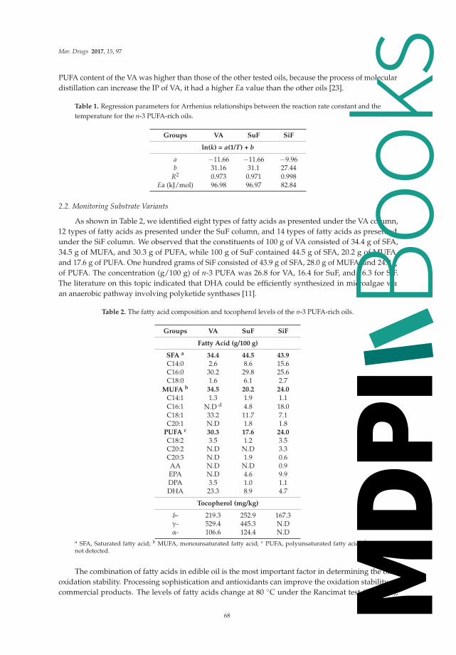
Mar. Drugs 2017, 15, 97
PUFA content of the VA was higher than those of the other tested oils, because the process of moleculardistillation can increase the IP of VA, it had a higher Ea value than the other oils [23].
Table 1. Regression parameters for Arrhenius relationships between the reaction rate constant and thetemperature for the n-3 PUFA-rich oils.
Groups VA SuF SiF
ln(k) = a(1/T) + b
a −11.66 −11.66 −9.96b 31.16 31.1 27.44
R2 0.973 0.971 0.998Ea (kJ/mol) 96.98 96.97 82.84
2.2. Monitoring Substrate Variants
As shown in Table 2, we identified eight types of fatty acids as presented under the VA column,12 types of fatty acids as presented under the SuF column, and 14 types of fatty acids as presentedunder the SiF column. We observed that the constituents of 100 g of VA consisted of 34.4 g of SFA,34.5 g of MUFA, and 30.3 g of PUFA, while 100 g of SuF contained 44.5 g of SFA, 20.2 g of MUFA,and 17.6 g of PUFA. One hundred grams of SiF consisted of 43.9 g of SFA, 28.0 g of MUFA, and 24.0 gof PUFA. The concentration (g/100 g) of n-3 PUFA was 26.8 for VA, 16.4 for SuF, and 16.3 for SiF.The literature on this topic indicated that DHA could be efficiently synthesized in microalgae viaan anaerobic pathway involving polyketide synthases [11].
Table 2. The fatty acid composition and tocopherol levels of the n-3 PUFA-rich oils.
Groups VA SuF SiF
Fatty Acid (g/100 g)
SFA a 34.4 44.5 43.9C14:0 2.6 8.6 15.6C16:0 30.2 29.8 25.6C18:0 1.6 6.1 2.7
MUFA b 34.5 20.2 24.0
C14:1 1.3 1.9 1.1C16:1 N.D d 4.8 18.0C18:1 33.2 11.7 7.1C20:1 N.D 1.8 1.8
PUFA c 30.3 17.6 24.0C18:2 3.5 1.2 3.5C20:2 N.D N.D 3.3C20:3 N.D 1.9 0.6AA N.D N.D 0.9EPA N.D 4.6 9.9DPA 3.5 1.0 1.1DHA 23.3 8.9 4.7
Tocopherol (mg/kg)
δ- 219.3 252.9 167.3γ- 529.4 445.3 N.Dα- 106.6 124.4 N.D
a SFA, Saturated fatty acid; b MUFA, monounsaturated fatty acid; c PUFA, polyunsaturated fatty acid; d N.D,not detected.
The combination of fatty acids in edible oil is the most important factor in determining the oil’soxidation stability. Processing sophistication and antioxidants can improve the oxidation stability ofcommercial products. The levels of fatty acids change at 80 ◦C under the Rancimat test (Figure 2),
68
Books
MDPI

Mar. Drugs 2017, 15, 97
with the difference in the PUFA amounts of the VA, SuF, and SiF blends being −16.60%, −13.11%, and−2.76% at the 100% oxidation level, and −29.43%, −24.08%, and −16.61% at the 125% oxidation level,mainly as a result of DHA degradation. The difference in the SFA amounts of the VA, SuF, and SiFblends were 1.01%, 4.11%, and 2.29% at the 100% oxidation level, and −0.67%, 4.34%, and 12.36% at the125% oxidation level. It is known that the thermal treatment of oils and fats generates hydroperoxidebreakdown of any fatty acids with a chain shorter than ten carbon atoms, such that such breakdowncan be considered a chemical indicator of the fat degradation grade [24]. This study was similar tosuch results, as SFA formation appeared to be correlated with PUFA loss, and with monounsaturatedfatty acid (MUFA) increases and decreases.
Figure 2. Percentage variations (g/100 g oil) of the (A) SFA, (B) MUFA, (C) PUFAs, and (D) totaltocopherol measured in the n-3 PUFA-rich oils.
Tocopherols are the most abundant antioxidants in n-3 PUFA concentrates, because of theircapacity to inhibit hydroperoxides and C-3 aldehydes [25]. In commercial products, tocopherol canbe used independently or in combination with other compounds, such as ascorbic acid palmitate,lecithin, and catechin, and has shown significantly in fish oil stabilization. On the other hand,the structure conformation also affects the physical property of tocopherol, as γ- and δ-tocopherolhave thermal resistance. Hydroperoxyl radical-scavenging activity occurs in the order of α- > β- > γ-> δ-tocopherol [26]. As shown in Figure 2, half of the total tocopherols of the tested n-3 PUFA-richoils was lost at the 50% oxidation level under the Rancimat test, while 90% of the total was lost atthe 100% oxidation level under the Rancimat test. The fact that the tocopherol content dropped veryquickly under the Rancimat test conditions could be due mainly to the very high susceptibility of thismolecule to oxidation to tocopherol quinones at high temperatures, which diminishes the protection ofunsaturated fatty acids against oxidation [27].
69
Books
MDPI

Mar. Drugs 2017, 15, 97
2.3. Monitoring Oxidation Products
Lipid oxidation products have negative impacts on the flavor and odor of sensory parameters,which can, in turn, have harmful effects on human health. The progress of lipid oxidation can beevaluated by the monitoring of a diverse series of primary, secondary, and tertiary oxidation productsover time. The quality standards of GOED require specific levels of acid value (AV, ≤3 mg KOH/g),POV (≤5 meq/kg), p-AV (≤20), and TOTOX (≤26) throughout the stated lifetime of a product [15].We found that the VA and SuF tested in this study met their stated label claims for GOED (Table 3).POV and AV are used in the industry’s on-line quality control index. POV has a significant correlationto the off-flavour compounds created during the initial oxidation. AV represents the content of freefatty acids which are easily oxidized to hydro-peroxides [28].
The EC regulations suggest that absorbances at 234 nm (K234), 270 nm (K270), and 280 nm(K280) be used to measure oils according to their oxidated products such as ethylenic diketones,conjugated ketodienes, and the dienal formation of conjugated dienes and trienes. K234 is a primaryoxidation index that has been found to be closely related to hydroperoxide content [29]. In addition,the measurement of UV absorbance at 270 and 280 nm has been used previously in analyzing edibleoils. Absorption at these wavelengths is mainly due to secondary oxidation. In a spectral analysisconducted for this study (Table 3), we found that the absorbances of 10 mg/mL of VA, SuF, and SiFwere 3.33, 14.79, and 7.58, respectively, at K234; 1.12, 0.66, and 1.78, respectively, at K270; and 1.08,0.51 and 1.48, respectively, at K280. Most of them increased as the oxidation levels increased. The K234of SuF had high values at the beginning, a finding which can be attributed to the residual oxidatedproducts or fat-soluble compounds that were produced from the simplified refining process.
Table 3. Initial quality characteristics of the n-3 PUFA-rich oils.
Groups VA SuF SiF
Quality Indicators
AV (mg KOH/g) 0.48 ± 0.02 0.49 ± 0.01 0.65 ± 0.01CVD (%) 0.22 ± 0.05 1.18 ± 0.17 0.58 ± 0.03
POV (meq/kg) 1.98 ± 0.27 4.12 ± 0.34 13.62 ± 0.42p-AV (meq/kg) 6.33 ± 0.71 15.12 ± 0.64 29.23 ± 1.84
TOTOX (meq/kg) 10.30 ± 0.92 23.26 ± 1.24 56.46 ± 2.11
Visible Spectra (10 mg/mL)
K234 a 3.33 ± 0.07 14.79 ± 0.14 7.58 ± 0.17K270 1.12 ± 0.01 0.66 ± 0.02 1.78 ± 0.04K280 1.08 ± 0.02 0.51 ± 0.01 1.48 ± 0.07
a K234, K270, and K280, specific absorption at 234, 270, and 280 nm.
In the literatures, hierarchical cluster was showed that could apply to grouping basis on quality,sensory attributes and refined level [30,31]. The AHC analysis was applied to identify clusters ofsamples with similar oxidation properties. Three main clusters were extracted (Figure 3). Cluster 1contained the 0–100% oxidation levels of the VA, the 0–75% oxidation levels of the SuF, and the 0–75%oxidation levels of the SiF. Cluster 2 contained the 100% oxidation levels of the SuF and SiF. Cluster 3contained the 125% oxidation levels of the VA, SuF, and SiF. These results showed that there aresignificant changes of quality in the IP of a given oil when it is subjected to the Rancimat test. Theseresults were similar to those of a previous study reported by our team, which oxidized volatiles of n-3PUFA [32].
With regard to the quality standard change at 80 ◦C under the Rancimat test, according to thePCA analysis, two dimensions were extracted and together account for approximately 91.51% ofthe variability from the original data (Figure 3). In our results, we observed p-AV (r: 0.92), TOTOX(r: 0.91), K270 (r: 0.88), and POV (r: 0.87) with PAC1; and K234 (r: 0.83) and conjugated dienes (CVD,r: 0.81) with PCA2. The POV and p-AV are measures of primary oxidation and secondary oxidation,
70
Books
MDPI

Mar. Drugs 2017, 15, 97
respectively. The TOTOX value gives an overall indication of the complete oxidation status of oil,and consists of the combination of the POV and the p-AV values to determine the oxidation levelof the oil. Correlations between the quality parameters and spectral analysis is that K234, with AV,POV, p-AV, and TOTOX is low (R < 0.5), while K270 and K280 with acid value (AV), POV, p-AV, andTOTOX is high (R > 0.7). These results were based on measuring the formation of secondary oxidationproducts; for example, p-AV and K270 are available to support the AV evaluation of frying oil quality,and aldehyde molecules were a common marker for K270, K280, and p-AV [28,33].
Figure 3. PCA plots of quality changes for the different oxidation levels of the n-3 PUFA-rich oils underthe Rancimat method; the circles represent the clusters detected with AHC analysis; the solid fill typehighlights the values with PCA.
3. Materials and Methods
3.1. Materials
Conventional algae oils (VA) were provided by VEDAN Enterprise Corporation (Taichung,Taiwan), and conventional fish oils from mackerel (SuF) were provided by SUN AGRICULTURE(Ilan, Taiwan). Additional fish oils (SiF) were provided by Sigma (Sigma-Aldrich, Taufkirchen,Germany). A fatty acid methyl ester standard (FAME) mixture, Supelco 37 Component FAMEMix, was purchased from Supelco (Sigma-Aldrich, Taufkirchen, Germany). Standards of α-, γ-,and δ- tocopherol were purchased from Merck (Darmstadt, Germany).
3.2. Rancimat Test
The oxidative stability index of the n-3 PUFA-rich oil was previously determined at four differenttemperatures (70 ◦C, 80 ◦C, 90 ◦C, and 100 ◦C) as the induction period (hours) that was recorded usinga Rancimat 743 apparatus and a 5 ± 0.05 g sample of oil with an air flow of 10 L/h. Then, oil sampleswere oxidized at 80 ◦C for periods of time that corresponded to 25%, 50%, 75%, 100%, and 125% oftheir respective induction periods. The oil samples that were used to determine the oxidative stabilitywere also analyzed for their volatile oxidation compounds. The IP of the n-3 PUFA-rich oils wereautomatically recorded and taken as the break point of the plotted curves (the intersection point of thetwo extrapolated parts of the curve).
71
Books
MDPI

Mar. Drugs 2017, 15, 97
3.3. Kinetic Data Analysis
The kinetic parameters were determined according to the method previously utilized as reportedin [17]. The IP of the oil samples were automatically recorded and taken as the break point of theplotted curves (the intersection point of the two extrapolated parts of the curve). A kinetic rate constantwas taken as the inverse of the IP (k, h−1).
Temperature coefficients (T Coeff, K−1) were determined from the slopes of the lines generatedby regressing ln(k) vs. the absolute temperature (T, K):
ln(k) = a(T) + b (1)
where a and b are the equation parameters.Activation energies (Ea, kJ/mol) and pre-exponential or frequency factors (A, h−1) were
determined from the slopes and intercepts, respectively, of the lines generated by regressing ln(k) vs.1/T using the Arrhenius equation:
ln(k) = ln(A) − (Ea/RT) (2)
where k is the reaction rate constant or reciprocal IP (h−1), and R is the molar gas constant(8.3143 J/mol K).
3.4. Analysis of Tocopherol
Each oil sample (0.1 g) was diluted with 2-propanol to a volume of 10 mL and filtered throughan MS nylon syringe filter with a 0.45 μm pore size directly to vials and then immediately analyzedusing an HPLC system. Aliquots of 10 μL of the filtrate were injected into the injection port andanalyzed with HPLC (Hitachi L-2130 pump, Hitachi, Tokyo, Japan). The remaining procedures werecarried out as previously reported [34] using an HPLC attached to a detector L-2400 UV and a HitachiL-2130 pump. An RP-18GP250 Mightysil column (l = 250 mm; i.d. = 4.6 mm; thickness = 0.32 μm;Kanto Chemical Co., Inc., Tokyo, Japan) was used for separation. The same mobile phase and elutionconditions were adopted. The calibration curves were, respectively, established for tocopherol byplotting the peak area vs. each corresponding concentration, from which quantitations of the standardswere achieved.
3.5. Fatty Acid Analysis
The n-3 PUFA-rich oil was analyzed for its fatty acid composition via GC/FID. The triacylglycerolswere converted to methyl esters using the AOCS Official Method Ce 2–66 [35]. The methyl esters wereseparated using a column that was coated with DB-23 (30 m × 0.25 mm × 0.25 μm, Agilent, Palo Alto,CA, USA), and helium was used as the carrier gas at a flow rate of 1.0 mL/min. The oven temperaturewas initially held for 8 min at 200 ◦C, and then increased at 10 ◦C/min to 220 ◦C and then held therefor 40 min. The FID was maintained at 270 ◦C, and the injector (split mode 1:40, 4 mm liner) wasmaintained at 250 ◦C. The contents of the fatty acids were determined using the normalization method,with heneicosanoic methyl ester used as an internal standard to quantitation.
3.6. Quality Analytical Determination
The POV and UV spectrophotometric were measured using the analytical methods describedin European Regulation EEC 2568/91 [36]. The CDA measures the formed from PUFA during lipidoxidation according to the AOCS method Ti la-64 [35]. The p-AV measures secondary oxidationproducts, such as 2-alkenal and 2,4-alkadienal. The p-AV of each sample was determined accordingto the AOCS method Cd 18–90 [35]. The AV was determined using a titration with 0.1 N potassiumhydroxide alcoholic solution.
72
Books
MDPI

Mar. Drugs 2017, 15, 97
3.7. Statistical Analysis
The data reported were obtained from triplicate measurements of each sample and were expressedas means. The data were subjected to an AHC analysis with squared Euclidean distances. Subsequently,the data were analyzed using PCA combined with VARIMAX rotation. For the AHC and PCA analysis,XLSTAT software (version 2010.2.01, Addinsoft Deutschland, Andernach, Germany) was used.
4. Conclusions
In this work, the Rancimat test was applied to the analysis of the oxidation properties of n-3PUFA-rich oil. The results of the present study indicated: (1) the degree of unsaturation in fatty acidsis not the only parameter for assessing oil quality, which is mainly influenced by commercialization.The oxidation stability of n-3 PUFA-rich oils is improved by commercialization, especially with respectto antioxidant protection; and (2) chemometric applications have also clarified the nature of thedifferences in correlation among oxidative and chemical parameters. The IP of primary and secondaryoxidation product formations represented the quality changes as detected via AHC analysis. Notall quality parameters increased linearly through PCA analysis, which can allow for the selection ofa proper and adequate method for a particular application. It is recommended for p-AV, TOTOX, K270,and POV to be used when carrying out quality control for n-3 PUFA- rich oils.
Acknowledgments: This work was supported by the Ministry of Science and Technology (NSC 105-2221-E-005-077),Taiwan.
Author Contributions: Kai-Min Yang carried out all the experiments Kai-Min Yang and Po-Yuan Chiang designedall of the experiments and analyzed the data; Kai-Min Yang and Po-Yuan Chiang wrote the manuscript.
Conflicts of Interest: The authors declare no conflict of interest.
Abbreviations
The following abbreviations are used in this manuscript:
SFA Saturated fatty acidsMUFA Monounsaturated fatty acidPUFA Polyunsaturated fatty acidAA Arachidonic acidEPA Eicosapentaenoic acidDPA Docosapentaenoic acidDHA Docosahexaenoic acidGOED Global Organization for EPA and DHAIP Induction periodEa Activation energiesPOV Peroxide valuep-AV Anisidine valueAV Acid valueCD Conjugated dienesTOTOX Total oxidation valuePCA Principal component analysisAHC Agglomerative hierarchical cluster
References
1. Pike, I.H. Fish oil: Supply and demand as a source of long-chain n-3 polyunsaturated fatty acids in thehuman diet. Eur. J. Lipid Sci. Technol. 2015, 117, 747–750. [CrossRef]
2. Nichols, P.D.; McManus, A.; Krail, K.; Sinclair, A.J.; Miller, M. Recent advances in omega-3: Health benefits,Sources, Products and bioavailability. Nutrients 2014, 9, 3727–3733. [CrossRef] [PubMed]
3. Kris-Etherton, P.M.; Harris, W.S.; Appel, L.J.; Committee, N. Fish consumption, fish oil, omega-3 fatty acids,and cardiovascular disease. Circulation 2002, 106, 2747–2757. [CrossRef] [PubMed]
73
Books
MDPI

Mar. Drugs 2017, 15, 97
4. Watanabe, N.; Onuma, K.; Fujimoto, K.; Miyake, S.; Nakamura, T. Long-term effect of an enteral diet witha different n-6/n-3 ratio on fatty acid composition and blood parameters in rats. J. Oleo Sci. 2011, 60, 109–115.[CrossRef] [PubMed]
5. Lim, D.K.; Garg, S.; Timmins, M.; Zhang, E.S.; Thomas-Hall, S.R.; Schuhmann, H.; Li, Y.; Schenk, P.M.Isolation and evaluation of oil-producing microalgae from subtropical coastal and brackish waters. PLoS ONE2012, 7, e40751. [CrossRef] [PubMed]
6. Opperman, M.; Marais, D.W.; Benadé, A.S. Analysis of omega-3 fatty acid content of South African fish oilsupplements. Cardiovasc. J. Afr. 2011, 22, 324–329. [CrossRef] [PubMed]
7. Facts, P. Global Market for EPA/DHA Omega-3 Products; Packaged Facts: Rockville, MD, USA, 2012.8. Adarme-Vega, T.C.; Thomas-Hall, S.R.; Schenk, P.M. Towards sustainable sources for omega-3 fatty acids
production. Curr. Opin. Biotechnol. 2014, 26, 14–18. [CrossRef] [PubMed]9. Adarme-Vega, T.C.; Lim, D.K.; Timmins, M.; Vernen, F.; Li, Y.; Schenk, P.M. Microalgal biofactories:
A promising approach towards sustainable omega-3 fatty acid production. Microb. Cell Fact. 2012, 11, 96.[CrossRef] [PubMed]
10. Kuratko, C.N.; Norman, S. Docosahexaenoic acid from algal oil. Eur. J. Lipid Sci. Technol. 2013, 115, 965–976.[CrossRef]
11. Martins, D.A.; Custódio, L.; Barreira, L.; Pereira, H.; Ben-Hamadou, R.; Varela, J.; Abu-Salah, K.M.Alternative sources of n-3 long-chain polyunsaturated fatty acids in marine microalgae. Mar. Drugs 2013, 11,2259–2281. [CrossRef] [PubMed]
12. Drusch, S.; Groß, N.; Schwarz, K. Efficient stabilization of bulk fish oil rich in long-chain polyunsaturatedfatty acids. Eur. J. Lipid Sci. Technol. 2008, 110, 351–359. [CrossRef]
13. Guillén, M.D.; Goicoechea, E. Toxic oxygenated α, β-unsaturated aldehydes and their study in foods:A review. Crit. Rev. Food Sci. Nutr. 2008, 48, 119–136. [CrossRef] [PubMed]
14. Guillén, M.D.; Cabo, N.; Ibargoitia, M.L.; Ruiz, A. Study of both sunflower oil and its headspace throughoutthe oxidation process. Occurrence in the headspace of toxic oxygenated aldehydes. J. Agric. Food Chem. 2005,53, 1093–1101. [CrossRef] [PubMed]
15. GOED. Available online: http://www.goedomega3.com/healthcare (accessed on 10 January 2016).16. Tan, C.; Man, Y.C.; Selamat, J.; Yusoff, M. Application of Arrhenius kinetics to evaluate oxidative stability
in vegetable oils by isothermal differential scanning calorimetry. J. Am. Oil Chem. Soc. 2001, 78, 1133–1138.[CrossRef]
17. Farhoosh, R.; Niazmand, R.; Rezaei, M.; Sarabi, M. Kinetic parameter determination of vegetable oil oxidationunder Rancimat test conditions. Eur. J. Lipid Sci. Technol. 2008, 110, 587–592. [CrossRef]
18. Chen, M.H.; Huang, T.C. Volatile and Nonvolatile Constituents and Antioxidant Capacity of Oleoresinsin Three Taiwan Citrus Varieties as Determined by Supercritical Fluid Extraction. Molecules 2016, 21, 1735.[CrossRef] [PubMed]
19. Robertson, G.L. Shelf life of packaged foods, its measurements and prediction. In Developing New FoodProducts for a Changing Marketplace; CRC Press: Boca Raton, FL, USA, 2000; pp. 329–353.
20. Rupasinghe, H.V.; Erkan, N.; Yasmin, A. Antioxidant protection of eicosapentaenoic acid and fish oiloxidation by polyphenolic-enriched apple skin extract. J. Agric. Food Chem. 2009, 58, 1233–1239. [CrossRef][PubMed]
21. Adhvaryu, A.; Erhan, S.; Liu, Z.; Perez, J. Oxidation kinetic studies of oils derived from unmodified andgenetically modified vegetables using pressurized differential scanning calorimetry and nuclear magneticresonance spectroscopy. Thermochim. Acta 2000, 364, 87–97. [CrossRef]
22. Yoshii, H.; Furuta, T.; Siga, H.; Moriyama, S.; Baba, T.; Maruyama, K.; Misawa, Y.; Hata, N.; Linko, P.Autoxidation kinetic analysis of docosahexaenoic acid ethyl ester and docosahexaenoic triglyceride withoxygen sensor. Biosci. Biotechnol. Biochem. 2002, 66, 749–753. [CrossRef] [PubMed]
23. Wang, W.; Li, T.; Ning, Z.; Wang, Y.; Yang, B.; Ma, Y.; Yang, X. A process for the synthesis of PUFA-enrichedtriglycerides from high-acid crude fish oil. J. Food Eng. 2012, 109, 366–371. [CrossRef]
24. De Leonardis, A.; Macciola, V. Heat-oxidation stability of palm oil blended with extra virgin olive oil.Food Chem. 2012, 135, 1769–1776. [CrossRef] [PubMed]
25. Kulås, E.; Ackman, R.G. Properties of α-, γ-, and δ-tocopherol in purified fish oil triacylglycerols. J. Am. OilChem. Soc. 2001, 78, 361–367. [CrossRef]
74
Books
MDPI

Mar. Drugs 2017, 15, 97
26. Fujisawa, S.; Kadoma, Y. Kinetic study of the radical-scavenging activity of vitamin E and ubiquinone.In Vivo 2005, 19, 1005–1011. [PubMed]
27. Verleyen, T.; Kamal-Eldin, A.; Dobarganes, C.; Verhé, R.; Dewettinck, K.; Huyghebaert, A. Modeling ofα-tocopherol loss and oxidation products formed during thermoxidation in triolein and tripalmitin mixtures.Lipids 2001, 36, 719–726. [CrossRef] [PubMed]
28. Navas, J.A.; Tres, A.; Codony, R.; Guardiola, F. Optimization of analytical methods for the assessment of thequality of fats and oils used in continuous deep fat frying. Grasas Aceites 2007, 58, 154–162.
29. Shahid Chatha, S.A.; Anwar, F.; Manzoor, M.; Rehman Bajwa, J.U. Evaluation of the antioxidant activity ofrice bran extracts using different antioxidant assays. Grasas Aceites 2006, 57, 328–335.
30. Navarro, T.; de Lorenzo, C.; Pérez, R. SPME analysis of volatile compounds from unfermented olivessubjected to thermal treatment. Anal. Bioanal. Chem. 2004, 379, 812–817. [CrossRef] [PubMed]
31. Tu, D.; Li, H.; Wu, Z.; Zhao, B.; Li, Y. Application of headspace solid-phase microextraction and multivariateanalysis for the differentiation between edible oils and waste cooking oil. Anal. Bioanal. Chem. 2014, 7,1263–1270. [CrossRef]
32. Yang, K.M.; Cheng, M.C.; Chen, C.W.; Tseng, C.Y.; Lin, L.Y.; Chiang, P.Y. Characterization of VolatileCompounds with HS-SPME from Oxidized n-3 PUFA Rich Oils via Rancimat tests. J. Oleo Sci. 2017, 66,113–122. [CrossRef] [PubMed]
33. Grau, A.; Guardiola, F.; Boatella, J.; Baucells, M.D.; Codony, R. Evaluation of lipid ultraviolet absorptionas a parameter to measure lipid oxidation in dark chicken meat. J. Agric. Food Chem. 2000, 48, 4128–4135.[CrossRef] [PubMed]
34. Shehata, A.B.; Rizk, M.S.; Farag, A.M.; Tahoun, I.F. Development of two reference materials for alltrans-retinol, retinyl palmitate, α- and γ-tocopherol in milk powder and infant formula. J. Food Drug Anal.2015, 23, 82–92. [CrossRef]
35. American Oil Chemists’ Society (AOCS). Official Methods and Recommended Practices of the American OilChemists’ Society; Firestone, D., Ed.; AOCS Press: Champaign, IL, USA, 1998.
36. Regulation, H. Commission Regulation (EEC) No. 2568/91 of 11 July 1991 on the characteristics of oliveoil and olive-residue oil and on the relevant methods of analysis Official Journal L 248, 5 September 1991.Off. J. L 1991, 248, 1–83.
© 2017 by the authors. Licensee MDPI, Basel, Switzerland. This article is an open accessarticle distributed under the terms and conditions of the Creative Commons Attribution(CC BY) license (http://creativecommons.org/licenses/by/4.0/).
75
Books
MDPI

marine drugs
Review
Marine Lipids on Cardiovascular Diseases andOther Chronic Diseases Induced by Diet: An InsightProvided by Proteomics and Lipidomics
Lucía Méndez 1,*, Gabriel Dasilva 1, Nùria Taltavull 2, Marta Romeu 2 and Isabel Medina 1
1 Instituto de Investigaciones Marinas (IIM-CSIC), Eduardo Cabello 6, E-36208 Vigo, Spain;[email protected] (G.D.); [email protected] (I.M.)
2 Unitat de Farmacologia, Facultat de Medicina i Ciències de la Salut, Universitat Rovira i Virgili,Sant Llorenç 21, E-43201 Reus, Spain; [email protected] (N.T.); [email protected] (M.R.)
* Correspondence: [email protected]; Tel.: +34-986-231930; Fax: +34-986-292762
Received: 16 May 2017; Accepted: 15 August 2017; Published: 18 August 2017
Abstract: Marine lipids, especially ω-3 polyunsaturated fatty acids (PUFAs) eicosapentaenoic acid(EPA) and docosahexaenoic acid (DHA), have largely been linked to prevention of diet-induceddiseases. The anti-inflammatory and hypolipidemic properties of EPA and DHA supplementationhave been well-described. However, there is still a significant lack of information about theirparticular mechanism of action. Furthermore, repeated meta-analyses have not shown conclusiveresults in support of their beneficial health effects. Modern “omics” approaches, namely proteomicsand lipidomics, have made it possible to identify some of the mechanisms behind the benefits ofmarine lipids in the metabolic syndrome and related diseases, i.e., cardiovascular diseases and type 2diabetes. Although until now their use has been scarce, these “omics” have brought new insightsin this area of nutrition research. The purpose of the present review is to comprehensively showthe research articles currently available in the literature which have specifically applied proteomics,lipidomics or both approaches to investigate the role of marine lipids intake in the prevention orpalliation of these chronic pathologies related to diet. The methodology adopted, the class of marinelipids examined, the diet-related disease studied, and the main findings obtained in each investigationwill be reviewed.
Keywords: marine lipids; EPA; DHA; proteomics; lipidomics; metabolic syndrome; cardiovasculardisease; type 2 diabetes
1. Introduction
1.1. Chronic Diseases Induced by Diet: A World Health Problem
The intake of westernized diets, which are rich in refined carbohydrates, cholesterol, saturatedand trans fats, and have an increased ratio of ω-6/ω-3 PUFAs, along with a sedentary lifestyle canquickly lead to the development of different pathologies and metabolic disorders [1]. These chronicdiseases related to diet, such as cardiovascular diseases (CVD), obesity, overweight, hypertension,type 2 diabetes, hyperlipidemia, hyperinsulinemia, osteoporosis, osteopenia or cancer, are consideredthe epidemic of modern societies. It is estimated that 20–30% of the adult population in these countriessuffers Metabolic Syndrome (MetS) [2]. The MetS is a compilation of risk factors associated with CVDand type 2 diabetes. These factors include dyslipidemia (decreased high-density lipoprotein (HDL)cholesterol levels and increased low-density lipoprotein (LDL) cholesterol and triglycerides levels),hyperglycemia, hypertension, insulin resistance and obesity. Inflammation and oxidative stress havealso been closely related to MetS and derived diseases [3].
Mar. Drugs 2017, 15, 258 76 www.mdpi.com/journal/marinedrugs
Books
MDPI

Mar. Drugs 2017, 15, 258
According to the Eurostat report of May 2016, the first two leading causes of death in Europewere CVD and cancer, well above respiratory diseases, which were the third most common causeof death [4]. The average in Europe was 132 deaths/100,000 inhabitants due to CVD in 2013, being350/100,000 in countries like Hungary or Slovakia, and less than 100/100,000 in Spain, Portugal orGreece. Data from the World Health Organization (WHO) in 2015 also showed that the leading causesof death in high-income countries were CVD and cancer, while infectious diseases were the leadingcause of death in low-income countries [5].
Therefore, the study of diet components and their relation to the progression of chronic diseasesand metabolic disorders has arisen as a field of great interest for the scientific community. Accordingly,the number of publications found in the Scopus database (www.scopus.com) which are focused on thistopic has exponentially grown, especially since 1990. Considering the search criteria: TITLE-ABS-KEY(nutrition OR diet) AND TITLE-ABS-KEY (“metabolic disorder” OR “metabolic alteration” OR obesityOR “metabolic syndrome” OR diabetes OR “cardiovascular disease” OR cholesterol OR insulin ORatherosclerosis OR inflammation OR “oxidative stress”), around 272,000 were found. From these, 75%are research articles and 14% are reviews. The rest of the published work consists of book chapters,notes, conferences, etc. In relation to the field of study, 76% of the publications belong to the fieldof medicine, 27% to biochemistry, and the rest of them are framed in the fields of agricultural andbiological sciences, nursing, pharmacology, toxicology and pharmaceutics, neuroscience, chemistry,immunology and microbiology, etc.
As a consequence of these investigations, huge quantities of bioactive compounds have beenfound to exert beneficial effects on human health. Among that, ω-3 PUFAs from marine origin havequickly gained more attention. Currently, there is considerable evidence that the intake of marine-originpolyunsaturated fatty acids (PUFAs), especially eicosapentaenoic acid (EPA) and docosahexaenoicacid (DHA), can help in the prevention/palliation of inflammatory processes and metabolic diseases,although their mechanisms of action are not entirely understood yet.
1.2. Marine Lipids as Bioactive Compounds against MetS and Chronic Diseases Induced by Diet
Marine lipids, especially EPA and DHA, have largely demonstrated their bioactivity in humanhealth. The interest in their intake arose from a series of pioneering studies in the Inuit Eskimopopulation during the 1960s and 1970s. These studies reported a lower incidence of cardiovascularpathologies in that population associated with a high intake of fish in their diet. Later, similarrelationships were discovered in human populations from other regions, particularly in Iceland andAlaska Natives [6].
In addition to these investigations, marine ω-3 PUFAs have been shown to alleviate metabolicdisorder symptoms, such as heart disease, diabetes, obesity and insulin resistance. Since the late 1950s,the number of publications found in the Scopus database (TITLE-ABS-KEY (“omega 3” OR “PUFA” OR“eicosapentaenoic” OR “docosahexaenoic” OR “marine fatty acids” OR “marine lipid” OR “marine oil”OR “fish oil” OR “fish lipid”) AND TITE-ABS-KEY (nutrition OR diet) AND TITLE-ABS-KEY (“metabolicdisorder” OR “metabolic alteration” OR “metabolic disease” OR obesity OR “metabolic syndrome” ORdiabetes OR “cardiovascular disease” OR cholesterol OR insulin OR atherosclerosis OR inflammationOR “oxidative stress”)), which have related the consumption of marine fatty acids or lipids to thesemetabolic disorders, has increased exponentially, mainly since the late 1970s. On the whole, more than13,100 publications can currently be found in the Scopus database. Sixty-five percent of these publicationsare research articles, 23% are reviews and the rest of them are book chapters, notes, conferences, etc.
These research articles have used a plethora of different approaches (from chemistry orbiochemistry to phycology) and a huge variety of methodologies to address this topic. As a consequence,the publications are categorized in very diverse fields of study, medicine (74%), biochemistry (30%),agricultural and biological sciences (20%), nursing (19%) and the rest of the articles are framed in thefields of pharmacology, toxicology and pharmaceutics, chemistry, neuroscience, immunology andmicrobiology, phycology, social sciences, mathematics, computer science, etc.
77
Books
MDPI

Mar. Drugs 2017, 15, 258
The aim of the present review is to comprehensively show those research articles currentlyavailable in the literature which have specifically used proteomics, lipidomics or both approaches toinvestigate the role of marine lipids intake in the prevention/palliation of chronic pathologies inducedby the consumption of westernized diets, namely MetS, CVD and type 2 diabetes. Our ultimateobjective is to highlight the usefulness of the application of these approaches to nutrition research,in order to discover the underlying mechanisms of the beneficial effects of marine lipids in humanmetabolic health, since its translation to practice via nutritional interventions is still in its infancy.
2. Omics for Unrevealing Mechanisms: Proteomics and Lipidomics
Almost all cellular processes, from gene expression to synthesis, degradation and protein activity,can be affected by diet and lifestyle. Therefore, nutrients and other food components (includingmarine lipids) may alter metabolic functions in cells in a quite complex way. This complex relationshipbetween nutrition and health makes nutrition research an ideal field for the application of systemsbiology approaches [7], as being holistic and integrated. Systems biology encompasses a broadrange of functional areas called “omics” These areas include genomics, transcriptomics, proteomics,metabolomics, lipidomics and bioinformatics [8]. This new outlook started as a result of the humangenome project in the early 2000s [9].
The number of publications found in the literature which incorporates the most novel omicsapproaches (proteomics and lipidomics) to the study of the beneficial effects of marine lipids againstMetS and their associated chronic diseases is still scarce. Proteomics and lipidomics studies appliedto nutrition research have to cope with a variety of difficulties ranging from experimental design,sample processing and optimization strategies to data analysis and identification. The need forhigh-sensitivity modern mass spectrometers combined with bioinformatics resources increases costsand requires highly qualified staff. In spite of these difficulties, the interest of the scientific communityin these approaches has been progressively growing, in parallel with analytical advancements in liquidchromatography, mass spectrometry and bioinformatics.
In the following subsections, we review the research articles currently available in the literaturewhich have used proteomics (Section 2.1), lipidomics (Section 2.2) or both (Section 2.3) to analyze theeffects of the consumption of marine lipids on the development and progression of human MetS aswell as their associated chronic diseases (CVD and type 2 diabetes).
2.1. Beneficial Effects of Marine Lipids Intake Assayed by Proteomics
Proteins are important mediators of biological activities in all living cellular units. Proteomics isthe large-scale study of proteins and therefore comprises the methodologies used for the study of aproteome [10]. The “proteome” a term firstly coined by Wilkins et al. in the early 2000s, is comprisedof all expressed proteins encoded by the genome of a cellular system, including all cellular proteinsand all protein species (isoforms and protein modifications) [11,12].
The study of how ω-3 PUFAs regulate proteins and metabolic pathways may significantlycontribute to understanding the putative mechanisms by which marine lipids elicit their beneficialhealth effects. In fact, nutrition research has lately adopted the proteomics tools to measure changesin the protein complement of a biological system. This adoption has enabled modeling of biologicalprocesses in response to dietary marine lipids, as well as the elucidation of novel biomarkers for healthor disease which are sensitive to such lipids [13]. There are limited studies which have addressedthe influence of marine lipids on proteome with the aim to investigate their effects against humanmetabolic disorders. These studies have mainly used classical two-dimensional gel electrophoresis(2-DE) combined with mass spectrometry (MS) to elucidate changes in metabolic pathways and targetproteins which may explain a certain beneficial effect on human health. This traditional methodologyhas made it possible to identify changes in several metabolic pathways, such as glucose and fatty acidmetabolism, oxidative stress, antioxidant defense mechanisms and redox status. The study of minorabundant proteins such as those participating in inflammatory pathways requires the introduction in
78
Books
MDPI

Mar. Drugs 2017, 15, 258
nutrition research of more quantitative and sensitive methods, like multiple reaction monitoring (MRM)and multiplexed immunoassays. Both strategies might be useful for the evaluation and validation ofnewly discovered candidate biomarkers in human biofluids.
By using Scopus database, 53 publications have been found following the search criteria:TITLE-ABS-KEY (“omega 3” OR “PUFA” OR “eicosapentaenoic” OR “docosahexaenoic” OR “marinefatty acids” OR “marine lipid” OR “marine oil” OR “fish oil” OR “fish lipid”) AND TITE-ABS-KEY(nutrition OR diet) AND TITLE-ABS-KEY (“metabolic disorder” OR “metabolic alteration” OR“metabolic disease” OR obesity OR “metabolic syndrome” OR diabetes OR “cardiovascular disease”OR cholesterol OR insulin OR atherosclerosis OR inflammation OR “oxidative stress”) ANDTITE-ABS-KEY (proteomic OR proteomics OR proteome). These publications, mainly research articles(56%) and reviews (30%), belong, in descending order of abundance, to the fields of biochemistry,genetics and molecular biology (63%) and medicine (58%), and to a lesser extent nursing, agriculturaland biological sciences, pharmacology, toxicology and pharmaceutics, immunology and microbiology,neuroscience, chemistry, etc.
In this review, only research articles focusing on proteomics are presented. The studies whichhave used ω-3 PUFAs from a vegetal origin, i.e., α-linolenic acid (ALA), have been excluded. Onlyarticles which have assayed the effect of marine lipids on human metabolic disorders related to diet(i.e., features of MeS, cardiovascular diseases and type 2 diabetes) and those that have used animal orcellular models to mimic these alterations are presented. Moreover, articles which have combined bothlipidomics and proteomics tools in the same research are shown jointly in Section 2.3.
According to these filters, 14 research articles have been found. All of them investigated the effect ofEPA or DHA or both on metabolic health. Most of these studies used bottom-up proteomics approaches,based on 2-DE coupled with MS protein identification. Novel gel-free proteomics techniques based onMS, such as shotgun proteomics, have scarcely been used yet, as noted in Table 1.
2.1.1. Proteomics in Clinical Trials
Two studies analyzed the effect of the supplementation of a mix of EPA and DHA on themodulation of the peripheral blood mononuclear cells (PBMCs) proteome after acute intake [14]or after 12 weeks of dietary intervention [15] in patients with MetS. In the first case, proteomicsanalysis identified five proteins related to cell signaling and interaction, DNA repair, cellular assemblyand organization, and cell morphology which were regulated by acute consumption of marine PUFAs.
In the second study, the prolonged intake of EPA and DHA regulated 17 proteins of PBMCsproteome involved in immunological diseases and inflammatory response, down-regulating proteinsdirectly related to oxidative stress, inflammation, endoplasmic reticulum stress and DNA repair. A thirdstudy, which was carried out with the same dietary intervention and proteomics tools, evaluated thechanges induced in subcutaneous white adipose tissue (WAT) proteome after 12 weeks of EPA andDHA intake [16]. Three proteins of glucose metabolism were found down-regulated by marine PUFAssupplementation. These proteins were correlated with lower systemic insulin resistance and improvedinsulin signaling in subcutaneous WAT of the MetS patients.
Also in humans, De Roos et al. [17] studied mechanisms involved in preventing the early onsetof coronary heart disease (CHD) through fish oil intake. Authors identified ten serum proteins thatwere down-regulated in healthy volunteers after the consumption of a daily dose of 3.5 g of fishoil for 6 weeks. These altered serum proteins, metabolically related to lipoprotein metabolism andinflammation, led to a significant shift towards the larger, more cholesterol-rich HDL2 particle whichmight imply that fish oil activated anti-inflammatory and lipid modulating mechanisms believed toimpede the early onset of CHD. This specific effect of fish oil on HDL metabolism has later beeninvestigated by Burillo et al. [18]. They compared the proteome of HDL before and after ω-3PUFAs intake for 5 weeks. Healthy smoker volunteers ingested a commercial mixture of marineand non-marine ω-3 fatty acids. The consumption of marine ω-3 PUFAs up-regulated seven proteinsrelated to the antioxidant, anti-inflammatory and anti-atherosclerotic properties of HDL. Likewise,
79
Books
MDPI

Mar. Drugs 2017, 15, 258
marine lipids down-regulated six proteins involved in the regulation of complement activation andacute phase response. Moreover, the modification of lipoprotein containing apoAI (LpAI) proteomesuggested that the protein changes found might have improved the functionality of the particle.
2.1.2. Proteomics in Animal Models and Cell Cultures
Besides clinical trials, several studies have used animal or cell models to investigate the role ofmarine ω-3 PUFAs in regulating cellular proteomes which are rather difficult to analyze in humans,such as the liver proteome. In a study carried out in C57BL/6 mice, Ahmed et al. [19] investigated theregulation of the liver proteome by a dietary intervention with 10% ω-3 PUFAs from menhaden oilfor 4 months. Proteomics data showed that fish oil up-regulated eight proteins related to lipid,carbohydrate, protein and one-carbon metabolism and the citric acid cycle, which involved anintegrated regulation of metabolic pathways by fish oil. This proteome modulation was correlatedwith a significant reduction of plasma triglycerides and free fatty acid (FFA) levels.
Likewise, other studies investigated the effect of fish oil when it was added to high fat, cholesteroland sucrose diets. The first study [20] evaluated changes in the liver mitochondrial subproteome,an organelle which plays a critical role in cell metabolism and the development of metabolic alterations,induced in rats by the supplementation with 10% fish oil of a high fat diet for 50 weeks as compared torats fed low fat diet. As a result, Wrzesinski et al. identified 54 mitochondrial proteins regulated byfish oil. These proteins were involved in fatty acid oxidation and amino acid metabolism as well as inthe increase of oxidative phosphorylation.
In a second research, De Roos et al. [21] studied the modulation of the liver proteome by fish oilsupplementation of a diet high in saturated fat and cholesterol (HFC) for 3 weeks in APOE*3 Leidentransgenic mice, a model for lipid metabolism and atherosclerosis. These authors identified up to44 proteins altered by fish oil as compared to HFC, which were mainly involved in glucose and lipidmetabolism, as well as oxidation and aging processes. These changes were correlated with lowerplasma and liver cholesterol and triglycerides levels as well as minor plasma FFA and glucose buthigher plasma insulin levels, revealing new insights into mechanisms by which these fish oils canregulate lipid metabolism and related pathways.
A third research [22] reported the physiological modulation of rat liver proteome induced by24-week supplementation with fish oil considering two different background diets: standard or highin fat and sucrose (HFHS). Méndez et al. demonstrated a different capacity of fish oil for regulatingliver proteins depending on the background diet (6 proteins into standard diet and 31 into HFHS diet).Proteome changes induced by fish oil, especially under HFHS diet, consisted of decreasing the level ofenzymes from lipogenesis and glycolysis, enhancing fatty acid beta-oxidation and insulin signalingand ameliorating endoplasmic reticulum stress. Moreover, the consumption of fish oil decreasedprotein oxidation and improved several biochemical parameters.
Other authors have used cell culture experiments to evaluate EPA and DHA effects on theproteome. Kalupahana et al. [23] investigated the effects of EPA on proteins from 3T3-L1 adipocytestreated with either EPA or arachidonic acid (ARA). EPA-treated cells presented higher levels of19 proteins involved in carbohydrate and fatty acid metabolism and several other proteins related tocellular metabolism including response to stress. Likewise, EPA treatment resulted in lower levelsof eight proteins belong to lipogenesis and other cellular metabolic processes such as cytoskeletonorganization and biogenesis.
Proteomics has also been employed to evaluate the differential effect of the main marine ω-3PUFAs. Mavrommantis et al. [24] analyzed the potentially different effects on the liver proteomeregulation exerted by the dietary supplementation either with fish oil (EPA and DHA) or DHA alonein apoE knockout mice (model of atherosclerosis) fed HFC diet for 2 weeks. Both DHA and fishoil regulated 35 liver proteins, mainly involved in the metabolism of lipoproteins and oxidativestress. However, their effects on the proteome were not the same, since four of these proteins weredifferentially modulated by DHA or fish oil. As a consequence, although both fish oil and DHA
80
Books
MDPI

Mar. Drugs 2017, 15, 258
could beneficially affect lipoprotein metabolism and oxidative stress, intervention with fish oil butnot with DHA resulted in significantly lower levels of hepatic soluble epoxide hydrolase, an enzymeclosely related to cardiovascular disease, as compared to control oil. This different behavior betweenEPA and DHA was also highlighted by Johnson et al. [25]. These authors evaluated the potentiallyindependent protective/reversal effect of dietary EPA or DHA against mitochondrial dysfunction inaging skeletal muscle, after diet supplementation of young or older C57BL/6 mice. Authors found that10-weeks of dietary supplementation with EPA but not with DHA partially attenuated the age-relateddecay in mitochondrial function. Thirty-nine mitochondrial proteins changed between old controland old EPA-treated mice. Thirty-two mitochondrial proteins changed between old control and oldDHA-treated mice. However, only three proteins for EPA supplementation and seven for DHA werecoincident with those proteins which were found to differ in young and old control mice. Authorsconcluded that neither EPA nor DHA attenuated the age-related drop in mitochondrial protein content,although these changes demonstrated that both EPA and DHA exerted some common biological effects(anticoagulation, anti-inflammatory, reduced FXR/RXR activation). Additionally, proteomics datashowed that EPA improved muscle protein quality, specifically by decreasing mitochondrial proteincarbamylation, a post-translational protein modification (PTM) that is driven by inflammation.
2.1.3. Proteomics for Studying Post-Translational Protein Modifications (PTMs)
Abundant evidence has shown that ω-3 PUFAs influence redox homeostasis [26] and severalresearchers have described both antioxidant and pro-oxidant properties for these fatty acids.Since diet-induced metabolic diseases are often associated with increased oxidative stress, thecharacterization of oxidative PTMs seems to be critical for understanding the mechanisms underlyingthe action of marine ω-3 PUFAs in theses pathologies. However, studies focused on the effectsof EPA and DHA in modulating protein quality, besides protein quantity, are scarce likely due tomethodological limitations.
Among the oxidative PTMs, protein carbonyl moieties formed in proteins (protein carbonylation),which are the more common oxidative PTMs, are considered as a major hallmark of oxidative proteindamage. Metabolic alterations have strongly been correlated with high levels of protein carbonylation.However, only a very few studies have used proteomics tools to identify protein carbonylation targetsin metabolic diseases and evaluate the potential effects that marine lipids exerted on them. A studyaddressed the effect of various dietary EPA and DHA ratios (1:1, 2:1, and 1:2, respectively) on proteincarbonylation from plasma, kidney, skeletal muscle, and liver [27] in rats after 13 weeks of dietaryintervention. Rats fed soybean (rich in ω-6 linoleic acid (LA)) or linseed oil (rich in ω-3 ALA) were usedas controls. Authors identified targets of protein carbonylation in all the analyzed tissues. The three fishoil ratios, especially the 1:1 EPA:DHA, exerted a selective-protective effect against carbonylation of sixproteins from plasma and liver, which was correlated with the improvement of biochemical features.
Jourmard-Cubizolles et al. [28] studied a specific oxidative PTM, namely 4-hydroxynonenal(4-HNE) protein adducts derived from PUFA peroxidation. These authors investigated themodulation of the aortic proteome in atherosclerotic prone (LDLR−/−) mice fed an atherogenic dietsupplemented with DHA for 20 weeks. Nineteen proteins were differentially regulated in the aorta ofDHA-supplemented group. Most of them were related to glucose or lipid metabolism, including theup-regulation of superoxide dismutase by DHA which suggested an impact on vascular antioxidantdefenses. This up-regulation was in agreement with data from the quantification of proteins with4-HNE adducts. None of the twelve different identified proteins with 4-HNE adducts enhanced theiroxidation in response to DHA supplementation. The articles cited in this section of the review aresummarized in Table 1.
81
Books
MDPI

Mar. Drugs 2017, 15, 258
Ta
ble
1.
Res
earc
har
ticl
esfo
und
inlit
erat
ure
whi
chus
edpr
oteo
mic
sto
assa
yhe
alth
mar
ine
lipid
effe
cts.
Refe
ren
ceM
ari
ne
Lip
ids
Inte
rven
tio
nE
xp
eri
men
tal
Mo
del
Pro
teo
mic
sT
oo
lsT
arg
et
Pro
teo
me
Main
Eff
ect
s
Cam
argo
etal
.,20
13[1
4]A
cute
inta
keof
EPA
/DH
A(1
.4:1
)H
uman
suff
erin
gM
etS
Qua
ntit
ativ
e2-
DE-
MS/
MS
PBM
Cs
5pr
otei
nsre
gula
ted
from
cell
sign
alin
gan
din
tera
ctio
n,D
NA
repa
ir,ce
llula
ras
sem
bly
and
orga
niza
tion
and
cell
mor
phol
ogy
Ran
gel-
Zúñ
iga
etal
.,20
15[1
5]EP
A/D
HA
(1.4
:1)f
or12
wee
ksH
uman
suff
erin
gM
etS
Qua
ntit
ativ
e2-
DE-
MS/
MS
PBM
Cs
17pr
otei
nsre
gula
ted
from
imm
unol
ogic
aldi
seas
esan
din
flam
mat
ory
resp
onse
,oxi
dati
vest
ress
,infl
amm
atio
n,en
dopl
asm
icre
ticu
lum
stre
ssan
dD
NA
repa
ir
Jim
énez
-Góm
ezet
al.,
2014
[16]
EPA
/DH
A(1
.4:1
)for
12w
eeks
Hum
ansu
ffer
ing
Met
SQ
uant
itat
ive
2-D
E-M
S/M
SW
hite
adip
ose
tiss
ue3
prot
eins
regu
late
dfr
omgl
ucos
em
etab
olis
m
De
Roo
set
al.,
2008
[17]
EPA
/DH
A(2
:1)f
or6
wee
ksH
ealt
hyhu
man
sQ
uant
itat
ive
2-D
E-M
S/M
SSe
rum
10pr
otei
nsre
gula
ted
from
lipop
rote
inm
etab
olis
man
din
flam
mat
ion
Buri
lloet
al.,
2012
[18]
0.6
g/d
EPA
and
DH
Afo
r5
wee
ksH
ealt
hysm
oker
shu
man
s2-
DIG
E-M
S/M
SH
DL
12pr
otei
nsre
gula
ted
rela
ted
toan
tiox
idan
t,an
ti-i
nflam
mat
ory
and
anti
-ath
eros
cler
otic
prop
erti
es,r
egul
atio
nof
com
plem
enta
ctiv
atio
nan
dac
ute
phas
ere
spon
se
Ahm
edet
al.,
2014
[19]
EPA
/DH
A(1
:1)f
or4
mon
ths
Hea
lthy
C57
BL/6
mic
eQ
uant
itat
ive
2-D
E-M
S/M
SLi
ver
11pr
otei
nsre
gula
ted
from
lipid
,car
bohy
drat
e,on
e-ca
rbon
,cit
ric
acid
cycl
ean
dpr
otei
nm
etab
olis
ms
Wrz
esin
skie
tal.,
2013
[20]
EPA
/DH
A(2
:1)f
or50
wee
ksW
ista
rra
tsfe
dH
FHS
diet
Qua
ntit
ativ
e2-
DE-
MS/
MS
Live
rm
itoc
hond
ria
54pr
otei
nsre
gula
ted
from
fatt
yac
idan
dam
ino
acid
met
abol
ism
s,fa
tty
acid
oxid
atio
nan
dox
idat
ive
phos
phor
ylat
ion
De
Roo
set
al.,
2005
[21]
EPA
/DH
A(2
:1)f
or3
wee
ksA
POE*
3Le
iden
tran
sgen
icm
ice
fed
HFC
diet
Qua
ntit
ativ
e2-
DE-
MS/
MS
Live
r44
prot
eins
regu
late
dfr
omgl
ucos
ean
dlip
idm
etab
olis
m,o
xida
tion
and
agin
gpr
oces
ses
Mén
dez
etal
.,20
17[2
2]EP
A/D
HA
(1:1
)for
28w
eeks
Wis
tar
Kyo
tora
tsfe
dH
FHS
diet
orST
Ddi
et2-
DIG
E-M
S/M
SiT
RA
Q-n
anoL
C-M
S/M
SLi
ver
6pr
otei
nsre
gula
ted
inST
Ddi
et31
prot
eins
regu
late
din
HFH
Sdi
etfr
omlip
ogen
esis
and
glyc
olys
is,f
atty
acid
beta
-oxi
dati
on,i
nsul
insi
gnal
ing,
oxid
ativ
est
ress
and
amel
iora
ting
endo
plas
mic
reti
culu
mst
ress
Kal
upah
ana
etal
.,20
10[2
3]EP
AC
ellc
ultu
re2-
DIG
E-M
S/M
S3T
3-L1
adip
ocyt
es
27pr
otei
nsre
gula
ted
from
carb
ohyd
rate
and
fatt
yac
idan
dce
llm
etab
olis
m,r
espo
nse
tost
ress
,lip
ogen
esis
,cyt
oske
leto
nor
gani
zati
onan
dbi
ogen
esis
Mav
rom
mat
iset
al.,
2010
[24]
EPA
/DH
A(1
.4:1
)or
DH
Afo
r2
wee
ksap
oEkn
ocko
utm
ice
fed
HFC
diet
Qua
ntit
ativ
e2D
E-M
S/M
SLi
ver
35pr
otei
nsre
gula
ted
from
oflip
opro
tein
sm
etab
olis
man
dox
idat
ive
stre
ss;4
ofth
emdi
ffer
entb
etw
een
DH
Aan
dfis
hoi
l
John
son
etal
.,20
15[2
5]0.
5%EP
Aor
0.5%
DH
Afo
r10
wee
ks6-
or24
-mon
ths
C57
BL/6
mic
eQ
uant
itat
ive
unta
rget
edna
noLC
-MS/
MS
Qua
dric
eps
mus
cle
39pr
otei
nsre
gula
ted
byEP
A-t
reat
edan
d32
prot
eins
regu
late
dby
DH
A-t
reat
edol
dm
ice
rela
ted
toan
tico
agul
atio
n,an
ti-i
nflam
mat
ory,
redu
ced
FXR
/RX
Rac
tiva
tion
EPA
decr
ease
prot
ein
carb
amyl
atio
n
Mén
dez
etal
.,20
13[2
7]EP
A:D
HA
1:1
or2:
1or
1:2
for
13w
eeks
Wis
tar
Kyo
tora
tsFT
SC-c
arbo
nylp
rote
inla
belin
gQ
uant
itat
ive
1DE-
and
2DE-
MS/
MS
Plas
ma,
kidn
ey,
skel
etal
mus
cle,
and
liver
6ca
rbon
ylat
edpr
otei
nta
rget
sre
gula
ted
by1:
1EP
A:D
HA
inpl
asm
aan
dliv
er
Jour
mar
d-C
ubiz
olle
set
al.,
2013
[28]
2%D
HA
for
20w
eeks
LDLR
−/−
mic
efe
dat
hero
scle
roti
cdi
etQ
uant
itat
ive
2DE-
MS/
MS
Aor
ta19
prot
eins
regu
late
dfr
omgl
ucos
ean
dlip
idm
etab
olis
ms
and
oxid
ativ
est
ress
12id
enti
fied
4-H
NE-
prot
eins
82
Books
MDPI

Mar. Drugs 2017, 15, 258
2.2. Beneficial Effects of Marine Lipids Intake Assayed by Lipidomics
In the era of genomics, transcriptomics and proteomics, metabolomics is becoming a criticalcomponent of the omics revolution and systems biology [9]. Among the four types of biologicalmolecules that compose the human body, i.e., nucleic acids, amino acids, carbohydrates and lipids, thestudy of lipid homeostasis has been attracting increasing interest, especially during the last decade.As a subfield of metabolomics, lipidomics addresses the large-scale study of lipids, including thedetailed characterization of lipid metabolites, their interactions and influence on biological systems.Lipidomics comprises the methodologies used for the study of a lipidome, which can be defined as thecomprehensive and quantitative description of a set of lipid species present in an organism [29].
In spite of a lack of a commonly accepted definition, lipids are hydrophobic or amphipathiccompounds of relatively small molecular weight. They include a vast plethora of different structureswhich play a critical role in cell physiology. Based on their chemical structure, lipids are dividedinto eight main classes: (a) fatty acyls, such as polyunsaturated fatty acids (PUFAs); (b) glycerolipids(GLs), such as triglycerides (TGs); (c) glycerophospholipids (GPs), also referred to as phospholipids(PLs), such as phosphatidylcholine; (d) sphingolipids (SPs), such as ceramides; (e) sterol lipids (STs)such as cholesterol; (f) prenol lipids (PRs); (g) saccharolipids (SLs); (h) polyketides (PKs). Some of thebiological functions of these lipids are energy storage and structural components of cellular membranes,cell signaling, endocrine actions and essential role in signal transduction, membrane trafficking andmorphogenesis [30].
Lipids are potent signaling molecules which can act as biosynthetic precursors of lipid mediators.In this review, the lipid mediators derived from ω-6 and ω-3 PUFAs, such as ARA, EPA, and DHA,are particularly noteworthy. These lipid mediators, which are generally known as eicosanoids(derivatives from the oxidation of C20 PUFAs, i.e., ARA, EPA, and dihomo-γ-linolenic acid (DGLA))and docosanoids (derivatives from the oxidation of C22 PUFAs, i.e., DHA and docosapentaenoic acid(DPA)), are bioactive compounds related to inflammatory processes [31]. While most of them areformed by the action of lipoxygenases (LOX), cyclooxygenases (COX), cytochrome P450 (CyP450),and several subsequent enzymes on PUFAs, several compounds, for instance, isoprostanes, are theconsequence of non-enzymatic process [32]. The main lipid mediators originated from ARA, EPA, andDHA are shown in Table 2.
The enormous complexity of lipidomes implies that lipidomics must consider the characterizationof thousands of pathways and networks which involve cellular lipids species and their interactionswith other molecules. Lipidomics is a valuable tool to investigate the influence of marine lipids onhealth. However, its use is still limited due to its novelty but also to several difficulties associated withdata interpretation, among other limitations [33]. In consequence, only a few studies have addressedthe effect of marine lipids on the lipidome in the context of human metabolic disorders. Some of themused lipidomics to characterize total lipid classes, mainly by using gas chromatography (GC)-MS orLC-MS, and others were focused on lipid mediators, mainly by using solid phase extraction (SPE)coupled to identification and quantification by LC-MS/MS.
By using Scopus database, 53 publications were found following these search criteria:TITLE-ABS-KEY (“omega 3” OR “PUFA” OR “eicosapentaenoic” OR “docosahexaenoic” OR “marinefatty acids” OR “marine lipid” OR “marine oil” OR “fish oil” OR “fish lipid” ) AND TITLE-ABS-KEY(nutrition OR diet) AND TITLE-ABS-KEY (“metabolic disorder” OR “metabolic alteration” OR“metabolic disease” OR obesity OR “metabolic syndrome” OR diabetes OR “cardiovascular disease”OR cholesterol OR insulin OR atherosclerosis OR inflammation OR “oxidative stress”) ANDTITLE-ABS-KEY (lipidomic OR lipidomics OR lipidome). These publications, mainly research articles(69%) and reviews (19%), belong, in descending order of abundance, to the fields of medicine (65%)and biochemistry, genetics and molecular biology (61%), and to a lesser extent nursing, agriculturaland biological sciences, pharmacology, toxicology and pharmaceutics, chemistry, immunology andmicrobiology, neuroscience, etc.
83
Books
MDPI

Mar. Drugs 2017, 15, 258
Ta
ble
2.
Mai
ncl
asse
sof
lipid
med
iato
rsde
rive
dfr
omar
achi
doni
cac
id(A
RA
),ei
cosa
pent
aeno
icac
id(E
PA)a
nddo
cosa
hexa
enoi
cac
id(D
HA
).
Fa
mil
yL
ipid
Me
dia
tors
fro
mA
RA
Lip
idM
ed
iato
rsfr
om
EP
AL
ipid
Me
dia
tors
fro
mD
HA
No
me
ncl
atu
reIs
om
ers
No
me
ncl
atu
reIs
om
ers
No
me
ncl
atu
reIs
om
ers
Mon
ohyd
roxy
sH
ETE
3-,5
-,8-
,9-,
11-,
12-,
15,1
8-,
19-a
nd20
HET
EH
EPE
5-,8
-,9-
,11-
,12-
,15
and
18H
EPE
HD
oHE
4-,7
-,8-
,10-
,11-
,13-
,14-
,16-
,17
-and
20H
DoH
E
Dih
ydro
xys
DiH
ET(D
iHET
rE)
5,6-
,8,9
-,11
,12-
and
14,1
5D
iHET
rED
iHET
E5,
6-,5
,12-
,5,1
5-,
8,15
-,14
,15-
and
17,1
8DiH
ETE
DiH
DPA
10,1
1-,1
4,21
-and
19,2
0DiH
DPA
Leuk
otri
enes
LT- 4
LTA
4,-B
4,-C
4,-D
4an
d–E
4LT
- 5LT
A5,
-B5,
-C5,
-D5
and
-E5
Trih
ydro
xys
(lip
oxin
s)LX
- 4LX
A4
and
–B4
LX- 5
LXA
5
Hyd
rope
roxi
des
HpE
TE5-
,8-,
9-,1
1-,1
2-,1
5-,1
9-an
d20
HpE
TEH
pEPE
5-,8
-,9-
,11-
,12-
,15
and
18H
pEPE
HpD
oHE
4-,7
-,8-
,10-
,11-
,13-
,14-
,16-
,17
-and
20H
pDoH
E
Epox
ides
EET
(EpE
TrE)
5,6-
,8,9
-,11
,12-
and
14,1
EET
EEQ
(EpE
TE)
8,9-
,11,
12-,
14,1
5-an
d17
,18E
EQED
P(E
pDPA
)7,
8-,1
0,11
-,13
,14,
16,1
7-an
d19
,20E
DP
Thro
mbo
xane
sTX
- 2TX
A2
and
-B2
TX- 3
TXA
3an
d-B
3
Pros
tagl
andi
nsPG
- 2PG
A2,
-B2,
-D2,
-E2,
-G2,
-H2,
-I2,
-J2
and
–F2α
PG- 3
PGA
3,-B
3,-C
3,-D
3,-E
3,-I
3,-H
3an
d–F
3α
Isop
rost
anes
IsoP
- 28i
soPG
J 2,-
A2,
-E2
and-
D2
IsoP
- 38-
,5-,
11-,
12-,
15-
and
18is
oPG
F 3α
Res
olvi
ns8-
,5-,
12an
d15
isoP
GF 2
αR
vER
vE1,
-E2
and
-E3
RvD
RvD
1,- 2
,-3
and
- 4
Neu
ropr
otec
tins
PDPD
1
Mar
esin
sM
aRM
aR2
(13,
14D
iHD
PA)7
-MaR
1
Ket
o-de
riva
tive
s
Ket
o-PG
oxoE
TE5-
,8-,
9-,1
1-,1
2-,1
5,19
-an
d20
oxoE
TE
84
Books
MDPI

Mar. Drugs 2017, 15, 258
In this section, the same inclusion criteria exposed above (Section 2.1) for proteomics articles isfollowed (i.e., only research articles focusing on lipidomics, which have assayed the effect of marinelipids on features of MetS, cardiovascular diseases and type 2 diabetes, excluding the studies whichhave used ω-3 PUFAs from vegetal origin, i.e., ALA). Likewise, works which have combined bothproteomics and lipidomics approaches will be presented in Section 2.3.
According to these filters, 35 research articles have been found and are summarized below.
2.2.1. Lipidomics in Clinical Trials
Several researches have used lipidomics tools in human clinical trials to gain insights of physiologicalmechanisms behind the evidence of the multiple beneficial health effects of fish consumption.
Some of them have investigated the modulation of plasma/serum lipidome by fish oil inhealthy subjects. Ottestad et al. [34] studied the plasma lipidomic profile in healthy subjects whichreceived either 8 g/day of fish oil from cod liver or high oleic sunflower oil for 7 weeks. Authorsidentified and quantified 260 different lipids in plasma, being 23 lipids significantly decreased and51 significantly increased by fish oils. Data analysis demonstrated that fish oil supplementation alteredlipid metabolism and increased the plasma proportion of PLs and TGs containing long-chain PUFAs.Therefore, the beneficial effects of fish oil supplementation could be explained in part by a remodelingof the plasma lipids. Rudkowska et al. [35] combined transcriptomics and metabolomics technologiesto investigate molecular and metabolic changes in healthy subjects underwent 6-week supplementationwith EPA and DHA. Authors measured 107 lipids in plasma, which were further subdivided into threedifferent classes: 15 sphingomyelins (SMs) and SM derivatives, 15 lysophosphatidylcholines (lysoPCs)and 77 glycerophosphatidylcholines (glyPCs). Results showed some gender differences in lipidomicprofiles between pre- and post- ω-3 supplementation, although the main differences were found as aresult of fish oil supplementation. These differences were principally due to changes in glyPCs, andoverall, results demonstrated that there was an increase in unsaturated fatty acids after the ω-3 PUFAssupplementation period. These and others data given by authors supported the cardioprotectiveeffects of the ω-3 PUFAs supplementation, although some of their mechanisms can be dependent ongender. The high variability in lipid profiles and lipidome responses to PUFAs supplementation wasaddressed in a third study performed by Nording et al. in healthy subjects. These authors used amulti-platform lipidomics approach to investigate both the consistent and inconsistent responses toa defined ω-3 intervention for 6 weeks [36]. Thus, Nording et al. evaluated the changes induced byω-3 intervention in total lipidomic plasma profile (including fatty acids, lipid classes and lipoproteindistribution) but also the effects of ω-3 on lipid mediators. Authors measured 7 lipid classes, anda total of 87 lipid mediators. Results showed significant changes in both total lipidomic and lipidmediator profiles after ω-3 supplementation, as well as a strong correlation between lipid mediatorprofiles and EPA and DHA incorporated into different lipid classes. However, authors found thatboth ω-3 and ω-6 fatty acid metabolites displayed a large degree of variation among the subjects;for instance, only the 50% of the subjects presented significantly decreased levels of PGE2, TXB2
and 12-HETE whereas the other 50% did not show any change or even increased levels. Specifically,12-HEPE showed high heterogeneity, decreasing up to 82% in some subjects and increasing up to 5%in others. This work pointed out the highly variable response to ω-3 fatty acids supplementationand the need for an in-depth lipidomic phenotype characterization in order to properly assess theireffectiveness against diseases.
Since lipidomic characterization requires an accurate determination of the huge range of lipid mediatorsand fatty acid derivatives, some authors have tried to shed light on this matter. Mas et al. [37] publishedthe development of a SPE-LC-MS/MS assay to measure resolvins and protectins families generatedfrom the ω-3 EPA and DHA in human blood after fish oil supplementation (4 g fish oil containing35% EPA and 25% DHA/day for 3 weeks). It was the first time that 17R/SHDHA RvD1 and RvD2
were detected in plasma/serum after oral ω-3 fatty acid supplementation. Authors found that thoseRvD1 and RvD2 were within the biological range of anti-inflammatory and pro-resolving activities
85
Books
MDPI

Mar. Drugs 2017, 15, 258
detected in isolated human leukocytes and in vivo studies in mice. This methodology was furtheremployed to examine the effect of short-term (5 days) ω-3 fatty acid supplementation. As compared tobaseline, ω-3 intake significantly increased plasma levels of RvE1, 18R/S-HEPE, 17R/S-HDHA, and14R/S-HDHA up to concentrations biologically active in healthy humans. Therefore, ω-3 PUFAs wereable to exhibit their anti-inflammatory action even after short interventions [38].
Furthermore, Keelan et al. [39] determined if the supplementation with EPA and DHA duringpregnancy could modify placental PUFAs composition and the accumulation of lipid mediators.In this case, only resolvins (RvD1, 17R-RvD1 and RvD2) and protectins from the D-series (PD1 and10S, 17SdiHDHA) and upstream precursors (18-HEPE and 17-HDHA) were measured. Authorsfound that the ω-3 PUFAs supplementation increased placental DHA levels, as well as the levelsof precursors 18-HEPE and 17-HDHA, but the concentration of EPA was not significantly increasedneither concentrations of RvD1, 17R-RvD1, RvD2 and PD1. Placental pro-resolving lipid mediatorlevels seemed to be modulated by maternal dietary PUFAs, although their biological significance inthe placenta remains unknown.
Besides healthy subjects, other authors have used lipidomics tools to evaluate changes inlipid homeostasis induced by ω-3 in patients suffering metabolic disorders. The modulation oflipid mediators by EPA and DHA was analyzed in plasma of humans with MetS [40]. Dietaryintervention consisted in a daily supplementation with EPA and DHA in the form of TGs for3 weeks. Plasma lipid mediators (i.e., 18-HEPE, E-series resolvins, 17-HDHA, D-series resolvins,14-HDHA, and maresin-1) from MetS volunteers and their healthy controls (at baseline and afterdietary intervention) were measured. Results showed that ω-3 PUFAs supplementation increasedE-series resolvins to a similar extent in MetS subjects and controls. However, only the healthy controlspresented increased concentrations of E- and D-series resolvin precursors and 14-HDHA in responseto ω-3 PUFAs supplementation. The action of EPA and DHA supplementation was also investigatedin hyperlipidemic men (cholesterol >200 mg/dL; triglyceride >150 mg/mL) after 12-weeks dailyintake [41]. Schuchardt et al. measured serum levels of 44 free hydroxy, epoxy and dihydroxyfatty acids and found that after supplementation, all subjects (including healthy controls) showedconsiderably elevated levels of EPA-derived lipid mediators and a less pronounced increment ofDHA-derived ones. However, the supplementation with higher amounts of DHA than EPA (DHA:EPA5:1) for 3 months induced a significant increase of pro-resolving DHA derivatives in plasma of obesewomen [42]. All these different results in the level of EPA and DHA derivatives seemed to be correlatedwith the EPA and DHA supplement content.
In another study, Lankinen et al. [43] investigated how fatty fish or lean fish in a diet affectserum lipidomic profiles in subjects with coronary heart disease after their consumption for 8 weeks.Lipidomic changes among groups were detected in at less 59 bioactive lipid plasma species, includingceramides, lysoPCs and diacylglycerols (DGs), which were found significantly diminished in the fattyfish group, whereas in the lean fish group cholesterol esters and specific long-chain TGs increasedsignificantly. Therefore, fatty fish intake reduced lipid species which are potential mediators oflipid-induced insulin resistance and inflammation, and these results might be associated with theprotective effects of fatty fish on the progression of atherosclerotic vascular diseases or insulinresistance. However, Midtbø et al. [44] demonstrated in mice fed western diets that the consumptionof farmed salmon, which had previously fed fish feed with a reduced ratio of ω-3/ω-6 PUFAs, ledto a selectively increased abundance of ARA in the liver PLs pool of the mice. This increment wasaccompanied by higher levels of hepatic ceramides and ARA-derived pro-inflammatory mediatorsand a reduced abundance of lipid mediators derived from EPA and DHA. Therefore, the studiesmade after fish consumption rather than fish oil have to consider the PUFA composition of fish to getproper conclusions.
Two articles have used enriched ω-3 PUFAs dairy products to test their effects on lipidome.In the first one, mildly hypertriacylglycerolemic subjects consumed yogurt supplemented with 3 gof EPA and DHA per day for 10 weeks [45]. Results showed that a daily intake of supplemented
86
Books
MDPI
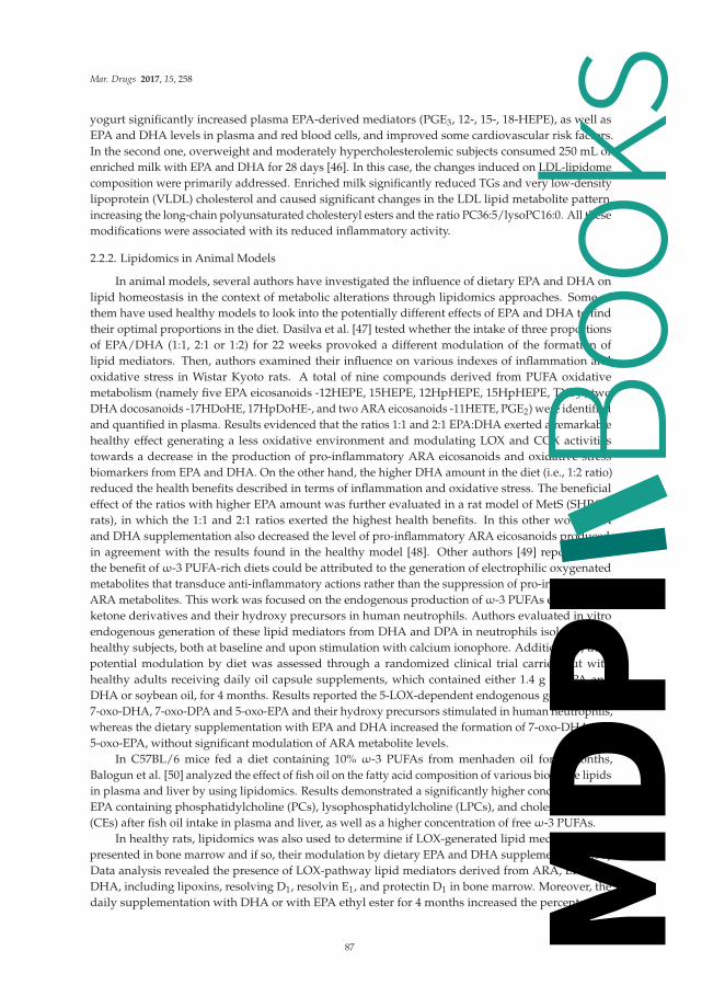
Mar. Drugs 2017, 15, 258
yogurt significantly increased plasma EPA-derived mediators (PGE3, 12-, 15-, 18-HEPE), as well asEPA and DHA levels in plasma and red blood cells, and improved some cardiovascular risk factors.In the second one, overweight and moderately hypercholesterolemic subjects consumed 250 mL ofenriched milk with EPA and DHA for 28 days [46]. In this case, the changes induced on LDL-lipidomecomposition were primarily addressed. Enriched milk significantly reduced TGs and very low-densitylipoprotein (VLDL) cholesterol and caused significant changes in the LDL lipid metabolite pattern,increasing the long-chain polyunsaturated cholesteryl esters and the ratio PC36:5/lysoPC16:0. All thesemodifications were associated with its reduced inflammatory activity.
2.2.2. Lipidomics in Animal Models
In animal models, several authors have investigated the influence of dietary EPA and DHA onlipid homeostasis in the context of metabolic alterations through lipidomics approaches. Some ofthem have used healthy models to look into the potentially different effects of EPA and DHA to findtheir optimal proportions in the diet. Dasilva et al. [47] tested whether the intake of three proportionsof EPA/DHA (1:1, 2:1 or 1:2) for 22 weeks provoked a different modulation of the formation oflipid mediators. Then, authors examined their influence on various indexes of inflammation andoxidative stress in Wistar Kyoto rats. A total of nine compounds derived from PUFA oxidativemetabolism (namely five EPA eicosanoids -12HEPE, 15HEPE, 12HpHEPE, 15HpHEPE, TXB3-, twoDHA docosanoids -17HDoHE, 17HpDoHE-, and two ARA eicosanoids -11HETE, PGE2) were identifiedand quantified in plasma. Results evidenced that the ratios 1:1 and 2:1 EPA:DHA exerted a remarkablehealthy effect generating a less oxidative environment and modulating LOX and COX activitiestowards a decrease in the production of pro-inflammatory ARA eicosanoids and oxidative stressbiomarkers from EPA and DHA. On the other hand, the higher DHA amount in the diet (i.e., 1:2 ratio)reduced the health benefits described in terms of inflammation and oxidative stress. The beneficialeffect of the ratios with higher EPA amount was further evaluated in a rat model of MetS (SHROBrats), in which the 1:1 and 2:1 ratios exerted the highest health benefits. In this other work, EPAand DHA supplementation also decreased the level of pro-inflammatory ARA eicosanoids produced,in agreement with the results found in the healthy model [48]. Other authors [49] reported thatthe benefit of ω-3 PUFA-rich diets could be attributed to the generation of electrophilic oxygenatedmetabolites that transduce anti-inflammatory actions rather than the suppression of pro-inflammatoryARA metabolites. This work was focused on the endogenous production of ω-3 PUFAs electrophilicketone derivatives and their hydroxy precursors in human neutrophils. Authors evaluated in vitroendogenous generation of these lipid mediators from DHA and DPA in neutrophils isolated fromhealthy subjects, both at baseline and upon stimulation with calcium ionophore. Additionally, theirpotential modulation by diet was assessed through a randomized clinical trial carried out withhealthy adults receiving daily oil capsule supplements, which contained either 1.4 g of EPA andDHA or soybean oil, for 4 months. Results reported the 5-LOX-dependent endogenous generation of7-oxo-DHA, 7-oxo-DPA and 5-oxo-EPA and their hydroxy precursors stimulated in human neutrophils,whereas the dietary supplementation with EPA and DHA increased the formation of 7-oxo-DHA and5-oxo-EPA, without significant modulation of ARA metabolite levels.
In C57BL/6 mice fed a diet containing 10% ω-3 PUFAs from menhaden oil for 4 months,Balogun et al. [50] analyzed the effect of fish oil on the fatty acid composition of various bioactive lipidsin plasma and liver by using lipidomics. Results demonstrated a significantly higher concentration ofEPA containing phosphatidylcholine (PCs), lysophosphatidylcholine (LPCs), and cholesteryl esters(CEs) after fish oil intake in plasma and liver, as well as a higher concentration of free ω-3 PUFAs.
In healthy rats, lipidomics was also used to determine if LOX-generated lipid mediators werepresented in bone marrow and if so, their modulation by dietary EPA and DHA supplementation [51].Data analysis revealed the presence of LOX-pathway lipid mediators derived from ARA, EPA andDHA, including lipoxins, resolving D1, resolvin E1, and protectin D1 in bone marrow. Moreover, thedaily supplementation with DHA or with EPA ethyl ester for 4 months increased the percentage of
87
Books
MDPI

Mar. Drugs 2017, 15, 258
DHA and EPA in bone marrow, and the proportion of LOX mediators biosynthesized from DHA orEPA, respectively. Given the potent bioactivities of the lipoxins, resolvins and protectins, their presenceand changes in their profile found after EPA and DHA ethyl ester supplementation may be of interestin bone marrow function and as a potential source of these mediators in vivo.
Several articles have used lipidomics to assay the protective effects of EPA and DHA againstmetabolic alterations induced by unhealthy diets. In rats, Taltavull et al. [52] investigated howsupplementation of a high fat and sucrose diet with both EPA and DHA modified the hepatic ceramideprofile triggered by the unhealthy diet in a dietary intervention of 24 weeks. Authors found that ω-3PUFAs reduced total liver ceramide content and altered ceramide profiles in pre-diabetic rats. They alsoobserved a significant positive linear correlation between long chain ceramide 18:1/18:0 and the HOMAindex, and negative between very long chain ceramides 18:1/24:0 and 18:1/20:0 and plasma insulinlevels and the HOMA index. Overall, these data may help explain the protective action of ω-3 PUFAsagainst liver insulin resistance induced by diet. Caesar et al. [53] used lipidomics tools to evaluate theregulation of lipid composition in mice liver and serum by dietary fish oil as compared to lard oil, butconsidering their interaction with gut microbiota. After 11 weeks, menhaden fish oil supplementationinduced significant changes in abundance of most lipid classes. The gut microbiota affected lipidcomposition by increasing hepatic levels of cholesterol and cholesteryl esters in mice fed high-fat larddiet but not in mice fed high-fat fish oil diet. These results highlighted that the regulation of hepaticcholesterol metabolism induced by gut microbiota was dependent on dietary lipid composition.
Animal models of obesity have also been used to investigate the modulation of lipid metabolismin the adipose tissue. Kuda et al. [54] identified cells producing lipid mediators in epididymalWAT of mice fed for 5 weeks obesogenic high-fat diet, which was supplemented or not with EPAand DHA. Results demonstrated selectively increased levels of anti-inflammatory lipid mediators inWAT in response to ω-3, reflecting either their association with adipocytes (endocannabinoid-relatedNdocosahexaenoylethanolamine) or with stromal vascular cells (pro-resolving lipid mediator protectinD1). In parallel, tissue levels of obesity-associated pro-inflammatory endocannabinoids were suppressed.Moreover, they found that adipose tissue macrophages (ATMs) were not the main producers ofprotectin D1 and that ω-3 PUFAs lowered lipid load in ATMs while promoting their less-inflammatoryphenotype. Besides these specific roles of various cell types in WAT, the kind of fat depots seemedto be also critical. In the abdominal (epididymal) fat but not in other fat depots, Flachs et al. [55]found a synergistic induction of the mitochondrial oxidative capacity and lipid catabolism aftercombining ω-3 PUFAs intake and caloric restriction in high-fat fed mice. This combination resulted inan increased oxidation of metabolic fuels in the absence of mitochondrial uncoupling, while low-gradeinflammation was suppressed, reflecting changes in tissue levels of anti-inflammatory lipid mediators,namely 15-deoxy-Δ(12,15)-prostaglandin J2 and protectin D1.
Lipidomics analysis [56] also revealed that ω-3 PUFAs supplementation could alleviate hepaticsteatosis in ob/ob mice, an obesity model of insulin resistance and fatty liver disease. ω-3 PUFAsinhibited the formation of ω-6 PUFAs derived eicosanoids while triggering the formation of ω-3PUFAs derived resolvins and protectins. Moreover, representative members of these lipid mediators,namely resolvin E1 and protectin D1, mimicked the insulin sensitizing and anti-steatotic effects of ω-3PUFAs and induced adiponectin expression to a similar extent that the antidiabetic drug rosiglitazone.These findings uncovered beneficial actions of ω-3 PUFAs and their bioactive lipid derivatives inpreventing obesity-induced insulin resistance and hepatic steatosis. Similar conclusions were obtainedby Kalish et al. [57] who demonstrated in a mouse model of steatosis that parental nutrition with fishoil-based lipid emulsions was associated with the production of anti-inflammatory and pro-resolvinglipid mediators. The preventive effect of EPA and DHA against necroinflammatory injury in liver wasalso investigated in mice fed high saturated fat diets containing either DHA or both EPA and DHAfor 5 weeks [58]. Both marine ω-3-rich diets induced an increased hepatic formation of DHA-derivedlipid mediators (i.e., 17S-hydroxy-DHA (17S-HDHA) and protectin D1), which was correlated withsignificant protection of liver injury. This work reported a potential role for DHA-derived products,
88
Books
MDPI

Mar. Drugs 2017, 15, 258
specifically 17SHDHA and protectin D1, in mediating the protective effects of dietary DHA againstnecroinflammatory liver injury.
The potential role of fish oil to prevent glomerulosclerosis in a rat model of MetS (JCR:LA-cp rats)via renal eicosanoid metabolism and lipidomics analysis was addressed by Aukema et al. [59]. MetS ratswere supplemented with 5% or 10% fish oil for 16 weeks. Dietary fish oil reduced glomerulosclerosisand albuminuria and the 11- and 12-HETE levels, as well as other (5-, 9- and 15-) HETE. Also, fish oilreduced endogenous renal levels of 6-keto PGF1α (PGI2 metabolite), thromboxane B2 (TXB2), PGF2α,and PGD2 by approximately 60% in rats fed 10% fish oil as compared to untreated MetS rats. Whereasin rats fed 5% fish oil, TXB2 decreased in 250% and PGF2a in 241%. These results suggested that dietaryfish oil might improve dysfunctional renal eicosanoid metabolism associated with kidney damageduring conditions of the MetS.
The impact of DHA supplementation on the profiles of PUFA oxygenated metabolites and theircontribution to atherosclerosis prevention were investigated by Gladine et al. [60]. The study wasconducted with atherosclerosis prone mice which received increasing doses of DHA (0%, 0.1%, 1%or 2% of energy) during 20 weeks. Targeted lipidomics analysis determined a significant modulationof EPA and DHA and their respective oxygenated metabolites in plasma and liver. Remarkably,hepatic F4-neuroprostanes were strongly correlated with the hepatic DHA level. The hepaticlevel of F4-neuroprostanes was the variable most negatively correlated with the plaque extent andplasma EPA-derived diols. Thus, oxygenated ω-3 PUFAs derivatives, particularly F4-neuroprostanes,were revealed as potential biomarkers of DHA-associated_atherosclerosis prevention which mightcontribute to the anti-atherogenic effects of DHA.
It is well known that oils from marine organisms have a different fatty acid composition anddiffer in their molecular composition. Fish oil has a high content of EPA and DHA mostly esterified toTGs, while in krill oil these fatty acids are mainly esterified to PLs. Considering that, Skorve et al. [61]studied the effects of these oils on the lipid content and fatty acid distribution in the various lipidclasses in liver and brain of mice. After 6 weeks of feeding a high-fat diet supplemented with fish oilor with krill oil, shotgun lipidomics showed that in both fish and krill oil fed mice, the TGs content inthe liver was more than doubled compared to control mice. The fatty acid distribution was affected bythe oils in both liver and brain with a decrease in the abundance of LA and ARA, and an increase inEPA and DHA in both study groups. LA decreased in all lipid classes in the fish oil group but withonly minor changes in the krill oil one. Differences were especially evident in some of the minor lipidclasses associated with inflammation and insulin resistance. Ceramides and DGs were decreased, andcholesteryl esters increased in the liver of the krill oil group, while plasmalogens were diminished inthe fish oil group. In the brain, DGs were decreased, more by krill than fish oil, while ceramides andlactosylceramides were increased, more by fish than krill oil. Changes in hepatic sphingolipids andARA fatty acid levels were higher in the krill oil group than in the fish oil one. These changes wereconsistent with a hypothesis that krill oil may have a stronger anti-inflammatory action and enhanceinsulin sensitivity more potently than fish oil.
2.2.3. Lipidomics in Cell Cultures
Finally, some authors have applied lipidomics approaches in cell cultures and in vitro assays todeeply analyze the metabolic effect of dietary marine ω-3 PUFAs. Polus et al. demonstrated that theaddition of EPA during differentiation of human subcutaneous adipose tissue stromal vascular fractioncells induced the formation of small lipid droplets and reduced the production of pro-inflammatorymediators in adipose tissue in comparison to ARA addition. These changes were the consequence ofthe production of anti-inflammatory eicosanoids derived from EPA [62].
The preventive effect of DHA at physiological doses against insulin resistance was investigated inC2C12 myotubes exposed to palmitate. DHA decreased protein kinase C activation, restored cellularacylcarnitine profile, insulin-dependent AKT phosphorylation and glucose uptake. Results showed thatDHA participated in the regulation of muscle lipid and glucose metabolism by preventing lipotoxicity,
89
Books
MDPI

Mar. Drugs 2017, 15, 258
inflammation and insulin resistance in skeletal muscle [63]. Likewise, in in vitro experiments,Ting et al. [64] investigated structural changes in cardiolipins after DHA or EPA supplementationand compared them to ARA treatment, using H9c2 cardiac myoblast as a cell model. Among the116 cardiolipin species with 36 distinct mass identified, the three PUFAs treatments differentiallyperturbed the fatty acyl chain compositions in the mitochondrial of the H9c2 cardiac myoblast,suggesting that both mitochondrial membrane composition and function were susceptible to exogenouslipids. Additionally, DHA supplementation correlated with an elevation of less unsaturated and ω-3cardiolipin species, which appeared to be a minor effect on EPA but not on ARA.
2.2.4. Marine Lipids and Other Bioactive Compounds Assayed by Lipidomics
Several articles have also used lipidomics to study the combined action of ω-3 PUFAswith other bioactive compounds which have shown potential benefits in subjects susceptible tocardiovascular diseases.
The protective effects of fatty fish consumption on the progression of insulin resistance weretested in combination with other products with recognized effect on glucose metabolism (whole grainand low postprandial insulin response grain products, and bilberries) in a clinical trial [65]. Plasmalipidomic profiles of people with impaired glucose metabolism and with at least two other features ofthe MetS were evaluated after 12 weeks of dietary intervention. Among the 364 characterized lipids inplasma, 25 changed significantly in the treated group, including multiple TGs incorporating the longchain ω-3 PUFAs. These results were supported by biochemical data and suggested an improvementof glucose metabolism and a beneficial effect in preventing type 2 diabetes in population groups atconsiderably higher risk of suffering it.
Other three articles found in literature have employed lipidomics tools to study the combinedaction of ω-3 PUFAs with other bioactive compounds which have shown potential benefits insubjects suffering CVD or at considerably high risk. The first one [66] was designed to assay thecombined effect of L-alanyl-L-glutamine and fish oil supplementation for 3 months on skeletal musclefunction and metabolism in patients with chronic heart failure. Patients were randomized to eitherL-alanyl-L-glutamine and PUFAs or placebo (safflower oil and milk powder). Regular uptake of thebioactive compounds led to the expected increase in unsaturated fatty acids. Moreover, the lipidomicanalysis revealed a decrease in circulating levels in total ceramides and two ceramide subspecies (C22:1and C20:1) induced by supplements at 4 weeks, which was not detectable in samples at 3 months.In another study, the combined effect of ω-3 fatty acids with Coenzyme Q10 was evaluated on plasmalipid mediators profile in patients with chronic kidney disease (CKD), which are highly predisposedto suffer CVD, partially due to their chronic inflammation [67]. Patients received a daily dose ofω-3 PUFAs, Coenzyme Q10 (CoQ), or both supplements for 8 weeks. Compounds as 18-HEPE,17-HDHA, RvD1, 17R-RvD1, and RvD2 were measured in plasma before and after the intervention.Results showed that ω-3 PUFAs but not CoQ significantly increased plasma levels of the upstreamprecursors of the E and D-series resolvins (18-HEPE and 17-HDHA, respectively) as well as RvD1.This finding may have important implications for limiting ongoing low-grade inflammation in CKD.Finally, Bondía-Pons et al. [68] investigated the effects of ω-3 PUFAs and polyphenol rich diets onplasma and HDL fraction lipidomic profiles in MetS patients. Authors compared the effects of dietscontained low or high ω-3 PUFAs (EPA and DHA) in combination with low or high of polyphenols,resulting in 4 isoenergetic diets, differing in their natural ω-3 PUFAs and polyphenols amount. Authorssuccessfully identified 350 and 293 lipid species in total plasma and HDL fraction samples respectively.Results showed that the two diets high in ω-3 PUFAs highly increased unsaturated long-chain TGsand EPA and DHA-containing PLs levels and decreased levels in total plasma of low unsaturatedPLs, and PCes, LysoPCs, and PCps with ARA in their structure. With regards to HDL, PCs and TGswith DHA or EPA in their structure increased after the consumption of high ω-3 PUFAs diets, whilePCes and PCps with ARA in their structure, and medium-chain PCs decreased. The diet high inboth ω-3 and polyphenols significantly reduced PCs and PEs levels, especially of those alkyl and
90
Books
MDPI

Mar. Drugs 2017, 15, 258
alkenyl ether lipids with 16:0 in their structure as well as saturated and low-unsaturated PCs and PEs.The study found a relevant association among lipidomics data, dietary and clinical/anthropometricvariables. The most remarkable and complex association among variables was observed after theintervention with the diet high in both ω-3 PUFAs and polyphenols. Different types of TGs werepositively or negatively associated with waist circumference, which was positively associated withinsulin levels. Glucose was positively associated with body weight, which was negatively associatedwith EPA. This latter association was only detected after feeding the combined diet, which may meanthat dietary polyphenols interacted with ω-3 PUFAs in the regulation of body weight in MetS subjects.Overall, data reflected different lipid rearrangements after a nutritional intervention with diets rich inω-3 PUFAs and polyphenols in these patients at a high CVD risk.
The promising cooperative effect between fish oil and polyphenols was further analyzedconsidering the ability of polyphenols as antioxidants to potentially ameliorate oxidative damage ofω-3 PUFAs when they are consumed together and to enhance their individual potential effects onmetabolic health through the modulation of fatty acids profiling and the formation of lipid mediators.Dasilva et al. [69] evaluated the effect of diet supplementation with EPA and DHA, grape polyphenolsor both in rats fed either standard or high fat and sucrose diets (a total of eight diets), on theinflammatory response and redox unbalance triggered by these unhealthy diets. Authors analyzedtotal fatty acid composition in the liver, plasma, adipose tissue, erythrocytes as well as circulatingFFA in plasma across experimental groups and calculated fatty acid desaturases (FADs) indexes(stearoyl-CoA desaturases SCD-16 and SCD-18 as well as desaturases Δ4, Δ5, and Δ6). Likewise, atotal of nine compounds derived from PUFA oxidative metabolism (namely five EPA eicosanoids, twoDHA docosanoids and two ARA eicosanoids) were identified and quantified in plasma. Data analysisreflected that the supplementation with fish oil led to an anti-inflammatory situation associated with alower ω-6/ω-3 index in plasma and membranes, a lower production of ARA pro-inflammatory lipidmediators, an up-regulation of desaturases related to EPA and DHA synthesis and a down-regulationof these desaturases to synthesize ARA. However, polyphenols bioactivity was influenced by thebackground diet. In a standard diet, they seemed to modulate enzymes towards an anti-inflammatoryand antioxidant response, and the combination with fish oil down-regulated Δ5D related with ARAsynthesis, decreased COX activity on ARA, enhanced the antioxidant enzymes and decreased total FFAin plasma. Similarly, the combination of both supplements also produced a significant improvement inthe antioxidant balance and oxidative stress in unhealthy diets. However, the efficacy of polyphenols toreduce inflammation was lower when were added to the unhealthy diet, and some pro-inflammatorypathways were found even up-regulated. Therefore, fish oil seemed to be the main responsible for theanti-inflammatory effects observed in the combined group in the unhealthy diet. The combinationof both bioactive supplements may improve the metabolic health in both background diets by actingon inflammation and oxidative stress pathways. A summary of the articles cited in this section of thereview is shown in Table 3.
91
Books
MDPI

Mar. Drugs 2017, 15, 258
Ta
ble
3.
Res
earc
har
ticl
esfo
und
inlit
erat
ure
whi
chus
edlip
idom
ics
toas
say
heal
thm
arin
elip
idef
fect
s.
Re
fere
nce
Ma
rin
eL
ipid
sIn
terv
en
tio
nE
xp
eri
me
nta
lM
od
el
Lip
ido
mic
sT
oo
lsT
arg
et
Lip
ido
me
Ma
inE
ffe
cts
Ott
esta
det
al.,
2012
[34]
0.7
g/da
yEP
Aan
d0.
9g/
day
DH
Afo
r7
wee
ksH
ealt
hyhu
man
sU
PLC
-MS
Plas
ma
Dec
reas
ed23
lipid
sIn
crea
sed
PLs
and
TG
sco
ntai
ning
EPA
and
DH
A
Rud
kow
ska
etal
.,20
13[3
5]1.
9g/
day
EPA
and
1.1
g/da
yD
HA
for
6w
eeks
Hea
lthy
hum
ans
MS
assa
yki
tPl
asm
aIn
crea
sed
glyP
Cs
inun
satu
rate
dFA
Nor
ding
etal
.,20
13[3
6]1.
9g/
day
EPA
and
1.5
g/da
yD
HA
for
6w
eeks
Hea
lthy
hum
ans
HPL
C-G
S-M
SSP
E-LC
-MS/
MS
Plas
ma
Incr
ease
din
corp
orat
ion
ofEP
Aan
dD
HA
into
7lip
idcl
asse
sH
igh
vari
abili
tyin
87lip
idm
edia
tors
mea
sure
d
Mas
etal
.,20
12[3
7]4
gfis
hoi
l/da
y(3
5%EP
Aan
d25
%D
HA
)for
3w
eeks
Hea
lthy
hum
ans
SPE-
LC-M
S/M
SPl
asm
a/se
rum
Mea
sure
dfo
rfir
stti
me
17R
/SH
DH
A,R
vD1,
and
RvD
2co
ncen
trat
ions
RvD
1an
dR
vD2
into
anti
-infl
amm
ator
yan
dpr
o-re
solv
ing
conc
entr
atio
nra
nge
Bard
enet
al.,
2014
[38]
4g
fish
oil/
day
(35%
EPA
and
25%
DH
A)f
or5
days
Hea
lthy
hum
ans
SPE-
LC-M
S/M
SPl
asm
aIn
crea
sed
RvE
1,18
R/S
-HEP
E,17
R/S
-HD
HA
and
14R
/S-H
DH
A
Kee
lan
etal
.,20
15[3
9]3.
7g/
day
(27.
7%EP
Aan
d56
.%D
HA
)fro
m20
preg
nanc
y-w
eek
Hea
lthy
preg
nant
wom
enG
CSP
E-LC
-MS/
MS
Plac
enta
Incr
ease
dD
HA
Incr
ease
d18
-HEP
Ean
d17
-HD
HA
Bard
enet
al.,
2015
[40]
1.4
gEP
A/d
ayan
d1
gD
HA
/day
inth
efo
rmof
trig
lyce
ride
sfo
r3
wee
ks.
Hum
ansu
ffer
ing
met
abol
icsy
ndro
me
SPE-
LC-M
S/M
SPl
asm
aIn
crea
sed
E-se
ries
reso
lvin
sin
Met
Spa
tient
san
dco
ntro
ls,i
nw
hich
also
incr
ease
dD
-ser
ies
reso
lvin
prec
urso
rsan
d14
-HD
HA
Schu
char
dtet
al.,
2014
[41]
1.14
g/da
yD
HA
and
1.56
g/da
yEP
Afo
r12
wee
ksH
yper
lipid
emic
men
SPE-
LC-M
S/M
SPl
asm
aIn
crea
sed
EPA
-der
ived
lipid
med
iato
rsLe
ssin
crea
sed
DH
A-d
eriv
edlip
idm
edia
tors
Polu
set
al.,
2016
[42]
3×(4
30m
gof
DH
Aan
d90
–150
mg
ofEP
A)/
day
for
3m
onth
sO
bese
wom
enG
C-M
SLC
-MS/
MS
Plas
ma
Incr
ease
dpr
o-re
solv
ing
DH
Ade
riva
tive
s
Lank
inen
etal
.,20
09[4
3]Fa
tty
orle
anfis
hfo
r8
wee
ksC
oron
ary
hear
tdi
seas
epa
tien
tsG
C-M
SU
PLC
-ESI
-MS
Plas
ma
Dec
reas
ed59
bioa
ctiv
elip
idsp
ecie
s(c
eram
ides
,lys
oPC
san
dD
Gs)
byfa
tty
fish
Incr
ease
dch
oles
tero
lest
ers
and
spec
ific
long
-cha
inT
Gs
byle
anfis
h
Mid
tbø
etal
.,20
15[4
4]Fa
rmed
salm
onfe
dw
itha
redu
ced
ratio
ofω
-3/ω
-6fo
r10
wee
ksC
57BL
/6Jm
ice
fed
wes
tern
diet
sLC
-MS/
MS
Live
r
Incr
ease
dA
RA
inPL
sIn
crea
sed
cera
mid
esIn
crea
sed
AR
A-d
eriv
edpr
o-in
flam
mat
ory
med
iato
rsD
ecre
ased
lipid
med
iato
rsde
rive
dfr
omEP
Aan
dD
HA
Daw
czyn
skie
tal.,
2013
[45]
3g
ofEP
Aan
dD
HA
(in
1:1
rati
o)/d
ayfo
r10
wee
ksM
ildly
hype
rtri
acyl
glyc
erol
emic
subj
ects
LC-M
S/M
SPl
asm
aR
edbl
ood
cells
Incr
ease
dEP
Aan
dD
HA
leve
lsin
plas
ma
and
red
bloo
dce
llsIn
crea
sed
plas
ma
EPA
-der
ived
med
iato
rs(P
GE 3
,an
d12
-,15
-and
18-H
EPE)
Padr
oet
al.,
2015
[46]
0.37
5EP
Aan
dD
HA
g/da
yfo
r28
days
Ove
rwei
ghta
ndm
oder
atel
yhy
perc
hole
ster
olem
icsu
bjec
ts
LC-M
S/M
SLD
LIn
crea
sed
long
-cha
inpo
lyun
satu
rate
dC
EsIn
crea
sed
rati
oPC
36:5
/lys
oPC
16:0
92
Books
MDPI

Mar. Drugs 2017, 15, 258
Ta
ble
3.
Con
t.
Re
fere
nce
Ma
rin
eL
ipid
sIn
terv
en
tio
nE
xp
eri
me
nta
lM
od
el
Lip
ido
mic
sT
oo
lsT
arg
et
Lip
ido
me
Ma
inE
ffe
cts
Das
ilva
etal
.,20
15[4
7]EP
A:D
HA
1:1
or2:
1or
1:2
for
13w
eeks
Wis
tar
Kyo
tora
tsSP
E-LC
-MS/
MS
Plas
ma
Dec
reas
edpr
o-in
flam
mat
ory
AR
Aei
cosa
noid
sby
1:1
and
2:1
rati
os
Das
ilva
etal
.,20
16[4
8]EP
A:D
HA
1:1
or2:
1or
1:2
for
wee
ksSH
RO
Bra
tsSP
E-LC
-MS/
MS
Plas
ma
Dec
reas
edpr
o-in
flam
mat
ory
AR
Aei
cosa
noid
sby
1:1
and
2:1
rati
os
Cip
ollin
aet
al.,
2014
[49]
1g/
day
EPA
and
0.4
g/da
yD
HA
for
4m
onth
sH
ealt
hyhu
man
sBM
Ere
acti
onBl
ood
neut
roph
ilsIn
crea
sed
7-ox
o-D
HA
and
5-ox
o-EP
A
Balo
gun
etal
.,20
13[5
0]EP
A:D
HA
1:1
for
4m
onth
sC
57BL
/6m
ice
LC-M
SPl
asm
aLi
ver
Incr
ease
dEP
Aco
ntai
ning
PCs,
LPC
s,an
dC
EsIn
crea
sed
free
ω-3
PUFA
s
Poul
sen
etal
.,20
08[5
1]0.
5g
DH
Aor
EPA
ethy
lest
er/k
gbo
dyw
eigh
t/da
y4
mon
ths
Spra
gue–
Daw
ley
rats
LC-M
S/M
SBo
nem
arro
wIn
crea
sed
EPA
and
DH
AIn
crea
sed
LOX
med
iato
rsbi
osyn
thes
ized
from
DH
Aan
dEP
A(l
ipox
ins,
reso
lvin
gD
1,re
solv
inE 1
and
prot
ecti
nD
1)
Talt
avul
leta
l.,20
16[5
2]EP
A/D
HA
(1:1
)for
24w
eeks
Wis
tar
Kyo
tora
tsfe
dH
FHS
diet
GS-
MS
SPE-
LC-M
S/M
SLi
ver
Dec
reas
edto
talc
eram
ides
Dec
reas
edlo
ngch
ain
cera
mid
e18
:1/1
8:0
Incr
ease
dve
rylo
ngch
ain
cera
mid
es18
:1/2
4:0
and
18:1
/20:
0
Cae
sar
etal
.,20
16[5
3]M
enha
den
fish
oil(
25.2
gEP
Aan
d18
.2g
DH
A/1
00g)
for
11w
eeks
C57
BL/6
mic
efe
dH
Fdi
etU
PLC
-MS
Seru
mLi
ver
Inte
ract
ion
with
gutm
icro
biot
ain
crea
sed
hepa
ticle
vels
ofch
oles
tero
land
chol
este
ryle
ster
sby
lard
butn
otby
fish
oil
Kud
aet
al.,
2016
[54]
4.3
mg
EPA
and
14.7
mg
DH
A/g
diet
for
5w
eeks
C57
BL/6
Jmic
efe
dob
esog
enic
HF
diet
SPE-
LC-M
S/M
SW
hite
adip
ose
tiss
ueIn
crea
sed
anti
-infl
amm
ator
ylip
idm
edia
tors
(end
ocan
nabi
noid
-rel
ated
Ndo
cosa
hexa
enoy
leth
anol
amin
e)an
dpr
o-re
solv
ing
lipid
med
iato
rpr
otec
tin
D1
Flac
hset
al.,
2011
[55]
46%
DH
Aan
d14
%EP
Afo
r5
wee
ksM
ice
fed
obes
ogen
icM
Fdi
etLC
-MS/
MS
Whi
tead
ipos
eti
ssue
Incr
ease
dan
ti-in
flam
mat
ory
lipid
med
iato
rs(1
5-de
oxy-
Δ(1
2,15
)-pr
osta
glan
din
J 2an
dpr
otec
tinD
1)in
epid
idym
alfa
t
Gon
zále
z-Pé
riz
etal
.,20
09[5
6]6
g/10
0gω
-3PU
FAs
for
5w
eeks
ob/o
bm
ice
(B6.
VLe
p/J)
SPE-
LC-M
S/M
SLi
ver
Inhi
bite
dfo
rmat
ion
ofω
-6PU
FAs
deri
ved
eico
sano
ids
Indu
ced
form
atio
nof
ω-3
PUFA
sde
rive
dre
solv
ins
and
prot
ecti
ns
Kal
ish
etal
.,20
13[5
7]Pa
rent
alnu
triti
onw
ithfis
hoi
l-bas
edlip
idem
ulsi
ons
C57
BL6/
Jmic
ehi
gh-c
arbo
hydr
ate
diet
LC-M
S/M
SLi
ver
Indu
ced
prod
ucti
onof
anti
-infl
amm
ator
yan
dpr
o-re
solv
ing
lipid
med
iato
rs
Gon
zále
z-Pé
riz
etal
.,20
06[5
8]1.
37%
DH
Aor
1.37
%EP
Aan
dD
HA
for
5w
eeks
129S
2/Sv
PasC
rlm
ice
fed
high
satu
rate
dfa
tdie
tsH
PLC
-GC
/MS
Live
rIn
crea
sed
DH
A-d
eriv
edlip
idm
edia
tors
(17S
-hyd
roxy
-DH
A(1
7S-H
DH
A)a
ndpr
otec
tinD
1by
both
supp
lem
enta
tion
s
Auk
ema
etal
.,20
13[5
9]5%
or10
%fis
hoi
lfor
16w
eeks
JCR
:LA
-cp
rats
LC-M
S/M
SK
idne
yD
ecre
ased
5-,9
-11-
,12-
and
15-H
ETE
Dec
reas
eden
doge
nous
rena
llev
els
of6-
keto
PGF 1
α,T
XB 2
,PG
F 2α
and
PGD
2
Gla
dine
etal
.,20
14[6
0]D
HA
(0%
,0.1
%,1
%or
2%of
ener
gy)f
or20
wee
ksLD
LR−/
−m
ice
GC
-MS
SPE-
LC-M
S/M
SPl
asm
aLi
ver
Incr
ease
dD
HA
Incr
ease
dF4
-neu
ropr
osta
nes
(DH
Ape
roxi
dize
dm
etab
olit
es)
Skor
veet
al.,
2015
[61]
Fish
oilo
rkr
illoi
lfor
6w
eeks
C57
BL/6
Jmic
efe
dH
Fdi
etG
C-M
SU
PLC
-MS/
MS
Live
rBr
ain
Dec
reas
edun
satu
rate
dfa
tty
acid
sby
fish
and
krill
oils
Dec
reas
edce
ram
ides
and
DG
sin
liver
and
brai
nby
krill
oil
Incr
ease
dC
Esby
krill
oili
nliv
erD
ecre
ased
plas
mal
ogen
sby
fish
oili
nliv
erIn
crea
sed
hepa
tic
sphi
ngol
ipid
san
dA
RA
fatt
yac
idle
vels
mor
eby
krill
than
fish
oili
nliv
erIn
crea
sed
cera
mid
esan
dla
ctos
ylce
ram
ides
mor
eby
fish
than
krill
oili
nbr
ain
93
Books
MDPI

Mar. Drugs 2017, 15, 258
Ta
ble
3.
Con
t.
Re
fere
nce
Ma
rin
eL
ipid
sIn
terv
en
tio
nE
xp
eri
me
nta
lM
od
el
Lip
ido
mic
sT
oo
lsT
arg
et
Lip
ido
me
Ma
inE
ffe
cts
Polu
set
al.,
2015
[62]
EPA
Cel
lcul
ture
GS-
MS
LC-M
S/M
S
Hum
ansu
bcut
aneo
usad
ipos
eti
ssue
stro
mal
vasc
ular
frac
tion
cells
Dec
reas
edpr
o-in
flam
mat
ory
med
iato
rsfr
omA
RA
Incr
ease
dan
ti-i
nflam
mat
ory
eico
sano
idfr
omEP
A
Cap
elet
al.,
2015
[63]
DH
AC
ellc
ultu
reG
C-F
IDLC
-MS/
MS
C2C
12m
yotu
bes
Res
tori
ngce
llula
rac
ylca
rnit
ine
profi
le
Ting
etal
.,20
15[6
4]EP
Aor
DH
AC
ellc
ultu
reLC
-MS/
MS
H9c
2ca
rdia
cm
yobl
ast
Elev
atio
nof
less
unsa
tura
ted
andω
-3ca
rdio
lipin
spec
ies
mai
nly
byD
HA
Lank
inen
etal
.,20
11[6
5]Fa
tty
fish
and
othe
rbi
oact
ive
com
poun
dsfo
r12
wee
ksM
etab
olic
synd
rom
epa
tien
tsU
PLC
-ESI
-MS
Plas
ma
25al
tere
dlip
ids,
incl
udin
gm
ulti
ple
TG
sin
corp
orat
ing
the
long
chai
nω
-3PU
FAs
Wu
etal
.,20
15[6
6]ω
-3PU
FA(6
.5g/
day)
and
L-a
lany
l-L
-glu
tam
ine
(8g/
day)
for
3m
onth
sPa
tien
tsw
ith
chro
nic
hear
tfai
lure
LC-M
SPl
asm
aSk
elet
alm
uscl
eIn
crea
sed
upta
keEP
Aan
dD
HA
Dec
reas
edto
talc
eram
ides
and
cera
mid
es22
:1an
d20
:1
Mas
etal
.,20
16[6
7]ω
-3fa
tty
acid
s(4
g),C
oenz
yme
Q10
(CoQ
)(20
0m
g)or
both
for
8w
eeks
Pati
ents
wit
hch
roni
cki
dney
dise
ase
LC-M
S/M
SPl
asm
aIn
crea
sed
8-H
EPE,
17-H
DH
Aan
dR
vD1
byω
-3PU
FAs
Bond
ia-P
ons
etal
.,20
14[6
8]0.
5%or
1.5%
tota
lene
rgy
inta
keEP
Aan
dD
HA
365
mg
or29
00m
gof
poly
phen
ols
Pati
ents
wit
hm
etab
olic
synd
rom
eU
PLC
-QTO
F-M
SPl
asm
aan
dH
DL
frac
tion
Incr
ease
dpl
asm
ahi
ghly
unsa
tura
ted
long
-cha
inT
Gs
and
EPA
and
DH
A-c
onta
inin
gPL
sby
ω-3
diet
sD
ecre
ased
plas
ma
low
unsa
tura
ted
PLs,
PCes
,Lys
oPC
san
dPC
psco
ntai
ning
AR
Aby
ω-3
diet
sIn
crea
sed
PCs
and
TG
sco
ntai
ning
DH
Aor
EPA
byω
-3di
ets
inH
DL
frac
tion
Dec
reas
edPC
esan
dPC
psco
ntai
ning
AR
Aan
dm
ediu
m-c
hain
PCs
byω
-3di
ets
inH
DL
frac
tion
Dec
reas
edPC
san
dPe
s,se
vera
lalk
ylan
dal
keny
leth
erlip
ids
cont
aini
ng16
:0an
dsa
tura
ted
and
low
-uns
atur
ated
PCs
and
PEs
bybo
thω
-3an
dpo
lyph
enol
sdi
et
Das
ilva
etal
.,20
17[6
9]EP
A/D
HA
(1:1
)Gra
pepo
lyph
enol
sfo
r24
wee
ksW
ista
rK
yoto
rats
fed
HFH
Sdi
etor
STD
diet
GS-
MS
SPE-
LC-M
S/M
S
Plas
ma
Live
rA
dipo
seti
ssue
Dec
reas
edω
-6/ω
-3in
dex
inpl
asm
aan
dm
embr
anes
byω
-3di
ets
Dec
reas
edA
RA
pro-
infla
mm
ator
ylip
idm
edia
tors
byω
-3di
ets
Incr
ease
dde
satu
rase
sre
late
dto
EPA
and
DH
Asy
nthe
sis
byω
-3di
ets
Dec
reas
edde
satu
rase
sre
late
dto
AR
Asy
nthe
sis
byω
-3di
ets
Com
bina
tion
ω-3
&po
lyph
enol
sco
oper
ativ
edo
wn-
regu
late
dΔ
5Dre
late
dw
ith
AR
Asy
nthe
sis,
decr
ease
dC
OX
acti
vity
onA
RA
and
tota
lFFA
inpl
asm
ain
toST
Dan
dH
FHS
diet
s
94
Books
MDPI

Mar. Drugs 2017, 15, 258
2.3. Beneficial Effects of Marine Lipids Intake Assayed by Both Proteomics and Lipidomics
The number of research articles which have combined both proteomics and lipidomics approachesto address the beneficial effects of marine lipids in metabolic alterations induced by unhealthy dietintake is scant yet. More articles have used genomics or transcriptomics tools, but gene expression ortranscription is not always correlated with protein levels or activities. The application of proteomicsand lipidomics approaches can help overcome this drawback and identify molecular pathways actuallyaffected by marine lipids. The integration of different omics seems essential to have a complete pictureof how marine lipids affect health. In spite of this observation, only three research articles which havecombined both proteomics and lipidomics are currently available.
The first study was carried out in healthy overweight men with mildly elevated plasma C-reactiveprotein concentrations. Bakker et al. [70] performed a dietary intervention with a mix of severalproducts selected for their evidence-based anti-inflammatory properties. That “anti-inflammatorydietary mix” consisted of fish oil, green tea extract, resveratrol, vitamin E, vitamin C, and tomatoextract. Regarding fish oil, subjects consumed 1200 mg cold water fish oil/day, composed of 380 mgEPA and 260 mg DHA, and 60 mg other ω-3 PUFAs. Authors measured inflammatory and oxidativestress defense markers in plasma and urine. Furthermore, 120 plasma proteins, 274 plasma metabolites(lipids, FFA, and polar compounds) and the transcriptomes of peripheral blood mononuclear cells andadipose tissue were also quantified. Therefore, this study combined proteomics, metabolipidomicsand transcriptomics tools. Plasma adiponectin concentrations increased by 7%, whereas C-reactiveprotein (principal inflammation marker) was unchanged. However, a multitude of subtle changeswas detected by an integrated analysis of the omics data, which indicated modulated inflammationof adipose tissue, improved endothelial function and oxidative stress, and increased liver fatty acidoxidation. Using the same subjects, Pellis et al. [71] determined their postprandial response to theconsumption of a standardized 500 mL high-fat dairy shake by using metabolomics and proteomicstools. During a 6 h time course after Postprandial Challenge Test (PCT), authors quantified severalplasma metabolites, 79 plasma proteins and 7 clinical biochemistry parameters (glucose, insulin,total FFA, total TGs, hsCRP, IL6, and TNFa). Among these, 31 had different responses over timebetween treated and control groups, revealing differences in amino acid metabolism, oxidative stress,inflammation, and endocrine metabolism. Results showed different short-term metabolic responses tothe PCT in subjects previously supplemented with the anti-inflammatory mix compared to the controls.Additional metabolic changes related to the dietary intervention were also detected as compared tonon-perturbed conditions.
In the third research [72] the obesogenic effect of diets heavily enriched in ω-6 PUFAs and poorin ω-3 PUFAs was assayed by combined proteomics and lipidomics. The study was carried outin aging mice, which had previously suffered a myocardial infarction, after 5 months of feedinga ω-6 enriched diet (ω-6:ω-3 442:1 ratio). Plasma proteomic profiling revealed higher VCAM-1,macrophage inflammatory protein-1 and D40 and myeloperoxidase in the ω-6 PUFAs group. Lipidomicanalysis showed higher levels of ARA and 12(S)-HETE and altered levels of inflammation-resolvingenzymes 5-LOX, COX-2, and heme oxygenase-1, which reflected that excess of ω-6 stimulate prolongedneutrophil trafficking and pro-inflammatory lipid mediators after myocardial infarction. Table 4summarizes the articles cited in this section of the review.
95
Books
MDPI

Mar. Drugs 2017, 15, 258
Ta
ble
4.
Res
earc
har
ticl
esfo
und
inlit
erat
ure
whi
chus
edbo
thpr
oteo
mic
san
dlip
idom
ics
toas
say
heal
thm
arin
elip
idef
fect
s.
Refe
ren
ceM
ari
ne
Lip
ids
Inte
rven
tio
nE
xp
eri
men
tal
Mo
del
Pro
teo
mic
san
dL
ipid
om
ics
To
ols
Targ
et
Pro
teo
me
an
dL
ipid
om
eM
ain
Eff
ect
s
Bakk
eret
al.,
2010
[70]
380
mg
EPA
and
260
mg
DH
Aan
dot
her
anti
-infl
amm
ator
yco
mpo
unds
for
5w
eeks
Hea
lthy
over
wei
ghtm
enH
uman
MA
PG
S-M
SLC
-MS/
MS
Plas
ma
Reg
ulat
edpl
asm
apr
otei
nsan
dpl
asm
am
etab
olit
es(l
ipid
s,fr
eefa
tty
acid
s,an
dpo
lar
com
poun
ds)
rela
ted
tom
odul
atio
nof
infla
mm
atio
n,im
prov
eden
doth
elia
lfun
ctio
n,ox
idat
ive
stre
ssan
din
crea
sed
fatt
yac
idox
idat
ion.
Pelli
set
al.,
2012
[71]
Post
pran
dial
resp
onse
inan
ti-i
nflam
mat
ory
mix
-sup
plem
ente
dm
enA
cute
inta
ke
Hea
lthy
over
wei
ghtm
enH
uman
MA
PG
S-M
SPl
asm
a31
regu
late
dpr
otei
nsan
dlip
ids
invo
lved
inam
ino
acid
met
abol
ism
,oxi
dati
vest
ress
,infl
amm
atio
nan
den
docr
ine
met
abol
ism
.
Lópe
zet
al.,
2015
[72]
ω-6
:ω-3
in44
2:1
rati
ofo
r5
mon
ths
Agi
ngC
57BL
/6Jm
ice
prev
ious
lysu
ffer
edm
yoca
rdia
linf
arct
ion
Prot
ein
imm
unob
lot
anal
ysis
LC-M
S/M
SPl
asm
a
Incr
ease
dV
CA
M-1
,mac
roph
age
infla
mm
ator
ypr
otei
n-1,
D40
and
mye
lope
roxi
dase
Incr
ease
dA
RA
and
12(S
)-H
ETE
and
alte
red
leve
lsof
infla
mm
atio
n-re
solv
ing
enzy
mes
5-LO
X,C
OX
-2,a
ndhe
me
oxyg
enas
e-1
96
Books
MDPI

Mar. Drugs 2017, 15, 258
3. Mechanisms behind the Beneficial Effects of Marine Lipids Assayed by Proteomicsand Lipidomics
The information obtained from both proteomics and lipidomics approaches has confirmedmechanisms proposed by genomics and transcriptomics data on the role of marine lipids indiet-induced metabolic diseases. But interestingly, these techniques have also provided new insightssuggesting the modulation of new molecular pathways and proteins.
Lipidomics tools have revealed the potential mechanisms related to the influence exerted on theregulation of lipid profiles in plasma/blood, tissues and membranes and lipid mediator synthesis bymarine lipids in human, animal and cell-model experiments. These lipid profiles and their derivativemetabolites from fatty acids have closely been associated with inflammation, oxidative stress and theendogenous antioxidant system [73,74].
Besides their known effects in decreasing plasma TGs and cholesterol levels, some of thelipidomics studies reported in this review have found a remodeling of the plasma/membrane/tissuelipids into PLs, TGs, lipoproteins and other lipid species of long chain PUFAs, including plasma FFAprofiles [34–36,39,43–48,50–53,60,61,63–66,68,69]. In general, the consumption of marine ω-3 led to areplacement of ARA with EPA and DHA in cell membranes and lipid species presented in plasma,erythrocytes and liver and adipose tissue, but also kidney and muscle. Such replacement provokedthe consequent enrichment on ω-3 long chain PUFAs accompanied by modulation of LOX and COXactivities, which was attributed to a competence mechanism, especially between ARA and EPA. Asa result, the production of lipid mediators was affected. Additionally, the uptake of ω-3 PUFAsmodulated the synthesis de novo of ARA through elongases and desaturases. In fact, lipidomicsdata from the liver, which is in charge of the de novo fatty acids synthesis, confirmed the preferentialsubstrate competition of Δ5D, which controls de novo synthesis of EPA and ARA, for ω-3 PUFAsover ω-6 PUFAs [44,50,53,60,61,69]. Marine lipids also regulated the formation of bioactive lipids suchas ceramides. The level of long chain ceramides associated with insulin resistance was found to bereduced in plasma [43,66] and liver [52,61] together to an increment of the concentration of very longchain ceramides which seemed to be IR protective.
The fatty acids modulation in the liver due to the consumption of marine lipids was reflected in thetotal fatty acids profile of plasma and adipose tissue [34–36,43,45–48,50,53,65,68,69]. Such modulationwas also observed in circulating plasma FFA and the incorporation of fatty acids into erythrocytemembranes [47,48,69]. Fatty acids released from the adipose tissue, which become circulating FFA inplasma revealed the influence of the diet and synthesis de novo in the accumulation of fat in adipocytes.In consequence, the ω6/ω3 ratio in plasma, circulating FFA, membranes and tissues was lower afterfeeding marine lipids. The ω6/ω3 ratio is an excellent clinical marker for cellular inflammation [75]and higher values are correlated with increased prevalence of chronic inflammatory diseases [76].
In agreement with these effects, lipidomics studies focused on the formation of lipid mediatorsfrom marine PUFAs demonstrated that the intake of these lipids promoted the generationof anti-inflammatory and pro-resolving lipid mediators derived from EPA and DHA whiledecreasing pro-inflammatory mediators derived from ARA [37–42,44,45,47–49,51,54–58,60,62,64,67,69].This anti-inflammatory response can be further due to the fact that ω-3 PUFAs, especially EPA, andω-6 PUFAs can compete for the same enzymes, including phospholipases, desaturases, lipoxygenasesand cyclooxygenases, resulting in a higher production of derived metabolites from EPA and DHA thanderived from ARA.
By using genomics and transcriptomics tools, some authors had previously reported thatPUFAs inhibited the nuclear factor kb (NF-κB) and reduced cytokine production because EPA andDHA can inhibit the binding between saturated fats and toll-like receptors of membranes by directcompetition. The binding to saturated fats would activate NF-κB gene transcription factor, whichinduces inflammatory responses through COX and cytokine synthesis [77]. Moreover, although itis not fully known, marine ω-3 PUFAs may also control gene expression by direct interaction withat least another 4 metabolic nuclear receptors: PPAR (peroxisome proliferator activated receptor),
97
Books
MDPI

Mar. Drugs 2017, 15, 258
LXR (liver X receptor), HNF-4α (hepatic nuclear factor 4) and farnesol X receptor (FXR). Likewise,marine PUFAs can reduce the levels of sterol regulatory element binding proteins (SREBPs) and thecarbohydrate response element binding protein (ChREBP) [78]. Therefore, the modulation of EPAand DHA levels and their outcomes revealed by lipidomics approaches may explain their actionsin regulating gene expression. In addition to these findings, the use of proteomics approaches hasconfirmed the influence of EPA and DHA on several pathways modulated by these transcriptionalfactors. Interestingly, proteomics data identified specific proteins with a pivotal role in these pathwayswhich were altered by marine lipids. In this regard, it is necessary to highlight that a direct correlationbetween the level of gene expression and the cellular content of proteins cannot always be found [79].Consequently, proteomics becomes a critical tool for understanding marine lipid actions on cellularmetabolism and for identifying biomarkers of modulation. Proteomics has specifically revealed thatthe influence of marine lipids intake is not limited to regulating cellular protein quantity but alsoprotein quality by controlling the formation of oxidative PTMs on proteins, mainly carbonyl moieties.The control of these oxidative PTMs plays a key role in understanding the mechanism behind thebeneficial effect of marine lipids in decreasing the risk of CVD and type 2 diabetes [80].
In the adipose tissue, aorta and especially liver, proteomics analysis revealed that ω-3 PUFAsfrom fish oil produced an improvement of lipid profiles and lower accumulation of fat through amechanism that involved the down-regulation of proteins participating in lipogenesis and glycolysiswhile causing the up-regulation of proteins involved in fatty acid beta-oxidation. Fish oil also showedan important action on proteins implicated in the urea cycle and protein metabolism. Additionally,EPA and DHA demonstrated to act on insulin signaling by modulating proteins such as proteasomesystem [16,19–24,27,28].
Proteomics also found a substantial up-regulation of the antioxidant system in blood/plasma andthe rest of tissue analyzed. In fact, fish oil induced higher levels of antioxidant enzymes, ameliorating ofendoplasmic oxidative stress and stimulating protein and DNA cellular system repair [14–23,25,27,28].Moreover, EPA and DHA altered protein carbonylation levels of specific liver proteins, demonstratingan additional mechanism of protein regulation by marine lipids, which is particularly interesting in theinvestigation of diseases induced by diet. It is important to point out that the impairment of normalredox homeostasis, and the consequent accumulation of oxidized biomolecules, has been linked tothe onset and/or development of a great variety of diet-induced diseases [81,82]. Fish oil reducedcarbonylation of proteins related to the antioxidant system such as albumin or 3-α-hydroxysteroiddehydrogenase in plasma and liver, respectively. Proteomics findings in liver demonstrated that EPAand DHA improved ammonia detoxification by decreasing carbonylation level of argininosuccinatesynthetase while increasing oxidation of aspartate aminotransferase. Finally, in skeletal muscle, fish oilintake exerted a protection from cellular dysfunction by ameliorating actin carbonylation level [27].
Therefore, proteomics and lipidomics can largely help understand some of the mechanismsbehind the beneficial effect of marine lipids against chronic disease induced by diet. These mechanismsare mainly related to competence from enzymes involved in lipid de novo synthesis and oxidation,modulation of anti-inflammatory and antioxidant pathways as well as protein homeostasis. Data alsoreflected that the effect of dietary marine lipids is closely dependent on their doses, the EPA andDHA ratio, diet components or health status of patients, among others. A schematic representation ofmechanisms and beneficial effects of EPA and DHA intake found by proteomics and lipidomics toolsis shown in Figure 1.
98
Books
MDPI

Mar. Drugs 2017, 15, 258
Figure 1. Schematic representation of mechanisms and beneficial effects of EPA and DHA intake foundby proteomics and lipidomics tools.
4. Concluding Remarks and Final Considerations
Proteomics and lipidomics approaches constitute valuable tools for studying the effects of marinelipids on metabolic health and disease. In spite of their recent application to nutrition research, theseomics have already allowed the identification of numerous metabolic molecules and pathways, tissuesand physiological processes which are modulated by the consumption of marine lipids. These findingsconfirm some of the mechanisms previously suggested by genomics and transcriptomics tools, but theyalso provide new insights revealing the existence of novel mechanisms and target molecules of greatinterest for the growing field of food bioactives and personalized nutrition. Proteomics and lipidomicscontribute to the discovering of new biomarkers of disease and support the optimal design of bothpreventive and palliative nutritional strategies against the pathologies in which marine lipids havepreviously demonstrated their beneficial effects. The progress on the characterization of mechanismsof action of marine lipids at proteomics and lipidomics levels might further identify other diseases inwhich marine lipids can exert a positive influence. Therefore, although the combination of proteomicsand lipidomics approaches is still scarce, it could be the key to understanding the mechanisms involvedin the beneficial effects of marine lipids. Such combination will offer a complete overview of cellularprocess contributing to clarify several controversial facts regarding the in vivo role of marine lipids.
Finally, it should be noted that the enormous complexity of proteomes and lipidomes togetherwith the scant knowledge available have limited the use of omics in the field of marine lipids.This inconvenience is especially significant for lipidomics due to the lack of information for predictingthe number of individual lipid molecules present in an organism. Additionally, it is necessary tomention the high variability of results derived from in vivo experiments as well as other caveats relatedto high instrumental costs or the need for highly qualified staff. The achievements on higher sensitivityand specificity of the modern MS developments, such as imaging MS or top-down approaches,constitute promising tools which can help solve these problems.
Acknowledgments: Spanish Ministry of Science and Innovation is acknowledged for grant AGL2013-49079-C2-1-R.The Consejo Superior de Investigaciones Científicas (CSIC) and the University of Santiago de Compostela(USC) are gratefully acknowledged for the doctoral fellowship to G.D. Language revision by Hannelore Lottis appreciated.
99
Books
MDPI

Mar. Drugs 2017, 15, 258
Author Contributions: L.M. performed search of the literature and wrote the review. I.M. conceived, revisedand corrected the paper. G.D., N.T., M.R. revised and corrected the paper. All authors agreed with the finalsubmitted version.
Conflicts of Interest: The authors declare no conflict of interest.
References
1. Cordain, L.; Eaton, S.B.; Sebastian, A.; Mann, N.; Lindeberg, S.; Watkins, B.A.; O’Keefe, J.H.; Brand-Miller, J.Origins and evolution of the Western diet: Health implications for the 21st century. Am. J. Clin. Nutr. 2005,81, 341–354. [PubMed]
2. Hutcheson, R.; Rocic, P. The metabolic syndrome, oxidative stress, environment, and cardiovascular disease:The great exploration. Exp. Diabetes Res. 2012, 2012, 271028. [CrossRef] [PubMed]
3. Grundy, S.M. Metabolic syndrome pandemic. Arterioscler. Thromb. Vasc. Biol. 2008, 28, 629–636. [CrossRef][PubMed]
4. Eurostat. News Releases, Product Code: 3-24052016-AP, Published on 24 May 2016. Available online:http://ec.europa.eu/eurostat/documents/2995521/7335847/3-24052016-AP-EN.pdf/4dd0a8ad-5950-4425-9364-197a492d3648 (accessed on 17 August 2017).
5. World Health Organization. The Top 10 Causes of Death. Available online: http://www.who.int/mediacentre/factsheets/fs310/en/index1.html (accessed on 17 August 2017).
6. Simopoulos, A.P. Essential fatty acids in health and chronic disease. Am. J. Clin. Nutr. 1999, 70, 560S–569S.[PubMed]
7. Moore, J.B.; Weeks, M.E. Proteomics and systems biology: Current and future applications in the nutritionalsciences. Adv. Nutr. 2011, 2, 355–364. [CrossRef] [PubMed]
8. Ganesh, V.; Hettiarachchy, N.S. Nutriproteomics: A promising tool to link diet and diseases in nutritionalresearch. Biochim. Biophys. Acta BBA Proteins Proteom. 2012, 1824, 1107–1117. [CrossRef] [PubMed]
9. Kussmann, M.; Raymond, F.; Affolter, M. OMICS-driven biomarker discovery in nutrition and health.J. Biotechnol. 2006, 124, 758–787. [CrossRef] [PubMed]
10. Tyers, M.; Mann, M. From genomics to proteomics. Nature 2003, 422, 193–197. [CrossRef] [PubMed]11. Wasinger, V.C.; Cordwell, S.J.; Cerpa-Poljak, A.; Yan, J.X.; Gooley, A.A.; Wilkins, M.R.; Duncan, M.W.;
Harris, R.; Williams, K.L.; Humphery-Smith, I. Progress with gene-product mapping of the Mollicutes:Mycoplasma genitalium. Electrophoresis 1995, 16, 1090–1094. [CrossRef] [PubMed]
12. Wilkins, M.; Gooley, A. Protein Identification in Proteome Projects. In Proteome Research: New Frontiers inFunctional Genomics; Wilkins, M., Williams, K., Appel, R., Hochstrasser, D., Eds.; Springer: Berlin/Heidelberg,Germany, 1997; pp. 35–64.
13. Sauer, S.; Luge, T. Nutriproteomics: Facts, concepts, and perspectives. Proteomics 2015, 15, 997–1013.[CrossRef] [PubMed]
14. Camargo, A.; Rangel-Zúñiga, O.A.; Peña-Orihuela, P.; Marín, C.; Pérez-Martínez, P.; Delgado-Lista, J.;Gutierrez-Mariscal, F.M.; Malagón, M.M.; Roche, H.M.; Tinahones, F.J.; et al. Postprandial changes in theproteome are modulated by dietary fat in patients with metabolic syndrome. J. Nutr. Biochem. 2013, 24,318–324. [CrossRef] [PubMed]
15. Rangel-Zúñiga, O.A.; Camargo, A.; Marín, C.; Peña-Orihuela, P.; Pérez-Martínez, P.; Delgado-Lista, J.;González-Guardia, L.; Yubero-Serrano, E.M.; Tinahones, F.J.; Malagón, M.M.; et al. Proteome from patientswith metabolic syndrome is regulated by quantity and quality of dietary lipids. BMC Genom. 2015, 16, 509.[CrossRef] [PubMed]
16. Jiménez-Gómez, Y.; Cruz-Teno, C.; Rangel-Zúñiga, O.A.; Peinado, J.R.; Pérez-Martínez, P.; Delgado-Lista, J.;García-Ríos, A.; Camargo, A.; Vázquez-Martínez, R.; Ortega-Bellido, M.; et al. Effect of dietary fatmodification on subcutaneous white adipose tissue insulin sensitivity in patients with metabolic syndrome.Mol. Nutr. Food Res. 2014, 58, 2177–2188. [CrossRef] [PubMed]
17. De Roos, B.; Geelen, A.; Ross, K.; Rucklidge, G.; Reid, M.; Duncan, G.; Caslake, M.; Horgan, G.; Brouwer, I.A.Identification of potential serum biomarkers of inflammation and lipid modulation that are altered by fishoil supplementation in healthy volunteers. Proteomics 2008, 8, 1965–1974. [CrossRef] [PubMed]
100
Books
MDPI

Mar. Drugs 2017, 15, 258
18. Burillo, E.; Mateo-Gallego, R.; Cenarro, A.; Fiddyment, S.; Bea, A.M.; Jorge, I.; Vázquez, J.; Civeira, F.Beneficial effects of omega-3 fatty acids in the proteome of high-density lipoprotein proteome. Lipids Health Dis.2012, 11, 116. [CrossRef] [PubMed]
19. Ahmed, A.A.; Balogun, K.A.; Bykova, N.V.; Cheema, S.K. Novel regulatory roles of omega-3 fatty acids inmetabolic pathways: A proteomics approach. Nutr. Metab. 2014, 11, 6. [CrossRef] [PubMed]
20. Wrzesinski, K.; León, I.R.; Kulej, K.; Sprenger, R.R.; Bjørndal, B.; Christensen, B.J.; Berge, R.K.; Jensen, O.N.;Rogowska-Wrzesinska, A. Proteomics identifies molecular networks affected by tetradecylthioacetic acidand fish oil supplemented diets. J. Proteom. 2013, 84, 61–77. [CrossRef] [PubMed]
21. De Roos, B.; Duivenvoorden, I.; Rucklidge, G.; Reid, M.; Ross, K.; Lamers, R.J.; Voshol, P.J.; Havekes, L.M.;Teusink, B. Response of apolipoprotein E*3-Leiden transgenic mice to dietary fatty acids: Combining liverproteomics with physiological data. FASEB J. 2005, 19, 813–815. [CrossRef] [PubMed]
22. Méndez, L.; Ciordia, S.; Fernández, M.S.; Juárez, S.; Ramos, A.; Pazos, M.; Gallardo, J.M.; Torres, J.L.;Nogués, M.R.; Medina, I. Changes in liver proteins of rats fed standard and high-fat and sucrose dietsinduced by fish omega-3 PUFAs and their combination with grape polyphenols according to quantitativeproteomics. J. Nutr. Biochem. 2017, 41, 84–97. [CrossRef] [PubMed]
23. Kalupahana, N.S.; Claycombe, K.; Newman, S.J.; Stewart, T.; Siriwardhana, N.; Matthan, N.; Lichtenstein, A.H.;Moustaid-Moussa, N. Eicosapentaenoic acid prevents and reverses insulin resistance in high-fat diet-inducedobese mice via modulation of adipose tissue inflammation. J. Nutr. 2010, 140, 1915–1922. [CrossRef] [PubMed]
24. Mavrommatis, Y.; Ross, K.; Rucklidge, G.; Reid, M.; Duncan, G.; Gordon, M.J.; Thies, F.; Sneddon, A.;De Roos, B. Intervention with fish oil, but not with docosahexaenoic acid, results in lower levels of hepaticsoluble epoxide hydrolase with time in apoE knockout mice. Br. J. Nutr. 2010, 103, 16–24. [CrossRef][PubMed]
25. Johnson, M.L.; Lalia, A.Z.; Dasari, S.; Pallauf, M.; Fitch, M.; Hellerstein, M.K.; Lanza, I.R. Eicosapentaenoicacid but not docosahexaenoic acid restores skeletal muscle mitochondrial oxidative capacity in old mice.Aging Cell 2015, 14, 734–743. [CrossRef] [PubMed]
26. Richard, D.; Kefi, K.; Barbe, U.; Bausero, P.; Visioli, F. Polyunsaturated fatty acids as antioxidants. Pharmacol. Res.2008, 57, 451–455. [CrossRef] [PubMed]
27. Méndez, L.; Pazos, M.; Gallardo, J.M.; Torres, J.L.; Pérez-Jiménez, J.; Nogués, R.; Romeu, M.; Medina, I.Reduced protein oxidation in Wistar rats supplemented with marine omega-3 PUFAs. Free Radic. Biol. Med.2013, 55, 8–20. [CrossRef] [PubMed]
28. Joumard-Cubizolles, L.; Gladine, C.; Gérard, N.; Chambon, C.; Brachet, P.; Comte, B.; Mazur, A. Proteomicanalysis of aorta of LDLR-/- mice given omega-3 fatty acids reveals modulation of energy metabolism andoxidative stress pathway. Eur. J. Lipid Sci. Technol. 2013, 115, 1492–1498. [CrossRef]
29. Wenk, M.R. The emerging field of lipidomics. Nat. Rev. Drug Discov. 2005, 4, 594–610. [CrossRef] [PubMed]30. Wymann, M.P.; Schneiter, R. Lipid signalling in disease. Nat. Rev. Mol. Cell Biol. 2008, 9, 162–176. [CrossRef]
[PubMed]31. Shearer, G.C.; Harris, W.S.; Pedersen, T.L.; Newman, J.W. Detection of omega-3 oxylipins in human plasma
and response to treatment with omega-3 acid ethyl esters. J. Lipid Res. 2010, 51, 2074–2081. [CrossRef] [PubMed]32. Murakami, M. Lipid mediators in life science. Exp. Anim. 2011, 60, 7–20. [CrossRef] [PubMed]33. Smith, P.K.; Krohn, R.I.; Hermanson, G.T.; Mallia, A.K.; Gartner, F.H.; Provenzano, M.D.; Fujimoto, E.K.;
Goeke, N.M.; Olson, B.J.; Klenk, D.C. Measurement of protein using bicinchoninic acid. Anal. Biochem. 1985,150, 76–85. [CrossRef]
34. Ottestad, I.; Hassani, S.; Borge, G.I.; Kohler, A.; Vogt, G.; Hyötyläinen, T.; Orešic, M.; Brønner, K.W.;Holven, K.B.; Ulven, S.M.; et al. Fish Oil Supplementation Alters the Plasma Lipidomic Profile and IncreasesLong-Chain PUFAs of Phospholipids and Triglycerides in Healthy Subjects. PLoS ONE 2012, 7, e42550.[CrossRef] [PubMed]
35. Rudkowska, I.; Paradis, A.M.; Thifault, E.; Julien, P.; Tchernof, A.; Couture, P.; Lemieux, S.;Barbier, O.; Vohl, M.C. Transcriptomic and metabolomic signatures of an n-3 polyunsaturated fatty acidssupplementation in a normolipidemic/normocholesterolemic Caucasian population. J. Nutr. Biochem. 2013,24, 54–61. [CrossRef] [PubMed]
36. Nording, M.L.; Yang, J.; Georgi, K.; Hegedus Karbowski, C.; German, J.B.; Weiss, R.H.; Hogg, R.J.; Trygg, J.;Hammock, B.D.; Zivkovic, A.M. Individual variation in lipidomic profiles of healthy subjects in response toomega-3 fatty acids. PLoS ONE 2013, 8, e76575. [CrossRef] [PubMed]
101
Books
MDPI

Mar. Drugs 2017, 15, 258
37. Mas, E.; Croft, K.D.; Zahra, P.; Barden, A.; Mori, T.A. Resolvins D1, D2, and other mediators of self-limitedresolution of inflammation in human blood following n-3 fatty acid supplementation. Clin. Chem. 2012, 58,1476. [CrossRef] [PubMed]
38. Barden, A.; Mas, E.; Croft, K.D.; Phillips, M.; Mori, T.A. Short-term n-3 fatty acid supplementation butnot aspirin increases plasma proresolving mediators of inflammation. J. Lipid Res. 2014, 55, 2401–2407.[CrossRef] [PubMed]
39. Keelan, J.A.; Mas, E.; D’Vaz, N.; Dunstan, J.A.; Li, S.; Barden, A.E.; Mark, P.J.; Waddell, B.J.; Prescott, S.L.;Mori, T.A. Effects of maternal n-3 fatty acid supplementation on placental cytokines, pro-resolving lipidmediators and their precursors. Reproduction 2015, 149, 171–178. [CrossRef] [PubMed]
40. Barden, A.E.; Mas, E.; Croft, K.D.; Phillips, M.; Mori, T.A. Specialized proresolving lipid mediators in humanswith the metabolic syndrome after n-3 fatty acids and aspirin. Am. J. Clin. Nutr. 2015, 102, 1357–1364.[CrossRef] [PubMed]
41. Schuchardt, J.P.; Schmidt, S.; Kressel, G.; Willenberg, I.; Hammock, B.D.; Hahn, A.; Schebb, N.H. Modulationof blood oxylipin levels by long-chain omega-3 fatty acid supplementation in hyper- and normolipidemicmen. Prostaglandins Leukot. Essent. Fat. Acids PLEFA 2014, 90, 27–37. [CrossRef] [PubMed]
42. Polus, A.; Zapala, B.; Razny, U.; Gielicz, A.; Kiec-Wilk, B.; Malczewska-Malec, M.; Sanak, M.;Childs, C.E.; Calder, P.C.; Dembinska-Kiec, A. Omega-3 fatty acid supplementation influences the wholeblood transcriptome in women with obesity, associated with pro-resolving lipid mediator production.Biochim. Biophys. Acta BBA Mol. Cell Biol. Lipids 2016, 1861, 1746–1755. [CrossRef] [PubMed]
43. Lankinen, M.; Schwab, U.; Erkkilä, A.; Seppänen-Laakso, T.; Hannila, M.L.; Mussalo, H.; Lehto, S.;Uusitupa, M.; Gylling, H.; Orešic, M. Fatty fish intake decreases lipids related to inflammation and insulinsignaling—A lipidomics approach. PLoS ONE 2009, 4, e5258. [CrossRef] [PubMed]
44. Midtbø, L.K.; Borkowska, A.G.; Bernhard, A.; Rønnevik, A.K.; Lock, E.-J.; Fitzgerald, M.L.; Torstensen, B.E.;Liaset, B.; Brattelid, T.; Pedersen, T.L.; et al. Intake of farmed Atlantic salmon fed soybean oil increaseshepatic levels of arachidonic acid-derived oxylipins and ceramides in mice. J. Nutr. Biochem. 2015, 26,585–595. [CrossRef] [PubMed]
45. Dawczynski, C.; Massey, K.A.; Ness, C.; Kiehntopf, M.; Stepanow, S.; Platzer, M.; Grün, M.; Nicolaou, A.;Jahreis, G. Randomized placebo-controlled intervention with n-3 LC-PUFA-supplemented yoghurt: Effectson circulating eicosanoids and cardiovascular risk factors. Clin. Nutr. 2013, 32, 686–696. [CrossRef] [PubMed]
46. Padro, T.; Vilahur, G.; Sánchez-Hernández, J.; Hernández, M.; Antonijoan, R.M.; Pérez, A.; Badimon, L.Lipidomic changes of LDL in overweight and moderately hypercholesterolemic subjects taking phytosterol-and omega-3-supplemented milk. J. Lipid Res. 2015, 56, 1043–1056. [CrossRef] [PubMed]
47. Dasilva, G.; Pazos, M.; García-Egido, E.; Gallardo, J.M.; Rodríguez, I.; Cela, R.; Medina, I. Healthy effectof different proportions of marine ω-3 PUFAs EPA and DHA supplementation in Wistar rats: Lipidomicbiomarkers of oxidative stress and inflammation. J. Nutr. Biochem. 2015, 26, 1385–1392. [CrossRef] [PubMed]
48. Dasilva, G.; Pazos, M.; García-Egido, E.; Pérez-Jiménez, J.; Torres, J.L.; Giralt, M.; Nogués, M.R.; Medina, I.Lipidomics to analyze the influence of diets with different EPA:DHA ratios in the progression of MetabolicSyndrome using SHROB rats as a model. Food Chem. 2016, 205, 196–203. [CrossRef] [PubMed]
49. Cipollina, C.; Salvatore, S.R.; Muldoon, M.F.; Freeman, B.A.; Schopfer, F.J. Generation and dietary modulationof anti-inflammatory electrophilic omega-3 fatty acid derivatives. PLoS ONE 2014, 9, e94836. [CrossRef][PubMed]
50. Balogun, K.A.; Albert, C.J.; Ford, D.A.; Brown, R.J.; Cheema, S.K. Dietary omega-3 polyunsaturated fattyacids alter the fatty acid composition of hepatic and plasma bioactive lipids in C57BL/6 mice: A lipidomicapproach. PLoS ONE 2013, 8, e82399. [CrossRef] [PubMed]
51. Poulsen, R.C.; Gotlinger, K.H.; Serhan, C.N.; Kruger, M.C. Identification of inflammatory and proresolvinglipid mediators in bone marrow and their lipidomic profiles with ovariectomy and omega-3 intake.Am. J. Hematol. 2008, 83, 437–445. [CrossRef] [PubMed]
52. Taltavull, N.; Ras, R.; Mariné, S.; Romeu, M.; Giralt, M.; Méndez, L.; Medina, I.; Ramos-Romero, S.; Torres, J.L.Protective effects of fish oil on pre-diabetes: A lipidomic analysis of liver ceramides in rats. Food Funct. 2016,7, 3981–3988. [CrossRef] [PubMed]
53. Caesar, R.; Nygren, H.; Orešic, M.; Bäckhed, F. Interaction between dietary lipids and gut microbiota regulateshepatic cholesterol metabolism. J. Lipid Res. 2016, 57, 474–781. [CrossRef] [PubMed]
102
Books
MDPI

Mar. Drugs 2017, 15, 258
54. Kuda, O.; Rombaldova, M.; Janovska, P.; Flachs, P.; Kopecky, J. Cell type-specific modulation of lipidmediator’s formation in murine adipose tissue by omega-3 fatty acids. Biochem. Biophys. Res. Commun. 2016,469, 731–736. [CrossRef] [PubMed]
55. Flachs, P.; Ruhl, R.; Hensler, M.; Janovska, P.; Zouhar, P.; Kus, V.; Macek Jilkova, Z.; Papp, E.; Kuda, O.;Svobodova, M.; et al. Synergistic induction of lipid catabolism and anti-inflammatory lipids in white fatof dietary obese mice in response to calorie restriction and n-3 fatty acids. Diabetologia 2011, 54, 2626–2638.[CrossRef] [PubMed]
56. González-Périz, A.; Horrillo, R.; Ferré, N.; Gronert, K.; Dong, B.; Morán-Salvador, E.; Titos, E.;Martínez-Clemente, M.; López-Parra, M.; Arroyo, V.; et al. Obesity-induced insulin resistance and hepaticsteatosis are alleviated by ω-3 fatty acids: A role for resolvins and protectins. FASEB J. 2009, 23, 1946–1957.[CrossRef] [PubMed]
57. Kalish, B.T.; Le, H.D.; Fitzgerald, J.M.; Wang, S.; Seamon, K.; Gura, K.M.; Gronert, K.; Puder, M. Intravenousfish oil lipid emulsion promotes a shift toward anti-inflammatory proresolving lipid mediators. Am. J.Physiol. Gastrointest. Liver Physiol. 2013, 305, G818–G828. [CrossRef] [PubMed]
58. González-Périz, A.; Planagumà, A.; Gronert, K.; Miquel, R.; López-Parra, M.; Titos, E.; Horrillo, R.; Ferré, N.;Deulofeu, R.; Arroyo, V.; et al. Docosahexaenoic acid (DHA) blunts liver injury by conversion to protectivelipid mediators: Protectin D1 and 17S-hydroxy-DHA. FASEB J. 2006, 20, 2537–2539. [CrossRef] [PubMed]
59. Aukema, H.M.; Lu, J.; Borthwick, F.; Proctor, S.D. Dietary fish oil reduces glomerular injury and elevatedrenal hydroxyeicosatetraenoic acid levels in the JCR:LA-cp rat, a model of the metabolic syndrome. Br. J. Nutr.2013, 110, 11–19. [CrossRef] [PubMed]
60. Gladine, C.; Newman, J.W.; Durand, T.; Pedersen, T.L.; Galano, J.M.; Demougeot, C.; Berdeaux, O.;Pujos-Guillot, E.; Mazur, A.; Comte, B. Lipid profiling following intake of the omega 3 fatty acid DHAidentifies the peroxidized metabolites F4-neuroprostanes as the best predictors of atherosclerosis prevention.PLoS ONE 2014, 9, e89393. [CrossRef] [PubMed]
61. Skorve, J.; Hilvo, M.; Vihervaara, T.; Burri, L.; Bohov, P.; Tillander, V.; Bjørndal, B.; Suoniemi, M.;Laaksonen, R.; Ekroos, K.; et al. Fish oil and krill oil differentially modify the liver and brain lipidome whenfed to mice. Lipids Health Dis. 2015, 14. [CrossRef] [PubMed]
62. Polus, A.; Kiec-Wilk, B.; Razny, U.; Gielicz, A.; Schmitz, G.; Dembinska-Kiec, A. Influence of dietary fattyacids on differentiation of human stromal vascular fraction preadipocytes. Biochim. Biophys. Acta Mol. CellBiol. Lipids 2015, 1851, 1146–1155. [CrossRef] [PubMed]
63. Capel, F.; Acquaviva, C.; Pitois, E.; Laillet, B.; Rigaudière, J.P.; Jouve, C.; Pouyet, C.; Gladine, C.; Comte, B.;Vianey Saban, C.; et al. DHA at nutritional doses restores insulin sensitivity in skeletal muscle by preventinglipotoxicity and inflammation. J. Nutr. Biochem. 2015, 26, 949–959. [CrossRef] [PubMed]
64. Ting, H.-C.; Chao, Y.-J.; Hsu, Y.-H.H. Polyunsaturated fatty acids incorporation into cardiolipin in H9c2cardiac myoblast. J. Nutr. Biochem. 2015, 26, 769–775. [CrossRef] [PubMed]
65. Lankinen, M.; Schwab, U.; Kolehmainen, M.; Paananen, J.; Poutanen, K.; Mykkänen, H.; Seppänen-Laakso, T.;Gylling, H.; Uusitupa, M.; Orešic, M. Whole grain products, fish and bilberries alter glucose and lipidmetabolism in a randomized, controlled trial: The Sysdimet study. PLoS ONE 2011, 6, e22646. [CrossRef][PubMed]
66. Wu, C.; Kato, T.S.; Ji, R.; Zizola, C.; Brunjes, D.L.; Deng, Y.; Akashi, H.; Armstrong, H.F.; Kennel, P.J.;Thomas, T.; et al. Supplementation of L-Alanyl-L-Glutamine and fish oil improves body composition andquality of life in patients with chronic heart failure. Circ. Heart Fail. 2015, 8, 1077–1087. [CrossRef] [PubMed]
67. Mas, E.; Barden, A.; Burke, V.; Beilin, L.J.; Watts, G.F.; Huang, R.C.; Puddey, I.B.; Irish, A.B.; Mori, T.A.A randomized controlled trial of the effects of n-3 fatty acids on resolvins in chronic kidney disease. Clin. Nutr.2016, 35, 331–336. [CrossRef] [PubMed]
68. Bondia-Pons, I.; Pöhö, P.; Bozzetto, L.; Vetrani, C.; Patti, L.; Aura, A.M.; Annuzzi, G.; Hyötyläinen, T.;Rivellese, A.A.; Orešic, M. Isoenergetic diets differing in their n-3 fatty acid and polyphenol content reflectdifferent plasma and HDL-fraction lipidomic profiles in subjects at high cardiovascular risk. Mol. Nutr.Food Res. 2014, 58, 1873–1882. [CrossRef] [PubMed]
69. Dasilva, G.; Pazos, M.; García-Egido, E.; Gallardo, J.M.; Ramos-Romero, S.; Torres, J.L.; Romeu, M.;Nogués, M.-R.; Medina, I. A lipidomic study on the regulation of inflammation and oxidative stress targetedby marine ω-3 PUFA and polyphenols in high-fat high-sucrose diets. J. Nutr. Biochem. 2017, 43, 53–67.[CrossRef] [PubMed]
103
Books
MDPI

Mar. Drugs 2017, 15, 258
70. Bakker, G.C.; van Erk, M.J.; Pellis, L.; Wopereis, S.; Rubingh, C.M.; Cnubben, N.H.; Kooistra, T.;van Ommen, B.; Hendriks, H.F. An antiinflammatory dietary mix modulates inflammation and oxidativeand metabolic stress in overweight men: A nutrigenomics approach. Am. J. Clin. Nutr. 2010, 91, 1044–1059.[CrossRef] [PubMed]
71. Pellis, L.; van Erk, M.J.; van Ommen, B.; Bakker, G.C.M.; Hendriks, H.F.J.; Cnubben, N.H.P.; Kleemann, R.;van Someren, E.P.; Bobeldijk, I.; Rubingh, C.M.; et al. Plasma metabolomics and proteomics profiling after apostprandial challenge reveal subtle diet effects on human metabolic status. Metabolomics 2012, 8, 347–359.[CrossRef] [PubMed]
72. López, E.F.; Kabarowski, J.H.; Ingle, K.A.; Kain, V.; Barnes, S.; Crossman, D.K.; Lindsey, M.L.; Halade, G.V.Obesity superimposed on aging magnifies inflammation and delays the resolving response after myocardialinfarction. Am. J. Physiol. Heart Circ. Physiol. 2015, 308, H269–H280. [CrossRef] [PubMed]
73. Warensjö, E.; Riserus, U.; Vessby, B. Fatty acid composition of serum lipids predicts the development of themetabolic syndrome in men. Diabetologia 2005, 48, 1999–2005. [CrossRef] [PubMed]
74. Filep, J.G. Resolution pathways in inflammation: The devil in the adipose tissues and in the details. Focuson “Diversity of lipid mediators in human adipose tissue depots”. Am. J. Physiol. Cell Physiol. 2013, 304,C1127–C1128. [CrossRef] [PubMed]
75. Sears, B. The Anti-Inflammation Zone; Regan Books: New York, NY, USA, 2005.76. McDaniel, J.C.; Massey, K.; Nicolaou, A. Fish oil supplementation alters levels of lipid mediators of
inflammation in microenvironment of acute human wounds. Wound Repair Regen. 2011, 19, 189–200.[CrossRef] [PubMed]
77. Bouwens, M.; van de Rest, O.; Dellschaft, N.; Bromhaar, M.G.; de Groot, L.C.; Geleijnse, J.M.; Müller, M.;Afman, L.A. Fish-oil supplementation induces antiinflammatory gene expression profiles in human bloodmononuclear cells. Am. J. Clin. Nutr. 2009, 90, 415–424. [CrossRef] [PubMed]
78. Davidson, M.H. Mechanisms for the Hypotriglyceridemic Effect of Marine Omega-3 Fatty Acids. Am. J. Cardiol.2006, 98 (Suppl. 1), 27–33. [CrossRef] [PubMed]
79. Krammer, J.; Digel, M.; Ehehalt, F.; Stremmel, W.; Fullekrug, J.; Ehehalt, R. Overexpression of CD36 andacyl-CoA synthetases FATP2, FATP4 and ACSL1 increases fatty acid uptake in human hepatoma cells. Int. J.Med. Sci. 2011, 8, 599–614. [CrossRef] [PubMed]
80. Ahmed, N.; Dobler, D.; Dean, M.; Thornalley, P.J. Peptide mapping identifies hotspot site of modificationin human serum albumin by methylglyoxal involved in ligand binding and esterase activity. J. Biol. Chem.2005, 280, 5724–5732. [CrossRef] [PubMed]
81. Ruskovska, T.; Bernlohr, D.A. Oxidative stress and protein carbonylation in adipose tissue—Implications forinsulin resistance and diabetes mellitus. J. Proteom. 2013, 92, 323–334. [CrossRef] [PubMed]
82. Keaney, J.F.; Larson, M.G.; Vasan, R.S.; Wilson, P.W.F.; Lipinska, I.; Corey, D.; Massaro, J.M.; Sutherland, P.;Vita, J.A.; Benjamin, E.J. Obesity and Systemic Oxidative Stress: Clinical Correlates of Oxidative Stress inThe Framingham Study. Arterioscler. Thromb. Vasc. Biol. 2003, 23, 434–439. [CrossRef] [PubMed]
© 2017 by the authors. Licensee MDPI, Basel, Switzerland. This article is an open accessarticle distributed under the terms and conditions of the Creative Commons Attribution(CC BY) license (http://creativecommons.org/licenses/by/4.0/).
104
Books
MDPI

marine drugs
Article
Antibacterial Effect of Eicosapentaenoic Acid againstBacillus cereus and Staphylococcus aureus:Killing Kinetics, Selection for Resistance,and Potential Cellular Target
Phuc Nguyen Thien Le 1 and Andrew P. Desbois 2,*
1 School of Biotechnology, International University—Vietnam National University HCMC, Block 6,Linh Trung Ward, Thu Duc District, Ho Chi Minh City 700000, Vietnam; [email protected]
2 Institute of Aquaculture, University of Stirling, Stirling FK9 4LA, UK* Correspondence: [email protected]; Tel.: +44-1786-467894
Received: 18 August 2017; Accepted: 23 October 2017; Published: 1 November 2017
Abstract: Polyunsaturated fatty acids, such as eicosapentaenoic acid (EPA; C20:5n-3), are attractinginterest as possible new topical antibacterial agents, particularly due to their potency and perceivedsafety. However, relatively little is known of the underlying mechanism of antibacterial actionof EPA or whether bacteria can develop resistance quickly against this or similar compounds.Therefore, the aim of this present study was to determine the mechanism of antibacterial actionof EPA and investigate whether bacteria could develop reduced susceptibility to this fatty acidupon repeated exposure. Against two common Gram-positive human pathogens, Bacillus cereus andStaphylococcus aureus, EPA inhibited bacterial growth with a minimum inhibitory concentration of64 mg/L, while minimum bactericidal concentrations were 64 mg/L and 128 mg/L for B. cereus andS. aureus, respectively. Both species were killed completely in EPA at 128 mg/L within 15 min at37 ◦C, while reduced bacterial viability was associated with increased release of 260-nm-absorbingmaterial from the bacterial cells. Taken together, these observations suggest that EPA likely killsB. cereus and S. aureus by disrupting the cell membrane, ultimately leading to cell lysis. Serial passageof the strains in the presence of sub-inhibitory concentrations of EPA did not lead to the emergenceor selection of strains with reduced susceptibility to EPA during 13 passages. This present studyprovides data that may support the development of EPA and other fatty acids as antibacterial agentsfor cosmetic and pharmaceutical applications.
Keywords: antibiotic resistance; antimicrobial; fish oil; free fatty acid; omega-3; wound infections
1. Introduction
The marine-derived polyunsaturated fatty acid (PUFA) eicosapentaenoic acid (EPA; C20:5 n-3) hasantimicrobial properties and there is increasing interest in developing fatty acids as new antibacterialagents, especially given the rise of bacterial pathogens with resistance against existing antibiotics [1–4].Similar to many other PUFAs, EPA exerts potent effects against Gram-positive species, includinghuman pathogens Bacillus cereus and Staphylococcus aureus [3]. S. aureus causes a multitude ofclinical problems from mild skin complaints, such as impetigo, to more serious soft tissue infections,osteomyelitis, and systemic bacteraemia [5]. Meanwhile, B. cereus is a well-known foodborne pathogenthat causes infections of the gastrointestinal tract, but this bacterium is also responsible for severeinfections of the eyes, lungs, cutaneous tissues, and central nervous system [6]. Importantly, bothpathogens can cause serious infections of wounds and surgical sites [5,6] and new effective treatmentoptions are highly desirable.
Mar. Drugs 2017, 15, 334 105 www.mdpi.com/journal/marinedrugs
Books
MDPI

Mar. Drugs 2017, 15, 334
In clinical and cosmetic applications, free fatty acids such as EPA could be applied topicallyto bolster the free fatty acids present naturally on the skin and mucosal surfaces as part of innateimmunity to protect against microbial infection [3,4,7–10]. In addition to antimicrobial activities,EPA exerts beneficial anti-inflammatory actions [11] and has other positive attributes that wouldsupport its development as a new topical antibacterial agent, including wound healing properties [12],potency and perceived safety [1,4,13], and a suspected lack of acquired bacterial resistance mechanismsagainst this and other fatty acids [14]. However, little is known of whether or not bacteria can developresistance quickly against this compound, or the underlying mechanisms of antibacterial action ofEPA [3]. Addressing these knowledge gaps may hasten the development of EPA and other fatty acidsas new topical antibacterial agents [1].
The aim of the present study was to characterize the antibacterial activity of EPA against twoGram-positive pathogens, B. cereus and S. aureus, and investigate whether the bacteria could developreduced susceptibility to this fatty acid upon repeated exposure and determine the possible mechanismof action.
2. Results
The susceptibility of B. cereus NCIMB 9373 and S. aureus Newman to EPA was assessed bybroth micro-dilution according to Clinical and Laboratory Standards Institute protocols [15,16].EPA demonstrated both growth inhibitory and bactericidal activities against B. cereus and S. aureus.The minimum inhibitory concentration (MIC) was 64 mg/L for EPA against both B. cereus and S. aureus,while the minimum bactericidal concentration (MBC) for B. cereus and S. aureus was 64 and 128 mg/L,respectively. In trials to determine kill kinetics at 128 mg/L EPA, no colonies formed by survivingcells were detected after plating 5 × 10⁵ colony forming units (CFU)/mL suspensions of B. cereus andS. aureus in Mueller-Hinton (MH) broth at 15 min or at subsequent sample times (Figure 1). As expected,some cell division occurred in the control suspensions in MH broth during the 4-h incubation (Figure 1).This experiment was repeated for cell suspensions prepared in phosphate-buffered saline (PBS) todetermine whether active bacterial growth was necessary for the killing activity of EPA, but again nocolonies formed by surviving cells were detected within 15 min and there was little change in CFU/mLduring the 4-h incubation in the control suspensions (Figure 1).
(a) (b)
Figure 1. Enumeration of colonies from cell suspensions of (a) Bacillus cereus NCIMB 9373 and(b) Staphylococcus aureus Newman in MH broth (�) or PBS ( ) exposed to eicosapentaenoic acid(EPA) at 128 mg/L compared to cells suspended in MH broth (�) or PBS (�) lacking EPA during 4 hat 37 ◦C, showing that no viable bacterial cells were detected in suspensions exposed to EPA within15 min. The detection limit was 2 log10 CFU/mL (dashed line). Data are geometric mean ± standarderror (not all error bars are visible); n = 4.
106
Books
MDPI

Mar. Drugs 2017, 15, 334
Next, to investigate the possibility to select experimentally for strains with reduced susceptibilityto EPA quickly, B. cereus and S. aureus were serially passaged 13 times in the presence of sub-inhibitoryconcentrations of this fatty acid in the wells of a 96-well microtitre plate. At each sub-passage,the contents of the wells used to inoculate the subsequent cultures were stored at −70 ◦C in a cryogenictube with 15% glycerol (v/v), so that the susceptibility of each passage isolate to EPA could bedetermined by MIC and compared to the parent strains. After passage of the B. cereus strain insub-inhibitory concentrations of EPA, the isolates from each of 13 passages and the parent strainshowed no change in susceptibility to EPA as all isolates had identical MIC and MBC values, indicatingthe lack of selection of B. cereus cells with reduced susceptibility to EPA (Figure 2). Similarly, the MICvalues of the corresponding S. aureus passage isolates were the same as the parent strain, though theMBC value of each passaged isolate (except for the final passage isolate) was lower than the MBC ofthe parent strain (Figure 2). Still, there was no evidence for the selection of S. aureus cells with reducedsusceptibility during repeated exposure to sub-inhibitory concentrations of EPA.
(a) (b)
Figure 2. Minimum inhibitory (solid lines) and bactericidal concentration (dashed lines) values ofthe parent (a) Bacillus cereus NCIMB 9373 and (b) Staphylococcus aureus Newman isolates and isolatescollected from each of 13 serial passages in sub-inhibitory concentrations of eicosapentaenoic acid(EPA), showing that susceptibility of the bacteria to EPA did not reduce during serial passage.
Finally, to determine the mechanism of antibacterial action of EPA, leakage of 260-nm(A260)-absorbing material from the bacterial cells in suspension was quantified after exposure toincreasing concentrations of EPA for 30 min, according to a protocol modified from Carson et al. [17].The detection of A260-absorbing material can indicate membrane perturbation and an increase inmembrane permeability, and these measurements were taken concomitant with bacterial viabilityassessments by plating of the cell suspensions on agar. The bacterial inoculums at the start of incubationwere 1.51 × 10⁹ ± 0.41 × 10⁹ CFU/mL (mean ± standard error) and 1.57 × 10⁹ ± 0.15 × 10⁹ CFU/mLfor B. cereus and S. aureus, respectively, and thus were considerably greater than used in the killingkinetics experiment above. The carrier solvent (ethanol) had little effect on bacterial viabilityand at the greatest concentration of ethanol (2.56%, v/v) the bacteria recovered at 30 min was1.31 × 109 ± 0.10 × 10⁹ CFU/mL and 2.79 × 10⁹ ± 0.05 × 10⁹ for B. cereus and S. aureus, respectively(data not shown). Control incubations in the presence of carrier solvent (ethanol) showed that negligiblequantities of A260-absorbing material were detected in cell-free filtrates (data not shown). However,leakage of A260-absorbing material was detected from B. cereus and S. aureus cell suspensions thathad been incubated in the presence of ≥64 mg/L EPA for 30 min, and greater concentrations of EPA
107
Books
MDPI

Mar. Drugs 2017, 15, 334
led to the detection of greater quantities of A260-absorbing material released from both species ofbacteria (Figure 3). Importantly, the increasing quantities of A260-absorbing material coincided withreductions in viable CFU/mL in the suspensions (Figure 3). Taken together, these observations suggestmembrane disruption and probable cell lysis of the bacterial cells by EPA.
(a) (b)
Figure 3. Enumeration of colonies from cell suspensions of (a) Bacillus cereus NCIMB 9373 and(b) Staphylococcus aureus Newman in PBS (�) and detection of 260-nm (A260)-absorbing material incell-free filtrates ( ) after exposure to increasing concentrations of eicosapentaenoic acid (EPA) during30 min at 37 ◦C, showing that the number of viable cells in suspension reduced concomitant to theamount of A260-absorbing material in cell-free filtrates, indicating likely cell membrane perturbationand cell lysis. The bacterial inoculums at the start of incubation were 1.51 × 10⁹ ± 0.41 × 10⁹ CFU/mL(mean ± standard error) and 1.57 × 10⁹ ± 0.15 × 10⁹ CFU/mL for B. cereus and S. aureus, respectively.The carrier solvent (ethanol) had little effect on bacterial viability and at the greatest concentrationof ethanol (2.56%, v/v) the bacteria recovered at 30 min was 1.31 × 10⁹ ± 0.10 × 10⁹ CFU/mLand 2.79 × 10⁹ ± 0.05 × 10⁹ for B. cereus and S. aureus, respectively (data not shown). Meanwhile,control incubations in the presence of carrier solvent (ethanol) showed that negligible quantities ofA260-absorbing material were detected in cell-free filtrates (data not shown). Data are mean (geometricmean for CFU values) ± standard error (not all error bars are visible); n = 3 for A260 values, n = 2 forCFU/mL determinations (detection limit was 2 log10 CFU/mL); note that the secondary y-axis (A260)scales differ for (a,b).
3. Discussion
EPA is antimicrobial and this property is being exploited in the development of new topicalcosmetics and pharmaceuticals [1–3]; however, relatively little is known for its antibacterialmechanisms or the ease with which it is possible to select for strains with reduced susceptibility.In this present study, EPA was observed to kill rapidly two species of Gram-positive pathogen,probably by causing cell lysis, and there was little evidence for the selection of strains with reducedsusceptibility after 13 passages.
In this present study, EPA inhibited the growth and killed both B. cereus and S. aureus atconcentrations similar to previous reports for these and other Gram-positive species [2,3,18,19].For B. cereus, MIC and MBC values were the same (64 mg/L) and killing was observed in PBS andculture medium within 15 min, indicating that actively dividing cells were not essential for bactericidalaction. In conjunction with evidence of leakage of A260-absorbing material from cells at growthinhibitory and bactericidal concentrations, these data support the likely catastrophic loss of bacterialcell membrane integrity once a concentration threshold of EPA is reached. Meanwhile, for S. aureus,there was a two-fold difference between MIC and MBC values of EPA (64 and 128 mg/L, respectively)and EPA killed cells within 15 min, which is consistent with previous reports [2,18]. Similar to B. cereus,there was no evidence of the need for actively dividing cells to exert antibacterial action, as EPA killed
108
Books
MDPI

Mar. Drugs 2017, 15, 334
S. aureus equally as effectively in PBS and MH broth. Notably, the leakage experiment showed that2.10 × 104 CFU/mL of S. aureus survived even at the greatest concentration of EPA (i.e., 512 mg/L)despite the release of A260-absorbing material being four times greater at this concentration thanobserved for B. cereus when no surviving cells were detected. This observation may derive fromdifferences in the physiology of the cell membranes of these species, and it could be that EPA causesdifferential effects on membrane permeability of the two bacteria. Moreover, S. aureus may tolerategreater membrane perturbation but remain viable (for longer at least), whereas similar disruption of theB. cereus membrane may be lethal. Indeed, the species-specific variations in the action of EPA observedin this present study are worthy of further investigation. In addition, the composition and quantityof A260-absorbing material (typically nucleic acids [17]) in the cytoplasmic components could differbetween the species and this also would need to be determined. Taken together, these observationssuggest possible concentration-dependent inhibitory and bactericidal mechanisms of action [4],and EPA probably affects cell membrane-associated metabolic systems or increases permeability,ultimately leading to cell lysis. This suggestion is consistent with other studies that have proposed thecell membrane to be the main site of action for antibacterial free fatty acids, with detrimental effectscaused through disruption of vital metabolic processes including cellular respiration [8,20–23] andnutrient uptake [24], or physical disturbance leading to increased permeability, leakage of cellularcomponents [8,23,25–28], and cell lysis (reviewed by Desbois and Smith [14]).
Certain bacteria intrinsically resist the actions of fatty acids and the cell wall of Gram-positivespecies can confer protection against free fatty acids [25]. Some bacteria increase cell wall synthesisor decrease cell surface hydrophobicity on exposure to free fatty acids [29–31]. The presence ofcell-membrane-stabilizing carotenoids that decrease fluidity may also reduce susceptibility to freeunsaturated fatty acids [32,33]. Still, there have been few studies on the selection of bacterial strainswith reduced susceptibility to antibacterial free fatty acids, particularly for Gram-positive species,and, to our knowledge, this present study is the first to perform serial passage to select for any strainswith reduced susceptibility to EPA. Previously, strains of Escherichia coli with reduced susceptibility tocaprylic acid (C8:0) or capric acid (C10:0) were selected successfully after 10 serial transfers on agarcontaining the fatty acids at sub-inhibitory concentrations [34], though the mechanisms underlyingthis phenomenon were not investigated further. Moreover, Petschow et al. [35] reported the isolationof Helicobacter pylori mutants on agar containing 10× MIC of the medium-chain length saturatedfatty acid lauric acid (C12:0) at a rate of 10−8, but the susceptibility of individual colonies wasnot subsequently confirmed. Additionally, Obonyo et al. [36] isolated H. pylori cells that resisteda previously bactericidal concentration of linolenic acid (C18:3 n-3) after just three sub-cultures ina sub-bactericidal concentration of this fatty acid. In contrast, Sun et al. [37] reported no change insusceptibility to lauric acid of H. pylori in response to serial passage six times in sub-inhibitoryconcentrations. Meanwhile, Lacey and Lord [38] were unable to select stable S. aureus strainswith reduced susceptibility to linolenic acid including mutants generated by chemical mutagenesis,and, elsewhere, no S. aureus strains resistant to linoleic acid (C18:2 n-6) were detected in a 5000 clonetransposon insertion library [30]. The opportunity for B. cereus to develop resistance to EPA may bereduced by MIC and MBC values being the same as the concentration inhibiting growth is close tothe concentration having a lethal effect. However, for S. aureus, culturable cells were detected after30 min exposure to EPA at 512 mg/L and there exists a difference between MIC and MBC values,meaning cells are inhibited but not killed at concentrations in this window, thus potentially permittingthe opportunity to select for spontaneous resistant mutants in the surviving population; nevertheless,this was not borne out in practice and serial passage of both bacteria in the presence of sub-inhibitoryconcentrations of EPA did not lead to the emergence or selection of strains with reduced susceptibility.These observations are consistent with the suggestion that it is more difficult to select for resistanceagainst compounds that exert their antibacterial action by acting on multiple cellular targets and thecell membrane. This present study provides some indication of the difficulty in rapidly selecting
109
Books
MDPI

Mar. Drugs 2017, 15, 334
for resistance against EPA, but future investigations will use further bacterial species and strains,undertake more passages, and employ more incremental sub-inhibitory PUFA concentrations.
To conclude, the data in this present study provide support for the development of EPA asa possible new antibacterial agent due to its favorable potency against Gram-positive pathogens,lack of rapid selection of bacterial strains with reduced susceptibility or resistance, and bactericidalmechanism of action.
4. Materials and Methods
4.1. Reagents and Bacteria
EPA (>99% purity) and culture media were purchased from Sigma-Aldrich Ltd. (Poole, Dorset,UK). An EPA stock was made in ethanol (≥99.5%) to 20 mg/mL and stored at −20 ◦C. All othersolutions and media were made with ultrapure deionized water (Option 3; Elga, High Wycombe,Bucks, UK) and were sterilized by autoclaving at 121 ◦C for 15 min or by filtration (polyethersulphone,0.22 μm; Millipore, Watford, UK). S. aureus Newman (gifted by Dr. Angelika Gründling, ImperialCollege London, UK) and B. cereus NCIMB 9373 were resuscitated on MH agar at 37 ◦C from 15%glycerol (v/v) stocks kept at −70 ◦C, and maintained thereafter at 4 ◦C.
4.2. Preparation of Bacterial Suspensions
Typically, bacterial suspensions were prepared from cultures that were inoculated with3–5 colonies into 5 mL MH broth in universal bottles and incubated (37 ◦C, 150 rpm) until lateexponential phase (determined by measuring the absorbance at 600 nm of the culture and comparingto growth curves constructed for each species; approximately 12 h). Next, bacterial cells were harvestedby centrifugation (2000× g, 10 min, 4 ◦C), washed twice with PBS (for 1 L: 8 g of NaCl, 0.2 g of KCl,1.78 g of Na2HPO4·2H2O, 0.24 g of KH2PO4; pH 7.4), and re-suspended to the desired CFU/mL inMH broth or PBS. The CFU/mL of the suspensions were checked by serially diluting 10 μL in PBS(in duplicate), plating on MH agar, incubating overnight (37 ◦C, 24 h), and performing CFU counts.
4.3. Assessing Antibacterial Potency
MIC values were determined by broth micro-dilution according to Clinical and LaboratoryStandards Institute protocol [15]. Briefly, EPA in MH broth was serially diluted in flat-bottomed96-well microtitre plates to final well concentrations of 4, 8, 16, 32, 64, 128, 256, and 512 mg/L (100 μLper well). Five microliters of bacterial suspension at 1 × 10⁷ CFU/mL (prepared in PBS as described inSection 4.2) was used to inoculate each well. Plates were sealed with Parafilm, incubated (37 ◦C, 24 h),and the lowest concentration that prevented bacterial growth visible to the naked eye was determinedto be the MIC. Control wells containing either MH broth only or the volume of ethanol equal to thegreatest volume in a test well were also inoculated; a non-inoculated MH broth well was also included.MBC was determined according to Clinical and Laboratory Standards Institute protocol [16] by plating20 μL from each well showing no visible growth at 24 h on to MH agar and incubating these plates forcolonies to form (37 ◦C, 24 h). Contents from individual wells were spread across a quarter of an agarplate and, as no attempt was made to wash away residual EPA, the effect of carryover preventingthe formation of colonies cannot be dismissed. The lowest concentration of EPA that killed ≥99.9%of the initial inoculum was determined to be the MBC. MIC and MBC values were determined fromduplicated series of wells.
4.4. Killing Kinetics
The times required for EPA at 128 mg/L to kill 5 × 10⁵ CFU/mL from exponential phase culturesof B. cereus and S. aureus were determined. Briefly, 200 μL of EPA at 128 mg/L in MH was preparedand dispensed into an Eppendorf tube, while control wells received an equal volume of carrier solvent(2.56 μL ethanol). Then, 10 μL of a cell suspension (at 1 × 10⁷ CFU/mL and prepared as described in
110
Books
MDPI

Mar. Drugs 2017, 15, 334
Section 4.2) was added to each tube, before the contents were mixed by inversion and then incubatedstatically at 37 ◦C. At 15 min, 30 min, 1 h, 2 h, and 4 h, the CFU/mL in each tube was determinedby serial dilution and plating of 10 μL of suspension as described in Section 4.2. Geometric meansand standard errors of these values were calculated from quadruplicate trials. The experiment wasrepeated in PBS to determine whether active bacterial growth was necessary for the killing activityof EPA.
4.5. Selection of Bacterial Strains with Reduced Susceptibility to EPA
To investigate the possibility to select experimentally for strains with reduced susceptibility toEPA quickly, bacteria were serially passaged in the presence of sub-inhibitory concentrations of thisfatty acid. Briefly, an MIC plate for each bacterium was prepared as described in Section 4.3, except thatthe final well concentrations of EPA were 16, 32, 64, and 128 mg/L. After 24–48 h incubation, 5 μL fromthe well showing growth at the greatest concentration of EPA was used to inoculate the wells of a newMIC plate set up such that the greatest EPA concentration was double that of the concentration in thewell from which the inoculum was taken. Meanwhile, the remainder of the well contents were storedat −70 ◦C in a cryogenic tube with 15% glycerol (v/v). This process was continued for 13 passagesfor each species of bacterium. Finally, the MICs against EPA of each passage isolate and the originalparent strain were determined as described in Section 4.3, once the cultures had been recovered fromcryogenic stocks by culturing on MH agar (37 ◦C, 24 h) as described in Section 4.1.
4.6. Leakage of A260-Absorbing Material from Bacterial Cells
To assess bacterial membrane perturbation and increasing membrane permeability, leakage of260-nm absorbing material was quantified from B. cereus and S. aureus cells in suspension after exposureto increasing concentrations of EPA for 30 min, according to a protocol modified from Carson et al. [17].These measurements were performed concomitant with bacterial viability assessments by plating cellsuspensions on agar. For this, EPA (solubilized in ethanol) was added to PBS and made up to 90 μLto give 16, 32, 64, 128, 256, and 512 mg/L (final volume concentrations after addition of inoculumbelow), while control tubes contained 90 μL PBS with ethanol concentrations corresponding to eachof the respective EPA-containing tubes. Two negative control tubes contained PBS only (no EPA orethanol). For 10 μL from each tube, A260 was determined on a NanoDrop 1000 spectrophotometer(ThermoScientific, Wilmington, DE, USA) and each of these values for the tube solutions served asthe ‘blank’ when readings were to be taken again from each tube at 30 min. After this, to each tubewas added 10 μL of bacterial suspension at 1.5 × 10¹⁰ CFU/mL, which had been prepared in PBS asdescribed in Section 4.2, except that the cells were derived from a 500-mL shake flask culture (37 ◦C,150 rpm, 24 h). Immediately, one of the negative control tubes was sampled to determine CFU/mLand the A260 of sterile-filtered supernatant. CFU/mL was determined by serial dilution and platingof 10 μL of suspension as described in Section 4.2, while the remaining 80 μL of the suspension wascentrifuged (2000× g, 10 min). Then the supernatant was collected and passed through a 0.22-μmsyringe filter (4 mm; Sterlitech, Washington, DC, USA), before the A260 of this filtrate was measuredon the NanoDrop against its respective ‘blank’. Meanwhile, all other tubes were incubated (37 ◦C,30 min). After incubation, the CFU/mL of unfiltered suspension and A260 of filtered suspension weredetermined for each tube as described above, however CFU/mL counts were performed only for theremaining negative control, each EPA-containing tube, and the tube containing the greatest volume ofcarrier solvent. Note that as the control incubations in the presence of carrier solvent (ethanol) showedthe presence of only negligible quantities of A260-absorbing material in cell-free filtrates (data notshown), these A260 values were not subtracted from the A260 value of each respective EPA treatmentreading. This experiment was repeated twice more, though CFU/mL was determined for only one ofthese further trials.
111
Books
MDPI

Mar. Drugs 2017, 15, 334
Acknowledgments: No external funding was received for this study.
Author Contributions: Andrew P. Desbois and Phuc Nguyen Thien Le conceived and designed the experiments;Phuc Nguyen Thien Le performed the experiments; Andrew P. Desbois and Phuc Nguyen Thien Le analyzed thedata; Andrew P. Desbois contributed reagents/materials/analysis tools; Andrew P. Desbois and Phuc NguyenThien Le wrote the paper.
Conflicts of Interest: The authors declare no conflict of interest.
References
1. Desbois, A.P. Potential applications of antimicrobial fatty acids in medicine, agriculture and other industries.Recent Pat. Antiinfect. Drug Discov. 2012, 7, 111–122. [CrossRef] [PubMed]
2. Desbois, A.P.; Lawlor, K.C. Antibacterial activity of long-chain polyunsaturated fatty acids againstPropionibacterium acnes and Staphylococcus aureus. Mar. Drugs 2013, 11, 4544–4557. [CrossRef] [PubMed]
3. Desbois, A.P. Antimicrobial properties of eicosapentaenoic acid (C20:5n-3). In Marine Microbiology: BioactiveCompounds and Biotechnological Applications; Kim, S.K., Ed.; Wiley-VCH Verlag GmbH & Co. KGaA: Hoboken,NJ, USA, 2013; pp. 351–367, ISBN 978-3-527-33327-1.
4. Sun, M.; Zhou, Z.; Dong, J.; Zhang, J.; Xia, Y.; Shu, R. Antibacterial and antibiofilm activitiesof docosahexaenoic acid (DHA) and eicosapentaenoic acid (EPA) against periodontopathic bacteria.Microb. Pathog. 2016, 99, 196–203. [CrossRef] [PubMed]
5. Crossley, K.B.; Jefferson, K.K.; Archer, G.L.; Fowler, V.G. (Eds.) Staphylococci in Human Disease, 2nd ed.;John Wiley & Sons Ltd.: Chichester, UK, 2009; ISBN 978-1-4443-0847-1.
6. Bottone, E.J. Bacillus cereus, a volatile human pathogen. Clin. Microbiol. Rev. 2010, 23, 382–398. [CrossRef][PubMed]
7. Brogden, N.K.; Mehalick, L.; Fischer, C.L.; Wertz, P.W.; Brogden, K.A. The emerging role of peptides andlipids as antimicrobial epidermal barriers and modulators of local inflammation. Skin Pharmacol. Physiol.2012, 25, 167–181. [CrossRef] [PubMed]
8. Parsons, J.B.; Yao, J.; Frank, M.W.; Jackson, P.; Rock, C.O. Membrane disruption by antimicrobial fattyacids releases low-molecular-weight proteins from Staphylococcus aureus. J. Bacteriol. 2012, 194, 5294–5304.[CrossRef] [PubMed]
9. Wang, Y.D.; Peng, K.C.; Wu, J.L.; Chen, J.Y. Transgenic expression of salmon delta-5 and delta-6 desaturasein zebrafish muscle inhibits growth of Vibrio alginolyticus and affects fish immunomodulatory activity.Fish Shellfish Immunol. 2014, 39, 223–230. [CrossRef] [PubMed]
10. Porter, E.; Ma, D.C.; Alvarez, S.; Faull, K.F. Antimicrobial lipids: Emerging effector molecules of innate hostdefense. World J. Immunol. 2015, 5, 51–61. [CrossRef]
11. Mullen, A.; Loscher, C.E.; Roche, H.M. Anti-inflammatory effects of EPA and DHA are dependentupon time and dose-response elements associated with LPS stimulation in THP-1-derived macrophages.J. Nutr. Biochem. 2010, 21, 444–450. [CrossRef] [PubMed]
12. Shingel, K.I.; Faure, M.P.; Azoulay, L.; Roberge, C.; Deckelbaum, R.J. Solid emulsion gel as a vehicle fordelivery of polyunsaturated fatty acids: Implications for tissue repair, dermal angiogenesis and woundhealing. J. Tissue Eng. Regen. Med. 2008, 2, 383–393. [CrossRef] [PubMed]
13. Nakatsuji, T.; Kao, M.C.; Fang, J.Y.; Zouboulis, C.C.; Zhang, L.; Gallo, R.L.; Huang, C.M. Antimicrobialproperty of lauric acid against Propionibacterium acnes: Its therapeutic potential for inflammatory acnevulgaris. J. Investig. Dermatol. 2009, 129, 2480–2488. [CrossRef] [PubMed]
14. Desbois, A.P.; Smith, V.J. Antibacterial free fatty acids: Activities, mechanisms of action and biotechnologicalpotential. Appl. Microbiol. Biotechnol. 2010, 85, 1629–1642. [CrossRef] [PubMed]
15. Clinical and Laboratory Standards Institute. Methods for Dilution Antimicrobial Susceptibility Tests forBacteria That Grow Aerobically; Approved Standard M07-A8; Clinical and Laboratory Standards Institute:Wayne, PA, USA, 2009.
16. Clinical and Laboratory Standards Institute. Methods for Determining Bactericidal Activity of AntimicrobialAgents; Approved Guideline M26-A Vol. 19 No. 18; Clinical and Laboratory Standards Institute:Wayne, PA, USA, 1999.
112
Books
MDPI

Mar. Drugs 2017, 15, 334
17. Carson, C.F.; Mee, B.J.; Riley, T.V. Mechanism of action of Melaleuca alternifolia (tea tree) oil onStaphylococcus aureus determined by time-kill, lysis, leakage, and salt tolerance assays and electron microscopy.Antimicrob. Agents Chemother. 2002, 46, 1914–1920. [CrossRef] [PubMed]
18. Shin, S.Y.; Bajpai, V.K.; Kim, H.R.; Kang, S.C. Antibacterial activity of eicosapentaenoic acid (EPA) againstfoodborne and food spoilage microorganisms. LWT Food Sci. Technol. 2007, 40, 1515–1519. [CrossRef]
19. Desbois, A.P.; Mearns-Spragg, A.; Smith, V.J. A fatty acid from the diatom Phaeodactylum tricornutum isantibacterial against diverse bacteria including multi-resistant Staphylococcus aureus (MRSA). Mar. Biotechnol.2009, 11, 45–52. [CrossRef] [PubMed]
20. Borst, P.; Loos, J.A.; Christ, E.J.; Slater, E.C. Uncoupling activity of long chain fatty acids. Biochim. Biophys. Acta1962, 62, 509–518. [CrossRef]
21. Boyaval, P.; Corre, C.; Dupuis, C.; Roussel, E. Effects of free fatty acids on propionic acid bacteria. Lait 1995,75, 17–29. [CrossRef]
22. Wojtczak, L.; Wieckowski, M.R. The mechanisms of fatty acid induced proton permeability of the innermitochondrial membrane. J. Bioenerg. Biomembr. 1999, 31, 447–455. [CrossRef] [PubMed]
23. Greenway, D.L.A.; Dyke, K.G.H. Mechanism of the inhibitory action of linoleic acid on the growth ofStaphylococcus aureus. J. Gen. Microbiol. 1979, 115, 233–245. [CrossRef] [PubMed]
24. Sheu, C.W.; Freese, E. Effects of fatty acids on growth and envelope proteins of Bacillus subtilis. J. Bacteriol.1972, 111, 516–524. [PubMed]
25. Galbraith, H.; Miller, T.B. Physiological effects of long chain fatty acids on bacterial cells and their protoplasts.J. Appl. Bacteriol. 1973, 36, 647–658. [CrossRef] [PubMed]
26. Thormar, H.; Isaacs, C.E.; Brown, H.R.; Barshatzky, M.R.; Pessolano, T. Inactivation of enveloped viruses andkilling of cells by fatty acids and monoglycerides. Antimicrob. Agents Chemother. 1987, 31, 27–31. [CrossRef][PubMed]
27. Quinn, P.J. Membranes as targets of antimicrobial lipids. In Lipids and Essential Oils as Antimicrobial Agents;Halldor, T., Ed.; John Wiley and Sons, Ltd.: Hoboken, NJ, USA, 2011; pp. 1–24, ISBN 978-0-470-74178-8.
28. Arouri, A.; Mouritsen, O.G. Membrane-perturbing effect of fatty acids and lysolipids. Prog. Lipids Res. 2013,52, 130–140. [CrossRef] [PubMed]
29. Clarke, S.R.; Mohamed, R.; Bian, L.; Routh, A.F.; Kokai-Kun, J.F.; Mond, J.J.; Tarkowski, A.; Foster, S.J.The Staphylococcus aureus surface protein IsdA mediates resistance to innate defenses of human skin.Cell Host Microbe 2007, 1, 199–212. [CrossRef] [PubMed]
30. Kenny, J.G.; Ward, D.; Josefsson, E.; Jonsson, I.M.; Hinds, J.; Rees, H.H.; Lindsay, J.A.; Tarkowski, A.;Horsburgh, M.J. The Staphylococcus aureus response to unsaturated long chain free fatty acids: Survivalmechanisms and virulence implications. PLoS ONE 2009, 4, e4344. [CrossRef] [PubMed]
31. Kohler, T.; Weidenmaier, C.; Peschel, A. Wall teichoic acid protects Staphylococcus aureus against antimicrobialfatty acids from human skin. J. Bacteriol. 2009, 191, 4482–4484. [CrossRef] [PubMed]
32. Chamberlain, N.R.; Mehrtens, B.G.; Xiong, Z.; Kapral, F.A.; Boardman, J.L.; Rearick, J.I. Correlation ofcarotenoid production, decreased membrane fluidity, and resistance to oleic acid killing inStaphylococcus aureus 18Z. Infect. Immun. 1991, 59, 4332–4337. [PubMed]
33. Xiong, Z.; Kapral, F.A. Carotenoid pigment levels in Staphylococcus aureus and sensitivity to oleic acid.J. Med. Microbiol. 1992, 37, 192–194. [CrossRef] [PubMed]
34. Marounek, M.; Skivanová, E.; Rada, V. Susceptibility of Escherichia coli to C2-C18 fatty acids. Folia Microbiol.2003, 48, 731–735. [CrossRef]
35. Petschow, B.W.; Batema, R.P.; Ford, L.L. Susceptibility of Helicobacter pylori to bactericidal properties ofmedium-chain monoglycerides and free fatty acids. Antimicrob. Agents Chemother. 1996, 40, 302–306.[PubMed]
36. Obonyo, M.; Zhang, L.; Thamphiwatana, S.; Pornpattananangkul, D.; Fu, V.; Zhang, L. Antibacterial activitiesof liposomal linolenic acids against antibiotic-resistant Helicobacter pylori. Mol. Pharm. 2012, 9, 2677–2685.[CrossRef] [PubMed]
113
Books
MDPI

Mar. Drugs 2017, 15, 334
37. Sun, C.Q.; O’Connor, C.J.; Roberton, A.M. Antibacterial actions of fatty acids and monoglycerides againstHelicobacter pylori. FEMS Immunol. Med. Microbiol. 2003, 36, 9–17. [CrossRef]
38. Lacey, R.W.; Lord, V.L. Sensitivity of staphylococci to fatty acids: Novel inactivation of linolenic acid byserum. J. Med. Microbiol. 1981, 14, 41–49. [CrossRef] [PubMed]
© 2017 by the authors. Licensee MDPI, Basel, Switzerland. This article is an open accessarticle distributed under the terms and conditions of the Creative Commons Attribution(CC BY) license (http://creativecommons.org/licenses/by/4.0/).
114
Books
MDPI

marine drugs
Article
Lipophilic Fraction of Cultivated Bifurcaria bifurcataR. Ross: Detailed Composition and In VitroProspection of Current ChallengingBioactive Properties
Sónia A. O. Santos 1,* , Stephanie S. Trindade 1, Catia S. D. Oliveira 1, Paula Parreira 2,
Daniela Rosa 2, Maria F. Duarte 2,3 , Isabel Ferreira 4,5, Maria T. Cruz 4,5, Andreia M. Rego 6,
Maria H. Abreu 6, Silvia M. Rocha 7 and Armando J. D. Silvestre 1
1 CICECO—Aveiro Institute of Materials and Department of Chemistry, University of Aveiro,3810-193 Aveiro, Portugal; [email protected] (S.S.T.); [email protected] (C.S.D.O.); [email protected] (A.J.D.S.)
2 Centro de Biotecnologia Agrícola e Agro-Alimentar do Alentejo (CEBAL)/Instituto Politécnico deBeja (IPBeja), 7801-908 Beja, Portugal; [email protected] (P.P.); [email protected] (D.R.);[email protected] (M.F.D.)
3 ICAAM—Instituto de Ciências Agrárias e Ambientais Mediterrânicas, Universidade de Évora,Pólo da Mitra, 7006-554 Évora, Portugal
4 CNC—Center for Neuroscience and Cell Biology, University of Coimbra, 3004-504 Coimbra, Portugal;[email protected] (I.F.); [email protected] (M.T.C.)
5 FFUC—Faculty of Pharmacy, University of Coimbra, 3000-548 Coimbra, Portugal6 ALGAplus—Prod. e Comerc. De Algas e Seus Derivados, Lda., 3830-196 Ílhavo, Portugal;
[email protected] (A.M.R.); [email protected] (M.H.A.)7 QOPNA and Department of Chemistry, University of Aveiro, 3810-193 Aveiro, Portugal; [email protected]* Correspondence: [email protected]; Tel.: +351-234-370-711; Fax: +351-234-370-084
Received: 20 September 2017; Accepted: 23 October 2017; Published: 1 November 2017
Abstract: Macroalgae have been seen as an alternative source of molecules with promisingbioactivities to use in the prevention and treatment of current lifestyle diseases. In this vein,the lipophilic fraction of short-term (three weeks) cultivated Bifurcaria bifurcata was characterizedin detail by gas chromatography–mass spectrometry (GC-MS). B. bifurcata dichloromethane extractwas composed mainly by diterpenes (1892.78 ± 133.97 mg kg−1 dry weight (DW)), followed by fattyacids, both saturated (550.35 ± 15.67 mg kg−1 DW) and unsaturated (397.06 ± 18.44 mg kg−1 DW).Considerable amounts of sterols, namely fucosterol (317.68 ± 26.11 mg kg−1 DW) were also found.In vitro tests demonstrated that the B. bifurcata lipophilic extract show antioxidant, anti-inflammatoryand antibacterial activities (against both Gram-positive and Gram-negative bacteria), using lowextract concentrations (in the order of μg mL−1). Enhancement of antibiotic activity of drug familiesof major clinical importance was observed by the use of B. bifurcata extract. This enhancement ofantibiotic activity depends on the microbial strain and on the antibiotic. This work represents the firstdetailed phytochemical study of the lipophilic extract of B. bifurcata and is, therefore, an importantcontribution for the valorization of B. bifurcata macroalgae, with promising applications in functionalfoods, nutraceutical, cosmetic and biomedical fields.
Keywords: Bifurcaria bifurcata; macroalgae; lipids; lipophilic compounds; diterpenes;antioxidant activity; anti-inflammatory activity; antibacterial activity
1. Introduction
Currently there is a remarkable demand for new bioactive molecules to treat diseases, caused bythe modern lifestyle and the growing exposure to industrial pollutants and environmental toxins.
Mar. Drugs 2017, 15, 340 115 www.mdpi.com/journal/marinedrugs
Books
MDPI

Mar. Drugs 2017, 15, 340
Antioxidant compounds have been widely researched, due to their protective action against damageinduced by oxidative stress, which is the major cause of diseases like cancer, diabetes, asthma,cardiovascular, or neurodegenerative problems [1], and of skin damage. Current lifestyle diseases havebeen also associated with inflammatory processes [2], which have also encouraged the scientificcommunity to search for new molecules with anti-inflammatory capacity. At the same time,the emergence and spread of antibiotic-resistant bacteria strains is a growing public health problem [3],which has led the scientific community to search for new antibacterial substitutes or combination ofdrugs that might overcome this resistance [4].
Nature has always been an important source of molecules with biological properties as such,or after chemical modification [5]. In recent years, marine resources have become alternative sourcesof several value-added compounds [6,7]. Macroalgae, due to their high biological and chemicaldiversity and fast-growing properties, are one of the most explored marine resources [8,9]. In fact,approximately 28.5 million tons of macroalgae (brown, red or green) were produced worldwide in2014, from both capture and aquaculture [10], with an estimated increase of 15%/year. This resourcehas been mainly explored for food consumption or for the production of specialty products, suchas alginic acid, carrageenan, agar, or colorants [7]. In this context, the exploitation of macroalgaehas been gaining increasing attention from several research groups around the world [11–13] andnotably in Portugal [14–18]. In fact, the exploitation of marine resources is also one of the criticalsectors for Portuguese development, given the extent and richness of its Exclusive Economic Zone(EEZ), being included in the Portuguese Research and Innovation Strategy for Smart Specializationfor 2014–2020 [19].
Amongst the macroalgae species (close to 10,000), only a limited number have been the object ofextensive studies due to their unique composition and consequent diversity of biological activitiesor health benefits [20]. Species belonging to the brown Dictyotaceae and Sargassaceae families havebeen studied in detail due to the presence of diterpenes, which have a wide range of biologicalproperties, such as antimicrobial or antitumoral [21]. Among those families, Bifurcaria bifurcata,which can only be found in the northeastern Atlantic coasts, from Morocco to northwestern Ireland,is known to biosynthesize components rarely found in other macroalgae species, namely linearditerpenes [22]. In fact, the presence of these components in B. bifurcata has been widely reported [23–27].In addition, several in vitro effects of B. bifurcata extracts, such as antimicrobial [28], antimitotic [29],or antiproliferative activity [30], have been attributed to the presence of diterpenes, although thisassumption has never been adequately complemented by a detailed study of the composition of thetested extracts. In fact, the relationship between the chemical composition (limited to the fatty acidsprofile) and the biological properties (namely, antimicrobial and antioxidant activity) of B. bifurcataextracts has been addressed in only one study [31].
It has been suggested that the diterpenic composition of B. bifurcata is largely dependent onabiotic factors and especially on its geographic origin [32]. Furthermore, there are no studies regardingthe lipophilic composition (and the diterpenic fraction in particular) of B. bifurcata cultivated innutrient-rich waters (Integrated Multi-Trophic Aquaculture systems (IMTA)) (or even from thePortuguese coast), where, due to the totally different biotic and abiotic growth conditions, substantialdifferences in composition might be anticipated.
In addition, most of the published studies using wild B. bifurcata have neglected the abundanceof diterpenic components, and the few studies that reported their quantification have focused onspecific fractions, not considering the lipophilic fraction as a whole [29,33,34]. The abundance of otherimportant classes of lipophilic compounds are also unknown, namely sterols, long-chain aliphaticalcohols and even fatty acids, for which only a single study can be found [31]. The limited numberof compounds identified in these studies can be overcome using gas chromatography coupled withmass spectrometry (GC-MS), which allows the detailed identification and quantification of complexmixtures of compounds found in the lipophilic extracts [35,36].
116
Books
MDPI

Mar. Drugs 2017, 15, 340
In this study the lipophilic fraction of B. bifurcata cultivated for a short period (three weeks)in a land-based IMTA system was detailed characterized by GC-MS analysis. In addition, in vitroassays were performed to evaluate the antioxidant, antibacterial and anti-inflammatory activities ofthis fraction. Experiments were also conducted aiming to access the synergistic antibiotic–B. bifurcatalipophilic extract bacterial growth inhibition by determining the minimum inhibitory concentration(MIC—the lowest concentration where no bacterial growth was detected).
2. Results and Discussion
2.1. Lipophilic Composition
The dichloromethane extraction yield of B. bifurcata accounted for 3.92 ± 0.09% (w/w), which isquite similar to the n-hexane extraction yield obtained for B. bifurcata from both aquaculture and wildcollected in Portuguese coast [37]. Additionally, the dichloromethane extraction yield was considerablyhigher than those observed previously for other Phaeophyta species [15] or for Chlorophyta orRhodophyta macroalgae [14].
The chemical composition of the B. bifurcata dichloromethane extract was studied in detailby GC-MS analysis. With the exception of the diterpenes family, all the lipophilic componentswere identified as their trimethylsilyl derivatives, after derivatization of the dichloromethaneextract. The identification of the main lipophilic extractives and the corresponding quantification issummarized in Table 1. In general, the extract was mainly composed of diterpenes, free fatty acids(C14–C22), long-chain aliphatic alcohols (C14–C28), and sterols, among other components. To the bestof our knowledge this is the first study reporting the analysis of the lipophilic fraction of B. bifurcataby GC-MS. Recently, Alves et al. [31] analyzed the dichloromethane extract of B. bifurcata by GC withflame ionization detector; however, only the presence of fatty acids was reported.
Table 1. Compounds identified in B. bifurcata dichloromethane extract expressed in mg g−1 of extractand in mg kg−1 of dry macroalgae 1.
Rt (min) Compound mg g−1 of Extract mg kg−1 of Dry Macroalgae
Fatty acids 2 24.18 ± 0.56 947.88 ± 21.94Saturated 14.04 ± 0.40 550.35 ± 15.67
31.0 Tetradecanoic acid 1.94 ± 0.04 76.23 ± 1.6933.5 Pentadecanoic acid 0.24 ± 0.02 9.45 ± 0.6036.0 Hexadecanoic acid 10.11 ± 0.43 396.43 ± 17.0138.2 Heptadecanoic acid 0.30 ± 0.02 11.81 ± 0.7840.5 Octadecanoic acid 1.05 ± 0.09 41.01 ± 3.4944.7 Docosanoic acid 0.24 ± 0.01 9.28 ± 0.4848.5 Tetracosanoic acid 0.16 ± 0.01 6.15 ± 0.20
Unsaturated 10.13 ± 0.47 397.06 ± 18.4435.3 Hexadec-9-enoic acid 1.08 ± 0.07 42.24 ± 2.7939.5 Octadeca-9,12-dienoic acid 1.04 ± 0.05 40.74 ± 2.0839.6 Octadeca-9,12,15-trienoic acid 1.47 ± 0.07 57.81 ± 2.7039.9 Octadec-9-enoic acid 5.13 ± 0.26 200.95 ± 10.3242.9 Eicosa-5,8,11,14,17-pentaenoic acid 0.91 ± 0.07 35.86 ± 2.5743.3 Eicosa-5,8,11-trienoic acid 0.02 ± 0.00 0.75 ± 0.0343.4 Eicosa-11,14-dienoic acid 0.07 ± 0.01 2.86 ± 0.05
ω-hydroxyacids 0.01 ± 0.00 0.47 ± 0.0255.3 22-Hydroxydocosanoic acid 0.01 ± 0.00 0.47 ± 0.02
Long-chain aliphatic alcohols 2 0.43 ± 0.03 17.03 ± 1.2929.0 Tetradecan-1-ol 0.10 ± 0.00 3.78 ± 0.0234.1 Hexadecan-1-ol 0.15 ± 0.01 5.89 ± 0.3838.7 Octadecan-1-ol 0.09 ± 0.00 3.41 ± 0.1958.9 Octacosan-1-ol 0.12 ± 0.00 4.78 ± 0.17
Sterols 2 10.37 ± 0.67 406.45 ± 26.1958.6 Cholesterol 0.19 ± 0.01 7.29 ± 0.2462.0 Desmosterol 1.11 ± 0.03 43.56 ± 1.1063.1 Fucosterol 8.10 ± 0.67 317.68 ± 26.1163.4 Campesterol 0.97 ± 0.04 37.92 ± 1.61
117
Books
MDPI

Mar. Drugs 2017, 15, 340
Table 1. Cont.
Rt (min) Compound mg g−1 of Extract mg kg−1 of Dry Macroalgae
Monoglycerides 2 0.89 ± 0.03 34.99 ± 1.1047.8 1-Monohexadecanoin 0.66 ± 0.03 25.82 ± 1.0150.9 1-Monooctadecenoin 0.13 ± 0.00 5.17 ± 0.1751.5 1-Monoeicosa-tetraenoin 0.10 ± 0.01 4.00 ± 0.31
Diterpenes 3 48.29 ± 3.42 1892.78 ± 133.9726.8 Neophytadiene 1.53 ± 0.08 59.86 ± 3.2932.4 Phytol 0.87 ± 0.07 34.02 ± 3.2534.0 Trans-geranylgeraniol 1.70 ± 0.16 66.53 ± 2.9334.5 6,7,9,10,11,12,14,15-Tetradehydrophytol 2.41 ± 0.88 94.35 ± 6.2936.5 6-Hydroxy-13-oxo-7,7′,10,11-didehydrophytol 16.27 ± 2.41 637.84 ± 34.4140.7 1-Acetyl-10,13-dioxo-6,7,11,11′,14,15-tridehydrophytol 25.51 ± 4.04 1000.17 ± 94.47
Other terpenic compounds 3 0.28 ± 0.00 10.79 ± 0.1826.6 6,10,14-Trimethyl-2-pentadecanone 0.28 ± 0.00 10.79 ± 0.18
Total 84.44 ± 4.14 3309.93 ± 162.25
1 Results are the average of the concordant values obtained from the triplicated extracts each injected twice(less than 5% variation between injections of the same aliquot and between triplicated dichloromethane extracts);2 Compounds identified as trimethylsilyl derivatives; 3 Compounds identified in the method without derivatization.
2.1.1. Diterpenes and Other Terpenoids
Several diterpenes (Figure 1) and other terpenoids were identified in B. bifurcata dichloromethaneextract, namely neophytadiene, phytol, trans-geranylgeraniol, 6,7,9,10,11,12,14,15-tetradehydrophytol,6-hydroxy-13-oxo-7,7′,10,11-didehydrophytol, 1-acetyl-10,13-dioxo-6,7,11,11′,14,15-tridehydrophytoland 6,10,14-trimethyl-2-pentadecanone.
Figure 1. Diterpenes and other major lipophilic compounds detected in B. bifurcata dichloromethane extract.
118
Books
MDPI

Mar. Drugs 2017, 15, 340
The fragmentation patterns of these components depend on the number and position of thedouble bonds, and on the nature of the substituent groups [23,25,26]. As an example, the mass spectraof neophytadiene and trans-geranylgeraniol are presented in Figure 2.
Figure 2. Mass spectra of (a) neophytadiene and (b) trans-geranylgeraniol.
The mass spectrum of neophytadiene presents a molecular ion [M]+ at m/z 278 and majorproduct ions at m/z 43 ([C3H7]+), 57 ([C4H9]+), 68, 82, 95 ([C7H11]+), 109 and 123 ([C9H15]+).Similarly, trans-geranylgeraniol presents a molecular ion [M]+ at m/z 290, and characteristic productions at m/z 69 ([C5H9]+), 81 ([C6H9]+), 121 ([C11H19]+) and 272 (M-H2O]+).
Diterpenes, accounting for 1892.78 ± 133.97 mg kg−1 DW, correspond to about 57% ofthe total amount of lipophilic compounds detected in B. bifurcata extract, with 1-acetyl-10,13-dioxo-6,7,11,11′,14,15-tridehydrophytol and 6-hydroxy-13-oxo-7,7′,10,11-didehydrophytol as the majorcomponents of this family, accounting for 1000.17 ± 94.47 and 637.84 ± 34.41 mg kg−1 DW, respectively.1-acetyl-10,13-dioxo-6,7,11,11′,14,15-tridehydrophytol and 6-hydroxy-13-oxo-7,7′,10,11-didehydrophytolwere already described as constituent of B. bifurcata collected in Brittany (France) [25,26], but noinformation was reported regarding its quantification. Considerable amounts of 6,7,9,10,11,12,14,15-tetradehydrophytol, trans-geranylgeraniol and neophytadiene were also found, and minor amountsof phytol were also detected. 6,7,9,10,11,12,14,15-Tetradehydrophytol was previously identifiedin B. bifurcata collected in Morocco [23], though no information has been provided concerning itsabundance, while, to the best of our knowledge, phytol and neophytadiene are reported, for the firsttime, as constituents of B. bifurcata. In opposition, geranylgeraniol has been one of the most reportedconstituents of this macroalga species [26,29,38]. Additionally, the abundance of this component in theB. bifurcata dichloromethane extract is in the range of those (0.0–1.5 mg g−1 DW) reported in a seasonalvariation study of the same species collected in Brittany (France) [39]. Notwithstanding, only traces ofthis diterpene were found in a geographical variation study of the diterpene composition of B. bifurcatacollected in Spain [33].
Despite the high abundance verified for all the detected diterpenes, these were not reported in someof the studies regarding the seasonal and geographical variation of diterpenes in B. bifurcata [29,33,34].
119
Books
MDPI

Mar. Drugs 2017, 15, 340
Diterpenes have been associated with a wide number of health benefits [40]. In particular,those from macroalgae have been reported to have antioxidant, antimicrobial [41], antifungal [42],anti-viral [43], or antitumoral activities [44].
Minor amounts (10.79 ± 0.18 mg kg−1 DW) of 6,10,14-trimethyl-2-pentadecanone were foundin B. bifurcata dichloromethane extract. This terpenoid and its derivatives have been described to beproduced by macroalgae from Cystoseiraceae family, namely Cystophora moniliformis [45,46].
2.1.2. Fatty Acids
Fatty acids were the second most abundant family of compounds detected in the lipophilicfraction of B. bifurcata, corresponding to 947.88 ± 21.94 mg kg−1 DW. Hexadecanoic acid (Figure 1)was the most abundant fatty acid (396.43 ± 17.01 mg kg−1 DW), followed by tetradecanoic(76.23 ± 1.69 mg kg−1 DW) and octadecanoic (41.01 ± 3.49 mg kg−1 DW) acids. The high abundance ofhexadecanoic and tetradecanoic acid in B. bifurcata was already reported [31], however, in considerablyhigher amounts than those found in this study.
Considerable amounts of unsaturated fatty acids were found in B. bifurcata (397.06± 18.44 mg kg −1 DW),representing about 42% of the total fatty acids. Octadec-9-enoic acid (Figure 1) was the mostabundant unsaturated fatty acid, accounting for 200.95 ± 10.32 mg kg−1 DW, followed by theω-3 octadeca-9,12,15-trienoic acid (57.81 ± 2.70 mg kg−1 DW) and the hexadec-9-enoic acid(42.24 ± 2.79 mg kg−1 DW). Significant amounts of the ω-3 fatty acid eicosa-5,8,11,14,17-pentaenoicacid were also found (35.86 ± 2.57 mg kg−1 DW), despite being 10 times lower than those reportedpreviously by Alves et al. [31] for B. bifurcata collected in Peniche coast, Portugal. Notwithstanding,the high abundance of this component contributes to the low ω-6/ω-3 ratio verified for this macroalga(~0.46), which is considerably lower than the maximum (4:1) recommended for a diet in order to preventthe development of inflammatory processes [47]. Additionally, this ratio is significantly lower thanthose verified for other macroalgae species [15,48], which were considered promising to be incorporatedin healthy diets. The high abundance of such components, widely associated with the prevention ordelay of chronic diseases, such as cancer, cardiovascular, or coronary problems [49], highlights thepotential of valorization of B. bifurcata also in food and/or nutraceutical industry. However, suchexploitation requires that more detailed studies should be performed, namely evaluating their sideeffects and toxicity.
2.1.3. Long-Chain Aliphatic Alcohols
Minor amounts of long-chain aliphatic alcohols (LCAA) were found in B. bifurcata dichloromethaneextract, accounting for 17.03 ± 1.29 mg kg−1 DW. To the best of our knowledge this is the first studyreporting the presence of these components in B. bifurcata. Hexadecan-1-ol was the most abundantLCAA detected, followed by octadecan-1-ol and tetradecan-1-ol.
2.1.4. Sterols
Significant amounts of sterols were found in the dichloromethane extract of B. bifurcata(406.45 ± 26.19 mg kg−1 DW). Other studies reported the total sterols content in this macroalgacollected in different countries [33,39]; however, only fucosterol has been described, with no furtherdetailed characterization. In fact, fucosterol (Figure 1) was the major sterol detected in B. bifurcatadichloromethane extract, accounting for 317.68 ± 26.11 mg kg−1 DW. Notwithstanding, this valuewas lower than the previous reported contents (1400–5900 mg kg−1 DW) [33,39]. The bioactivitiesassigned to this phytosterol have been widely described in literature, such as its capacity tomodulate cholesterol levels [50], its anti-inflammatory potential [51], or its dermo-protectiveeffect [52]. Considerable amounts of desmosterol (43.56 ± 1.10 mg kg−1 DW) and campesterol(37.92 ± 1.61 mg kg−1 DW) were also found, which is in accordance with results observed for otherPhaeophyta macroalgae [15]. Minor amounts of cholesterol were also detected.
120
Books
MDPI

Mar. Drugs 2017, 15, 340
2.1.5. Monoglycerides
Monoglycerides were also detected in B. bifurcata dichloromethane extract, representingonly a small fraction of the total lipophilic compounds identified (34.99 ± 1.10 mg kg−1 DW).To our knowledge, this is also the first study reporting the monoglycerides profile of B. bifurcata.1-Monohexadecanoin was the most abundant compound detected from this family, representing almost74% of the total monoglycerides. Minor amounts of 1-monooctadecenoin (5.17 ± 0.17 mg kg−1 DW)and 1-monoeicosa-tetraenoin (4.00 ± 0.31 mg kg−1 DW) were also found.
2.2. Antioxidant Activity
The antioxidant activity of B. bifurcata dichloromethane extract was evaluated by both DPPHand ABTS in vitro assays. Table 2 presents the obtained results, expressed as the amount of extractneeded to decrease the DPPH• and ABTS+• concentrations by 50% (IC50), as well as in mg of ascorbicacid (AAE) and trolox (TE) equivalents per g of dry weight. The IC50 values for ascorbic acid andBHT (for DPPH assay) and for trolox (for ABTS assay) were also estimated and are reported in Table 2for comparative purposes. B. bifurcata lipophilic extract showed antioxidant activity against bothradicals, although a higher activity was observed against ABTS+•, with the respective IC50 accountingfor 116.25 ± 2.54 μg mL−1, representing 23.10 ± 0.51 mg of trolox equivalents g−1 DW.
Table 2. Antioxidant activity of B. bifurcata dichloromethane extract expressed as IC50 values (μg mL−1)and in mg of ascorbic acid (AAE)/trolox equivalents (TE) g−1 of dry weight.
Heading DPPH Assay ABTS Assay
IC50 mg AAE g−1 DW IC50 mg TE g−1 DW
B. bifurcata 365.57 ± 10.04 11.18 ± 0.30 116.25 ± 2.54 23.10 ± 0.51Ascorbic acid 4.08 ± 0.05BHT 14.32 ± 0.69Trolox 2.68 ± 0.07
To our knowledge, this is the first study reporting the antioxidant activity of a B. bifurcata extractagainst ABTS+•. In addition, the IC50 value determined for DPPH assay (365.57 ± 10.04 μg mL−1) wasquite similar to that described before for a dichloromethane extract of B. bifurcata collected from rockpools in Portuguese coast [31]. Notwithstanding, B. bifurcata lipophilic extracts showed IC50 valuesagainst both radicals significantly lower than those determined for ascorbic acid or trolox (Table 2).
2.3. Anti-Inflammatory Activity
In order to study the capacity of B. bifurcata dichloromethane extract to modulate nitric oxideproduction, an in vitro model of inflammation consisting of macrophages stimulated with LPS wasperformed. Concomitantly, cell viability was evaluated by the resazurin-based assay (Figure 3) inorder to select concentrations with bioactivity and without cytotoxicity. B. bifurcata lipophilic extractshowed a slightly but statistically significant cytotoxic effect at 100 μg mL−1, when compared toLPS-treated cells.
The effect of B. bifurcata extract on NO production was analyzed by measuring the accumulationof nitrites in the culture medium, using the Griess assay. RAW 264.7 cells were stimulated with LPSin the presence or absence of B. bifurcata extract for 24 h. B. bifurcata extract inhibited LPS-inducedNO production in a concentration-dependent manner (Figure 4). This extract markedly inhibitedLPS-induced NO production to 6% and 40% at 50 and 25 μg mL−1, respectively.
121
Books
MDPI
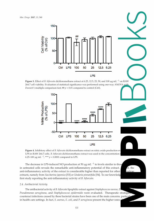
Mar. Drugs 2017, 15, 340
Figure 3. Effect of B. bifurcata dichloromethane extract at 6.25, 12.5, 25, 50, and 100 μg mL−1 on RAW264.7 cell viability. Evaluation of statistical significance was performed using one-way ANOVA withDunnett’s multiple comparison test; ## p < 0.01 compared to control (Ctrl).
Figure 4. Inhibitory effect of B. bifurcata dichloromethane extract on nitric oxide production evoked byLPS in RAW 264.7 cells. B. bifurcata dichloromethane extract was used in the concentration range of6.25–100 μg mL−1, **** p < 0.0001 compared to LPS.
The decrease in LPS-induced NO production at 50 μg mL−1 to levels similar to those observedin untreated cells reveals the remarkable anti-inflammatory potential of this extract. In fact, theanti-inflammatory activity of the extract is considerable higher than reported for other macroalgaeextracts, namely from Saccharina japonica [53] or Undaria pinnatifida [54]. To our knowledge, this is thefirst study reporting the anti-inflammatory activity of B. bifurcata.
2.4. Antibacterial Activity
The antibacterial activity of B. bifurcata lipophilic extract against Staphylococcus aureus, Escherichiacoli,Pseudomonas aeruginosa, and Staphylococcus epidermidis were evaluated. Therapeutic strategies tocounteract infections caused by these bacterial strains have been one of the main concerns, particularlyin health care settings. In fact, S. aureus, E. coli, and P. aeruginosa present the higher rates of antibiotic
122
Books
MDPI

Mar. Drugs 2017, 15, 340
resistance, while S. epidermidis has been considered an opportunistic pathogen, being the most commonsource of infections on indwelling medical devices [55]. Thus, in this study, the antimicrobial efficacyof four antibiotics representing drug families of major clinical importance, such as aminoglycosides(Gent: Gentamicin), tetracyclines (Tetra: Tetracycline), macrocyclics (Rif: Rifampicin), and β-lactamsantibiotics, such as aminopenicillins, (Amp: Ampicillin) were evaluated in combination with theB. bifurcata lipophilic extract.
Results regarding the antibacterial activity of the B. bifurcata dichloromethane extract arepresented in Table 3. Activity against S. aureus ATCC®6538 (MIC = 1024 μg mL−1), S. aureusATCC®43300 (MIC = 2048 μg mL−1) and E. coli (MIC = 2048 μg mL−1) was observed. No growthinhibition of S. epidermidis and P. aeruginosa PAO1 was verified in the range of concentrations tested(MIC > 2048 μg mL−1). B. bifurcata extract showed inhibition against both Gram-negative andGram-positive bacteria, in opposition to that observed by Alves et al. [31], which only verified activityof a B. bifurcata dichloromethane extract against Gram-negative bacteria. Additionally, these authorshave not reported growth inhibition against the same E. coli strain. Notwithstanding the fact that theextraction conditions in both studies were not the same, the macroalgae origins are distinct, which mayalter the metabolite composition and therefore their bioactivities, highlighting the importance ofcontrolling the growth conditions of macroalgae in order to maximize their valorization.
Table 3. Antibacterial activity of B. bifurcata dichloromethane extract and synergistic effects withdifferent antibiotics expressed in MIC (μg mL−1).
Bacteria Ext Rif Rif+ Ext Gent Gent+ Ext Amp Amp+ Ext Tetra Tetra + Ext
MIC (μg mL−1)
E. coli ATCC®25922 2048 32 16 >256 <2 32 128 18 <2Staphylococcus aureus ATCC®43300 2048 16 <2 >256 16 128 256 >256 <2Staphylococcus aureus ATCC®6538 1024 64 64 32 16 256 512 16 8Pseudomonas aeruginosa PAO1 >2048 32 - 2 - 16 - 16 -Staphylococcus epidermidis >2048 32 - 512 - >2048 - 32 -
Key: Ext: Extract; Rif: Rifampicin; Gent: Gentamicin; Amp: Ampicillin; Tetra: Tetracycline.
The synergism of B. bifurcata dichloromethane extract with antibiotics was also evaluated againstthe same bacterial panel. This approach has been suggested as a promising tool to overcome thebacterial resistance that has been developed to most of the antibiotic classes recommended for theirtreatment [3,4,56]; to our knowledge, no similar study has previously been reported for this macroalga.The synergetic activity (antibiotic + extract) screening was performed against the bacterial strainsfor which a new antibiotic MIC was possible to determine, namely for E. coli ATCC®25922, S. aureusATCC®43300 and S. aureus ATCC®6538 (Table 3). Interestingly, the combination of the extract withgentamicin or tetracycline drastically decreased the antibiotic MIC against the three strains under study,enabling it to be effective at considerably lower concentrations. Similar behavior was observed forboth E. coli ATCC®25922 and S. aureus ATCC®43300 when the extract was combined with rifampicin.These observations suggest that the use of B. bifurcata lipophilic fraction may be considered withina coadjutant/synergistic approach to conventional antibiotherapy, enabling a superior therapeuticpotential against the above-mentioned bacteria.
Concerning the interaction between B. bifurcata dichloromethane extract and ampicillin,an antagonism effect was observed, with MIC values increasing considerably. This may be linked toalterations in the antibiotic structure induced by the presence of the extract in solution, leading to itspartial inactivation.
3. Material and Methods
3.1. Sample
Bifurcaria bifurcata R. Ross was cultivated in the land-based aquaculture system of ALGAplus, Lda,at Ria de Aveiro (Portugal, 40◦36′43” N, 8◦40′43” W). The company operates under the IMTA concept,
123
Books
MDPI

Mar. Drugs 2017, 15, 340
using solely filtered nutrient-rich water from a fishpond to produce seaweed biomass. The culturestarting material was obtained at Aguda beach (Portugal, 41◦2′38” N, 8◦39′10” W) in May 2014 andgrown by vegetative propagation during three weeks at constant conditions. The biomass was washedwith seawater to remove salts, epiphytes, and/or microorganisms and dried at 25 ◦C until it reached atotal moisture content of 12%. The samples were transformed into flakes (1–2 mm), packed, and storedin hermetic bags in the company’s storage room.
3.2. Lipophilic Compounds Extraction
Three aliquots (20 g) of lyophilized macroalgae samples were Soxhlet extracted withdichloromethane (Sigma Chemical Co., Madrid, Spain) for 9 h. Solvent was evaporated to dryness,lipophilic extracts weighted and the results were expressed in percent of dry weight material (% DW).Dichloromethane was selected as a fairly specific solvent for lipophilic extractives isolation foranalytical purposes only.
3.3. GC-MS Analysis
The analysis of the lipophilic extracts followed two distinct methodologies: the first suitablefor trimethylsilyl (TMS) derivatizable compounds and the second for diterpenic compounds,as described below.
3.3.1. Analysis of Trimethylsilyl Derivatizable Compounds
Before GC-MS analysis, two aliquots of each dried extract (20 mg each) and an accurate amountof internal standard (tetracosane, 0.25–0.50 mg, Sigma Chemical Co., Madrid, Spain) were dissolved in250 μL of pyridine (Sigma Chemical Co.).
The compounds containing hydroxyl and carboxyl groups were converted into TMS ethers andesters, respectively, by adding 250 μL of N,O-bis(trimethylsilyl)trifluoroacetamide (Sigma ChemicalCo.) and 50 μL of trimethylchlorosilane (Sigma Chemical Co.), standing the mixture at 70 ◦C for30 min [57]. The derivatized extracts were analyzed by GC-MS following previously describedmethodologies [15,35] on a GCMS-QP2010 Ultra (Shimadzu, Kyoto, Japan), equipped with a DB–1J&W capillary column (30 m × 0.32 mm inner diameter, 0.25 μm film thickness). The chromatographicconditions were as follows: initial temperature, 80 ◦C for 5 min; temperature gradient, 4 ◦C min−1;final temperature, 260 ◦C; temperature gradient, 2 ◦C min−1; final temperature, 285 ◦C for 8 min;injector temperature, 250 ◦C; transfer-line temperature, 290 ◦C; split ratio, 1:33.
Compounds were identified as TMS derivatives by comparing their mass spectra withtwo commercial GC-MS spectral libraries (Wiley 275 and U.S. National Institute of Science andTechnology (NIST14) ), their characteristic retention times obtained under the described experimentalconditions [15,35], and by comparing their mass spectra fragmentation profiles with published data orby injection of standards.
For semi-quantitative analysis, GC-MS was calibrated with pure reference compounds,representative of the major lipophilic extractive families (cholesterol, hexadecanoic acid andnonadecan-1-ol (Sigma Chemical Co.)) relative to tetracosane. The respective response factors werecalculated as an average of six GC-MS runs. Each one of the three extracts prepared from macroalgaewas injected in duplicate (n = 6). The presented results are the average of the concordant valuesobtained (less than 5% variation between injections of the same aliquot and between triplicatedextracts of the same macroalgae).
3.3.2. Analysis of diterpenes
Diterpenes were identified and quantified by the analysis of the B. bifurcata extract withoutderivatization. About 10 mg of extract were dissolved in 1100 μL of dichloromethane with 0.8 mg ofinternal standard (n-hexadecane (Supelco, Bellefonte, PA, USA)).
124
Books
MDPI

Mar. Drugs 2017, 15, 340
The extracts were injected in the same GC-MS equipment as described above and thechromatographic conditions were as follows: initial temperature, 80 ◦C for 5 min; temperature gradient,5 ◦C min−1, final temperature 200 ◦C, temperature gradient, 2 ◦C min−1, final temperature 240 ◦C;temperature gradient, 5 ◦C min−1, final temperature 285 ◦C for 8 min; injector temperature, 250 ◦C;transfer-line temperature, 290 ◦C; split ratio, 1:40. All other conditions were the same as describedabove. Diterpenes were identified by comparing their mass spectra fragmentation profile with library(Wiley 275 and U.S. National Institute of Science and Technology (NIST14)) and with the characteristicfragmentation pathway described in literature for these components [23,25,26].
Semi-quantitative analysis was carried out determining the response factor (an average ofsix GC-MS runs) of a representative standard, namely phytol (Sigma Chemical Co.), relative ton-hexadecane. Each one of the three aliquots were injected in duplicate (n = 6).
3.4. Antioxidant Activity
3.4.1. DPPH Assay
The antioxidant activity of the B. bifurcata lipophilic extract was measured by their hydrogen-donatingor radical scavenging ability using the stable free radical 2,2-diphenyl-1-picrylhydrazyl (DPPH•)(Sigma Chemical Co.), following a previously described procedure [35]: 0.25 mL of DPPH• (0.8 mMin methanol (Fluka Chemie, Madrid, Spain)) were added to 1.00 mL of the aqueous solutionof the B. bifurcata dichloromethane extract and 2.75 mL of methanol. The extract concentrationranged between 61.9 and 556.9 μg mL−1. After 30 min of incubation in the dark, at roomtemperature, the absorbance was determined at 517 nm, using a Shimadzu UV-1800 spectrophotometer(Kyoto, Japan). Standards of ascorbic acid (Fluka Chemie) and 3,5-di-tert-4-butylhydroxytoluene (BHT)(Sigma Chemical Co. were used with concentrations ranging from 0.7 to 5.6 μg mL−1 and 5.0 to75.8 μg mL−1. Duplicate measurements of three extracts were carried out (n = 6). The antioxidantactivity was expressed in IC50 values, defined as the inhibitory concentration of the extract required todecrease the initial DPPH radical concentration by 50%, as well as in g of ascorbic acid equivalents perkg of dry weight (g AAE kg−1 DW).
3.4.2. ABTS Assay
The ABTS assay is based on the scavenging of the 2,2′-azino-bis(3-ethylbenzothiazoline-6-sulphonicacid) radical cation, ABTS+• (Sigma Chemical Co.) converting it into a colorless product. In thistest, ABTS+• cation was generated by reacting 50 mL of ABTS 7 mM solution with 25 mL ofpotassium persulfate (Fluka Chemie) 2.45 mM, following a methodology described before with minormodifications [35]. This mixture was then incubated in the dark, at room temperature, for 16 h.Before usage, the ABTS+• solution was diluted with methanol to obtain an absorbance of 0.700 ± 0.02at 734 nm. 30 μL of B. bifurcata dichloromethane extract or trolox (Aldrich Chemical Co., Madrid,Spain), used as reference, were mixed with 3 mL of ABTS+• solution, obtaining final concentrations of47.9–239.5 μg mL−1 and 1.0–5.0 μg mL−1, respectively. The absorbance was measured at 734 nm usinga Shimadzu UV-1800 spectrophotometer (Kyoto, Japan). Duplicate measurements of three extractswere performed.
The antioxidant activity was expressed as IC50 values (extract or trolox concentration providing50% of ABTS+• inhibition) as well as in mg of trolox equivalents per g of dry weight (mg TE g−1 DW).
3.5. Anti-Inflammatory Activity
Test solutions of B. bifurcata dichloromethane extract (100 mg mL−1) were prepared in ethanoland diluted in a culture medium (prepared as described below). Ethanol concentrations ranged from0.007 to 0.1% (v/v), corresponding to extract concentrations between 6.25 and 100 μg mL−1.
125
Books
MDPI

Mar. Drugs 2017, 15, 340
3.5.1. Cell Culture
Raw 264.7, a mouse leukaemic monocyte macrophage cell line from American Type CultureCollection (ATCC TIB-71), kindly supplied by Dr. Otília Vieira (Centro de Neurociências e BiologiaCelular, Universidade de Coimbra, Coimbra, Portugal), was cultured in Dulbecco’s ModifiedEagle Medium supplemented with 10% non-inactivated fetal bovine serum, 100 U mL−1 penicillin,and 100 μg mL−1 streptomycin at 37 ◦C in a humidified atmosphere of 95% air and 5% CO2.During the experiments, cells were monitored by microscope observation in order to detect anymorphological changes.
3.5.2. Determination of Cell Viability
Assessment of metabolically active cells was performed using a resazurin (a nonfluorescent bluedye) based assay [58]. Briefly, cell duplicates were plated at a density of 0.1 × 10⁶/well, in a 96 wellplate and allowed to stabilize overnight. Following this period, cells were either maintained in culturemedium (control) or pre-incubated with B. bifurcata extract diluted in culture medium for 1 h, and lateractivated with 50 ng mL−1 of the Toll-like receptor 4 agonist lipopolysaccharide (LPS) for 24 h. After thetreatments, resazurin solution (50 μM in culture medium) was added to each well and incubated at37 ◦C for 1 h, in a humidified atmosphere of 95% air and 5% CO2. Viable cells are able to reduceresazurin into resorufin (fluorescent pink) and, hence, their number correlates with the magnitudeof dye reduction. Quantification of resorufin was performed on a Biotek Synergy HT plate reader(Biotek, Winooski, VT, USA) at 570 nm, with a reference wavelength of 620 nm.
3.5.3. Measurement of Nitrite Production by Griess Reagent
The production of nitric oxide (NO) was measured by the accumulation of nitrite in the culturesupernatants of cells treated with or without B. bifurcata dichloromethane extract (6.25–100 μg mL−1)in the presence or absence of LPS, using a colorimetric reaction with the Griess reagent [59].Briefly, 170 μL of culture supernatants were diluted with equal volumes of the Griess reagent[0.1% (w/v) N-(1-naphthyl)-ethylenediamine dihydrochloride and 1% (w/v) sulphanilamide containing5% (w/v) H3PO4] and maintained during 30 min, in the dark. The absorbance at 550 nm was measuredin a Biotek Synergy HT plate reader. Culture medium was used as blank and nitrite concentration wasdetermined from a regression analysis using serial dilutions of sodium nitrite as standard (0.5–50 μM).
• Statistical Analysis
Statistical analysis was performed using GraphPad Prism 6.0 for Mac OS X (GraphPadSoftware, San Diego, CA, USA). For each experiment, the results are expressed as mean ± SDof, at least, three independent experiments. Evaluation of statistical significance was performedusing one-way ANOVA with Dunnett’s multiple comparison test. Values of p < 0.05 were consideredstatistically significant.
3.6. Antibacterial Activity
3.6.1. Bacterial Strains
Bacterial stocks of Gram-positive, Staphylococcus aureus ATCC®6538, S. aureus ATCC®43300 andS. epidermidis and Gram-negative Escherichia coli ATCC®25922 and Pseudomonas aeruginosa PAOI werekept in Brucella Broth (Fluka Chemie, Madrid, Spain) with 20% (v/v) glycerol (Sigma, Madrid, Spain)at −80 ◦C.
Pre-inoculum was prepared by stocks defrosting at room temperature, suspension in MuellerHinton Broth (MHB; Liofilchem, Roseto degli Abruzzi, Italy) followed by a 6 h incubation at 37 ◦C,220 rpm. Afterwards, bacteria were streaked onto Mueller Hinton Agar (MHA; Liofilchem, Rosetodegli Abruzzi, Italy) and incubated overnight at 37 ◦C. Then, bacteria were harvested with sterile
126
Books
MDPI

Mar. Drugs 2017, 15, 340
peptone water (Liofilchem), washed twice by centrifugation at 2700 rpm and bacterial pellet suspendedin MHB.
3.6.2. Growth Kinetics
For each strain, growth curves were performed in three independent experiments in order todetermine the respective exponential phase. Colonies from MHA were harvested with sterile peptonewater and bacterial pre-inoculum was prepared as mentioned above. The initial optical density of eachbacterial strain was adjusted to 0.04 in the referred media (λ = 600 nm). T-flasks (Starsted, Numbrecht,Germany) containing media and bacterial inoculums were incubated with agitation (220 rpm) at 37 ◦C.At different time points, samples were taken and the optical density was measured at λ = 600 nm.
The correlation between the obtained optical density values with the number of colony-formingunits per mL (CFU mL−1) was accessed. For CFU mL−1 determination, bacteria were harvested fromliquid media at different time points; serial dilutions were done in peptone water (10−2 until 10−7) and10 μL of each dilution plated in MHA. Incubation was done as previously described and the numberof CFUs was determined after 24 h. Experiments were performed twice and in triplicate (n = 6 foreach experiment).
3.6.3. Minimal Inhibitory Concentration (MIC) Determination
MIC assays were performed in accordance to the Clinical and Laboratory Standards Institute(CLSL) guidelines [60]. Briefly, bacterial cells in exponential growth phase (about 2 h of incubation,previously defined in Section 3.6.2) were suspended in MHB (Fluka Chemie) and the B. bifurcatadichloromethane extract was dissolved in dimethyl sulfoxide (DMSO; AppliChem, Darmstadt,Germany) to a final stock concentration of 50 mg mL−1. The antibacterial performance of the extractwas screened using the microbroth dilution method in a range of concentrations from 8 to 2048 μg mL−1
in 96 well-plates (Starsted, Numbrecht, Germany) [61]. The MIC was qualitatively determined after24 h of incubation, at 37 ◦C, by addition of 3-(4,5-dimethylthiazol-2-yl)-2,5-diphenyltetrazoliumbromide (MTT, Calbiochem® supplied by Millipore, Madrid, Spain) with slight adaptations to theprotocol described by Ellof et al. [62]. The use of MTT, metabolized to a formazan chromophore bythe viable cells, was required for MIC determination due to the coloration of the extract that turnedthe naked eye MIC visualization impracticable. The following controls were performed: (i) solventcontrol: bacterial cultures with 4% (v/v) of DMSO, which represents the maximum amount of solventadded to the cultures, to infer its effect on bacterial growth; (ii) pure cultures (only bacterial inoculum);and (iii) culture media. Experiments were performed twice and in triplicate (n = 6 for each experiment).
3.6.4. Synergistic Assays
The synergistic potential of the extract was accessed by conjugation of the extract with antibiotics,namely: rifampicin. gentamicin, ampicillin, and tetracycline (all supplied by Sigma, Madrid, Spain)in a concentration range between 2 and 512 μg mL−1; against the same bacterial panel. B. bifurcataextract was used at the respective MIC concentration, calculated as described above. Antibiotics MIC,when used alone or in extract + antibiotic combination were determined as mentioned above.Experiments were performed twice and in triplicate (n = 6 for each experiment).
4. Conclusions
The complete study of the lipophilic fraction of B. bifurcata from a Portuguese aquaculturesystem was characterized in detail by GC-MS. B. bifurcata dichloromethane extract wascomposed mainly of diterpenes (1892.78 ± 133.97 mg kg−1 DW), followed by fatty acids(947.88 ± 21.94 mg kg−1 DW). Considerable amounts of sterols (406.45 ± 26.19 mg kg−1 DW) were alsofound, with fucosterol accounting for 317.68 ± 26.11 mg kg−1 DW. The sterols, long-chain aliphaticalcohols, and monoglycerides profile of B. bifurcata is reported for the first time. Additionally, twoditerpenes, phytol and neophytadiene, are also reported for the first time as constituents of
127
Books
MDPI

Mar. Drugs 2017, 15, 340
B. bifurcata. This extract showed relevant antioxidant activities against both DPPH and ABTSradicals. Additionally, antibacterial activity was shown to be strain-dependent, with activity beingverified against both Gram-positive (S. aureus) and Gram-negative (E. coli) strains. B. bifurcata extractwas shown to increase antibiotic antimicrobial activity, with this effect being microbial strain- andantibiotic-dependent. This study provides the basis for further exploitation of this alga’s biomoleculesfor current antimicrobial chemotherapeutics, showing the practical utility of marine natural productsto sensitize pathogenic bacteria, increasing their susceptibility. In addition, the studied extract showedpromising anti-inflammatory activity, which is reported for the first time. It is important to pointout that all biological activities were expressed using low extract concentrations, on the order of μgmL−1. This study is, therefore, an important contribution for the valorization of B. bifurcata macroalgafrom aquaculture systems, with promising applications in the functional food, nutraceutical, cosmetic,and biomedical fields, obviously considering the development of eco-friendly methodologies to extractthis lipophilic fraction.
Acknowledgments: The authors wish to thank to FCT-Portugal (Fundação para a Ciência e Tecnologia)and POPH/FSE for the postdoctoral grant to S.A.O.S. (SFRH/BPD/84226/2012), PhD grant to I.F.(SFRH/BD/110717/2015), for CICECO-Aveiro Institute of Materials, POCI-01-0145-FEDER-007679 (FCT Ref.UID/CTM/50011/2013), QOPNA research unit (FCT UID/QUI/00062/2013) financed by national funds throughthe FCT/MEC and when applicable co-financed by FEDER under the PT2020 Partnership Agreement and CNCfinanced by the European Regional Development Fund (ERDF), through the Centro 2020 Regional OperationalProgram: project CENTRO-01-0145-FEDER-000012-HealthyAging2020, the COMPETE 2020—OperationalProgram for Competitiveness and Internationalization, and the Portuguese national funds via FCT—Fundaçãopara a Ciência e a Tecnologia, I.P.: project POCI-01-0145-FEDER-007440. The work was also supported by FEDERfunds through the Operational Program Competitiveness Factors—COMPETE and national funds by FCT underthe strategic project UID/NEU/04539/2013. M.F.D. also acknowledges FCT for the financial support under theProject UID/AGR/00115/2013. Part of the work of ALGAplus team was funded by the project SEACOLORS,LIFE13 ENV/ES/000445; the team also wishes to thank intern Raquel Silva.
Author Contributions: S.A.O.S. contributed to the GC-MS analysis, collected and interpreted all the data,and wrote the manuscript. S.S.T. and C.S.D.O. performed the extractions, extracts derivatization, GC-MS analysis,and antioxidant activity tests. P.P., D.R. and M.F.D. performed the antibacterial activity analysis and helped inthe data interpretation. I.F. and M.T.C. performed the anti-inflammatory activity analysis and helped in the datainterpretation. M.H.A. and A.M.R. contributed the cultivation of the algae and information on the processesfor obtaining biomass. Also, they provided information on previous results from this species at ALGAplus andreviewed the manuscript; S.M.R. and A.J.D.S. (together with S.A.O.S.) were responsible for the conception anddesign of the work, also performing a critical revision of the manuscript. All authors revised the article andapproved the version to be submitted.
Conflicts of Interest: The authors declare no conflict of interest. The funding sponsors had no role in the designof the study; in the collection, analyses, or interpretation of data; in the writing of the manuscript, and in thedecision to publish the results.
References
1. Reuter, S.; Gupta, S.C.; Chaturvedi, M.M.; Aggarwal, B.B. Oxidative stress, inflammation, and cancer:How are they linked? Free Radic. Biol. Med. 2010, 49, 1603–1616. [CrossRef] [PubMed]
2. Coussens, L.M.; Werb, Z. Inflammation and cancer. Nature 2002, 420, 860–867. [CrossRef] [PubMed]3. Davies, J.; Davies, D. Origins and evolution of antibiotic resistance. Microbiol. Mol. Biol. Rev. 2010, 74,
417–433. [CrossRef] [PubMed]4. Hemaiswarya, S.; Kruthiventi, A.K.; Doble, M. Synergism between natural products and antibiotics against
infectious diseases. Phytomedicine 2008, 15, 639–652. [CrossRef] [PubMed]5. Mishra, B.B.; Tiwari, V.K. Natural products: An evolving role in future drug discovery. Eur. J. Med. Chem.
2011, 46, 4769–4807. [CrossRef] [PubMed]6. Blunt, J.W.; Copp, B.R.; Keyzers, R.A.; Munro, M.H.G.; Prinsep, M.R. Marine natural products. Nat. Prod. Rep.
2014, 31, 160–258. [CrossRef] [PubMed]7. Carvalho, L.G.; Pereira, L. Review of marine algae as source of bioactive metabolites. In Marine Algae
Bodiversity, Taxonomy, Environmental Assessment, and Biotechnology; Pereira, L., Neto, J.M., Eds.; CRC Press:Boca Raton, FL, USA, 2014; pp. 195–227.
128
Books
MDPI
Mar. Drugs 2017, 15, 340
8. Plaza, M.; Cifuentes, A.; Ibanez, E. In the search of new functional food ingredients from algae. Trends FoodSci. Technol. 2008, 19, 31–39. [CrossRef]
9. Van Hal, J.W.; Huijgen, W.J.J.; López-Contreras, A.M. Opportunities and challenges for seaweed in thebiobased economy. Trends Biotechnol. 2014, 32, 231–233. [CrossRef] [PubMed]
10. FAO Yearbook of Fishery Statistics. Available online: http://www.fao.org/3/a-i5716t.pdf (accessed on4 April 2017).
11. Martins, C.D.L.; Ramlov, F.; Nocchi Carneiro, N.P.; Gestinari, L.M.; dos Santos, B.F.; Bento, L.M.; Lhullier, C.;Gouvea, L.; Bastos, E.; Horta, P.A.; et al. Antioxidant properties and total phenolic contents of some tropicalseaweeds of the Brazilian coast. J. Appl. Phycol. 2012, 25, 1179–1187. [CrossRef]
12. Fleurence, J.; Gutbier, G.; Mabeau, S.; Leray, C. Fatty acids from 11 marine macroalgae of the French Brittanycoast. J. Appl. Phycol. 1994, 6, 527–532. [CrossRef]
13. Tabarsa, M.; Rezaei, M.; Ramezanpour, Z.; Robert Waaland, J.; Rabiei, R. Fatty acids, amino acids, mineralcontents, and proximate composition of some brown seaweeds. J. Phycol. 2012, 48, 285–292. [CrossRef][PubMed]
14. Santos, S.A.O.; Vilela, C.; Freire, C.S.R.; Abreu, M.H.; Rocha, S.M.; Silvestre, A.J.D. Chlorophyta andRhodophyta macroalgae: A source of health promoting phytochemicals. Food Chem. 2015, 183, 122–128.[CrossRef] [PubMed]
15. Santos, S.A.O.; Oliveira, C.S.D.; Trindade, S.S.; Abreu, M.H.; Rocha, S.S.M.; Silvestre, A.J.D. Bioprospectingfor lipophilic-like components of five Phaeophyta macroalgae from the Portuguese coast. J. Appl. Phycol.2016, 1–8. [CrossRef]
16. Lopes, G.; Sousa, C.; Bernardo, J.; Andrade, P.B.; Valentão, P.; Ferreres, F.; Mouga, T. Sterol profiles in18 macroalgae of the Portuguese coast. J. Phycol. 2011, 47, 1210–1218. [CrossRef] [PubMed]
17. Andrade, P.B.; Barbosa, M.; Matos, R.P.; Lopes, G.; Vinholes, J.; Mouga, T.; Valentão, P. Valuable compoundsin macroalgae extracts. Food Chem. 2013, 138, 1819–1828. [CrossRef] [PubMed]
18. Melo, T.; Alves, E.; Azevedo, V.; Martins, A.S.; Neves, B.; Domingues, P.; Calado, R.; Abreu, M.H.;Domingues, M.R. Lipidomics as a new approach for the bioprospecting of marine macroalgae—Unravelingthe polar lipid and fatty acid composition of Chondrus crispus. Algal Res. 2015, 8, 181–191. [CrossRef]
19. Fundação para a Ciência e a Tecnologia Estratégia de Investigação e Inovação para uma EspecializaçãoInteligente 2014–2020. Available online: https://www.fct.pt/gabestudosestrategia/ENEI/docs/ENEI_Julho2014_aposconsulta_VF_completa.pdf (accessed on 27 February 2017).
20. Gupta, S.; Abu-Ghannam, N. Bioactive potential and possible health effects of edible brown seaweeds.Trends Food Sci. Technol. 2011, 22, 315–326. [CrossRef]
21. Bourgougnon, N.; Stiger-Pouvreau, V. Chemodiversity and Bioactivity within Red and Brown MacroalgaeAlong the French coasts, Metropole and Overseas Departements and Territories. In Handbook of MarineMacroalgae; John Wiley & Sons, Ltd.: Chichester, UK, 2011; pp. 58–105. ISBN 9781119977087.
22. Muñoz, J.; Culioli, G.; Köck, M. Linear diterpenes from the marine brown alga Bifurcaria bifurcata: A chemicalperspective. Phytochem. Rev. 2013, 12, 407–424. [CrossRef]
23. Culioli, G.; Daoudi, M.; Ortalo-magne, A.; Valls, R.; Piovetti, L. (S)-12-Hydroxygeranylgeraniol-derivedditerpenes from the brown alga Bifurcaria bifurcata. Phytochemistry 2001, 57, 529–535. [CrossRef]
24. Valls, R.; Piovetti, L.; Banaigs, B.; Archavlis, A.; Pellegrini, M. (S)-13-hydroxygeranylgeraniol-derivedfuranoditerpenes from Bifurcaria bifurcata. Phytochemistry 1995, 39, 145–149. [CrossRef]
25. Ortalo-Magné, A.; Culioli, G.; Valls, R.; Pucci, B.; Piovetti, L. Polar acyclic diterpenoids from Bifurcaria bifurcata(Fucales, Phaeophyta). Phytochemistry 2005, 66, 2316–2323. [CrossRef] [PubMed]
26. Culioli, G.; Daoudi, M.; Mesguiche, V.; Valls, R.; Piovetti, L. Geranylgeraniol-derived diterpenoids from thebrown alga Bifurcaria bifurcata. Phytochemistry 1999, 52, 1447–1454. [CrossRef]
27. Göthel, Q.; Muñoz, J.; Köck, M. Formyleleganolone and bibifuran, two metabolites from the brown algaBifurcaria bifurcata. Phytochem. Lett. 2012, 5, 693–695. [CrossRef]
28. Biard, J.; Verbist, J.; Letourneux, Y.; Floch, R. Cétols Diterpeniques à Activité Antimicrobienne deBifurcaria bifurcata. Planta Med. 1980, 40, 288–294. [CrossRef] [PubMed]
29. Valls, R.; Banaigs, B.; Piovetti, L.; Archavlis, A.; Artaud, J. Linear diterpene with antimitotic activity from thebrown alga Bifurcaria bifurcata. Phytochemistry 1993, 34, 1585–1588. [CrossRef]
129
Books
MDPI

Mar. Drugs 2017, 15, 340
30. Moreau, D.; Thomas-Guyon, H.; Jacquot, C.; Jugé, M.; Culioli, G.; Ortalo-Magné, A.; Piovetti, L.; Roussakis, C.An extract from the brown alga Bifurcaria bifurcata induces irreversible arrest of cell proliferation in anon-small-cell bronchopulmonary carcinoma line. J. Appl. Phycol. 2006, 18, 87–93. [CrossRef]
31. Alves, C.; Pinteus, S.; Simões, T.; Horta, A.; Silva, J.; Tecelão, C.; Pedrosa, R. Bifurcaria bifurcata: A keymacro-alga as a source of bioactive compounds and functional ingredients. Int. J. Food Sci. Technol. 2016, 51,1638–1646. [CrossRef]
32. Le Lann, K.; Rumin, J.; Cérantola, S.; Culioli, G.; Stiger-Pouvreau, V. Spatiotemporal variations of diterpeneproduction in the brown macroalga Bifurcaria bifurcata from the western coasts of Brittany (France).J. Appl. Phycol. 2014, 26, 1207–1214. [CrossRef]
33. Daoudi, M.; Bakkas, S.; Culioli, G.; Ortalo-Magné, A.; Piovetti, L.; Guiry, M.D. Acyclic diterpenes and sterolsfrom the genera Bifurcaria and Bifurcariopsis (Cystoseiraceae, Phaeophyceae). Biochem. Syst. Ecol. 2001, 29,973–978. [CrossRef]
34. Valls, R.; Mesguiehe, V.; Pellegrini, M.; Pellegrini, L.; Banaigs, B. Variation de la composition en diterpènesde Bifurcaria bifurcata sur les côtes atlantiques françaises. Acta Bot. Gall. 1995, 142, 119–124. [CrossRef]
35. Touati, R.; Santos, S.A.O.; Rocha, S.M.; Belhamel, K.; Silvestre, A.J.D. Retama sphaerocarpa: An unexploitedand rich source of alkaloids, unsaturated fatty acids and other valuable phytochemicals. Ind. Crops Prod.2015, 69, 238–243. [CrossRef]
36. Vilela, C.; Santos, S.A.O.; Oliveira, L.; Camacho, J.F.; Cordeiro, N.; Freire, C.S.R.; Silvestre, A.J.D. The ripepulp of Mangifera indica L.: A rich source of phytosterols and other lipophilic phytochemicals. Food Res. Int.2013, 54, 1535–1540. [CrossRef]
37. Silva, R.G. Estudos em Bifurcaria bifurcata: Aquacultura, Atividade Antioxidante e Antifungica; Universidade deCoimbra: Coimbra, Portugal, 2014.
38. Culioli, G.; Mesguiche, V.; Piovetti, L.; Valls, R. Geranylgeraniol and geranylgeraniol-derived diterpenesfrom the brown alga Bifurcaria bifurcata (Cystoseiraceae). Biochem. Syst. Ecol. 1999, 27, 665–668. [CrossRef]
39. Culioli, G.; Ortalo-Magné, A.; Richou, M.; Valls, R.; Piovetti, L. Seasonal variations in the chemicalcomposition of Bifurcaria bifurcata (Cystoseiraceae). Biochem. Syst. Ecol. 2002, 30, 61–64. [CrossRef]
40. González-Burgos, E.; Gómez-Serranillos, M.P. Terpene compounds in nature: A review of their potentialantioxidant activity. Curr. Med. Chem. 2012, 19, 5319–5341. [CrossRef] [PubMed]
41. Gouveia, V.; Seca, A.M.L.; Barreto, M.C.; Pinto, D.C.G.A. Di- and sesquiterpenoids from Cystoseira genus:Structure, intra-molecular transformations and biological activity. Mini Rev. Med. Chem. 2013, 13, 1150–1159.[CrossRef] [PubMed]
42. Manzo, E.; Ciavatta, M.L.; Bakkas, S.; Villani, G.; Varcamonti, M.; Zanfardino, A.; Gavagnin, M. Diterpenecontent of the alga Dictyota ciliolata from a Moroccan lagoon. Phytochem. Lett. 2009, 2, 211–215. [CrossRef]
43. Abrantes, J.L.; Barbosa, J.; Cavalcanti, D.; Pereira, R.C.; Frederico Fontes, C.L.; Teixeira, V.L.;Moreno Souza, T.L.; Paixão, I.C.P. The effects of the diterpenes isolated from the Brazilian brown algaeDictyota pfaffii and Dictyota menstrualis against the herpes simplex type-1 replicative cycle. Planta Med.2010, 76, 339–344. [CrossRef] [PubMed]
44. Awad, N.E. Bioactive Brominated Diterpenes from the Marine Red Alga Jania rubens (L.) Lamx. Phyther. Res.2004, 279, 275–279. [CrossRef] [PubMed]
45. Ravi, B.; Murphy, P.; Lidgard, R.; Warren, R.; Wells, R. C18 terpenoid metabolites of the brown algaCystophora moniliformis. Aust. J. Chem. 1982, 35, 171–182. [CrossRef]
46. Reddy, P.; Urban, S. Linear and cyclic C18 terpenoids from the southern Australian marine brown algaCystophora moniliformis. J. Nat. Prod. 2008, 71, 1441–1446. [CrossRef] [PubMed]
47. Wall, R.; Ross, R.P.; Fitzgerald, G.F.; Stanton, C. Fatty acids from fish: The anti-inflammatory potential oflong-chain omega-3 fatty acids. Nutr. Rev. 2010, 68, 280–289. [CrossRef] [PubMed]
48. Kumari, P.; Kumar, M.; Gupta, V.; Reddy, C.R.K.; Jha, B. Tropical marine macroalgae as potential sources ofnutritionally important PUFAs. Food Chem. 2010, 120, 749–757. [CrossRef]
49. Chen, B.; McClements, D.J.; Decker, E.A. Design of foods with bioactive lipids for improved health. Annu. Rev.Food Sci. Technol. 2013, 4, 35–56. [CrossRef] [PubMed]
50. Hoang, M.-H.; Jia, Y.; Jun, H.; Lee, J.H.; Lee, B.Y.; Lee, S.-J. Fucosterol is a selective liver X receptormodulator that regulates the expression of key genes in cholesterol homeostasis in macrophages, hepatocytes,and intestinal cells. J. Agric. Food Chem. 2012, 60, 11567–11575. [CrossRef] [PubMed]
130
Books
MDPI

Mar. Drugs 2017, 15, 340
51. Jung, H.A.; Jin, S.E.; Ahn, B.R.; Lee, C.M.; Choi, J.S. Anti-inflammatory activity of edible brown algaEisenia bicyclis and its constituents fucosterol and phlorotannins in LPS-stimulated RAW264.7 macrophages.Food Chem. Toxicol. 2013, 59, 199–206. [CrossRef] [PubMed]
52. Hwang, E.; Park, S.-Y.; Sun, Z.; Shin, H.-S.; Lee, D.-G.; Yi, T.H. The protective effects of fucosterol againstskin damage in UVB-irradiated human dermal fibroblasts. Mar. Biotechnol. 2014, 16, 361–370. [CrossRef][PubMed]
53. Islam, M.N.; Ishita, I.J.; Jin, S.E.; Choi, R.J.; Lee, C.M.; Kim, Y.S.; Jung, H.A.; Choi, J.S. Anti-inflammatoryactivity of edible brown alga Saccharina japonica and its constituents pheophorbide a and pheophytin a inLPS-stimulated RAW 264.7 macrophage cells. Food Chem. Toxicol. 2013, 55, 541–548. [CrossRef] [PubMed]
54. Hwang, J.-H.; Oh, Y.-S.; Lim, S.-B. Anti-inflammatory activities of some brown marine algae inLPS-stimulated RAW 264.7 cells. Food Sci. Biotechnol. 2014, 23, 865–871. [CrossRef]
55. Otto, M. Staphylococcus epidermidis—The “accidental” pathogen. Nat. Rev. Microbiol. 2009, 7, 555–567.[CrossRef] [PubMed]
56. Lewis, K. Platforms for antibiotic discovery. Nat. Rev. Drug Discov. 2013, 12, 371–387. [CrossRef] [PubMed]57. Freire, C.S.R.; Silvestre, A.J.D.; Neto, C.P.; Cavaleiro, J.A.S. Lipophilic extractives of the inner and outer barks
of Eucalyptus globulus. Holzforschung 2002, 56, 372–379. [CrossRef]58. O’Brien, J.; Wilson, I.; Orton, T.; Pognan, F. Investigation of the Alamar Blue (resazurin) fluorescent dye for
the assessment of mammalian cell cytotoxicity. Eur. J. Biochem. 2000, 267, 5421–5426. [CrossRef] [PubMed]59. Green, L.C.; Wagner, D.A.; Glogowski, J.; Skipper, P.L.; Wishnok, J.S.; Tannenbaum, S.R. Analysis of nitrate,
nitrite, and [15N]nitrate in biological fluids. Anal. Biochem. 1982, 126, 131–138. [CrossRef]60. CLSI. Performance Standards for Antimicrobial Susceptibility Testing; Twenty-Fifth Informational Supplement;
Clinical and Laboratory Standards Institute: Wayne, PA, USA, 2015; ISBN 1562387855.61. Wiegand, I.; Hilpert, K.; Hancock, R.E.W. Agar and broth dilution methods to determine the minimal
inhibitory concentration (MIC) of antimicrobial substances. Nat. Protoc. 2008, 3, 163–175. [CrossRef][PubMed]
62. Ellof, J. A sensitive and quick microplate method to determine the minimal inhibitory concentration of plantextracts for bacteria. Planta Med. 1998, 64, 711–713. [CrossRef] [PubMed]
© 2017 by the authors. Licensee MDPI, Basel, Switzerland. This article is an open accessarticle distributed under the terms and conditions of the Creative Commons Attribution(CC BY) license (http://creativecommons.org/licenses/by/4.0/).
131
Books
MDPI

marine drugs
Article
Chemical Profiling and Bioactivity of Body WallLipids from Strongylocentrotus droebachiensis
Alexander N. Shikov 1,*, Into Laakso 2, Olga N. Pozharitskaya 1, Tuulikki Seppänen-Laakso 3,
Anna S. Krishtopina 1, Marina N. Makarova 1 , Heikki Vuorela 2 and Valery Makarov 1
1 Saint-Petersburg Institute of Pharmacy, Leningrad Region, Vsevolozhsky District, Kuzmolovo P 245,188663 Saint-Petersburg, Russia; [email protected] (O.N.P.); [email protected] (A.S.K.);[email protected] (M.N.M.); [email protected] (V.M.)
2 Division of Pharmaceutical Biosciences, Faculty of Pharmacy, University of Helsinki, P.O. Box 56(Viikinkaari 5E), FI-00014 Helsinki, Finland; [email protected] (I.L.); [email protected] (H.V.)
3 VTT Technical Research Centre of Finland Ltd., P.O. Box 1000 (Tietotie 2), FI-02044 VTT Espoo, Finland;[email protected]
* Correspondence: [email protected]; Tel.: +7-812-603-2432
Received: 31 August 2017; Accepted: 15 November 2017; Published: 24 November 2017
Abstract: The lipids from gonads and polyhydroxynaphthoquinone pigments from body walls ofsea urchins are intensively studied. However, little is known about the body wall (BW) lipids.Ethanol extract (55 ◦C) contained about equal amounts of saturated (SaFA) and monounsaturatedfatty acids (MUFA) representing 60% of total fatty acids, with myristic, palmitic and eicosenoic acidsas major SaFAs and MUFAs, respectively. Non-methylene-interrupted dienes (13%) were composed ofeicosadienoic and docosadienoic acids. Long-chain polyunsaturated fatty acids (LC-PUFA) includedtwo main components, n6 arachidonic and n3 eicosapentaenoic acids, even with equal concentrations(15 μg/mg) and a balanced n6/n3 PUFA ratio (0.86). The UPLC-ELSD analysis showed that a greatmajority of the lipids (80%) in the ethanolic extract were phosphatidylcholine (60 μg/mg) andphosphatidylethanolamine (40 μg/mg), while the proportion of neutral lipids remained lowerthan 20%. In addition, alkoxyglycerol derivatives—chimyl, selachyl, and batyl alcohols—werequantified. We have assumed that the mechanism of action of body wall lipids in the present study isvia the inhibition of MAPK p38, COX-1, and COX-2. Our findings open the prospective to utilize thislipid fraction as a source for the development of drugs with anti-inflammatory activity.
Keywords: sea urchin; body wall lipids; inhibition of p38 MAPK; COX; GC-MS; UPLC-ELSD
1. Introduction
Sea urchins have wide distribution and they play an important role in ecosystem of bothshallow and deeper waters of the ocean. A number of species of sea urchin are intensively utilizedin food, pharmaceutical, and cosmetic industries. In 2015, the total world production of ediblesea urchin was 71,229 tons [1]. Green sea urchin, Strongylocentrotus droebachiensis, is an ediblespecies of the phylum Echinodermata, which is a typical inhabitant of the polar region of Russia,including the Barents Sea. The gonads are delicacies in many parts of the world and considered ashighly valued seafood. Several studies have indicated that the gonads of S. droebachiensis are rich inimportant bioactive compounds like polyunsaturated fatty acids (PUFA), phospholipids, tocopherols,sterols [2–4], carotenoids [2,5], and amino acids [6,7]. Extract of gonad tissue has also revealed effectiveanti-inflammatory and antidiabetic properties [4].
After removal of gonads, the residual shells and spines (body wall, BW) of sea urchins whichaccount for more than 40% of total body weight are discarded as waste. Previous studies have alsoshown that sea urchin BW contain polyhydroxynaphthoquinones. These pigments have evoked
Mar. Drugs 2017, 15, 365 132 www.mdpi.com/journal/marinedrugs
Books
MDPI

Mar. Drugs 2017, 15, 365
renewed interest as a promising source for the development of drugs. A number of bioactivities havebeen found, for example antiallergic [8], antidiabetic [9], antihypertensive [10], anti-inflammatory [11],antioxidant [12–14], cardioprotective [15], and hypocholesterolemic [16] effects.
Total concentration of pigments in sea urchin body wall is quite low (1.2–1.6 mg/g) [17], however,a purification method has been recently reported in order to improve the yield of shell pigments [18].The inner layer of the body wall, on the contrary, is covered with a biomembrane, consisting of lipids.However, their profile has not been described yet. Marine lipids are unique sources of essential fattyacids, phospholipids, sterols, and alkoxyglycerols, and they have a broad pharmacological activity.
The aim of this study was to analyze the ethanolic extract of lipids underlying the body wall (BW)of green sea urchin by using gas chromatography-mass spectrometry (GC-MS) and ultra-performanceliquid chromatography (UPLC-ELSD). In addition, the anti-inflammatory potential of BW lipids wasinvestigated in vitro.
2. Results
2.1. Fatty Acid Composition of Body Wall (BW) Lipids
The ethanolic (95%) extract, which was prepared at 55 ◦C for 3 h, contained about equal amounts ofsaturated (SaFA) and monounsaturated fatty acids (MUFA) representing 60% of total fatty acids. Myristic,palmitic, and eicosenoic acids were the main SaFAs and MUFAs, respectively (Table 1 and Figure 1).The principal MUFA is unusual and this isomer is suggested to be 20:1n15. Among non-methylene-interrupteddienes (NMID, 13%), eicosadienoic (20:2) and docosadienoic (22:2) acids were characteristic. They are typicalconstituents of sea urchin among which 20:2Δ5,11 often appears as the most abundant isomer. In addition,n12 and n5 isomers of 18:1, 20:3n9 and cyclopropaneoctanoic and -decanoic acid 2-octyl methyl estersaccounted 10 μg/mg.
The composition of long-chain polyunsaturated fatty acids (LC-PUFA) was characterized bytwo major components, n6 arachidonic (20:4n6; AA) and n3 eicosapentaenoic acids (20:5n3; EPA),with equal concentrations (15 μg/mg) and a balanced n6/n3 PUFA ratio (0.86). These LC-PUFAspossess important properties, since they act as the precursors of eicosanoids.
A very low amount of docosahexaenoic acid (22:6n3, DHA) was typical for BW lipid extract(Table 1), as well as the high proportion of free (22%) vs. esterified fatty acids. Quantitatively, free AAcovered 1/3 of total AA and free EPA 1/4 of total EPA, respectively, reflecting decomposition of boundfatty acids during extraction procedure. The relatively high proportion of LC-PUFAs, n6 AA, and n3EPA (20%), would suggest that the extract is rich in phospholipids.
Table 1. The concentrations (μg/mg; mean ± SD, n = 3) and relative amounts (%) of bound and freefatty acids in ethanolic extract of BW lipids of sea urchin determined by GC-MS.
Fatty Acids Bound Fatty Acids as FAME % FFA %
C10:0–C13:0 0.4 ± 0.1 0.3 - -C14:0 15.1 ± 0.7 9.6 3.7 ± 0.1 8.4C15:0 1.5 ± 0.1 0.9 0.5 ± 0.1 1.1C16:0 21.9 ± 1.2 13.9 5.0 ± 0.2 11.4C18:0 4.0 ± 0.2 2.5 2.1 ± 0.1 4.8C19:0 1.0 ± 0.1 0.6 - -C20:0 0.6 ± 0.1 0.4 - -
Σ SaFAΣ 44.5 ± 2.1 28.2 11.3 ± 0.3 25.7C14:1n5 1.1 ± 0.1 0.7 - -C16:1n9 0.9 ± 0.1 0.6 - -C16:1n7 7.8 ± 0.6 4.9 1.9 ± 0.1 4.3C16:1n5 4.5 ± 0.2 2.8 - -C18:1n9 3.0 ± 0.2 1.9 1.4 ± 0.1 3.2
133
Books
MDPI

Mar. Drugs 2017, 15, 365
Table 1. Cont.
Fatty Acids Bound Fatty Acids as FAME % FFA %
C18:1n7 4.2 ± 0.2 2.7 2.9 ± 0.1 6.6C20:1n15 16.7 ± 0.8 10.6 - -C20:1n9 5.2 ± 0.3 3.3 8.2 ± 0.4 18.6C20:1n7 1.3 ± 0.1 0.8 - -C22:1n9 4.3 ± 0.2 2.7 - -Σ MUFA 49.0 ± 1.3 31.0 14.4 ± 0.4 32.720:2Δ5,11 12.2 ±1.2 7.7 - -20:2Δ5,13 3.0 ± 0.5 1.9 - -22:2Δ7,13 0.9 ± 0.2 0.6 - -22:2Δ7,15 4.7 ± 0.4 3.0 - -Σ NMID 20.9 ± 1.9 13.2 - -C18:2n6 1.5 ± 0.1 0.9 5.2 ± 0.2 11.8C20:2n6 1.9 ± 0.1 1.2 - -C20:3n6 0.9 ± 0.1 0.6 - -C20:4n6 15.8 ± 0.6 10.0 7.3 ± 0.1 16.6
Σ n6 PUFA 20.1 ± 1.0 12.7 12.5 ± 0.8 28.4C18:3n3 1.2 ± 0.1 0.8 - -C18:4n3 2.3 ± 0.1 1.5 - -C20:3n3 2.3 ± 0.2 1.5 - -C20:4n3 0.6 ± 0.1 0.4 - -C20:5n3 15.2 ± 0.7 9.6 5.8 ± 1.0 13.2C22:5n3 0.2 ± 0.1 0.1 - -C22:6n3 1.7 ± 0.1 1.1 - -
Σ n3 PUFA 23.5 ± 1.1 14.9 5.8 ± 1.0 13.2Σ Fatty acids 158.0 ± 5.3 100.0 44.0 ± 1.8 100.0n6/n3 PUFA 0.86 2.16
FAME, fatty acid methyl ester; FFA, free fatty acid; SaFA, saturated fatty acid; MUFA, mono-unsaturated fatty acid;NMID, non-methylene-interrupted diene; PUFA, polyunsaturated fatty acid.
Figure 1. GC-MS analysis of transesterified and trimethylsilylated (TMS) fatty acids from ethanolicextract of BW lipids of sea urchin. Total ion (TIC) and extracted ion chromatogram (m/z 205) showsTMS derivatives of alkylglycerols (AOG; (1) 16:0-AOG, (2) 18:1-AOG, and (3) 18:0-AOG) and sterols(peaks 4–11, TIC): (4–5) unidentified sterols, (6) cholesterol, (7) desmosterol, (8) cholecalciferol asshoulder, (9) campesterol, (10) stigmasterol, and (11) clionasterol. Heptadecanoic acid (as FAME andTMS derivative) was used as internal standard (IS). Other peaks represent FAMEs and TMS ethers.
134
Books
MDPI

Mar. Drugs 2017, 15, 365
2.2. Sterols and Alkoxyglycerols (AOG)
Sterol and AOG samples were analyzed by GC-MS as TMS-derivatives. BW lipid extract containedmainly cholesterol (50 μg/mg) (Figure 1 and Table 2). Minor non-cholesterol sterols includeddesmosterol, campesterol, stigmasterol, and clionasterol (gamma-sitosterol) which is a commonconstituent in oyster, for example [19]. For GC-MS analyses from abundant AOG sources, purifiedsamples from unsaponifiable fraction have been used. Because of low concentration, extracted ionchromatogram (m/z 205) of silylated AOG was taken to confirm the peak location. This fragment isformed after cleavage between carbons 1 and 2 of the glycerol moiety [20]. Quantified AOG derivativeswere chimyl (C16:0), selachyl (C18:1) and batyl alcohols (C18:0).
Table 2. Sterol and alkoxyglycerol (AOG) content (μg/mg; mean ± SD, n = 3) of ethanolic extract ofBW lipids of sea urchin. Sterols and AOGs were determined as TMS ethers by GC-MS.
Sterols and AOGs μg/mg
Cholesterol 50.3 ± 2.8Non-cholesterol sterols * 10.6 ± 0.5
C16:0-AOG 1.0 ± 0.1C18:1-AOG 0.4 ± 0.1C18:0-AOG 0.3 ± 0.1
Σ Alkoxyglycerols 1.7 ± 0.2
* Non-cholesterol sterols include desmosterol, campesterol, stigmasterol, clionasterol, and two unidentified sterols(Figure 1).
2.3. Lipid Classes
The UPLC-ELSD analyses from the ethanol extract of BW lipids, shown in Figure 2 and Table 3,demonstrate high abundance of phospholipids (PL, 80%), especially those of phosphatidylcholine(PC, 60 μg/mg) and phosphatidylethanolamine (PE, 40 μg/mg). The proportion of neutral lipidsremained less than 20%. It is clear that ethanol extracts polar PLs better than neutral lipids (NL) liketriacylglycerols and cholesteryl esters. The fatty acid profile with relatively high content of PUFAsindicates that the fatty acids have mostly originated from PLs. Lysophosphatidylcholine (LPC) elutedlate as a broad peak and covered about 8% of total lipids.
Figure 2. UPLC-ELSD chromatogram of BW lipids of ethanol (95%) extract of sea urchin. UPLC, ultra-performance liquid chromatography; ELSD, evaporative light scattering detector; WE, wax ester,SE, steryl ester; TG, triacylglycerol; Chol, cholesterol; ip, impurity; Cer, ceramide; CL, cardiolipin;PE, phosphatidylethanolamine; PI, phosphatidylinositol; PS, phosphatidylserine; PC, phosphatidylcholine;SPH, sphingomyelin; LPC, lysophosphatidylcholine.
135
Books
MDPI

Mar. Drugs 2017, 15, 365
Table 3. Concentration of major lipid classes (μg/mg, mean ± SD; n = 3) and their relative amounts (%)in ethanolic extract of BW lipids of sea urchin. Analyses were carried out by UPLC-ELSD.
Lipid Classes μg/mg %
WE + SE 14.6 ± 0.7 8.4TG 15.9 ± 1.0 9.1
Σ Neutral lipids 30.5 ± 1.6 17.5PE 38.6 ± 1.1 22.2
PI + PS 27.8 ± 0.1 16.0PC 63.3 ± 1.7 36.4
LPC 13.5 ± 0.5 7.8Σ Phospholipids 143.2 ± 0.7 82.5
Σ Total lipids 173.7 ± 2.3 100.0
WE, wax ester; SE, steryl ester; TG, triacylglycerol; PE, phosphatidylethanolamine; PS, phosphatidylserine;PI, phosphatidylinositol; PC, phosphatidylcholine; LPC, lysophosphatidylcholine.
2.4. Bioactivity of Body Wall (BW) Lipids
Inflammation is a part of the body’s normal response to infection and injury, extreme orinappropriate inflammation, on the contrary, is linked to the pathobiology of several diseases [21].The anti-inflammatory effect of S. droebachiensis lipids of the extract was assessed in the humanmononuclear U937 cells stimulated with lipopolysaccharide (LPS). The stimulation of U937 resultedin direct activation of MAPK p38. The results (Table 4) revealed that BW lipids dose-dependentlyinhibited MAPK p38. The most effective dose of the lipid extract was 0.033 μg/mL. In addition,BW lipids were clearly more potent than a specific MAPK p38 inhibitor SB203580 (1.88 μg/mL) atthe doses of 0.0037–0.1 μg/mL. The COX-1 and COX-2 isoenzymes were inhibited by BW lipidsdose-dependently with IC50 = 15.7 μg/mL and 21 μg/mL, respectively.
Table 4. Effect of body wall (BW) lipids on the phosphorylation of MAPK p38 in the human mononuclearU937 cells Mean ± SEM, (n = 6).
Sample, Concentration Percentage of MAPK p38 (%)
Intact cells (no stimulation with LPS) 23.0 ± 1.2Control cells stimulated with LPS (1 μg/mL) 100
SB203580 (1.88 μg/mL) + LPS 30.0 ± 1.7BWL (10 μg/mL) + LPS 59.0 ± 1.3BWL (5 μg/mL) + LPS 53.0 ± 1.9BWL (1 μg/mL) + LPS 49.0 ± 0.9
BWL (0.5 μg/mL) + LPS 38.0 ± 1.6BWL (0.1 μg/mL) + LPS 17.0 ± 1.5
BWL (0.033 μg/mL) + LPS 12.0 ± 0.5BWL (0.011 μg/mL) + LPS 21.0 ± 1.7BWL (0.0037μg/mL) + LPS 27.0 ± 0.7BWL (0.0012 μg/mL) + LPS 38.0 ± 1.6BWL (0.0004 μg/mL) + LPS 52.0 ± 1.2
3. Discussion
The fatty acid composition of the ethanol extract of body wall (BW) lipids of sea urchin(Table 1 and Figure 1) was consistent with literature including non-methylene interrupted dienes(NMID) and unusual cyclopropane derivatives [2,22,23]. The principal PUFAs were n6 arachidonic(AA) and n3 eicosapentaenoic acids (EPA). The proportion docosahexanoic acid (22:6n3, DHA) in BWlipids, on the contrary, was very low as has been reported also by others [2,22]. In our previous study,gonads of sea urchin contained about 50 μg/mg of EPA and DHA [4]. Analysis of lipid classes showedtwo major phospholipids—i.e., phosphatidylcholine and phosphatidylethanolamine. This data is ingeneral agreement with the phospholipids profile of gonads of S. droebachiensis [7].
136
Books
MDPI

Mar. Drugs 2017, 15, 365
Fats in human diet are responsible for severe health problems because of long-term intake ofunbalanced proportions of SaFA, MUFA, n6, and n3 PUFA. The n6 and n3 PUFA intake favors too muchn6 PUFA. This presupposes an adequate intake of PUFA precursors (n6 linoleic and n3 alpha-linolenicacids) to form long-chain LC-PUFAs (n6-AA and n3-EPA), which, in turn, act as eicosanoid precursorswhich determine the balance and effects of eicosanoids in the body [24]. Arachidonic acid is convertedto thromboxane-type eicosanoids via cyclo-oxygenase enzyme, while EPA is converted to prostacycline,antagonizing the conversion of AA to eicosanoids. This would enhance anti-aggregatory andanti-inflammatory conditions [25].
From a biological point of view, the biomembrane covering the inner layer of the body wallplays an important protective role for survival of the sea urchin. Damage of the body wall willfollow with inflammation. Mitogen-activated protein kinases (MAPKs) are among the most importantmolecules in the signaling pathways among which MAPK p38 signaling plays an essential rolein regulating cellular processes, especially inflammation [26]. In our study, we observed 88% ofMAPK p38 inhibition by body wall (BW) lipids at a very low dose of 0.033 μg/mL (Table 4).The inhibition of MAPK p38 might be attributed to the different active compounds of BW lipids.Ait-Said et al. [27] established that EPA, unlike DHA, failed to inhibit nuclear factor-κB (NF-κB)activation, and suppressed MAPK p38 phosphorylation in IL-1β stimulated human pulmonarymicrovascular endothelial cells. The anti-inflammatory properties of EPA in LPS-stimulated BV2microglia cells were mediated by downregulation of NF-κB and MAPKs such as ERK, p38, JNK,and Akt activation [28].
Cyclooxygenase-2 (COX-2) isoenzyme could be induced by a wide range of proinflammatoryagents. Prostaglandin-dependent amplification of COX-2 is hypothesized to be an important part ofsustained proliferative and chronic inflammatory conditions [29]. EPA as well as DHA effectivelyinhibited COX-2 expression in LPS-stimulated HUVEC endothelial cells [30]. Recently, we havereported that lipid rich fraction from gonads of S. droebachiensis inhibited COX-2 with IC50 = 49 μg/mL,but was not effective against COX-1 isoform [4]. It is important to note that, in our current study,body wall lipids were more effective and inhibited COX-2 in lower dose and inhibited COX-1 withIC50 = 15.7 μg/mL.
Low amounts of ether-bonded alkoxyglycerols were also found in ethanolic extract of BW lipids(Figure 2 and Table 3). The total amount of AOG was less than 0.2% which would correspond aboutthe level in human milk and plasma lipids, for example [31]. By using the present UPLC-ELSDmethod, however, it was not possible to detect glycerophospholipid-based alkyl- and alkenylacyllipids, since their analysis first requires the separation of phospholipid subclasses. These lipids, suchas glycerophosphatidylethanolamine and -choline, are known to have important activities.
Biological activity of alkoxyglycerols has been known already more than half a century. Some ofthe activities, such as anti-inflammatory effects and protection against radiation damage, are stillunder study together with the more recent interest in the possible cell-signaling properties ofphospholipid-based ether-bonded compounds. These lipids have shown multiple pharmacologicalactivities such as anti-cancer [32], wound healing [33], and immunostimulatory effects [34]. It hasbeen demonstrated that AOG differentially modulate LPS-mediated MAPK and NF-κB signaling inadipocytes and that they do not activate signaling in the absence of LPS. Saturated alkyl chain increasedLPS-mediated activation of the MAPK signaling, which could cause the expression of inflammatorygenes. Conversely, unsaturated alkyl chain decreased LPS-mediated activation of the MAPK andNF-κB signaling [35].
Taking into account all these aspects, we can hypothesize that the lipids containing n3 PUFA,especially EPA, could contribute anti-inflammatory activity. Besides the content of neutral lipidalkoxyglycerols, it is necessary to confirm the occurrence of alkyl- and alkenylacyl lipids and fatty acidcompositions of individual phospholipid classes in BW lipid extracts.
137
Books
MDPI

Mar. Drugs 2017, 15, 365
4. Material and Methods
4.1. Sample Preparation and Extraction of Lipids
Green sea urchins, Strongylocentrotus droebachiensis, were harvested in the Barents Sea in 2016by divers. After removal of gonads, coelomic fluid, and internal organs, the shells with spines werewashed in cold tap water, dried at 4 ◦C for two days and stored in a dark place. The shells with spines(20 g) were ground and macerated with 160 mL of 95% ethanol for 3 h with constant stirring at 55 ◦C.The extract was filtered and evaporated into dryness by rotary evaporator (IKA RV 10; IKA®-WerkeGmbH & Co. KG, Staufen, Germany). The yield of the body wall lipids was 1.2%.
4.2. Chemical Analyses
4.2.1. Analysis of Fatty Acids, Sterols and Alkoxyglycerols by GC-MS
Analyses of bound and free (FFA) fatty acids were carried out by using the method describedpreviously [4]. Shortly, the lipid extract, spiked with internal standards (IS) TG(17:0/17:0/17:0) andFFA 17:0, were transesterified with 0.5 N sodium methoxide at 45 ◦C for 5 min. After acidification,fatty acid methyl esters (FAMEs) as well as FFAs were extracted with petroleum ether. The methodenabled the esterification of bound fatty acids from neutral lipids and also from phospholipids [36].The mixtures of FAMEs and FFAs were used as reference substances.
The analyses were performed on an Agilent 7890A GC mounted with Gerstel MPS injectionsystem and an Agilent 5975C mass selective detector. The column was an Agilent FFAP silica capillarycolumn (25 m × 0.2 mm × 0.3 μm) and helium was used as the carrier gas. The oven temperatureraised from 70 ◦C to 235 ◦C, with a total run time of 30 min. The temperatures of the injector and MSsource were 220 and 230 ◦C, respectively, and the data were collected in EI mode (70 eV) at a massrange of m/z 40–600.
After analyzing the composition of FAMEs by GC-MS, the same samples were derivatized todetermine the contents of FFAs, cholesterol, and minor sterols. Analyses of alkoxyglycerols (AOG) weredone according to a previous method [31]. Samples were evaporated, re-dissolved in dichloromethane,and silylated with MSTFA [N-Methyl-N-(trimethylsilyl)-trifluoroacetamide] (Pierce, Rockford, IL, USA)at 80 ◦C for 20 min. TMS-derivatives were analyzed on an Rtx-5-ms column (15 m × 0.25 mm × 0.25 μm)(Restek, Bellefonte, PA, USA). The split ratio was 20:1 and the oven temperature was programmed to gofrom 70 (1 min) to 270 ◦C at a rate of 10 ◦C/min, the total run time was 30 min. The data was collectedby MSD ChemStation software (Agilent Technologies, Inc., Santa Clara, CA, USA). Identification ofthe compounds was based on retention times of reference substances, library comparisons (The Wiley®
Registry of Mass Spectral Data, John Wiley and Sons, Inc., New York, NY, USA; NIST 08 spectral library,National Institute of Standards and Technology, Gaithersburg, MD, USA) and on literature data.
4.2.2. Analysis of Lipid Classes by UPLC-ELSD
The same lipid extract as above (without derivatization) was analyzed on a Waters AcquityTM
H-class UPLC (ultra-performance liquid chromatograph) equipped with an evaporative light scatteringdetector (ELSD) by modifying previous conditions [37]. Separation of the lipid classes was carriedout on a Waters Spherisorb silica column (3 μm, 100 × 2.1 mm I.D.). The gradient solvent systemconsisted of (A) iso-octane-tetrahydrofurane (99:1), (B) 2-propanol-dichloromethane (3:2) and (C)2-propanol-water (1:1) with an analysis time of 20 min. The temperature of the drift tube was 40 ◦Cand air flow 50 psi. The multigradient system started from 100% A, the proportion of A decreasedto 32%, that of B increased to 52% and simultaneously that of C (containing water) increased to16%. Stable retention times were obtained by keeping continuous cycle running. The flow rate was0.800 mL/min and the injection volume 2 μL. The temperature of the sample manager was 10 ◦C.
138
Books
MDPI

Mar. Drugs 2017, 15, 365
4.3. Biological Assays
4.3.1. Cell Lines and Cell Culture
The human mononuclear U937 cells were purchased from the Russian Collection of Cell Culture(Institute of Cytology of Russian Academy of Science, Saint-Petersburg, Russia), and maintainedat 37 ◦C in a humidified 95% air and 5% CO2 in RPMI1640 supplemented with 2 mM glutamine,10% heat-inactivated FBS, 100 U/mL penicillin, and 100 μg/mL streptomycin. BW lipids weredissolved in dimethyl sulfoxide (DMSO) as a stock solution at a 10 mg/mL concentration, and the stocksolution was then diluted with the medium to the desired concentration prior to use. Cells derived fromthe freeze-down batch were thawed, grown, and seeded (106 cells per well) onto 12-well tissue cultureplates and cultured in medium for 24 h. The cells were then stimulated with 1 μg/mL Escherichia coliLPS (Sigma-Aldrich, St. Louis, MO, USA) at 37 ◦C for 1 h. After that the cells were treated withSB203580 (Sigma-Aldrich, St. Louis, MO, USA) and various concentrations of BWL at 37 ◦C for 1 h.
4.3.2. Western Blotting
Cells were washed in cold (4 ◦C) phosphate-buffered saline (PBS; 0.5 mol/L sodium phosphate,pH 7.5) and separated by centrifugation (Hermle Labortechnik, Germany) at 1500 rpm−1 for 5 min at4 ◦C, harvested by gentle scraping, and used to prepare total protein or nuclear extracts. Cells weretreated with lysis buffer—1 mol/L Tris-HCl pH 7.5, 1.5 mol/L NaCl, 10% Triton X-100, 0.2 mol/LNa3VO4, 1 mol/L NaF, 0.2 mol/L EDTA, phenylmethylsulphonyl fluoride (PMSF), Abcam’s proteaseinhibitor cocktail, and Abcam’s phosphatase inhibitor cocktail—for 20 min at 4 ◦C. The lysates werethen clarified by centrifugation at 15,000 rpm−1 for 15 min at 4 ◦C and the supernatant was collected.
The protein concentrations of the extracts were determined using the [38] with an XMarkspectrophotometer (Bio-Rad, Hercules, CA, USA). For Western blot analysis, 40 μg of proteinwere desaturated by boiling with Laemmli buffer (5 min at 100 ◦C) and subjected to 4–14%SDS-polyacrylamide gels, and transferred to nitrocellulose membrane membranes (Bio-Rad) byelectroblotting. The membranes were blocked with 5% non-fat dry milk in PBS with Tween 20 buffer(PBS-T) (Tris-HCl (pH 7.5), 1.5 mol NaCl, and 0.1% Tween 20) for 1 h at room temperature. Membraneswere then incubated overnight at 4 ◦C with the primary antibodies, probed with enzyme-linkedsecondary antibodies, and visualized using a chemiluminescent detection with LumiGLO® reagent(Cell Signaling Technology, Danvers, MA, USA) according to the manufacturer’s instructions.After detection, the membranes were scanned (Epson Perfection V330 Photo, Seiko Epson Corporation,Nagano, Japan) and processed with Scion Image software (Alpha 4.0.3.2, Scion, Fredrick, MD, USA).The band intensities were used for calculations. Phospho-p38 MAPK antibody, rabbit, p38 MAPK XPrabbit mAb, β-actin rabbit mAb, and anti-rabbit IgG, HRP-linked antibody were from Cell SignalingTechnology (Danvers, MA, USA).
4.3.3. Assessment of Cyclooxygenase Activity
Inhibition of human recombinant cyclooxygenase COX-1 and COX-2 (Cayman Chemical,Ann Arbor, MI, USA) was assessed according to the manufacturer’s instructions. Indomethacin(1 μg/mL) from Sigma (St. Louis, MO, USA) was used as reference. The BWL was dissolved in DMSOprior to analysis.
4.4. Statistical Analysis
Data were analyzed using Statistica version 10.0. All biological assay data are presented asmean ± SEM or mean ± SD. Differences among groups were evaluated by one way analysisof variance (ANOVA) and post-hoc Tukey’s test. In all comparisons, p < 0.05 was accepted asstatistically significant.
139
Books
MDPI

Mar. Drugs 2017, 15, 365
5. Conclusions
To the best of our knowledge, this is the first time when the profile of body wall lipids is reported.It can be assumed that the mechanism of action of body wall lipids in the present study is via the inhibitionof MAPK p38, COX-1, and COX-2. Our findings open the potential to utilize this lipid fraction as a sourcefor the development of drugs with anti-inflammatory activity. Further- more, the anti-inflammatoryproperties of the lipid extract may be useful for ameliorating neuro- degenerative diseases, as well assuppressing LPS-induced shock.
Acknowledgments: This study was done without financial support. The cost to publish in open access will bepay from the budget of Division of Pharmaceutical Biosciences, Faculty of Pharmacy, University of Helsinki.
Author Contributions: A.N.S., O.N.P., V.M. and H.V. conceived and designed the experiments; A.N.S., O.N.P.,A.S.K., I.L. and T.S.-L. performed the experiments; A.N.S., I.L. and T.S.-L. analyzed the data; M.M., V.M. and H.V.contributed to data interpretation and discussion; A.N.S., O.N.P., I.L. and T.S.-L. wrote the paper with all otherscontributing in editing and revision.
Conflicts of Interest: The authors declare no conflict of interest.
References
1. FAO. Global Production Statistics (1950–2015). Available online: http://www.fao.org/fishery/statistics/global-production/query/en (accessed on 21 June 2017).
2. Liyana-Pathirana, C.; Shahidi, F.; Whittick, A.; Hooper, R. Lipid and lipid soluble components of gonads ofgreen sea urchin (Strongylocentrotus droebachiensis). J. Food Lipids 2002, 9, 105–126. [CrossRef]
3. Kalogeropoulos, K.; Mikellidi, A.; Nomikos, T.; Chiou, A. Screening of macro- and bioactive micro-constituentsof commercial finfish and sea urchin eggs. LWT-Food Sci. Technol. 2012, 46, 525–531. [CrossRef]
4. Pozharitskaya, O.N.; Shikov, A.N.; Laakso, I.; Seppänen-Laakso, T.; Makarenko, I.E.; Faustova, N.M.;Makarova, M.N.; Makarov, V.G. Bioactivity and chemical characterization of gonads of green sea urchinStrongylocentrotus droebachiensis from Barents Sea. J. Funct. Foods 2015, 17, 227–234. [CrossRef]
5. Matsuno, T.; Tsushima, M. Carotenoids in sea urchins. Dev. Aquac. Fish. Sci. 2001, 32, 115–138.6. Dincer, T.; Cakli, S. Chemical composition and biometrical measurements of the Turkish sea urchin
(Paracentrotus lividus, Lamarck, 1816). Crit. Rev. Food Sci. Nutr. 2007, 27, 21–26. [CrossRef]7. Shikov, A.N.; Laakso, I.; Pozharitskaya, O.N.; Makarov, V.G.; Hiltunen, R. Phospholipids and amino-acid
composition of eggs of sea urchin from Barents Sea. Planta Med. 2012, 78, 1146. [CrossRef]8. Pozharitskaya, O.N.; Shikov, A.N.; Makarova, M.N.; Ivanova, S.A.; Kosman, V.M.; Makarov, V.G.; Bazgier, V.;
Berka, K.; Otyepka, M.; Ulrichová, J. Antiallergic effects of pigments isolated from green sea urchin(Strongylocentrotus droebachiensis) shells. Planta Med. 2013, 79, 1698–1704. [CrossRef] [PubMed]
9. Kovaleva, M.A.; Ivanova, S.A.; Makarova, M.N.; Pozharitskaya, O.N.; Shikov, A.N.; Makarov, V.G. Effect ofa complex preparation of sea urchin shells on blood glucose level and oxidative stress parameters in type IIdiabetes model. Eksp. Klin. Farmakol. 2013, 76, 27–30. [PubMed]
10. Agafonova, I.G.; Bogdanovich, R.N.; Kolosova, N.G. Assessment of nephroprotective potential ofhistochrome during induced arterial hypertension. Bull. Exp. Biol. Med. 2015, 160, 223–227. [CrossRef][PubMed]
11. Talalaeva, O.S.; Mishchenko, N.P.; Bryukhanov, V.M.; Zverev, Y.F.; Fedoreyev, S.A.; Lampatov, V.V.;Zharikov, A.Y. The influence of histochrome on exudative and proliferative phases of the experimentalinflammation. Bull. Sib. Branch RAMS 2012, 32, 28–31.
12. Kuwahara, R.; Hatate, H.; Yuki, T.; Murata, H.; Tanaka, R.; Hama, Y. Antioxidant property ofpolyhydroxylated naphthoquinone pigments from shells of purple sea urchin Anthocidaris crassispina.LWT-Food Sci. Technol. 2009, 42, 1296–1300. [CrossRef]
13. Pozharitskaya, O.N.; Ivanova, S.A.; Shikov, A.N.; Makarov, V.G. Evaluation of free radical-scavengingactivity of sea urchin pigments using HPTLC with post-chromatographic derivatization. Chromatographia2013, 76, 1353–1358. [CrossRef]
14. Powell, C.; Hughes, A.D.; Kelly, M.S.; Conner, S.; McDougall, G.J. Extraction and identification of antioxidantpolyhydroxynaphthoquinone pigments from the sea urchin, Psammechinus miliaris. LWT-Food Sci. Technol.2014, 59, 455–460. [CrossRef]
140
Books
MDPI

Mar. Drugs 2017, 15, 365
15. Anufriev, V.P.; Novikov, V.L.; Maximov, O.B.; Elyakov, G.B.; Levitsky, D.O.; Lebedev, A.V.; Sadretdinov, S.M.;Shvilkin, A.V.; Afonskaya, N.I.; Ruda, M.Y.; et al. Synthesis of some hydroxynaphthazarins and theircardioprotective effects under ischemia-reperfusion in vivo. Bioorg. Med. Chem. Lett. 1998, 8, 587–592.[CrossRef]
16. Lakeev, Y.V.; Kosykh, V.A.; Kosenkov, E.I.; Novikov, V.L.; Lebedev, A.V.; Repin, V.S. Effect of naturaland synthetic antioxidants (polyhydroxynaphthaquinones) on cholesterol metabolism in cultured rabbithepatocytes. Bull. Exp. Biol. Med. 1992, 114, 1611–1614. [CrossRef]
17. Amarowicz, R.; Synowiecki, J.; Shahidi, F. Chemical composition of shells from red (Strongylocentrotus franciscanus)and green (Strongylocentrotus droebachiensis) sea urchin. Food Chem. 2012, 133, 822–826. [CrossRef]
18. Krishtopina, A.S.; Urakova, I.N.; Pozharitskaya, O.N.; Razboeva, E.V.; Kosman, V.M.; Makarov, V.G.; Shikov, A.N.Optimization of polyhydroxynaphtoquinone extraction from shells of Strongylocentrotus droebachiensis sea urchins.Pharm. Chem. J. 2017, 51, 407–410. [CrossRef]
19. Phillips, K.M.; Ruggio, D.M.; Exler, J.; Patterson, K.Y. Sterol composition of shellfish species commonlyconsumed in the United States. Food Nutr. Res. 2012, 56, 19831. [CrossRef] [PubMed]
20. Bordier, C.G.; Sellier, N.; Foucault, A.P.; Le Goffic, F. Purification and characterization of deep sea sharkCentrophorus squamosus liver oil 1-O-alkylglycerol ether lipids. Lipids 1996, 31, 521–528. [CrossRef] [PubMed]
21. Calder, P.C. n-3 polyunsaturated fatty acids, inflammation, and inflammatory diseases. Am. J. Clin. Nutr.2006, 83, S1505–S1519.
22. Takagi, T.; Eaton, C.A.; Ackman, R.G. Distribution of fatty acids in lipids of the common Atlantic sea urchinStrongylocentrotus droebachiensis. Can. J. Fish. Aquat. Sci. 1980, 37, 195–202. [CrossRef]
23. Castell, J.D.; Kennedy, E.J.; Robinson, S.M.C.; Parsons, G.J.; Blair, T.J.; Gonzales-Duran, E. Effect of dietarylipids on fatty acid composition and metabolism in juvenile green sea urchin (Strongylocentrotus droebachiensis).Aquaculture 2004, 242, 417–436. [CrossRef]
24. Lands, W.E.M. Diets could prevent many diseases. Lipids 2003, 38, 317–321. [CrossRef] [PubMed]25. Lands, B. A critique of paradoxes in current advice on dietary lipids. Prog. Lipid Res. 2008, 47, 77–106.
[CrossRef] [PubMed]26. Yang, Y.; Kim, S.C.; Yu, T.; Yi, Y.S.; Rhee, M.H.; Sung, G.H.; Yoo, B.C.; Cho, J.Y. Functional roles of p38
mitogen-activated protein kinase in macrophage-mediated inflammatory responses. Mediat. Inflamm. 2014.[CrossRef] [PubMed]
27. Ait-Said, F.; Elalamy, I.; Werts, C.; Gomard, M.T.; Jacquemin, C.; Couetil, J.P.; Hatmi, M. Inhibition byeicosapentaenoic acid of IL-1β-induced PGHS-2 expression in human microvascular endothelial cells:Involvement of lipoxygenase-derived metabolites and p38 MAPK pathway. Biochim. Biophys. Acta 2003,1631, 77–84. [CrossRef]
28. Moon, D.O.; Kim, K.C.; Jin, C.Y.; Han, M.H.; Park, C.; Lee, K.J.; Park, Y.M.; Choi, Y.H.; Kim, G.Y.Inhibitory effects of eicosapentaenoic acid on lipopolysaccharide-induced activation in BV2 microglia.Int. Immunopharmacol. 2007, 7, 222–229. [CrossRef] [PubMed]
29. Bagga, D.; Wang, L.; Farias-Eisner, R.; Glaspy, J.A.; Reddy, S.T. Differential effects of prostaglandin derived fromω-6 and ω-3 polyunsaturated fatty acids on COX-2 expression and IL-6 secretion. Proc. Natl. Acad. Sci. USA2003, 100, 1751–1756. [CrossRef] [PubMed]
30. Lee, S.; Kim, H.J.; Chang, K.C.; Baek, J.C.; Park, J.K.; Shin, J.K.; Choi, W.J.; Lee, J.H.; Paik, W.Y. DHA and EPAdown-regulate COX-2 expression through suppression of NF-κB activity in LPS-treated human umbilicalvein endothelial cells. Korean J. Physiol. Pharmacol. 2009, 13, 301–307. [CrossRef] [PubMed]
31. Seppänen-Laakso, T.; Laakso, I.; Hiltunen, R. Rapid determination of unsubstituted alkylglyceryl ethers bygas chromatography-mass spectrometry. J. Chromatogr. 1990, 530, 94–101. [CrossRef]
32. Pugliese, P.T.; Jordan, K.; Cederberg, H.; Brohult, J. Some biological actions of alkylglycerols from shark liveroil. J. Altern. Complement. Med. 1998, 4, 87–99. [CrossRef] [PubMed]
33. Pedrono, F.; Martin, B.; Leduc, C.; Le Lan, J.; Saïag, B.; Legrand, P.; Moulinoux, J.P.; Legrand, A.B. Naturalalkylglycerols restrain growth and metastasis of grafted tumors in mice. Nutr. Cancer 2004, 48, 64–69.[CrossRef] [PubMed]
34. Iannitti, T.; Palmieri, B. An update on the therapeutic role of alkylglycerols. Mar. Drugs 2010, 8, 2267–2300.[CrossRef] [PubMed]
141
Books
MDPI

Mar. Drugs 2017, 15, 365
35. Zhang, M.; Sun, S.; Tang, N.; Cai, W.; Qian, L. Oral administration of alkylglycerols differentiallymodulates high-fat diet-induced obesity and insulin resistance in mice. Evid.-Based Complement. Altern.2013, 2013, 834027. [CrossRef] [PubMed]
36. Seppänen-Laakso, T.; Laakso, I.; Hiltunen, R. Analysis of fatty acids by gas chromatography, and its relevanceto research on health and nutrition. Anal. Chim. Acta 2002, 465, 39–62. [CrossRef]
37. Seppänen-Laakso, T.; Laakso, I.; Vanhanen, H.; Kiviranta, K.; Lehtimäki, T.; Hiltunen, R. Major humanplasma lipid classes determined by quantitative high-performance liquid chromatography, their variationand associations with phospholipid fatty acids. J. Chromatogr. B 2001, 754, 437–445. [CrossRef]
38. Bradford, M.M. A rapid and sensitive method for the quantification of microgram quantities of proteinutilizing the principle of protein-dye binding. Anal. Biochem. 1976, 72, 248–254. [CrossRef]
© 2017 by the authors. Licensee MDPI, Basel, Switzerland. This article is an open accessarticle distributed under the terms and conditions of the Creative Commons Attribution(CC BY) license (http://creativecommons.org/licenses/by/4.0/).
142
Books
MDPI

marine drugs
Article
The Marine Fungi Rhodotorula sp. (Strain CNYC4007)as a Potential Feed Source for Fish Larvae Nutrition
M. Barra 1,2, A. Llanos-Rivera 1, F. Cruzat 1,2,3, N. Pino-Maureira 1,2,3 and
R. R. González-Saldía 1,2,3,*
1 Marine Biotechnology Unit, Department of Oceanography, Faculty of Natural and Oceanographic Sciences,Universidad de Concepción, Casilla 160-C, 4030000 Concepción, Chile; [email protected] (M.B.);[email protected] (A.L.-R.); [email protected] (F.C.); [email protected] (N.P.-M.)
2 Center for Oceanographic Research COPAS Sur-Austral, Universidad de Concepción, Casilla 160-C,4030000 Concepción, Chile
3 Doctoral Program in Aquatic Living Resources (MaReA), Faculty of Natural and Oceanographic Sciences,University of Concepción, Casilla 160-C, 4030000 Concepción, Chile
* Correspondence: [email protected]; Tel.: +56-41-2204520
Received: 31 August 2017; Accepted: 16 November 2017; Published: 1 December 2017
Abstract: Fish oil is used in the production of feed for cultured fish owing to its high polyunsaturatedfatty acid content (PUFA). The over-exploitation of fisheries and events like “El Niño” are reducingthe fish oil supply. Some marine microorganisms are considered potentially as alternative fatty acidsources. This study assesses a strain of Rhodotorula sp. (strain CNYC4007; 27% docosahexaenoicacid (DHA) of total fatty acids), as feed for fish larvae. The total length and ribonucleic acid(RNA)/deoxyribonucleic acid (DNA) ratio of Danio rerio larvae was determined at first feedingat six and 12 days old (post-yolk absorption larvae). Larvae fed with microencapsulated Rhodotorulasp. CNYC4007 had a significantly higher RNA/DNA ratio than control group (C1). At six dayspost-yolk absorption group, the RNA/DNA ratio of larvae fed with Rhodotorula sp. bioencapsulatedin Brachionus sp. was significantly higher than control group fed with a commercial diet high in DHA(C2-DHA). Finally, at 12 days post-yolk absorption, the RNA/DNA ratio was significantly higher inlarvae fed with Rhodotorula sp. CNYC4007 and C2-DHA (both bioencapsulated in Artemia sp. nauplii)than in control group (C1). These results suggest that Rhodotorula sp. CNYC4007 can be an alternativesource of DHA for feeding fish at larval stage, providing a sustainable source of fatty acids.
Keywords: Rhodotorula; DHA; EPA; Danio rerio; larvae nutrition; RNA/DNA ratio
1. Introduction
Marine resources for fishmeal and fish oil have been exploited beyond maximum sustainableyields [1], and the rate of exploitation is increasing by 8.8% per year [2]. One of the fish oil uses isthe production of feed for aquaculture [3], an area that has expanded significantly, resulting in therising demand for fish oil, which has consequently increased the price of this product [4]. Fish oil isused because of its high nutritional value and essential polyunsaturated fatty acid content (PUFA),among which are docosahexaenoic acid (C22:6, DHA), eicosapentaenoic acid (C20:5, EPA) anddocosapentaenoic acid (C22:5, DPA), [5]. Freshwater fish generally have sufficient elongase anddesaturase activities to produce these fatty acids from the 18C precursor. On the contrary, marine fishhave a very limited capacity to synthesize these fatty acids [6], hence a strict requirement for long-chainPUFA, eicosapentaenoic, docosahexaenoic and arachidonic acids (essential fatty acid; EFA). Therefore,when these fatty acids are not synthesized in fish they should be incorporated into the diets of thelarval-to-adult stages of the species that are of commercial interest [7,8]. Their presence is importantfor normal growth, reproduction, metabolic functions, feeding efficiency and immunocompetence [9].
Mar. Drugs 2017, 15, 369 143 www.mdpi.com/journal/marinedrugs
Books
MDPI
Mar. Drugs 2017, 15, 369
Specifically, DHA has a high biological value during larval development and is selectively incorporatedin neural tissue, contributing to pigmentation and visual acuity [10]. Lipids are maternally suppliedand sustain both embryonic and yolk sac larvae development, but when the yolk absorption iscompleted and first feeding is started an exogenous source of PUFA is required [3]. Then, at the timeof first feeding the larvae should receive a diet that covers all the nutritional requirements, so foraquaculture species the research focus has been to improve the nutritional content of artificial diets forlarvae [7,11], as diets lacking in nutritional content result in low survival rates [12].
Fatty acids have also been encapsulated in rotifers (Brachionus sp.) and Artemia sp. nauplii [13,14],these organisms allow for the passing of essential nutrients to the larvae [15,16]. Morphological andhistological parameters, as well as molecular ratios like the ribonucleic acid (RNA)/deoxyribonucleicacid (DNA) ratio, have been used to determine if effectively delivered nutrients improve the nutritionalcondition of the target species [17]. Theoretically, RNA levels in cells vary in relation to proteinsynthesis [18], while DNA concentrations remain constant even during starvation [19]. Therefore,the RNA/DNA ratio is a potential indicator of cellular protein synthesis and growth [17].
The declining supply of traditional sources of essential fatty acids and their high cost has led to thesearch for alternative sources [20], among which are vegetable seed oils [21] that contain alpha-linolenicacid (ALA, 18:3 ω-3), a precursor of DHA and EPA that is assimilated, but not necessarily transformed,into fatty acids [22]. However, it has not been fully possible to enhance these oils for their use inaquaculture as anti-nutritional components have been found in them that affect lipid homeostasis andenergy metabolism [23], along with weakening immune response [24]. Another alternative to fishoil is single-cell oils (SCO) from one-cell organisms like bacteria, algae, fungoid protists and marinefungi, which have great potential because of their high essential long-chain unsaturated fatty acidcontent [25,26]. Marine fungi has also been described as producers of carotenoids, omega-3 fatty acids,including DHA and EPA [27], and omega-6 acids like arachidonic acid (20:04 ω6, AA) [28].
Given their characteristics, strains of marine microorganisms constitute as alternative sources thatcan partially or totally replace fish oil for the nutrition of fish [25,29,30]. For example, Schizochytrium sp.,a strain that produces a high level of DHA (28% of its dry weight), has been highly studied because ithas been proven with juvenile Salmo salar that the complete replacement of fish oil with this strain doesnot negatively affect growth, immune response, and digestibility of nutrients [25]. Its use with othermarine species under intensive cultivation like Sparus aurata [31] has also been tested and with positiveresults. Schizochytrium sp. has therefore been proposed as a sustainable source of fatty acids [25].
A strain of Rhodotorula sp. (CNYC4007) from the Humboldt Current System off Central Chile wasisolated and grown in a bioreactor. The strain, which has high levels of DHA, EPA and carotenoids [27],could be considered as a suitable source of omega-3 for the aquaculture demand. In reference to theabove the aim of this study was to evaluate the use of strain CNYC4007 for feeding Danio rerio larvae.The zebrafish (D. rerio) has been widely studied in various fields of biology [32], and in recent yearshas been proposed as a possible model in studies of nutrition and growth in fish [33], especially asa model in aquaculture research [34]. However, despite potential limitations due to species-specificdifferences in fatty acid metabolism, zebrafish is a useful tool in the initial screening of new suppliesfor larval nutrition with lower cost and rearing times than the use of aquaculture species [35]. To date,research has been provided on the anti-nutritional aspects of some alternative ingredients [36] andindividualized the requirements for mineral and trace element requirements that have been establishedfor adequate larval development and growth in zebrafish that could benefit other fish species [16].Therefore, we used zebrafish larvae as a model in the first step to determine the potential use ofRhodotorula sp. (CNYC4007) in larval nutrition according to the usual feeding protocols implementedin larval rearing.
144
Books
MDPI

Mar. Drugs 2017, 15, 369
2. Results
2.1. RNA/DNA Ratio as a Proxy for the Nutritional Condition of Danio rerio
The total DNA content per individual did not present a significant difference among live larvae upto the 5th day five of the experiment, when the larvae were 10 days old (Kruskal-Wallis test, p = 0.0565).As well, no significant differences were observed for the same parameter among the treatmentsduring the days of the assay (Figure 1). However, there was 100% mortality rate by 6th day of thelarvae in starvation (11 days old), while there was 100% survival rate among the larvae that were fed.The quantity of RNA in larval tissue (Figure 1) progressively increased over the course of the treatmentwith the standard feed, presenting significant differences throughout the assay (one-way analysis ofvariance (ANOVA), F-statistic = 31.09, p-value = 4.18 × 10⁻⁵). In contrast, RNA content in the tissue ofthe larvae under the starvation treatment decreased over the course of the assay. Comparing RNAcontent when the larvae were 10 days old (Figure 1), a significant difference between larvae fed with thestandard feed (C1) and those in starvation (one-way analysis of variance (ANOVA), F-statistic = 11.6,p-value = 0.00362) was found. The average of the RNA/DNA ratio of the treatment with the standardfeed (Figure 2) when larvae were 10 days old (5th day of the treatment) was 0.81 ± 0.12, and thestarvation treatment had a lower average (0.71 ± 0.06). This ratio presents significant differencesbetween the treatments (two-way ANOVA, F-statistic = 5.873, p-value = 0.0210).
Figure 1. Average nucleic acid content (ng) versus larvae age of Danio rerio fed with standard feedcompared to treatment with larvae in starvation. Each point represents the average of four larvaeper treatment. * p < 0.05.
Figure 2. Ribonucleic acid (RNA)/deoxyribonucleic acid (DNA) ratio versus test of larvae of Danio reriofed with standard feed compared to that of larvae in starvation. The daily average is based on fourlarvae per treatment. * p < 0.05.
145
Books
MDPI

Mar. Drugs 2017, 15, 369
2.2. Assay with Six-Day-Old Larvae (First-Feeding Larvae, One Day Post-Yolk Absorption)
There were significant differences among the three treatments (one-way ANOVA,F-statistic = 1809, p-value = 2 × 10⁻¹⁶) by the final day of the assay (Figure 3). A significantly greateraverage larval length (7.7 ± 0.3 mm) was obtained with the control feed (control group) than withthe microencapsulated Rhodotorula sp. CNYC4007 and with the flour lyophilized of Rhodotorula sp.CNYC4007 (7.0 ± 0.2 mm and 7.1 ± 0.2 mm, respectively; Tukey test; p-value = 0.000125 for bothtreatments). There were no significant differences in the total length between larvae fed on flour andthose fed on microencapsulated Rhodotorula sp. CNYC4007 (Tukey test, p = 0.3641). With respect to themolecular analysis, the three treatments did not present any significant differences over the course ofthe assay (Kruskal-Wallis p = 0.148), except for the final day (Figure 4) when the RNA/DNA ratiosin the larvae in both the treatments with microencapsulated Rhodotorula sp. CNYC4007 and withlyophilized flour (which had the same ratios; 0.8 ± 0.14) were higher than that of the larvae in thecontrol group (0.53 ± 0.09).
Figure 3. Total length versus larvae age of Danio rerio at first feeding larvae. The daily average is basedon six larvae per treatment. * p < 0.05.
Figure 4. RNA/DNA ratio versus larvae age of Danio rerio at first feeding larvae. The daily averagedaily is based on six larvae per treatment. * p < 0.05.
146
Books
MDPI
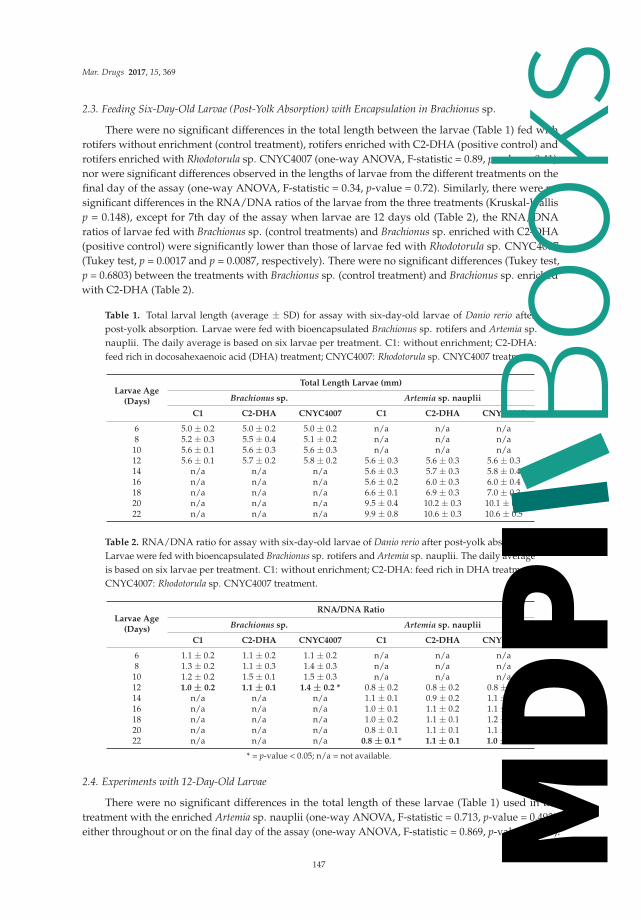
Mar. Drugs 2017, 15, 369
2.3. Feeding Six-Day-Old Larvae (Post-Yolk Absorption) with Encapsulation in Brachionus sp.
There were no significant differences in the total length between the larvae (Table 1) fed withrotifers without enrichment (control treatment), rotifers enriched with C2-DHA (positive control) androtifers enriched with Rhodotorula sp. CNYC4007 (one-way ANOVA, F-statistic = 0.89, p-value = 0.41),nor were significant differences observed in the lengths of larvae from the different treatments on thefinal day of the assay (one-way ANOVA, F-statistic = 0.34, p-value = 0.72). Similarly, there were nosignificant differences in the RNA/DNA ratios of the larvae from the three treatments (Kruskal-Wallisp = 0.148), except for 7th day of the assay when larvae are 12 days old (Table 2), the RNA/DNAratios of larvae fed with Brachionus sp. (control treatments) and Brachionus sp. enriched with C2-DHA(positive control) were significantly lower than those of larvae fed with Rhodotorula sp. CNYC4007(Tukey test, p = 0.0017 and p = 0.0087, respectively). There were no significant differences (Tukey test,p = 0.6803) between the treatments with Brachionus sp. (control treatment) and Brachionus sp. enrichedwith C2-DHA (Table 2).
Table 1. Total larval length (average ± SD) for assay with six-day-old larvae of Danio rerio afterpost-yolk absorption. Larvae were fed with bioencapsulated Brachionus sp. rotifers and Artemia sp.nauplii. The daily average is based on six larvae per treatment. C1: without enrichment; C2-DHA:feed rich in docosahexaenoic acid (DHA) treatment; CNYC4007: Rhodotorula sp. CNYC4007 treatment.
Larvae Age(Days)
Total Length Larvae (mm)
Brachionus sp. Artemia sp. nauplii
C1 C2-DHA CNYC4007 C1 C2-DHA CNYC4007
6 5.0 ± 0.2 5.0 ± 0.2 5.0 ± 0.2 n/a n/a n/a8 5.2 ± 0.3 5.5 ± 0.4 5.1 ± 0.2 n/a n/a n/a10 5.6 ± 0.1 5.6 ± 0.3 5.6 ± 0.3 n/a n/a n/a12 5.6 ± 0.1 5.7 ± 0.2 5.8 ± 0.2 5.6 ± 0.3 5.6 ± 0.3 5.6 ± 0.314 n/a n/a n/a 5.6 ± 0.3 5.7 ± 0.3 5.8 ± 0.416 n/a n/a n/a 5.6 ± 0.2 6.0 ± 0.3 6.0 ± 0.418 n/a n/a n/a 6.6 ± 0.1 6.9 ± 0.3 7.0 ± 0.320 n/a n/a n/a 9.5 ± 0.4 10.2 ± 0.3 10.1 ± 0.722 n/a n/a n/a 9.9 ± 0.8 10.6 ± 0.3 10.6 ± 0.5
Table 2. RNA/DNA ratio for assay with six-day-old larvae of Danio rerio after post-yolk absorption.Larvae were fed with bioencapsulated Brachionus sp. rotifers and Artemia sp. nauplii. The daily averageis based on six larvae per treatment. C1: without enrichment; C2-DHA: feed rich in DHA treatment;CNYC4007: Rhodotorula sp. CNYC4007 treatment.
Larvae Age(Days)
RNA/DNA Ratio
Brachionus sp. Artemia sp. nauplii
C1 C2-DHA CNYC4007 C1 C2-DHA CNYC4007
6 1.1 ± 0.2 1.1 ± 0.2 1.1 ± 0.2 n/a n/a n/a8 1.3 ± 0.2 1.1 ± 0.3 1.4 ± 0.3 n/a n/a n/a10 1.2 ± 0.2 1.5 ± 0.1 1.5 ± 0.3 n/a n/a n/a12 1.0 ± 0.2 1.1 ± 0.1 1.4 ± 0.2 * 0.8 ± 0.2 0.8 ± 0.2 0.8 ± 0.214 n/a n/a n/a 1.1 ± 0.1 0.9 ± 0.2 1.1 ± 0.316 n/a n/a n/a 1.0 ± 0.1 1.1 ± 0.2 1.1 ± 0.318 n/a n/a n/a 1.0 ± 0.2 1.1 ± 0.1 1.2 ± 0.220 n/a n/a n/a 0.8 ± 0.1 1.1 ± 0.1 1.1 ± 0.122 n/a n/a n/a 0.8 ± 0.1 * 1.1 ± 0.1 1.0 ± 0.1
* = p-value < 0.05; n/a = not available.
2.4. Experiments with 12-Day-Old Larvae
There were no significant differences in the total length of these larvae (Table 1) used in thetreatment with the enriched Artemia sp. nauplii (one-way ANOVA, F-statistic = 0.713, p-value = 0.493),either throughout or on the final day of the assay (one-way ANOVA, F-statistic = 0.869, p-value = 0.43).
147
Books
MDPI

Mar. Drugs 2017, 15, 369
However, by the final day there were significant differences in the RNA/DNA ratio (Table 2) betweenthe control group and the group treatment with nauplii enriched with C2-DHA and with the strainCNYC4007 (Tukey test, p-value = 0.0003 and p-value = 0.005, respectively). Finally, there were nosignificant differences on the final day of assay between the treatment with Artemia sp. nauplii enrichedwith C2-DHA and that with nauplii enriched with CNYC4007 (Tukey test, p-value = 0.39).
2.5. DHA and EPA Enrichment of Artemia sp. nauplii
There was a significant high DHA concentration (Figure 5) in nauplii enriched with the strainCNYC4007 (Rhodotorula sp.) compared to those without enrichment or enriched with C2-DHA(one-way ANOVA, p-value = 0.0036). There were no significant differences in DHA concentrations(p-value > 0.05) between nauplii without enrichment (control treatment) and those fed with C2-DHA.The same was observed with EPA concentrations, which were significantly high for nauplii fed withthe strain CNYC4007 (Rhodotorula sp.) compared to those of nauplii without enrichment or enrichedwith C2-DHA (one-way ANOVA, p-value = 0.00017). There were no significant differences in EPAconcentrations (p-value > 0.05) between Artemia sp. nauplii without enrichment (control treatment)and those fed with C2-DHA.
Figure 5. Average DHA and eicosapentaenoic acid (EPA) concentrations (± S.D.) in Artemia sp. naupliiwithout enrichment (C1), fed feed rich in DHA (C2-DHA) and fed with the strain Rhodotorula sp.CNYC4007. * p-value < 0.05.
3. Discussion
The use of strains of marine microorganisms in commercial fish feed and other applications,such as a source of food for human consumption, requires in vivo validation. The first step is to ruleout toxic effects and then verify the nutritional contribution to the species under study. The conditionindex of Fulton [37] is generally used for organisms that are sufficiently large to be easily weighed andmeasured. However, with fish larvae the determination of difference in a very small body mass tends tolack precision. The present work studied the effect of the strain CNYC4007 of the marine basidiomyceteRhodotorula sp. [27] in the RNA/DNA ratio, which was used as a proxy for the nutritional state ofDanio rerio larvae. The larvae were fed in their different stages of development and the strain wasadministered in three forms (flour, microencapsulated, and bioencapsulated).
The results show the efficacy of the RNA/DNA method to evaluate the nutritional condition oflarvae in the experiments carried out, given that it effectively distinguished between the conditions ofthe larvae that were fed and those in starvation (Figures 1 and 2). This method is relevant and has beenused above all with animals at the size scale of zooplankton [22,38] and fish larvae [17,39,40]. This ratiois considered a metabolic index and has been used as a measurement of individual growth rate andnutritional state [18,41]. The increase in RNA concentrations in larvae fed with standard feed (Figure 1)suggests there was protein synthesis and with this a progressive increase in larval size throughoutthe assay, as has been observed with other species like Sardina pilchardus, Engraulis encrasicolus,
148
Books
MDPI

Mar. Drugs 2017, 15, 369
Atherina presbyter and Paralichthys orbignyanus in different development stages [42]. This patternconcurs with that described by Chung and Segnini [43], who found that RNA levels were higher inrainbow trout larvae that were fed, as opposed to those in starvation. In the present work, average DNAlevels per individual were observed to be constant during technical standardization assays (Figure 1).It has been described that DNA levels do not vary under stress, whether caused by starvation orenvironmental conditions [44]. Coincident with our results, Ben Khemis et al. [17] proposed thatRNA/DNA is a useful molecular tool to evaluate larval condition in aquaculture.
In all of the assays the RNA/DNA ratio in the larval stages of Danio rerio ranged between 0.7 and 2.These values were similar to those obtained in Oncorhynchus mykiss (1.5–2.5) [45]. It was observed thatthe ratio was sensitive to changes in the diet composition; this concurs with what has been described inrainbow trout, where the RNA/DNA ratio of larvae varied in response to distinct protein compositionsincluded in their diet (40–50%) [45]. There were no differences among the feeding treatments whenonly considering larval length, although there were differences in nutritional state.
The first feeding assay with Danio rerio larvae indicates that the strain Rhodotorula sp. CNYC4007can be used effectively for this purpose. Larvae fed with flour and microcapsules had similar lengths(Figure 3) and were in a similar nutritional condition (Figure 4), indicating that the two formsof administrating feed can be considered as appropriate. The microencapsulated feed containedstabilizers (maltodextrine and capsule) that protected the fatty acids against moisture, light andother environmental factors [46], which because of storage or contact with water can deteriorate thefeed [47]. Furthermore, the preparation of microcapsules only required 4% of the microorganismsbiomass required for the flour lyophilized with Rhodotorula sp. CNYC4007 and its physical propertiesallowed the feed to remain longer on the surface of the water, which facilitates feeding the larvae,in contrast to the flour, which decants rapidly. Thus, the microencapsulation method to administerRhodotorula sp. (CNYC4007) is recommended for first-feeding the larvae because it is more stable,offers better protection of fatty acids and is more bioavailable than the lyophilized flour method(100% of flour), the latter thus requiring a large quantity of microorganisms to achieve the same effect.
As the larvae grow their mouth size and nutritional requirements increase, because of which thequantity and nutritional composition of the feed must be modified [48]. Because of this, in aquaculture,especially with marine species, live prey such as rotifers and Artemia sp. nauplii are used. However,these have low levels of polyunsaturated fatty acids. Rotifers have 4.19% arachidonic acid and 2.29%docosapentaenoic acid of total fatty acids, with the absence of DHA [16], and Artemia sp. naupliihave low levels of EPA with the absence of DHA [47]. Consequently, it is necessary to enrich theseorganisms for use as larval feed [13]. With larvae at six days post-yolk absorption (six days old),Rhodotorula sp. CNYC4007 was bioencapsulated in rotifers. The results indicate that the strain is nottoxic for Brachionus sp., Artemia sp. nauplii or zebrafish larvae (0% mortality in the assay). In the caseof larvae, its presence in the digestive tract was confirmed and larvae presented a better molecularcondition than larvae fed on rotifers without enrichment. This concurs with the results obtained withbioencapsulation in rotifers of Schizochytrium mangrovei, a marine fungoid used to enrich live prey [14].By comparing the fatty acid content of rotifers with and without enrichment, the authors observedthat the rotifers absorbed significant levels of the DHA present in the lyophilized of S. mangrovei [14].Barclay and Zeller [49] also found the DHA content in rotifers fed with Schizochytrium sp. wassignificantly higher than that of control rotifers fed with beer yeast. Schizochytrium sp. is one ofthe ingredients included in the formulation of feed, termed in this study as C2-DHA, which wasconsidered as a positive control in experiments in the present work. Both Schizochytrium mangrovei [14]and the strain Rhodotorula sp. CNYC4007, used in this experiment, have high percentages of DHA(31.53% and 27%, respectively, of total fatty acids).
The incorporation of the bioencapsulated strain CNYC4007 in feed resulted in significantlyhigher concentrations of polyunsaturated fatty acids in Artemia sp. nauplii than in nauplii fedwith bioencapsulated C2-DHA (Figure 5). Consequently, Rhodotorula sp. CNYC407, as well asSchizochytrium sp. [50], represent alternative species for enriching nauplii, even though the percentages
149
Books
MDPI

Mar. Drugs 2017, 15, 369
of DHA and EPA as part of the total of fatty acids differ between the two species (27% and 43% ofDHA; 7.2% and 2.8% of EPA for Rhodotorula sp. and Schizochytrium sp., respectively). With larvae at12 days post-yolk absorption, the RNA/DNA ratio of larvae in the control treatment was lower thanthat of larvae in the treatment with Artemia sp. nauplii enriched with feed rich in DHA (C2-DHA) andwith the strain Rhodotorula sp. CNYC4007. The bioencapsulation of highly unsaturated fatty acids ofthe omega-3 series in Artemia sp. nauplii has been shown to improve growth and survival of marinefish larvae [51], which concurs with what was observed with the enrichment of Artemia sp. with thestrain studied in this work.
Essential fatty acid requirements vary qualitatively and quantitatively with environmental originand during the ontogeny of fish, with early developmental stages and broodstock being criticalperiods [3]. Specifically, as there is evidence that n-3 HUFA (highly unsaturated fatty acids) andDHA may be more important and, possibly, essential in the larvae of some species of freshwater fishcompared with adults. In marine species, larvae are characterized by having a greater requirementfor n-3 HUFA than juvenile and pre-adult fish, although there are relatively few species where therequirements at larval and juvenile stages can be directly compared. Notwithstanding the foregoing,scarce data exists in relation to EFA requirements (expressed as % dry diet), in larvae and early juvenilefish, with values that ranged between 1 to 5% [3], described the fatty acid requirements (n-3/n-6) inzebrafish larvae using larval growth as a proxy, and concluded that a diet with a low n-3/n-6 ratiomaximizes growth. This finding is coincident with the requirement of typical warm water species,namely, a higher demand for n-6 PUFA, for example, tilapia.
While this study is based mainly on the use of the RNA/DNA ratio as a proxy for nutritionalcondition, there are other methods to validate results that could be used in future studies, such asthe expression of genes related to growth, which can provide new tools for analyzing growth infish [52]. The levels of gene expression related to myogenesis and ATP concentrations in rainbow troutare drastically reduced in response to starvation [50]. The analysis of microarrays also shows thatstarvation in rainbow trout also decreases the levels of gene expression related to lipid metabolismand immune response [53].
All the results obtained indicate that the marine basidiomycete strain Rhodotorula sp. CNYC4007represents a potential feed and/or supplement for first feeding of zebrafish. Additionally, it canbe incorporated in species like rotifers and Artemia sp. nauplii to be bioencapsulated to potentiallyimprove the nutritional state of larvae. All trials carried out in our study considered a comparisonbetween Rhodotorula sp. and commercial sources of fatty acid (formulated diets or emulsions) atdifferent larval stages. In each case, zebrafish larvae fed with Rhodotorula sp. showed at leastequal growth and nutritional condition than the larval treatment with the commercial alternative.These results are similar in magnitude to those reported in species with differences in fatty acidmetabolism such as Salmo salar parr and Sparus aurata larvae, where fish oil replacement with alternativesources of DHA (Schistochytrium sp. and Crypthecodinium cohnii) was suggested [25,31]. These resultsare relevant, as in other species, nutritional studies on zebrafish have determined positive or negativeinfluences of some food compounds [34] and correspond to the first step in the screening process fornovel ingredients or additives with a potential use in aquaculture [35].
Previous studies on Rhodotorula yeast have proposed it as a source of carotenoids [54],bioactive substances [55] and lipids, but just for aliphatic18-carbon atom fatty acid chains [56].The Rhodotorula sp. strain CNYC4007 is the only Rhodotorula species that has been reported as a DHAand EPA producer and to the best of our knowledge this is the first work in which a Rhodotorula yeasthas been used as feed. Finally, this study seeks to contribute in the search for a sustainable source ofPUFAs for feeding fish [57] to avoid the use of fish oil from over-exploited marine resources.
150
Books
MDPI

Mar. Drugs 2017, 15, 369
4. Materials and Methods
4.1. Obtaining and Producing the Strain Rhodotorula sp. CNYC4007
The strain used in this study has a 97% identity for the gene 18S ribosomal RNA (rRNA) ofRhodotorula sp. [27]. It was extracted from Caleta Maule, Biobío Region, Chile, and is stored in theNational Collection of Yeast Cultures in the UK with the code CNYC4007. Two preparations wereobtained from the strain and used in the experiments: lyophilized flour and microencapsulated,which were prepared with atomization drying using two stabilizers (maltodextrin and capsule),following the protocols established by Pino et al. [58]. Formulation and proximate composition ofthe strain Rhodotorula sp. and commercial diets C1 (Mikrovit Hi-Protein, Silesia, Poland) y C2-DHA(Algamac, Poland) used in experiments are detailed in Table 3.
Table 3. Proximate composition of the diets used as a control (C1), positive control (C2-DHA) and thestrain Rhodotorula sp. CNYC4007.
Components (%) C1 C2-DHA CNYC4007 Microencapsulated CNYC4007 Meal
Protein 49 17.6 n/a n/aCarbohydrate n/a 15.9 92.5 36.0
Fat 8.5 56.2 0.3 7.6Nitrogen compounds n/a n/a 1.4 6.1
Fiber 3 n/a 1.2 1.7Moisture 6 2.1 3.7 83.1
Ash n/a 8.2 0.5 7.6Calories (kcal) n/a 640 378.2 169.8
n/a = not available.
4.2. Obtaining and Maintaining Danio rerio Larvae
A breeding stock of D. rerio wild type strain was maintained at 27 ± 1 ◦C with a 12:12 h light:darkphotoperiod and constant filtration. Males and females of the same age were kept together in aglass aquarium with a density not exceeding four to five fish/L. The fish were fed twice daily withArtemia sp. nauplii (Utah strain; Aquafauna Bio-Marine Inc., Hawthorne, CA, USA) and TetraMintropical flakes. To obtain the larvae needed for the experiments, spawning was induced at three-dayintervals by placing plastic receptacles in the culture systems. The fertilized eggs were removed fromthe receptacles and washed twice, first with a mixture of 1× reconstituted saline solution (E3 medium:NaCl2 19 mM; KCl 9 mM; CaCl2 16 mM; MgSO4 17 mM; Merck & Co., Inc., Kenilworth, NJ, USA) andmethylene blue 0.01% (w/v) and then with 1× E3 medium. The healthy embryos were incubated in atemperature controlled camera (27 ◦C) until hatching occurred.
In preliminary observations on early development of zebra fish larvae we assessed the exactmoment when the larvae had the adequate mouth size to begin the feeding with rotifers and Artemia sp.nauplii. At six days post-fertilization the larvae had completed yolk absorption and were used infirst-feeding experiments (larvae at six days post-yolk absorption) or continued maturing until reachingthe phase required for the subsequent experiments, when the larvae were 12 days old (larvae at 12 dayspost-yolk absorption). Plastic wells (700 mL) were used as rearing chambers, and installed in theaquariums. To ensure optimal water quality, the wells were equipped with mesh walls to allow forwater circulation. The remains of feed and fecal matter were regularly removed from the bottomof the wells. Wells of different sizes were used for the feeding experiments according to larval size.The wells were placed in a 50-L tank at 26 ◦C, with constant oxygenation and a light/darkness ratio14:10. The water was partially changed every two days to maintain optimal quality (total ammonianitrogen (TAN) = 0.2 mg L−1; conductivity = 201.9 μS cm−1, pH = 7.5, dissolved oxygen = 7.32 mg L−1).
151
Books
MDPI

Mar. Drugs 2017, 15, 369
4.3. Validation of the RNA/DNA Method for Danio rerio Larvae
An assay was conducted with first-feeding larvae (one day post-yolk absorption), when thelarvae were 6 days old, to validate the RNA/DNA method to compare larvae fed with a standardcommercial feed (C1; Mikrovit Hi-Protein Table 3) and larvae in starvation. Three larvae from eachtreatment (group) were randomly selected every day to determine RNA and DNA concentrations.The experiment lasted six days, when larvae were 11 days old, given that by this day there was 100%mortality of the larvae in starvation, because of which the statistical analysis was applied only up tothe tenth day.
4.4. Estimating Larval Growth
Individual larvae were removed from the wells for analysis in all of the experiments.Cold anesthesia was used for observation and measurement. Larvae were placed individually in apetri dish over a gel pack bar frozen at −20 ◦C. Observations and measurements were made under astereoscopic magnifying glass equipped with a digital camera (Canon EOS REBEL T3, Canon U.S.A.,Inc., Huntington, NY, USA). Photographs of the larvae were analyzed using Image Pro-Plus 6.0software, Media Cybernetics, Inc., Rockville, MD, USA. Magnifications of 10× or 12× was used todetermine the total length depending on the size of the larvae. The distance from the mouth to theposterior point of the notochord was measured for pre-flexion larvae, while the distance from themouth to the hypural plate (standard length) was used for more developed larvae. After measurement,the larvae were immediately placed in Eppendorf tubes and stored at −80 ◦C.
4.5. Experiments with First-Feeding Age Larvae
4.5.1. Feeding with Rhodotorula sp. CNYC4007 Lyophilized in Flour and in Microcapsules
Thirty first-feeding age larvae (one day post-yolk absorption), when the larvae were six daysold, they were placed in 700-mL wells. The experiment involved three treatments: (1) larvae fedwith standard commercial feed (control group; C1); (2) larvae fed with flour lyophilized of thestrain Rhodotorula sp. CNYC4007; and (3) larvae fed with flour microencapsulated of Rhodotorula sp.CNYC4007 (Table 1). A preliminary experiment established a biomass of 30 mg (for 30 larvae) as anadequate daily ration of commercial feed and the lyophilized flour and 150 mg for microencapsulate.The experiment lasted nine days during which unconsumed feed was removed daily. Every second daythree larvae per replicate were removed, measured (detail point 4) and stored at −80 ◦C individuallyfor subsequent molecular analysis (detail point 7).
4.5.2. Larval Feeding with Bioencapsulation in Brachionus sp.
Rotifers used in this experiment were enriched following the protocol of Estudillo del Castillo et al. [16].The assay involved three treatments with respective replicates: (1) larvae fed with non-enriched rotifers(control group; C1); (2) larvae fed with rotifers enriched with a commercial feed high in DHA (C2-DHA);and (3) larvae fed with rotifers enriched with flour lyophilized of Rhodotorula sp. CNYC4007. Each replicateinvolved 20 larvae in a 300-mL well with 0.2-mm mesh walls that allowed for the flow of water whilepreventing rotifers from escaping. On a daily basis, 1.000 enriched rotifers and 1.000 without enrichmentwere applied according to the treatment, following the individual ration. Six randomly selected larvae(three per replicate) were removed from each treatment, then observed, measured (see point 4) and storedindividually at −80 ◦C for subsequent molecular analysis (see point 7).
4.6. Experiments with Larvae at 12 Days Post-Yolk Absorption
To carry out this experiment Artemia sp. (Utah strain; Aquafauna Bio-Marine Inc.) nauplii wereenriched with Rhodotorula sp. CNYC4007 in the form of lyophilized flour. The time and conditionsof the enrichment were defined by modifying the protocols from Silva [48]. Six hundred Artemia sp.
152
Books
MDPI

Mar. Drugs 2017, 15, 369
nauplii (8 h post-hatching) were placed in 15-mL tubes containing 5 mL of seawater. The tubes wereincubated with continuous light and strong aeration at 26 ◦C. Three mg of C2-DHA or 3 mg of flourlyophilized with CNYC4007 were added, respectively, to the two tubes. The tubes containing naupliiwith and without enrichment were kept under the same conditions. The enrichment time was 16 h,after which the nauplii were sieved (88 μm), washed and re-suspended in 2 mL of distilled water toproceed with feeding the larvae.
The experimental design involved three treatments: (1) Larvae fed with Artemia sp. naupliiwithout enrichment (control group; C1); (2) Larvae fed with Artemia sp. nauplii enriched with C2-DHA(positive control group); and (3) Larvae fed with Artemia sp. nauplii enriched with flour lyophilizedwith Rhodotorula sp. CNYC4007. Thirty larvae were placed in 700-mL containers (3 replicatesper treatment). The experiment lasted nine days. Every second day three larvae per replicate wereremoved, observed, measured and then stored individually at −80 ◦C for subsequent molecularanalysis. In all the trials (flour, rotifers and Artemia sp. nauplii) at the moment of photographingeach larvae the presence of food in the gut was visually verified and then stored for posteriormolecular analysis.
4.7. Obtaining the RNA/DNA Ratio Used as a Proxy for the Condition of Danio rerio Larvae
The protocol from Clemmensen [59], with modifications, was used to extract larval nucleic acid.The larvae were treated with Tris-EDTA (Ethylenediaminetetraacetic acid) buffer (Tris-HCl 0.05 M;NaCl 0.1 M; EDTA 0.01 M at pH 8.0) and SDS (sodium dodecyl sulfate) at 20% p/v, after whichproteinase K (20 mg/mL) was added and homogenized with a glass swab until a uniform solutionwas observed. The solution was then centrifuged for one minute and placed in a thermo-regulatedbath (50 ◦C) for 10 min, after which the samples were transferred to −20 ◦C for 20 min. The procedureof heating and cooling the samples was repeated three times, following which the samples werecentrifuged. Chloroform, phenol and isoamyl alcohol (24:25:1) was added and homogenized in anextraction chamber. After centrifugation for 15 min, the supernatant was transferred to a sterileEppendorf tube.
Nucleic acids were determined separately using the Quantifluor ONE dsDNA System(Promega Co., Fitchburg, WI, USA) for DNA and the Quantifluor RNA System (Promega Co.,Fitchburg, WI, USA) for RNA. The samples were prepared according the suppliers protocols.Fluorescence was measured in a multi-detection microplate reader (BioTek, Winooski, VT, USA,model FL×800) under a blue optical channel with excitation of 492 nm and an emission of 540 nm.
4.8. Analysis of Fatty Acid Content in Artemia sp. nauplii
Polyunsaturated fatty acids were extracted from Artemia sp. nauplii by saponification reactionand quantified by HPLC following the methodology described by Li et al. [60]. To do this, 100 mgof Artemia sp. nauplii without enrichment, enriched with C2-DHA and flour lyophilized from thestrain of Rhodotorula sp. CNYC4007 was used, each in triplicate. One mL of NaOH at 0.5 M in 96% ofethanol was added to the samples and homogenized with an Ultra Turrax for one minute. Sampleswere centrifuged at 7000 rpm for 5 min to eliminate solid residues after cell rupture. One mL of HCl at0.6 N and 3 mL of ethyl acetate (LiChrosolv®, Rochester, NY, USA) was added to the supernatant andagitated with vortex for one minute and incubated for 30 min at room temperature. The treated sampleswere dried by a current of nitrogen (N2(g)) to eliminate the organic solvent, lyophilized to eliminate theremains of water and stored at −20 ◦C until subsequent chromatographic analysis. Polyunsaturatedfatty acids were quantified with a HPLC VWR™ HITACHI (VWR International Ltd., Lutterworth, UK)with an organizer (model L-2000), an ultraviolet (UV) detector (model L-2400), gradient editing pump(model L-2100/2130) and a 15-cm-by-4.6-mm LC-18 column (Supelco®, Sigma-Aldrich, Inc., Darmstadt,Germany). The mobile phase consisted of a flow of 1 mL per min−1 of gradient A (25% acetonitrile),to 50% of gradient B (100% acetonitrile), with a flow of 2 mL per min−1 for the first 15 min and then1 mL per min−1 for another 15 min. DHA and EPA were identified through the construction of their
153
Books
MDPI

Mar. Drugs 2017, 15, 369
respective calibration curves using HPLC grade standards (Sigma-Aldrich®, Darmstadt, Germany)with an injection volume of 10 μL [27].
4.9. Statistical Analysis
The Statistica 10 program was used to analyze the data obtained. In the first step the homogeneityof variance (Bartlett, London, UK) and normality of data (Kolmogorov-Smirnov test) was analyzed.A two-way ANOVA and multiple Tukey comparison test was applied for total larval length, DNA andRNA concentrations and the RNA/DNA ratio. The treatments were compared on the final day of theassay. The Kruskal-Wallis test was applied to data that did not present homogeneity of variance ornormality. Mean was considered significant when probability was less than 0.05 (p < 0.05).
Acknowledgments: The authors are grateful for funding from COPAS Sur-Austral CONICYT PIA PFB31 ofUniversidad de Concepción. They also would like to thank the Marine Biotechnology Unit and the ProjectVIU120023 and VIU 15E0053 of Concurso Nacional de Valorización Universitaria, FONDEF of CONICYT.
Author Contributions: R. R. González-Saldía, N. Pino-Maureira, A. Llanos-Rivera y M. Barra conceived anddesigned the experiments; M. Barra performed the experiments; A. Llanos-Rivera, M. Barra y F. Cruzat analyzedthe data; F. Cruzat also help to standardized RNA/DNA method; N. Pino-Maureira provided the biomass andmicroencapsulated of Rhodotorula sp. CNYC4007 for experiment; R. R. González-Saldía wrote the paper.
Conflicts of Interest: The authors declare no conflict of interest.
References
1. Food and Agriculture Organization (FAO). El Estado Mundial de la Pesca y Acuicultura; Departamento dePesca y Acuicultura: Roma, Italy, 2004; pp. 1–115.
2. Food and Agriculture Organization (FAO). El Estado Mundial de la Pesca y Acuicultura; Departamento dePesca y Acuicultura: Roma, Italy, 2012; pp. 1–251.
3. Tocher, D.R. Fatty acid requirement in ontogeny of marine and freshwater fish. Aquac. Res. 2010, 41, 717–732.[CrossRef]
4. Food and Agriculture Organization (FAO). El Estado Mundial de la Pesca y Acuicultura; Departamento dePesca y Acuicultura: Roma, Italy, 2008; pp. 1–280.
5. Hardy, R.W.; Higgs, D.A.; Lalla, S.P.; Tacon, A.G.J. Alternative dietary protein and lipid sources for sustainableproduction of salmonids. Fisken og Havet 2001, 8, 44.
6. Izquierdo, M.S.; Turkmen, S.; Montero, D.; Zamorano, M.J.; Afonso, J.M.; Karalazos, V.;Fernández-Palacios, H. Nutritional programming through broodstock diets to improve utilization of verylow fishmeal and fish oil diets in gilthead sea bream. Aquaculture 2015, 449, 18–26. [CrossRef]
7. Izquierdo, M.; Socorro, J.; Arantzamendi, L.; Hernandez-Cruz, C. Recent advances in lipid nutrition in fishlarvae. Fish Physiol. Biochem. 2000, 22, 97–107. [CrossRef]
8. Hardy, R.W. Utilization on plant proteins in fish diets: Effects of global demand and supplies of fishmeal.Aquac. Res. 2010, 41, 770–776. [CrossRef]
9. Arslan, M.; Sirkecioglu, N.; Bayir, A.; Arslan, H.; Aras, M. The Influence of substitution of dietary fish oilwith different vegetable oils on performance and fatty acid composition of brown trout, Salmo trutta. Turk. J.Fish. Aquat. Sci. 2012, 12, 575–583. [CrossRef]
10. Díaz, N. Efecto de la Relación EPA/DHA en Larvas de Puye (Galaxias maculatus, Jenyns. 1842), Cultivadas enDiferentes Salinidades. Tesis de Grado, Facultad de Acuicultura y Ciencias Veterinarias, Universidad Católicade Temuco, Temuco, Chile, 2004; p. 70.
11. Kaushik, S.; Georga, I.; Koumoundouros, G. Growth and body composition of zebrafish (Danio rerio)larvae fed a compound feed from first feeding onward: Toward implications on nutrients requirements.Zebrafish 2011, 8, 87–95. [CrossRef] [PubMed]
12. Li, K.; Ostensen, M.; Attramadal, K.; Winge, P.; Sparstad, T.; Bones, A.; Vadstein, O.; Kjørsvik, E.; Olsen, Y.Gene regulation of lipid and phospholipid metabolism in Atlantic cod (Gadus morhua) larvae. Comp. Biochem.Physiol. Part B 2015, 190, 16–26. [CrossRef] [PubMed]
13. Hamre, K.; Harboe, T. Artemia enriched with high n-3 HUFA may give a large improvement in performanceof Atlantic halibut (Hippoglossus hippoglossus L.) larvae. Aquaculture 2008, 277, 239–243. [CrossRef]
154
Books
MDPI

Mar. Drugs 2017, 15, 369
14. Estudillo del Castillo, C.; Gapasin, R.; Leaño, E. Enrichment potential of HUFA-rich thraustochytridSchizochytrium mangrovei for the rotifer Brachionus plicatilis. Aquaculture 2009, 293, 57–61. [CrossRef]
15. Satuino, C.; Hirayama, K. Fat-soluble vitamin requirements of the rotifer Brachionus plicatilis. In The First AsianFisheries Forum; Maclean, L., Dizon, L.B., Hosillos, L.V., Eds.; Asian Fisheries Society: Manila, Philippines,1986; pp. 619–622.
16. Hawkyard, M.; Sæle, Ø.; Nordgreen, A.; Langdon, C.; Hamre, K. Effect of iodine enrichment of Artemia sp ontheir nutritional value for larval zebrafish (Danio rerio). Aquaculture 2001, 316, 37–43. [CrossRef]
17. Ben Khemis, I.; de la Noue, J.; Audet, C. Feeding larvae of winter flounder Pseudopleuronectes americanus(Walbaum) with live prey or microencapsulated diet: Linear growth and protein, RNA and DNA content.Aquac. Res. 2000, 31, 377–386. [CrossRef]
18. Robinson, S.; Ware, D. Ontogenetic development of growth rates in larval Pacific herring, Clupea harengus pallasi,measured with RNA/DNA ratios in the Strait of Georgia, British Columbia. Can. J. Fish. Aquat. Sci. 1988,45, 1422–1429. [CrossRef]
19. Chícharo, M.; Chícharo, L. RNA: DNA Ratio and Other Nucleic Acid Derived Indices in Marine Ecology.Int. J. Mol. Sci. 2008, 9, 1453–1471. [CrossRef] [PubMed]
20. Wing, N.; Campbell, P.; Dick, J.; Bell, J. Interactive effect of dietary palm oil concentration and watertemperature on lipid digestibility in rainbow trout, Oncorhynchus mykiss. Lipids 2003, 38, 1031–1038.
21. Turchini, G.; Torstensen, B.; Ng, W. Fish oil replacement in finfish nutrition. Rev. Aquac. 2009, 1, 10–57.[CrossRef]
22. Valenzuela, A.; Sanhueza, J.; De la Barra, F. El aceite de pescado: Un desecho industrial transformado en unproducto de alto valor comercial. Grasas Aceites 2011, XXII, 84–98.
23. Morais, S.; Pratoomyot, J.; Taggart, J.; Bron, J.; Guy, D.; Gordon, J.; Tocher, D. Genotype-specific responses inAtlantic salmon (Salmo salar) subject to dietary fish oil replacement by vegetable oil: A liver transcriptomicanalysis. BMC. Genom. 2011, 12, 255–259. [CrossRef] [PubMed]
24. Kiron, V. Fish immune system and its nutritional modulation for preventive health care. Anim. Feed Sci.2012, 173, 111–133. [CrossRef]
25. Miller, M.; Nichols, P.; Carter, C. Replacement of fish oil with thraustochytrid Schizochytrium sp L oil inAtlantic salmon parr (Salmo salar L) diets. Comp. Biochem. Physiol. Part A 2007, 148, 382–392. [CrossRef][PubMed]
26. Armenta, R.E.; Valentine, M.C. Single-Cell Oils as a Source of Omega-3 Fatty Acids: An Overview of RecentAdvances. J. Am. Oil Chem. Soc. 2013, 90, 167–182. [CrossRef]
27. Pino, N.L.; Socias, C.; González, R.R. Marine fungoid producers of DHA, EPA and carotenoids from centraland southern Chilean marine ecosystems. Rev. Biol. Mar. Oceanogr. 2015, 50, 507–520. [CrossRef]
28. Iida, I.; Nakahara, T.; Yocochi, T.; Kamisaka, Y.; Yagi, H.; Yamaoka, M.; Suzuki, O. Improvement ofdocosahexaenoic acid production in a culture of Thraustochytrium aureum by medium optimization.J. Ferment. Bioeng. 1996, 81, 76–78. [CrossRef]
29. Akanbi, T.O.; Barrow, C.J. Candida antarctica lipase A effectively concentrates DHA from fish andthraustochytrid oils. Food Chem. 2017, 229, 509–516. [CrossRef] [PubMed]
30. Fan, K.W.; Chen, F.; Jones, E.B.G.; Vrijmoed, L.L.P. Eicosapentaenoic and docosahexaenoic acids productionby and okara-utilizing potential of thraustochytrids. J. Ind. Microbiol. Biotechnol. 2001, 27, 199–202. [CrossRef][PubMed]
31. Ganuza, E.; Benitez-Santana, T.; Atalah, E.; Vega-Orellana, O.; Ganga, R.; Izquierdo, M. Crypthecodinium cohniiand Schizochytrium sp as potential substitutes to fisheries-derived oils from seabream (Sparus aurata)microdiets. Aquaculture 2008, 277, 109–116. [CrossRef]
32. Alestrom, P.; Holter, J.; Nourizadeh-Lillabadi, R. Zebrafish in functional genomics and aquatic biomedicine.Trends Biotechnol. 2006, 24, 15–21. [CrossRef] [PubMed]
33. Ulloa, P.; Iturra, P.; Neira, R.; Araneda, C. Zebrafish as a model organism for nutrition and growth:Towards comparative studies of nutritional genomics applied to aquacultured fishes. Rev. Fish Biol. Fish.2011, 21, 649–666. [CrossRef]
34. Ribas, L.; Piferrer, F. The zebrafish (Danio rerio) as a model organism, with emphasis on applications forfinfish aquaculture research. Rev. Aquac. 2014, 6, 209–240. [CrossRef]
35. Ulloa, P.E.; Medrano, J.F.; Feijoo, C.G. Zebrafish as animal model for aquaculture nutrition research.Front. Genet. 2014, 5, 313. [CrossRef] [PubMed]
155
Books
MDPI

Mar. Drugs 2017, 15, 369
36. Hedrera, M.I.; Galdames, J.A.; Jimenez-Reyes, M.F.; Reyes, A.E.; Avendaño-Herrera, R.; Romero, J.;Feijóo, C.G. Soybean Meal Induces Intestinal Inflammation in Zebrafish Larvae. PLoS ONE 2013, 8, e69983.[CrossRef] [PubMed]
37. Fulton, T. Rate of Growth of Sea-Fishes; Neill & Co.: Edinburgh, UK, 1902; Volume 1, p. 20.38. Ikeda, T.; San, F.; Yamaguchi, A.; Matsuishi, T. RNA: DNA ratios of calanoid copepods from the epipelagic
through abyssopelagic zones of the North Pacific Ocean. Aquat. Biol. 2007, 1, 99–108. [CrossRef]39. Bulow, J.F. RNA-DNA ratios as indicators of growth rates in fish: A review. In The Age and Growth of Fish;
Summerfelt, R.C., Hall, G.E., Eds.; The Iowa State University Press: Ames, IA, USA, 1987; pp. 45–64.40. Caldarone, E.; Onge-Burns, J.; Buckley, L. Relationship of RNA/DNA ratio and temperature to growth in
larvae of Atlantic cod Gadus morhua. Mar. Ecol. Prog. Ser. 2003, 262, 229–240. [CrossRef]41. Buckley, L. RNA-DNA ratio: An index of larval fish growth in the sea. Mar. Biol. 1984, 80, 291–298. [CrossRef]42. Olivar, M.; Díaz, M.; Chícharo, A. Tissue effect on RNA: DNA ratios of marine fish larvae. Sci. Mar. 2009,
73, 171–182. [CrossRef]43. Chung, K.; Segnini, M. RNA-DNA ratio as physiological condition of rainbow trout fry fasted and fed.
Ital. J. Zool. 1998, 65, 517–519. [CrossRef]44. Narayan, S.; Archana, L.; Ranjan, R. Effects of dietary protein concentrations on growth and RNA: DNA ratio
of rainbow trout (Oncorhynchus mykiss) cultured in Nuwakot district of Nepal. Int. J. Fish. Aquat. Stud. 2014,1, 184–188.
45. Moreau, D.; Rosenberg, M. Microstructure and fat extractability in microcapsules based on whey proteins ormixtures of whey proteins and lactose. Food Struct. 1993, 12, 457–468.
46. Habi Mat Dian, N.; Sudin, N.; Affandi, Y. Characteristics of microencapsulated palm-based oil as affected bytype of wall material. J. Sci. Food Agric. 1996, 70, 422–426. [CrossRef]
47. Navarro, J.; Henderson, J.; McEvoy, L.; Bell, M.; Amat, F. Lipid conversions during enrichment of Artemia.Aquaculture 1999, 174, 155–166. [CrossRef]
48. Silva, A. Cultivo de Peces Marinos; Departamento de Acuicultura, Facultad de Ciencias del Mar,Universidad Católica del Norte: Coquimbo, Chile, 2005.
49. Barclay, W.; Zeller, S. Nutritional enhancement of n-3 and n-6 fatty acids in rotifers and Artemia nauplii byfeeding spray-dried Schizochytrium sp. J. World Aquac. Soc. 1996, 27, 314–322. [CrossRef]
50. Johansen, K.A.; Overturf, K. Alterations in expression of genes associated with muscle metabolism andgrowth during nutritional restriction and refeeding in rainbow trout. Comp. Biochem. Physiol. Part B 2006,144, 119–127. [CrossRef] [PubMed]
51. Izquierdo, M.; Arakawa, T.; Takeuchi, R.; Haroun, R.; Watanabe, T. Effect of ω-3 HUFA levels in Artemia ongrowth of larval Japanese flounder (Paralicthys olilaceus). Aquaculture 1992, 105, 73–82. [CrossRef]
52. Masuda, Y.; Oku, H.; Okumura, T.; Nomura, K.; Kurokawa, T. Feeding restriction alters expression of someATP related genes more sensitively than the RNA/DNA ratio in zebrafish, Danio rerio. Comp. Biochem.Physiol. Part B 2009, 152, 287–291. [CrossRef] [PubMed]
53. Salem, M.; Silverstain, J.; Rexroad, C.; Yao, J. Effect of starvation on global gene expression and proteolysis inrainbow trout (Oncorhynchus mykiss). BMC. Genom. 2007, 8, 328. [CrossRef] [PubMed]
54. Frengova, G.I.; Beshkova, D.M. Carotenoids from Rhodotorula and Phaffia: Yeasts of biotechnologicalimportance. J. Ind. Microbiol. Biotechnol. 2009, 36, 163–180. [CrossRef] [PubMed]
55. MacDonald, M.C.; Arivalagan, P.; Barre, D.E.; MacInnis, J.A.; D’Cunha, G.B. Rhodotorula glutinisPhenylalanine/Tyrosine Ammonia Lyase Enzyme Catalyzed Synthesis of the Methyl Ester ofpara-Hydroxycinnamic Acid and its Potential Antibacterial Activity. Front. Microbiol. 2016, 7, 281. [CrossRef][PubMed]
56. Taskin, M.; Ortucu, S.; Aydogan, M.N.; Arslan, N.P. Lipid production from sugar beet molasses undernon-aseptic culture conditions using the oleaginous yeast Rhodotorula glutinis TR29. Renew. Energy 2016,99, 198–204. [CrossRef]
57. Sprague, M.; Betancor, M.B.; Tocher, D.R. Microbial and genetically engineered oils as replacements for fishoil in aquaculture feeds. Biotechnol. Lett. 2017, 39, 1599–1609. [CrossRef] [PubMed]
58. Pino, N.; Gómez, C.; González, R.R. Proceso de Obtención de Biomasa del Fungoide Marino CNYC 4007, Uso de laBiomasa y Suplemento Alimenticio que Contiene Esta Biomasa; Solicitud CL201402916; Instituto de PropiedadIndustrial: Santiago, Chile, 2014.
156
Books
MDPI

Mar. Drugs 2017, 15, 369
59. Clemmensen, C. RNA/DNA ratios of laboratory reared and wild herring larvae determined with a highlysensitive fluorescence method. J. Fish Biol. 1989, 35, 331–333. [CrossRef]
60. Li, Z.; Gu, T.; Kelder, B.; Kopchick, J. Analysis of fatty acids in mouse cells using Reversed-PhaseHigh-Performance Liquid. Chromatographia 2001, 54, 463–467. [CrossRef]
© 2017 by the authors. Licensee MDPI, Basel, Switzerland. This article is an open accessarticle distributed under the terms and conditions of the Creative Commons Attribution(CC BY) license (http://creativecommons.org/licenses/by/4.0/).
157
Books
MDPI

Books
MDPI

MDPI AG
St. Alban-Anlage 66
4052 Basel, Switzerland
Tel. +41 61 683 77 34
Fax +41 61 302 89 18
http://www.mdpi.com
Marine Drugs Editorial Office
E-mail: [email protected]
http://www.mdpi.com/journal/marinedrugs
Books
MDPI

Books
MDPI

MDPI St. Alban-Anlage 66 4052 Basel Switzerland
Tel: +41 61 683 77 34 Fax: +41 61 302 89 18
www.mdpi.com ISBN 978-3-03842-800-8
Books
MDPI