marine maladies
72
JULY 2016 | WWW.THE-SCIENTIST.COM MARINE MALADIES THE PATHOGENIC EFFECTS OF WARMER, MORE ACIDIC OCEANS WHAT CAUSES TYPE 2 DIABETES? STEM CELLS’ COOPERATION WITH THE IMMUNE SYSTEM USING COMPOSITE ENDPOINTS PLUS FROM BENCHWORK TO BIOINFORMATICS
-
Upload
khangminh22 -
Category
Documents
-
view
2 -
download
0
Transcript of marine maladies

JULY 2016 | WWW.THE-SCIENTIST.COM
MARINE MALADIESTHE PATHOGENIC EFFECTS OF WARMER, MORE ACIDIC OCEANS
WHAT CAUSES TYPE 2 DIABETES?
STEM CELLS’ COOPERATION
WITH THE IMMUNE SYSTEM
USING COMPOSITE ENDPOINTS
PLUS FROM BENCHWORK
TO BIOINFORMATICS

www.biotek.com
Augmented Microscopy™
Lionheart™ FX Automated Live Cell Imager enables superior digital microscopy with high resolution images up to 100x. From simple fixed cell assays and slide scanning to advanced, environmentally controlled time-lapse movies and 3D spheroid formation imaging, Lionheart FX and Gen5 3.0 Software provide qualitative and quantitative data in a compact automated microscopy system. Visit www.lionheartfx.com
capture analyze annotate video
Cellular compartments are automatically segmented and analyzed. Annotation tools are available.
Includes temperature, gas, humidity control and a movie maker for live cell time-lapse imaging.

Class 1 Laser Product.For Research Use Only. Not for use in diagnostic or therapeutic procedures. © 2016 BD. BD, the BD Logo and BD FACSMelody are trademarks of Becton, Dickinson and Company. 23-18464-00 MC6471
Learn more about the Difference of One at bd.com/Simple-Sort
ONE RESEARCHER, ONE SORTER, ONE CELL, MANY DISCOVERIES. BD is dedicated to developing easy-to-use cell sorting technologies that simplify accurate and reliable flow cytometry. The BD FACSMelody™ cell sorter introduces a powerful combination of high performance, reproducible results and automated ease of use from a brand whose integrated flow cytometry portfolio and rigorous standards you can trust. BD FACSMelody is an affordable cell sorter that requires minimal training making it an ideal solution to advance your research. Its software guides the operator through every step, with a system sort readiness of less than 15 minutes for optimal timeliness. Designed to improve efficiency and throughput, it comes with the full suite of BD service and support to help you maximize your investment. Learn more about the one cell sorter that is easy to learn, to use and to maintain. Discover the difference one company can make. Discover the new BD.

Announcing The Scientist’s annual Top 10 Innovations Competition
SUBMITTING YOUR INNOVATION
HAS NEVER BEEN EASIER
Submit your cutting-edge, life-sciences technology innovation for consideration by a panel of expert judges.
The winners will be the subject of a feature article in the December 2016 issue of The Scientist.
• An “innovation” is defi ned as any product that researchers use in a lab: machines, instruments, tools, cell lines, custom-made molecular probes and labels, software, apps, etc.
• Products released on or after October 1, 2015 are eligible.
• Entries accepted from April 11 to August 16, 2016.
For further information, contact us at: [email protected]
www.the-scientist.com/top10enter online

3 07.2016 | THE SCIENTIST
ContentsTHE SCIENTIST THE-SCIENTIST.COM VOLUME 30 NUMBER 7
JULY 2016
Features ON THE COVER: © CINOBY/GETTY IMAGES
XL
CA
TL
IN S
EA
VIE
W S
UR
VE
Y;
© S
CO
TT
CA
MA
ZIN
E/S
CIE
NC
E S
OU
RC
E;
© F
AN
AT
IC S
TU
DIO
/GE
TT
Y I
MA
GE
S
36Cellular TeamworkUnderstanding interactions between the immune system and stem cells could pave the way for successful stem cell–based regenerative therapies.BY WALEED RAHMANI, SARTHAK SINHA,
AND JEFF BIERNASKIE
30Pinpointing the CauseInsulin resistance and high levels of insulin and lipids all precede the development of type 2 diabetes. Which metabolic factor is to blame?BY BARBARA E. CORKEY
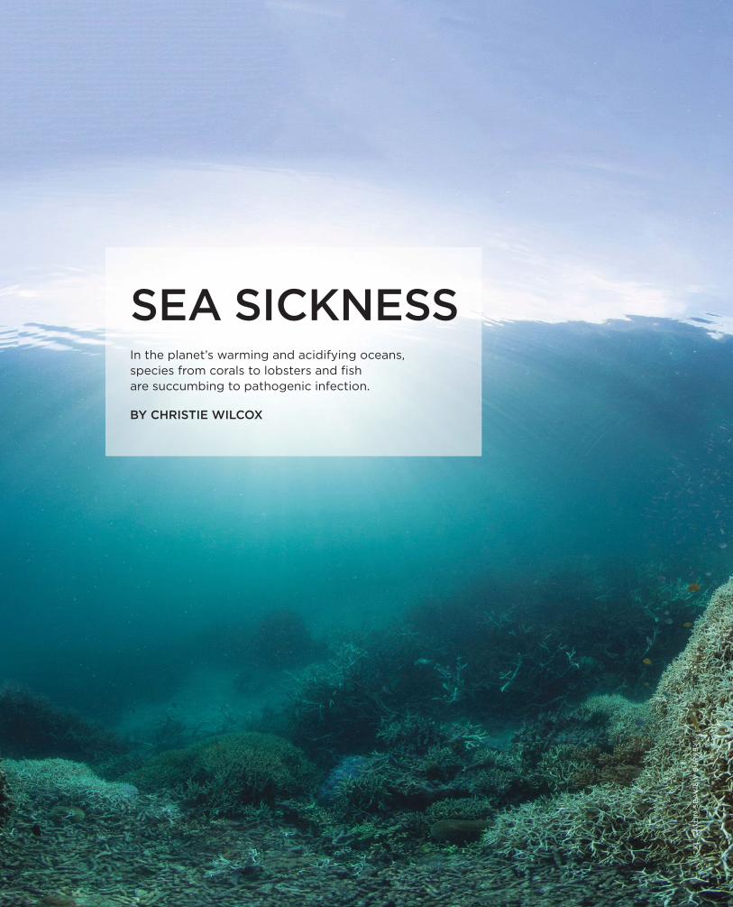
22Sea SicknessIn the planet’s warming and acidifying oceans, species from corals to lobsters and fish are succumbing to pathogenic infection.BY CHRISTIE WILCOX

BioResearch
CytoSMART™ System Live Cell Imaging – The Smart Way
View your cell culture via cloud access on smart phone, tablet or computer.© 2015 Lonza Walkersville, Inc.
Small, Easy and AffordableSized and priced for virtually any lab and budget, the CytoSMART™ System has been developed for live cell imaging and monitoring. The system is set up within minutes. Via innovative cloud technology, your cell culture is just one click away – monitor your cells anytime, anywhere.
Watch CytoSMART™ live in action!www.lonza.com/cytosmart

5 07.2016 | THE SCIENTIST
JULY 2016
Department Contents
ED
WA
RD
DE
LO
NG
, D
AV
ID K
AR
L,
NA
NC
Y H
UL
BIR
T;
PH
OT
O B
Y T
OM
KL
EIN
DIN
ST/
© W
OO
DS
HO
LE
OC
EA
NO
GR
AP
HIC
IN
ST
ITU
TIO
N;
WIK
IME
DIA
CO
MM
ON
S
10 FROM THE EDITOR
The Tides of ChangeMarine pathogens flourish in oceans that are warmer and more acidic.BY MARY BETH ABERLIN
12 FREEZE FRAME
Selected Images of the Day from the-scientist.com
14 NOTEBOOK
Babe Sleeps with the Fishes; Double Talk; Hidden Reefs; Office Mates
21 MODUS OPERANDI
Bound and TaggedAn RNA-editing approach reveals targets of RNA-binding proteins in specific cells.BY RUTH WILLIAMS
44 THE LITERATURE
Archaea in seafloor sediments generate acetate that is consumed by other seabed microorganisms; differential consumption of amino acids by marine bacterioplankton; multicellularity, cooperation, and cheating
46 PROFILE
Sounds from the SeasBehavioral ecologist Peter Tyack studies the social structures and behaviors of whales and dolphins, recording and analyzing their acoustic communications.BY ANNA AZVOLINSKY
49 SCIENTIST TO WATCH
Tessa Hill: Climate TrackerBY CATHERINE OFFORD
50 LAB TOOLS
Pointing in the Right DirectionComposite endpoints are increasingly used to boost the statistical sensitivity of clinical trials without ballooning costs. But there’s a right way and a wrong way to do it, experts say.BY SARAH C.P. WILLIAMS
54 LAB TOOLS
Exome ExercisesIdentifying and understanding the genetic components of rare diseasesBY KELLY RAE CHI
58 CAREERS
Bench to BioinformaticsIn today’s data-heavy research environment, wet-lab scientists can benefit from learning new computational skills.BY ESTHER LANDHUIS
62 READING FRAMES
Sugar and SocietyWhat the biomedical community gets wrong about race and metabolic syndromeBY ANTHONY RYAN HATCH
63 CAPSULE REVIEWS
BY BOB GRANT
68 FOUNDATIONS
Myth Becomes Reality, 1874BY CATHERINE OFFORD
IN EVERY ISSUE
9 CONTRIBUTORS
11 SPEAKING OF SCIENCE
64 THE GUIDE
65 RECRUITMENT
68�
46
44

6 THE SCIENTIST | the-scientist.com
Coming in August
JULY 2016
Online Contents
AS ALWAYS, FIND BREAKING NEWS EVERY DAY, AND LEAVE YOUR COMMENTS ON INDIVIDUAL STORIES ON OUR WEBSITE.
HERE’S WHAT YOU’LL FIND IN NEXT MONTH’S ISSUE:
• Human accelerated regions of the genome: What can they tell us?
• Recent human evolution
• Autism: looking at genes, neurons, and the developing brain
• The ins and outs of imaging software
• A profile of Clyde A. Hutchison III
AND MUCH MORE © L
AN
CE
WIL
LIS
; ©
IS
TO
CK
.CO
M/A
ND
RE
A I
ZZ
OT
TI;
© I
ST
OC
K.C
OM
/VA
LE
NT
YN
VO
LK
OV
; W
IKIM
ED
IA C
OM
MO
NSVIDEO
Oysters at RiskClimate change is causing ocean acidification, and shellfish, such as oysters, are one of many sea creatures affected.
VIDEO
Long-Distance CallsMarine scientist Peter Tyack expresses the beauty of marine mammal communication.
VIDEO
Amazonian ReefSee footage from the expedition that explored a coral reef hiding beneath the massive, muddy plume at the mouth of the Amazon River.
THIS MONTH AT THE-SCIENTIST.COM:

012.A1.0116.A © 2016 Eppendorf AG.Watch video!
New: 4 liter capacity!
More CapacityCentrifuge 5920 R
>
>> Benchtop centrifuge with
>
extraordinary high capacity in a very compact and ergonomic

EDITORIAL ADVISORY BOARD
Roger Beachy Donald Danforth Plant Science Center
Steven A. Bent Foley and Lardner LLP
Deborah Blum University of Wisconsin
Annette Doherty Pfizer Global Research and Development
Kevin Horgan GE Healthcare
Steve Jackson University of Cambridge
Simon Levin Princeton University Center for BioComplexity
Edison Liu Genome Institute of Singapore
Peter Raven Missouri Botanical Garden
Joseph Schlessinger Yale University School of Medicine
J. Craig Venter J. Craig Venter Institute
Marc Vidal Dana Farber Cancer Institute Harvard University
H. Steven Wiley Biomolecular Systems Pacific Northwest National Laboratory
Alastair J.J. Wood Symphony Capital
SUBSCRIPTION RATES & SERVICES In the United States & Canada individual subscriptions: $39.95. Rest of the world: air cargo add $25.
For assistance with a new or existing subscription please contact us at:
Phone: 847.763.9519 Fax: 847.763.-9674 E-mail: [email protected] Mail: The Scientist, PO Box 2015, Skokie, Illinois 60076
For institutional subscription rates and services visit www.the-scientist.com/info/subscribe or e-mail [email protected]
LIST RENTALS Contact Statlistics, Jennifer Felling at 203-778-8700 or [email protected]
REPRINTS Contact Lee Denton at [email protected]
PERMISSIONS For photocopy and reprint permissions, contact Copyright Clearance Center at www.copyright.com
415 Madison Avenue, Suite 1508, New York, NY 10017E-mail: [email protected]
POSTMASTER: Send address changes to The Scientist, PO Box 2015, Skokie, Illinois 60076. Canada Publications Agreement #40641071 The Scientist is indexed in Current Contents, Science Citation Index, BasicBIOS IS, and other databases. Articles published in The Scientist reflect the views of their authors and are not the official views of the publication, its editorial staff, or its ownership. The Scientist is a registered trademark of LabX Media Group Inc. The Scientist® (ISSN 0890-3670) is published monthly.
Advertising Office: The Scientist, 415 Madison Avenue, Suite 1508, New York, NY 10017. Periodical Postage Paid at New York, NY, and at additional mailing offices.
EDITORIAL
EDITOR-IN-CHIEFMary Beth [email protected]
SENIOR EDITORSJef [email protected]
Kerry [email protected]
ONLINE MANAGING EDITORTracy [email protected]
CONTRIBUTING EDITORAlla Katsnelson
COPY EDITORAnnie Gottlieb
CORRESPONDENTSAnna Azvolinsky Tanya Lewis Ruth Williams
INTERNCatherine Offord
DESIGN
AND PRODUCTION
ART DIRECTORLisa [email protected]
GRAPHIC DESIGNERErin [email protected]
MANAGEMENT
AND BUSINESS
PRESIDENTBob [email protected]
GENERAL MANAGERKen [email protected]
MANAGING PARTNERMario Di [email protected]
PUBLISHERRobert S. D’[email protected]
ADVERTISING,
MARKETING,
ADMINISTRATION
SENIOR ACCOUNT EXECUTIVESMid-West U.S., Eastern CanadaMelanie [email protected]
Ashley Haire (Munro)[email protected]
West U.S. and Western Canada, Pacific RimKaren Evans [email protected]
ACCOUNT EXECUTIVESNortheast U.S.Anita [email protected]
Southeast U.S., Europe, TS CareersNicole [email protected]
SENIOR DIRECTOR, CREATIVE SERVICESSusan Harrison Uysharrisonuy@ the-scientist.com
DIRECTOR, CREATIVE SERVICESVince [email protected]
TECHNICAL EDITOR, CREATIVE SERVICESElizabeth [email protected]
AUDIENCE DEVELOPMENT MANAGERBrian [email protected]
EVENTS MANAGERCayley [email protected]
ADMINISTRATOR, BUSINESS DEVELOPMENT Aoife [email protected]
CUSTOMER [email protected]

9 07.2016 | THE SCIENTIST
ContributorsAlthough Barbara Corkey has been working on metabolic disease research since her teens, it wasn’t until her 40s that she began graduate school. Nonetheless, “I have essentially been doing independent research all my life,” she explains. “I’ve always been in a lab, and I’ve always been focused on metabolic disease.” Corkey earned her PhD in 1981 researching branched-chain amino acid metabolism at the University of Pennsylvania and joined the faculty at Boston University in 1986. Since then, she’s focused on fat metabolism and the role of insulin. “If there’s one goal I’ve always had, it is to cure diabetes,” she says. Corkey currently serves as director of the Obesity Research Center at Boston Medical Center, and her awards include the American Diabetes Associa-tion’s Banting Medal for Scientific Achievement.
In “Pinpointing the Cause” on page 30, Corkey explores possible factors contributing to type 2 diabetes.
Understanding how tissues regenerate following injury has been the focus of Jeff Biernaskie’s research for nearly two decades. After earning a BSc in neuroscience at the University of Leth-bridge, Alberta, in 1998, Biernaskie began a PhD at Memorial University, Newfoundland, where he studied how neural stem cells in the brain respond to injury and rehabilitation. The work involved training rats to use limbs affected by stroke—a task achieved with M&Ms, he explains, because “rats really like chocolate.” After a postdoc at the Hospital for Sick Children in Toronto, Bier-naskie started his own lab at Calgary University’s veterinary school in 2012, and has broadened his research to study tissue regeneration and stem cell function in both skin and the nervous system.
Waleed Rahmani developed a fascination with stem cells as an undergraduate at the University of Victoria, British Columbia, and a research placement in 2009 with Andras Nagy at the University of Toronto convinced him to pursue it further. “Part of my work involved developing an online data-base of transgenic mice used in stem cell research,” he recalls. “I was exposed to many regenerative medicine studies. It really captured my interest.” After graduating, Rahmani moved to Jeff Biernaskie’s lab at the University of Calgary to work on skin stem cells and helped discover a bipotent dermal stem cell within adult hair follicles. Having recently completed his PhD, Rahmani is now attending medical school at Calgary and plans to enter the field of regenerative medicine.
When Sarthak Sinha’s family emigrated to Canada from India, Sinha says he was exposed to all sorts of opportunities he hadn’t had back home. He took part in various science fairs and competi-tions, and by the age of 14 had joined Jeff Biernaskie’s lab as a high school research assistant. “From there, I’ve always been interested in stem cells and regenerative medicine,” says Sinha, who has con-tinued to work in Biernaskie’s lab on a series of projects examining the role of stem cells in wound repair. Already an author on two published papers, Sinha is now a second-year undergraduate at the University of Toronto and hopes to become a clinician scientist.
Rahmani, Sinha, and Biernaskie discuss the importance of understanding interactions between stem cells and the immune system in “Cellular Teamwork” on page 36.
While an undergraduate in philosophy at Dartmouth College, Anthony Ryan Hatch took a part-time job as an administrative assistant at Emory University in Atlanta. The work offered him research experience on a health intervention project targeting black youth and kindled an inter-est in the ties between race and public health. For his MA and PhD in sociology at the University of Maryland, College Park, Hatch focused on the causes of health inequality. “The more I read, the more I kept seeing claims being made by scientists that reminded me of old-school scientific rac-ism,” he says. Exploring perceptions of race in biomedical science, he spent five years as a profes-sor at the Georgia State University before joining the faculty at Wesleyan University in Connecti-cut in 2015.
Hatch traces the relationships between race and metabolic syndrome in “Sugar and Society” on page 62, an essay based on his new book Blood Sugar: Racial Pharmacology and Food Justice in Black America.
JULY 2016

10 THE SCIENTIST | the-scientist.com
FROM THE EDITOR
AN
DR
ZE
J K
RA
UZ
E
Marine pathogens flourish in oceans that are warmer and more acidic.
BY MARY BETH ABERLIN
The Tides of Change
July always puts me in mind of the doo wop refrain: Summertime, summertime, sum sum summertime. My family decamped to the Jer-
sey Shore every summer, and the months I spent there left me with a lifelong love of the ocean: beach-combing for hours to see what the sea had spit onto the sand, surfcasting for bluefish or yummier blow-fish to cook for dinner. Later, on trips to warmer climes, snorkeling on the Great Barrier Reef and diving in the Caribbean revealed technicolor coral reefs and fishes as astonishingly beautiful as the marine organisms pictured on this month’s cover.
But the oceans of my youth mean something different to me now—less mysterious on some lev-els, yet even more fascinating. Much more is known about their denizens, thanks to marine scien-tists and advances in research technology: under-sea exploration vehicles can probe to extraordinary depths; worldwide expeditions have collected speci-mens ranging in size from viruses to whales.
Unfortunately, a lot of what marine biologists are learning is alarming. In “Sea Sickness” (page 22), science writer Christie Wilcox reports on the effects of rising ocean temperatures and acidity on a wide range of sea creatures. “The barrier reef north of Cairns will not look again how it did in my lifetime,” Terry Hughes, director of the Australian Resource Coun cil’s Centre of Excellence for Coral Reef Stud-ies, told Wilcox about the worst coral bleaching event ever to hit the northern part of the Great Barrier Reef. And the warming and acidifying seas appear to be contributing to a spike in diseased animals from sea stars to lobsters, from fishes to humans.
Other articles in this issue also focus on marine research. Behavioral ecologist Peter Tyack, this month’s profilee (“Sounds from the Seas,” page 46), has spent four decades listening to whales sing and dolphins communicate with their companions, and studying how cetaceans have recomposed their songs to deal with man-made noise—from ship engines, oil rigs, and naval sonar. Scientist to Watch Tessa Hill, of the University of California, Irvine, plots the corrosive effects of ocean acidification on mussels growing in the lab or in tidal pools on Cali-
fornia’s coast (page 49). See also reports of recent publications on the role of archaea in sea-floor sediments (page 44) and a method for studying amino acid assimilation by coastal bacterioplank-ton (page 45), and read about deep reefs discovered off the mouth of the Amazon River and elsewhere (page 17) and the 1874 documentation of a complete giant squid specimen (page 68).
As coral reefs offer habitats for an incredible variety of species, special niches in the human body harbor the tissue-specific stem cells necessary for replacing old or damaged cells. And similar to the symbioses so vital to life in reefs, the participation of the body’s immune cells is necessary to keep those tissue-renewal processes running smoothly. Read about it in “Cellular Teamwork” (page 36).
In this month’s third feature, on type 2 diabetes, Boston University School of Medicine’s Barbara Cor-key (“Pinpointing the Cause,” page 30) proposes that reducing elevated levels of lipids and insulin can yield better diabetes therapies. Rather than target-ing insulin resistance, treatment strategies should shift to those that protect pancreatic beta cells, Cor-key suggests.
Both Lab Tools articles focus on statistics and data mining. “Pointing in the Right Direction” (page 50) examines the use and misuse of compos-ite endpoints in clinical trials and how to make such endpoints meaningful. “Exome Exercises” (page 54) reviews three software tools aimed at making it eas-ier to identify and understand genes and variants involved in rare genetic diseases.
If you visit or swim in one of Earth’s oceans this sum sum summertime, I hope this issue will have you thinking about all that’s going on under the sea.

11 07.2016 | THE SCIENTIST
Speaking of Science
QUOTES©
AN
DR
EA
DA
NT
I/S
HU
TT
ER
ST
OC
K.C
OM
Further understanding of genetic blueprints could come from construction of large, gigabase-sized animal and plant genomes, including the human genome, which would in turn drive development of tools and methods to facilitate large-scale synthesis and editing of genomes. To this end, we propose the Human Genome Project–Write (HGP-write).
—New York University geneticist Jef Boeke and 24 others, in a Science Perspective about the launching of a new
genomic-scale engineering project (June 3)
There are only limited ethical concerns about synthesizing segments of DNA for laboratory experiments. But whole-genome, whole-organism synthesis projects extend far beyond current scientific capabilities, and immediately raise numerous ethical and philosophical red f lags.—Francis Collins, director of the National Institutes of Health,
speaking about the ethical pitfalls of HGP-write (June 2)
[Viruses], not lions, tigers or bears, sit masterfully above us on the food chain of life, occupying a role as alpha predators who prey on everything and are preyed upon by nothing.
—Claus Wilke and Sara Sawyer, virologists at the University of Texas at Austin andthe University of Colorado Boulder, respectively, in a recent eLife commentary on how
viruses drive evolution and adaptation in human and other mammalian genomes (May 17)
I think it’s ethically dubious to run the Olympics when you’ve got an epidemic of a virus that we don’t understand very well.
—New York University bioethicist Arthur Caplan, warning of the potential dangers to visitors and athletes attending this year’s Olympics in Rio de Janeiro
as the Zika virus continues to spread around the Americas (June 3)
If I would be an athlete competing, from what I’ve read, I would be more concerned about the pollution in the water than Zika.
— Alessandro Vespignani, physicist at Northeastern University in Boston, on the fears being expressed about holding the Olympics this summer
in Rio de Janeiro (June 3)
In the tale of Chicken Little, reasonable farm animals could disagree about whether the sky was falling, but no one had any misconceptions about what the petite poulet was chirping about. The discussion of reproducibility needs its own lingua franca.
—Journalists Ivan Oransky and Adam Marcus, writing in STAT abouta recent Science Translational Medicine paper that bemoans a lack of consensus
on defi ning “reproducibility” (June 1)

12 THE SCIENTIST | the-scientist.com
Caught on CameraSelected Images of the Day from the-scientist.com
FREEZE FRAME
MULLET MARBLESEach of these spheres is a developing sea mullet (Mugil cephalus) embryo.Posted: April 18, 2016
»
PRETTY POLYPSThe stony twists and
turns of this giant brain coral (Colpophyllia natans)
are formed from calcium carbonate secreted
by tiny polyps. Posted: January 14, 2016
»
WATER WORLDThree orbicular batfi sh (Platax orbicularis) swim through mangrove roots off Raja Ampat, Indonesia.Posted: April 29, 2016
»
RAKING IT INBarnacles use long, wispy appendages,
imaged here using confocal microscopy, to sweep passing plankton into their shells.
Posted: February 2, 2016
»

DEGRADING DEEPWATERMarine bacteria helped break down the hydrocarbons in this slick caused by the Deepwater Horizon oil spill in the Gulf of Mexico in 2010.Posted: May 17, 2016
»
ORIGIN STORYGill arches, situated below the eyes in this
skeletal preparation of a late-stage little skate (Leucoraja erinacea) embryo, may provide
clues about limb evolution in tetrapods.Posted: April 20, 2016
»
Water World: University of Miami Rosenstiel School of Marine and Atmospheric Science Underwater Photography Contest Best Overall Image, Beth Watson, Mangroves and Orbicular Batfish (Platax
orbicularis), Raja Ampat, Indonesia; Pretty Polyps: Royal Society Publishing Photography Competition 2015, Evan D’Alessandro; Mullet Marbles: Nikon 2015 Photomicrography Competition, Hannah
Sheppard-Brennand; Raking it in: 2014 Olympus Bioscapes Competition, Igor Siwanowicz; Degrading Deepwater: Dr. Luke McKay, Biofilm Center of Montana State University; Origin Story: J. Andrew Gillis

14 THE SCIENTIST | the-scientist.com
NotebookNEWS AND ANALYSIS
G.S
AN
DE
RS
ON
, L
.S.
BE
LL
, P
LOS
ON
E,
11:E
014
910
7, 2
016
.
Babe Sleeps with the FishesWhen forensic entomologist Gail Ander-son gave talks to police officers about what insects can do to dead bodies, inevitably somebody would ask her what happens to bodies dumped in the ocean. But with almost no work done on human decompo-sition in marine environments, “I couldn’t give them an answer,” the Simon Fraser University researcher says.
Around 2000, Robert Teather, a decorated officer in British Colum-bia’s Royal Canadian Mounted Police (RCMP) and founder of its Underwater Recovery Team, asked Anderson why she didn’t take matters into her own hands
and do some marine studies herself. The answer, she told him, was simply a mat-ter of resources. “I said, ‘I’d love to, but you’d need boats, and you’d need divers,’” she recalls. “And he said, ‘Gail, we have boats [and] divers.’”
So Anderson teamed up with the RCMP, along with the Canadian Coast Guard, Canadian Amphibious Search Team, and the Vancouver Aquarium, to place freshly killed pigs, straight from the butcher, in British Columbia’s Howe Sound and watch what happened. The team found that decomposition was influenced by sed-iment type and whether the carcasses, which were tethered to the bottom, floated or sank—and that the deterioration of the carcasses generally resembled what could be extrapolated from investigations of
human remains that had been recovered from the ocean (Int J Legal Med, 118:206-09, 2004). “It was really interesting but it was obviously limiting,” Anderson says, noting that divers could only check on the pigs every few days, and even those efforts depended on the weather. Moreover, they could only run the experiment at relatively shallow depths of up to 15 meters.
Anderson didn’t pursue the matter further until several years later, after giving a talk at the University of Vic-toria. “I was presenting my usual mur-der and maggots stuff,” she says, but she
JULY 2016
PORK DINNERS: Gail Anderson’s experimental setup included two pig carcasses, one caged and one uncaged, sunk to waiting scavengers on the ocean floor.

15 07.2016 | THE SCIENTIST
also briefly mentioned the work she had done with pig carcasses in Howe Sound. After her lecture, marine biologist Ver-ena Tunnicliffe approached her with an offer she couldn’t refuse: “[She] said, ‘I’m about to put a camera on the bot-tom of the ocean. Would you like to put a pig under it?’” Anderson recalls.
Tunnicliffe had helped develop Ocean Networks Canada’s Victoria Experimental Network Under the Sea (VENUS) obser-vatory, an underwater lab controlled from the surface via fiber optic cables. VENUS was the perfect opportunity for Anderson to test what happens to pig carcasses at 100 meters deep in the Saanich Inlet and to monitor decomposition 24/7. “It was terribly exciting,” Anderson says. “I was able to control my camera on my laptop [from] anywhere in the world.”
Anderson and her colleagues ran the experiment with a single pig carcass per year for three consecutive years. The first two carcasses were quickly scavenged by crustaceans, including larger species such as squat lobsters (Munida quadri-spina), Alaskan prawns (Pandalus platy-ceros), and Dungeness crabs (Metacarci-nus magister), as well as small amphipods commonly called sea lice (Orchomenella obtusa). But the third carcass stuck around, seemingly unchanged, for nearly three months. Then, suddenly, the scav-engers swooped in and skeletonized the pig’s body (PLOS ONE, 9:e110710, 2014).
“The driving force in the whole thing was oxygen,” Anderson explains. The researchers had placed the weighted car-casses in a spot that was, at times, almost completely devoid of oxygen, but would receive flushes of oxygenated water. The first two carcasses were deployed at times of relatively high oxygen lev-els, while the third was placed when the water was almost completely anoxic. Then, following a seasonal freshwater flush, the crustaceans and other organ-isms showed up. “Suddenly I turned the camera and there was a sea of fish—the oxygen had come back,” Anderson says.
Given the dramatic effect of the local conditions, Anderson and her colleagues decided they needed to repeat the experi-
ment in different locations. Taking advan-tage of another VENUS node in the Strait of Georgia, the researchers sank a pig to 300 meters and watched with anticipa-tion. “Within half an hour there were a whole bunch of sharks,” Anderson recalls; the carcass was completely devoured. “It was a hit video,” she joked, but it didn’t tell them much about decomposition in the absence of predators.
So the researchers developed a cage system and sent down two more car-casses, one in the cage and one uncaged. The sharks came again, but for whatever reason they were not as efficient as this time, biting at the uncaged carcass but leaving it relatively intact. Then came the amphipods. “Within a couple of hours, the carcasses were covered,” says Anderson, who affectionately refers to the amphipods as “maggots of the sea.” Within a few days, the amphipods had stripped the carcasses of flesh (PLOS ONE, 11:e0149107, 2016).
“The interesting thing is, you can skeletonize a dead body in just a few days,” says Achim Reisdorf, a paleontol-ogist at the University of Basel’s Insti-tute of Geology and Paleontology who has collaborated with Anderson in the past but was not involved in these stud-ies. “That’s really, really fast, and that’s a really important result of the study.”
The amphipods also appeared to repel all other animals, even the sharks, which would take a bite of the pig carcass and then immediately spit it out and send the amphipods shooting through their gill slits. And it may not just be the amphipods’ presence and bites that drive away other organisms; Anderson suspects that the tiny crustaceans were somehow causing the decline in local oxygen levels that she and her colleagues recorded. Unlike Saan-ich Inlet, the Strait of Georgia is normally well oxygenated, “but when the amphi-
pods were there, oxygen just dropped like a rock,” she says. “I think that was driving the [other] animals away.”
This finding may be of use to foren-sic scientists, Anderson notes. Although more work is needed, she speculates that there might be ways to detect that change in oxygen levels to help recov-ery divers locate a body. She also won-ders if the amphipods’ chomping might be audible with special equipment. “On land, you can hear [maggots] when you get close,” she says. “So I suspect you’re going to be able to hear the action of the little amphipods.” Meanwhile, her col-laborator Lynne Bell is examining the pigs’ skeletal remains to look for physical evidence of the feeding frenzy. “Marks made by insects [can be] mistaken for torture,” says Anderson. “It’s very impor-tant to be able to understand these kinds of things.”
“I believe that studies like this are very valuable to the field of forensic sci-ence, as they allow forensic experts to increase the accuracy of their postmor-tem interval calculations,” says Michael Humphreys, a forensic scientist at the Solano County, California, Sheriff ’s Office who has studied decomposition patterns in freshwater environments. Of course, he notes, the processes will likely differ based on climate and other factors, so similar studies should be car-ried out in different areas and at differ-ent times of year.
Anderson couldn’t agree more. “Ocean Networks Canada has gone into much deeper water, and that is our hope next.” —Jef Akst
Double Talk For years, doctors and teachers warned bilingual parents not to expose their young children to a second language, for fear they would suffer language confu-sion and language delays. New research is helping pediatricians and parents bet-ter understand the benefits of learning a second language, even for children who have yet to utter their first word.
The interesting thing is, you can skeletonize a dead body in just a few days. —Achim Reisdorf, University of Basel

16 THE SCIENTIST | the-scientist.com
AN
DR
ZE
J K
RA
UZ
E
NOTEBOOK
University of Delaware speech-lan-guage pathologist Aquiles Iglesias has helped develop tests to measure whether bilingual children, raised in environments where English and Spanish are used, truly have language delays, or are simply limited in their exposure to English. Pinpointing the difference has long perplexed educa-tors. Iglesias says research now shows con-clusively that bilingualism does not cause a language delay in children (Am J Speech-Lan guage Path, 23:574-86, 2014). “If you look at both languages simultaneously—like what we try to do in our assessment—then these kids are functioning fine,” he says. “If typically developing one-and-a-half-year-old kids have 50 words, for these dual-language learners they might have 50 words, but they’re not all in one language.”
With the old theory of speech delay cast in doubt, scientists are uncovering previously underappreciated benefits of bilingualism: a host of neural and health benefits for bilingual adults, including improved ability to recover from stroke and an increased volume of gray matter (Cereb Cortex, doi:10.1093/cercor/bhv152, 2015). And recent research has suggested
that the neural and cognitive advan-tages may start much earlier than adult-hood, perhaps even in preverbal children exposed to more than one language.
One study, from Naja Ferjan Ramirez and colleagues at the University of Washington, found that 11-month-old babies who have yet to utter their first words but are exposed to bilingual fam-ily members exhibit stronger brain activ-ity in areas related to executive function compared with babies raised in monolin-gual home environments.
Ferjan Ramirez says the babies were tested using a completely noninvasive and silent brain recording technique called MEG (magnetoencephalography), which maps brain activity by measuring magnetic changes in neural tissue. “We put them in this high chair, and we played language sounds for them. Some of these sounds were common to both Spanish and English, some were specific to Span-ish and some were specific to English.”
Ferjan Ramirez says her results showed that the brains of the monolin-gual babies were specialized to process the sounds of English while the bilin-
gual babies were trained to process the sounds of both languages equally (Dev Sci, doi:10.1111/desc.12427, 2016). “We looked across the entire brain surface to see where in the brain do these two groups differ. In the bilingual babies we see stronger responses in the prefron-tal and orbitofrontal cortex. And these areas are known to be involved in execu-tive functioning.”
Executive functioning is critical to real-life tasks including the ability to shift attention, to plan or juggle multiple tasks simultaneously, and to think more flexibly. Bilingual babies may there-fore have a leg up in this era of constant technology and the temptation to be dis-tracted or to multitask.
Ferjan Ramirez’s study was the first to use MEG to do whole-brain analyses comparing activity patterns in the brains of bilingual and monolingual babies in response to speech. The work indicates that young babies are practicing tasks related to executive function even before they can speak. Ferjan Ramirez says the results sug-gested that not only is second-language exposure good for children, but early child-hood is an ideal time for them to learn.
Other research also points to the ben-efits of speaking two tongues. In the lab of Diane Poulin-Dubois at Concordia Uni-versity in Montreal, Canada, 39 bilingual toddlers were compared with 43 monolin-gual toddlers when they were 24 months old and then again when the children were 31 months old. Cristina Crivello, a gradu-ate student in Poulin-Dubois’s lab, says the bilingual toddlers performed better than the monolingual toddlers on tasks assess-ing selective attention and cognitive flexi-bility (J Exp Child Psych, 141:121-32, 2016). “Selective attention refers to focusing on relevant information while ignoring dis-
In the bilingual babies we see stronger responses in the prefrontal and orbito-frontal cortex. —Naja Ferjan Ramirez,
University of Washington

17 07.2016 | THE SCIENTIST
PH
ILIP
TE
RR
Y G
RA
HA
M/W
IKIM
ED
IA C
OM
MO
NS
; JE
SS
E A
LL
EN
AN
D R
OB
ER
T S
IMM
ON
/WIK
IME
DIA
CO
MM
ON
S
tracting information, and cognitive flexi-bility refers to the mental ability to switch between thinking about two different con-cepts,” Crivello says. That could have real-world consequences: a child with high selective-attention abilities could have an easier time reading a book in a noisy class-room, for example, she says.
Their results also showed that the more toddlers engaged in language switching—as indicated by an increase in doublets in their vocabulary (e.g., “dog” in English and “chien” in French)—over a seven-month period, the more benefits they accrued in mental flexibility.
To assess the children’s conflict-inhibition abilities, the study partici-pants were asked to put little blocks in a little bucket and big blocks in a big bucket, then told to do the opposite. By performing this “reverse-categorization task,” the children were being forced to ignore certain conflicting information—namely the size of a block relative to the size of a bucket. That challenge of selec-tion and inhibition, Crivello says, is simi-lar to the choices a bilingual baby’s brain must make regularly. When a person speaks two languages, she explains, the dominant theory in the literature is that “when you’re using one of them, you’re inhibiting the other one because both languages are constantly activated.” That is especially true in cases where a word is more easily accessible in one language, but the brain must inhibit that impulse and instead reach for the word in the other language. Since bilingual kids are practiced at this form of inhibition, they tend to perform better on such tasks. The greater the overlap in a child’s two vocabularies, Crivello adds, the greater the inhibition that must take place, so the greater the cognitive benefits. —Elizabeth Fieldler
Hidden ReefsIn 2012, Fabiano Thompson boarded the US Navy research vessel Atlantis and set out into the Atlantic Ocean. He and his colleagues were aiming for a patch of
water off the coast of Brazil about 80 to 180 kilometers from the mouth of the Amazon River. Their mission: to find a previously unexplored reef system located in the unlikeliest of places.
“If you look at textbooks, they say that reefs do not form at large river mouths such as the Amazon,” says Thompson, a micro-biologist at the Federal University of Rio de Janeiro in Brazil. “We’re talking about a river that is exporting over 300,000 cubic meters of water per second into the ocean.” Large volumes of muddy freshwater—according to accepted wisdom—disrupt the preferred habitats of marine reef-building organisms, including corals and coralline algae such as rhodoliths.
But Thompson, along with Carlos Rezende of the State University of North-ern Rio de Janeiro and colleagues in Bra-zil and the U.S., had reason to believe the textbooks were wrong. “We had two leads to base our research on,” Thomp-son says. “The first was a study published in 1977, describing the presence of fish and sponges at the mouth of the Amazon River. The second was a publication from 1999 . . . reporting the presence of corals.”
During the 2012 expedition, and a sub-sequent trip in 2014, the team deployed a barrage of equipment, including a multi-beam echosounder to map the ocean floor—30 meters to 120 meters down—coring machines to remove samples, and trawl nets to collect larger animals. Ear-lier this year, they reported their findings:
a 9,500 square kilometer carbonate reef system, spread along a 50-kilometer by 1,000-kilometer corridor parallel to the coast. The reef comprises mostly rhodo-liths and sponges, the team found, and supports a bustling community of fish and crustaceans, all thriving beneath the murky plume of the Amazon River (Sci Adv, 2:e1501252, 2016).
Despite the inevitable astonishment that met this discovery, the fact that the Amazon reef has remained “hidden from view” until now is perhaps not so surpris-ing, says marine biologist J. Murray Rob-erts of Heriot Watt University in the U.K. “We know very little about what’s at the bottom of the ocean, even in areas that are relatively well studied like the Ama-zon river basin,” he says. Until the 1990s, it was generally accepted that reefs—particularly those built by corals—were
AMAZONIAN SURPRISE: Fabiano Thompson and colleagues have started mapping a large coral reef (top) beneath the murky plume of water flowing from the Amazon into the Atlantic Ocean (bottom).

18 THE SCIENTIST | the-scientist.com
NOTEBOOK
restricted to shallow warm water with high light penetration. Indeed, such reefs, which Roberts refers to as “the ones we see on our holidays,” have traditionally been the main focus of research. But per-spectives have been evolving as technol-ogy has taken science deeper into the world’s oceans.
“Practically, it’s got easier,” says marine geoscientist Veerle Huvenne, whose work involves mapping complex deep-sea habi-tats with the U.K.’s National Oceanography Centre. Along with echosounders like the one Atlantis used to map the topology of the ocean floor, “we also have robotic vehi-cles that we can send down to the seabed that allow us to have a much closer inspec-tion of those reefs,” she explains.
Tethered remote operated vehicles (ROVs), for example, allow researchers to explore environments from the safety of a research vessel using a hand-held con-troller. And free-swimming autonomous underwater vehicles (AUVs) following pre-programmed routes can cover large areas to survey potential reef locations. “First it’s a matter of finding what’s there,” says Huvenne. “Second, it’s understanding. When you understand, you can start pre-dicting other places these [reefs] will be.”
Progress has been significant in the last 20 years. Now it’s known that in contrast to the well-studied light-loving corals of the shallow tropics, many coralline algae and coral species—including several found in the Amazon reef—are adapted to meso-photic, or mid-light, conditions, while so-called “cold-water” corals build exten-sive reefs in the darkness of the deep sea, sometimes thousands of meters under-water or at latitudes far removed from the tropics. Roberts’s team, for instance, located a large Lophelia coral reef that has been growing for almost 4,000 years just a few kilometers off Mingulay Island in the Outer Hebrides, Scotland (Coral Reefs, 24:654-69, 2005); other assemblages have been identified as far north as the Cana-dian Arctic.
These discoveries, plus increasingly sophisticated surveys, are helping scientists understand the distribution and diverse ecology of the world’s reefs, says Harvey
Mudd College’s Andrea Quattrini, a coral researcher who has assisted in mapping the seafloor along the eastern coast of the U.S. and the Gulf of Mexico. It’s quite pos-sible, she adds, that the Amazon reef won’t be the last surprise in the understudied gloomy regions between around 30 meters and 150 meters. “In that ‘twilight zone,’ as it’s known, I think the more we explore the more we’re going to find long stretches of these mesophotic reefs all over the world.”
As they’re being discovered, document-ing and protecting these hidden reefs is becoming a priority. Bottom trawling by fishing boats slices through the seabed, pos-ing a serious threat to deep-sea habitats; the Amazon reef system, home to signifi-cant fisheries of red snapper and spiny lob-ster, faces possible damage from drilling by oil companies. And reefs at all depths are at risk from ocean acidification weakening their carbonate skeletons, as Roberts and colleagues recently demonstrated in Loph-elia pertusa (Proc R Soc B, doi:10.1098/rspb.2015.0990, 2015). Understanding reef distribution has become a crucial prerequi-site for projections and conservation efforts.
For Federal University’s Thompson, that means getting back on a boat. “We proposed a system with over 9,000 square kilometers, but were able to navigate and map only 10 percent of this,” he says. “We’ll have to go back there many times, refine our maps and put more pixels in our photographs. We’ve got our work cut out for many, many years!”
—Catherine Offord
Office MatesAlthough we normally associate micro-bial assemblages with mammalian guts, it turns out buildings have microbiomes too.
For the last decade, Scott Kelley, a biolo-gist at San Diego State University, and his fellow pioneers in the growing field of built-environment microbiology have been studying factors that shape those microbial communities and how they affect the health of people who work and live in them. “Westerners spend about 90 percent of their time indoors,” says Kelley. “We would like to know exactly what’s in there and who’s growing. Is it dangerous? How different is it from the outside?”
As a postdoc in the lab of Norman Pace at the University of Colorado Boul-der, Kelley spent his days sampling things like shower curtains and pools. As he and his colleagues catalog the bac-teria and fungi populating our homes, offices, and hospitals, they’ve highlighted the importance of variables such as geog-raphy (PLOS ONE, 7:e37849, 2012) and building ventilation (ISME J, 6:1469-79, 2012). But they have run up against one very important and hard-to-isolate vari-able: building materials. The question of whether different types of indoor surfaces or building materials favor some micro-bial communities over others has com-plicated indoor-microbiome research because building material can’t always be separated from other variables, such as location or usage. One would be hard-pressed, for example, to find an office with carpet on a surface other than the floor, or ceiling tiles that ever have con-tact with the bottoms of people’s feet. “It’s hard to decouple all these different fac-tors,” says Sean Gibbons, a postdoc who studies human microbiomes at MIT. He says most indoor-microbiome research has not tried to control for the types or locations of surfaces sampled within a given room.
Kelley and colleagues at Northern Arizona University (NAU) and the Uni-versity of Toronto took on the challenge. They designed an experiment to zero in on the influences of geographic loca-tion, position within a room (floor, wall, or ceiling), occupants of the room, and type of building material. For a year, 11 people in Toronto, San Diego, and Flag-staff, Arizona, worked in offices with
In that “twilight zone,” as it’s known, I think the more we explore the more we’re going to find long stretches of these mesophotic reefs all over the world.
—Andrea Quat trini, Harvey Mudd College
19 07.2016 | THE SCIENTIST
KA
TE
LA
UE
; JE
FF
KL
INE
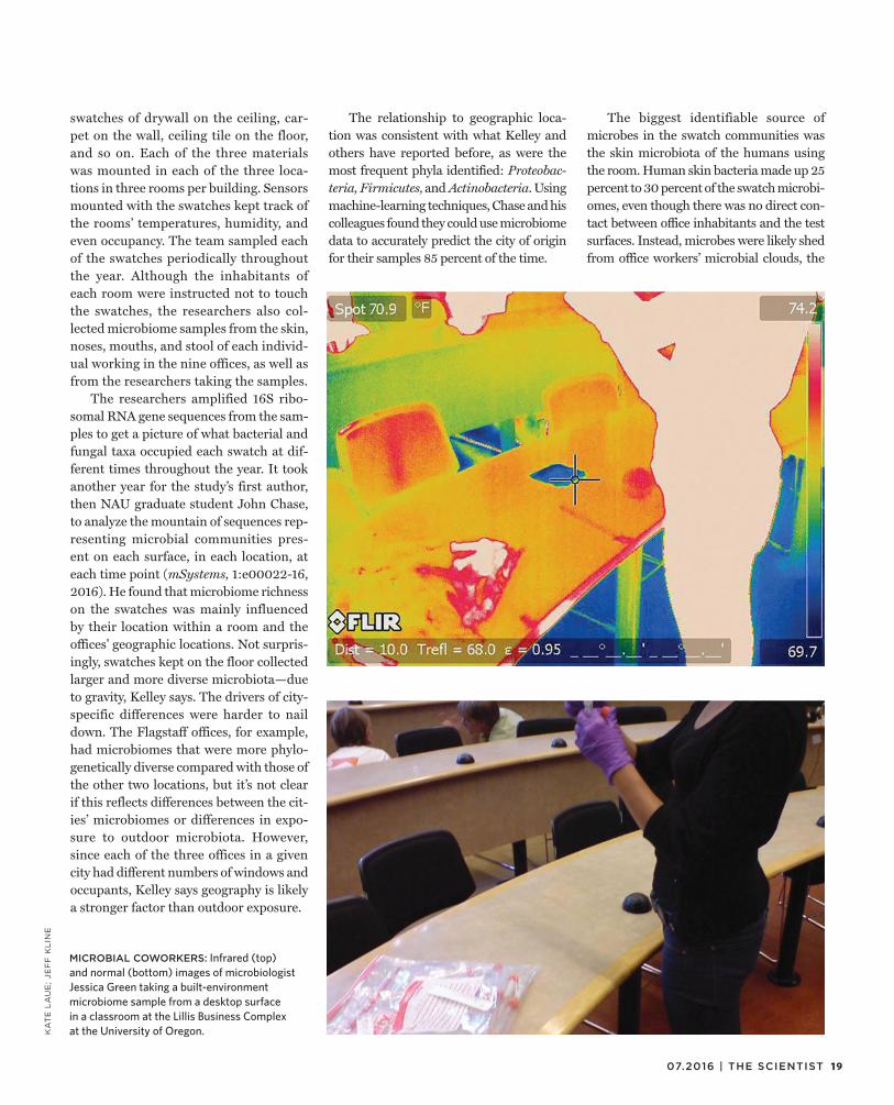
swatches of drywall on the ceiling, car-pet on the wall, ceiling tile on the floor, and so on. Each of the three materials was mounted in each of the three loca-tions in three rooms per building. Sensors mounted with the swatches kept track of the rooms’ temperatures, humidity, and even occupancy. The team sampled each of the swatches periodically throughout the year. Although the inhabitants of each room were instructed not to touch the swatches, the researchers also col-lected microbiome samples from the skin, noses, mouths, and stool of each individ-ual working in the nine offices, as well as from the researchers taking the samples.
The researchers amplified 16S ribo-somal RNA gene sequences from the sam-ples to get a picture of what bacterial and fungal taxa occupied each swatch at dif-ferent times throughout the year. It took another year for the study’s first author, then NAU graduate student John Chase, to analyze the mountain of sequences rep-resenting microbial communities pres-ent on each surface, in each location, at each time point (mSystems, 1:e00022-16, 2016). He found that microbiome richness on the swatches was mainly influenced by their location within a room and the offices’ geographic locations. Not surpris-ingly, swatches kept on the floor collected larger and more diverse microbiota—due to gravity, Kelley says. The drivers of city-specific differences were harder to nail down. The Flagstaff offices, for example, had microbiomes that were more phylo-genetically diverse compared with those of the other two locations, but it’s not clear if this reflects differences between the cit-ies’ microbiomes or differences in expo-sure to outdoor microbiota. However, since each of the three offices in a given city had different numbers of windows and occupants, Kelley says geography is likely a stronger factor than outdoor exposure.
The relationship to geographic loca-tion was consistent with what Kelley and others have reported before, as were the most frequent phyla identified: Proteobac-teria, Firmicutes, and Actinobacteria. Using machine-learning techniques, Chase and his colleagues found they could use microbiome data to accurately predict the city of origin for their samples 85 percent of the time.
The biggest identifiable source of microbes in the swatch communities was the skin microbiota of the humans using the room. Human skin bacteria made up 25 percent to 30 percent of the swatch microbi-omes, even though there was no direct con-tact between office inhabitants and the test surfaces. Instead, microbes were likely shed from office workers’ microbial clouds, the
MICROBIAL COWORKERS: Infrared (top) and normal (bottom) images of microbiologist Jessica Green taking a built-environment microbiome sample from a desktop surface in a classroom at the Lillis Business Complex at the University of Oregon.

NOTEBOOK
constellation of fungi, bacteria, and viruses that encircles each one of us. Regardless of which surfaces the microbes landed on, once settled they stayed largely the same. In fact, the main variable the team was inter-ested in—building material—had no influ-ence on microbiome composition or rich-ness. “These surfaces are just sort of inert,” says Gibbons; built environments are pretty much just passive accumulators of microbes. “It’s like we’re living in these inert boxes.” And the microbiology that is present is largely derived from humans, “so we’re sort of stewing in our own microbial soup. That might be a bad thing.”
Aside from things like toxic mold, Gibbons says, the relationship between built-environment microbiomes and human health is largely undefined. For example, it’s unclear how built-envi-ronment microbiome compositions mesh with the hygiene hypothesis—the idea that a dearth of exposure to out-
door microbes contributes to higher incidences of autoimmune and aller-gic disease. “[Built environments] act as barriers between us and the types of microbial diversity we should be exposed to, especially during childhood when our immune systems are developing,” says Gibbons. It’s not clear whether some indoor microbiomes could exacerbate or remedy this issue.
Although his study provides a good springboard from which to launch fur-ther indoor microbiome research, Kelley is not ready to discount building material as an important variable just yet. “If it’s normally a dry indoor place, we’re seeing that you’re not getting much difference between the materials,” he says. “Once they get wet, though, that’s a totally open question.” He and his colleagues intend to test which microbes will populate build-ing materials once they get wet or start to break down.
According to Jessica Green, director of the Biology and the Built Environment Center at the University of Oregon, build-ing-material composition may matter for high-touch areas, such as countertops in hospitals. “We know that in health-care environments, design commonly steers away from carpets,” she says. “There is some evidence to suggest that there’s a lower risk of infection in a high-copper environment, for example.”
It is also possible that building mate-rial could influence microbial gene expression or viability. “[16S ribosomal RNA gene sequencing] gives you a pic-ture of all microbes that are on a sur-face, whether they are dead, dormant, or alive,” says Green. Focusing on living or metabolically active communities could give totally different results. “There are all kinds of directions that this research could go.”
—Amanda B. Keener

2107.2016 | THE SCIENTIST
MODUS OPERANDI
AT A GLANCE
RNA-binding proteins (RBPs) influence, among other things, the stability, splicing, nuclear export, and translation of their target transcripts. Identifying RBP-RNA interactions is thus of great
value for understanding gene expression, cellular functions, and more.A common method for identifying RBP targets is to cross-link and
immunoprecipitate the RBP-RNA complexes from cell extracts. However, says Roy Parker of the University of Colorado Boulder, a persistent prob-lem “is how to do this on [small] specialized subsets of cells.” After all, explains Michael Rosbash of Brandeis University in Massachusetts, “one can simply not do biochemistry on tiny amounts of material.”
Rosbash and his team have thus come up with what he calls “a geneticist’s work-around.” In his team’s approach, called Targets of RNA-binding proteins Identified By Editing (TRIBE), RBPs are fused to the catalytic domain of an RNA-editing enzyme from fruit flies. The enzyme converts adenosine nucleotides into inosines, which
appear as guanosines in sequencing readouts. Identifying the RBP targets, then, is a simple case of extracting and sequencing RNA and looking for aberrant Gs (Cell, 165:742-53, 2016).
The team has successfully applied the technique to three differ-ent RBPs and has shown it to work with as few as 150 fly neurons, though Rosbash suggests it could work with even fewer. “In princi-ple, it’s totally compatible with single cells,” he says.
Marvin Wickens of the University of Wisconsin–Madison and col-leagues devised a similar technique called RNA Tagging. The method fuses RBPs to an enzyme that adds uridines to the ends of associated transcripts (Nat Methods, 12:1163-70, 2015).
“Both [approaches] have pluses and minuses,” says Parker, who is experimenting with the two techniques in his own lab. And in the future, he suggests, it may even be possible to combine them, enabling researchers “to look at two [RBPs] at once.”
An RNA-editing approach reveals targets of RNA-binding proteins in specific cells.
BY RUTH WILLIAMS
Bound and Tagged
TAGGING TECHNIQUE
RNA Tagging
TRIBE
RNA-BINDING PROTEIN FUSED WITH
Poly(U) polymerase PUP-2 from C. elegans
Catalytic domain of RNA-editing enzyme ADAR from D. melanogaster
ENZYME FUNCTION
Adds uridines at the 3’ end of bound RNAs
Converts adenosines to inosines at sites targeted by RNA-binding proteins
SAMPLE PROCESSING FOR SEQUENCING
A specialized cDNA library is made with “U-select” primers, which home in on U-tags.
Standard RNA extraction and preparation protocols for RNA-seq
NEW GAMES OF TAG: The TRIBE method for identifying RNAs that interact with ribosome-binding proteins (RBPs) fuses an RBP of interest to the catalytic domain (CD) of an RNA-editing enzyme. When the RBP-CD fusion protein binds to the RBP’s specific RNA targets, the CD converts adenine nucleotides to inosine nucleotides. Extraction and RNA sequencing thus reveals those RNAs with altered sequences 1 . RNA Tagging fuses an RBP of interest to an enzyme that attaches chains of uridines (U) to the ends of the RBP’s target RNAs. The poly-U tails are then used to identify targets during RNA sequencing 2 .
mRNA
© G
EO
RG
E R
ET
SE
CK
mRNA
TRIBE RNA TAGGING
SINGLE-CELL ANALYSIS
Theoretically possible, but because U-tag library protocol is not a standard approach for high-throughput sequencing, it may need tweaking.
Possible in principle; TRIBE uses the same standard deep-sequencing technique that has been applied to single cells.
CDRBP
PUPRBP
poly-U tailAdenine Inosine
1 2
22 THE SCIENTIST | the-scientist.com
SEA SICKNESSIn the planet’s warming and acidifying oceans, species from corals to lobsters and fi share succumbing to pathogenic infection.
BY CHRISTIE WILCOX
XL
CA
TL
IN S
EA
VIE
W S
UR
VE
Y

A DEATHLY PALLOR: More than half of the northern Great Barrier Reef’s corals, such as
those at this Lizard Island site, have succumbed to bleaching, a loss of their algal symbionts that
leaves them vulnerable to infection.
07.2016 | THE SCIENTIST 23
24 THE SCIENTIST | the-scientist.com
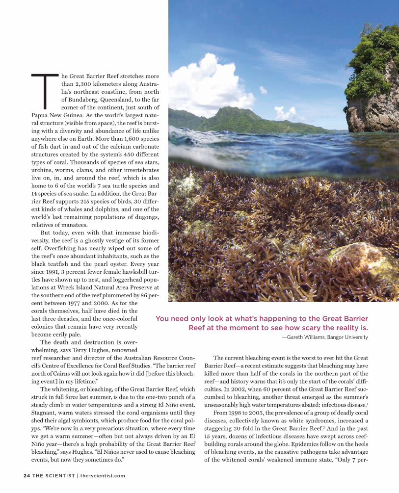
The Great Barrier Reef stretches more than 2,300 kilometers along Austra-lia’s northeast coastline, from north of Bundaberg, Queensland, to the far corner of the continent, just south of
Papua New Guinea. As the world’s largest natu-ral structure (visible from space), the reef is burst-ing with a diversity and abundance of life unlike anywhere else on Earth. More than 1,600 species of fish dart in and out of the calcium carbonate structures created by the system’s 450 different types of coral. Thousands of species of sea stars, urchins, worms, clams, and other invertebrates live on, in, and around the reef, which is also home to 6 of the world’s 7 sea turtle species and 14 species of sea snake. In addition, the Great Bar-rier Reef supports 215 species of birds, 30 differ-ent kinds of whales and dolphins, and one of the world’s last remaining populations of dugongs, relatives of manatees.
But today, even with that immense biodi-versity, the reef is a ghostly vestige of its former self. Overfishing has nearly wiped out some of the reef ’s once abundant inhabitants, such as the black teatfish and the pearl oyster. Every year since 1991, 3 percent fewer female hawksbill tur-tles have shown up to nest, and loggerhead popu-lations at Wreck Island Natural Area Preserve at the southern end of the reef plummeted by 86 per-cent between 1977 and 2000. As for the corals themselves, half have died in the last three decades, and the once-colorful colonies that remain have very recently become eerily pale.
The death and destruction is over-whelming, says Terry Hughes, renowned reef researcher and director of the Australian Resource Coun-cil’s Centre of Excellence for Coral Reef Studies. “The barrier reef north of Cairns will not look again how it did [before this bleach-ing event] in my lifetime.”
The whitening, or bleaching, of the Great Barrier Reef, which struck in full force last summer, is due to the one-two punch of a steady climb in water temperatures and a strong El Niño event. Stagnant, warm waters stressed the coral organisms until they shed their algal symbionts, which produce food for the coral pol-yps. “We’re now in a very precarious situation, where every time we get a warm summer—often but not always driven by an El Niño year—there’s a high probability of the Great Barrier Reef bleaching,” says Hughes. “El Niños never used to cause bleaching events, but now they sometimes do.”
The current bleaching event is the worst to ever hit the Great Barrier Reef—a recent estimate suggests that bleaching may have killed more than half of the corals in the northern part of the reef—and history warns that it’s only the start of the corals’ diffi-culties. In 2002, when 60 percent of the Great Barrier Reef suc-cumbed to bleaching, another threat emerged as the summer’s unseasonably high water temperatures abated: infectious disease.1
From 1998 to 2003, the prevalence of a group of deadly coral diseases, collectively known as white syndromes, increased a staggering 20-fold in the Great Barrier Reef.2 And in the past 15 years, dozens of infectious diseases have swept across reef-building corals around the globe. Epidemics follow on the heels of bleaching events, as the causative pathogens take advantage of the whitened corals’ weakened immune state. “Only 7 per-
You need only look at what’s happening to the Great Barrier Reef at the moment to see how scary the reality is.
—Gareth Williams, Bangor University

25 07.2016 | THE SCIENTIST
XL
CA
TL
IN S
EA
VIE
W S
UR
VE
Y/G
LO
BA
LC
OR
AL
BL
EA
CH
ING
.OR
G
cent of [Great Barrier] reefs are completely free of bleaching,” says Hughes. In all likelihood, these corals are now vulnerable to the plethora of pathogenic bacteria, fungi, and viruses lurk-ing in sediments and seawater.
And corals are not alone. Warming and acidifying oceans appear to be contributing to an uptick in diseases among other species, too. From 2013 to 2015, an unprecedented outbreak of sea star wasting disease decimated populations of 20 dif-ferent species from Mexico to Alaska, killing 90 percent of the sea stars in some areas. Since 2000, young Caribbean lobsters have been falling victim to a viral infection that leaves them with no energy to move or eat. Oysters 3 and abalone 4 have been plagued by Vibrio bacteria, and numerous fish species are regularly attacked by the protozoan Ichthyophonus.5 In many of these cases, the disease outbreaks have been linked to cli-mate change.
“We have a narrow window of opportunity to quickly reduce greenhouse gas emissions before the degradation of reefs becomes irreversible,” says Hughes.
Heat strokeScientists use satellites to track the daily temper-ature of the planet both over land and across the seas. These data are then averaged over different time scales to determine how hot, globally speak-ing, a given month, year, or decade is. Each of the past 15 years (2001–2015) have been among the hottest 16 years on record (since 1880); 2014 and 2015 shattered temperature records. And if the first few months of 2016 are any indication, this year will make those years seem cool by compari-son: this April marked the 12th consecutive record-breaking month.
In the oceans, surface temperatures have increased at an average rate of 0.12 °C per decade since 1976—triple the rate of warming that occurred in the 75 years before that (0.04 °C per decade). And the warming is hastening: global ocean temperatures in 2016 have been 0.82 °C (1.48 °F) above average and 0.21 °C (0.38 °F) hot-ter than 2015, making them the hottest waters since record-keeping began 137 years ago.
According to Drew Harvell, an expert on marine infectious diseases and professor of ecology at Cornell University, the effects of climate change are “a double whammy” because they simultane-
ously help pathogens while harming their hosts. Because of the particulars of their environmental preferences, “a lot of marine bacteria, viruses, and fungi grow better at warmer temperatures,” she explains. At the same time, the animals they infect are weak-ened by the hotter temps. “It’s a perfect storm of trouble.”
The gooey mess that remains of the sea stars in the Pacific from Mexico to Alaska is a prime example. Beginning in 2013, “melted” sea stars began appearing along the US West Coast en masse. The animals, which made their way to the shallow tide pools that become exposed at low tides, were infected with what scientists have dubbed sea star–associated densovirus, the caus-ative agent of sea star wasting disease. A little more than two weeks after infection with the virus, lesions appear and the sea star’s arms fall off, leaving behind a slimy, decaying disc. The virus isn’t new; it has been around for at least 70 years, and there have been smaller densovirus outbreaks before, affecting just one or two sea star species in a localized area. But the recent epidemic dwarfed previous events, hitting 20 different species along thou-sands of miles of coastline—a catastrophe made possible by ris-
A QUICK BLEACH: In just three months—from December 2014 (left) to February 2015 (right)—the corals
off the coast of American Samoa were stripped of their algal symbionts, turning the reef white.

26 THE SCIENTIST | the-scientist.com
MELTING AWAY: Starting in 2013, a densovirus spread rapidly along the North American west coast, from Mexico to Alaska, killing as many as 90 percent of the sea stars in some areas. Following infection, lesions begin to appear on the animals and their arms begin to fall off. Sea star wasting disease appears to be increasing in frequency and severity thanks to rising sea-surface temperatures.
ing sea surface temperatures, according to research by Harvell lab graduate student Morgan Eisenlord.
Surveying more than 6,500 ochre stars (Pisaster ochraceus) at 16 widespread sites between December 2013 and July 2015, Eisenlord and her colleagues watched as 80 percent of the adults disappeared. When the researchers modeled how the disease pat-terns correlated with various environmental variables, they dis-
covered a strong link between wasting and heat. For every 1 °C increase in temperature, the probability of disease increased by 1.30. Laboratory experiments confirmed these results—the warmer the water, the more quickly the echinoderms succumbed to infection.6
Charlotte Eve Davies, a postdoctoral fellow at the National Autonomous University of Mexico, has seen the same pattern in
TE
MP
ER
AT
UR
E D
ATA
: N
CD
C.N
OA
A.G
OV
; A
CID
IFIC
AT
ION
DA
TA:
IPC
C,
20
07/
GR
ID-A
RE
ND
AL
; S
EA
STA
RS
: M
EL
ISS
A M
INE
R/U
C S
AN
TA C
RU
Z
1880 20151950
-0.5
-0.4
-0.3
-0.2
-0.1
0.0
0.1
0.2
0.3
0.4
0.5
0.6
0.7
0.8
GLOBAL OCEAN TEMPERATURE ANOMALIES JULY 1880–2015
1985
300 199
0
199
5
200
0
200
5
1985
199
0
199
5
200
0
200
5
320
340
360
380
8.06
8.08
8.10
8.12
8.14
GLOBAL OCEAN ACIDIFICATION 1985–2005
Oceanic CO2 concentration, atm Ocean water acidity, pH
AN
OM
ALY
(°C
)

27 07.2016 | THE SCIENTIST
ered, and Maine fishermen worry that their waters—which sup-port a $465.9-million-a-year lobster fishery—are in ESD’s path of destruction as the oceans continue to warm.8
Farther south, in the Caribbean, Davies is now tracking a dis-ease threatening another lobster fishery. In the state of Quintana Roo, Mexico, more than 2,600 families rely on Caribbean spiny lobster fishing. But their livelihood is threatened by Panulirus argus virus 1 (PaV1), which enters the hemolymph (arthropod blood) and drains it of essential oxygen-carrying cells, turning the clear fluid white. This causes the lobsters to become extremely lethargic, unable to eat or move. Eventually the animals starve to death. The virus, first discovered in 2000,9 infects 60 percent or more of the spiny lobsters (Panulirus argus) in some areas of the Caribbean. And once again, laboratory studies suggest that temperature is playing a big role: when kept in warmer waters, lobsters develop more-active and more-intense infections, while cooler waters reduce the pathogen’s virulence.
“It is thought that PaV1 is becoming an important source of mortality for juveniles,” says Davies. “If PaV1 continues to spread, it could have significant effects on the health of Carib-bean reefs as a whole, as well as on the valuable Caribbean lob-ster fishery.”
The acid testRising global temperatures are largely due to the increase in atmospheric carbon dioxide (CO2) that primarily stems from automobile and industrial emissions. But higher atmospheric CO2 doesn’t just warm the planet; it also lowers the pH of sea-water by reacting with H2O to form carbonic acid. The ocean has become 30 percent more acidic in the last 200 years and, as with temperature, the rate of change is accelerating.10
Calcifying organisms, including corals and the coralline algae that paint the reefs’ surfaces and cement their structures, are par-ticularly vulnerable to ocean acidification. The lower pH makes it more difficult for these species to produce the calcium carbon-ate structures that form the foundation of the reef, and extreme acidification speeds the dissolution of existing carbonate struc-tures, dissolving the very foundations upon which corals build their homes.
Because acidification stresses these reef-building organisms separately from temperature-related effects, it is thought that the combination of changes will present a worst-case scenario for coral and coralline algae. “I think many people assume global climate change impacts, such as ocean warming and acidifica-tion, will have additive or synergistic affects on disease impacts to reefs,” says Gareth Williams, a lecturer in the School of Ocean Sciences, Bangor University, U.K. But as scientists delve deeper into marine epidemiology, they are discovering that the reality is far more complex.
In 2009, El Niño conditions led to an outbreak of coralline fungal disease (CFD), which afflicts coralline algae, in Palmyra Atoll, southwest of Hawaii. Once it has infiltrated its host, the fun-gus radiates outward, leaving patches of dead coral and bare rock
LETHARGIC LOBSTERS: The hemolymph of Caribbean lobsters infected with the Panuliris argus virus 1 (PaV1) is drained of essential oxygen-carrying cells, causing it to turn white (top right) and making the crustaceans extremely lethargic to the point that they cannot eat or move. The virus can also cause discoloration and fouling of the carapace (middle and bottom). And just as with sea star–associated densovirus, the pathogen seems to be growing increasingly virulent as global ocean temperatures rise.
crustacean diseases. In the 1990s, after nearly a decade of excep-tionally warm summers, the lobster fisheries operating in the waters off Connecticut, Massachusetts, New York, and Rhode Island collapsed, at least in part due to the emergence of epizootic shell disease (ESD).7 The fatal bacterial infection creates holes in infected lobsters’ shells, preventing the animals from properly molting. The southern New England fishery has still not recov-
TO
P:
BE
HR
ING
ER
ET
AL
., D
IS A
QU
AT
OR
G,
94
:15
3–1
60
, 2
011
; M
IDD
LE
: C
OU
RT
ES
Y O
F D
ON
AL
D B
EH
RIN
GE
R;
BO
TT
OM
: JE
FF
SH
IEL
DS
, V
IRG
INIA
IN
ST
ITU
TE
OF
MA
RIN
E S
CIE
NC
E
28 THE SCIENTIST | the-scientist.com
© I
ST
OC
K.C
OM
/TA
MM
Y6
16
in its wake. Studying the outbreak’s destruction, Williams and his colleagues found that the temperature and acidification of the water worked against each other. Higher temperatures increased the disease’s prevalence and lethality, while more-acidic waters, though they stressed and weakened its algal host, also slowed the fungus’s spread.11
“Such complex, interactive effects between global climate change stressors on disease dynamics are important to consider if we are to accurately predict the response of coral reef commu-nities to future climate change,” says Williams.
Unfortunately, for most marine diseases, the role of acidifica-tion hasn’t been well studied. “The component of climate change that we have stressed so much is temperature because it’s just such a pervasive influence,” says Harvell. “Ocean acidification is a whole other matter. We know almost nothing about its poten-tial role or ability to affect diseases. It’s a big knowledge gap that needs to be filled.”
Furtive fish infectionsStudying any marine organism is inherently challenging, as humans require expensive equipment to spend any amount of time beneath the waves. But some species are easier to examine than others. Sea stars and lobsters don’t travel much, and corals don’t relocate once they’ve settled down, so returning to them time after time to evaluate their health is fairly straightforward. Fish, on the other hand, can travel great distances rather quickly. “One day, a herring school might be 50 miles from where it was yesterday, and so it if dies, first of all, you may not even see the mortality event, and if you do happen to see it, you don’t really have a good feel for the scale,” says fishery biologist Paul Hersh-berger, station leader of the US Geological Survey’s Marrowstone Marine Field Station in Nordland, Washington.
Hershberger recalls studying an outbreak of the unicellu-lar parasite Ichthyophonus among king salmon spawning in the Yukon River in the early 2000s; about 30 percent of the fish were infected. As the fish swam upstream, their symptoms worsened, until just before they got to the spawning grounds, when diseased fish died. And when they die, they sink, Hershberger says. “We could never find the diseased fish in the river because they’d die and sink to the bottom, and the water was the color of coffee with too much cream in it,” he says. “At one point I was actually stand-ing in the water, knee-deep, watching a fish die right in front of me. It would come to the surface and kind of roll over, and I’d try to grab it, and then it’d go down to the bottom. That was going on for about a half an hour, and I kept trying to grab it, and eventu-ally it died and floated downstream. I never got it.”
Because of the challenges in tracking fish and their diseases, researchers have little historical data on the frequency or scale of disease outbreaks in wild fishes, so it’s impossible to say whether such maladies are increasing due to climate change. But some studies suggest that warming waters are likely to alter the status quo.12 “Fish immune systems are extremely dictated by tempera-ture,” says Hershberger. “We see the difference of a couple degrees
PREDICTING MARINE OUTBREAKSScientists want to predict outbreaks of marine disease before they happen. With the appropriate tools, says Cornell University postdoc Jeffrey Maynard, “we can mobilize resources, generate political and social will, target research and monitoring, and potentially implement actions that reduce anthropogenic stressors that may interact synergistically with temperature.”
Already, scientists have developed methods to predict coral disease outbreaks based on temperature. Twice a week, for example, the National Oceanographic and Atmospheric Administration’s Coral Reef Watch uses a predictive algorithm and real-time NOAA satellite measurements of sea-surface temperatures around the world to ascertain areas at imminent risk for bleaching. Scientists and ecosystem managers receive alerts at the earliest signs of trouble.
Terry Hughes, director of the Australian Research Council Centre of Excellence for Coral Reef Studies, relies on similar meteorological predictions to monitor the risks affronting life in the Great Barrier Reef. “I formed a task force in November last year, when it was obvious that the coming austral summer was going to be very hot, with El Niño conditions—hot, calm, few cyclones—that favor bleaching,” says Hughes. Christened the National Coral Bleaching Taskforce, the network consists of 10 institutions working together to collaborate on reef research and share data, responsibilities, and resources. So when the most recent event began, Australia had “an industrial-scale response to the bleaching,” Hughes says. “We have done aerial surveys of bleaching on over 1,000 reefs, underwater surveys on more than 150 reefs to measure bleaching and mortality of corals, and collected samples to examine the physiological and cellular and molecular responses of corals, fish, and other organisms.”
Data collected in the field will be fed back into NOAA’s algorithms for Coral Reef Watch and into an Australian version written by the Bureau of Meteorology to improve the models’ accuracy. Right now, such tools are still in their infancy, so they aren’t perfect coral fortunetellers. “The predictions for bleaching by NOAA and the [Australian Bureau of Meteorology] wax and wane, and like any other meteorological forecast, they don’t always get it right, especially early on,” says Hughes.
Pioneering such efforts is ReefTemp, an experimental product created by researchers from the Australian Research Council’s Centre of Excellence for Coral Reef Studies. ReefTemp works much as Coral Reef Watch does, but for coral white syndromes. So rather than predicting bleaching, it predicts disease outbreaks—whether they follow on the heels of a bleaching event or not. Its predictive algorithms integrate satellite data with variables linked to the disease to forecast what areas are at high risk for outbreaks.
And ReefTemp is just the beginning; the ultimate goal is to create a suite of tools that can predict marine disease outbreaks, from Vibrio to PaV1 and beyond. “This is a very new research area,” says Maynard. “There are many host-pathogen systems with links to temperature for which predictive tools could be developed.”

29 07.2016 | THE SCIENTIST
centigrade turning on or off certain immune-response genes and making fish more or less susceptible to pathogens.”
Interestingly, the response is opposite to that of corals and other invertebrates: in fish, the expression of genes that help fight viral infections is enhanced at warmer temperatures. “So the fish become less susceptible because their immune system is ramped up and ready to deal,” says Hershberger. How this plays out in the face of pathogens that also benefit from the change in tempera-ture remains to be seen.
Beyond temperature and pH changes, Hershberger points to yet another effect of climate change: the alteration of global cur-rents, which may affect patterns of plankton movement and accu-mulation. “Many of these plankton assemblages likely serve as the intermediate host for some of these fish pathogens,” he says. “Unfortunately, we’re just not there yet in our understanding of the parasite life cycles in the marine environment to be able to predict which ones are going to be the winners.”
A human elementClimate change may do more than unleash scores of diseases that attack marine organisms. Lurking in ocean waters are pathogens, such as V. parahaemolyticus and V. vulnificus, which can infect people, either through open wounds on the body or via consump-tion of contaminated and undercooked seafood, such as raw oys-ters. With an unsettling case fatality rate above 50 percent,13 Vib-rio bacterial infections are the leading cause of seafood-related deaths in the United States.14
Vibrio outbreaks are becoming more frequent and are occur-ring in areas where they previously haven’t. “If you asked 10 years ago, you would not have heard of Vibrio cases occurring in the North Sea,” says Rachel Noble, director of the University of North Carolina’s Institute for the Environment field site in Morehead City. “Nor would you hear of them occurring in the northern bays of Norway, Sweden, or Finland.” But now, those waters, too, have changed enough to allow these pathogenic species to flourish. “They’re very opportunistic,” says Noble. As global temperatures rise, the seasonal range of these bacteria might also expand from their typical May-to-October time frame, increasing the poten-tial for virulent infections to cause even more deaths worldwide.
Whether it’s Vibrio in humans, viruses in lobsters, white syn-drome in corals, or wasting disease in sea stars, marine pathogens are flourishing in today’s changing oceans. Ensuring a healthy and sustainable future for the world’s marine ecosystems relies on additional research into the factors that drive disease outbreaks.
Although the US Congress has repeatedly threatened to reduce the budgets of major granting agencies such as the National Sci-ence Foundation and the National Institutes of Health, proposed legislation might lessen the financial burden of marine monitor-ing and responses to disease outbreaks that threaten the sustain-ability of marine species or the health of ocean ecosystems. In 2015, Representative Denny Heck (D-WA) introduced the Marine Disease Emergency Act, which would allow such outbreaks to be declared emergencies. In particular, the act stipulates the cre-ation of a Marine Disease Emergency Fund to pay for disease-response efforts. If such legislation is signed into law, it could pro-vide much-needed support in the fight against outbreaks.
“You need only look at what’s happening to the Great Barrier Reef at the moment to see how scary the reality is,” says Bangor University’s Williams. “But we cannot lose sight of the fact that much of this could still be within our control.”
Christie Wilcox is a freelance science writer living in Honolulu, Hawaii. Hitting bookshelves next month is her new book Venom-ous: How Earth’s Deadliest Creatures Mastered Biochemistry on her up-close encounters with the world’s most notorious species and the secrets they can reveal about evolution and disease.
References
1. J.A. Maynard et al., “Predicting outbreaks of a climate-driven coral disease in the Great Barrier Reef,” Coral Reefs, 30:485-95, 2011.
2. B.L. Willis et al., “Coral disease on the great barrier reef,” in E. Rosenberg, Y. Loya, eds., Coral Health and Disease (Berlin and Heidelberg: Springer, 2004), 69-104.
3. R.A. Elston et al., “Re-emergence of Vibrio tubiashii in bivalve shellfish aquaculture: Severity, environmental drivers, geographic extent and management,” Dis Aquat Organ, 82:119-34, 2008.
4. M.-A. Travers et al., “Influence of temperature and spawning effort on Haliotis tuberculata mortalities caused by Vibrio harveyi: An example of emerging vibriosis linked to global warming,” Global Change Biol, 15:1365-76, 2009.
5. N. Okamoto et al., “Relationships between water temperature, fish size, infective dose and Ichthyophonus infection of rainbow trout,” B Jpn Soc Sci Fish, 53:581-84, 1987.
6. M.E. Eisenlord et al., “Ochre star mortality during the 2014 wasting disease epizootic: Role of population size structure and temperature,” Phil Trans R Soc B, 371:20150212, 2016.
7. R.A. Wahle et al., “Distinguishing disease impacts from larval supply effects in a lobster fishery collapse,” Mar Ecol Prog Ser, 376:185-92, 2009.
8. J. Maynard et al., “Improving marine disease surveillance through sea temperature monitoring, outlooks and projections,” Phil Trans R Soc B, 371:20150208, 2016.
9. D.C. Behringer et al., “A review of the lethal spiny lobster virus PaV1 – ten years after its discovery,” P 62nd Gulf Caribbean Fish Ins, 62:370-75, 2009.
10. B. Hönisch et al., “The geological record of ocean acidification,” Science, 335:1058-63, 2012.
11. G.J. Williams et al., “Ocean warming and acidification have complex interactive effects on the dynamics of a marine fungal disease,” P Roy Soc B Bio, 281:20133069, 2014.
12. C.A. Burge et al., “Climate change influences on marine infectious diseases: Implications for management and society,” Ann Rev Mar Sci, 6:249-77, 2014.
13. N.A. Daniels, “Vibrio vulnificus oysters: Pearls and perils,” Clin Infect Dis, 52:788-92, 2011.
14. C. Baker-Austin et al., “Emerging Vibrio risk at high latitudes in response to ocean warming,” Nat Climate Change, 3:73-77, 2013.
The component of climate change that we have stressed so much is temperature because it’s just such a pervasive influence. Ocean acidification is a whole other matter.
—Drew Harvell, Cornell University

30 THE SCIENTIST | the-scientist.com
CR
ED
IT L
INE
Pinpointing the Cause
Insulin resistance and high levels of insulin and lipids all precede the development of type 2 diabetes.
Which metabolic factor is to blame?
BY BARBARA E. CORKEY
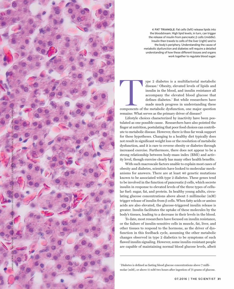
31 07.2016 | THE SCIENTIST
T ype 2 diabetes is a multifactorial metabolic disease.1 Obesity, elevated levels of lipids and insulin in the blood, and insulin resistance all accompany the elevated blood glucose that defines diabetes.† But while researchers have made much progress in understanding these
components of the metabolic dysfunction, one major question remains: What serves as the primary driver of disease?
Lifestyle choices characterized by inactivity have been pos-tulated as one possible cause. Researchers have also pointed the finger at nutrition, postulating that poor food choices can contrib-ute to metabolic disease. However, there is thus far weak support for these hypotheses. Changing to a healthy diet typically does not result in significant weight loss or the resolution of metabolic dysfunction, and it is rare to reverse obesity or diabetes through increased exercise. Furthermore, there does not appear to be a strong relationship between body-mass index (BMI) and activ-ity level, though exercise clearly has many other health benefits.
With such macroscale factors unable to explain most cases of obesity and diabetes, scientists have looked to molecular mech-anisms for answers. There are at least 40 genetic mutations known to be associated with type 2 diabetes. These genes tend to be involved in the function of pancreatic cells, which secrete insulin in response to elevated levels of the three types of cellu-lar fuel: sugar, fat, and protein. In healthy young adults, circu-lating glucose concentrations above about 5 millimolar (mM) trigger release of insulin from cells. When fatty acids or amino acids are also elevated, the glucose-triggered insulin release is greater. Insulin facilitates the uptake of these molecules by the body’s tissues, leading to a decrease in their levels in the blood.
To date, most researchers have focused on insulin resistance, or the failure of insulin-sensitive cells in muscle, fat, liver, and other tissues to respond to the hormone, as the driver of dys-function in this feedback cycle, assuming the other metabolic changes observed in type 2 diabetics to be symptoms of such flawed insulin signaling. However, some insulin-resistant people are capable of maintaining normal blood glucose levels, albeit
« FAT TRIANGLE: Fat cells (left) release lipids into the bloodstream. High lipid levels, in turn, can trigger
the release of insulin from pancreatic cells (middle). Insulin then travels to cells of the liver (right) and to
the body’s periphery. Understanding the cause of metabolic dysfunction and diabetes will require a detailed
understanding of how these different tissues and organs work together to regulate blood sugar.
DA
NIE
LA
MA
LID
E,
NA
TIO
NA
L H
EA
RT,
LU
NG
, A
ND
BL
OO
D I
NS
TIT
UT
E;
NIH
; ©
SC
OT
T C
AM
AZ
INE
/SC
IEN
CE
SO
UR
CE
; ©
IS
TO
CK
.CO
M/M
AX
PD
IA
† Diabetes is defined as fasting blood glucose concentrations above 7 milli-
molar (mM), or above 11 mM two hours after ingestion of 75 grams of glucose.

32 THE SCIENTIST | the-scientist.com
by producing higher-than-normal levels of insulin. Moreover, if insulin resistance leads to metabolic dysfunction, then increas-ing levels of insulin should restore metabolic homeostasis. But treating insulin-resistant patients with drugs such as sulfonyl-ureas or injected insulin is actually followed by greater meta-bolic imbalance.
In light of these findings, it’s time to begin considering what other metabolic correlates of diabetes might be driving factors. Elevated levels of lipids and insulin—both of which are seen prior to and at the onset of type 2 diabetes—are top candidates. Hyperlipidemia, the state of persistently high circulating levels of lipids called triglycerides, stimulates insulin secretion, lead-ing to elevated insulin levels, or hyperinsulinemia. Inducing hyperinsulinemia in animal models can lead to insulin resis-tance and obesity.2
Prevention and treatment of type 2 diabetes depends on cor-rectly determining the cause of metabolic failure. In fact, two available drugs (metformin and thiazolidinediones) developed to treat insulin resistance may actually work by lowering lipids. In combination with mounting evidence from our group and others that lowering circulating levels of insulin and lipids can reverse metabolic dysfunction in rodent models, researchers must now consider possible causes other than insulin resistance and try tar-geting these factors for new diabetes treatments.
Exploring hyperlipidemia and hyperinsulinemia Obesity is accompanied by an uptick in circulating triglycer-ides and free fatty acids that come from increased adipose tissue mass. Triglycerides stored in fat cells (adipocytes) are broken down into fatty acids, and these lipids can enter cells, where
© 2
016
AN
DR
EA
CH
AR
ES
T &
KA
RIN
A M
ET
CA
LF
Lipids and insulin play important roles in blood sugar regulation, and altered levels of either could kick start metabolic dysfunction. AN INTRICATE DANCE
Excess fat deposits on the body can release triglycerides and free fatty acids into the blood, causing hyperlipidemia.
As the lipids arrive at the pancreas, they are converted to free fatty acids and taken up by cells, where they join with coenzyme A to form long-chain acyl-CoA. This can trigger insulin secretion, leading to hyperinsulinemia. As more insulin in secreted, cells may begin to run low on their insulin reserves and insulin synthesizing capacity, such that they are not able to fully respond to the next surge in glucose.
ADIPOCYTE
Triglyceride
Free fatty acids
BLOOD VESSEL
To pancreas
From fat tissue
To liver
Fat tissue
BETA CELL
Lipase
Pancreas
Free fatty
acids
Coenzyme A
Insulin secreted
Long-chain acyl-CoA

33 07.2016 | THE SCIENTIST
they generate signals to increase insulin secretion, resulting in elevated blood insulin levels. This signaling cascade is typi-cally initiated inside the cell by the attachment of coenzyme A (CoA) to the fatty acids, forming long-chain acyl-CoA. Acyl-CoA itself is a well-established and potent signaling molecule and is the precursor of other important signaling molecules such as diglycerides and monoglycerides. In pancreatic cells, acyl-CoA has been shown to directly stimulate insulin exocytosis, change membrane ion channel activity, and influence Ca2+ handling.3 (See illustration below.)
To explore the importance of long-chain acyl-CoA in the development of diabetes, researchers studying rodent models have replaced dietary long-chain triglycerides with medium-chain triglycerides, which are rapidly oxidized in the mitochon-dria and thus do not generate cytoplasmic acyl-CoAs to stimulate insulin release. When a pair of researchers at McGill University in Quebec tried this treatment in mice, the animals had lower fasting insulin secretion and restored ability to respond to stimu-latory glucose. They experienced no weight gain or increased fat mass, and they did not suffer from impaired glucose tolerance, which typically accompanies a high-fat diet. In addition, mice that consumed medium-chain triglycerides were satiated more quickly than animals fed long-chain fats, reducing their overall food consumption.4
Stimulating fat burning to decrease lipid stores can also reduce circulating lipids and cytoplasmic long-chain acyl-CoA. Activating the transcription factor PPAR increases expres-sion of enzymes required for long-chain acyl CoA oxidation. In rodent models of high-fat diet–induced obesity, treatment with the PPAR agonist fenofibrate effectively stimulated fat burning. In obese mice, fenofibrate reduced circulating lev-els of fatty acids and triglycerides while reversing hyperinsu-linemia and hyperglycemia; in rats, fenofibrate also increased metabolic rate.5
These findings point to hyperlipidemia as a driving force in the development of metabolic dysfunction: by reducing levels of circulating lipids, researchers have successfully stunted the devel-opment of diabetes in animal models. But there is also evidence that hyperinsulinemia is the initiating defect that leads to obe-sity, hyperlipidemia, and insulin resistance. In 2000, Christian Weyer, then at the Clinical Diabetes and Nutrition Section at the National Institutes of Health, and colleagues found that hyperin-sulinemia precedes and predicts the development of diabetes in Pima Indians.6 Their findings fit with several previous studies of people in other ethnic groups prone to obesity, such as Mexican Americans and Nauruans, that also concluded hyperinsulinemia predicts diabetes.7,8,9,10
Other evidence for hyperinsulinemia as a cause of diabetes comes from gastric bypass surgery, an effective treatment for some patients with type 2 diabetes. In 2011, Walter Pories and G. Lynis Dohm at East Carolina University published a review document-ing that, following surgery, patients experienced a decrease in fast-ing insulin levels, along with decreases in blood glucose and res-olution of diabetes within a week—well before patients showed significant weight loss.11 Inhibiting insulin secretion in rats can prevent the development of the metabolic abnormalities induced by a high-sucrose diet;12 in vitro, curbing insulin secretion can keep human islets from deteriorating and becoming ineligible for islet transplantation.13
As for how hyperinsulinemia might cause metabolic problems, it has been known since the 1970s that insulin can downregulate its own receptor. When insulin binds its receptor, the cell internalizes the complex, digesting the attached insulin and recycling most of
By reducing levels of circulating lipids, researchers have successfully stunted the devel opment of diabetes in animal models.
As insulin from the pancreas binds to receptors in the liver, those receptors get internalized and the insulin generates signals to stop gluconeogenesis. High levels of insulin can exhaust the receptors, such that the cells of the liver (and muscle and fat) become insulin resistant, unable to respond to spikes in insulin and continuing to make glucose even though blood sugar levels are inappropriately high.
From pancreas
HEPATOCYTELiver
Insulin processed
Receptor recycled to cell membrane
Insulin

34 THE SCIENTIST | the-scientist.com
© D
AV
ID M
. P
HIL
LIP
S/S
CIE
NC
E S
OU
RC
E
the receptors to the membrane surface. At chronically high insulin levels, such recycling can exhaust the receptors, such that there are few on the cell surface to respond to further increases in insulin. Moreover, my group recently showed that hypersecretion of insulin can deplete the insulin reserves of cells in vitro. This leaves the cells unable to fully respond to a surge in glucose—a precursor to
-cell failure. In this case, we exposed the cells to excess glucose and fat to cause basal insulin secretion, while inhibition of secretion preserved the cells’ insulin content.14 Partial inhibition of insulin secretion in obese mice with elevated fasting insulin did not result in increased glucose levels, nor did the animals fare worse on a glu-cose tolerance test, indicating that elevated insulin was not neces-sary to maintain normal levels of circulating glucose.15
Thus, evidence exists from in vitro studies and work in ani-mal models that metabolic health and insulin secretory perfor-mance improve by preventing hyperinsulinemia or lowering the amount of ingested long-chain fatty acids. These approaches are now moving into clinical testing, with some early success.
Predicting and treating diabetes New diabetes therapies are desperately needed. Although the cur-rent standard of care—daily administration of insulin or drugs such as various sulfonylureas that trigger increased insulin release from
cells—is sufficient to control metabolic disarray, there are many untoward near- and long-term side effects. I and others contend that some of the standard therapies adopted by medical practi-tioners may actually be causing metabolic dysfunction; further increasing or stimulating insulin secretion or insulin levels in the presence of hyperinsulinemia may accelerate -cell deterioration.
Insulin secreted by the pancreas travels first to the liver, where it suppresses hepatic glucose production and is degraded. As a result, blood insulin levels entering the liver are three times higher than the concentrations that ultimately reach the periphery. This may cause hepatic insulin resistance to occur before muscle insulin resistance. To deliver adequate insulin supplies to the liver to overcome such insulin resistance, then, requires the administration of three times more insulin than is normally found in the periphery. Such excess insulin worsens muscle insulin resistance and promotes triglyceride synthesis, leading to increases in body fat and weight gain that fur-ther perpetuate metabolic dysfunction. High insulin levels may also promote cell growth and proliferation, increasing one’s risk of cancer.
SACS OF FAT: Fat cells, such as the one shown here, contain droplets full of lipids. These lipids can be released into the bloodstream, causing the high levels of circulating fats (hyperlipidemia) associated with metabolic dysfunction.

35 07.2016 | THE SCIENTIST
Several small studies have assessed the effects of inhibiting insulin secretion to preserve -cell insulin content in obese and prediabetic subjects. A study in healthy men showed a lower glucose level during an oral glucose tolerance test following a single dose of NN414, a small molecule that inhibits release of insulin from cells;16 another study documented improved glucose-stimulated insulin secretion in diabet-ics after seven days of treatment with diazoxide, which has the same effect as NN414.17 Paired with exogenous insulin to maintain normal glucose levels, decreasing -cell stimulation may also help preserve the remaining -cell function in type 1 diabetics, whose metabolic dys-function stems from an autoimmune attack on the pancreatic islets.18 (See “Taming Autoimmunity,” The Scientist, June 2016.)
A few human studies have indicated that reversing hyper-lipidemia can also stall the development of diabetes. As seen in animal models, consumption of medium-chain triglycer-ides (such as those found in palm kernel oil and coconut oil), instead of long-chain triglycerides (such as those in olive oil), increased energy expenditure, satiety, and fat loss in obese humans.4 More-extensive, longer trials are needed to identify patients who would benefit from such dietary intervention. Experimentally reducing carbohydrates in the diet of predia-betic and diabetic patients is also a promising strategy, as car-bohydrates are necessary to form triglycerides, and most cells burn fat if glucose is not available.
PPAR agonists, particularly fenofibrate, have been incon-sistent in their effects in human trials. But bezafibrate, a fibrate that interacts with all three PPAR isoforms ( , , and ), has con-sistently lowered triglycerides and improved glucose handling in diabetics.19,20,21 It should be noted, however, that most fibrate studies were designed to assess cardiac outcomes; the use of fibrates specifically to prevent deterioration of metabolic health before the onset of overt diabetes has not been studied in humans.
Unfortunately, the field has been slow to adopt hyperlipidemia and hyperinsulinemia as prime targets for diabetes therapy. It’s diffi-cult to change scientific thinking, and most researchers are still stuck on insulin resistance as the ultimate molecular cause of metabolic dysfunction. Yet modern treatments often worsen prognosis. The time has come to focus on ways to protect the cell, and research is now revealing just how to do that. Diverse stem cell therapies in development that are designed to stimulate the production of new
cells could also improve pancreatic function. We must follow the science as it leads us in new directions, and thoroughly test some of these novel approaches that have begun to show promise.
Barbara E. Corkey is Zoltan Kohn Professor of Medicine, Direc-tor of the Obesity Research Center and Vice Chair for Research in the Department of Medicine at Boston University School of Medicine. She would like to express special appreciation to
Dylan Thomas, Marie McDonnell, Richard Corkey, and Stan-ley Schwartz for their valuable suggestions on this article.
References
1. S.S. Schwartz et al., “The time is right for a new classification system for diabetes: Rationale and implications of the cell–centric classification schema,” Diabetes Care, 39:179-86, 2016.
2. S. Del Prato et al., “Effect of sustained physiologic hyperinsulinaemia and hyperglycaemia on insulin secretion and insulin sensitivity in man,” Diabetologia, 37:1025-35, 1994.
3. B.E. Corkey, J.T. Deeney, “Acyl CoA regulation of metabolism and signal transduction,” Prog Clin Biol Res, 321:217-32, 1990.
4. M.P. St-Onge, P.J. Jones, “Physiological effects of medium-chain triglycerides: Potential agents in the prevention of obesity,” J Nutr, 132:329-32, 2002.
5. F.P. Mancini et al., “Fenofibrate prevents and reduces body weight gain and adiposity in diet-induced obese rats,” FEBS Lett, 491:154-58, 2001.
6. C. Weyer et al., “A high fasting plasma insulin concentration predicts type 2 diabetes independent of insulin resistance: Evidence for a pathogenic role of relative hyperinsulinemia,” Diabetes, 49:2094-101, 2000.
7. G. Gulli et al., “The metabolic profile of NIDDM is fully established in glucose-tolerant offspring of two Mexican-American NIDDM parents,” Diabetes, 41:1575-86, 1992.
8. S.M. Haffner et al., “Decreased insulin secretion and increased insulin resistance are independently related to the 7-year risk of NIDDM in Mexican-Americans,” Diabetes, 44:1386-91, 1995.
9. S. Lillioja et al., “Insulin resistance and insulin secretory dysfunction as precursors of non–insulin-dependent diabetes mellitus. Prospective studies of Pima Indians,” N Engl J Med, 329:1988-92, 1993.
10. R.A. Sicree et al., “Plasma insulin response among Nauruans: Prediction of deterioration in glucose tolerance over 6 yr,” Diabetes, 36:179-86, 1987.
11. W.J. Pories, G.L. Dohm, “Diabetes: Have we got it all wrong?” Diabetes Care, 35:2438-42, 2012.
12. R. Gutman et al., “Diazoxide prevents the development of hormonal and metabolic abnormalities present in rats fed a sucrose rich diet,” Horm Metab Res, 17:491-94, 1985.
13. K. Maedler et al., “Glucose- and interleukin-1 -induced cell apoptosis requires Ca2+ influx and extracellular signal–regulated kinase (ERK) 1/2 activation and is prevented by a sulfonylurea receptor 1/inwardly rectifying K+ channel 6.2 (SUR/Kir6.2) selective potassium channel opener in human islets,” Diabetes, 53:1706-13, 2004.
14. K.A. Erion et al., “Chronic exposure to excess nutrients left-shifts the concentration dependence of glucose-stimulated insulin secretion in pancreatic cells,” J Biol Chem, 290;16191-201, 2015.
15. R. Alemzadeh, K.M. Tushaus, “Modulation of adipoinsular axis in prediabetic zucker diabetic fatty rats by diazoxide,” Endocrinology, 145:5476-84, 2004.
16. M. Zdravkovic et al., “The effects of NN414, a SUR1/Kir6.2 selective potassium channel opener, in healthy male subjects,” J Clin Pharmacol, 45:763-72, 2005.
17. E. Qvigstad et al., “Nine weeks of bedtime diazoxide is well tolerated and improves beta-cell function in subjects with Type 2 diabetes,” Diabet Med, 21:73-76, 2004.
18. M.A. Radtke et al., “Six months of diazoxide treatment at bedtime in newly diagnosed subjects with type 1 diabetes does not influence parameters of cell function and autoimmunity but improves glycemic control,” Diabetes Care, 33:589-94, 2010.
19. J.H. Flory et al., “Antidiabetic action of bezafibrate in a large observational database,” Diabetes Care, 32:547-51, 2009.
20. H. Tenenbaum et al., “Long-term effect of bezafibrate on pancreatic beta cell function and insulin resistance in patients with diabetes,” Atherosclerosis, 194:265-71, 2007.
21. T. Teramoto et al., “Effects of bezafibrate on lipid and glucose metabolism in dyslipidemic patients with diabetes: The J-BENEFIT study,” Cardiovasc Diabetol, 11:29, 2012.
Standard therapies adopted by medical practitioners may actually be causing metabolic dysfunction.


37 07.2016 | THE SCIENTIST
© F
AN
AT
IC S
TU
DIO
/GE
TT
Y I
MA
GE
S
Understanding interactions between the immune system and stem cells could pave the way for successful stem cell–based regenerative therapies.
BY WALEED RAHMANI, SARTHAK SINHA, AND JEFF BIERNASKIE
Cellular Teamwork
W e may perceive ourselves as static beings, but the cells of our bodies are in constant flux.
The outer layers of our skin and intesti-nal tract are replaced every few weeks; red blood cells circulate in our bodies for about 100 days before they are replaced; cells in our liver and fat are longer lived—more than a year for a liver cell, 10 years on average for a fat cell—but still turn over repeatedly during our lifetimes. More slowly, up to half our heart cells may be replaced during a normal lifespan. And, of course, when healthy tissue is lost due to injury, new cells are made to patch up the damage. What are the biological pro-cesses responsible for normal cell turnover and organ homeostasis? What controls proper repair after injury? What allows organisms like the salamander to regen-erate an amputated limb while humans form scars and struggle to regrow much simpler structures, such as hair?
These and other questions are the target of ongoing research in the field of
regenerative medicine. But what we do know, and have known for nearly half a century, is that stem cells are crucial play-ers. Stem cells self-renew to maintain their numbers and differentiate into the special-ized cell types that make up our tissues and organs—a function that becomes espe-cially important after stress or injury. The ultimate goal of regenerative medicine is to harness stem cells’ regenerative poten-tial to treat and even cure many of the diseases besetting society today. Despite progress in understanding the potential of these multipotent cells, the unfortunate reality is that we remain far from cures. One possible reason for this is scientists’ failure to sufficiently consider what goes on within the biological environment sur-rounding the stem cell.
For years, stem cell biologists have focused their attention on the intrinsic properties of stem cells to understand what gives them the ability to self-renew and differentiate into a range of cell types. While these investigations have uncov-
ered a collection of genes and proteins responsible for a cell’s “stemness,” the role of the microenvironment, also known as the stem cell niche, was largely ignored. But neighboring cells, secreted proteins, the extracellular matrix, circulating met-abolic signals such as oxygen and glucose, and diverse physical parameters, such as shear stress and tissue stiffness, can all affect the behavior of stem cells.
One of the best-studied examples of mammalian stem cell environments is the intestinal stem cell (ISC) niche. The small intestine’s epithelium is the fastest self-renewing tissue in the body due to ISCs’ exceptionally rapid rates of cell division and the rapid migration of their differen-tiated progeny out of the stem cell niche. But the system would not work without
Diverse immune cells have been caught in the act of manipulating stem cell behavior.

CR
ED
IT L
INE
IMMUNE CELLS HELP OUT
During puberty, as hormones trigger the maturation of the rudimentary mammary ducts, macrophages and other immune cells migrate to the ducts’ tips, where they support rapid proliferation and duct branching.
Following an acute injury to the skeletal muscle, local and infiltrating immune cells remove damaged tissue, while T cells help spur the generation of new muscle cells.
Macrophages enhance the formation of collagen fibrils that support duct growth.
M1 macrophages and fibro/adipocyte progenitors (FAPs) clear damaged muscle fibers.
T cells secrete a protein called amphiregulin that stimulates muscle stem
cells called satellite cells to differentiate into myoblasts.
Neural stem cell
Neural precursor
Microglial cell
T cell
Neuroblast
Immature neuron
Mature neuron
Microglia phagocytize excess newborn neurons that have undergone apoptosis.
BRAIN
MAMMARY GLANDS
MUSCLE
Adipocytes
Collagen fibrils
Skeletal muscle
Myoblast
FAPs
Amphiregulin
M1 macrophage
Satellite cell
Damaged fibers
Hippocampus
The cells of the mammalian immune system do more than just fight off pathogens; they are also important players in stem cell function and are thus crucial for maintaining homeostasis and recovering from injury. Here are a few examples.
As new neurons differentiate from neural stem cells in the hippocampus, T cells and microglia are recruited to the neurogenic site. Following injury, macrophages stimulate remyelination of neurons.
Microglia
Dead neuron
Mature neurons integrate into hippocampal circuits
or undergo apoptosis.
HOMEOSTATIC NEUROGENESIS

39 07.2016 | THE SCIENTIST
CR
ED
IT L
INE
M1 macrophages help clear debris after a brain injury. They then transition into M2 macrophages, which secrete a protein called activin-A, which facilitates the differentiation of oligodendrocyte progenitor cells (OPCs) into oligodendrocytes, neural-support cells that are responsible for myelination of neural cell axons.
Mast cells secrete serine proteases, which stimulate duct branching.
M1 macrophages also spur the proliferation of muscle progenitor cells called myoblasts.
Macrophages
Proliferating body cells
Mast cell
Duct branching
Serine proteases
Myoblast proliferation
New muscle fibers
T cells
M1 macrophage
M2 macrophage
OPCs
Activin-A
Oligodendrocyte
Damaged neurons
the help of Paneth cells, one of four dif-ferentiated cell types produced by ISCs, which remain in the niche and secrete essential proteins that are critical for ISC survival. Indeed, the genetic inactivation of Paneth cells results in a near-total loss of ISCs.1
In addition to niche-specific cells, stem cells regularly interact with the body’s mobile and diffuse army of immune cells. Traditionally regarded as the pri-mary line of defense against pathogenic invaders, the immune system is now also recognized as essential for tissue homeo-stasis and healing, even in the absence of infection. Diverse immune cells have even been caught in the act of manipulating stem cell behavior.
The precise roles that immune cells play in the stem cell niche is context dependent. Whether macrophages and T cells ensure homeostasis, promote regen-eration (e.g., regrowth of liver tissue after a partial hepatectomy), or mediate scar-forming tissue repair depends on the spe-cies, its developmental stage, the organ or tissue in question, the severity of injury, and the availability of a stem cell pool. Which molecules immune cells secrete, and the effect the cells have on regenera-tion, can also change drastically depend-ing on the organism and tissue. In some cases, immune cells may even work against the body, supporting the growth and spread of cancer. Understanding the immune system’s role in stem cell biology may help clinicians and scientists better respond to injuries or homeostatic imbal-ances, as well as develop stem cell thera-pies to treat diverse ailments, from anemia to multiple sclerosis, muscular dystrophy, and heart failure.
Maintaining homeostasisAn integral part of homeostasis in diverse tissues is the continuous replacement of differentiated cell types. Research is now showing that the immune cells residing within the stem cell niche are essential to this process. For example, specialized macrophages in the bone marrow remain in direct contact with a red blood stem cell called an erythroblast. Without this direct
INJURY
Mammary stem cell
© I
KU
MI
KA
YA
MA
/ST
UD
IO K
AY
AM
A

40 THE SCIENTIST | the-scientist.com
cell-cell contact, erythroblasts are not able to mature properly and repopulate the blood with new red blood cells, a defi-ciency that can lead to hemolytic anemia.2
Immune cells are also critical for the development of mammary glands during puberty. At birth, mammary glands con-sist of fat pads with rudimentary ducts descending from the nipple. At the start of puberty, ovarian hormones trigger the bifurcation and elongation of the ductal structures towards the outer edges of the fat pad while diverse immune cells—mast cells, eosinophils, and macrophages—migrate to the region around the ducts’ tips. Genetic or pharmacological dis-ruption of mast cells and macrophages in mice has revealed that these immune cells are critical for rapid proliferation and normal duct branching during puberty. Mast cells secrete protein-degrading ser-ine proteases, which are necessary for the breakdown and reorganization of collagen fibers surrounding the developing ducts, for example,3 while macrophages phago-cytize apoptotic cell debris and directly act on mammary stem cells through an unknown mechanism.4,5
Another organ that relies on immune cells to regulate normal cell turnover is the brain. Once believed to occur only during embryonic and late gestational stages in mammals, neurogenesis is now known to occur throughout adult life in the dentate gyrus of the hippocampus and the subven-tricular zone of the lateral ventricle, two locations where neural stem cells reside. (See “Brain Gain,” The Scientist, October 2015.) And investigations of the cellular mechanisms regulating adult neurogen-esis have revealed that immune cells play crucial roles in hippocampal-dependent learning and memory.
Researchers at the Weizmann Insti-tute of Science in Israel have shown that hippocampal neurogenesis in rodents, induced by housing the animals in enriched environments, was associ-ated with the recruitment of T cells and microglia (macrophages of the brain and spinal cord). Immune-deficient mice, on the other hand, exhibited impaired hip-pocampal neurogenesis that led to poor
results in spatial learning and memory tasks.6 It is still not clear how immune cells influence the neural stem cell niche dur-ing hippocampal neurogenesis. However, because only a small subset of newborn neurons integrate into the hippocam-pal circuitry, with the majority undergo-ing death by apoptosis, it is believed that microglia shape hippocampal neurogene-sis by rapidly phagocytizing the apoptotic newborn neurons.7
Studies of the bone marrow, mammary gland, and brain reveal that stem cells’ immune niches play an important role in maintaining homeostasis in our organs, ensuring a stable equilibrium between cell overpopulation and atrophy under normal conditions. But what about when homeo-stasis is disturbed?
Dealing with injuryPerhaps the best-understood example of immune- and stem-cell cooperation is in skeletal muscle following an acute injury. Tissue repair begins with the removal of damaged muscle fibers by local and infil-trating immune cells. Rare, circulating immune cells called eosinophils instruct resident progenitor cells known as fibro/adipogenic progenitors (FAPs) to gener-ate the fibroblasts and fat cells that deposit collagen and secrete growth factors to support muscle fiber regeneration.8 Con-currently, T cells secrete a protein called amphiregulin, which instructs resident muscle stem cells known as satellite cells to differentiate into new muscle cells and replace the lost muscle fibers.9
Such immune–stem cell interactions are not restricted to skeletal muscle, but have been observed across many organs in mice. During chronic liver injury, mac-rophages secrete a protein called Wnt3a, which drives the differentiation of local liver stem cells into mature liver cells.10 In the colon, macrophages are recruited
to activate intestinal stem cell prolifera-tion and regenerate wounded intestinal epithelium.11 And in the nervous system, recent work has shown that following injury, anti-inflammatory M2 macro-phages are essential for efficient replace-ment of the myelin sheath, an insulating layer of fatty substance that facilitates the transmission of action potentials along the axons of neurons. Specifically, the macro-phages secrete a protein called activin-A
that triggers oligodendrocyte progenitor cells (OPCs) to differentiate into oligo-dendrocytes, neural support cells that are responsible for myelination.12
A particularly interesting system in which researchers have explored the rela-tionship between stem cells and immune cells is the hair follicle, one of the few mammalian tissues capable of continuous regeneration throughout life. Last year, the University of Southern California’s Cheng-Ming Chuong and his colleagues showed that macrophages are responsi-ble for the regrowth of a new hair follow-ing plucking.13 When researchers plucked hairs off the backs of mice, they found that damaged hair follicles beneath the skin’s surface secrete, in unison, a pro-tein called CCL2. In response to this dis-tress signal, macrophages migrated up the CCL2 gradient and toward hair folli-cles, where they secreted a protein called tumor necrosis factor (TNF), which instructed hair follicle stem cells to pro-duce new hair.
It’s becoming clear that immune cells are an important component of stem cell niches across the body, with crucial roles in injury-induced regeneration. Theo-retically, targeting certain immune cells should promote healing. However, the great diversity and heterogeneity found within each immune cell population have made it difficult to develop effective ther-apies. More research is needed to suffi-
It’s becoming clear that immune cells are an important component of stem cell niches across the body, with crucial roles in injury-induced regeneration.

41 07.2016 | THE SCIENTIST
ciently discriminate among subpopula-tions of immune cells and to understand which cells must be targeted to elicit the desired effect in injured tissues.
Crosstalk interruptedCommunication between immune cells and stem cells does not always do the body good; at times, cell interactions can result in fibrosis and organ dysfunction. In mouse models of chronic muscle dam-age approximating Duchenne muscular dystrophy (DMD), immune cell infiltra-tion and FAP activity are abnormally pro-longed, while the reparative capacity of satellite stem cells is diminished. These abnormalities, a result of genetic defects in the dystrophin gene, lead to exces-sive and disorganized collagen deposi-tion, ultimately causing fibrosis and loss of muscle function. Why does this hap-pen? The answer may have to do with how infiltrating macrophages communi-cate with FAPs.
Last year, researchers at the Uni-versity of British Columbia in Canada showed that, in healthy muscle regenera-tion, FAP numbers dramatically increase three days after an acute injury but quickly drop to pre-injury levels by day five.14 It turns out that macrophages are directly responsible for the quick decline in FAP numbers; the immune cells secrete TNF, which binds to FAPs and signals them to undergo apoptosis. In the mouse model of DMD, however, macrophages increase the production of another protein called transforming growth factor b1 (TGFb1). Unlike TNF, TGFb1 instructs FAPs to sur-vive longer and differentiate into the col-lagen-secreting cells that, when present in excess, cause muscle fibrosis and dysfunc-tion. Treatment with nilotinib, a US Food and Drug Administration–approved ther-apy for the treatment of a drug-resistant form of leukemia, reduced muscle fibrosis in the mice by blocking the adverse effects of TGFb1.
Immune cells can get especially dangerous when they start supporting the survival and metastasis of tumors by interacting with cancer stem cells (CSCs), a small subset of tumor cells that self-renew and generate the major-ity of cells within tumor masses. Many traditional cancer therapies discrimi-nately kill actively dividing CSCs and their progeny, but slow-dividing CSCs remain untouched, enabling relapse and even metastasis. Scientists are now rac-ing to better understand and target CSCs. Intriguingly, the key to success may lie in our own immune system.
The most abundant immune cell within the tumor microenvironment is the macrophage. While biologists once suspected that macrophages provided anti-tumor immunity, we now know that the tumor microenvironment is enriched with signals that rewire these cells into tumor-associated macrophages (TAMs), which actually fuel the cancer’s survival, malignancy, invasiveness, and drug resis-tance. Lactic acid, for example, supports lung cancer and melanoma growth by converting normal macrophages into TAMs that produce high levels of vascu-lar endothelial growth factor (VEGF) to promote tumor vascularization, as well as enzymes that support nitrogen metab-olism, increasing tumor cell prolifera-tion.15 Indeed, many clinical studies have demonstrated that increased macrophage density is strongly correlated with poor prognoses in thyroid, breast, lung, and liver cancers.
Recent research has suggested that some CSCs encourage the transformation of normal macrophages into TAMs. Last year, for example, a team led by research-ers at the Cleveland Clinic found that CSCs in glioblastomas, a highly malignant brain cancer, secrete a potent chemoattractant called periostin that instructs blood-derived macrophages to migrate into the tumor, where they are converted into TAMs. In a mouse model of glioblastoma, genetically silencing periostin reduced the number of TAMs within the tumor, inhib-ited tumor growth, and extended the ani-mals’ survival.16
IMMUNE CELLS DO HARMSometimes, immune- and stem-cell interactions do not promote homeostasis or healing, but instead lead to further damage or disease. The most dramatic example is the cooperation of tumor-associated macrophages (TAMs) and cancer stem cells (CSCs) to drive cell proliferation and tumor malignancy and invasiveness, as well as drug resistance.
GLIOBLASTOMA
CSC
TAM factors
Blood vessel
In glioblastomas, a highly malignant brain cancer, CSCs secrete a potent chemoattractant called periostin that instructs blood-derived macrophages to migrate to the tumor, where they are converted into TAMs.
TAMs then secrete factors to promote tumor growth and progression.
Periostin
TAM
© I
KU
MI
KA
YA
MA
/ST
UD
IO K
AY
AM
A
Macrophage
Tumor vascularization

42 THE SCIENTIST | the-scientist.com
© I
ST
OC
K.C
OM
/AL
EX
BE
LO
ML
INS
KY
Researchers are now exploring ways to more effectively prevent macrophages from infiltrating and acquiring this tumor-supportive identity, and to dis-rupt the ongoing crosstalk between CSCs and TAMs. A 2013 study of mouse pan-creatic cancer showed that inhibiting CSF1R and CCR2, macrophage recep-tors key for migration and survival, decreased the total number of pancreatic CSCs, enhanced chemotherapeutic effi-ciency, and inhibited metastasis.17 And when human patients were treated with a drug targeting CSF1R, patients had sig-nificantly fewer TAMs at tumor sites and improved clinical outcomes.18
Tissue-resident stem cells’ remarkable ability to self-renew while also giving rise to diverse mature cell types is critical for our existence. In order to carry out their inherent roles in tissue maintenance and regeneration, these stem cells rely on sig-nals provided by diverse cell types, includ-ing immune cells, within the local and sys-
temic environments. We are at the dawn of understanding the complex and dynamic roles of the immune system’s many cell types and their functional relationships with stem cells—a feat that will be criti-cal to harnessing the power of stem cells to treat or cure disease.
Waleed Rahmani is an MD/PhD student in the Department of Comparative Biol-ogy and Experimental Medicine at the University of Calgary, Canada. Sarthak Sinha is an undergraduate neuroscience student at the University of Toronto. Jeff Biernaskie is an assistant professor of stem cell biology and regenerative medi-cine at the University of Calgary, where he holds the inaugural Calgary Firefight-ers Burn Treatment Society Chair in Skin Regeneration and Wound Healing within the Faculty of Veterinary Medicine. He is a member of the Alberta Children’s Hospi-tal Research Institute and the Hotchkiss Brain Institute.
References
1. T. Sato et al., “Paneth cells constitute the niche for Lgr5 stem cells in intestinal crypts,” Nature, 469:415-18, 2011.
2. A. Chow et al., “CD169+ macrophages provide a niche promoting erythropoiesis under homeostasis and stress,” Nat Med, 19:429-36, 2013.
3. J.N. Lilla, Z. Werb, “Mast cells contribute to the stromal microenvironment in mammary gland branching morphogenesis,” Dev Biol, 337:124-33, 2010.
4. V. Gouon-Evans et al., “Postnatal mammary gland development requires macrophages and eosinophils,” Development, 127:2269-82, 2000.
5. D.E. Gyorki et al., “Resident macrophages influence stem cell activity in the mammary gland,” Breast Cancer Res, 11:R62, 2009.
6. Y. Ziv et al., “Immune cells contribute to the maintenance of neurogenesis and spatial learning abilities in adulthood,” Nat Neurosci, 9:268-75, 2006.
7. A. Sierra et al., “Microglia shape adult hippocampal neurogenesis through apoptosis-coupled phagocytosis,” Cell Stem Cell, 7:483-95, 2010.
8. J.E. Heredia et al., “Type 2 innate signals stimulate fibro/adipogenic progenitors to facilitate muscle regeneration,” Cell, 153:376-88, 2013.
9. D. Burzyn et al., “A special population of regulatory T cells potentiates muscle repair,” Cell, 155:1282-95, 2013.
10. L. Boulter et al., “Macrophage-derived Wnt opposes Notch signaling to specify hepatic progenitor cell fate in chronic liver disease,” Nat Med, 18:572-79, 2012.
11. S.L. Pull et al., “Activated macrophages are an adaptive element of the colonic epithelial progenitor niche necessary for regenerative responses to injury,” PNAS, 102:99-104, 2005.
12. V.E. Miron et al., “M2 microglia and macrophages drive oligodendrocyte differentiation during CNS remyelination,” Nat Neurosci, 16:1211-18, 2013.
13. C.-C. Chen et al., “Organ-level quorum sensing directs regeneration in hair stem cell populations,” Cell, 161:277-90, 2015.
14. D.R. Lemos et al., “Nilotinib reduces muscle fibrosis in chronic muscle injury by promoting TNF-mediated apoptosis of fibro/adipogenic progenitors,” Nat Med, 21:786-94, 2015.
15. O.R. Colegio et al., “Functional polarization of tumour-associated macrophages by tumour-derived lactic acid,” Nature, 513:559-63, 2014.
16. W. Zhou et al., “Periostin secreted by glioblastoma stem cells recruits M2 tumour-associated macrophages and promotes malignant growth,” Nat Cell Biol, 17:170-82, 2015.
17. J.B. Mitchem et al., “Targeting tumor-infiltrating macrophages decreases tumor-initiating cells, relieves immunosuppression and improves chemotherapeutic responses,” Cancer Res, 73:1128-41, 2013.
18. C.H. Ries et al., “Targeting tumor-associated macrophages with anti-CSF-1R antibody reveals a strategy for cancer therapy,” Cancer Cell, 25:846-59, 2014.
Understanding the immune system’s role in stem cell biology may help clinicians and scientists better respond to injuries or homeostatic imbal ances, as well as develop stem cell therapies to treat diverse ailments.

TS Webinars
ONDEMAND Disease in a Dish: New Tools for Understanding Cellular Systems
Cell-based model systems are powerful tools for studying signaling pathways, deciphering disease mechanisms, and predicting drug activities. Advances in stem cell technology provide new opportunities for in vitro modeling and screening in a physiologically relevant environment that is consistent and replicable. In this webinar, our panel of experts discusses emerging technologies for disease modeling in regenerative medicine and stem cell therapy.
DOO YEON KIM, PhD Assistant Professor of NeurologyGenetics and Aging Research UnitMassachusetts General HospitalHarvard Medical School
JASON SPENCE, PhD Assistant ProfessorDepartment of Internal Medicine, GastroenterologyDepartment of Cell and Developmental BiologyUniversity of Michigan
WEBINAR SPONSORED BY:
WATCH NOW! www.the-scientist.com/diseaseinadish
JAMIE FREEMAN, PhD Product ManagerBioproductionHorizon Discovery
WEBINAR SPONSORED BY:NATHAN LEWIS, PhD Assistant ProfessorDepartment of PediatricsUniversity of California, San Diego
TOPICS COVERED:
• 3-D cell culture models for Alzheimer’s disease and intestinal development• Techniques and tips for studying pathogenesis in culture
ONDEMAND Bioproduction: Advances in Cell Line Engineering
Bioproduction involves the growth and manipulation of large quantities of cells under tightly controlled conditions mandated by good manufacturing practices (GMP), while maintaining quality, reproducibility, and cost. Innovative technologies and strategies are being employed to optimize scale-up to high-throughput production. In this webinar, our panel of experts discusses strategies and workflows for screening and optimizing cell lines for bioproduction.
WATCH NOW! www.the-scientist.com/bioproduction
TOPICS COVERED:
• Genome-editing approaches for bioproduction• Analysis and leverage of screening and omics data to guide cell line engineering for biotherapeutic development
RUSKINN

44 THE SCIENTIST | the-scientist.com
The LiteratureEDITOR’S CHOICE PAPERS
© T
HO
M G
RA
VE
S
THE PAPER
Y. He et al., “Genomic and enzymatic evi-dence for acetogenesis among multiple lineages of the archaeal phylum Bathyar-chaeota widespread in marine sediments,” Nat Microbiol, 16035, 2016.
Microbial ecologist Fengping Wang was not looking for archaea. She just couldn’t stop finding them. A couple of years ago at Shanghai Jiao Tong University, Wang was investigating the composition of marine sediments, expecting to turn up primarily bacteria. Instead, she says, “I kept detect-ing archaea. Repeatedly, they were com-ing up in almost every sample I studied.”
Wang was puzzled. Archaea have tra-ditionally been thought of as extremo-philes, living unusual lifestyles in unusual environments, such as in hot springs or the Arctic. Even though that view is changing as more and more studies identify archaea in diverse habitats, the organisms’ abun-dance in these sedimentary samples, col-lected from the Gulf of California, was a surprise to Wang, and she wondered whether perhaps they were performing an as yet undiscovered ecosystem function.
Along with collaborators at Shen-zhen University in China and the Woods Hole Oceanographic Institution in Mas-sachusetts, Wang’s group in Shanghai used metagenomics to identify most of the archaea in their sediment samples as mem-bers of a phylum called Bathyarchaeota, newly established in 2013 by Wang and her colleagues and previously known as the Miscellaneous Crenarchaeotic Group. Analyzing the genomes of these archaea revealed several sequences that appeared to code for proteins involved in carbon cycling processes including, unexpectedly, acetogenesis—the production of acetate
from inorganic carbon, a crucial step in biogeochemical cycling in marine car-bon reservoirs.
“This was really surprising to us,” says Wang. “Acetogenesis is a very important pathway, especially in anaerobic environ-ments, as acetate is a source of organic car-bon which can be used to fuel other organ-isms, or for methane production. It was thought that bacteria were the major players.”
The researchers were keen to learn more about Bathyarchaeota’s role in aceto-genesis, but they faced challenges studying the microbes directly in the lab because the organisms are notoriously slow-growing and difficult to cultivate. So to test whether the Bathyarchaeota gene sequences were in fact functional, the team “did a little bit of enzyme characterization,” Wang says. The researchers cloned one of the genes that appeared to be involved in acetogenesis, expressed it in E. coli, and through bio-chemical assays showed that the resulting
enzyme could catalyze a step in the aceto-genesis pathway. “We found that they can really perform acetogenesis,” says Wang, adding that now, scientists “will have to rethink the roles of archaea.”
“It’s a really very interesting study because it highlights the importance of archaea in geochemical cycles,” says micro-biologist Bettina Siebers of the University of Duisberg-Essen, Germany, who was not involved in the work. “It helps us to under-stand what’s really going on in marine sed-iments.” The next step, she adds, will be to try to grow these cells in the lab to perform studies on their function.
Plans to do just that are underway in Wang’s lab, alongside the development of approaches to characterize the archaea’s carbon cycling activity in situ. “We need to prove that the acetogenesis really occurs in the environment,” Wang says, “not just in the lab or in the genome.”
—Catherine Offord
EDITOR’S CHOICE IN MICROBIOLOGY
�Secret Cyclers
PLAYING A PART: Members of the phylum Bathyarchaeota are abundant in marine sediments, where they take up carbon from organic compounds in the surrounding environment. A recent study has shown that they also have the potential to fix inorganic carbon—in the form of carbon dioxide (CO2)—to generate acetate, an important fuel for other sediment-dwelling organisms such as methanogens (which include marine archaea) and heterotrophic bacteria. The products of these organisms feed back into the carbon cycle in marine sediments, where they may be consumed by other microbes.
Carbon fixation
Methanogens
CO2
Acetyl CoA
CO2
CH4
SEA
SEDIMENT
Heterotrophic bacteria
and archaea
HydrolysisAcetogenesis
Acetate
Bathyarchaeon
CH3COO–
Carbon-containing detritus, e.g.,
cellulose, proteins

45 07.2016 | THE SCIENTIST
ER
IC B
AS
TIA
AN
S;
ED
WA
RD
DE
LO
NG
, D
AV
ID K
AR
L,
NA
NC
Y H
UL
BIR
T
MEAL TIME: Ocean-dwelling microbes chow down on marine carbon sources at rates that are taxon dependent (false-color micrograph).
KIN KINKS: Sporulating fungal colonies (orange) are rarer in strains evolved under low-relatedness conditions due to the evolution of cheaters (tan).
MICROBIOLOGY
Assessing AssimilationTHE PAPER
S. Bryson et al., “Proteomic stable isotope probing reveals taxonomically distinct patterns in amino acid assimilation by coastal marine bacterioplankton,” mSystems, doi:10.1128/mSystems.00027-15, 2016.
THE FIXERS
Marine systems fix about 50 gigatons of carbon each year, of which about half is processed by heterotrophic microbial communities. But relatively little is known about the role of individual taxa in the assimilation and metabolism of carbon compounds. So Samuel Bryson, a graduate student in Ryan Mueller’s lab at Oregon State University, set out to test a method that could improve taxon-specific measurements of carbon uptake and usage.
PROTEOMIC PROBE
Bryson and his colleagues added 13C-labeled amino acids to seawater samples of bacterioplankton collected at two different locations off the western coast of the U.S. The team then used metagenomics and mass spectrometry to analyze various bacterial orders’ incorporation of 13C into peptides to see “who is taking up what,” says Bryson.
HEAVY LIFTERS
The researchers found that some orders, such as Alteromonadales, incorporated 13C-amino acids into biomass more rapidly than more-dominant orders like Rhodobacteriales and Flavobacteriales. “I think it’s using some really cutting-edge technology to get at an important question in microbial oceanography,” says David Kirchman, a marine microbiologist at the University of Delaware, adding that data on how bacteria share the job of carbon assimilation will help scientists better understand carbon cycling in marine systems.
COMPLICATING MATTERS
So far, the method has “confirmed and validated other predictions and observations” about taxonomic differences in amino acid assimilation, says Bryson. The next step will be to increase the complexity of the experiments, by “looking at how taxa respond across a variety of substrates,” he says. —Catherine Offord
MICROBIOLOGY
Curbing CheatersTHE PAPER
E. Bastiaans et al., “Experimental evolution reveals that high related-ness protects multicellular cooperation from cheaters,” Nat Commun, 7:11435, 2016.
CHEATING COOPERATION
Genetic similarity among cells is probably an important factor for suc-cessful multicellularity, as suggested by studies of varying relatedness among the fruiting bodies of myxobacteria or Dictyostelium—which form by aggregation of cells, not by clonal expansion from a single cell. That work showed cheating can increase with decreased relatedness , but in these models it has not been possible to make comparisons in which relatedness is the only variable.
FAMILIAL FUNGI
Duur Aanen of Wageningen University in the Netherlands and his grad-uate student Eric Bastiaans found a model to overcome that problem: the bread mold Neurospora crassa. These filamentous fungi grow either clonally—by mitosis—or by fusion with less-related cells, which cre-ates chimeric strands. Bastiaans and Aanen evolved 31 generations of a mutant line unable to fuse—which yielded highly related progeny—and a standard strain, with lower relatedness due to fusion. They found that more cheaters arose in the standard strain, which had a lower spore yield because the cheaters neglected their somatic functions.
EXPLAINING EVOLUTION
David Queller of Washington University in St. Louis, who has done similar work in Dictyostelium, praises the creative use of Neurospora and calls the work a “significant advance.” It’s a rare experimental-evolution study of cooperation, and “the only one that did a proper low-related-ness and high-relatedness treatment,” Queller says.
SOCIETAL IMPACT
Aanen plans to study the dynamics of fungal cultures in which cheating and cooperative cells coexist—work that might help explain the structure of human organizations with too few team players. “There are so many interesting parallels in human societies,” he says. —Jenny Rood

46 THE SCIENTIST | the-scientist.com
PROFILE
PH
OT
O B
Y T
OM
KL
EIN
DIN
ST/
© W
OO
DS
HO
LE
OC
EA
NO
GR
AP
HIC
IN
ST
ITU
TIO
N
In 1974, during the spring semester of his junior year at Har-vard University, Peter Tyack noticed a summer job posting tacked up on the bulletin board of the undergraduate biol-
ogy office. “It said, ‘Do you want to clean pigeon cages, train homing pigeons, and join a project studying how pigeons navi-gate?’” says the marine mammal biologist. Tyack’s answer was an emphatic yes. “I leapt at this first opportunity to do field-work.” In high school, Tyack had worked at a start-up medical devices company in Palo Alto. The office job was a stark contrast to time spent hiking and mountain climbing, and he was itching to spend time in the field
Under the supervision of animal behaviorist Charles Walcott, Tyack spent that summer in Lincoln, Massachusetts, securing tiny magnetic coils to the heads of homing pigeons. Depending on the orientation of the battery’s polarity, the pigeons would either fly home or 180 degrees from home under overcast skies when the sun was not visible, Walcott and his students found. “It was an early and really clean experiment on the impact of magnetic field on navigation,” says Tyack.
That summer job was fortuitous because it led Tyack to study marine mammal communication. Next door to the house where Walcott and his students worked, Roger Payne—who in 1967, along with his then wife Katharine, had famously discovered that humpback whales sing songs—was analyzing whale-song record-ings. Tyack would go over to Payne’s house every few weeks to sing madrigals. One evening, Payne answered a phone call from the New York Zoological Society, where he worked during the rest of the year. The staff member on the other end told Payne that they needed to hire a caretaker for their whale field site in Patagonia. Tyack enthusiastically volunteered.
Taking a year off from Harvard, he flew to Patagonia and traveled to the Peninsula Valdez in Argentina. “It’s 42° south latitude. As a comparison, Boston is 42° north latitude. There were only a few hundred people living there, and there was this incredible richness of marine wildlife,” he says. Using a hydro-phone he installed a kilometer offshore, Tyack recorded the sounds of bottlenose dolphins as they swam past the research camp. He assembled the electronics for the underwater record-
ers himself, learning on the go. “There were just bits and pieces from disposable buoys donated by the US Navy. I pirated them to make the equipment I needed. That’s field biology—you have to learn how to solve whatever problem comes at you.” When he returned to Boston, Tyack wrote his senior thesis about how the pattern of the animals’ calls changed depending on other nearby dolphins, people, or boats. The experience made Tyack realize he wanted to do fieldwork as a career.
Here, Tyack discusses the whale equivalent of “the cocktail party effect,” how studying whales has shown him the limits of humans’ imagination, and the excitement a day in the field can bring.
TYACK TRAINS Hands on learning. Tyack was part of the Sputnik generation, a time when there was an emphasis on the creative process in science. “In elementary school, what grabbed my attention was the hour a day we spent doing a science study project. It was very much capturing the experience first and then working out what was going on,” says Tyack. “We collected water from a puddle, looked under a lens, and saw protozoans; we explored how the volume of a gas changes its temperature.”
Sparking an interest. Tyack’s father was a historian, and his mother was a psycholinguist who studied language learning disabil-ities. His parents met through singing, and Tyack says he grew up with a love of acoustics and music. His family lived in Boston until he was 5 years old, and his parents, avid sailors, frequently took the young Tyack ocean sailing. He encountered his first whale at age 5, while sailing with his parents and a family friend who was blind. “The man could hear the whale at sea before anyone could see any-thing. Within a few minutes we sighted the whale, which is the first time I ever saw one.” The family moved to Portland, Oregon, where Tyack hiked and did mountain climbing. “Living on the West Coast lodged my initial interest in field biology,” he says.
In good company. Tyack entered Harvard University as a freshman in 1972. He was initially interested in neuroscience, but after taking a freshman course with E.O. Wilson, who was studying sociobiology and evolution at the time, he decided to major in biological anthropology. In his sophomore year, Tyack read a New Yorker article that featured William Schevill, a founder of the field of marine mammal acoustics and the first to record and study underwater sounds of marine mammals. The article noted that Schevill’s office, in the attic of Harvard’s Museum of Comparative Zoology, was impossible to find. “It took me three trips of exploring the attic to find it, but I finally
Behavioral ecologist Peter Tyack studies the social structures and behaviors of whales and dolphins, recording and analyzing their acoustic communications.
BY ANNA AZVOLINSKY
Sounds from the Seas
“Right whales have moved from being basses to being tenors to avoid all of the low-frequency noise from shipping.”

47 07.2016 | THE SCIENTIST
CR
ED
IT L
INE
PETER TYACK Professor of Marine Mammal BiologyScottish Oceans Institute, School of BiologyUniversity of St AndrewsFife, Scotland, U.K.
Greatest Hits• Provided evidence that, similar to those of songbirds,
humpback whale songs play a role in mating • Found that humpback whales have a wide range of songs
and are able to make complex and rapid changes to their songs—evidence that the marine mammal is capable of vocal learning
• Demonstrated that individual dolphins produce a signature whistle and showed that the animals can readily imitate the whistles of other dolphins
• Pioneered a sound-orientation tag that, when attached to a dolphin, is able to identify the animal producing vocalization in a group of captive animals, and a digital acoustic-recording tag to measure the vocal responses of wild cetaceans
• Raised awareness of how oil rigs, boats, and sonar disturb marine mammals
found the office. I took a reading course with him on marine mammal behavior and very quickly focused on bioacoustics.”
A research vision. After his senior year, Tyack continued to work with Payne and went to Hawaii to capture recordings of humpback whales. Because humpbacks usually sing only when alone, it was easier to study their vocal communication than those of dolphins. “Dolphins travel in a group, and you can capture the group sound, but it’s very difficult to identify which individual is making a sound and which one is responding. I’ve spent my career trying to solve that problem,” says Tyack. Land animals give visual cues when making a sound, and our ears can locate where a sound is coming from in air. But underwater, cetaceans provide no visual cues, and our ears do not sense the direction of a sound. Tyack observed that a humpback would continue to sing when it surfaced, but the sound would lose its low-frequency energy. Then the whale would take a breath through its blowhole and dive back down, continuing to sing. This disruption resulted in a change in frequency that could identify the singer.
TYACK TACKLES Whale serenade. While in Hawaii, Tyack applied to graduate programs and chose to study animal behavior at Rockefeller University, entering grad school in 1977. He wanted to continue the humpback whale work and chose two advisors: Donald Griffin, who was among the first to study cognitive awareness in animals, and Peter Marler, who studied the singing behavior and patterns of birds. Tyack also continued to work with Payne, an adjunct professor at Rockefeller. “Roger was my practical field advisor, while Griffin never went into the field, but he explored a set of intellectual questions. That was helpful in pulling me to think broadly and to not get caught up in the minutiae of a single field project.”
Tyack would spend January to May in Maui, Hawaii, following the movements and recording the songs of humpback whales. He observed that lone whales (it was difficult to determine their sex) do most of the singing, and only during breeding season, and pro-posed that the males sing to females to communicate their readi-ness to mate and to other males to communicate a readiness to compete. Along with Roger and Katharine Payne, Tyack also found that the whales sang different variations of songs and that the songs changed throughout the breeding season, providing some of the first examples of complex changes in animal vocalization. “The animals are constantly changing the song while singing, and the only way that can happen is by vocal learning, which is rare among mammals,” says Tyack.

48 THE SCIENTIST | the-scientist.com
PROFILE
Sound engineer. In 1982, Tyack began a postdoctoral fellowship at the Woods Hole Oceanographic Institution with William Watkins, which lasted three years, and then transitioned into a permanent research position there. “The field then was limited in methodology, and it was helpful to be in a place that was centered on solving measurement problems at sea, even though I had very few colleagues in animal behavior there,” he says. He designed a device—an underwater microphone, circuitry, and light-emitting diodes that lit up depending on the strength of the animal call—that could be attached to captive dolphins’ heads to identify which dolphin in a group is vocalizing. Working at an aquarium on Cape Cod, Tyack showed that each animal produces a signature whistle and can precisely mimic other animals’ whistles. More recently Tyack, along with acoustical engineer Mark Johnson, built a recording device that can attach to large marine mammals in the wild and record their sounds. “Mark was a very creative engineer and added accelerometers and magnetometers to the device to record the animal’s orientation, which gives us a much richer view of how the animal is moving along with its sounds,” says Tyack. Over the course of 15 years, he developed smaller versions of the tags that can be used to track dolphins. The tags last up to 24 hours, recording tens of gigabytes of data. “The tags are tools to understand how these animals communicate with one another, a phenomenon I’m still exploring to this day,” says Tyack.
Unintended consequences. Those devices, it turned out, could also be used as an acoustic dosimeter to study the impact of man-made noises in the ocean. “As we developed these tags, evidence emerged that sounds of naval sonars could cause massive numbers of deep-diving beaked whales to strand and die in Greece and the Bahamas. It seemed that there was something causing the animals to respond to the sonar in a way that caused them to strand,” says Tyack. Using the tags, Tyack and his team studied beaked whales in the Bahamas, finding that the whales use echolocation to forage for prey at a depth of a kilometer or more, a “crazy place for a mammal to make a living.” After experiments studying how beaked whales respond to sonar, the National Marine Fisheries Service and the Navy reached an agreement to lower the threshold at which sonar was predicted to disrupt the behavior of the animals.
Not-so-white noise. In 2011, after 29 years at Woods Hole, Tyack moved to the University of St Andrews in Scotland. He continues to study the effect of man-made noises on the ocean’s marine mammals. “The beaked whale problem was a canary in the coal mine—an unexpected and dramatic problem that does not seem to be true for all marine mammals, but that has alerted us to the potential impact of chronic noise on these animals. But chronic noise is very difficult to study. How can you tell that a whale a hundred miles from another whale is not responding because of another noise?” Tyack, along with his then graduate student Susan Parks, has used the acoustic recording tags to show that the animals can compensate for the interfering noise. “It turns out that right whales have their own version of the cocktail party effect. The louder the interfering noise, the louder the whales begin to call to compensate for the noise. The animals don’t
just live with a reduced range; they must have an effective range to which they want to communicate, but they will modify their signals to do that,” says Tyack. The animals also compensate by changing the wave frequency of their call or by repeating their message. “These whales have increased the frequency of their calls in the 20th century by significant amounts. They have moved from being basses to being tenors to avoid all of the low-frequency noise from shipping.” Tyack is now trying to understand the impact of these behavioral and physiological responses to noise. “What does this do to reproduction and survival? We don’t know the answer to this difficult question.”
TYACK THRIVESGood old days. “Originally, to capture whale sounds, we used analog electronics that showed the amount of energy at different frequencies by burning marks onto special paper. In the original humpback song analysis, Katharine [Payne] had to analyze the 10-minute songs by stitching together pieces of paper with 1.2-second data increments. It was a nightmare. By the time I worked on my PhD, there were digital machines that converted the audio into the energy at different frequencies that were captured on 35-mm photographic paper. Now, any student with a laptop can download a program and do this analysis instantly with almost no effort.”
Thrill of the unknown. “What I’ve always loved about fieldwork is that you have no idea what the day will bring, what you will discover. In Argentina, it was much more contemplative; I was alone much of the time. But in Hawaii, for example, it was a team of 15 to 20 to man the boats and the shore station. I like peace and quiet, but it taught me how much you can do with a team that functions well.”
Risky business. “I was always comfortable in the water and on boats. I always loved swimming and became certified to dive as an undergraduate. The water and boats can be big hurdles for people wanting to get into marine biology. I remember discussions with primate evolutionary biologists at a time in the 1970s when Stanford students were held hostage by an African rebel group, and researchers risked infection with tropical disease. And they would say to me, ‘Oh my god, you have to get out on a boat?’ I was just incredulous. Here they were the ones taking real risks. Meanwhile, getting on a boat was no risk at all.”
A problem of scale. “The problem with studying whales is that they live on a much larger scale than we do. The animals can be separated by kilometers, and sitting in a boat your field of view is way too small. When we look from the boat, the scope over which our senses are operating [is] so much smaller that it takes a leap of the imagination to [comprehend] that another whale can hear a whale’s call a hundred kilometers away. These are ocean basin–scale animals that are swimming from Hawaii to Alaska, solving orientation and navigation problems over the whole North Pacific. But that is hard to grasp, an impoverishment of our imagination.”

SCIENTIST TO WATCH
Growing up on the Pacific coast, Tessa Hill developed a fascination with the sea and its wildlife. In the late ’90s,
eager to see another part of the country—and another ocean—she moved to Eckerd College in Saint Petersburg, Florida, to study marine science. There, she became inter-ested in the relationship between oceans and environmental change. “I wanted to learn more about the Earth’s climate system,” she says. “How it operated in the past, and how we might be modifying that system today.”
In 1999, Hill moved back west for a PhD at the University of California, Santa Bar-bara, where she worked with paleocean-ographer James Kennett to document the contribution of ocean sources of methane to climate change throughout Earth’s his-tory. “She’s very capable of choosing ques-tions of major significance,” Kennett says of Hill, adding that on the methane project, “she just jumped right in.” During her dis-sertation work, Hill discovered that methane gas leaves a signature in the fossilized shells of Foraminifera—amoeboid protists found in marine sediments—that can be used to track changes in methane levels in the world’s oceans through time.1
Graduating in 2004, she moved to the University of California, Davis, to investigate more-current climate trends. “I was interested in asking questions about modern impacts on the ocean,” she says. “I think it’s hard to be a climate scientist studying Earth’s natural cli-mate system and not start to wonder how we are impacting things.”
As a postdoc in the lab of biogeochem-ist Howie Spero, Hill measured shorter-term changes in oceanic environments via deep-sea corals, which show tree ring–like annual growth patterns. “Her research program was extremely successful,” Spero says, noting that it wasn’t long before UC Davis offered her a faculty position. Since then, he adds, “she’s
really been the glue, so to speak, that’s held a lot of the research together at the frontier between earth science and biology.”
That research has been carried out on UC Davis’s campus and 100 miles west at the university’s Bodega Marine Laboratory, where Hill’s group reconstructs paleocli-mates and assesses the effect of current and expected geochemical changes in the ocean on ecologically and commercially important members of marine communities.
In 2011, for example, her group showed that mussels grown in the lab in seawater containing CO2 levels predicted for 2100 made thinner, more fragile shells.2 And ear-lier this year, she and her colleagues found that when algae and seagrasses living in tidal pools switch off oxygen-producing pho-tosynthesis for the night, the pools become corrosive to shell-growing animals, indicat-ing that coastal creatures could become early victims of ocean acidification.3 Impor-tantly, Hill says, “we’re starting to connect the dots between what we see in the lab and what we see in the real world.”
Beyond her academic research, Hill has also been recognized for her outreach work—she was named a public engagement fellow by the Leshner Leadership Institute at the American Association for the Advancement of Science in December—and collaborates not only with other scientists but with policy mak-ers and aquaculture industrialists. In May, she visited the White House to receive a Presidential Early Career Award for Scientists and Engineers (PECASE) in recognition of innovative research and commitment to community service.
“I don’t really want to be in the business of documenting decline,” Hill explains. “Rather, I would like to do good science that can be used by people to make better decisions.”
REFERENCES1. T.M. Hill et al., “Foraminifera as indicators
of methane-rich environments: A study of modern methane seeps in Santa Barbara Channel, California,” Mar Micropaleontol, 49:123-38, 2003. (Cited 80 times)
2. B. Gaylord et al., “Functional impacts of ocean acidification in an ecologically critical foundation species,” J Exp Biol, 214:2586-94, 2011. (Cited 115 times)
3. L. Kwiatkowski et al., “Nighttime dissolution in a temperate coastal ocean ecosystem increases under acidification,” Sci Rep, 6:22984, 2016. (Cited 2 times)
Associate Professor, Department of Earth and Planetary Sciences and Bodega Marine Laboratory, University of California, Davis. Age: 38
BY CATHERINE OFFORD
Tessa Hill: Climate Tracker
© N
AT
AN
D C
OD
Y G
AN
TZ

50 THE SCIENTIST | the-scientist.com
LAB TOOLS
© B
RY
AN
SA
TAL
INO
Composite endpoints are increasingly used to boost the statistical sensitivity of clinical trials without ballooning costs. But there’s a right way and a wrong way to do it, experts say.
BY SARAH C.P. WILLIAMS
Pointing in the Right Direction
It’s a moment every clinical researcher dreads: you crunch the numbers for an upcoming trial and realize you’ll need
to recruit tens of thousands of partici-pants to show a statistically significant effect for the therapy you’re testing. You don’t see any way to change most of your variables linked to trial size. But what if you change your endpoints?
In recent years, a growing number of clinical trials have used composite end-points—multiple events all treated as one endpoint—as a way to boost the power of a study so that fewer participants are needed. “Say you’re designing a study to look at heart attacks, and it looks like you’ll need 40,000 patients,” says Joshua Stolker, a cardiologist at Mercy Clinic in Saint Louis. “But if you use a com-bined endpoint that considers both heart attacks and hospitalizations, suddenly you only need 20,000. Then you add in revascularization surgeries, and you only need 5,000 patients.”
In that hypothetical example, researchers who chose to combine all three outcomes would be testing whether their intervention changed the number of heart attacks patients experienced, the number of hospitalizations, or the number of revascularization surger-ies they required. The study would need fewer patients to reach a firm conclusion because increasing the number of pos-sible outcomes in their endpoint makes it more likely that one of them would occur in any given patient. But the design lim-its the study’s conclusion, because a com-posite endpoint lumps together all the outcomes, making it hard to conclude which outcome is affected by the inter-vention. The drug may have decreased the number of surgeries but not heart attacks or hospitalizations, for example,
or affected any other combination of the three measures.
“It can be really hard to understand these studies,” says Lisa Schwartz, a pro-fessor of medicine and a medical commu-nication researcher at Dartmouth College. “A drug reduces your chances of this or this or that; what does that really mean?”
Stolker, Schwartz, and other experts in statistics and study design say that composite endpoints are overused—or, at the very least, often improperly used. The Scientist asked these experts for their advice on proper design and interpreta-tion of the statistical approach. Here’s what they said.
Weigh the importance of endpointsBecause it’s so hard to determine which outcomes are truly affected by a trial that uses composite endpoints, Stolker says it’s key to select appropriate endpoints
before a study begins. His number one piece of advice: choose endpoints that are relatively similar in how clinically important they are to patients.
“We have so many trials that are being driven by endpoints that don’t matter very much,” says Stolker. “It’s kind of a chronic pet peeve of lots of us doing outcomes research.”
In cardiology, for instance, multiple endpoints can be used to evaluate drugs or procedures that treat heart disease. Researchers can measure the frequency of procedures such as bypasses or angio-plasties that patients receive to treat ves-sel blockages; the number of strokes or heart attacks patients experience; how many patients are hospitalized; rates of death; or in the case of many stud-ies with composite endpoints, all of the above. But would patients be equally apt to take a drug with side effects if its only benefit was a slight downtick in the rate

51 07.2016 | THE SCIENTIST
Read The Scientist on your iPad!
© B
RY
AN
SA
TAL
INO
of hospitalizations, but not in the rate of strokes, heart attacks, or death?
That question plays out constantly in the field, says Stolker. The 2006 DREAM trial testing rosiglitazone (Avandia) con-cluded that the drug was effective for a composite endpoint combining death and new diabetes diagnoses (Lancet, 368:1096-105). The results left clini-cians and patients wondering whether the drug improved only one of the two drastically different outcomes and, if so, which one.
Curious how patients rank the rela-tive value of commonly used cardiac end-points, Stolker and his colleagues sur-veyed 785 cardiovascular patients and 164 authors of recent clinical trials about which negative outcomes they considered most important for an intervention to reduce (Circulation, 130:1254-61, 2014). While clinicians rated death as the top outcome to prevent, many patients saw it differently. “If you’re doing a treatment for people in their 80s, maybe the only thing that matters is reducing strokes,” says Stolker. “They don’t care if they’re hospitalized five times, but they’re terri-fied of becoming a vegetable.”
So what do Stolker’s ranking results have to do with composite endpoints? Studies with composite endpoints are typically designed to give equal weight to all endpoints. But that means you must select endpoints that are—from a patient’s perspective—equal. Stolker sug-gests surveying a small group of patients before a trial to get a sense of whether they view your endpoints as relatively
similar in importance. Or, he says, you could use weighted endpoints, adding a statistical twist to the standard idea of composite endpoints. In that case, different values are assigned to differ-ent outcomes within a composite end-point, rather than treating all outcomes as statistically identical. But using this approach could require more patients and complicate the analysis.
“Weighted endpoints are an idea that’s been very slow to catch on, but they really make sense,” Stolker says. If a trial combines endpoints of strokes and hos-pitalizations, you could assign more value to occurrences of strokes, and less to hospitalizations. “This can really change the way you’d interpret the study and whether it’s positive or negative to do one therapy over another,” he says.
Decompose effectsEven choosing appropriate, equally important endpoints doesn’t fully solve the problem. By their nature, composite endpoints often leave unanswered ques-tions about the effects of an intervention. Are the outcomes influenced similarly by the intervention, or does one outcome increase and another decrease? It’s a par-ticular issue when using the common composite endpoint “event-free survival,” says radiation oncologist Loren Mell of the University of California, San Diego.
“Event-free survival, or overall sur-vival, is a composite endpoint because it includes death from multiple different causes,” says Mell. In cancer, he explains,
52 THE SCIENTIST | the-scientist.com
LAB TOOLS
© B
RY
AN
SA
TAL
INO
that means deaths included in a study could be deaths from the cancer itself or from the toxicity of a strong systemic drug. “For a researcher, it’s a very differ-ent story if a drug is completely inert or if it’s effective but the survival is being off-set by toxicity,” he says.
Mell and his colleagues looked at 158 studies linking patterns of gene expres-sion to cancer outcome and using event-free survival as an endpoint (BMC Pro-ceedings, 9 (Suppl 1):A17, 2015). Only 15 of the studies, or about 10 percent, spe-cifically reported the effects on both can-cer and noncancer events. That means that in 90 percent of the studies, readers can’t conclude whether the genes studied have an effect on cancer or on death from another cause, says Mell. “It’s this prob-lem of saying, ‘I showed that X affects either A or B and therefore X affects A.’ It’s a logical fallacy that is repeated in the scientific literature again and again.”
Mell’s advice: if you’re using event-free survival or overall survival as a com-posite endpoint, do the extra statistics to show the effect of your gene of inter-est or new therapy on different events. Depending on the details of your study, you may not have enough participants to show a statistically significant effect of each outcome, but you could still crunch the numbers to offer a glimpse of what the results suggest.
Keep it simpleIn some fields, there are reasons beyond reducing costs and trial sizes to use com-
posite endpoints. In multifaceted dis-eases like Alzheimer’s, for example, com-posite endpoints are required to capture the complexity of disease.
“Depending on where people are in the process of disease, certain cognitive areas are impacted more than others,” says Alette Wessels, a neuropsycholo-gist who leads the development of out-comes measures for Alzheimer’s trials at Eli Lilly. “And then, as you can imagine, there is variability between subjects.”
So a single endpoint—measuring memory, for instance—might not cap-ture the severity of Alzheimer’s in all patients. Combining endpoints that reflect memory, executive functioning, and language could capture cognitive deficits more completely.
Yet problems in interpreting com-posite endpoints still arise, says Wes-sels. “There are a lot of different com-posites; a lot of researchers come up with their own version by combining different items. And data comparison or results comparison is very difficult if everyone is doing something different.” There are three different tools used to assess Alzheimer’s patients, she explains, and each contains many data points collected by testing patients and inter-viewing them or their caregiver. Many researchers create their own composite endpoints by picking and choosing dif-ferent items out of the three scales. Ide-ally, Wessels says, researchers in a field like Alzheimer’s should settle on a single, accepted composite endpoint to use in a broad range of studies.
Whether you’re using a composite endpoint that has been developed by oth-ers or combining outcomes into a new composite, Wessels says simplicity is important. “The more statistical manipu-lation you’re doing when you’re combin-ing things, the more difficult it is, at the end of the day, to figure out what’s driv-ing any treatment effect.”
Communicate clearlyMost problems with composite end-points, says Schwartz, could be solved by better discussions of endpoints in
research papers. In 2010, Schwartz analyzed a collection of studies from various fields that used composite endpoints (BMJ, 341:c3920). Only one of 40 papers included a discus-sion of how the authors chose com-ponents of the endpoint; 13 of the papers had inconsistent definitions of their composite, making it unclear what outcomes were included. More-over, among the 16 trials that had a statistically significant composite at the end, 11 misleadingly used lan-guage implying that the intervention affected an individual component of the composite.
Authors commonly use “and” when discussing their results, Schwartz says. “But if you have a composite, it reduces [the risk of ] this, this, or that. It doesn’t reduce this, this, and that. It’s very subtle language but it’s very important.” Incorrect language and wording in research papers can be propagated through media coverage of a paper and lead to faulty news articles about the research.
“The clearest thing to say is that a drug affects the chance of any one of these things happening,” Schwartz sug-gests. “Don’t give the message that it affects all of these things.” She also echoes what others have suggested about teasing apart the effects within a composite. “Give people a sense about where you’re most confident the effect was,” she says.

Exploring Life, Inspiring Innovation
Each issue contains feature articles on hot new trends in science, profiles of top-notch researchers, reviews of the latest tools and technologies, and much, much more. The Scientist’s website features award-winning life science news coverage, as well as features, profiles, scientist-written opinions, and a variety of multimedia content, including videos, slide shows, and infographics.
VISITSUBSCRIBEwww.the-scientist.comwww.the-scientist.com/subscribe
GET YOUR
FREESUBSCRIPTION
TODAY

54 THE SCIENTIST | the-scientist.com
LAB TOOLS
Exome Exercises
RO
BIN
SO
N E
T A
L.,
GE
NO
ME
RE
S,
24
:34
0–4
8,
20
14.
Identifying and understanding the genetic components of rare diseases
BY KELLY RAE CHI
Last fall, the conclusion of the 1000 Genomes Project revealed 88 mil-lion variants in the human genome.
What most of them mean for human health is unclear. Of the known associa-tions between a genetic variant and dis-ease, many are still tenuous at best. How can scientists determine which genes or genetic variants are truly detrimental?
Patients with rare diseases are often caught in the crosshairs of this uncer-tainty. By the time they have their genome, or portions of it, sequenced, they’ve endured countless physician visits and tests. Sequencing provides some hope for an answer, but the pro-cess of uncovering causal variants on which to build a treatment plan is still one of painstaking detective work with many false leads. Even variants that are known to be harmful show no effects in some individuals who harbor them, says Adrian Liston, a translational immunologist at the University of Leu-ven in Belgium who works on disease gene discovery.
Exome sequencing, which cov-ers the 1 percent to 2 percent of the genome that codes for protein, typi-cally turns up some 30,000 genetic variants, which need to be carefully assessed. Advances in bioinformatics tools have allowed researchers to rap-idly whittle numerous variants or genes down to a manageable list. From there, other web-based platforms are help-ing investigators build a case for causa-tion. These steps are important, Liston says, because testing a gene candidate in animal models or cell lines consumes a vast amount of resources.
The Scientist spoke with developers and users of these tools—all of them freely available and each of which takes
a slightly different tack to build a case for causation.
EXOMISERwww.sanger.ac.uk/science/tools/exomiserLaunched in 2014, Exomiser is an open-source Java software package that fil-ters and prioritizes candidate genes and variants from whole-exome or whole-genome sequencing data, with a spe-
cial focus on phenotypic data. The pro-gram is a suite of various algorithms developed by the Monarch Initiative, a cross-institutional collaboration that builds bioinformatics tools to help sci-entists more easily navigate phenotypes, diseases, model systems, and genes in translational research.
HOW IT WORKS: Users enter a patient’s clinical features and exome into the pro-
CROSS COMPARE: Each model organism has its own vocabulary that researchers use to describe an array of characteristics. The Monarch Initiative has mapped phenotype descriptions used in model systems to human clinical features. The Initiative’s Exomiser software employs this mapping strategy to help users better understand human genetic disorders by widening the pool of gene-function associations.
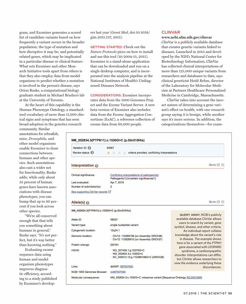
55 07.2016 | THE SCIENTIST
gram, and Exomiser generates a scored list of candidate variants based on how frequently a variant occurs in the broader population; the type of mutation and how disruptive it may be; and potentially related genes, which may be implicated in a particular disease or clinical feature. What sets Exomiser and other Mon-arch Initiative tools apart from others is that they also employ data from model organisms to predict whether a mutation is involved in the person’s disease, says Orion Buske, a computational biology graduate student in Michael Brudno’s lab at the University of Toronto.
At the heart of this capability is the Human Phenotype Ontology, a standard-ized vocabulary of more than 11,000 clin-ical signs and symptoms that has seen broad adoption in the genetics research community. Similar annotations for zebrafish, mice, Drosophila, and other model organisms enable Exomiser to draw connections between humans and other spe-cies. Such annotations also cast a wider net for functionality, Buske adds; while only about 35 percent of human genes have known asso-ciations with disease phenotypes, you can bump that up to 80 per-cent if you look across other species.
“We’re all conserved enough that that tells you something about humans in general,” Buske says. “It’s not per-fect, but it’s way better than knowing nothing.”
Evaluating exome sequence data using human and model organism phenotypes improves diagnos-tic efficiency, accord-ing to a study published by Exomiser’s develop-
ers last year (Genet Med, doi:10.1038/gim.2015.137, 2015).
GETTING STARTED: Check out the Nature Protocols piece on how to install and use this tool (10:2004-15, 2015). Exomiser is a stand-alone application that can be downloaded and run on a single desktop computer, and is incor-porated into the analysis pipeline at the National Institutes of Health’s Undiag-nosed Diseases Network.
CONSIDERATIONS: Exomiser incorpo-rates data from the 1000 Genomes Proj-ect and the Exome Variant Server. A new beta version of Exomiser also includes data from the Exome Aggregation Con-sortium (ExAC), a reference collection of exome data from 60,000 people.
CLINVARwww.ncbi.nlm.nih.gov/clinvarClinVar is a publicly available database that curates genetic variants linked to diseases. Launched in 2013 and devel-oped by the NIH’s National Center for Biotechnology Information, ClinVar has collected clinical interpretations of more than 125,000 unique variants from researchers and databases to date, says clinical geneticist Heidi Rehm, director of the Laboratory for Molecular Medi-cine at Partners Healthcare Personalized Medicine in Cambridge, Massachusetts.
ClinVar takes into account the inex-act nature of determining a gene vari-ant’s effect on health, with one research group saying it is benign, while another says it’s more serious. In addition, the categorizations themselves—for exam-
QUERY AWAY: NCBI’s publicly available database ClinVar allows
users to search by variant, gene symbol, disease, and other criteria.
An individual report collates knowledge about the variant’s role
in disease. The example shown here is for a variant of the PTPN11
gene associated with LEOPARD syndrome, a cardiomyopathic
disorder. Interpretations can differ, but ClinVar allows researchers to
view the evidence underlying these discordances.

LAB TOOLS
ple, “likely pathogenic”—are more clearly defined and standardized in the tool.
HOW IT WORKS: ClinVar uses a star-based system to rate the review level of a given variant’s supposed (or interpreted) role in disease. A four-star rating is the highest, meaning the variant has been through a review process with multiple experts in the community weighing in on its interpretation and the support-ing evidence. The upside of this detailed review process is that users can trust three- and four-star variants, Rehm says. However, only a small subset of the
variants in the ClinVar database (3,800) fit into these categories.
More often, variants receive one star—usually based on a single submission pro-viding an interpretation and rules for inter-pretation—or no stars, indicating that the submitter did not provide their interpreta-tion criteria and attest to a comprehensive review of supporting evidence. One chal-lenge facing the field is that most of the clinically significant variants (83 percent) in the ClinVar repository are unique to a particular family or are very rare, according to an analysis published last year by Rehm (N Engl J Med, 372:2235-42, 2015).
GETTING STARTED: To learn how to take full advantage of ClinVar, check out a recent detailed primer geared for users (Curr Protoc Hum Genet, doi:10.1002/0471142905.hg0816s89, 2016). A YouTube video explains the dif-ferent search options. Because the use-fulness of the tool relies on submissions, Rehm encourages labs to share data. A submission wizard tool can be found on ClinVar’s site.
CONSIDERATIONS: Although ClinVar aims to be everything you need, it isn’t just yet. That’s mainly because the database relies on voluntary submissions. “We’re still trying to convince all of the journals to require [sharing on] ClinVar as part of the publication,” Rehm says. She and her col-leagues are working to mandate submis-sion from clinical laboratories as well.
In the meantime, Rehm consults a variety of sources to research the poten-tial clinical significance of candidate vari-ants identified in patients’ genetic data. These include the Human Genetic Muta-tion Database (HGMD), which collects all of the variants that have been published in the literature. Although HGMD is poorly curated, “it’s at least helpful in trying to find the publications where your variant might be reported,” she says. She also still digs into disease-specific databases, some of them archival, for particular variants.
MATCHMAKER EXCHANGEwww.matchmakerexchange.orgMatchmaker Exchange is a network that connects the stand-alone databases focused on linking human genes and clin-ical features. Today the platform draws on three existing databases and will incor-porate more in the future. In addition to collating information on gene–disease links, Matchmaker Exchange aims to join together researchers who are working on rare disease cases to share information and potentially collaborate. The ultimate goal is to help researchers build a more solid case for a causal gene and publish on it, because many rare disease gene can-didates languish in the lab unpublished, notes Rehm, who led the development of
GENERAL TIPS: Whole-genome vs. whole-exome? Whole-exome sequencing is almost always the better choice, because it costs much less. Annotation of variants in noncoding regions of the genome is still not up to scratch, so most of that data are still unusable, says the University of Leuven’s Adrian Liston. Still, a head-to-head comparison of the approaches published last year found that whole-genome sequencing is more powerful for detecting candidates for rare diseases (PNAS, 112:5473-78, 2015).
Use reference populations as similar as possible to your patient. Large reference sets like the 1000 Genomes Project may not serve as adequate filters for your case or cohort, if your population is not represented in these databases (e.g., if you are studying a relatively isolated village that is not sampled in one of these big projects), so you’ll have to start building your own reference database or find someone else who has one.
Analyze families whenever possible. Sequence the exomes of parents and, ideally, a healthy sibling. “This is essential if you’re going to look for de novo mutations,” Liston says.
Read and read more. Bioinformatics tools should be used to help you narrow down the candidates to a list that you can manage by reading papers on those genes.
Prioritization is a team effort. Taking a short list of candidates down from 10 to just 1 or 2 is a subjective decision-making process. Assemble a team of experts, including those well versed in a particular disease process, to help you decide which candidates to take further. A team like this should meet multiple times throughout the process, making decisions about, for example, when to sequence a particular variant (Sanger validation) in additional family members, says Christopher Cassa of Brigham and Women’s Hospi tal in Boston.
Build a case for publication. When he’s reviewing papers, Liston looks for a second, independent family that is affected by the same variant and gene as the original patient. If you don’t have one, you’ll need to pursue other supporting evidence, such as from cell lines or animal models.
56 THE SCIENTIST | the-scientist.com

57 07.2016 | THE SCIENTIST
CO
UR
TE
SY
OF
OR
ION
BU
SK
E
the platform. The October 2015 issue of Human Mutation details the platform’s capabilities and utility (36:915-1019).
HOW IT WORKS: Choose among Match-maker Exchange’s founding members:
GeneMatcher. Create an entry for the gene you’re interested in. If two people create an entry with the same gene name, the database (which is otherwise not searchable) will send them both an email. As of May, 4,459 genes had been submit-ted by 1,675 users from 55 countries. More than 5,200 matches have been made on some 1,200 genes. “This brings together people, knowing they can talk more in detail about the features of the patient [and] about variants of the gene they are studying,” says GeneMatcher codeveloper Nara Sobreira, an assistant professor of pediatrics at Johns Hopkins Medicine in
Baltimore, Maryland. Patients can also use the tool, as can researchers using animal models, she adds.
PhenomeCentral. This tool for clinicians and scientists is phenotype-focused, allowing users investigating rare and unnamed disorders to be paired based on Human Phenotype Ontology vocabu-lary. It also incorporates the Exomiser software package to filter and prioritize genes for matching with other cases.
DECIPHER. This web-based database pulls together a variety of bioinformat-ics tools to help clinicians interpret vari-ants and to pair up cases on the basis of shared variant and clinical data. There are several ways to make matches in the data-base. For example, nonregistered users can search DECIPHER’s open-access patient records covering 56,000 phenotypes,
1,200 sequence variants, and 28,000 copy number variants and contact the data sub-mitter. Or you can set up your own project to share data from consenting patients and match with other researchers.
More databases are coming soon, includ-ing patient portals such as PEER, PatientKind, and ClinGen’s Genome-Connect, as well as Monarch Initiative’s model organism–focused database.
GETTING STARTED: You need to create a single log-in to any one of the three data-bases that are currently part of the net-work. Pick the one that best fits the data you have and the questions you want to ask. Creating an entry takes roughly 10 minutes, and you can elect which other databases you want to run your query against, Buske says.
CONSIDERATIONS: An ideal time to use Matchmaker Exchange is fairly late in the process, when you have a candidate gene or two for an individual or family that you are fairly certain is causal, but you want to find that second family to strengthen your case.
“That’s important because if every-one put seven or eight variants in Match-maker Exchange, the chance that you hit some other person that has a case is sub-stantial,” says Christopher Cassa, a genet-icist at Brigham and Women’s Hospi-tal in Boston. More hits bring more false positives, though. Cassa and his group have developed a tool called Rare Disease Match, which uses data from the Exome Aggregation Consortium to help predict the probability of observing a gene based on chance (Hum Mutat, 36:998-1003, 2015). They hope to integrate this tool with Matchmaker Exchange.
Despite the challenge of false-posi-tives, Matchmaker Exchange stakehold-ers hope the tool can eventually be use-ful earlier in the workflow, Buske says, where, for example, matches can be made with less-complete data. The plat-form also aims to support whole-exome searching rather than single variants or genes of interest, though there are more privacy issues to iron out.
PAIRING UP: Matchmaker Exchange’s founding members are PhenomeCentral, GeneMatcher, and DECIPHER (dotted arrows), but the platform will soon allow researchers to query an even larger number of databases. Each takes a slightly different approach to understanding rare genetic diseases.
ClinGen Genome Connect
Phenome Central
Monarch
LOVD
BROAD RDAP
PEER RD Connect
GENESIS/ GEM.app
Undiagnosed Diseases Network
Café Variome
Gene Matcher
DECIPHER
EFFORTS HAVE DIFFERENT DATA/FOCUS
Variants, diseases
Genes,phenotypes (PhenoDB)
Phenotypes (HPO),exomes,
genes
Model organisms
Variants
Diseases
Diseases, exomes
Variants (CNVs),
phenotypes (HPO)

58 THE SCIENTIST | the-scientist.com
CAREERS
© I
ST
OC
K.C
OM
/NIC
OL
AS
_
Adelaide Rhodes had no idea a tiny crustacean would fuel such a big career shift. About a decade ago,
as a postdoc at the University of Wash-ington, she was researching copepods—microscopic organisms that convert unsat-urated fatty acids into the omega-3 fats that make salmon a healthy meal. They’re “what fish eat to get fat,” Rhodes says. Dur-ing an aquaculture boom, she began hunt-ing for genes involved in the fat-converting process. Trouble was, very few researchers studied copepod genetics. Back in 2005, Rhodes’s searches for “copepod and lipids” on the DNA Data Bank of Japan, European Nucleotide Archive, and GenBank yielded no results. When she searched “crusta-cean,” she got a list of some 50 genes, but none were related to lipid metabolism.
Undeterred, Rhodes broadened her search to include insect genes, then designed primer sets and ran countless PCR assays to check if those same genes were found in copepods. She also went around at meet-ings asking other researchers if they had copepod data or sequences to share. Rhodes eventually identified two potential copepod desaturases—enzymes that introduce dou-ble bonds into fatty acid chains. However, she couldn’t confirm whether those genes are specific to copepods, because there weren’t enough publicly available crusta-cean genomes for comparison.
These days, she wouldn’t have that problem. When researchers identify a new genomic sequence, they can use modern computing and bioinformatics tools to check for its presence in related species’ genomes with just a few keystrokes. And as technical advances yield unmanage-able amounts of data across diverse fields, more wet-lab scientists are turning to bio-informatics to make sense of their results. Online courses, workshops, and a grow-
ing community of bioinformatics-savvy researchers are now available to help sci-entists better understand available data-analysis tools or create their own—or even to persuade them to leave the bench alto-gether for a computing career.
After her struggles to identify copepod genes involved in omega-3 fatty acid pro-duction, Rhodes went on to do two addi-tional postdocs: at Smithsonian Marine Station in Fort Pierce, Florida, and at Texas A&M University–Corpus Christi. Knee-deep in computational analyses by then, she returned to school and com-pleted a master’s degree in bioinformat-ics at Johns Hopkins University in 2012.
Rhodes, who is now a researcher and bioinformatics trainer at Oregon State University, wasn’t alone in choosing to make this career shift. A recent survey by the jobs and recruiting site Glassdoor.
com rated “data scientist” as the best job of 2016, and in 2012 Harvard Business Review called it the “sexiest job of the 21st century.” But even if you’re not looking to change paths, a little bioinformatics know-how can still be helpful in the lab.
Collaborate with codersAs an immunology postdoc at Vir-ginia Tech, Raquel Hontecillas-Magarzo worked with mice and did molecular biol-ogy experiments. She then spent two years doing benchwork at the Spanish Institute for Research and Agriculture in Madrid before returning to Virginia Tech’s Bio-complexity Institute as an assistant pro-fessor. So when the university assembled a team to develop computational models for studying human immunity to gut patho-gens, Hontecillas-Magarzo was tapped for her expertise in experimental design. The
In today’s data-heavy research environment, wet-lab scientists can benefit from learning new computational skills.
BY ESTHER LANDHUIS
Bench to Bioinformatics

59 07.2016 | THE SCIENTIST
team included life scientists, physicists, bioinformaticians, and software engi-neers—about a 50:50 mix of experimen-tal and computational researchers.
At a weeklong symposium on compu-tational immunity in summer 2014, Hon-tecillas-Magarzo and other immunologists learned how computational tools could deepen their analysis of wet-lab data and suggest new hypotheses that might not seem intuitive based on the literature. Nowadays, Hontecillas-Magarzo uses computer simulations to model the behav-ior of immune cells during infection by Helicobacter pylori, a bacterium that can cause ulcers. She and her colleagues define the simulation’s parameters based on experimental data—for instance, the level of T-cell activity measured on the third day of an H. pylori infection in a mouse. Recently, a sensitivity analysis using this model suggested that anti-inflammatory macrophages may help maintain mucosal integrity and prevent stomach epithelial cells from dying during H. pylori infection. These in silico analyses don’t reveal under-lying mechanisms. However, they can show that “if you change one [element], it has a significant effect on the other,” which helps inform decisions on what to vali-date in bench experiments, says Hontecil-las-Magarzo. She is currently conducting mouse studies to follow up on the macro-phage/epithelial link.
Even without access to local courses or symposia, wet-lab researchers can gain familiarity with computational and bio-informatics methods by arranging col-laborations with research groups whose members have that expertise, suggests Josep Bassaganya-Riera, who he directs Virginia Tech’s Nutritional Immunology and Molecular Medicine Laboratory, which includes Hontecillas-Magarzo’s lab. Researchers specifically interested in computational immunology can
find links to books, tutorials, and other resources at this Virginia Tech site (www.modelingimmunity.org/education).
To analyze data with interdisciplin-ary teams, “you don’t need to be an expert in computational tools or bioin-formatics or math,” Hontecillas-Magarzo says. However, “you need some level of understanding. You need to understand some of their terminology.”
Back to schoolKathleen Fisch, a computational biologist at the University of California, San Diego, got her first taste of bioinformatics from evolutionary biologist Craig Moritz as a University of California, Berkeley, under-graduate using geographic software to map climatic niches of hummingbirds. But it wasn’t formal instruction. “He’d say, ‘Here are some data points. Go play with the software,’” Fisch says. Then, while working on her PhD at UC Davis, Fisch continued dabbling with computational tools—using a program called Structure to detect population structure from micro-satellite DNA markers and SPAGeDi (Spa-tial Pattern Analysis of Genetic Diversity) to assess the genetic diversity of endan-gered smelt populations in the San Fran-cisco Bay Delta.
But those software packages were developed by others; Fisch wanted to create her own. She started by learning Python and R, two widely used program-ming languages. “I bought a bunch of books and hardly looked at them,” Fisch
jokes. Instead, she immersed herself in online courses through Coursera. Five days a week, Fisch logged onto Coursera to watch lectures, puzzle over problems, and get feedback from fellow students. At first the “computational stuff seems intimidating,” says Fisch, “but it’s totally within your grasp if you have time to dedi-cate to it.” (The Python and R classes were free when Fisch took them five years ago, though Coursera now charges $79–$99 per course for similar offerings. Upon successful completion, students earn elec-tronic course certificates that can be added to their LinkedIn profile.)
Researchers can also learn computa-tional basics by attending Data Carpen-try and Software Carpentry courses. Soft-ware Carpentry runs about 100 two-day workshops around the world each year, teaching core skills for research comput-ing through short tutorials and practi-cal exercises. All instruction is done via live coding. While Software Carpentry is mostly aimed at researchers who are already doing some data analysis and programming, its sister organization, Data Carpentry, is good for those who are just beginning the transition from spread-sheets to R, Python, and command-line data analysis.S
HA
WN
O’N
EIL
BUGGING OUT: Emily Bellis, an Oregon State University graduate student, discusses methods to sort sequences belonging to different organ-isms in symbiotic systems at the Bioinformatics Users Group (BUG) biweekly lunchtime meeting.
You don’t need to be an expert in computational tools or bioin formatics or math. —Raquel Hontecillas-Magarzo,
Virginia Tech

60 THE SCIENTIST | the-scientist.com
CAREERS
PH
OT
O P
RO
VID
ED
BY
MIE
P
With more computational skills under her belt, Fisch decided to leave the bench entirely and work with Scripps Research Institute bioinformatician Andrew Su, whose lab builds and applies tools to use crowdsourcing for genetics and genom-ics. As a Scripps postdoc, Fisch learned how to analyze next-generation sequenc-ing data on different platforms and has collaborated with multiple research groups on projects ranging from preci-sion medicine studies in breast cancer to systems biology analyses of osteoarthritis. “Working with lots of PIs and collabora-tors, I was able to get exposed to pretty much all the next-gen sequencing types,” Fisch says. In the fall of 2014, she took a job at UC San Diego, where she currently works at the Institute of Genomic Medi-cine developing an open-source platform to automate multi-omics data analysis pipelines on computer clusters and in the cloud.
Community supportAs she began taking Coursera classes to learn Python and R, Fisch also turned to help from colleagues and an online community forum called StackOver-flow, where she picked up the basics of a command-line language called bash. Although self-teaching on a “need to
know” basis was probably not as com-prehensive as a formal college course, “it was enough to get me off the ground,” Fisch told The Scientist. The collection of Python “recipes” on GitHub.com, a public code repository, is another good resource for bioinformatics code snip-pets and concepts.
Another postdoc in Su’s lab at Scripps, Tim Putman, also waded into bioinfor-matics with help from a supportive com-munity. When he began his PhD research at Oregon State University (OSU) in 2010, Putman conducted cell biology experiments to study the pathogenesis of Chlamydia infection. But sequencing bacterial genomes and doing compara-tive genomics quickly hooked Putman on the analysis side of the research. To do that kind of work, he needed to navi-gate the Linux environment to pull files from other servers, extract the data he wanted, and run Python and R scripts to reformat the results to work with the lab’s algorithms.
Putman picked up some command-line basics from other members of his lab. He also took a Python workshop offered on campus through OSU’s Cen-ter for Genome Research and Biocomput-ing. Another big help was OSU’s bioin-formatics users group (BUG). This group
of life scientists, bioinformaticians, com-puter scientists, mathematicians, and engineers meets every other week to chat over lunch about metagenomics, struc-tured query language (SQL), and other computational challenges. The primary goal of BUG is “getting people into the same room to chat about what they’re learning and what they’re struggling with,” says Shawn O’Neill, one of OSU’s bioinformatics trainers.
Indeed, Putman sometimes found others in the room who had answers for his nagging problems. “A big thing I learned from BUG people was how to configure and debug the command-line tools and set up my environment,” he says. “This can be a big hurdle to some-one new to computer science.”
UC Davis also provides opportunities for bioinformaticians to share struggles and solutions, through a forum called the Data Intensive Biology program. The ses-sions are organized by Titus Brown, a key leader of a grassroots movement to have bioinformaticians train each other. Some of the discussions are broadcast online, so that interested researchers outside the UC Davis campus can participate. And some attendees, including Rhodes, meet periodically to hash out new course mate-rials and teaching methodologies.
It was during one of these UC Davis workshops that Rhodes learned about several techniques for hybrid de novo assembly of Illumina data. So when she returned to OSU and Putman com-plained to her that there were no refer-ence sequences with which to align his own bacterial genomes, she encouraged him to look into new tools. “It was great because she was out learning about cut-ting edge stuff from the leaders in the field and then bringing it back to researchers at OSU,” Putman says.
Now at Scripps, Putman is putting his bioinformatics experiences to good
SUMMER CRAM: In 2014, the Center for Mod-eling Immunity to Enteric Pathogens (MIEP) at Virginia Tech hosted a Modeling Mucosal Immu-nity summer symposium that combined morning lectures with afternoon hands-on work.
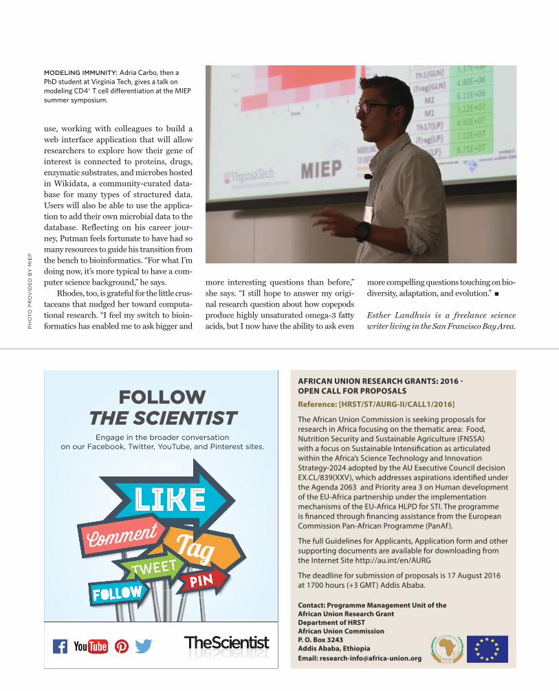
Engage in the broader conversation on our Facebook, Twitter, YouTube, and Pinterest sites.
FOLLOW THE SCIENTIST
PH
OT
O P
RO
VID
ED
BY
MIE
P
use, working with colleagues to build a web interface application that will allow researchers to explore how their gene of interest is connected to proteins, drugs, enzymatic substrates, and microbes hosted in Wikidata, a community-curated data-base for many types of structured data. Users will also be able to use the applica-tion to add their own microbial data to the database. Reflecting on his career jour-ney, Putman feels fortunate to have had so many resources to guide his transition from the bench to bioinformatics. “For what I’m doing now, it’s more typical to have a com-puter science background,” he says.
Rhodes, too, is grateful for the little crus-taceans that nudged her toward computa-tional research. “I feel my switch to bioin-formatics has enabled me to ask bigger and
more interesting questions than before,” she says. “I still hope to answer my origi-nal research question about how copepods produce highly unsaturated omega-3 fatty acids, but I now have the ability to ask even
more compelling questions touching on bio-diversity, adaptation, and evolution.”
Esther Landhuis is a freelance science writer living in the San Francisco Bay Area.
MODELING IMMUNITY: Adria Carbo, then a PhD student at Virginia Tech, gives a talk on modeling CD4+ T cell diff erentiation at the MIEP summer symposium.
AFRICAN UNION RESEARCH GRANTS: 2016 - OPEN CALL FOR PROPOSALS
Reference: [HRST/ST/AURG-II/CALL1/2016]
The African Union Commission is seeking proposals for research in Africa focusing on the thematic area: Food, Nutrition Security and Sustainable Agriculture (FNSSA) with a focus on Sustainable Intensification as articulated within the Africa’s Science Technology and Innovation Strategy-2024 adopted by the AU Executive Council decision EX.CL/839(XXV), which addresses aspirations identified under the Agenda 2063 and Priority area 3 on Human development of the EU-Africa partnership under the implementation mechanisms of the EU-Africa HLPD for STI. The programme is financed through financing assistance from the European Commission Pan-African Programme (PanAf ).
The full Guidelines for Applicants, Application form and other supporting documents are available for downloading from the Internet Site http://au.int/en/AURG
The deadline for submission of proposals is 17 August 2016 at 1700 hours (+3 GMT) Addis Ababa.
Contact: Programme Management Unit of the African Union Research GrantDepartment of HRSTAfrican Union CommissionP. O. Box 3243Addis Ababa, EthiopiaEmail: [email protected]
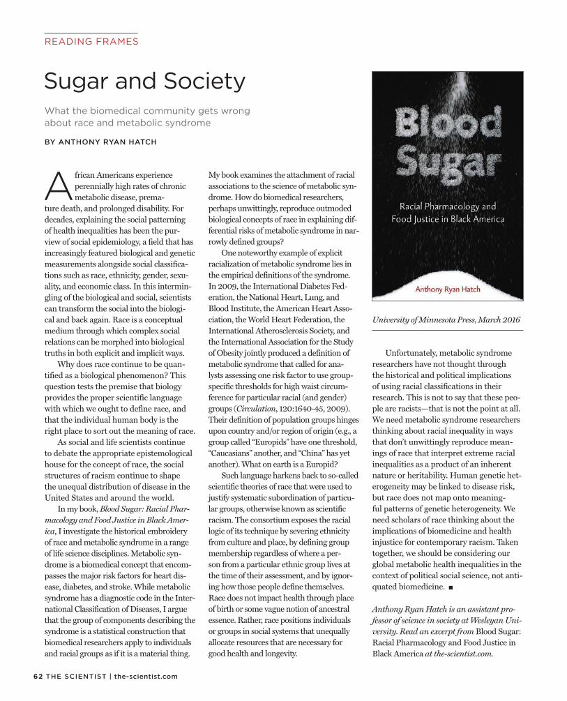
62 THE SCIENTIST | the-scientist.com
READING FRAMES
What the biomedical community gets wrong about race and metabolic syndrome
BY ANTHONY RYAN HATCH
Sugar and Society
African Americans experience perennially high rates of chronic metabolic disease, prema-
ture death, and prolonged disability. For decades, explaining the social patterning of health inequalities has been the pur-view of social epidemiology, a field that has increasingly featured biological and genetic measurements alongside social classifica-tions such as race, ethnicity, gender, sexu-ality, and economic class. In this intermin-gling of the biological and social, scientists can transform the social into the biologi-cal and back again. Race is a conceptual medium through which complex social relations can be morphed into biological truths in both explicit and implicit ways.
Why does race continue to be quan-tified as a biological phenomenon? This question tests the premise that biology provides the proper scientific language with which we ought to define race, and that the individual human body is the right place to sort out the meaning of race.
As social and life scientists continue to debate the appropriate epistemological house for the concept of race, the social structures of racism continue to shape the unequal distribution of disease in the United States and around the world.
In my book, Blood Sugar: Racial Phar-macology and Food Justice in Black Amer-ica, I investigate the historical embroidery of race and metabolic syndrome in a range of life science disciplines. Metabolic syn-drome is a biomedical concept that encom-passes the major risk factors for heart dis-ease, diabetes, and stroke. While metabolic syndrome has a diagnostic code in the Inter-national Classification of Diseases, I argue that the group of components describing the syndrome is a statistical construction that biomedical researchers apply to individuals and racial groups as if it is a material thing.
My book examines the attachment of racial associations to the science of metabolic syn-drome. How do biomedical researchers, perhaps unwittingly, reproduce outmoded biological concepts of race in explaining dif-ferential risks of metabolic syndrome in nar-rowly defined groups?
One noteworthy example of explicit racialization of metabolic syndrome lies in the empirical definitions of the syndrome. In 2009, the International Diabetes Fed-eration, the National Heart, Lung, and Blood Institute, the American Heart Asso-ciation, the World Heart Federation, the International Atherosclerosis Society, and the International Association for the Study of Obesity jointly produced a definition of metabolic syndrome that called for ana-lysts assessing one risk factor to use group-specific thresholds for high waist circum-ference for particular racial (and gender) groups (Circulation, 120:1640-45, 2009). Their definition of population groups hinges upon country and/or region of origin (e.g., a group called “Europids” have one threshold, “Caucasians” another, and “China” has yet another). What on earth is a Europid?
Such language harkens back to so-called scientific theories of race that were used to justify systematic subordination of particu-lar groups, otherwise known as scientific racism. The consortium exposes the racial logic of its technique by severing ethnicity from culture and place, by defining group membership regardless of where a per-son from a particular ethnic group lives at the time of their assessment, and by ignor-ing how those people define themselves. Race does not impact health through place of birth or some vague notion of ancestral essence. Rather, race positions individuals or groups in social systems that unequally allocate resources that are necessary for good health and longevity.
Unfortunately, metabolic syndrome researchers have not thought through the historical and political implications of using racial classifications in their research. This is not to say that these peo-ple are racists—that is not the point at all. We need metabolic syndrome researchers thinking about racial inequality in ways that don’t unwittingly reproduce mean-ings of race that interpret extreme racial inequalities as a product of an inherent nature or heritability. Human genetic het-erogeneity may be linked to disease risk, but race does not map onto meaning-ful patterns of genetic heterogeneity. We need scholars of race thinking about the implications of biomedicine and health injustice for contemporary racism. Taken together, we should be considering our global metabolic health inequalities in the context of political social science, not anti-quated biomedicine.
Anthony Ryan Hatch is an assistant pro-fessor of science in society at Wesleyan Uni-versity. Read an excerpt from Blood Sugar: Racial Pharmacology and Food Justice in Black America at the-scientist.com.
University of Minnesota Press, March 2016

63 07.2016 | THE SCIENTIST
CAPSULE REVIEWS
Serendipity: An Ecologist’s Quest to Understand NatureJames A. EstesUniversity of California Press, May 2016
Part memoir and part ecology primer, Serendip-ity—the latest book from celebrated marine ecolo-gist James Estes—is an insightful reminder that when observing nature,
there is always much more than meets the eye. Estes relates his epiphany that apex predators play key roles in kelp for-est ecosystems, an insight he gleaned in 1971 while studying sea otters in Alaskan waters. “In the absence of sea otter pre-dation, sea urchins had increased in size and number, and the larger and more abundant urchins had eaten the kelp,” he writes. “This was my ‘aha moment,’ a pro-found realization that would set a path for the remainder of my life.”
Estes took that revelation and ran with it, building a conceptual framework and a career that would see him apply his understanding of ecology to numerous marine ecosystems around the world. His message in Serendipity is clear: the com-plexities of the natural world will only be revealed to thoughtful, dedicated sci-entists who seek not only to catalog and observe nature, but to truly understand it.
Complexity: The Evolution of Earth’s Biodiversity and the Future of HumanityWilliam C. BurgerPrometheus Books, June 2016
Another accomplished biologist, William Burger, curator emeritus at Chica-go’s Field Museum of Nat-ural History, has written a fascinating book about evolution and the future
of our planet at the hands of its most successful—and destructive—species. In Complexity, Burger describes the intri-cate interplay between species that took Earth’s biota from single to multicellu-
lar organisms, resulting in the evolution of all manner of biological complexity and the rich tapestry of biodiversity that blankets the Earth today.
Central to Burger’s sweeping history of life on Earth is the theme of coopera-tion. From endosymbiosis to the develop-ment of cooperative species such as ants and bees, working together is a hallmark of biology, he explains.
And interaction between species and individuals continues to mold the fate of our planet. Human culture helped us become a dominant species, but overcon-sumption and continuing conflict now threaten the very fabric of our world. Burger is not optimistic: “Regrettably, I am a pessimist who sees modern indus-trial society as unsustainable and ‘human nature’ incapable of diminishing its bio-logical and cultural appetites,” he writes. “Also, I’m worried that, since people cause trouble, more people will inevitably cause more trouble.”
The Human Superorganism: How the Microbiome Is Revolutionizing the Pursuit of a Healthy LifeRodney DietertDutton, July 2016
With each passing day, researchers are learning more about the intricate ways that humans interact with our microbial copi-lots. And now, immunolo-gist Rodney Dietert has
written a definitive book on what we know and what we have yet to discover about the fascinating world of our microbiome.
In The Human Superorganism, Dietert expresses the view that well-meaning medical science has gotten it wrong for decades. “Wonderful scientific discoveries significantly reduced infant mortality, lengthened life spans, and drove medical technologies,” he writes. “However, the fundamental approach to human biology behind these advances unintentionally ushered in an epidemic of diseases afflicting humanity in the twenty-first century.” By treating the
human body as a pure organism and by focusing so intently on the human genome, biologists failed to appreciate the fact that individuals are not pure—nor are they individual.
Dietert argues that the rash of non-communicable diseases—cancer, obe-sity, and others—that plagues humanity is a direct result of this misplaced per-spective. He advocates for a perceptual shift in which humans are viewed more like ecosystems that support a teeming microcosm of microbial partners.
Love and Ruin: Tales of Obsession, Danger, and Heartbreak from The Atavist MagazineEdited by Evan RatliffW.W. Norton & Company, July 2016
From the Web pages of The Atavist magazine comes this compendium of award-winning, long-form nonfiction, edited by Evan Ratliff, who cofounded the Atavist
multimedia publishing platform in 2009. The centerpiece of Love and Ruin is
the story of the same name, which won the 2014 National Magazine Award for best feature writing. That piece, written by James Verini, touches on the imper-iled archeological artifacts of war-torn Afghanistan, but other stories in the book focus more squarely on the life sciences.
Leslie Jamison’s “52 Blue” is a story about the trials and tribulations of marine biologists and the blue whale who sang at a frequency (52 hertz) that made his song unhearable by his fellow leviathans. And the story nicely encap-sulates what sets writing in The Atavist apart from the quick-hit click bait that has become so common in online news. Jamison explores the people and person-alities behind the science. She takes time to describe in vivid detail their motiva-tions, challenges, and triumphs. Readers are sure to feel as much, if not more, for the story’s human devotees of the whale as they are for the hapless cetacean itself.
—Bob Grant
64 THE SCIENTIST | the-scientist.com
The
Gui
de Your Partner in Immunotherapy Research
Lonza is committed to supporting immunotherapy research by providing cells, media, and both large- and small-volume transfection solutions to help your transition from research to therapy. If you’re ready for the clinic, our cell therapy services could prove to be the solution for successful development, manufacturing and testing of your cell-based therapeutics.
Contact us today for more information.
LONZA WALKERSVILLE, [email protected] www.lonza.com/immunotherapy
Celase® GMP For Adipose Stem Cell Isolation Celase® GMP, an avian and mammalian tissue-free collagenase and neutral protease blend produced under GMP by Cytori, is now available from Worthington for adipose stem cell isolation.A single, sterile vial digests up to 280g of adipose tissue with best-in-class GMP quality and shelf life up to 72 months. Currently included in human IDE applications approved by the U.S. FDA.
WORTHINGTON BIOCHEMICAL CORP.730 Vassar Ave, Lakewood, NJ 08701Ph: 800.445.9603/732.942.1660 • Fax: 800.368.3108/732.942.9270Worthington-Biochem.com
New Stand-Alone Imager for Gels and BlotsThe new ChemiDoc-ItTS3 Imager is an upgradeable, chemiluminescence imager for high sensitivity imaging of Western blots, gels, dyes and stains. It is a stand-alone system with an integrated 15.6 touch screen, which includes UVP's TS3 Software for automated, image acquisition, and enhancement. The imager features a choice of deeply cooled, cameras and lenses for high performance image capture in low light applications.
UVP, LLC(800) [email protected]://www.uvp.com
Label-Free Automated Cell Migration AssayThe ECIS (Electric Cell-Substrate ImpedanceSensing) Wound Healing Assay replaces thetraditional “scratch” or “scrape” assay. Instead of disrupting the cell layer mechanically with a toothpick, needle or pipette tip and followingthe migration of cells to “heal” the wound with a microscope, electric signals are used to both wound and monitor the healing process. Additionally, the ECM protein coating on the electrode is not damaged (or modified) if using the electric fence method.
APPLIED BIOPHYSICSPhone: (866) 301-3247www.biophysics.com
System for DNA Damage Measurement CometChip®
• This high-throughput platform is developed to simultaneously treat and measure DNA damage induced by different treatments, or among different cell types on a single slide using the Comet assay• Extends the productivity of the 96-well format Comet Assay even further• A single 96-well CometChip®can produce 20,000 data points per chip, based on an average of 208 cells imaged per well
AMSBIOwww.amsbio.com/comet-chip.aspx
iPSC-Derived Astrocyte Kits • New options include the iPSC-Derived Astrocyte Progenitor Kit and iPSC-Derived Mature Astrocyte Kit• Include cryopreserved cells, optimized media, and supplements• Offer researchers a complete culture system that is easy to use and promotes long-term cell viability for use in disease modeling and drug discovery applications
AXOL BIOSCIENCEwww.axolbio.com

Meetings:Genome Engineering: The CRISPR/Cas RevolutionAugust 17 - 20 Jennifer Doudna, Maria Jasin, Jonathan Weissman
Regulatory & Non-Coding RNAsAugust 23 - 27 Victor Ambros, Elisa Izaurralde, Nicholas Proudfoot
The PI3K-mTOR-PTEN Network in Health & DiseaseAugust 30 - September 3
Translational Control
Epigenetics & ChromatinSeptember 13 - 17 Shelley Berger, Juerg Mueller, Yang Shi
Axon Guidance, Synapse Formation & RegenerationSeptember 20 - 24 abstracts due July 1
Mechanisms of AgingSeptember 26 - 30 abstracts due July 25
Germ CellsOctober 4 - 8 abstracts due July 15
Biological Data ScienceOctober 26 - 29 abstracts due August 12
Transposable Elements
Neurodegenerative Diseases: Biology & Therapeutics
Blood Brain BarrierDecember 7 - 10 abstracts due September 23
CSHL 2016 Upcoming Meetings & Courses
photo: Symposium 2015 BBQwww.cshl.edu/meetings
Courses:Programming for BiologyOctober 10 - 25
X-Ray Methods in Structural BiologyOctober 10 - 25
Cereal GenomicsOctober 19 - 25
Computational & Comparative Genomics
Antibody Engineering & Phage Display
Advanced Sequencing Technologies & Applications
Immersive Approaches to Biological Data VisualizationDecember 1 - 10Kelly Gaither, Matthew Vaughn
The Genome Access Course
Professional Development:
Charla Lambert, Stephen Matheson
www.cshl.edu/meetings

We are pleased to announce that The Scientist won a 2015 FOLIO Eddie award for our July 2014 issue and garnered an honorable mention in the Eddies Digital category for our online news coverage. FOLIO awards are presented annually and the Eddies recognize editorial excellence.
WINNER
B-to-B, Full issue-ScienceJuly 2014
HONORABLEMENTION
B-to-B, News Coverage
MOVING ASSAYS TO THE CLINIC
Next GenerationSUMMIT
EIGHTH ANNUAL August 23 - 26, 2016Grand Hyatt,Washington, DC
Cambridge Healthtech Institute250 First Avenue, Suite 300, Needham, MA 02494www.healthtech.com
NextGenerationDx.com
Setting the stage for the great minds of
together
diagnostics to advance medicine
Register Early for Maximum Savings:Mention Keycode 1680P10


68 THE SCIENTIST | the-scientist.com
FOUNDATIONS
WIK
IME
DIA
CO
MM
ON
S
BY CATHERINE OFFORD
Myth Becomes Reality, 1874
In Portugal Cove, Newfoundland, a small fishing boat was attacked in the fall of 1873. One of the boat’s occupants—so
the story goes—saw vast tentacles rising up from the water and, in an act of heroism, hacked a cou-ple off. Boat freed, the fisher-men headed back to shore.
The anglers fed one ten-tacle to a dog, according to some accounts; the other, measuring 19 feet in length, they carried to nearby St. John’s, to the home of min-ister and amateur naturalist Moses Harvey. “Harvey was Presbyterian Irish, incredi-bly homesick for Ireland, and had lost himself in all things natural,” says Matthew Gavin Frank, who explored Har-vey’s life and essays on New-foundland’s flora and fauna in his 2014 book Preparing the Ghost. “He was known in St. John’s in the mid- and late 1800s as just being crazy after all things from the land and the sea.” Harvey bought the tentacle for $10, says Frank, and estimated the creature it came from to be 72 feet long.
A subject of caution-ary tales rather than scien-tific inquiry, the giant squid was still very much consid-ered part of mythology, Frank says. But the following year, another group of fisherman in Logy Bay near St. John’s port brought Harvey some-thing unequivocally convinc-ing: a whole giant squid that had died thrashing in their nets. “These fisherman had obviously heard that Harvey had paid $10 for a tentacle and thought, ‘Well, goodness,
what will he pay for the entire thing?’” Frank says. “The answer was also $10.”
Harvey took the specimen back to his house at 3 Devon Row and draped its
27-foot-long body over a curtain rod above the bathtub. He arranged its tentacles, set up a photograph of the cephalopod hang-ing from the rail, and then relegated the
animal to a vat of brine in the back yard.
News of the squid quickly spread, and “around the globe, Harvey’s photograph was immediately, even by scien-tists, referred to as ‘the prob-lem of the giant squid,’” Frank says. “The problem, of course, was that now it was real.” While scientists attempted to reconcile this bizarre crea-ture with their view of the ani-mal kingdom, others, includ-ing American showman and circus founder P.T. Barnum, began bidding for the carcass.
In the end, Harvey sent his squid to Yale University zoologist Addison Emery Ver-rill, who was horrified by the specimen’s condition, Frank says. “The carcass had shrunk, desiccated a bit, and some of the suckers had fallen off.” But Verrill went on to publish the world’s first accurate illustra-tions and descriptions of this hitherto mythical creature.
Still today, nearly 150 years after the photograph was taken, little is known about the ecol-ogy of the giant squid. Rarely seen in action, this animal lives at depths of more than 500 meters (1,640 feet), and resists even the most dedicated researchers’ attempts to find it. “The photograph stands as a testament to the fact that this thing still remains, so many years out, so incredibly mysteri-ous,” says Frank. “It’s a source of both frustration and wonder.”
LAID OUT: The first photograph of most of a giant squid, now displayed in the Smithsonian National Museum of Natural History, was taken for amateur naturalist Moses Harvey of St. John’s, Newfoundland, in 1874. The 27-foot-long carcass (along with this photo of its arms draped on a rail over a bathtub) made its way to zoologist Addison Emery Verrill, who took detailed notes on the specimen. “The tub is 38½ inches in diameter,” he noted on the image. “On the club of the long arm there is a marginal row of small suckers on each side alternating with the larger ones.” Harvey displayed more awe. “I knew that I had in my possession what all the savants in the world did not,” he wrote in his journal. “A photograph could not lie and would silence the gainsayers.”

HOW MUCH DO YOU EARN?
www.the-scientist.com/salarysurveytake the survey
Announcing The Scientist’s annual Life Sciences Salary Survey
Help us compile the most current salary data for life scientists.
Take 5 minutes to tell us how you’re doing.
In November—just before your year-end reviews—we’ll publish results, and break them down by specialization, geographic location, degree, job title, and more.
Fill out the survey today, and encourage your friends to do the same.

The life science business of Merck KGaA, Darmstadt, Germany operates as MiliporeSigma in the U.S. and Canada.
Immobilon is a registered trademark and MilliporeSigma is a trademark of Merck KGaA, Darmstadt, Germany. BS-GEN-16-12214 5/2016 © 2016 EMD Millipore Corporation, Billerica, MA USA
Exercise your mem-brain.
Exercise your choice.No single transfer membrane gives you the best signal-to-noise ratio for all your proteins of interest. That’s why MilliporeSigma, the original Western blotting experts and inventors of Immobilon® PVDF membrane, gives you multiple membrane options.
Help your brain make the right membrane choice.
Remember:
Nitrocellulose was not designed for protein blotting.
PVDF has higher binding capacity than nitrocellulose which means generally higher sensitivity.
more sensitive.Request a sample of Immobilon® PVDF,
the intelligent membrane choice.
www.emdmillipore.com/immobilon
Rethink Western Blotting