multipurpose cellular lipids with emerging roles in cell death
13
Cell Death & Differentiation (2019) 26:781–793 https://doi.org/10.1038/s41418-018-0269-2 REVIEW ARTICLE Phosphoinositides: multipurpose cellular lipids with emerging roles in cell death Thanh Kha Phan 1 ● Scott A Williams 1 ● Guneet K Bindra 1 ● Fung T Lay 1 ● Ivan K. H Poon 1 ● Mark D Hulett 1 Received: 5 September 2018 / Revised: 18 December 2018 / Accepted: 19 December 2018 / Published online: 11 February 2019 © ADMC Associazione Differenziamento e Morte Cellulare 2019 Abstract Phosphorylated phosphatidylinositol lipids, or phosphoinositides, critically regulate diverse cellular processes, including signalling transduction, cytoskeletal reorganisation, membrane dynamics and cellular trafficking. However, phosphoinosi- tides have been inadequately investigated in the context of cell death, where they are mainly regarded as signalling secondary messengers. However, recent studies have begun to highlight the importance of phosphoinositides in facilitating cell death execution. Here, we cover the latest phosphoinositide research with a particular focus on phosphoinositides in the mechanisms of cell death. This progress article also raises key questions regarding the poorly defined role of phosphoinositides, particularly during membrane-associated events in cell death such as apoptosis and secondary necrosis. The review then further discusses important future directions for the phosphoinositide field, including therapeutically targeting phosphoinositides to modulate cell death. Facts ● Phosphoinositides are spatiotemporally enriched mem- brane lipids, and their turnover is dynamically, yet tightly, regulated. ● Phosphoinositides were initially characterised as second messengers for major signalling pathways. ● Phosphoinositides were later found to act as docking lipids to recruit and modulate actin cytoskeleton remodelling and membrane dynamics. ● Involvement of phosphoinositides in cell death had been poorly defined, often overshadowed by their traditional role as second messengers and regulators of cytoskeleton-membrane interactions. ● Recent reports support emerging importance of phos- phoinositides in pyroptosis, necroptosis, host defense peptide-induced necrosis, NETosis and autophagic cell death. Open questions ● Considering the importance of phosphoinositides in membrane and cytoskeleton dynamics, what are their roles in morphological changes during cell death, such as apoptosis? ● Do other lytic forms of cell death, including secondary necrosis, require phosphoinositides and what are the phosphoinositide-binding effectors? ● Can we target phosphoinositides to modulate cell death execution for therapeutic development? Introduction Phosphoinositides are derived from phosphatidylinositol (PI), comprising a diacylglycerol moiety linked to a D-myo- inositol ring via a phosphodiester linkage (Fig 1a). The inositol hydroxyls at positions 3, 4 or 5 are reversibly conjugated with phosphate groups, resulting in monopho- sphorylated [PI(3)P, PI(4)P and PI(5)P], bisphosphorylated [PI(3,4)P 2 ), PI(3,5)P 2 and PI(4,5)P 2 ] and trisphosphorylated [PI(3,4,5)P 3 ] derivatives. These seven interconvertible Edited by: D. Rubinsztein * Thanh Kha Phan [email protected] * Mark D Hulett [email protected] 1 Department of Biochemistry and Genetics, La Trobe Institute for Molecular Science, La Trobe University, Melbourne, Australia 1234567890();,: 1234567890();,:
-
Upload
khangminh22 -
Category
Documents
-
view
0 -
download
0
Transcript of multipurpose cellular lipids with emerging roles in cell death

Cell Death & Differentiation (2019) 26:781–793https://doi.org/10.1038/s41418-018-0269-2
REVIEW ARTICLE
Phosphoinositides: multipurpose cellular lipids with emerging rolesin cell death
Thanh Kha Phan 1● Scott A Williams1 ● Guneet K Bindra 1
● Fung T Lay 1● Ivan K. H Poon 1
● Mark D Hulett 1
Received: 5 September 2018 / Revised: 18 December 2018 / Accepted: 19 December 2018 / Published online: 11 February 2019© ADMC Associazione Differenziamento e Morte Cellulare 2019
AbstractPhosphorylated phosphatidylinositol lipids, or phosphoinositides, critically regulate diverse cellular processes, includingsignalling transduction, cytoskeletal reorganisation, membrane dynamics and cellular trafficking. However, phosphoinosi-tides have been inadequately investigated in the context of cell death, where they are mainly regarded as signallingsecondary messengers. However, recent studies have begun to highlight the importance of phosphoinositides in facilitatingcell death execution. Here, we cover the latest phosphoinositide research with a particular focus on phosphoinositides in themechanisms of cell death. This progress article also raises key questions regarding the poorly defined role ofphosphoinositides, particularly during membrane-associated events in cell death such as apoptosis and secondary necrosis.The review then further discusses important future directions for the phosphoinositide field, including therapeuticallytargeting phosphoinositides to modulate cell death.
Facts
● Phosphoinositides are spatiotemporally enriched mem-brane lipids, and their turnover is dynamically, yettightly, regulated.
● Phosphoinositides were initially characterised as secondmessengers for major signalling pathways.
● Phosphoinositides were later found to act as dockinglipids to recruit and modulate actin cytoskeletonremodelling and membrane dynamics.
● Involvement of phosphoinositides in cell death had beenpoorly defined, often overshadowed by their traditionalrole as second messengers and regulators ofcytoskeleton-membrane interactions.
● Recent reports support emerging importance of phos-phoinositides in pyroptosis, necroptosis, host defense
peptide-induced necrosis, NETosis and autophagic celldeath.
Open questions
● Considering the importance of phosphoinositides inmembrane and cytoskeleton dynamics, what are theirroles in morphological changes during cell death, suchas apoptosis?
● Do other lytic forms of cell death, including secondarynecrosis, require phosphoinositides and what are thephosphoinositide-binding effectors?
● Can we target phosphoinositides to modulate cell deathexecution for therapeutic development?
Introduction
Phosphoinositides are derived from phosphatidylinositol(PI), comprising a diacylglycerol moiety linked to a D-myo-inositol ring via a phosphodiester linkage (Fig 1a). Theinositol hydroxyls at positions 3, 4 or 5 are reversiblyconjugated with phosphate groups, resulting in monopho-sphorylated [PI(3)P, PI(4)P and PI(5)P], bisphosphorylated[PI(3,4)P2), PI(3,5)P2 and PI(4,5)P2] and trisphosphorylated[PI(3,4,5)P3] derivatives. These seven interconvertible
Edited by: D. Rubinsztein
* Thanh Kha [email protected]
* Mark D [email protected]
1 Department of Biochemistry and Genetics, La Trobe Institute forMolecular Science, La Trobe University, Melbourne, Australia
1234
5678
90();,:
1234567890();,:
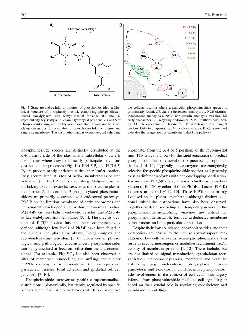
phosphoinositide species are distinctly distributed at thecytoplasmic side of the plasma and subcellular organellemembranes where they dynamically participate in variousdistinct cellular processes (Fig. 1b). PI(4,5)P2 and PI(3,4,5)P3 are predominantly enriched at the inner leaflet, particu-larly accumulated at sites of active membrane-associatedactivities [1]. PI(4)P is located along Golgi-endosomaltrafficking axis, on exocytic vesicles and also at the plasmamembrane [2]. In contrast, 3-phosphorylated phosphoino-sitides are primarily associated with endosomal pathways:PI(3)P on the limiting membrane of early endosomes andintraluminal vesicles contained within multivescular bodies,PI(3,4)P2 on non-clathrin endocytic vesicles, and PI(3,5)P2at late endolysosomal membranes [3, 4]. The precise loca-tion of PI(5)P pool(s) has not been comprehensivelydefined, although low levels of PI(5)P have been found inthe nucleus, the plasma membrane, Golgi complex andsarco/endoplasmic reticulum [5, 6]. Under certain physio-logical and pathological circumstances, phosphoinositidescan be synthesised at locations other than those aforemen-tioned. For example, PI(4,5)P2 has also been observed atsites of membrane remodelling and ruffling, the nuclearmRNA splicing factor compartment (nuclear speckles),perinuclear vesicles, focal adhesion and epithelial cell-celljunctions [7–10].
Phosphoinositide turnover at specific compartmentaliseddistributions is dynamically, but tightly, regulated by specifickinases and antagonistic phosphatases which add or remove
phosphates from the 3, 4 or 5 positions of the myo-inositolring. This critically allows for the rapid generation of productphosphoinositides or removal of the precursor phosphoino-sitides [1, 4, 11]. Typically, these enzymes are catalyticallyselective for specific phosphoinositide species, and generallyexist as different isoforms with non-overlapping localisation.For instance, PI(4,5)P2 is synthesised chiefly by phosphor-ylation of PI(4)P by either of three PI(4)P 5-kinase (PIP5K)isoforms (α, β and γ) [7–10]. These PIP5Ks are mainlylocalised on the plasma membrane, although distinct addi-tional subcellular distributions have also been observed.Together, spatially restricting and temporally governing thephosphoinositide-metabolising enzymes are critical forphosphoinositide metabolic turnover at dedicated membranecompartments and to a particular stimulation.
Despite their low abundance, phosphoinositides and theirmetabolism are crucial to the precise spatiotemporal reg-ulation of key cellular events, where phosphoinositides canserve as second messengers or modulate recruitment and/oractivity of membrane proteins [1, 12]. Τhese include, butare not limited to, signal transduction, cytoskeleton reor-ganisation, membrane dynamics, membrane and vesiculartrafficking (e.g. endocytosis, phagocytosis, macro-pinocytosis and exocytosis). Until recently, phosphoinosi-tide involvement in the context of cell death was largelyinferred from phosphoinositide-mediated cell signalling orbased on their crucial role in regulating cytoskeleton andmembrane remodelling.
Fig. 1 Structure and cellular distribution of phosphoinositides. a Che-mical structure of phosphatidylinositol, comprising phosphodiester-linked diacylglycerol and D-myo-inositol moieties. R1 and R2represent any acyl (fatty acid) chain. Hydroxyl at positions 3, 4 and 5 ofD-myo-inositol ring are readily phosphorylated, giving rise to sevenphosphoinositides. b Localisation of phosphoinositides on plasma andorganelle membrane. This distribution map is exemplary, only showing
the cellular location where a particular phosphoinositide species isprominently found. CE clathrin-dependent endocytosis, NCE clathrin-independent endocytosis, NCV non-clathrin endocytic vesicles, EEearly endosomes, RE recycling endosomes, MVB multivesicular bod-ies, LE late endosomes, L lysosome, ER endoplasmic reticulum, Nnucleus, GA Golgi apparatus, SV secretory vesicles. Black arrow (→)indicates the progression of membrane trafficking pathway
782 T. K. Phan et al.

Phosphoinositides in the context of celldeath via classical activities
Second messengers in signal transduction
Phosphoinositides have been well-defined as second mes-sengers of transmembrane signalling. PI(4,5)P2, in particular,is an essential intermediate for two important pathways:phosphoinositide-specific phospholipase C (PLC)/diacylgly-cerol (DAG)/inositol-1,4,5-trisphosphate (IP3) pathway andclass I phosphoinositide 3-kinase (PI3K)/protein kinase B(Akt) pathway (Fig. 2). Typically, stimulation of G-protein-coupled receptors and receptor tyrosine kinases leads toactivation of PLC, that via its PI(4,5)P2-binding Pleckstrinhomology (PH) domain, hydrolyses PI(4,5)P2 to generateDAG and IP3 and thus initiating two-armed intracellularsignalling cascade. DAG remains at the plasma membrane to
facilitate activation and membrane localisation of proteinkinase C (PKC) family members, whereas cytosolic IP3binds the IP3 receptor and activates a ligand-gated calciumchannel on smooth ER surface, triggering calcium releasefrom intracellular storage [13–15]. In contrast, the PI3K/Aktsignalling cascade canonically begins with the phosphor-ylation of PI(4,5)P2 by PI3K to yield PI(3,4,5)P3, an essentialeffector of Akt signalling [16]. The resultant PI(3,4,5)P3promotes translocation of phosphoinositide-dependent kinase1 (PDK1) and Akt to the plasma membrane, leading to Aktphosphorylation of threonine-308 and its partial activation[16]. Complete Akt activation requires additional phos-phorylation of serine-473, achievable by multiple proteins,including phosphoinositide-dependent kinase 2 (PDK2),mechanistic target of rapamycin complex (mTORC),integrin-linked kinase (ILK) or DNA-dependent proteinkinase (DNA-PK) [17, 18]. Subsequently, downstream
Fig. 2 Phosphoinositides as a second messengers. PI(4,5)P2 mediatestwo major transmembrane signalling pathways: PLC-DAG-IP3 andPI3K-Akt. Activated receptor recruits and induce class I PI3K tophosphorylate PI(4,5)P2. The resultant PI(3,4,5)P3 then recruits Aktand its activator kinases PDK1 and PDK2 (or mTORC2, ILK) can alsoto the plasma membrane, leading to complete phosphorylation of Akt.Alternatively, Akt can also be non-canonically recruited by PI(3,4)P2.Subsequently, Akt regulates downstream effectors through its acti-vated kinase activity. PLC, on the other hand, cleaves PI(4,5)P2 intoDAG and IP3 upon stimulation. DAG remains at the plasma membrane
to recruit and activate PKC to modulate its downstream effectors viaits regulatory phosphorylation. IP3 translocates to ER to excite calciumchannels, causing calcium flux into cytoplasm, initiating calciumsignalling via calcium binding effectors. PI3K phosphoinositide 3-kinases, PLC phosphoinositide-specific phospholipase C, DAG dia-cylglycerol, IP3 inositol 1,4,5-trisphosphate, PKC protein kinase C,Akt protein kinase B, PDK1 phosphoinositide-dependent kinase 1,PDK2 phosphoinositide-independent kinase 2, ER endoplasmic reti-culum, mTORC2 mechanistic target of rapamycin complex 2, ILKintegrin-linked kinase (ILK)
Phosphoinositides: multipurpose cellular lipids with emerging roles in cell death 783

transduction of DAG/PKC, IP3/calcium and Akt signallingindispensably orchestrate many cellular and physiologicalprocesses including, but are not limited to, cell proliferation,cell growth and survival, cell division, cell migration,immune response, muscle contraction, memory and learning[19–21].
Other phosphoinositides, such as PI(3,4)P2 and PI(5)P,although historically considered as intermediate productsof PI(3,4,5)P3 and PI(4,5)P2 metabolism, respectively, havealso emerged to be second messengers themselves. Chal-lenging the exclusive PI(3,4,5)P3/PI3K/Akt signalling axis,mounting evidence has suggested the importance of PI(3,4)P2 in directly recruiting Akt, hence executing a distinct armof the PI3K pathway [22]. Understanding of PI(3,4,5)P3and PI(3,4)P2 cross-talk in regulating, balancing and con-textualising Akt activity, however, undoubtedly requiresfuture investigation. PI-derived PI(5)P is reportedly also animportant second messenger for different cellular pro-cesses, depending on its production pathway [23–26]. PI(5)P generated by direct phosphorylation of PI, via phos-phoinositide 5-kinase PIKfyve catalytic activity, is required
for antiviral immunity, via interferon regulatory factor 3(IRF3)-TANK-binding kinase (TBK1) axis [24]. In addi-tion, PI(5)P can also be generated ‘indirectly’ through acomplex series of steps involving class III PI3K (generat-ing PI(3)P from PI), then PIKfyve (converting PI(3)P toPI(3,5)P2) and finally phosphoinositide 3-phosphatasemyotubularin-related protein-3 (MTMR3), depho-sphorylating PI(3,5)P2 into PI(5)P). PI(5)P produced bythis indirect pathway is involved in cell migration signal-ling (via PI(5)P-recruited and activated exchange factors T-cell lymphoma invasion and metastasis 1 (Tiam1) and Ras-related C3 botulinum toxin substrate (Rac1)) [23, 25, 26].
The role of phosphoinositides in cell death, survival andproliferation is often deduced from the abovementionedphosphoinositide-mediated signalling pathways. Forinstance, the PI3K-Akt signalling pathway critically confersprotection against apoptosis and autophagy, hence essentialfor cell survival (Fig. 3). Activated Akt, through its kinaseactivity, induces either inhibitory or stimulatory phosphor-ylation of components of the apoptotic machinery. Forexample, the phosphorylated nuclear Forkhead transcription
Fig. 3 Phosphoinositide-mediated signalling pathways in cell deathand survival. Proteins involved in (pro-)apoptosis are in dark grey box,anti-apoptosis and survival in green box, autophagy in maroon boxand NETosis in orange box. Legends for arrows/lines: kinase-
catalysed PIP synthesis (curl arrow), PIP-mediated membranerecruitment (dashed arrow), direct inhibition (blunt-ended solid line),direct activation (solid arrow), stimulatory phosphorylation (bluearrow), inhibitory phosphorylation (red arrow)
784 T. K. Phan et al.

factors (FoxO/FH) are exported to cytosol and subsequentlydegraded via ubiquitin-proteasome-dependent pathway,resulting in the repression of FOX-activated pro-apoptoticgene expression [27, 28]. Akt also inhibits pro-apoptoticproteins, including p73-mediated apoptotic Yes-associatedprotein (YAP) [29] and caspase cascade-stimulating Bcl-2-associated death promoter (BAD) and caspase 9 [30].Conversely, Akt-mediated phosphorylation positively reg-ulates pro-survival/anti-apoptotic gene expression, via NF-κB induction (for c-Myb and Bcl-xL expression) [31] orcAMP response element binding protein (CREB) andCREB-binding protein (for Bcl-2 expression) [32, 33]. Inaddition, Akt indirectly inhibits p53-dependent apoptosisvia apoptotic murine double minute 2 (Mdm2)-mediateddegradation of p53 [34]. In addition, the PI3K-Akt signal-ling downregulates autophagy via activation of downstreamAkt effector mTOR kinase which is a negative regulator ofautophagy proteins [35].
Intriguingly, despite the well-characterised pro-survivalrole, PI3K-Akt as well as PLC-PKC cascades are also twomajor signalling pathways driving NETosis (Fig. 3), a morerecently described form of programmed neutrophil death thatresults in the formation of neutrophil extracellular traps(NETs). Phorbol myristate acetate or microbial infection (e.g.Leshmania amazonesis, Candida albicans) trigger the signaltransduction through PI3K and PLC, leading to reactiveoxygen species release or increased intracellular calcium[36–38]. These key events in turn activate myeloperoxidase,neutrophil elastase and protein-arginine deiminase type4 to promote chromatin decondensation and ultimatelyforming NETs to ensnare invading pathogens [37].The discovery of the phosphoinositide-mediated pathwaysin transducing NETotic death signal has added anotherlayer of complexity into functional pleiotropy of phosphoi-nositides. It remains to be determined how these pathwaysfine tune the opposing outcomes in different cellularcontexts.
Actin cytoskeleton and membrane dynamics
Tightly regulated and precisely coordinated changes in thecell membrane and cytoskeleton are central to many aspectsof cell physiology, such as membrane-associated and actin-dependent motility, membrane trafficking, cell polarity, cellsignalling, cell division, and other morphogenetic pro-cesses. The physical and mechanistic links between theactin network and cell membrane are essential for thesecritical changes, which includes membrane detachment/attachment, actin polymerisation/depolymerisation, mem-brane curvature and membrane protrusion [39]. Phosphoi-nositides, especially PI(4,5)P2 and PI(3,4,5)P3, associatewith multiple signal transductions, actin-binding proteinsand membrane-remodelling machineries, hence they
spatiotemporally and interconnectedly control the organi-sation and dynamics of actin cytoskeleton and plasmamembrane [11, 39].
In addition to the aforementioned phosphoinositide sig-nalling, the actin cytoskeleton remodelling and actin-binding proteins are also controlled by various master reg-ulators, among which are small GTPases (Fig. 4). The Rho-family small GTPases (e.g. Rac1, cell division controlprotein 42 homologue Cdc42), are in turn regulated by PI(3,4,5)P3-recruited guanine nucleotide exchange factors(GEFs) in formation of focal adhesion complexes, extensionof migratory pseudopods, or specialised actin filamentassembly such as stress fibres [40, 41]. PI(3,4,5)P3, and themore rigorously characterised PI(4,5)P2, are also consideredas direct-positive regulators of actin filament formation, bydirectly interacting with actin-binding proteins at cellularand subcellular organelle membranes in a localconcentration-dependent manner [39, 40]. Binding to PI(4,5)P2 typically leads to conformational change-associated
Fig. 4 Regulation of actin remodelling by phosphoinositides PI(3,4,5)P2, synthesised from PI(4,5)P2 upon receptor-mediated stimulation ofPI3K via Ras GTPase, recruits GEFs to plasma membrane. GEFs thenfacilitate GTP loading on Rho family GTPases Cdc42 and Rac,leading to activation of actin nucleators WASPs and, subsequently,Arp2/3 complex. PI(4,5)P2 directly binds to and activates proteinmediating membrane-cytoskeleton interaction (e.g. ERM proteins) orinhibits actin depolymerising factor ADF/cofilin, capping proteins (e.g.CapZ) and severing proteins (e.g. gelsolin). PI3K phosphoinositide3-kinases, Ras Rat sarcoma GTPase, Cdc42 cell division controlprotein 42 homologue, Rac Ras-related C3 botulinum toxin substrate,GEF guanine nucleotide exchange factor, WASP Wiskott-Aldrichsyndrome protein, Arp2/3 actin-related proteins 2/3 complex, ERMerzin, radixin and moesin, ADF actin depolymerising factor
Phosphoinositides: multipurpose cellular lipids with emerging roles in cell death 785

activation of proteins that are involved in promoting actinfilament assembly such as nucleators Wiskott-Aldrich syn-drome proteins (WASPs) and actin-related protein 2/3complex (Arp 2/3), rulers, stabilisers and membraneanchoring. Conversely, the PI(4,5)P2 interaction can inhibitactin depolymerising factor ADF/cofilin, capping (CapZ),bundlers, crosslinkers and severing proteins as well as otheractin disassembly inducers, such as profilin [39]. The PI(4,5)P2:actin-binding protein interactions are generallymediated through canonical PI(4,5)P2 binding domains,such as PH domain and band 4.1-ezrin-radixin-moesin(FERM), or defined basic amino acid-rich clusters/motifs ontheir sequences [42, 43]. For instance, ERM family pro-teins, which directly connect the actin cytoskeleton to theplasma membrane, characteristically contain an N-terminalFERM domain and C-terminal actin-binding site. Aninteraction between these two domains effectively estab-lishes autoinhibition, rendering these proteins inactive. Thesubsequent binding of PI(4,5)P2 to the FERM domain leadsto their activation by exposing the actin-binding site [44]. Incontrast, gelsolin binds PI(4,5)P2 through two basic aminoacid-rich regions which also overlap with actin-bindingsites, resulting in the blocking of its actin binding, and thusactin-severing and filament end-capping activity [45].
Dynamic membrane remodelling and generation ofmembrane curvatures are also mechanistically associatedwith phosphoinositide-mediated actin reorganisation. Forexample, mainly PI(4,5)P2 and PI(4)P) binding activity ofcertain actin-associated proteins, such as Bin-Amphiphysin-Rvs (BAR) domain superfamily, epsin/ATP180 NH2-term-inal homology (ENTH/ANTH) domain family and dyna-min, directly influences membrane remodelling includingmembrane protrusions or invaginations by manipulatingphosphoinositide-rich membranes [46, 47]. The binding ofBAR domain-containing proteins, such as amphiphysin 2,to phosphoinositides promotes their membrane binding andbending to create rigid scaffolds and affects the degree ofintrinsic curvature and mechanistic variability [46–49].Similarly, phosphoinositide binding via ENTH domainsalso enables membrane interactions of epsins as well asinduce folding of distorted N-terminal extension into anamphipathic α-helix, an important structure for ENTH-mediated membrane bending/deformation [50, 51]. Theinteraction between dynamin and PI(4,5)P2 through its PHdomain not only allows its membrane translocation, but alsomediates membrane scission via PI(4,5)P2 clustering [52].
Through the modulation of membrane and cytoskeletondynamics, phosphoinositides could be involved in mor-phological changes of dying cells, such as membraneblebbing (formation of circular membrane bulges) [39, 53].The membrane blebs are locally disrupted corticalcytoskeleton-plasma membrane interaction and/or increasedinternal hydrostatic pressure, modulable by PI(4,5)P2-
regulated ERM family proteins [39, 53]. Hence, elevatedplasma membrane PI(4,5)P2 concentration suppresses blebformation by strengthening actin-membrane interaction,whilst its depletion causes membrane blebbing due to theloss of adhesion energy between plasma membrane andcytoskeleton [39]. Pseudomonas aeuginosa phospholipase-like cytotoxin ExoU hydrolyses PI(4,5)P2 acutely causesdisrupted focal adhesion, disorganised cytoskeleton struc-ture, membrane blebbing and eventually cell lysis due toloss of membrane integrity [54, 55]. Similar membrane-bleb-associated cell death was also reported for Gambier-discus toxicus maitotoxin, allegedly due to its PI(4,5)P2phosphodiesterase activity [56, 57]. Membrane blebbing isalso a hallmark of apoptotic cell death, formed upon Rhokinase (ROCK1)-dependent actin-myosin contractionthrough phosphorylation of myosin light chain [53]. Cor-tical contraction-induced increase in cytoplasmic hydro-static pressure and associated disruption of ERM-mediatedmembrane-cytoskeleton interaction are believed to be thedriving forces of bleb formation during apoptosis [58–60].However, the role of phosphoinositides, including PI(4,5)P2, in apoptotic bleb formation remains largely speculative.Furthermore, as apoptotic blebs go through cycles of for-mation, expansion and retraction, a dynamic machinery tomodulate membrane-actin cortex network should be inplace [60]. The trigger and progression of such machinery isstill relatively unknown. As crucial players in regulatingmembrane and cytoskeleton dynamics, it will be interestingto decipher the importance of phosphoinositides in theapoptotic bleb-cycling machinery.
Emerging roles of phosphoinositides indirectly facilitating cell death
Considering that phosphoinositides crucially mediatenumerous cellular processes, one would expect they wouldhave greater and a more direct influence on cell death thanmerely being second messenger lipids of PI3K-Akt signal-ling or modulating membrane and actin dynamics. How-ever, only recently have phosphoinositides been recognisedfor their roles in regulating cell death. A number ofimpactful studies have reported their essential functions inmediating the execution of lytic cell death (pyroptosis,necroptosis and defensin-induced necrosis), autophagic celldeath, and NETosis (Fig. 5), whilst a few others havelimitedly addressed this in the context of apoptosis.
Autophagic cell death
Autophagy, also known as autophagocytosis, is a destruc-tive mechanism to degrade and recycle unnecessary ordysfunctional cellular components [61, 62]. Autophagy is
786 T. K. Phan et al.
often characterised by the formation of cytoplasmic autop-hagic vesicles (autophagosomes) that sequester disposedconstituents targeted for digestion in autolysosomes [63,64]. Physiologically, the tight regulation of autophagy isrequired for proper cellular homoeostasis. Autophagy alsoprovides a defense mechanism by killing ingested bacteria[65, 66]. Under certain developmental, pathophysiological(e.g. post-ischaemic injury, starvation) or chemical (e.g.vintexin treatment) conditions, autophagy can result inautophagic cell death (ACD) [67–69]. Typical character-istics of ACD include membrane rupture, numerousautophagolysosomes, and enlargement of major cellularorganelles [70–72]. Mechanistically, ACD is the lethal
outcome of disturbed autophagic homoeostasis via exces-sive self-consumption of cellular content, uncontrolledmitophagy and autosis (Na+/K+-ATP-dependent ACD)[68, 69].
ACD strictly depends on autophagy machinery, whichrequires the production of PI(3)P by Vps34 kinase com-plexes, disruptions of which, demonstrated by geneticinterference and pharmacological inhibitions, inhibit theautophagic pathway [73–77]. PI(3)P fundamentally providesdocking platforms on the ER cytoplasmic membrane forautophagy effectors, and possibly mediates the fusion ofautophagosomes and lysosomes to form autolysosomes [77].Particularly in humans, PI(3)P is reported to specifically
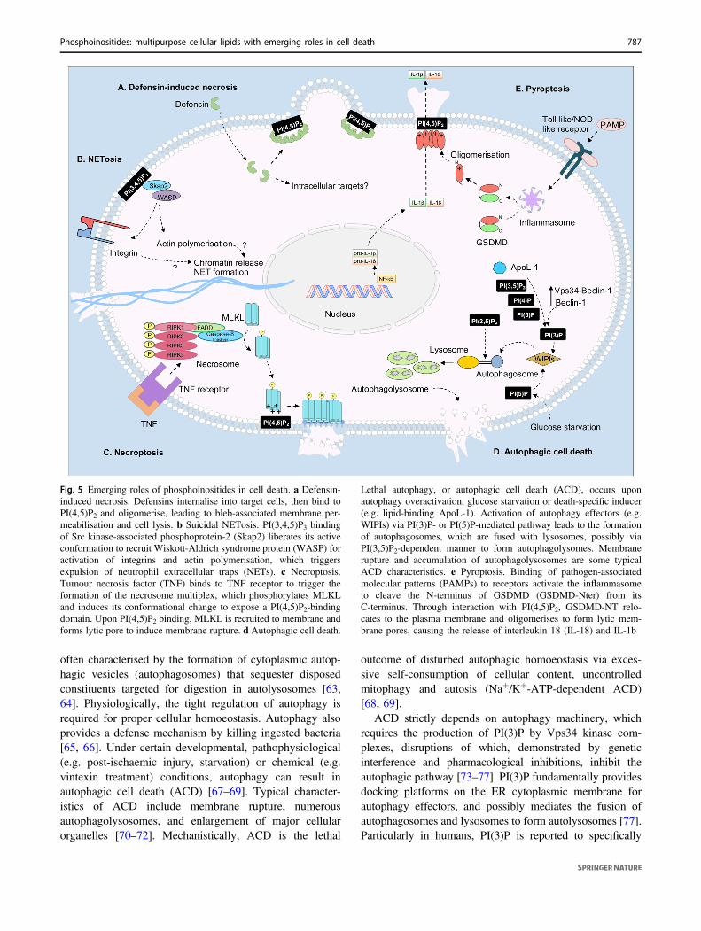
Fig. 5 Emerging roles of phosphoinositides in cell death. a Defensin-induced necrosis. Defensins internalise into target cells, then bind toPI(4,5)P2 and oligomerise, leading to bleb-associated membrane per-meabilisation and cell lysis. b Suicidal NETosis. PI(3,4,5)P3 bindingof Src kinase-associated phosphoprotein-2 (Skap2) liberates its activeconformation to recruit Wiskott-Aldrich syndrome protein (WASP) foractivation of integrins and actin polymerisation, which triggersexpulsion of neutrophil extracellular traps (NETs). c Necroptosis.Tumour necrosis factor (TNF) binds to TNF receptor to trigger theformation of the necrosome multiplex, which phosphorylates MLKLand induces its conformational change to expose a PI(4,5)P2-bindingdomain. Upon PI(4,5)P2 binding, MLKL is recruited to membrane andforms lytic pore to induce membrane rupture. d Autophagic cell death.
Lethal autophagy, or autophagic cell death (ACD), occurs uponautophagy overactivation, glucose starvation or death-specific inducer(e.g. lipid-binding ApoL-1). Activation of autophagy effectors (e.g.WIPIs) via PI(3)P- or PI(5)P-mediated pathway leads to the formationof autophagosomes, which are fused with lysosomes, possibly viaPI(3,5)P2-dependent manner to form autophagolysomes. Membranerupture and accumulation of autophagolysosomes are some typicalACD characteristics. e Pyroptosis. Binding of pathogen-associatedmolecular patterns (PAMPs) to receptors activate the inflammasometo cleave the N-terminus of GSDMD (GSDMD-Nter) from itsC-terminus. Through interaction with PI(4,5)P2, GSDMD-NT relo-cates to the plasma membrane and oligomerises to form lytic mem-brane pores, causing the release of interleukin 18 (IL-18) and IL-1b
Phosphoinositides: multipurpose cellular lipids with emerging roles in cell death 787

recruit Autophagy-linked FYVE protein (Alfy), WD-repeatdomain phosphoinositide-interacting proteins (WIPIs)and FYVE domain-containing protein 1 (DFCP1) thatsequesters target cargo to the autophagic machinery andforms an isolation compartment, eventually maturing intoautophagosomes [78–80]. Similar to most phosphoinositide-regulating processes, PI(3)P turnover in autophagy byphosphatidylinositol 3-kinase Vps34-Beclin-1 and phos-phatase Jumpy is critical for the initiation and termination ofautophagy [74, 81]. Non-canonically, autophagy can beexecuted via Vps34-independent (hence PI(3)P-indepen-dent) pathways, mediated by PIKfyve-synthesised PI(5)P viaknown PI(3)P effectors, such as WIPI proteins. The non-canonical PI(5)P-dependent pathway can compensate theabsence of PI(3)P, e.g. due to defective Vps34, or overrulePI(3)P in particular circumstances, such as glucose starva-tion [82, 83]. It also been reported that PI(3,5)P2 is requiredfor completion of basal autophagy, particularly in mousemodels of neurodegeneration. PI(3,5)P2-deficient miceexhibit neuronal loss and accumulation of autophagy inter-mediates, possibly due to the lack of proper PI(3,5)P2-gov-erned autophagosome-lysosome fusion and/or lysosomefunction [83, 84].
A shift from basal autophagy to ACD has been linkedwith hyperactivation of Vps34-Beclin-1, overexpression ofBeclin-1, downregulation of the molecular brakes of Beclin-1,hyperactivation of non-canonical autophagic pathways andcertain death-specific factors (e.g. apolipoprotein 1 ApoL-1)[67, 68, 85]. ApoL-1, a BH3-only lipid-binding protein, caninduce ACD, speculatively via disruption of phosphatidicacid-mTOR interaction and inhibition mitochondrialcardiolipin-associated apoptosis [85]. However, in this study,the protein-lipid overlay assay also indicated that ApoL-1binds to phosphoinositides, particularly PI(3)P, PI(4)P, PI(5)P, PI(3,5)P2 at comparable affinity to PA and CL [85].Whether the binding of ApoL-1 to phosphoinositides altersthe lipid homoeostasis leading to ACD as suggested by theauthors [85], or a more sophisticated phosphoinositide-associated mechanism of ApoL-1-induced ACD, requiresfurther elucidation.
NETosis
Suicidal NETosis is a novel form of neutrophil cell death,characterised by the formation of NETs that follows DNAdecondensation and plasma membrane rupture [86, 87].NET release forms a novel host defense mechanism againstinvading microbial pathogens. Though the execution andregulation of NETosis are still not fully understood, it isemerging that PI(3,4,5)P3-mediated recruitment and acti-vation of NETotic effectors, including Src kinase-associatedphosphoprotein-2 (Skap2), WASPs and integrins [88].While initial studies showed that Skap2 adopts an
autoinhibitory conformation in steady-state neutrophils,upon stimulation, interactions with PI(3,4,5)P3 via its PHdomain induces a switch to the active conformation [89].Then, Skap2 can form a complex with WASPs and trans-locate to the inner leaflet of the plasma membrane, leadingto the downstream activation of integrins [88, 89]. InSkap2−/− neutrophils reconstituted with a Skap2 R140Mmutant, which has been previously shown to impair PI(3,4,5)P3 binding to the PH domain [89], both Skap2 andWASPs are unable to translocate to the plasma membraneand, consequentially, resulting in failed NETosis. Thesefindings suggest the importance of direct PI(3,4,5)P3-mediated membrane recruitment in directing NETosis.
Pyroptosis
In order to respond to bacterial infection and danger signals,cells can undergo pyroptosis, a programmed cell deathassociated with membrane lysis and inflammatory cytokinerelease [90]. During pyroptosis, activated caspase-1 orcaspase-11 cleaves the effector protein gasdermin D(GSDMD), liberating its N-terminal (GSDMD-Nter) fromthe autoinhibitory C-terminal domain (GSDMD-Cter)[91–93]. The GSDMD-Nter then relocates to the plasmamembrane, through its preferential interaction with PI(4,5)P2. In fact, GSDMD-Nter harbours clusters of positivelycharged and hydrophobic residues, a common featureobserved in PI(4,5)P2-binding motifs, within its α1 helixand the β1–β2 loop [94]. Upon PI(4,5)P2 binding, GSDMD-Nter promptly inserts into plasma membrane, oligomerisesand forms large transmembrane pores to trigger inflamma-tory cytokine (interleukins 18 and 1β) release and, subse-quently, membrane lysis [93, 95]. A recent time-lapseatomic force microscopy study has highlighted the crucialrole of PI(4,5)P2 throughout GSDMD assembly progress,not only governing its membrane targeting step but alsoaccelerating membrane insertion and ensuring correct poretopology [94].
Necroptosis
Necroptosis is another inflammatory and lytic form ofprogrammed cell death, occurring upon tumour necrosisfactor receptor stimulation and caspase inhibition [96, 97].Unlike pyroptosis, necroptosis is however distinctivelycharacterised by caspase independence, membrane damageand release of inflammatory damage-associated molecularpatterns [96, 97]. The GSDMD counterpart for necroptosisis mixed lineage kinase-like (MLKL) pseudokinase, struc-turally consisting of a N-terminal bundle (NB) domainfused by a brace region to a C-terminal pseudokinasedomain [96, 97]. During necroptosis, MLKL is phos-phorylated by a multiprotein complex, called necrosome,
788 T. K. Phan et al.

leading to its transition from dormant monomeric to brace-mediated and activated oligomeric confirmation [98]. TheMLKL oligomer is initially recruited to the plasma mem-brane through its low-affinity PI(4,5)P2 binding provided byclusters of basic residues within NB domain [99, 100].Subsequent robust membrane association progresses ashigher-affinity PI(4,5)P2 binding sites are exposed anddisplace the brace [100]. Mutation of basic residues at theputative binding sites suggests PI(4,5)P2 interaction is par-ticularly essential for the lytic activity. However, it remainsto determine how PI(4,5)P2-bound MLKL causes mem-brane rupture, possibly by its membrane destabilisation,pore formation or osmosis imbalance induced by its cationchannel-like activity [98–101].
Defensin-induced necrosis
In addition to pyroptosis and necroptosis, defensin-inducednecrosis represents a parallelly novel form of PI(4,5)P2-dependent lytic cell death. Defensins are small cationicpeptides of host innate immunity displaying broad-spectrumantimicrobial and immunomodulatory activities [102].Additionally, recent studies have reported on membrano-lytic effect of ornamental tobacco defensin NaD1 [103],tomato defensin TPP3 [104] and human β-defensin 3(HBD-3) [105] via PI(4,5)P2-mediated membrane permea-bilisation. At sub-micromolar concentrations, these defen-sins promptly enter mammalian cell followed by binding toPI(4,5)P2 at inner membrane leaflet, thus destabilising theplasma membrane, causing bulge-like membrane structures(blebs) and eventually cell lysis. Supported by site-directedmutagenesis and crystallographic studies, the PI(4,5)P2interaction was mapped to analogous cysteine-flanked, β-strand-spanning, cationic clusters (33HCSKILRR40 inNaD1, 37HCSKLQRK42 in TPP3 and 32KCSTRGRK39 inHBD-3), which resemble the K/R-X(3,7)-K-X-K/R-K/Rmotif found in nuclear PI(4,5)P2-binding proteins [106].Strikingly, NaD1 can form an arch-shaped oligomer with PI(4,5)P2, stabilised by an extensive network of protein-protein and protein-lipid interactions. The oligomerisationpropensity may increase NaD1 avidity for PI(4,5)P2, andresultant tight NaD1:PI(4,5)P2 complex would efficientlysequester PI(4,5)P2, hence disrupting plasma membrane[103]. Interestingly, the membranolytic defensins show agreat specificity towards tumour cells, likely due to certainmorphological changes of plasma membranes upon tumourtransformation to influence robust growth, motility, inva-sion and metastasis, as opposed to normal cells. These mayinclude increased negatively charged phospholipid,increased membrane surface area and possibly increasedphosphoinositide levels [105]. More recently, the oligo-meric defensin:PI(4,5)P2 complex has also been reported forhuman β-defensin 2 (HBD-2). Though structurally and
mechanistically different to the abovementioned defensins,the HBD-2:PI(4,5)P2 complex is also be crucial for C.albicans membrane permeabilisation, directly related to itsantifungal property [107]. These findings accentuate aninterspecies conserved PI(4,5)P2-dependent mechanismamong membrane-targeting innate immune molecules,mediating the necrosis of altered-self (such as tumour cells)and invading pathogens.
Apoptosis and secondary necrosis
Some early studies demonstrated a direct, yet opposing,action of phosphoinositides in mediating apoptosis, a non-lytic, non-inflammatory, caspase-dependent programmedcell death. PI(4,5)P2 acts as a direct inhibitor of apoptosisinitiator caspases 8 and 9 and their common effector cas-pase 3, independently of PI3K and Akt [108]. Increased PI(4,5)P2 levels by PIP5Kα overexpression exacerbatesapoptotic suppression; and PIP5Kα is actually cleaved bycaspase 3 in vitro at a consensus cleavage site, mutation ofwhich prevents PIP5Kα inactivation and enhances apop-tosis in vivo [108]. In contrast, PI(4,5)P2, in aconcentration-dependent fashion, essentially activatescation channel P2X7 current upon ATP stimulation, andindispensably induces P2X7-mediated cell death, particu-larly apoptosis, in endothelial cells, T cells and macro-phages [109]. It is uncertain why such discrepancy existsfor the role of phosphoinositides in apoptosis, althoughcell-type dependency could be part of the explanation. Tothis point, however, the direct involvement of phosphoi-nositides in apoptosis remains largely underexploreddespite apoptosis being the most well-studied cell deathprogram.
Recent breakthroughs in apoptosis demonstrate thatapoptosis is associated with three finely tuned downstreamevents with distinct membrane morphologies including: (i)the previously discussed membrane blebbing, (ii) formationof thin apoptotic membrane protrusion causing blebseparation and (iii) protrusion fragmentation to formmembrane-bound extracellular vesicles, called apoptoticbodies [110–112]. The entire process, collectively known asapoptotic cell disassembly, is an important physiological orpathophysiological phenomenon downstream of apoptosis.Resultant apoptotic bodies mediate efficient phagocyticremoval of apoptotic cells as well as aid intercellularcommunication [110]. In line with the suggestion earlier,the role of membrane-actin dynamics-regulating phosphoi-nositides, particularly PI(4,5)P2 and PI(3,4,5)P3, in med-iating morphological changes during apoptotic celldisassembly definitely require further investigation.
Intriguingly, in the events of impaired clearance,apoptotic cells can progress into secondary necrosis due toprogressive loss of plasma membrane integrity. Though
Phosphoinositides: multipurpose cellular lipids with emerging roles in cell death 789

long regarded as an unregulated progress, recent reportssuggest caspase 3-mediated cleavage of gasdermin E(GSDME, also known as DFNA5) predisposing apoptosisto membrane rupture via similar mechanisms to MLKLand GSDMD [113, 114]. GSDME knockout apoptoticcells fail to lyse, but instead extensively fragment to formapoptotic bodies. Once cleaved N-terminal fragment ofGSDME translocates to the plasma membrane and indu-ces membrane permeabilisation [113, 114]. Thoughdetailed membrane recruitment and interaction ofGSDME is unknown, based on its relatedness to GSDMD,one can logically propose a role for phosphoinositides inGSDME-induced secondary necrosis.
Exploiting phosphoinosititdes totherapeutically modulate cell death
With phosphoinositides emerging to critically integrate intoan array of cell death pathways, developingphosphoinositide-targeting therapeutics could be useful inmodulating cell death pathways, whether it be an inhibitingor promoting cell death. Phosphoinositide-binding celldeath effectors (such as the defensins, GSDMD and MLKL)or related molecular mimetics could be exploited in thefuture to combat a range of diseases related to bothmicrobial pathogens and cancer by inducing cell deaththrough phosphoinositide interactions. This could prove aneffective way of overcoming the ability of some cells,particularly in cancer, to evade programmed cell death, asdirect treatment with effector molecule would enable bypassof the upstream signalling pathways.
In contrast, the opposite effect could be achieved bytargeting phosphoinositides with therapeutics designed toprevent interactions with cell death effector molecules. Thisapproach would be relevant in disease settings associatedwith unwanted cell death and inflammation (such as inautoimmune diseases), as reducing cell death could bebeneficial. For example, in multiple sclerosis, whereunwarranted pyroptosis contributes to neuroinflammationand demylenation, preventing the translocation of GSDMD-Nter to the plasma membrane by blocking PI(4,5)P2 couldbe useful in treating the disease [115]. It is therefore pos-sible that other cell death-associated diseases, e.g. neuro-degenerative diseases, can be limited by blockingautophagic cell death can using PI(3)P and PI(5)P-seques-tering agents.
Concluding remarks
The last few years have seen a remarkable increase in reportsof phosphoinositide-facilitated cell death, emphasising the
importance of phosphoinositides in many important aspectsof life and death within the cell. Direct PI(3)P, PI(3,5)P3, PI(4,5)P2 and PI(3,4,5)P3 function in mediating autophagic,lytic and NETotic forms of cell death essentially contributeto the homoeostasis, development and host defensemechanisms in many disease settings by preventing patho-gen/tumour cell growth and alerting host immune response.These seminal findings however require additional in vivoevidence to strengthen the emerging importance of phos-phoinositides in the context of cell death. Future investiga-tions should also further delineate the death effector-phosphoinositide interactions and how such interactionsmolecularly execute different forms of cell death. It is worthstudying how these pathways differentially are employedand/or cross-talk in different immune cells and against var-ious infections/altered-self. There may also other cell deathsettings possibly recruiting phosphoinositides, particularlyPI(4,5)P2, for their enactment including apoptotic cell dis-assembly, secondary necrosis and necrosis by other lytichost defense peptides and molecules such as venoms. Takentogether, phosphoinositides, in particular PI(4,5)P2, mayhave potential as novel targets for combatting infection andcancer. The ability of PI(4,5)P2 to orchestrate lytic cell deaththrough the recruitment and activation of death effectors isindeed an attractive feature to manipulate cell death fortherapeutic benefit.
Compliance with ethical standards
Conflict of interest The authors declare that they have no conflict ofinterest.
Publisher’s note: Springer Nature remains neutral with regard tojurisdictional claims in published maps and institutional affiliations.
References
1. Di Paolo G, De Camilli P. Phosphoinositides in cell regulationand membrane dynamics. Nature. 2006;443:651–7.
2. Dickson EJ, Jensen JB, Hille B. Golgi and plasma membranepools of PI(4)P contribute to plasma membrane PI(4,5)P2 andmaintenance of KCNQ2/3 ion channel current. Proc Natl AcadSci USA. 2014;111:E2281–90.
3. Sbrissa D, Ikonomov OC, Fu Z, Ijuin T, Gruenberg J, TakenawaT, et al. Core protein machinery for mammalian phosphatidyli-nositol 3,5-bisphosphate synthesis and turnover that regulates theprogression of endosomal transport. Novel Sac phosphatase joinsthe ArPIKfyve-PIKfyve complex. J Biol Chem. 2007;282:23878–91.
4. Cullen PJ, Carlton JG. Phosphoinositides in the mammalianendo-lysosomal network. Subcell Biochem. 2012;59:65–110.
5. Sarkes D, Rameh LE. A novel HPLC-based approach makespossible the spatial characterization of cellular PtdIns5P andother phosphoinositides. Biochem J. 2010;428:375–84.
6. Grainger DL, Tavelis C, Ryan AJ, Hinchliffe KA. The emergingrole of PtdIns5P: another signalling phosphoinositide takes itsplace. Biochem Soc Trans. 2012;40:257–61.
790 T. K. Phan et al.

7. van den Bout I, Divecha N. PIP5K-driven PtdIns(4,5)P2 synthesis: regulation and cellular functions. J Cell Sci. 2009;122(Pt 21):3837–50.
8. Ling K, Bairstow SF, Carbonara C, Turbin DA, Huntsman DG,Anderson RA. Type I gamma phosphatidylinositol phosphatekinase modulates adherens junction and E-cadherin traffickingvia a direct interaction with mu 1B adaptin. J Cell Biol. 2007;176:343–53.
9. Giudici ML, Lee K, Lim R, Irvine RF. The intracellular locali-sation and mobility of Type Igamma phosphatidylinositol 4P 5-kinase splice variants. FEBS Lett. 2006;580:6933–7.
10. Gericke A, Leslie NR, Losche M, Ross AH. PtdIns(4,5)P2-mediated cell signaling: emerging principles and PTEN as aparadigm for regulatory mechanism. Adv Exp Med Biol.2013;991:85–104.
11. Balla T. Phosphoinositides: tiny lipids with giant impact on cellregulation. Physiol Rev. 2013;93:1019–137.
12. Falkenburger BH, Jensen JB, Dickson EJ, Suh BC, Hille B.Phosphoinositides: lipid regulators of membrane proteins. JPhysiol. 2010;588(Pt 17):3179–85.
13. Berridge MJ, Irvine RF. Inositol trisphosphate, a novel secondmessenger in cellular signal transduction. Nature. 1984;312:315–21.
14. Meldrum E, Parker PJ, Carozzi A. The PtdIns-PLC superfamilyand signal transduction. Biochim Biophys Acta. 1991;1092:49–71.
15. Katan M. The control of inositol lipid hydrolysis. Cancer Surv.1996;27:199–211.
16. Rameh LE, Cantley LC. The role of phosphoinositide 3-kinaselipid products in cell function. J Biol Chem. 1999;274:8347–50.
17. Osaki M, Oshimura M, Ito H. PI3K-Akt pathway: its functionsand alterations in human cancer. Apoptosis. 2004;9:667–76.
18. Hemmings BA, Restuccia DF. PI3K-PKB/Akt pathway. ColdSpring Harb Perspect Biol. 2012;4:a011189.
19. Yu JS, Cui W. Proliferation, survival and metabolism: the role ofPI3K/AKT/mTOR signalling in pluripotency and cell fatedetermination. Development. 2016;143:3050–60.
20. Heras-Sandoval D, Perez-Rojas JM, Hernandez-Damian J,Pedraza-Chaverri J. The role of PI3K/AKT/mTOR pathway inthe modulation of autophagy and the clearance of proteinaggregates in neurodegeneration. Cell Signal. 2014;26:2694–701.
21. Cain RJ, Ridley AJ. Phosphoinositide 3-kinases in cell migra-tion. Biol Cell. 2009;101:13–29.
22. Li H, Marshall AJ. Phosphatidylinositol (3,4) bisphosphate-specific phosphatases and effector proteins: a distinct branch ofPI3K signaling. Cell Signal. 2015;27:1789–98.
23. Oppelt A, Lobert VH, Haglund K, Mackey AM, Rameh LE,Liestol K, et al. Production of phosphatidylinositol 5-phosphatevia PIKfyve and MTMR3 regulates cell migration. EMBO Rep.2013;14:57–64.
24. Kawasaki T, Takemura N, Standley DM, Akira S, Kawai T. Thesecond messenger phosphatidylinositol-5-phosphate facilitatesantiviral innate immune signaling. Cell Host Microbe. 2013;14:148–58.
25. Haugsten EM, Oppelt A, Wesche J. Phosphatidylinositol 5-phosphate is a second messenger important for cell migration.Commun Integr Biol. 2013;6:e25446.
26. Viaud J, Lagarrigue F, Ramel D, Allart S, Chicanne G, CeccatoL, et al. Phosphatidylinositol 5-phosphate regulates invasionthrough binding and activation of Tiam1. Nat Commun. 2014;5:4080.
27. Zhang X, Tang N, Hadden TJ, Rishi AK. Akt, FoxO and reg-ulation of apoptosis. Biochim Biophys Acta. 2011;1813:1978–86.
28. Tzivion G, Dobson M, Ramakrishnan G. FoxO transcriptionfactors; regulation by AKT and 14-3-3 proteins. Biochim Bio-phys Acta. 2011;1813:1938–45.
29. Basu S, Totty NF, Irwin MS, Sudol M, Downward J. Aktphosphorylates the Yes-associated protein, YAP, to induceinteraction with 14-3-3 and attenuation of p73-mediated apop-tosis. Mol Cell. 2003;11:11–23.
30. Cardone MH, Roy N, Stennicke HR, Salvesen GS, Franke TF,Stanbridge E, et al. Regulation of cell death protease caspase-9by phosphorylation. Science. 1998;282:1318–21.
31. Kane LP, Shapiro VS, Stokoe D, Weiss A. Induction of NF-kappaB by the Akt/PKB kinase. Curr Biol. 1999;9:601–4.
32. Du K, Montminy M. CREB is a regulatory target for the proteinkinase Akt/PKB. J Biol Chem. 1998;273:32377–9.
33. Pugazhenthi S, Nesterova A, Sable C, Heidenreich KA, BoxerLM, Heasley LE, et al. Akt/protein kinase B up-regulates Bcl-2expression through cAMP-response element-binding protein. JBiol Chem. 2000;275:10761–6.
34. Ogawara Y, Kishishita S, Obata T, Isazawa Y, Suzuki T, TanakaK, et al. Akt enhances Mdm2-mediated ubiquitination anddegradation of p53. J Biol Chem. 2002;277:21843–50.
35. Kim J, Kundu M, Viollet B, Guan KL. AMPK and mTORregulate autophagy through direct phosphorylation of Ulk1. NatCell Biol. 2011;13:132–41.
36. Papayannopoulos V. Neutrophil extracellular traps in immunityand disease. Nat Rev Immunol. 2018;18:134–47.
37. DeSouza-Vieira T, Guimaraes-Costa A, Rochael NC, Lira MN,Nascimento MT, Lima-Gomez PS, et al. Neutrophil extracellulartraps release induced by Leishmania: role of PI3Kgamma, ERK,PI3Ksigma, PKC, and [Ca2+]. J Leukoc Biol. 2016;100:801–10.
38. Zawrotniak M, Bochenska O, Karkowska-Kuleta J, Seweryn-Ozog K, Aoki W, Ueda M, et al. Aspartic proteases and majorcell wall components in Candida albicans trigger the release ofneutrophil extracellular traps. Front Cell Infect Microbiol.2017;7:414.
39. Saarikangas J, Zhao H, Lappalainen P. Regulation of the actincytoskeleton-plasma membrane interplay by phosphoinositides.Physiol Rev. 2010;90:259–89.
40. Egami Y, Taguchi T, Maekawa M, Arai H, Araki N. SmallGTPases and phosphoinositides in the regulatory mechanisms ofmacropinosome formation and maturation. Front Physiol.2014;5:374.
41. Benard V, Bohl BP, Bokoch GM. Characterization of rac andcdc42 activation in chemoattractant-stimulated human neu-trophils using a novel assay for active GTPases. J Biol Chem.1999;274:13198–204.
42. Toker A. The synthesis and cellular roles of phosphatidylinositol4,5-bisphosphate. Curr Opin Cell Biol. 1998;10:254–61.
43. Kabachinski G, Yamaga M, Kielar-Grevstad DM, Bruinsma S,Martin TF. CAPS and Munc13 utilize distinct PIP2-linkedmechanisms to promote vesicle exocytosis. Mol Biol Cell.2014;25:508–21.
44. Barret C, Roy C, Montcourrier P, Mangeat P, Niggli V. Muta-genesis of the phosphatidylinositol 4,5-bisphosphate (PIP(2))binding site in the NH(2)-terminal domain of ezrin correlateswith its altered cellular distribution. J Cell Biol. 2000;151:1067–80.
45. Yu FX, Sun HQ, Janmey PA, Yin HL. Identification of apolyphosphoinositide-binding sequence in an actin monomer-binding domain of gelsolin. J Biol Chem. 1992;267:14616–21.
46. Lemmon MA. Membrane recognition by phospholipid-bindingdomains. Nat Rev Mol Cell Biol. 2008;9:99–111.
47. Gallop JL, McMahon HT. BAR domains and membrane cur-vature: bringing your curves to the BAR. Biochem Soc Symp.2005;72:223–31.
Phosphoinositides: multipurpose cellular lipids with emerging roles in cell death 791

48. Wang Q, Kaan HY, Hooda RN, Goh SL, Sondermann H.Structure and plasticity of Endophilin and Sorting Nexin 9.Structure. 2008;16:1574–87.
49. Frost A, Perera R, Roux A, Spasov K, Destaing O, Egelman EH,et al. Structural basis of membrane invagination by F-BARdomains. Cell . 2008;132:807–17.
50. Ford MG, Mills IG, Peter BJ, Vallis Y, Praefcke GJ, Evans PR,et al. Curvature of clathrin-coated pits driven by epsin. Nature.2002;419:361–6.
51. Itoh T, Koshiba S, Kigawa T, Kikuchi A, Yokoyama S, TakenawaT. Role of the ENTH domain in phosphatidylinositol-4,5-bispho-sphate binding and endocytosis. Science. 2001;291:1047–51.
52. Bethoney KA, King MC, Hinshaw JE, Ostap EM, Lemmon MA.A possible effector role for the pleckstrin homology (PH) domainof dynamin. Proc Natl Acad Sci USA. 2009;106:13359–64.
53. Zhang Y, Chen X, Gueydan C, Han J. Plasma membranechanges during programmed cell deaths. Cell Res. 2018;28:9–21.
54. Sato H, Frank DW. Intoxication of host cells by the T3SSphospholipase ExoU: PI(4,5)P2-associated, cytoskeletal collapseand late phase membrane blebbing. PLoS ONE. 2014;9:e103127.
55. Tyson GH, Hauser AR. Phosphatidylinositol 4,5-bisphosphate isa novel coactivator of the Pseudomonas aeruginosa cytotoxinExoU. Infect Immun. 2013;81:2873–81.
56. Bernard V, Laurent A, Derancourt J, Clement-Durand M, PicardA, Le Peuch C, et al. Maitotoxin triggers the cortical reaction andphosphatidylinositol-4,5-bisphosphate breakdown in amphibianoocytes. Eur J Biochem. 1988;174:655–62.
57. Estacion M, Schilling WP. Maitotoxin-induced membraneblebbing and cell death in bovine aortic endothelial cells. BMCPhysiol. 2001;1:2.
58. Lorentzen A, Bamber J, Sadok A, Elson-Schwab I, Marshall CJ.An ezrin-rich, rigid uropod-like structure directs movement ofamoeboid blebbing cells. J Cell Sci. 2011;124(Pt 8):1256–67.
59. Wickman G, Julian L, Olson MF. How apoptotic cells aid in theremoval of their own cold dead bodies. Cell Death Differ.2012;19:735–42.
60. Charras GT, Hu CK, Coughlin M, Mitchison TJ. Reassembly ofcontractile actin cortex in cell blebs. J Cell Biol. 2006;175:477–90.
61. Takeshige K, Baba M, Tsuboi S, Noda T, Ohsumi Y. Autophagyin yeast demonstrated with proteinase-deficient mutants andconditions for its induction. J Cell Biol. 1992;119:301–11.
62. Ichimura Y, Kirisako T, Takao T, Satomi Y, Shimonishi Y,Ishihara N, et al. A ubiquitin-like system mediates protein lipi-dation. Nature. 2000;408:488–92.
63. Nara A, Mizushima N, Yamamoto A, Kabeya Y, Ohsumi Y,Yoshimori T. SKD1 AAA ATPase-dependent endosomal trans-port is involved in autolysosome formation. Cell Struct Funct.2002;27:29–37.
64. Mizushima N, Ohsumi Y, Yoshimori T. Autophagosome for-mation in mammalian cells. Cell Struct Funct. 2002;27:421–9.
65. Ogawa M, Yoshimori T, Suzuki T, Sagara H, Mizushima N,Sasakawa C. Escape of intracellular Shigella from autophagy.Science. 2005;307:727–31.
66. Gutierrez MG, Master SS, Singh SB, Taylor GA, Colombo MI,Deretic V. Autophagy is a defense mechanism inhibiting BCGand Mycobacterium tuberculosis survival in infected macro-phages. Cell . 2004;119:753–66.
67. Bhardwaj M, Paul S, Jakhar R, Khan I, Kang JI, Kim HM, et al.Vitexin confers HSF-1 mediated autophagic cell death by acti-vating JNK and ApoL1 in colorectal carcinoma cells. Onco-target. 2017;8:112426–41.
68. Bialik S, Dasari SK, Kimchi A. Autophagy-dependent celldeath - where, how and why a cell eats itself to death. J Cell Sci.2018;131:18.
69. Liu Y, Shoji-Kawata S, Sumpter RM Jr., Wei Y, Ginet V, ZhangL, et al. Autosis is a Na+,K+-ATPase-regulated form of celldeath triggered by autophagy-inducing peptides, starvation, andhypoxia-ischemia. Proc Natl Acad Sci USA. 2013;110:20364–71.
70. Tsujimoto Y, Shimizu S. Another way to die: autophagic pro-grammed cell death. Cell Death Differ. 2005;12(Suppl 2):1528–34.
71. Liu Y, Levine B. Autosis and autophagic cell death: the dark sideof autophagy. Cell Death Differ. 2015;22:367–76.
72. Green DR, Levine B. To be or not to be? How selectiveautophagy and cell death govern cell fate. Cell . 2014;157:65–75.
73. Takatsuka C, Inoue Y, Matsuoka K, Moriyasu Y. 3-methyladenine inhibits autophagy in tobacco culture cellsunder sucrose starvation conditions. Plant Cell Physiol. 2004;45:265–74.
74. Juhasz G, Hill JH, Yan Y, Sass M, Baehrecke EH, Backer JM,et al. The class III PI(3)K Vps34 promotes autophagy andendocytosis but not TOR signaling in Drosophila. J Cell Biol.2008;181:655–66.
75. Eskelinen EL, Prescott AR, Cooper J, Brachmann SM, Wang L,Tang X, et al. Inhibition of autophagy in mitotic animal cells.Traffic. 2002;3:878–93.
76. Blommaart EF, Krause U, Schellens JP, Vreeling-Sindelarova H,Meijer AJ. The phosphatidylinositol 3-kinase inhibitors wort-mannin and LY294002 inhibit autophagy in isolated rat hepa-tocytes. Eur J Biochem. 1997;243:240–6.
77. Axe EL, Walker SA, Manifava M, Chandra P, Roderick HL,Habermann A, et al. Autophagosome formation from membranecompartments enriched in phosphatidylinositol 3-phosphate anddynamically connected to the endoplasmic reticulum. J Cell Biol.2008;182:685–701.
78. Walker S, Chandra P, Manifava M, Axe E, Ktistakis NT. Makingautophagosomes: localized synthesis of phosphatidylinositol 3-phosphate holds the clue. Autophagy. 2008;4:1093–6.
79. Jeffries TR, Dove SK, Michell RH, Parker PJ. PtdIns-specificMPR pathway association of a novel WD40 repeat protein,WIPI49. Mol Biol Cell. 2004;15:2652–63.
80. Filimonenko M, Isakson P, Finley KD, Anderson M, Jeong H,Melia TJ, et al. The selective macroautophagic degradation ofaggregated proteins requires the PI3P-binding protein Alfy. MolCell. 2010;38:265–79.
81. Vergne I, Roberts E, Elmaoued RA, Tosch V, Delgado MA,Proikas-Cezanne T, et al. Control of autophagy initiation byphosphoinositide 3-phosphatase Jumpy. EMBO J. 2009;28:2244–58.
82. Vicinanza M, Korolchuk VI, Ashkenazi A, Puri C, Menzies FM,Clarke JH, et al. PI(5)P regulates autophagosome biogenesis.Mol Cell. 2015;57:219–34.
83. Hasegawa J, Strunk BS, Weisman LS. PI5P and PI(3,5)P2:minor, but essential phosphoinositides. Cell Struct Funct.2017;42:49–60.
84. Ferguson CJ, Lenk GM, Meisler MH. Defective autophagy inneurons and astrocytes from mice deficient in PI(3,5)P2. HumMol Genet. 2009;18:4868–78.
85. Wan G, Zhaorigetu S, Liu Z, Kaini R, Jiang Z, Hu CA. Apoli-poprotein L1, a novel Bcl-2 homology domain 3-only lipid-binding protein, induces autophagic cell death. J Biol Chem.2008;283:21540–9.
86. Dabrowska D, Jablonska E, Garley M, Ratajczak-Wrona W,Iwaniuk A. New aspects of the biology of neutrophil extra-cellular traps. Scand J Immunol. 2016;84:317–22.
87. Brinkmann V, Reichard U, Goosmann C, Fauler B, Uhlemann Y,Weiss DS, et al. Neutrophil extracellular traps kill bacteria.Science. 2004;303:1532–5.
792 T. K. Phan et al.
88. Boras M, Volmering S, Bokemeyer A, Rossaint J, Block H,Bardel B, et al. Skap2 is required for beta2 integrin-mediatedneutrophil recruitment and functions. J Exp Med. 2017;214:851–74.
89. Swanson KD, Tang Y, Ceccarelli DF, Poy F, Sliwa JP, Neel BG,et al. The Skap-hom dimerization and PH domains comprise a3’-phosphoinositide-gated molecular switch. Mol Cell. 2008;32:564–75.
90. Fink SL, Cookson BT. Apoptosis, pyroptosis, and necrosis:mechanistic description of dead and dying eukaryotic cells.Infect Immun. 2005;73:1907–16.
91. Shi J, Zhao Y, Wang K, Shi X, Wang Y, Huang H, et al.Cleavage of GSDMD by inflammatory caspases determinespyroptotic cell death. Nature. 2015;526:660–5.
92. Kayagaki N, Stowe IB, Lee BL, O’Rourke K, Anderson K,Warming S, et al. Caspase-11 cleaves gasdermin D for non-canonical inflammasome signalling. Nature. 2015;526:666–71.
93. Ding J, Wang K, Liu W, She Y, Sun Q, Shi J, et al. Pore-formingactivity and structural autoinhibition of the gasdermin family.Nature. 2016;535:111–6.
94. Liu Z, Wang C, Rathkey JK, Yang J, Dubyak GR, Abbott DW,et al. Structures of the Gasdermin D C-terminal domains revealmechanisms of autoinhibition. Structure. 2018;26:778–84 e3.
95. Liu X, Zhang Z, Ruan J, Pan Y, Magupalli VG, Wu H, et al.Inflammasome-activated gasdermin D causes pyroptosis byforming membrane pores. Nature. 2016;535:153–8.
96. Wang H, Sun L, Su L, Rizo J, Liu L, Wang LF, et al. Mixedlineage kinase domain-like protein MLKL causes necroticmembrane disruption upon phosphorylation by RIP3. Mol Cell.2014;54:133–46.
97. Sun L, Wang H, Wang Z, He S, Chen S, Liao D, et al. Mixedlineage kinase domain-like protein mediates necrosis signalingdownstream of RIP3 kinase. Cell . 2012;148:213–27.
98. Cai Z, Jitkaew S, Zhao J, Chiang HC, Choksi S, Liu J, et al.Plasma membrane translocation of trimerized MLKL protein isrequired for TNF-induced necroptosis. Nat Cell Biol. 2014;16:55–65.
99. Dondelinger Y, Declercq W, Montessuit S, Roelandt R, Gon-calves A, Bruggeman I, et al. MLKL compromises plasmamembrane integrity by binding to phosphatidylinositol phos-phates. Cell Rep. 2014;7:971–81.
100. Quarato G, Guy CS, Grace CR, Llambi F, Nourse A, RodriguezDA, et al. Sequential engagement of distinct MLKLphosphatidylinositol-binding sites executes necroptosis. MolCell. 2016;61:589–601.
101. Xia B, Fang S, Chen X, Hu H, Chen P, Wang H, et al. MLKLforms cation channels. Cell Res. 2016;26:517–28.
102. Phan TK, Lay FT, Hulett MD. Importance of phosphoinositidebinding by human beta-defensin 3 for Akt-dependent cytokineinduction. Immunol Cell Biol. 2018;96:54–67.
103. Poon IKH, Baxter AA, Lay FT, Mills GD, Adda CG, Payne JA,et al. Phosphoinositide-mediated oligomerization of a defensininduces cell lysis. eLife. 2014;3:e01808.
104. Baxter AA, Richter V, Lay FT, Poon IK, Adda CG, Veneer PK,et al. The tomato defensin tpp3 binds phosphatidylinositol (4,5)-bisphosphate via a conserved dimeric cationic grip conformationto mediate cell lysis. Mol Cell Biol. 2015;35:1964–78.
105. Phan TK, Lay FT, Poon IK, Hinds MG, Kvansakul M, HulettMD. Human beta-defensin 3 contains an oncolytic motif thatbinds PI(4,5)P2 to mediate tumour cell permeabilisation. Onco-target. 2016;7:2054–69.
106. Lewis AE, Sommer L, Arntzen MO, Strahm Y, Morrice NA,Divecha N, et al. Identification of nuclear phosphatidylinositol4,5-bisphosphate-interacting proteins by neomycin extraction.Mol Cell Proteom. 2011;10:M110 003376.
107. Jarva M, Phan TK, Lay FT, Caria S, Kvansakul M, Hulett MD.Human beta-defensin 2 kills Candida albicans through phos-phatidylinositol 4,5-bisphosphate-mediated membrane permea-bilization. Sci Adv. 2018;4:eaat0979.
108. Mejillano M, Yamamoto M, Rozelle AL, Sun HQ, Wang X, YinHL. Regulation of apoptosis by phosphatidylinositol 4,5-bisphosphate inhibition of caspases, and caspase inactivation ofphosphatidylinositol phosphate 5-kinases. J Biol Chem. 2001;276:1865–72.
109. Zhao Q, Yang M, Ting AT, Logothetis DE. PIP(2) regulates theionic current of P2X receptors and P2X(7) receptor-mediated celldeath. Channels. 2007;1:46–55.
110. Atkin-Smith GK, Poon IKH. Disassembly of the dying:mechanisms and functions. Trends Cell Biol. 2017;27:151–62.
111. Atkin-Smith GK, Tixeira R, Paone S, Mathivanan S, Collins C,Liem M, et al. A novel mechanism of generating extracellularvesicles during apoptosis via a beads-on-a-string membranestructure. Nat Commun. 2015;6:7439.
112. Jiang L, Tixeira R, Caruso S, Atkin-Smith GK, Baxter AA,Paone S, et al. Monitoring the progression of cell death and thedisassembly of dying cells by flow cytometry. Nat Protoc.2016;11:655–63.
113. Rogers C, Fernandes-Alnemri T, Mayes L, Alnemri D, CingolaniG, Alnemri ES. Cleavage of DFNA5 by caspase-3 duringapoptosis mediates progression to secondary necrotic/pyroptoticcell death. Nat Commun. 2017;8:14128.
114. Wang Y, Gao W, Shi X, Ding J, Liu W, He H, et al. Che-motherapy drugs induce pyroptosis through caspase-3 cleavageof a gasdermin. Nature. 2017;547:99–103.
115. McKenzie BA, Mamik MK, Saito LB, Boghozian R, MonacoMC, Major EO, et al. Caspase-1 inhibition prevents glialinflammasome activation and pyroptosis in models of multiplesclerosis. Proc Natl Acad Sci USA. 2018;115:E6065–E74.
Phosphoinositides: multipurpose cellular lipids with emerging roles in cell death 793