Intraocular and serum cytokine profiles in patients with intermediate uveitis
8
Intraocular and serum cytokine profiles in patients with intermediate uveitis Natasa Vidovic Valentincic, 1 Jolanda D.F. de Groot-Mijnes, 3,4 Aleksandra Kraut, 1 Peter Korosec, 2 Marko Hawlina, 1 Aniki Rothova 4 1 Eye Hospital, University Medical Centre, Ljubljana, Slovenia; 2 University Hospital for Pulmonary and Allergic diseases, Golnik, Slovenia; 3 Department of Virology, University Medical Center Utrecht, The Netherlands; 4 Department of Ophthalmology, University Medical Center Utrecht, The Netherlands Molecular Vision 2011; 17:2003-2010 <http://www.molvis.org/molvis/v17/a218> Received 1 February 2011 | Accepted 14 July 2011 | Published 20 July 2011 © 2011 Molecular Vision 2003 Purpose: To study the intraocular and serum cytokine and chemokine profile in patients with intermediate uveitis (IU) at various stages of inflammatory activity. Methods: Institutional, prospective association study. Paired aqueous humor (AqH) and serum samples were collected from 36 consecutive IU patients and 10 controls. The concentrations of interleukin (IL)-1β, IL-6, IL-8, IL-10, IL-12p70, tumor necrosis factor (TNF)-α, CC - chemokine ligand 5/regulated upon activation normal T-cell expressed, and secreted (CCL5/RANTES), CC - chemokine ligand 3/macrophage inflammatory protein 1alpha (CCL3/MIP-1α), CCL4/MIP-1β, and CC - chemokine ligand 2/monocyte chemotactic protein - 1 (CCL2/MCP-1) were measured in both AqH and serum by multiplex immunoassay. Main outcome measures were serum and intraocular levels of the analyzed cyto- and chemokines. Results: Patients with IU had higher serum levels of TNF-α than non-uveitic controls (p<0.0001), whereas their AqH TNF-α levels did not show a difference (p=0.323). IU patients had higher intraocular levels of IL-1β, IL-6, IL-8, IL-10, IL-12p70 and CCL2/MCP-1 than the controls (p=0.020, 0.001, <0.0001, 0.005, 0.003, and 0.003, respectively). Active stages of IU were characterized by higher levels of IL-6, IL-8, CCL5/RANTES and CCL2/MCP-1 (p=0.003, <0.0001, 0.033, and 0.033, respectively). Higher levels of IL-6 and IL-8 were found in IU patients with cystoid macular edema (CME) compared to non-CME IU patients (p=0.026 and 0.012, respectively). Significant positive correlations between various observed mediators were present in the AqH of IU patients only. Conclusions: Significantly elevated concentrations of multiple intraocular cytokines were found in IU patients, especially IL-6 and IL-8 in those with CME and active disease. In serum elevated TNF-α levels were observed in IU patients. Our findings improve the understanding of the pathogenesis of IU and contribute to the identification of factors which may contribute to the activity of IU. Intermediate uveitis (IU) represents a chronic type of uveitis which usually manifests at young adult age with the vitreous and peripheral retina as the major sites of inflammation. The cause and pathogenesis of IU are not known. The associated systemic diseases include mostly multiple sclerosis (MS) and sarcoidosis; however the majority of cases are idiopathic [1]. It is not clear whether the inflammation in idiopathic cases is limited solely to the eye or whether there is (subclinical) inflammatory activity elsewhere in the body. Cytokines and chemokines play a major role in the pathogenesis and persistence of intraocular inflammation [2]. In this prospective study we compared the intraocular and serum cytokine profiles in 36 patients with IU and related the laboratory results to clinical features and various stages of inflammatory activity. Correspondence to: Nataša Vidovič Valentinčič, Eye Hospital, University Medical Centre, Grabloviceva 46, Ljubljana, Slovenia; Phone: +386 1 522 1900; FAX: +386 1 522 1960; email: [email protected] METHODS This was an institutional, prospective association study performed between 2007 and 2009. Paired aqueous humor (AqH) and serum samples were collected from 36 consecutive IU patients in various stages of inflammatory activity and from 10 control patients with cataract and no uveitis. AqH and serum samples were collected from the control patients during cataract surgery. AqH sampling was done with a standardized procedure described by van der Lelij et al. [3]. In our uveitis patients AqH samples were taken with the help of a head magnifying lens while the patient lay supine on an operating chair. A lid speculum was used to spread the eyelids. Local anesthesia was given with Alcaine (proparacaine hydrocloride 0.5%; Alcon, Fort Worth, TX).The ocular surface was sterilized with povidone iodine and irrigated with 0.9% NaCl. The eye was fixated firmly at limbus with Fluid Analysis Set (FAS) tweezers (L.KLEIN AG, Biel, Switzerland). A corneal pre- incision was made and up to 200 μl of AqH was aspirated with a 27 gauge tuberculin syringe. Ten control AqH samples were obtained during cataract surgery before the initial incision was
Transcript of Intraocular and serum cytokine profiles in patients with intermediate uveitis
Intraocular and serum cytokine profiles in patients withintermediate uveitis
Natasa Vidovic Valentincic,1 Jolanda D.F. de Groot-Mijnes,3,4 Aleksandra Kraut,1 Peter Korosec,2Marko Hawlina,1 Aniki Rothova4
1Eye Hospital, University Medical Centre, Ljubljana, Slovenia; 2University Hospital for Pulmonary and Allergic diseases, Golnik,Slovenia; 3Department of Virology, University Medical Center Utrecht, The Netherlands; 4Department of Ophthalmology,University Medical Center Utrecht, The Netherlands
Molecular Vision 2011; 17:2003-2010 <http://www.molvis.org/molvis/v17/a218>Received 1 February 2011 | Accepted 14 July 2011 | Published 20 July 2011
© 2011 Molecular Vision
2003
Purpose: To study the intraocular and serum cytokine and chemokine profile in patients with intermediate uveitis (IU)at various stages of inflammatory activity.Methods: Institutional, prospective association study. Paired aqueous humor (AqH) and serum samples were collectedfrom 36 consecutive IU patients and 10 controls. The concentrations of interleukin (IL)-1β, IL-6, IL-8, IL-10, IL-12p70,tumor necrosis factor (TNF)-α, CC - chemokine ligand 5/regulated upon activation normal T-cell expressed, and secreted(CCL5/RANTES), CC - chemokine ligand 3/macrophage inflammatory protein 1alpha (CCL3/MIP-1α), CCL4/MIP-1β,and CC - chemokine ligand 2/monocyte chemotactic protein - 1 (CCL2/MCP-1) were measured in both AqH and serumby multiplex immunoassay. Main outcome measures were serum and intraocular levels of the analyzed cyto- andchemokines.Results: Patients with IU had higher serum levels of TNF-α than non-uveitic controls (p<0.0001), whereas their AqHTNF-α levels did not show a difference (p=0.323). IU patients had higher intraocular levels of IL-1β, IL-6, IL-8, IL-10,IL-12p70 and CCL2/MCP-1 than the controls (p=0.020, 0.001, <0.0001, 0.005, 0.003, and 0.003, respectively). Activestages of IU were characterized by higher levels of IL-6, IL-8, CCL5/RANTES and CCL2/MCP-1 (p=0.003, <0.0001,0.033, and 0.033, respectively). Higher levels of IL-6 and IL-8 were found in IU patients with cystoid macular edema(CME) compared to non-CME IU patients (p=0.026 and 0.012, respectively). Significant positive correlations betweenvarious observed mediators were present in the AqH of IU patients only.Conclusions: Significantly elevated concentrations of multiple intraocular cytokines were found in IU patients, especiallyIL-6 and IL-8 in those with CME and active disease. In serum elevated TNF-α levels were observed in IU patients. Ourfindings improve the understanding of the pathogenesis of IU and contribute to the identification of factors which maycontribute to the activity of IU.
Intermediate uveitis (IU) represents a chronic type ofuveitis which usually manifests at young adult age with thevitreous and peripheral retina as the major sites ofinflammation. The cause and pathogenesis of IU are notknown. The associated systemic diseases include mostlymultiple sclerosis (MS) and sarcoidosis; however the majorityof cases are idiopathic [1]. It is not clear whether theinflammation in idiopathic cases is limited solely to the eyeor whether there is (subclinical) inflammatory activityelsewhere in the body.
Cytokines and chemokines play a major role in thepathogenesis and persistence of intraocular inflammation [2].In this prospective study we compared the intraocular andserum cytokine profiles in 36 patients with IU and related thelaboratory results to clinical features and various stages ofinflammatory activity.
Correspondence to: Nataša Vidovič Valentinčič, Eye Hospital,University Medical Centre, Grabloviceva 46, Ljubljana, Slovenia;Phone: +386 1 522 1900; FAX: +386 1 522 1960; email:[email protected]
METHODSThis was an institutional, prospective association studyperformed between 2007 and 2009. Paired aqueous humor(AqH) and serum samples were collected from 36 consecutiveIU patients in various stages of inflammatory activity andfrom 10 control patients with cataract and no uveitis. AqH andserum samples were collected from the control patients duringcataract surgery.
AqH sampling was done with a standardized proceduredescribed by van der Lelij et al. [3]. In our uveitis patientsAqH samples were taken with the help of a head magnifyinglens while the patient lay supine on an operating chair. A lidspeculum was used to spread the eyelids. Local anesthesia wasgiven with Alcaine (proparacaine hydrocloride 0.5%; Alcon,Fort Worth, TX).The ocular surface was sterilized withpovidone iodine and irrigated with 0.9% NaCl. The eye wasfixated firmly at limbus with Fluid Analysis Set (FAS)tweezers (L.KLEIN AG, Biel, Switzerland). A corneal pre-incision was made and up to 200 µl of AqH was aspirated witha 27 gauge tuberculin syringe. Ten control AqH samples wereobtained during cataract surgery before the initial incision was
Patients with IU were selected from a database of uveitispatients from the Eye Hospital of the University MedicalCentre in Ljubljana, Slovenia. Our patients with IUrepresented a consecutive series of patients who visited ourclinic between 2007 and 2009 and were subjected to AqHanalysis. The study protocol was approved by the local ethicscommittee at the Ministry of Health, Republic of Slovenia,and signed informed consent was obtained from each patient.The study was performed in accordance with the Declarationof Helsinki.
The diagnosis of IU was made according to the diagnosticcriteria of the standardization of uveitis nomenclature forReporting Clinical Data [4]. There were 36 patients with amale-to-female ratio of 1.0:1.1. Mean age at onset of IU was38 years, median age at sampling was 44 years. Fifteenpatients had quiescent IU and 21 patients had active IU at thetime of sampling. Quiescence of IU was defined as the absenceof active inflammation (with the exception of sporadic cellsin the vitreous) together with the absence of cystoid macularedema (CME), snowballs, and snow banking. In addition, tobe classified as quiescent IU, patients had not receivedsystemic treatment or periocular injections for at least 1 year.Seven patients had CME. Our series included 5 patients withmultiple sclerosis, 3 patients with sarcoidosis and 1 with Lymedisease. During the sampling, 10 patients were on systemictherapy, but none of them had received anti-tumor necrosisfactor (anti-TNF) treatment.
The concentration of inflammatory mediators wasmeasured by the Cytometric Bead Arrays method (BDBiosciences, San Diego, CA) and included the measurementof interleukin (IL)-1β, IL-6, IL-8, IL-10, IL-12p70, TNF-α,Chemokine (C-C motif) ligand 5, also known as Regulatedupon Activation, Normal T-cell Expressed and Secreted(CCL5/RANTES), CCL3/ Macrophage Inflammatory Protein−1α (MIP-1α), CCL4/MIP-1β, and CCL2/MCP-1 in pairedAqH and serum of all subjects.
AqH (50 µl) and 50 µl of serum were used for cytokineanalysis. Flow cytometry was performed using aFACSCalibur flow cytometer (Becton, Dickinson andCompany, Frankling Lakes, MD). Data were acquired andanalyzed using Cytometric Bead Array (CBA; Becton,Dickinson and Company) software. Concentrations above orbelow the detection limit were given as the highest or lowestdetectable value. For statistical analysis concentrations belowthe detection limit were converted to a value of 0.5×, thelowest point of the calibration curve [5].
SPSS 15.0.1 for Windows (SPSS Inc., Chicago, IL) wasused for statistical analysis. The Mann–Whitney U-test wasused for nonparametric comparison of the geometric meansof the different groups. For analysis of correlations between
inflammatory mediators the Spearman's Rho test was used. Ap value of <0.05 was considered statistically significant.
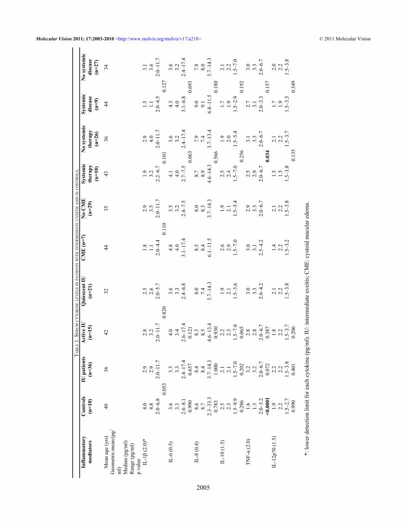
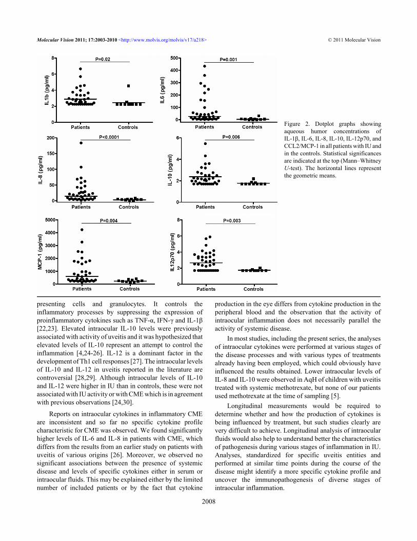
RESULTSSerum cytokine and chemokine levels: Serum cytokine andchemokine levels are given in Table 1. Serum TNF-α levelswere significantly higher in IU patients than in controls(p<0.0001, Figure 1). No other differences in serum cytokinelevels between IU and non-uveitic controls were noted. Serumcytokine and chemokine levels did not differ between activeand non-active IU patients, between IU patients with andwithout CME, and between the IU patients with or withoutsystemic disease. However, patients on systemic therapy hadlower serum levels of TNF-α than patients who were not ontreatment (p=0.034).Aqueous humor cytokine and chemokine levels: Intraocularcytokine levels of patients with IU and controls are given inTable 2. In most IU patients the cytokine and chemokine levelswere higher in the ocular fluids than in the corresponding sera;the levels of the two most abundant mediators, IL-6 and IL-8,exceeded those of serum in 23/26 (88%; p<0.0001) and 24/36(66%; p=0.038), respectively. In the controls, the intraocularlevels of IL-1β, IL-8, IL-10 and IL-12p70 were lower than inserum. Intraocular samples of IU patients had significantlyhigher levels of IL-1β (p=0.02), IL-6 (p=0.001), IL-8(p<0.001), IL-10 (p=0.005), IL-12p70 (p=0.003), and CCL2/MCP-1 (p=0.003) compared to the controls (Figure 2). Nodifferences in intraocular cytokines levels were noted betweenpatients with IU-associated systemic disease and idiopathicIU.
Active IU was characterized by higher levels ofintraocular IL-6 (p=0.003), IL-8 (p<0.0001), CCL5/RANTES(p=0.033), and CCL2/MCP-1 (p=0.033) compared to IU inremission; borderline significance was observed for IL-10(p=0.062). No significant associations were found for theother measured cytokines (Table 2). The IU patients onsystemic therapy had similar levels of intraocular cytokinesas the IU patients who were without systemic treatment; inparticular no differences were noted for intraocular TNF-αlevels. The presence of CME in IU patients was associatedwith higher intraocular levels of IL-6 and IL-8 (p=0.026 andp=0.012, respectively), whereas a borderline difference wasobserved for IL-10 (p=0.053).Correlations: No significant correlations of cytokines andchemokines were observed between the AqH and the serum .However, significant positive correlations were observed inthe AqH of IU patients between IL-6 and IL-8 (p=<0.0001,r=0.8), IL-1β and IL-12p70 (p<0.001, r=0.7), IL12p70 andTNF-α (p<0.001, r=0.6), IL-10 and TNF-α (p<0.0001, r=0.7),and between RANTES and IL-6 (p=0.001, r=0.6) and IL-8(p<0.0001, r=0.6), respectively. No significant correlationscould be detected for other analyzed cytokines andchemokines in serum of IU and control patients.
Molecular Vision 2011; 17:2003-2010 <http://www.molvis.org/molvis/v17/a218> © 2011 Molecular Vision
2004
made. Samples were stored immediately at −80 °C in sterilescrew-cap tubes and thawed immediately before analysis.
TAB
LE 1
. SER
UM
CY
TOK
INE
LEV
ELS I
N PA
TIEN
TS W
ITH
INTE
RM
EDIA
TE U
VEI
TIS A
ND
IN C
ON
TRO
LS.
Infla
mm
ator
ym
edia
tors
Con
trol
s(n
=10)
IU p
atie
nts
(n=3
6)A
ctiv
e IU
(n=1
5)Q
uies
cent
IU(n
=21)
CM
E (n
=7)
No
CM
E(n
=29)
Syst
emic
ther
apy
(n=1
0)
No
syst
emic
ther
apy
(n=2
6)
Syst
emic
dise
ase
(n=9
)
No
syst
emic
dise
ase
(n=2
7)
Mea
n ag
e (y
rs)
4036
4232
4435
4336
4434
Geo
met
ric m
ean (
pg/
ml)
Med
ian
(pg/
ml)
Ran
ge (p
g/m
l)p
valu
eIL
-1β
(2.0
)*4.
04.
82.
0–6.
8
2.9
2.9
2.0–
11.7
2.8
3.2
2.0–
11.7
2.5
2.6
2.0–
5.7
1.8
1.1
2.0–
4.4
2.9
3.5
2.0–
11.7
1.9
3.2
2.2–
6.7
2.9
4.0
2.0–
11.7
1.5
1.1
2.0–
4.5
3.1
3.6
2.0–
11.7
0.
053
0
.820
0
.110
0.10
10.
127
IL-6
(0.5
)3.
63.
32.
6–8.
1
3.3
3.3
2.4–
17.4
4.0
3.4
2.6–
17.4
3.6
3.3
2.4–
6.8
4.8
4.0
3.1–
17.4
3.5
3.2
2.4–
7.5
4.1
4.0
2.7–
7.5
3.6
3.2
2.4–
17.4
4.3
4.0
3.1–
6.8
3.6
3.2
2.4–
17.4
0.
990
0.65
70.
121
0.06
30.
693
IL-8
(0.4
)8.
68.
72.
3–33
.3
8.4
8.4
3.7–
14.3
8.3
8.5
4.6–
13.4
8.0
7.4
3.7–
14.3
8.5
8.4
6.1–
11.5
8.0
8.3
3.7–
14.3
8.7
8.9
4.6–
14.3
7.9
7.4
3.7–
13.4
9.0
9.1
6.8–
11.5
7.8
8.0
3.7–
14.3
0.
783
1.00
00.
930
0.56
60.
180
IL-1
0 (1
.5)
2.5
2.3
1.5–
9.9
2.1
2.1
1.5–
7.0
2.2
2.3
1.5–
7.0
1.9
2.1
1.5–
3.6
2.6
2.9
1.5–
7.0
1.9
2.1
1.5–
5.4
2.5
2.4
1.5–
7.0
1.9
2.0
1.5–
5.4
1.7
1.9
1.5–
2.9
2.1
2.2
1.5–
7.0
0.
286
0.20
20.
065
0.25
60.
192
TNF-
α (2
.0)
1.6
1.3
2.0–
3.2
3.2
3.2
2.0–
6.7
2.8
2.8
2.0–
6.7
3.0
3.3
2.0–
4.2
3.0
3.1
2.5–
4.2
2.9
3.3
2.0–
6.7
2.5
2.9
2.0–
6.7
3.1
3.3
2.0–
6.7
2.7
3.1
2.0–
3.3
3.0
3.3
2.0–
6.7
<0
.000
10.
072
0.38
70.
034
0.15
7IL
-12p
70 (1
.5)
1.9
2.2
1.5–
2.7
2.2
2.2
1.5–
3.8
1.8
2.2
1.5–
3.7
2.1
2.2
1.5–
3.8
1.4
2.2
1.5–
3.2
2.1
2.2
1.5–
3.8
1.5
1.5
1.5–
3.8
2.1
2.2
1.5–
3.7
1.7
1.9
1.5–
3.5
2.0
2.2
1.5–
3.8
0.
990
0.40
10.
206
0.13
50.
349
*:
low
er d
etec
tion
limit
for e
ach
cyto
kine
(pg/
ml).
IU: i
nter
med
iate
uve
itis;
CM
E: c
ysto
id m
acul
ar e
dem
a.
Molecular Vision 2011; 17:2003-2010 <http://www.molvis.org/molvis/v17/a218> © 2011 Molecular Vision
2005
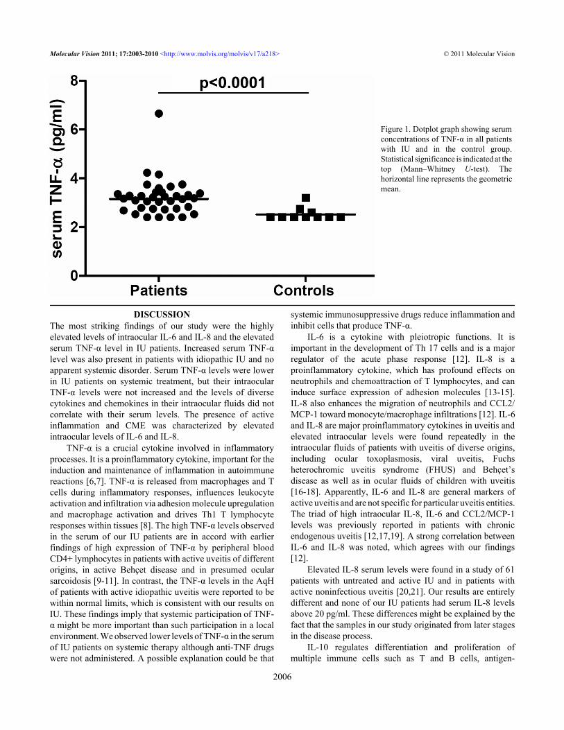
DISCUSSIONThe most striking findings of our study were the highlyelevated levels of intraocular IL-6 and IL-8 and the elevatedserum TNF-α level in IU patients. Increased serum TNF-αlevel was also present in patients with idiopathic IU and noapparent systemic disorder. Serum TNF-α levels were lowerin IU patients on systemic treatment, but their intraocularTNF-α levels were not increased and the levels of diversecytokines and chemokines in their intraocular fluids did notcorrelate with their serum levels. The presence of activeinflammation and CME was characterized by elevatedintraocular levels of IL-6 and IL-8.
TNF-α is a crucial cytokine involved in inflammatoryprocesses. It is a proinflammatory cytokine, important for theinduction and maintenance of inflammation in autoimmunereactions [6,7]. TNF-α is released from macrophages and Tcells during inflammatory responses, influences leukocyteactivation and infiltration via adhesion molecule upregulationand macrophage activation and drives Th1 T lymphocyteresponses within tissues [8]. The high TNF-α levels observedin the serum of our IU patients are in accord with earlierfindings of high expression of TNF-α by peripheral bloodCD4+ lymphocytes in patients with active uveitis of differentorigins, in active Behçet disease and in presumed ocularsarcoidosis [9-11]. In contrast, the TNF-α levels in the AqHof patients with active idiopathic uveitis were reported to bewithin normal limits, which is consistent with our results onIU. These findings imply that systemic participation of TNF-α might be more important than such participation in a localenvironment. We observed lower levels of TNF-α in the serumof IU patients on systemic therapy although anti-TNF drugswere not administered. A possible explanation could be that
systemic immunosuppressive drugs reduce inflammation andinhibit cells that produce TNF-α.
IL-6 is a cytokine with pleiotropic functions. It isimportant in the development of Th 17 cells and is a majorregulator of the acute phase response [12]. IL-8 is aproinflammatory cytokine, which has profound effects onneutrophils and chemoattraction of T lymphocytes, and caninduce surface expression of adhesion molecules [13-15].IL-8 also enhances the migration of neutrophils and CCL2/MCP-1 toward monocyte/macrophage infiltrations [12]. IL-6and IL-8 are major proinflammatory cytokines in uveitis andelevated intraocular levels were found repeatedly in theintraocular fluids of patients with uveitis of diverse origins,including ocular toxoplasmosis, viral uveitis, Fuchsheterochromic uveitis syndrome (FHUS) and Behçet’sdisease as well as in ocular fluids of children with uveitis[16-18]. Apparently, IL-6 and IL-8 are general markers ofactive uveitis and are not specific for particular uveitis entities.The triad of high intraocular IL-8, IL-6 and CCL2/MCP-1levels was previously reported in patients with chronicendogenous uveitis [12,17,19]. A strong correlation betweenIL-6 and IL-8 was noted, which agrees with our findings[12].
Elevated IL-8 serum levels were found in a study of 61patients with untreated and active IU and in patients withactive noninfectious uveitis [20,21]. Our results are entirelydifferent and none of our IU patients had serum IL-8 levelsabove 20 pg/ml. These differences might be explained by thefact that the samples in our study originated from later stagesin the disease process.
IL-10 regulates differentiation and proliferation ofmultiple immune cells such as T and B cells, antigen-
Figure 1. Dotplot graph showing serumconcentrations of TNF-α in all patientswith IU and in the control group.Statistical significance is indicated at thetop (Mann–Whitney U-test). Thehorizontal line represents the geometricmean.
Molecular Vision 2011; 17:2003-2010 <http://www.molvis.org/molvis/v17/a218> © 2011 Molecular Vision
2006

T AB
LE 2
. AQ
UEO
US H
UM
OR C
YTO
KIN
E LE
VEL
S IN
PATI
ENTS
WIT
H IN
TER
MED
IATE
UV
EITI
S AN
D IN
CO
NTR
OLS
.In
flam
mat
ory
med
iato
rsC
ontr
ols
(n=1
0)IU
pat
ient
s(n
=36)
Act
ive
dise
ase
(n=1
5)Q
uies
cent
dis
ease
(n=2
1)C
ME
(n=7
)N
o C
ME
(n=2
9)Sy
stem
ic th
erap
y(n
=10)
No
syst
emic
ther
apy
(n=2
6)Sy
stem
ic d
isea
se(n
=9)
No
syst
emic
dise
ase
(n=2
7)
Mea
n ag
e (y
rs)
4036
4232
4435
4336
4434
Geo
met
ric m
ean
(pg/
ml)
Med
ian
(pg/
ml)
Ran
ge (p
g/m
l)p
valu
eIL
-1β
(2.0
)*1.
41.
12.
2–4.
5
2.3
2.5
2.2–
6.7
2.1
2.1
2.2–
6.7
2.5
2.2
2.2–
4.6
2.5
2.5
2.2–
6.7
2.2
2.6
2.2–
5.7
2.3
2.5
2.2–
6.7
2.3
2.7
2.2–
5.7
2.5
4.0
2.2–
4.5
2.2
3.2
2.2–
6.6
0.
020
0.27
90.
845
0.87
60.
641
IL-6
(0.5
)3.
34.
80.
6–31
.0
27.5
24.5
2.0–
433.
1
66.5
57.3
5.6–
433.
1
14.6
15.4
2.1–
160.
6
86.7
64.4
5.6–
433.
1
20.8
20.7
2.0–
294.
0
40.1
34.6
5.6–
358.
4
23.8
21.8
2.1–
433.
1
32.9
42.6
2.0–
295.
0
25.9
21.5
2.3–
433.
10.
001
0.00
30.
026
0.39
30.
693
IL-8
(0.4
)2.
63.
80.
4–7.
9
14.2
15.6
1.0–
84.0
34.0
25.8
9.2–
183.
9
7.6
5.9
1.0–
57.1
40.4
25.8
15.5
–83.
9
11.0
9.2
1.0–
106.
7
26.2
24.4
3.8–
183.
9
11.2
10.3
1.0–
106.
7
17.6
26.8
2.9–
68.4
13.2
13.6
1.0–
183.
9
<0.
0001
<0.
0001
0.01
20.
074
0.56
5IL
-10
(1.5
)1.
12.
61.
7–2.
2
2.0
2.8
1.7–
14.7
2.5
3.0
1.7–
14.7
1.7
2.5
1.7–
5.5
2.5
2.8
1.7–
4.3
1.9
2.0
1.7–
14.7
2.1
2.6
1.7–
3.6
1.9
2.0
1.7–
14.7
2.1
2.5
1.7–
5.5
2.0
2.1
1.7–
14.7
0.
005
0.06
20.
054
0.25
60.
641
TNF-
α (2
.0)
2.0
2.8
2.4–
3.3
2.4
2.6
2.4–
5.4
2.7
2.8
2.4–
5.2
2.2
2.8
2.4–
5.4
3.1
3.3
2.4–
5.2
2.2
2.7
2.4–
5.4
3.2
3.0
2.4–
5.2
2.9
2.6
2.4–
5.3
2.3
2.7
2.4–
5.2
2.3
3.0
2.4–
5.4
0.
323
0.46
20.
094
0.13
50.
247
IL-1
2p70
(1.5
)1.
00.
91.
7–2.
0
2.1
2.7
1.7–
5.9
2.1
2.5
1.7–
5.9
2.2
3.0
1.7–
4.8
2.8
2.5
1.7–
5.9
2.0
2.8
1.7–
4.8
2.4
3.0
1.7–
5.9
2.1
2.5
1.7–
4.8
2.5
3.0
1.7–
4.8
2.0
2.5
1.7–
5.9
0.
003
0.50
50.
480
0.52
00.
565
CC
L5/R
AN
TES
(0.0
2)1.
81.
51.
1–3.
9
3.6
2.3
0.0–
76.5
6.3
3.7
0.5–
59.4
2.1
2.0
0.0–
76.5
8.4
8.5
2.0–
59.4
2.9
2.1
0.0–
76.5
4.5
5.7
0.0–
43.0
3.3
2.1
0.5–
76.5
8.9
8.5
0.5–
76.5
2.7
2.3
0.0–
59.4
0.
091
0.03
30.
078
0.35
50.
192
CC
L3/M
IP-1
α (4
.0)
3.2
3.1
4.1–
9.1
4.0
4.9
4.1–
15.2
4.5
5.3
4.1–
8.0
3.7
4.8
4.1–
15.3
3.9
4.2
4.1–
8.0
4.1
5.0
4.1–
15.3
4.2
4.7
4.1–
8.0
4.0
5.2
4.1–
15.3
5.0
5.0
4.1–
15.3
3.8
4.8
4.1–
8.0
0.
812
0.20
20.
907
0.93
10.
368
CC
L4/M
IP-1
β (3
.0)
27.9
24.3
19.0
–43.
8
34.6
32.3
16.4
–190
.9
35.5
34.4
16.4
–85.
0
34.0
30.2
16.9
–190
.4
33.8
34.4
16.4
–85.
0
34.8
32.0
16.9
–190
.4
34.5
32.3
16.4
–190
.4
35.1
31.8
18.4
–85.
0
39.7
32.7
16.9
–190
.4
33.1
30.5
16.4
–85.
0
0.35
00.
657
0.87
60.
821
0.72
0C
CL2
/MC
P1 (3
.0)
235.
923
2.4
140.
3–41
5.5
595.
741
7.5
143.
4–42
30.1
888.
684
6.7
214.
6–32
70.0
447.
731
6.9
143.
1–42
30.1
857.
484
6.7
222.
7–25
36.0
545.
636
1.0
143.
1–42
30.1
767.
978
1.0
214.
6–32
70.0
540.
335
6.3
143.
1–42
30.1
901.
095
9.9
143.
1–42
30.1
519.
036
3.9
200.
1–32
70.0
0.
003
0.03
30.
236
0.32
00.
157
*:
low
er d
etec
tion
limit
for e
ach
cyto
kine
(pg/
ml).
IU: i
nter
med
iate
uve
itis;
CM
E: c
ysto
id m
acul
ar e
dem
a.
Molecular Vision 2011; 17:2003-2010 <http://www.molvis.org/molvis/v17/a218> © 2011 Molecular Vision
2007
presenting cells and granulocytes. It controls theinflammatory processes by suppressing the expression ofproinflammatory cytokines such as TNF-α, IFN-γ and IL-1β[22,23]. Elevated intraocular IL-10 levels were previouslyassociated with activity of uveitis and it was hypothesized thatelevated levels of IL-10 represent an attempt to control theinflammation [4,24-26]. IL-12 is a dominant factor in thedevelopment of Th1 cell responses [27]. The intraocular levelsof IL-10 and IL-12 in uveitis reported in the literature arecontroversial [28,29]. Although intraocular levels of IL-10and IL-12 were higher in IU than in controls, these were notassociated with IU activity or with CME which is in agreementwith previous observations [24,30].
Reports on intraocular cytokines in inflammatory CMEare inconsistent and so far no specific cytokine profilecharacteristic for CME was observed. We found significantlyhigher levels of IL-6 and IL-8 in patients with CME, whichdiffers from the results from an earlier study on patients withuveitis of various origins [26]. Moreover, we observed nosignificant associations between the presence of systemicdisease and levels of specific cytokines either in serum orintraocular fluids. This may be explained either by the limitednumber of included patients or by the fact that cytokine
production in the eye differs from cytokine production in theperipheral blood and the observation that the activity ofintraocular inflammation does not necessarily parallel theactivity of systemic disease.
In most studies, including the present series, the analysesof intraocular cytokines were performed at various stages ofthe disease processes and with various types of treatmentsalready having been employed, which could obviously haveinfluenced the results obtained. Lower intraocular levels ofIL-8 and IL-10 were observed in AqH of children with uveitistreated with systemic methotrexate, but none of our patientsused methotrexate at the time of sampling [5].
Longitudinal measurements would be required todetermine whether and how the production of cytokines isbeing influenced by treatment, but such studies clearly arevery difficult to achieve. Longitudinal analysis of intraocularfluids would also help to understand better the characteristicsof pathogenesis during various stages of inflammation in IU.Analyses, standardized for specific uveitis entities andperformed at similar time points during the course of thedisease might identify a more specific cytokine profile anduncover the immunopathogenesis of diverse stages ofintraocular inflammation.
Figure 2. Dotplot graphs showingaqueous humor concentrations ofIL-1β, IL-6, IL-8, IL-10, IL-12p70, andCCL2/MCP-1 in all patients with IU andin the controls. Statistical significancesare indicated at the top (Mann–WhitneyU-test). The horizontal lines representthe geometric means.
Molecular Vision 2011; 17:2003-2010 <http://www.molvis.org/molvis/v17/a218> © 2011 Molecular Vision
2008
Our patients with IU exhibited a consistent pattern of fivehighly-elevated immune mediators (IL-6, IL-8, IL-10,IL-12p70, and CCL2/MCP-1; each p<0.003). We cannotconclude that this combination is actually characteristic for IUbecause we lack data of other specific uveitis entities arelacking. However this pattern clearly differs fromobservations for FHUS, ocular Behçet disease or sarcoiduveitis [17,31-33].
In conclusion, our results reveal the intraocularproduction of multiple cytokines and chemokines during IU,some of which were linked to disease activity. High serumlevels of TNF-α, independent of the presence of associatedsystemic disease, were typical, but, decreased underimmunosuppressive treatment. The identification ofproinflammatory molecules involved in the disease processesmay lead to the development and implementation of newtherapies. At present, TNF-α can be targeted, along with IL-6,IL-8, and CCL2/MCP-1 [34-36].
Our findings improve the understanding of thepathogenesis of IU and contribute to the identification offactors, which are involved in active IU.
ACKNOWLEDGMENTSThe authors did not receive any financial or material supportfor the research and the work. Study is a part of the researchprogram ARRS P3–0333. The authors declare that there areno conflicts of interest
REFERENCES1. Wakefield D, Chang JH. Epidemiology of uveitis. Int
Ophthalmol Clin 2005; 45:1-13. [PMID: 15791154]2. Curnow SJ, Murray PI. Inflammatory mediators of uveitis:
cytokines and chemokines. Curr Opin Ophthalmol 2006;17:532-7. [PMID: 17065921]
3. Van der Lelij A, Rothova A. Diagnostic anterior chamberparacentesis in uveitis: a safe procedure? Br J Ophthalmol1997; 81:976-9. [PMID: 9505822]
4. Jabs DA, Nussenblatt RB, Rosenbaum JT, Standardization ofUveitis Nomenclature (SUN) Working Group.Standardization of uveitis nomenclature for reporting clinicaldata. Results of the First International Workshop. Am JOphthalmol 2005; 140:509-16. [PMID: 16196117]
5. Sijssens KM, Rijkers GT, Rothova A, Stilma JS, SchellekensPA, de Boer JH. Cytokines, chemokines and soluble adhesionmolecules in aqueous humor of children with uveitis. Exp EyeRes 2007; 85:443-9. [PMID: 17662277]
6. Vassalli P. The pathophysiology of tumor necrosis factors.Annu Rev Immunol 1992; 10:411-52. [PMID: 1590993]
7. Suhler EB, Smith JR, Giles TR, Lauer AK, Wertheim MS, KurzDE, Lim L, Mackensen F, Pickard TD, Rosenbaum JT.Infliximab therapy for refractory uveitis: 2-year results of aprospective trial. Arch Ophthalmol 2009; 127:819-22.[PMID: 19506209]
8. Jap A, Chee SP. Immunosuppressive therapy for oculardiseases. Curr Opin Ophthalmol 2008; 19:535-40. [PMID:18854699]
9. Murphy CC, Duncan L, Forrester JV, Dick AD. SystemicCD4(+) T cell phenotype and activation status in intermediateuveitis. Br J Ophthalmol 2004; 88:412-6. [PMID: 14977779]
10. Santos Lacomba M, Marcos Martín C, Santos Lacomba M,Marcos Martín C, Gallardo Galera JM, Gómez Vidal MA,Collantes Estévez E, Ramírez Chamond R, Omar M. Aqueoushumor and serum tumor necrosis factor-alpha in clinicaluveitis. Ophthalmic Res 2001; 33:251-5. [PMID: 11586057]
11. Akdeniz N, Esrefoglu M. Keleş MS, Karakuzu A, Atasoy M.Serum interleukin-2, interleukin-6, tumour necrosis factor-alpha and nitric oxide levels in patients with Behçet’s disease.Ann Acad Med Singapore 2004; 33:596-9. [PMID:15531955]
12. Yoshimura T, Sonoda KH, Ohguro N, Ohsugi Y, Ishibashi T,Cua DJ, Kobayashi T, Yoshida H, Yoshimura A. Involvementof Th17 cells and the effect of anti-IL-6 therapy inautoimmune uveitis. Rheumatology 2009; 48:347-55.[PMID: 19164426]
13. De Vos AF, Hoekzema R, Kijlstra A. Cytokines and uveitis, areview. Curr Eye Res 1992; 11:581-97. [PMID: 1505201]
14. Dinarello CA. IL-18: A TH1-inducing, proinflammatorycytokine and new member of the IL-1 family. J Allergy ClinImmunol 1999; 103:11-24. [PMID: 9893178]
15. Nussenblatt RB. Uveitis. Fundamentals and Clinical Practice(3rd ed). Philadelphia, Penn: Elsevier; 2004.
16. Perez VL, Papaliodis GN, Chu D, Anzaar F, Christen W, FosterCS. Elevated levels of interleukin 6 in the vitreous fluid ofpatients with pars planitis and posterior uveitis: theMassachusetts eye & ear experience and review of previousstudies. Ocul Immunol Inflamm 2004; 12:193-201. [PMID:15385196]
17. Curnow SJ, Falciani F, Durrani OM, Rauz S, Wallace GR,Salmon M, Cheung CM, Ross EJ, Wloka K, Murray PI.Multiplex bead immunoassay analysis of aqueous humorreveals distinct cytokine profiles in uveitis. InvestOphthalmol Vis Sci 2005; 46:4251-9. [PMID: 16249505]
18. Santos Lacomba M, Marcos Martín C, Gallardo Galera JM,Collantes Estévez E, Ramírez Chamond R, Omar M, GomezVidal A. Aqueous humor and serum interleukin-6 in patientswith uveitis. Arch Soc Esp Oftalmol 2001; 76:345-50.[PMID: 11438864]
19. Lahmar I, Abou-Bacar A, Abdelrahman T, Kairallah M, Speeg-Schatz C, Bourcier T, Sauer A, Villard O, Pfaff AW, MousliM, Garweg JG, Candolfi E, Guinard M, Babba H, Ben YahiaS. Cytokine profiles in toxoplasmic and viral uveitis. J InfectDis 2009; 199:1239-49. [PMID: 19302012]
20. Klok AM, Luyendijk L, Zaal MJW, Rothova A, Hack CE,Kijlstra A. Elevated serum IL-8 levels are associated withdisease activity in idiopathic intermediate uveitis. Br JOphthalmol 1998; 82:871-4. [PMID: 9828768]
21. Kramer M, Monselise Y, Bahar I, Cohen Y, Weinberger D,Goldenberg-Cohen N. Serum cytokine levels in active uveitisand remission. Curr Eye Res 2007; 32:669-75. [PMID:17852191]
22. Asadullah K, Sterry W, Volk HD. Interleukin-10 therapy.Review of a new approach. Pharmacol Rev 2003;55:241-69. [PMID: 12773629]
23. O'Garra A, Vieira PL, Vieira P, Goldfeld AE. IL-10-producingand naturally- occurring CD4+ Tregs: limiting collateraldamage. J Clin Invest 2004; 114:1372-8. [PMID: 15545984]
Molecular Vision 2011; 17:2003-2010 <http://www.molvis.org/molvis/v17/a218> © 2011 Molecular Vision
2009
http://www.ncbi.nlm.nih.gov/entrez/query.fcgi?cmd=Retrieve&db=PubMed&dopt=abstract&list_uids=9505822
http://www.ncbi.nlm.nih.gov/entrez/query.fcgi?cmd=Retrieve&db=PubMed&dopt=abstract&list_uids=1590993
http://www.ncbi.nlm.nih.gov/entrez/query.fcgi?cmd=Retrieve&db=PubMed&dopt=abstract&list_uids=1505201
http://www.ncbi.nlm.nih.gov/entrez/query.fcgi?cmd=Retrieve&db=PubMed&dopt=abstract&list_uids=9893178
24. Ongkosuwito JV, Feron EJ, van Doornik CE, Van der Lelij A,Hoyng CB, La Heij EC, Kijlstra A. Analysis ofimmunoregulatory cytokines in ocular fluid samples frompatients with uveitis. Invest Ophthalmol Vis Sci 1998;39:2659-65. [PMID: 9856775]
25. Takase H, Futagami Y, Yoshida T, Mochizuki M, Kamoi K,Sugita S, Imai Y. Cytokine profile in aqueous humor and seraof patients with infectious or noninfectious uveitis. InvestOphthalmol Vis Sci 2006; 47:1557-61. [PMID: 16565392]
26. van Kooij B, Rothova A, Rijkers GT, deGroot Mijnes J. Distinctcytokine and chemokine profiles in the aqueous humor ofpatients with uveitis and cystoid macular edema. Am JOphthalmol 2006; 142:192-4. [PMID: 16815285]
27. Yoshida A, Koide Y, Uchijima M, Yoshida TO. IFN gammainduces IL12 mRNA expression by a murine macrophage cellline, J774. Biochem Biophys Res Commun 1994;198:857-61. [PMID: 7906942]
28. el-Shabrawi Y, Livir-Rallatos C, Christen W, Baltatzis B,Foster CS. High levels of interleukin-12 in the aqueous humorand vitreous of patients with uveitis. Ophthalmology 1998;105:1659-63. [PMID: 9754174]
29. Akpek EK, Maca SM, Christen WG, Foster CS. Elevatedvitreous interleukin-10 level is not diagnostic of intraocular-central nervous system lymphoma. Ophthalmology 1999;106:2291-5. [PMID: 10599659]
30. El-Asrar AMA, Struyfb S, Kangavea D, Al-Obeidana SS,Opdenakkerb G, Geboesch K, Van Damme J. Cytokineprofiles in aqueous humor of patients with different clinical
entities of endogenous uveitis. Clin Immunol 2011;139:177-84. [PMID: 21334264]
31. Muhaya M, Calder V, Towler HM, Shaer B, McLauchlan M,Lightman S. Characterization of T cells and cytokines in theaqueous humour (AH) in patients with Fuchs' heterochromiccyclitis (FHC) and idiopathic anterior uveitis (IAU). Clin ExpImmunol 1998; 111:123-8. [PMID: 9472671]
32. Ahn JK, Yu HG, Chung H, Park YG. Intraocular cytokineenvironment in active Behçet uveitis. Am J Ophthalmol 2006;142:429-34. [PMID: 16935587]
33. Xu H, Rizzo LV, Silver PB, Casi RR. Uveitogenicity isassociated with a Th1-like lymphokine profile: cytokine-dependent modulation of early and committed effector T cellsin experimental autoimmune uveitis. Cell Immunol 1997;178:69-78. [PMID: 9184700]
34. Hohki S, Ohguro N, Haruta N, Nakai K, Terabe F, Serada S,Fujimoto M, Nomura S, Kawahata H, Kishimoto T, Naka T.Blockade of interleukin-6 signaling suppresses experimentalautoimmune uveoretinitis by the inhibition of inflammatoryTh17 responses. Exp Eye Res 2010; 91:162-70. [PMID:20420831]
35. Barnes PJ. New molecular targets for the treatment ofneutrophilic diseases. J Allergy Clin Immunol 2007;119:1055-62. [PMID: 17353033]
36. Brennan F, Beech J. Update on cytokines in rheumatoidarthritis. Curr Opin Rheumatol 2007; 19:296-301. [PMID:17414959]
Molecular Vision 2011; 17:2003-2010 <http://www.molvis.org/molvis/v17/a218> © 2011 Molecular Vision
Articles are provided courtesy of Emory University and the Zhongshan Ophthalmic Center, Sun Yat-sen University, P.R. China.The print version of this article was created on 15 July 2011. This reflects all typographical corrections and errata to the articlethrough that date. Details of any changes may be found in the online version of the article.
2010
http://www.ncbi.nlm.nih.gov/entrez/query.fcgi?cmd=Retrieve&db=PubMed&dopt=abstract&list_uids=9856775
http://www.ncbi.nlm.nih.gov/entrez/query.fcgi?cmd=Retrieve&db=PubMed&dopt=abstract&list_uids=7906942
http://www.ncbi.nlm.nih.gov/entrez/query.fcgi?cmd=Retrieve&db=PubMed&dopt=abstract&list_uids=9754174
http://www.ncbi.nlm.nih.gov/entrez/query.fcgi?cmd=Retrieve&db=PubMed&dopt=abstract&list_uids=9472671