Inflammatory Cytokine Alterations in Schizophrenia: A Systematic Quantitative Review
Upload
independentCategory
view
0download
0
Cytokine Transcriptional Events during Helper T Cell Subset Differentiation By James A. Lederer,* Victor L. Perez,* Lori DeslLoches,~ So Mee Kim,* Abul K. Abbas,* and Andrew H. Lichtman*
From the *Department of Surgery and the *Immunology Research Division, Department of Pathology, Brigham and Women's Hospital and Harvard Medical School, Boston, Massachusetts 02115
Summary The molecular basis for changes in cytokine expression during T helper (Th) cell subset differ- entiation is not well understood. We have characterized transcriptional events related to cyto- kine gene expression in populations of naive T cell receptor-transgenic T cells as they are driven in vitro toward Th l or Th2 phenotypes by interleukin (IL)-12 or IL-4 treatment, re- spectively. Quantitative reverse transcriptase-polymerase chain reaction analysis of cytokine transcripts indicates that interferon (IFN) ~/, IL-4, and IL-2 m R N A are expressed with distinct kinetics after naive T cells are stimulated with antigen and either IL-4 or IL-12. IFN-~/mR.NA appears as early as 6 h m IL-12-treated cultures, IL-4 appears only after 48 h in lL-4--treated cultures, and IL-2 is equivalently expressed in both types of cultures. Analyses were performed to determine if there were any differences in activation of lL-2 or IL-4 transcription factors that accompanied Thl versus Th2 differentiation. These studies demonstrated that signal transducer and activator of transcription 6 (STAT6) binds to a sequence in the IL-4 promoter and that this STAT6-binding site can support IL-4--dependent transcription of a linked heterologous pro- moter. Prolonged activation of STAT6 is characteristic of populations undergoing Th2 differ- entiation. Furthermore, STAT6 is activated in an autocrine manner when differentiated Th2 populations are stimulated by antigen receptor ligation. Th l populations derived from IL-12 plus antigen treatment of naive T cells remain responsxve to IL-4 as indicated by induction of STAT6 and IL-4 mRNA. These data indicate that Th l and Th2 differentiation represents the combination of different, apparently independently regulated transcriptional events. Further- more, among transcription factors that bind to the IL-4 or IL-2 promoters, STAT6 is the one whose activation distinguishes Th2 versus Th l development.
T he development of effector populations of CD4 + Th cells that produce distinct cytokines is of central im-
portance in infectious, allergic and autoimmune diseases (1, 2). Thl cells, which secrete IL-2 and IFN-% are responsible for phagocyte-dependent protective immunity and tissue injury in many organ-specific autoimmune diseases. Th2 cells, which produce IL-4, IL-5, and IL-10, are involved in the development of allergies and in defense against helmin- thic parasites. In addition, the cytokines produced by Th2 cells, especially IL-4 and IL-10, may be critical for down- regulating or suppressing inflammation associated with Thl-mediated immune responses. Experimental evidence indicates that these polarized subsets of differentiated Th cells arise from a common precursor, which is a naive CD4 + T cell that produces small amounts of IL-2 and no detectable IFN-~/or IL-4 when stimulated for the first time
J. Lederer and V. Perez contributed equally to this paper.
in vitro (3, 4). The presence of exogenous cytokines at the time of primary antigenic stimulation profoundly influ- ences which differentiation pathway a naive T cell will fol- low. Thus, IL-4 is required to drive differentiation of naive cells into Th2 effectors. IL-12 and IFN-~/are the principal cytokines that induce Th l differentiation. There as some evidence that these cytokines, particularly IL-4 and IL-12, act directly on the T cells and not on APCs. It is clear that IL-4 has a dominant effect over IL-12, causing Th2 differ- entiation even when both cytokines are present (2, 5).
Very little is known about the molecular events that un- derlie T cell subset differentiation. Studies with T cell lines indicate that production of cytokines such as IL-2 and IL-4 is controlled primarily by transient activation of transcrip- tion induced by the binding of nuclear factors to 5'-regula- tory regions of the cytokine genes (6-9). The regulation of IFN-~/expression may be more complex, involving meth- ylation of DNA and posttranscriptional events (10, 11). We have previously shown that in Th l and Th2 clones, the ac-
397 j. Exp. Med. �9 The Rockefeller University Press �9 0022-1007/96/08/397/10 $2.00 Volume 184 August 1996 397-406
on February 18, 2015
jem.rupress.org
Dow
nloaded from
Published August 1, 1996

tivity o f exogenously introduced IL-2 and IL-4 promoter constructs correlates with IL-2 and IL-4 product ion (6). Furthermore, we found that T C R - i n d u c i b l e nuclear trans- location o f the p50/p65 heterodimer o f nuclear factor-KB (NF-KB) 1 occurs in IL-2-produc ing T h l and Th0 clones, but not in Th2 clones. It is, therefore, important to estab- lish if bulk populations o f differentiated T h l and Th2 cells show distinct patterns of cytokine gene transcription, and whether this correlates with the activation and nuclear translocation o f specific transcription factors.
In this paper we describe our initial studies to determine changes in cytokine gene transcription and transcription factors during the differentiation o f naive CD4 + T cells to T h l and Th2 populations. The nmdel system we have used is the in vitro antigen plus cytokine st imulat ion o f naive T cells from a TCR- t ransgen ic mouse line. Such T cells have been shown to develop into populations o f T h l and Th2 cells by stimulation with antigen and IL-12 or IL-4, respectively (12-14). Our experiments show that patterns of cytokine gene expression are clearly different within 48 h o f stimulation, and changes in IL-2, IL-4, and I F N - y gene expression occur at distinct t ime points. Furthermore, we have identified a functionally active site in the IL-4 pro- moter that specifically binds the IL-4- induced transcription factor signal transducer and activator o f transcription 6 (STAT6). STAT6 activation consistently and uniquely cor- relates with IL-4- induced Th2 differentiation.
Materials and Methods
Animals. TCR-transgenic mice expressing the AND TCR spe- cific for pigeon cytochrome c (PCC) peptide 81-104 in associa- tion with I-E k (15) were bred in our virus-free animal facility. All animals were maintained in accordance with the guidelines of the Committee on Animals of the Harvard Medical School and those prepared by the Committee on Care and Use of Laboratory Ani- mals of the Institute of Laboratory Resources, National Research Council (DHEW Publication No. [NIH] FS-23). Transgenic TCR expression was screened by PCR as previously described (5).
Auuicen, Cytokiues, and Antibodies. The antigen used in all stud- ies was the COOH-terminal 81-104 peptide of PCC and was ob- tained from the peptide synthesis facility of the Center for Neu- rologic Diseases, Brigham and Women's Hospital. Supernatant containing recombinant murine IL-4 was generated from the I3L6 cell line transfected with a constitutively expressed routine IL-4 eDNA (a gift of Dr. Robert Tepper, Massachusetts General Hospital, Boston, MA). Recombinant murine IL-12 was a gift of Dr. Stan Wolf (Genetics Institute, Cambridge, MA). Supernatant containing recombinant murine IL-2 was obtained from the IL-2 gene-transfected X63 cell lme (16). The hybridoma producing anti-IL-4 mAb, 11Bll, obtained from Dr. William Paul (Na- tional Institutes of Health, Bethesda, MD) was used to produce ascites in nude mice.
~ Abbreviations used itt this paper: CAT, chloramphenicol acetyl tranferase; CF, competitive fragment; EMSA, electrophoretic mobility shift assay; NFAT, nuclear factor of activated T cells; NF-KB, nuclear factor KB; PCC, pigeon cytochrome c; RT, reverse transcriptase; STAT6, signal transducer and activator of transcription 6.
Culture Conditions. CD4; T cells were purified from pooled lymph nodes and spleens of TCR-transgenic mice by comple- ment-mediated lysis of MHC class II-, CD8- and JllD-express- ins cells, followed by depletion of remaining B cells by panning on anti-Is--coated dishes, as previously described (5). These T cell populations were >95% CD3+CD4 +. T cell differentiation was induced by culturing 2 X 10⁵ purified TCR-transgenic T cells with 2 X 10⁶ APCs, 1 I.zM PCC peptide 81-104, and either lL-12 (10 ng/ml) or IL-4 (1,000 U/ml) in 1 ml of medium. APCs were splenic B cells purified from littermate B10.BR mice by anti-Is panning and were treated with 50 ~g/ml mitomycin C at 37~ for 30 rain, as described (5). All cultures were carried out in RPMI 1640 supplemented with 1 mM L-glutatnine, sodiunt pyruvate, nonessential amino acids, 5 • 1(1 5 2-met and 10% heat-inactivated FCS (all from GIBCO BRL, Gaithersburg, MD).
ELISA Determination of Secreted IL-2, IL-4, and IFN-% Culture supematants were assayed for the presence of IL-2, IL-4, and IFN-',/ using ELISA reagents from PharMingen (San Diego, CA).
Quantitation of Cytokine Transcripts by Reverse Tra,scriptase- PCR. After various times in culture, cells were lysed with Ultra- spec RNA extraction solution (Biotecx, Houston, TX), RNA was precipitated with isopropanol, washed, and reverse tran- scribed using oligo d(T)12-18 by standard methods (17). Quanti- tative PCR was performed by using a competitive fragment (CF) that contains various cytokine eDNA sequences that yield differ- ent size PCR-amplification products than do endogenous cyto- kine cDNAs (18). The [3-actin eDNA concentration of each sam- ple was first determined using [3-actin--specific primers by keeping the cDNA concentration constant and adding serial dilutions of CF. Amplified products were separated on an agarose gel and stained with ethidiurn bromide to determine the serial dilution of CF that gave amplified products from the CF and eDNA of equal intensity. The cDNA samples were normalized to equal [3-actin eDNA concentrations, amplified with IL-2-, IFN-'y- and IL-4- specific primers, and products were separated on agarose gels. The eDNA concentrations were then determined from the dilution of CF that yielded an equal amount of amplified product as had the eDNA. The eDNA concentrations were calculated as attonmles of cDNA transcripts divided by femtomoles of input [3-actin cDNA.
Electrophoretic Gel Mobility Shift Assays. Electrophoretic gel mo- bility shift assays (EMSA) for transcription factors were pertbrmed as previously described (6). Briefly, oligonucleotides that corre- spond to the consensus DNA-binding elements in the nmrine IL-2 promoter (NFIL-2A, AP-1, NF-KB, nuclear factor of acti- vated T cells [NFAT]) and IL-4 promoter (STAT6, IL-4 NFAT) were synthesized for use as probes in EMSA using a DNA syn- thesizer (model 381A; Applied Biosystems, Inc., Foster City, CA) or they were purchased from Integrated DNA Technologies, Inc. (Coralville, IA). These oligonucleotides included:
NFIL-2A 5 ' -TATGTGTAATATGTAAAA-3 ' AP-1 5 ' -AATTCCAGAGAGTCATCAGA-3' NF-KB 5 ' -ACCAAGAGGGATTTCACCTAAATC-3 ' NFAT 5 ' -AAGAGGAAAATTTGTTTCATACAG-3' STAT6 5 ' -TGATTTCACAGGAAAATT-3 ' IL-4 NFAT 5 ' -ATAAAATTTTCCAATGTAAA-3'
The complementary single-stranded oligonucleotides were also syn- thesized and annealed to the above listed oligonucleotides, and the double-stranded oligonucleotides were end-labeled with .g_32p_ ATP (DuPont NEN Research Products, Wilmington, DE) using T4 polynucleotide kinase (Pharmacia LKB Biotechnology, Inc., Piscataway, NJ) for use as probes in EMSA. Labeled oligonucle- otide was separated from unincorporated "y-32p-ATP using spin-
398 Cytokine Gene Transcription during Thl and Th2 Differentiation
on February 18, 2015
jem.rupress.org
Dow
nloaded from
Published August 1, 1996

10 columns (Clontech, Palo Alto, CA). Nuclear protein extracts were prepared as described (6). Nuclear extract (5 ~g protein) was incubated for 20 min at room temperature in binding buffer (10 mM Tris-HC1, pH 7.9, 0.1 mM EDTA, 2 mM dithiothreitol, 5% glycerol, and 100 mM KC1) with --,10,000 cpm of 32p-labeled oligonucleotides. The binding reactions were then mixed with 6X gel-loading buffer (0.25% bromophenol blue, 0.25% xylene cyanol FF, and 30% glycerol), and protein-DNA complexes were separated from free oligonucleotide probes by electrophoresis through a 9% nondenaturing polyacrylamide gel. After electro- phoresis, the gels were dried under vacuum and prepared for au- toradiography. Control experiments using 100-fold molar excess unlabeled oligonucleotide probes demonstrated that the shifted protein-DNA complexes were specific for each respective oligo- nucleotide probe used in this study. For supershifts, normal rabbit serum or antisera specific for murine STAT4 or STAT6 were added to the binding reactions.
Functional Analysis of the IL-4 Promoter STA T6 Site by Transient Transfection. Upper and lower strand oligonucleotides containing the IL-4 promoter STAT6 site sequence with 5'-phosphorylated TCGA overhangs were purchased from Integrated DNA Tech- nologies, Inc., annealed, ligated to SalI-cut pBLCAT2 reporter construct (19), and used to transform Escherichia coil Plasmid miniprep DNA from transformants were screened by hybridiza- tion to a 32p-labeled STAT6 oligonucleotide probe, and positive clones were sequenced. The 10g and 14g clones identified in this manner contain one and two tandem copies of the STAT6-bind- ing site, respectively. These constructs or the pBLCAT2 parent plasmid were transfected into the TA3 B-lymphoblastoid line (20) by electroporation using a Gene Pulsar instrument (Bio-Rad Laboratories, Richmond, CA). Transfection was performed with 40 I.tg of DNA and 20 X 10 v cells as described previously (6). Electroporated cells were cultured in 5 ml culture medium for 48 h. IL-4 (500 U/ml) was added to some cells for the final 24 h. Cyto- plasmic lysates were prepared and chloramphenicol acetyl trans- ferase (CAT) assays were performed as described (6).
Tab le 1. Cytokine Expression After Restimulation
Results
Kinetics of IL-2, IFN-% and IL-4 Gene Expression during Th 1 and Th2 Cell Differentiation. In the initial set o f ex- periments, we wished to determine when during in vitro differentiation T cells acquired cytokine transcription pro- files characteristic o f T h l and Th2 subsets. To first establish that cytokine secretion correlated with m R N A levels, we examined TCR- t ransgen ic CD4 § T cells that had been cultured with antigen, splenic B cell APCs, and either IL-12 or IL-4 for 6 d, in order to induce differentiation. As shown in Table 1, restimulation o f cells from 6-d IL -12 - treated primary cultures resulted in IL-2 and I F N - y pro- duction, but no detectable IL-4 product ion. In contrast, cells from IL-4- t rea ted primary cultures produced IL-4, but no detectable I F N - y and very little IL-2 upon restimu- lation with antigen. Thus, as expected, IL-12 and IL-4 in- duce distinct patterns o f T h l / T h 2 differentiation. Because some experiments addressing the induction o f IL-2 and IL-4 transcription factors required the use o f a n t i - C D 3 as a st im- ulus, we also needed to examine the phenotype o f IL -12 - and IL-4- t rea ted T cell populations when restimulated with ant i -CD3. Table 1 shows that an t i -CD3- induced IL-4 product ion was observed only in IL-4- t rea ted cells. Sur- prisingly, an t i -CD3 restimulation induced significant levels o f IFN- ' , /p ro te in in T cells from IL-4- t rea ted primary cul- tures. This was seen in three separate experiments. Further- more, there was some IL-2 detectable in an t i -CD3- res t im- ulated T cells from both IL -12 - and IL-4--treated primary cultures.
Cytokine transcript levels as determined by quantitative reverse transcriptase ( R T ) - P C R using competi t ive P C R - MIMICs were also distinctly different in these T h l and Th2 populations. Upon restimulation, T h l cells induced
Cytokine Cytokine mRNA expression (atto- moles/femtomole [3-actin mRNA)
Cytokine added Restimulation in 1~ culture conditions IL-2 IFN-y 1L-4 IL-2 IFN-y IL-4
IL-12
IL-4
U/ml APCs 0 0 0 0.33 100 n.d.
APCs + PCC peptide 133 878 0 3.3 10,000 n.d.
None 0 0 0 n.d. < 1 n.d.
Anti-CD3 34 664 0 3.5 100 n.d.
APCs 0 0 0 n.d. n.d. n.d.
APCs + PCC peptide 2 0 60 0.33 1 33
None 0 0 0 n.d. < 1 n.d.
Anti-CD3 5 466 51 3.5 10 100
Cytokine secretion and mRNA expression in Thl and Th2 populations of TCR-transgenic T cells. Naive T cells from AND mice were cultured with splenic B cell APCs, PCC (81-104) peptide (1 ~M), and either IL-4 (1,000 U/ml) or IL-12 (10 ng/ml) as indicated, for 6 d. T cells were then harvested, washed, and recultured in microwells with splenic B cell APCs minus or plus PCC peptide, no stinmlus, or plate-bound anti-CD3. RNA was extracted after 18 h for RT-PCR analysis of cytokine transcripts. Quantities of cytokine mRNA were calculataed from competitive PCR shown in Fig. 1. Identical cultures were left for 48 h, after which supernatants were collected and analyzed for cytokine protein by ELISA. n.d., none detected.
399 Lederer et al.
on February 18, 2015
jem.rupress.org
Dow
nloaded from
Published August 1, 1996
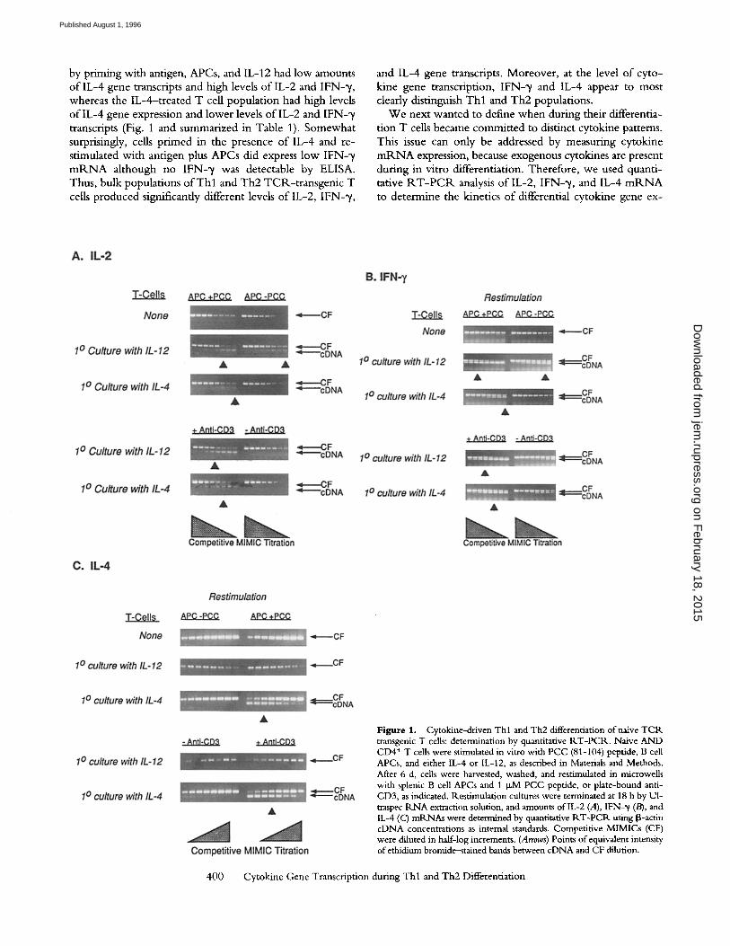
by priming with antigen, APCs, and IL-12 had low amounts o f IL-4 gene transcripts and high levels o f IL-2 and I F N - % whereas the IL-4--treated T cell populat ion had high levels o f l L - 4 gene expression and lower levels o f l L - 2 and IFN-21 transcripts (Fig. 1 and summarized in Table 1). Somewhat surprisingly, cells pr imed in the presence o f IL-4 and re- stimulated with antigen plus APCs did express low IFN- 'y m R N A although no IFN-2r was detectable by ELISA. Thus, bulk populations o f T h l and Th2 TCR-t ransgenic T cells produced significantly different levels o f IL-2, IFN-~ ,
and IL-4 gene transcripts. Moreover , at the level o f cyto- kine gene transcription, IFN-~/ and IL-4 appear to most clearly distinguish T h l and Th2 populations.
W e next wanted to define when during their differentia- tion T cells became commit ted to distinct cytokine patterns. This issue can only be addressed by measuring cytokine m R N A expression, because exogenous cytokines are present during in vitro differentiation. Therefore, we used quanti- tative R.T-PCR. analysis o f IL-2, I F N - % and IL-4 m_RNA to determine the kinetics o f differential cytokine gene ex-
Figure 1. Cytokine-driven Thl and Th2 differentiation of naive TCR transgenic T cells: determination by quantitative RT-PCP.. Naive AND CD4 + T cells were stimulated in vitro with PCC (81-104) peptide, B cell APCs, and either IL-4 or IL-12, as described in Materials and Methods. After 6 d, cells were harvested, washed, and restimulated in microwells with splenic B cell APCs and 1 I.LM PCC peptide, or plate-bound anti- CD3, as indicated, l~.estimulation cultures were terminated at 18 h by U1- traspec RNA extraction solution, and amounts of IL-2 (A), [FN-~/(B), and IL-4 (C) mR.NAs were determined by quantitative RT-PCR using iB-actin cDNA concentrations as internal standards. Competitive MIMICs (CF) were diluted in half-log increments. (Arrows) Points of equivalent intensity of ethidium bromide-stained bands between cDNA and CF dilution.
400 Cytokine Gene Transcription during Thl and Th2 Differentiation
on February 18, 2015
jem.rupress.org
Dow
nloaded from
Published August 1, 1996

pression in differentiating Th populations (Fig. 2). The re- suits show that IL-12 enhanced IFN-~ /mt (NA expression as early as 6 h after initial stimulation, and that IFN-~/gene expression remained significantly higher in the IL-12- treated cultures than in the untreated or IL-4-treated cul- tures throughout the differentiation process. A striking ob- servation was that IL-4 gene expression was not detected until 48 h after stimulation of naive T cells with antigen and IL-4. The expression of IL-2 was not significantly af- fected b~, IL-12 and IL-4 treatment, even at 48 h when IL-4 gene expression was the highest in the IL-4-treated stimu- lation cultures.
These results indicate that changes in expression of IL-2, IL-4, and IFN-~/ genes during subset differentiation are temporally distinct events, suggesting that they may be reg- ulated in different ways. In vitro differentiation of naive T cells is associated with clearly polarized patterns of IL-4 and IFN-',/ gene transcription within 48 h. Early transcription of IL-2 is not distinct in these differentiating T cells, and the diminished expression of the IL-2 gene in Th2 popula- tions is only observed after restimulating cells that have dif- ferentiated in the presence of IL-4.
Analysis of lL-2 and IL-4 Promoter-specific Transcription Fac- tors in Th 1 and Th2 Cell Populations. The expression of cy- tokine genes is controlled, in part, by sets of transcription factors that bind to cytokine promoters. We conducted EMSA analyses in order to determine if the distinct patterns of cytokine gene transcription in T cell populations under- going Th l versus Th2 differentiation could be attributed to distinct patterns of transcription factor activation. The well characterized regulatory elements within the IL-2 gene promoter include AP-1, NFIL-2A (Oct-1/OAP-40), NFAT,
and NF-KB (9). The IL-4 promoter contains multiple NFAT sites that have been shown to be involved in regula- tion of IL-4 gene expression (7, 21). Recently, several groups have described a transcription factor that becomes tyrosine phosphorylated and rapidly translocates from the cytoplasm to the nucleus in response to IL-4-induced sig- nals. This factor, called STAT6 or IL-4STAT, binds to sites in the 5' regulatory regions of several IL-4-responsive genes that have an inverted repeat spaced by three nucle- otides that is characteristic of several described cytokine re- sponse- or STAT factor-binding sites (22-24). Compari- son of these sequences with the murine IL-4 promoter sequences revealed a potential binding site located at -153 to -167 from the transcriptional initiation site within the routine IL-4 promoter. This site partially overlaps a previ- ously identified NFAT-binding site (7). Thus, for the EMSA assays in these studies, we used oligonucleotide probes corresponding to the AP- I - , NFIL-2A (Oct - l / OAP-40)-, NFAT-, and NF-KB-binding sequences of the routine IL-2 gene promoter, and the NFAT- and STAT6- binding sites from the murine IL-4 promoter.
When we compared cells from IL-4- and IL-12-treated cultures, we observed no significant difference in the nu- clear presence of IL-2 promoter-binding factors, including AP-1, NFAT, NFIL-2A, and NF-KB. This was true early during T cell differentiation, i.e., 48 h after stimulation of naive T cells with IL-12 or IL-4 (Fig. 3), and even in cells that had been differentiated for 6 d and were restimulated (Fig. 4). Parallel assays for cytokine secretion by 6-d differ- entiated populations showed that Th l and Th2 differentia- tion had occurred in these IL-12- and IL-4-treated cul- tures, respectively (data not shown). Although we have
E
0.6"
0.5-
0.4-
0.3-
0.2-
0.1
0
I ' - , L - l ~ " ~ t e d ~ltures - 4 - r 11..-4 tr.eated ~jltures -0- _ I
IL-2
6 24 48 96
160
'~ 11111
q
0
IFN-7
I I I 0
6 24 48 96
RNA Isolation Time (Hours)
16
12 10 8-q 6~ 4 - . ~
2q
6
IL-4
i ~ I m I m
24 48 96
Figure 2. Kinetics of cytokine mRNA expression in differentiating CD4 § T cells: quantitative I:(T-PCt( analysis. Differentiation cultures were set up as described in Fig. 1. RNA was isolated from the cultures at the indicated time points and reverse transcribed IL-2, IFN-~, and IL-4 mI(NA was ana- lyzed hy quantitative I~T-PCIk.
401 Lederer et al.
on February 18, 2015
jem.rupress.org
Dow
nloaded from
Published August 1, 1996

Figure 3. Nuclear transcription factors in differentiating Thl and Th2 populations. TCR transgenic T cells were cultured with splenic B cell APCs, antigen, and either IL-12 or IL-4, as described in Materials and Methods. After 48 h of culture, nuclear extracts were prepared and ana- lyzed by EMSA for the presence of DNA-binding proteins using oligonu- cleotide probes containing binding sites for the indicated factors.
previously observed a lack of NF-KB activation in T C R - stimulated Th2 clones that do not transcribe the IL-2 gene, the finding here that NF-KB activation does occur in stim- ulated Th2-1ike populations is consistent with the fact that these cells do express IL-2 transcripts (Fig. 1 A). The lack of TC1K-inducible NF-KB activation may be a phenotype of cells that have undergone multiple rounds of differentiation.
Among the IL-4 promoter -b inding proteins, we found no difference in nuclear proteins that bind to an IL-4 NFAT site, but there was a distinct difference in EMSA patterns using an oligonucleotide probe corresponding to the puta- tive STAT6-binding sequence in the mouse IL-4-promoter. STAT6 protein was detected in the nuclei of IL-4-treated T cells at 48 h after initiation of cultures, the time point during differentiation when we first detect IL-4 mlKNA (Fig. 2). Supershift analysis using a murine STAT6-specific antibody indicated that STAT6 was in fact present in the IL-4- induced gel-shift complex detected with this probe. The ant i -STAT6 serum, but not ant i -STAT4 or preim- mune sera, supershifted complexes of the probe and nu- clear proteins from IL-4-treated TCR-transgeuic T cells (Fig. 5), as well as complexes formed with nuclear extracts from ant i-CD3-treated TCR-transgenic Th2 populations (not shown). Kinetic analysis showed that nuclear STAT6 was also detectable as early as 24 h after culture initiation (Fig. 6), indicating that STAT6 activation precedes IL-4 gene transcription. Thus, antigen stimulation of a naive population of T cells in the presence of IL-4 leads to the induction of STAT6 proteins before and during the time of
Figure 4. Nuclear transcription factors in restimulated Thl and Th2 cells. Identical cultures to those described in Fig, 3 were continued for 6 d, at which time cells were harvested and restimulated with anti-CD3. Nu- clear extracts were prepared from these restimulation cultures 3 h after initiation, and were analyzed by EMSA.
differentiation when IL-4 gene transcript levels peak. IL-4 induction of STAT6 is generally a transient event with nu- clear STAT6 waning after 2 h (24, and data not shown). Interestingly, the presence of nuclear STAT6 at either 24 or 48 h appeared to be dependent on both the presence of IL-4 and antigen stimulation, suggesting that other signals besides those induced by IL-4 were required for sustained activation of STAT6 (Fig. 3 and 6). No STAT6 was de- tectable in cells from IL-12-treated primary cultures. Nu- clear STAT6 was also induced by ant i -CD3 restimulatiou of cells from IL-4-treated primary cultures, but not from IL-12-treated primary cultures (Fig. 4).
Figure 5. Identification of STAT6 in gel-shift complexes using a probe with a routine IL-4 promoter sequence. TCR-transgenic T cells were cultured with splenic B cell APCs, antigen, and IL-4 for 6 d, as described in Figs. 3 and 4, and then restmmlated with 500 U/lnl IL-4 for 30 min followed by nuclear extract preparation. EMSA analyses were perfonned using these extracts and the STAT6 probe corresponding to a sequence in the nmrine IL-4 promoter (see Materials and Methods). Antisera specific for normal rabbit serum, murine STAT4 (negative controls), or murine STAT6 were added to the binding reactions, as indicated. (arrowhead) Lo- cation of the STAT6-containing complexes.
402 Cytokine Gene Transcription during Thl and Th2 Differentiation
on February 18, 2015
jem.rupress.org
Dow
nloaded from
Published August 1, 1996
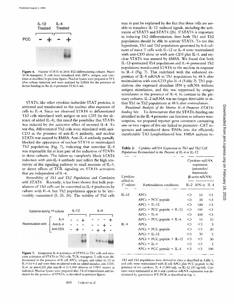
Figure 6. Nuclear STAT6 in 24-h Th2-differentiating cultures. Naive TCR-transgenic T cells were stimulated with APes, antigen, and cyto- kines as described in previous figures. Nuclear lysates were prepared at 24 h after culture initiation and were analyzed by EMSA for the presence of factors binding to the IL-4 promoter STAT6 site.
STAT6, like other cytokine-inducible STAT proteins, is activated and translocated to the nucleus after exposure of cells to IL-4. Since we observed STAT6 in differentiated Th2 cells stimulated with antigen or ant i -CD3 (in the ab- sence of added IL-4), this raised the possibility that STAT6 was induced by the autocrine effect of secreted IL-4. To test this, differentiated Th2 cells were stimulated with anti- CD3 in the presence of anti-IL-4 antibody, and nuclear STAT6 was assayed by EMSA. Anti-IL-4 antibody partially blocked the appearance of nuclear STAT6 in restimulated Th2 populations (Fig. 7), indicating that autocrine IL-4 was responsible for at least part of the induction of STAT6 in these cultures. The failure to completely block STAT6 induction with anti-IL-4 antibody may reflect the high sen- sitivity of this signaling pathway to small amounts of IL-4, or direct effects of T C R signaling on STAT6 activation that are independent of IL-4.
Reversibility of Till and Th2 Populations and Correlation with S T A T 6 . Recently, it has been shown that bulk pop- ulations of T h l cells can be converted to IL-4 producers by culture with IL-4, but Th2 populations appear to be irre- versibly committed (5, 25, 26). The stability of Th2 cells
Figure 7. Exogenous IL-4 activation of STAT6 in Thl cells and auto- crine activation of STAT6 in Th2 cells. TCR-transgenic T cells were dif- ferentiated in the presence of B cell APCs, antigen, and either IL-12 or IL-4 for 6 d and were then recultured with no added stimulus, anti-CD3, IL-4, or anti-CD3 plus anti-IL-4 (1/1,000 dilution of 11B11 ascites) as indicated. Nuclear lysates were prepared after 3 h ofrestimulation and an- alyzed for the presence of STAT6, as described in previous figures.
may in part be explained by the fact that these cells are un- able to transduce IL-12- induced signals, including the acti- vation of STAT3 and STAT4 (26). If STAT6 is important in inducing Th2 differentiation, then both T h l and Th2 populations should be able to activate STAT6. To test this hypothesis, T h l and Th2 populations generated by 6-d cul- tures of naive T cells with IL-12 or IL-4 were restimulated with ant i -CD3 alone or with ant i -CD3 plus IL-4, and nu- clear STAT6 was assessed by EMSA. We found that both IL-12-pretreated T h l populations and IL-4-pretreated Th2 populations translocated STAT6 to the nucleus in response to IL-4 (Fig. 7). This correlated with the enhanced ex- pression of IL-4 m R N A in T h l populations by 48 h after restimulation with ant i -CD3 plus IL-4 (Table 2), T h l pop- ulations also expressed abundant IFN-~/ m R N A without antigen stimulation, and this was suppressed by antigen stimulation in the presence of IL-4. In contrast to the pri- mary cultures, IL-2 m R N A was no longer detectable in ei- ther T h l or Th2 populations at 48 h after restimulation.
Functional Analysis of the Murine IL-4 Promoter S T A T6- binding Site. To demonstrate that the STAT6-binding site identified in the IL-4 promoter can function to enhance tran- scription, we prepared reporter gene constructs containing one or two copies of this site linked to promoter- CAT se- quences and introduced these DNAs into the efficiently transfectable TA3 lymphoblastoid line. EMSA analyses in-
Table 2. Cytokine mRNA Expression in Th I and Th2 Cell Populations Restimulated in the Presence of lL-4 or IL-12
Cytokine added in 1~ culture Restimulation conditions
Cytokine mRNA expression (attomoles/ femtomole
[3-actin mRNA)
IL-2 IFN-~/ IL-4
IL-12
IL-4
APCs <3 10 <3
APCs + PCC peptide <3 30 <3
APCs + IL-12 <3 100 <3
APCs + PCC peptide + IL-12 <3 100 <3
APCs + IL-4 <3 100 <3
APCs + PCC peptide + IL-4 <3 10 10
APCs <3 <3 3
APCs + PCC peptide <3 <3 30
APCs + IL-12 <3 30 3
APCs + PCC peptide + IL-12 <3 <3 30
APCs + IL-4 <3 <3 3
APCs + PCC peptide + IL-4 <3 <3 100
Thl and Th2 populations were derived in vitro as described in Table 1, and cells were restimulated with B cell APCs plus PCC peptide in the presence of no cytokine, IL-4 (1,000/ml), or IL-12 (10 ng/ml). Cul- tures were terminated at 48 h and cytokine mRNA expression was de- termined by quantitative RT-PCR as described in Fig. 1.
403 Lederer et al.
on February 18, 2015
jem.rupress.org
Dow
nloaded from
Published August 1, 1996

Table 3. Functional analysis of the IL-4 Gene 5" STA T6-binding Site
Plasmid Number of STAT6 sites Fold induction by IL-4
pBLCAT2 None 0.6
10g 1 1.0 14g 2 4.0
TA3 cells were transfected with the indicated CAT-reporter plasmids by electroporation. The cells were stimulated with 1,000 U/ml IL-4 at 24 h, and CAT activity was assayed by phase extraction liquid scintilla- tion (see Matenals and Methods) in cell lysates prepared at 48 h. Fold induction was calculated as counts per minute from IL-4-treated cells divided by Counts per minute from untreated cells. A representative experiment is shown. [L-4 induction of CAT activity in cells trans- (ected with the 14g plasmid was observed in three independent experi- ments.
dicated that IL-4 treatment leads to STAT6 activation in these cells (data not shown). The data in Table 3 indicate that the IL-4 promoter STAT6 site does support IL -4 - enhanced transcription o f a linked reporter gene.
Discuss ion
These studies were undertaken to characterize changes in transcriptional regulation o f cytokine genes during Th cell subset differentiation. The kinetics o f IL-4, IL-2, and I F N - y m R N A expression that we observed in differentiat- ing cultures o f C D 4 + T cells indicates temporally indepen- dent regulation o f the expression of different cytokine genes. IFN-~, transcripts appear early, within 24 h, in populations differentiating toward a Th l phenotype. In contrast, IL-4 transcripts do not appear until 48 h in cells differentiating towards a Th2 phenotype. IL-2 transcripts are detectable at 6 h and no difference is seen in the appearance o f IL-2 transcripts in primary cultures differentiating toward either Thl or Th2 phenotypes. This observation is not surprising since it is known that IL-2 is required for both differentia- tion pathways (27). Thus, there does not appear to be a sin- gle switch event during subset differentiation that affects multiple cytokine genes at the same time.
The only clear difference we observed in nuclear tran- scription factors between cultures o f differentiating CD4 + cells was the presence o f STAT6 in cells driven toward the Th2 phenotype. STAT6 has been identified as a protein that binds to D N A sequences found in the promoters o f IL-4-responsive genes (22, 24) A c D N A encoding a pro- tein that binds to the IL-4 response element in the human FcyP, l gene has been cloned and sequenced (14). The pre- dicted protein product o f this c D N A shares features with other STAT family members and it contains regions that can apparently bind to the cytoplasmic tail o f IL-4R. STAT6 activation is thought to involve association o f la- tent monomers with the IL-41:k, tyrosine phosphorylation, dimerization, and translocation to the nucleus, where the dinners bind to response elements in the promoters o f vari-
ous genes. Recently, it has been reported that mice with targeted disruptions o f the STAT6 gene are incapable o f mounting Th2 responses (28-30). These studies confirm the essential role o f IL-4 signaling in promoting Th2 differ- entiation, but they do not indicate how IL-4 may induce T cells to become competent at transcribing their own IL-4 genes. The identification o f a functional STAT6-binding site in the IL-4 promoter reported here, and the correlation o f STAT6 activation with Th2 differentiation, implicate that STAT6 is involved directly with IL-4 transcriptional activity during Th2 differentiation.
Gel shift assays using nuclear extracts from IL-4-treated THP-1 cells and a sequence from the human F c y R l gene demonstrated rapid indcution o f STAT6 activity, within 5 min, and a rapid decay, within 2 h (24). We have seen similar kinetics in mouse T cells, using an oligonucleotide probe with a murine IL-4 promoter sequence (Lederer, J.A., and A.H. Lichtman, unpublished observation). A longer half-life for nuclear STAT6 was reported in HepG2 cells using an interferon response factor 1 y activation site (GAS) probe, which binds a variety o f STAT family mem- bers (11). We found that STAT6 was present in the nuclei of IL-4-treated cells at 24 and 48 h as well as in restimu- lated Th2 cells derived from 6-d IL-4-treated cultures. Thus, STAT6 remains consistently activated before and during the onset o f IL-4 gene transcription in these cul- tures. The sustained presence o f nuclear STAT6 days after the initial exposure to IL-4, as well as the antigen-depen- dence o f STAT6 activation after 24 h, suggests that other signals besides those provided by the exogenously added IL-4 are responsible for maintaining STAT6 activation. Clearly, autocrine IL-4 production can induce STAT6 ac- tivation, as we observed in restimulated Th2 cells. Thus, it is possible that autocrine IL-4 can contribute to the tran- scriptional regulation o f the IL-4 gene after the effects o f exogenous sources o f IL-4 have waned.
A recently reported study with TCR-transgenic T cells has demonstrated that there is an extinction o f IL-12 sig- naling in populations differentiating toward the Th2 phe- notype (16). IL- 12-signaling pathways involve tyrosine phos- phorylation o f STAT3 and STAT4. T cells undergoing Th2 differentiation may fail to generate the active forms o f these transcription factors at the same time that they gener- ate active STAT6 in response to exogenous and autocrine IL-4. Thus, it is possible that an abundance o f STAT3 and STAT4 in the nucleus o f a T cell at the time of antigen stimulation favors commitment to a Th l phenotype, whereas a relative abundance o f STAT6 favors Th2 differentiation. Since the presence o f STAT6 correlates with Th2 differen- tiation, it is important to determine the functional signifi- cance of STAT6 binding to the IL-4 promoter. We are currently using mutational analyses and reporter gene trans- fections to formally test the role o f STAT6 in transcrip- tional activation o f the IL-4 gene. To date, there is no evi- dence indicating that STAT3 or STAT4 bind to regulatory sequences o f the IFN-3' or IL-2 genes and regulate their transcription.
The lack o f correlation o f subset differentiation with
404 Cytokine Gene Transcription during Thl and Th2 Differentiation
on February 18, 2015
jem.rupress.org
Dow
nloaded from
Published August 1, 1996
other transcription factors, including AP-1, N F - A T , and NFIL-2A, indicates that these factors may not be key de- terminants o f early commi tmen t towards a T h l or Th2 phenotype. This finding is consistent with the observation that IL-2 gene transcription is not modulated early during the T cell differentiation process and that upon restimula-
tion, both T h l and Th2 populations have the capacity to express IL-2. Perhaps multiple rounds o f stimulation by chronic antigen exposure lead to a more pronounced seg- regation o f IL-2 expression in Th cells similar to what has been observed in T cell clones and in T h l and Th2 popu- lations after multiple rounds o f restimulation (6, 31).
This work was supported by American Cancer Society grant IM666 (A.H. Lichtman), National Institutes of Health grant AI-25022 (A.K. Abbas), The Arthritis Foundation (J.A. Lederer), and U.S. Public Health Ser- vice Training Grant T32HL07627 (V.L. Perez).
Address correspondence to Dr. Andrew Licthman, Immunology Research Division, Department of Pathol- ogy, Brigham and Women's Hospital, 221 Longwood Avenue, Boston, MA 02115-5814.
Received for publication 5July 1995 and in revised form 9 May 1996.
References 1. Romagnani, S. 1994. Lymphocyte production by human T
cells in disease states. Annu. Rev. Immunol. 12:227-257. 2. Seder, R.A., and W.E. Paul 1994. Acquisition of lympho-
kine-producing phenotype by CD4+ T cells. Annu. Rev. Im- munol. 12:635--673.
3. Rocken, M., J.H. Saurat, and C. Hauser. 1992. A common precursor for CD4 § T cells producing IL-2 or IL-4. J. Immu- nol. 148:1031-1036.
4. Kamogawa, u L.A. Minasi, S.R. Carding, K. Bottomly, and R.A. Flave11. 1993. The relationship of IL-4- and IFN gamma- producing T cells studied by lineage ablation of IL-4-producing cells. Cell 75:985-995.
5. Perez, V.L., J.A. Lederer, A.H. Lichtman, and A.K. Abbas. 1995. Stability of Thl and Th2 populations. Int. Immunol. 7:1179-1186.
6. Lederer, J.A., J.S. Liou, M.D. Todd, L.H. Glimcher, and A.H. Lichtman. 1994. Regulation of cytokine gene expres- sion in T helper cell subsets..]. Immunol. 152:77-86.
7. Szabo, S.J., J.S. Gold, T.L. Murphy, and K.M. Murphy. 1993. Identification of cis-acting regulatory elements control- ling interleukin-4 gene expression in T cells: roles for NF-Y and NF-ATc. Mol. Cell. Biol. 13:4793-4805.
8. Todd, M.D., M.J. Grusby, J.A. Lederer, E. Lacy, A.H. Licht- man, and L.H. Glimcher. 1993. Transcription of the interleu- kin 4 gene is regulated by multiple promoter elements. J. Exp. Med. 177:1663-1674.
9. Crabtree, G.R. 1989. Contingent genetic regulatory events in T lymphocyte activation. Science (Wash. DC). 243:355-361.
10. Young, H.A., J.Y. Ghosh, J.A. Lederer, A.H. Lichtman, J.R. Gerard, L. Penix, C.B. Wilson, A.J. Melvin, M.E. McGim, D.B. Lewis, and D.D. Taub. 1994. Differentiation of T helper phenotypes by analysis of the methylation state of the IFN-y gene.J. Immunol. 153:3603-3610.
11. Penix, U, W.M. Weaver, Y. Pang, H.A. Young, and C.B. Wilson. 1993. Two essential regulatory elements in the hu- man interferon gamma promoter confer activation specific expression in T cells.J. Exp. Med 178:1483-1496.
12. Hsieh, C.-S., A.B. Heimburger, J.S. Gold, A. O'Garra, and K.M. Murphy. 1992. Differential regulation o fT helper phe- notype development by interleukins 4 and 10 in an ~x13 T cell receptor transgenic system. Proc. Natl. Acad. Sci. USA. 89:
405 Lederer et al.
6065--6069. 13. Seder, R.A., R. Gazzinelli, A. Sher, and W.E. Paul. 1993.
lnterleukin 12 acts directly on CD4 + T cells to enhance priming for interferon gamma production and diminishes in- terleukin 4 inhibition of such priming. Proc. Natl. Acad, Sci. USA. 90:10188-10192.
14. McKnight, A.J., V.L. Perez, C.M. Shea, G.S. Gray, and A.K. Abbas. 1994. Costimulator dependence oflymphokine secre- tion by naive and activated CD4 + T lymphokines from T cell receptor transgenic mice. J. lmmunol. 153:5220-5225.
15. Kaye, J., M.-L. Hsu, M.E. Sauron, C. Jameson, N.R.J. Gas- coigne, and S. Hedrick. 1989. Selective development of CD4 + T cells in transgenic mice expressing a class II MHC-restricted antigen receptor. Nature (Lond.). 341:746-748.
16. Karasuyama, H., and F. Melchers. 1988. Establishment of mouse cell lines which constitutively secrete large quantities of interleukin 2,3,4 or 5 using modified cDNA expression vectors. Eur.J. Immunol. 18:97-104.
17. Ausubel, F.M. 1992. Short Protocols in Molecular Biology. 2nd ed. Greene Publishing Associates and John Wiley & Sons, New York.
18. Platzer, C., G. Richter, K. Uberla, W. Muller, H. Blocker, T. Diamantstein, and T. Blankenstein. 1992. Analysis of cy- tokine mRNA levels in interleukin-4 transgenic mice by quantitative polymerase chain reaction. Eur. J. Immunol. 22: 1179-1184.
19. Luckow, B., and G. Schutz. 1987. CAT constructions with multiple unique restriction sites for the functional anlaysis of eukaryotic promoters and regulatory elements. Nucleic Acids Res. 15:5490-5493.
20. Glimcher, L.H., T. Hamano, R. Asofsky, D.H. Sachs, M. Pierres, L.E. Samelson, S.O. Sharrow, and W.E. Paul. 1983. IA mutant functional antigen-presenting cell lines. J. lmmu- nol. 130:2287-2294.
21. Chuvpilo, S., C. Schomberg, R. Gerwig, A. Heinfling, R. Reeves, F. Grummt, and E. Serfling. 1993. Multiple closely- linked NFAT/octamer and HMG ICY) binding sites are part of the interleukin-4 promoter. Nucleic Acids Res. 21:5694- 5704.
22. Schindler, C., H. Kashleva, A. Pernis, R. Pine, and P. Roth- man. 1994. STF-IL-4: a novel IL-4-induced signal transduc-
on February 18, 2015
jem.rupress.org
Dow
nloaded from
Published August 1, 1996
ing factor. EMBO (Eur. Mol. Biol. Organ. J. 13:1350-1356. 23. Hou, J., U. Schinder, W.J. Henzel, T.C. Ho, M. Brasseur,
and S.L. McKnight. 1994. An interleukin-4--induced tran- scription factor: IL-4 Stat. Science (Wash. DC). 265:1701- 1707.
24. Kotanides, H., and N.C. Reich. 1993. Requirement of ty- rosine phosphorylation for rapid activation of a DNA binding factor by IL-4. Science (Wash. DC). 262:1265-1267.
25. Mocci, S., and R.L. Coffman. 1995. Induction ofa Th2 pop- ulation from a polarized leishmania-specific Thl population by in vitro culture with IL-4. J. Immunol. 154:3779-3787.
26. Szabo, S.J., N.G. Jacobson, A.S. Dighe, U. Gubler, and K.M. Murphy. 1995. Developmental commitment to the Th2 lin- eage by extinction oflL-12 signaling. Immunity. 2:665-675.
27. Seder, R.A., R.N. Germain, P.S. Linsley, and W.E. Paul. 1994. CD28-mediated costimulation of interleukin-2 (IL-2) production plays a critical role in T cell priming for IL-4 and interferon 3' production.J. Exp. Med. 179:299-304.
28. Shimoda, K., J. Deursen, M.Y. Sangster, S. Sarawar, R.T. Carson, R.A. Tripp, C. Chu, F.W. Quelle, T. Nosaka, D.A.A. Vignali, P.C. Doherty, G. Grosveld, W.E. Paul, and J.N. Ihle. 1996. Nature (Lond.). Lack of IL-4-induced Th2 response and IgE class switching in mice with disrupted Stat6 gene. 380:630-633.
29. Takeda, K., T. Tanaka, W. Shi, M. Matsumoto, M. Minami, S.-I. Kashiwamura, K. Nakanishi, N. Yoshida, T. Kishimoto, and S. Akira. 1996. Essential role of Stat6 in IL-4 signalling. Nature (Lond.). 380:627-630.
30. Kaplan, M.H., U. Schnidler, S.T, Smiley, and M.J. Grusby. 1996. Stat6 is required for mediating responses to IL-4 and for the development of Th2 cells. Immunity. 4:313-319.
31. Bucy, R.P., A. Panoskaltsis-Mortari, G.-Q. Huang, J. Li, L. Karr, M. Ross, J.H. Russel, K.M. Murphy, and C.T. Weaver. 1994. Heterogeneity of single cell cytokine gene ex- pression in clonal T cell populations.J. Exp. Med. 180:1251- 1262.
406 Cytokine Gene Transcription during Thl and Th2 Differentiation
on February 18, 2015
jem.rupress.org
Dow
nloaded from
Published August 1, 1996
Copyright © 2022 FDOKUMEN