Individual T Helper Cells Have a Quantitative Cytokine Memory
Upload
independentCategory
view
0download
0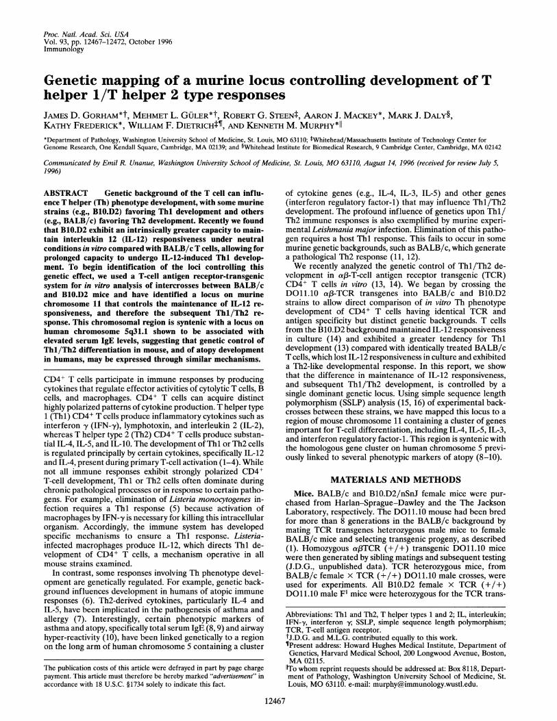
Proc. Natl. Acad. Sci. USAVol. 93, pp. 12467-12472, October 1996Immunology
Genetic mapping of a murine locus controlling development of Thelper 1/T helper 2 type responsesJAMES D. GORHAM*t, MEHMET L. GULER*t, ROBERT G. STEENt, AARON J. MACKEY*, MARK J. DALY§,KATHY FREDERICK*, WILLIAM F. DIETRICHfT, AND KENNETH M. MURPHY*II*Department of Pathology, Washington University School of Medicine, St. Louis, MO 63110; tWhitehead/Massachusetts Institute of Technology Center forGenome Research, One Kendall Square, Cambridge, MA 02139; and §Whitehead Institute for Biomedical Research, 9 Cambridge Center, Cambridge, MA 02142
Communicated by Emil R. Unanue, Washington University School of Medicine, St. Louis, MO 63110, August 14, 1996 (received for review July 5,1996)
ABSTRACT Genetic background of the T cell can influ-ence T helper (Th) phenotype development, with some murinestrains (e.g., B1O.D2) favoring Thl development and others(e.g., BALB/c) favoring Th2 development. Recently we foundthat B1O.D2 exhibit an intrinsically greater capacity to main-tain interleukin 12 (IL-12) responsiveness under neutralconditions in vitro compared with BALB/c T cells, allowing forprolonged capacity to undergo IL-12-induced Thl develop-ment. To begin identification of the loci controlling thisgenetic effect, we used a T-cell antigen receptor-transgenicsystem for in vitro analysis of intercrosses between BALB/cand B1O.D2 mice and have identified a locus on murinechromosome 11 that controls the maintenance of IL-12 re-sponsiveness, and therefore the subsequent Thl/Th2 re-sponse. This chromosomal region is syntenic with a locus onhuman chromosome 5q31.1 shown to be associated withelevated serum IgE levels, suggesting that genetic control ofThl/Th2 differentiation in mouse, and of atopy developmentin humans, may be expressed through similar mechanisms.
CD4+ T cells participate in immune responses by producingcytokines that regulate effector activities of cytolytic T cells, Bcells, and macrophages. CD4+ T cells can acquire distincthighly polarized patterns of cytokine production. T helper type1 (Thl) CD4+ T cells produce inflammatory cytokines such asinterferon y (IFN-,y), lymphotoxin, and interleukin 2 (IL-2),whereas T helper type 2 (Th2) CD4+ T cells produce substan-tial IL-4, IL-5, and IL-10. The development of Thl or Th2 cellsis regulated principally by certain cytokines, specifically IL-12and IL-4, present during primary T-cell activation (1-4). Whilenot all immune responses exhibit strongly polarized CD4+T-cell development, Thl or Th2 cells often dominate duringchronic pathological processes or in response to certain patho-gens. For example, elimination of Listeria monocytogenes in-fection requires a Thl response (5) because activation ofmacrophages by IFN-,y is necessary for killing this intracellularorganism. Accordingly, the immune system has developedspecific mechanisms to ensure a Thl response. Listeria-infected macrophages produce IL-12, which directs Thl de-velopment of CD4+ T cells, a mechanism operative in allmouse strains examined.
In contrast, some responses involving Th phenotype devel-opment are genetically regulated. For example, genetic back-ground influences development in humans of atopic immuneresponses (6). Th2-derived cytokines, particularly IL-4 andIL-5, have been implicated in the pathogenesis of asthma andallergy (7). Interestingly, certain phenotypic markers ofasthma and atopy, specifically total serum IgE (8, 9) and airwayhyper-reactivity (10), have been linked genetically to a regionon the long arm of human chromosome 5 containing a cluster
of cytokine genes (e.g., IL-4, IL-3, IL-5) and other genes(interferon regulatory factor-1) that may influence Thl/Th2development. The profound influence of genetics upon Thl/Th2 immune responses is also exemplified by murine experi-mental Leishmania major infection. Elimination of this patho-gen requires a host Thl response. This fails to occur in somemurine genetic backgrounds, such as BALB/c, which generatea pathological Th2 response (11, 12).We recently analyzed the genetic control of Thl/Th2 de-
velopment in a13-T-cell antigen receptor transgenic (TCR)CD4+ T cells in vitro (13, 14). We began by crossing theDO11.10 af3-TCR transgenes into BALB/c and B1O.D2strains to allow direct comparison of in vitro Th phenotypedevelopment of CD4+ T cells having identical TCR andantigen specificity but distinct genetic backgrounds. T cellsfrom the B1O.D2 background maintained IL-12 responsivenessin culture (14) and exhibited a greater tendency for Thldevelopment (13) compared with identically treated BALB/cT cells, which lost IL-12 responsiveness in culture and exhibiteda Th2-like developmental response. In this report, we showthat the difference in maintenance of IL-12 responsiveness,and subsequent Thl/Th2 development, is controlled by asingle dominant genetic locus. Using simple sequence lengthpolymorphism (SSLP) analysis (15, 16) of experimental back-crosses between these strains, we have mapped this locus to aregion of mouse chromosome 11 containing a cluster of genesimportant for T-cell differentiation, including IL-4, IL-5, IL-3,and interferon regulatory factor-1. This region is syntenic withthe homologous gene cluster on human chromosome 5 previ-ously linked to several phenotypic markers of atopy (8-10).
MATERIALS AND METHODS
Mice. BALB/c and BlO.D2/nSnJ female mice were pur-chased from Harlan-Sprague-Dawley and the The JacksonLaboratory, respectively. The DO11.10 mouse had been bredfor more than 8 generations in the BALB/c background bymating TCR transgenes heterozygous male mice to femaleBALB/c mice and selecting transgenic progeny, as described(1). Homozygous aJ3TCR (+/+) transgenic DO11.10 micewere then generated by sibling matings and subsequent testing(J.D.G., unpublished data). TCR heterozygous mice, fromBALB/c female x TCR (+/+) DO11.10 male crosses, wereused for experiments. All B1O.D2 female x TCR (+/+)DO11.10 male F1 mice were heterozygous for the TCR trans-
Abbreviations: Thl and Th2, T helper types 1 and 2; IL, interleukin;IFN--y, interferon y; SSLP, simple sequence length polymorphism;TCR, T-cell antigen receptor.tJ.D.G. and M.L.G. contributed equally to this work.SPresent address: Howard Hughes Medical Institute, Department ofGenetics, Harvard Medical School, 200 Longwood Avenue, Boston,MA 02115."To whom reprint requests should be addressed at: Box 8118, Depart-ment of Pathology, Washington University School of Medicine, St.Louis, MO 63110. e-mail: [email protected].
12467
The publication costs of this article were defrayed in part by page chargepayment. This article must therefore be hereby marked "advertisement" inaccordance with 18 U.S.C. §1734 solely to indicate this fact.
12468 Immunology: Gorhamet al.
genes. Male TCR transgenic F1 mice were crossed withBALB/c female mice to generate backcrossed mice (BC1), ofwhich one-half carried the TCR transgenes. Prior to use in invitro experiments, heterozygous TCR transgenic BC1 pups
were identified by anti-clonotypic KJ1-26 staining of periph-eral blood lymphocytes, as described (1). Mice were housed inpathogen clean facilities at the Washington University MedicalCenter.
Tissue Culture Media and Peptide. Cultures were main-tained in Iscove's modified Dulbecco's Eagle's medium sup-
plemented with 10% fetal calf serum (HyClone), 2 mML-glutamine, 0.1 mM sodium pyruvate, 0.1 mM MEM nones-
sential amino acids, 100 units/ml penicillin, 100,ug/ml strep-tomycin, and 50,uM 2-mercaptoethanol. The antigenic peptideOVA 323-339 was synthesized as described (2) and purified byHPLC.
Transgenic T-Cell Purification and Culture. CD4+ T cellsfrom TCR transgenes heterozygous mice were prepared fromperipheral lymph nodes of 5-7-week-old mice as described(13). T cells (1.25 x 10⁵ per well) were stimulated in 1-mlcultures with 0.3p,M OVA peptide presented by I-Ad express-
ing BALB/c splenocytes [2000 rad (1 rad = 0.01 Gy), 2.5 x 10⁶per well], and expanded at 72 hr 3-fold into fresh medium. Ondays 7-10, T cells were harvested, washed, and restimulated ina secondary stimulation at 1.25 x 10⁵ per well with antigen-presenting cell and 0.3,uM OVA peptide, without or withrecombinant murine IL-12 [5 units/ml; the kind gift of S. F.Wolf (Genetic Institute, Cambridge, MA)]. For assessment ofimmediate production of IFN-'y in response to IL-12 (assay I),antigen was presented by the I-Ad expressing B-cell hybridomaTA3 (10,000 rad, 2.5 x 10⁵ per well) (2), which does notproduce IL-12 or IFN-,y (14). Supernatants were collectedafter an additional 48 hr and IFN--y quantified by capture
ELISA (1). For assessment of the ability to develop the Thlphenotype in response to IL-12 in the secondary stimulation(assay II), BALB/c splenocytes were used as antigen-presenting cell and cells expanded at 72 hr 3-fold into freshmedium. On days 7-10, the T cells were harvested, washed, andrestimulated in a tertiary stimulation at 1.25 x 10⁵ per wellwith BALB/c splenocytes and antigen, without added IL-12.IFN--y was measured in 48-hr supernatants.
Genotyping. High molecular weight genomic DNA was
prepared from mouse tails and SSLP mapping analysis per-
formed using standard procedures (16). Markers, selected on
the basis of predicted polymorphism between the BALB/c andC57BL/6 strains, were first confirmed to be polymorphic forthe DO11.10 mouse (in the BALB/c background) and theB10.D2/nSnJ strain. We used a total of 197 polymorphic SSLPmarkers distributed over the entire genome, at about 10-centimorgan (cM) intervals. A list of these markers is availableupon request from the authors.
Linkage Analysis. MAPMAKER (17) was used to construct allthe genetic maps. To analyze IFN-y production as a quanti-tative trait, the data was transformed by taking the logarithmof the absolute phenotypic values, as is required for the analysis(18). The "penetrance scan," a newly developed mappingfunction for MAPMAKER/QTL, is ideally suited for phenotypeslike affectation status for a disease or trait. Trait values are
expressed as 0 or 1 (BALB/c- or F,-like) to optimize a set ofpenetrances (a probability of affectation, or trait = 1) for eachgenotypic class, yielding a logarithm of odds (lod) score
representing how much more likely the data are to have arisendue to the effect of a quantitative trait locus with the optimizedset of penetrances than to the effect of chance (under the nullhypothesis that the penetrances for each genotypic class are
equal and that no quantitative trait locus is present). The new
function has been implemented in the newest version ofMAPMAKER/QTL (not yet released). Test versions will soon beavailable via anonymous ftp to genome.wi.mit.edu.
RESULTSIn Vitro Analysis of Genetic Influences on Thl/Th2 Devel-
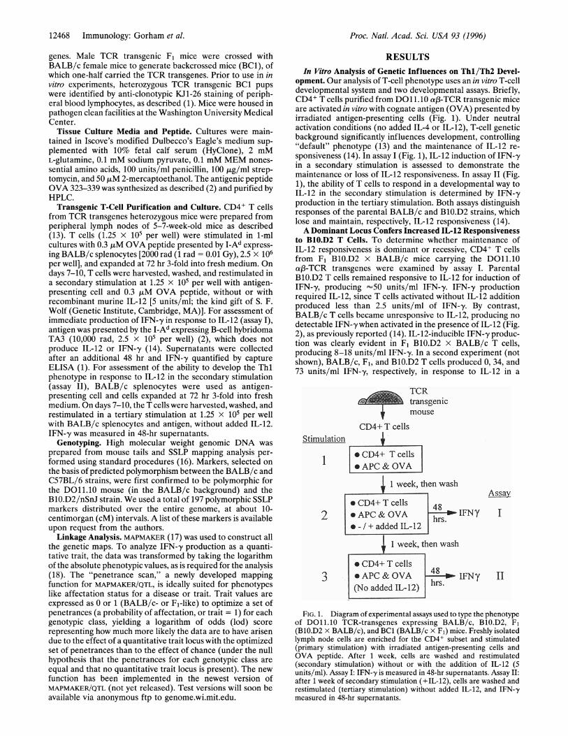
opment. Our analysis of T-cell phenotype uses an in vitro T-celldevelopmental system and two developmental assays. Briefly,CD4+ T cells purified from DO11.10 a13-TCR transgenic miceare activated in vitro with cognate antigen (OVA) presented byirradiated antigen-presenting cells (Fig. 1). Under neutralactivation conditions (no added IL-4 or IL-12), T-cell geneticbackground significantly influences development, controlling"default" phenotype (13) and the maintenance of IL-12 re-sponsiveness (14). In assay I (Fig. 1), IL-12 induction of IFN-,yin a secondary stimulation is assessed to demonstrate themaintenance or loss of IL-12 responsiveness. In assay II (Fig.1), the ability of T cells to respond in a developmental way toIL-12 in the secondary stimulation is determined by IFN-,yproduction in the tertiary stimulation. Both assays distinguishresponses of the parental BALB/c and B10.D2 strains, whichlose and maintain, respectively, IL-12 responsiveness (14).A Dominant Locus Confers Increased IL-12 Responsiveness
to B10.D2 T Cells. To determine whether maintenance ofIL-12 responsiveness is dominant or recessive, CD4+ T cellsfrom F1 B10.D2 x BALB/c mice carrying the D011.10af3-TCR transgenes were examined by assay I. ParentalB10.D2 T cells remained responsive to IL-12 for induction ofIFN-y, producing "50 units/ml IFN-,y. IFN-,y productionrequired IL-12, since T cells activated without IL-12 additionproduced less than 2.5 units/ml of IFN-,y. By contrast,BALB/c T cells became unresponsive to IL-12, producing no
detectable IFN-y when activated in the presence of IL-12 (Fig.2), as previously reported (14). IL-12-inducible IFN-,y produc-tion was clearly evident in F1 B10.D2 x BALB/c T cells,producing 8-18 units/ml IFN-,y. In a second experiment (notshown), BALB/c, F1, and B10.D2 T cells produced 0, 34, and73 units/ml IFN-,y, respectively, in response to IL-12 in a
TCR
transgenic
mouse
CD4+ T cellsStimulation
* CD4+ T cells* APC & OVA
1 week, then wash
* CD4+ T cells 482 *APC & OVA hrs IFNy
* - / + added IL- 12
1ee,ehe wash
AssaI
* CD4+ T cells3 *APC&OVA 48 l IFNy
(No added IL-12) hrs.
FIG. 1. Diagram of experimental assays used to type the phenotypeof D011.10 TCR-transgenes expressing BALB/c, B10.D2, F1(B1O.D2 x BALB/c), and BC1 (BALB/c x Fj) mice. Freshly isolatedlymph node cells are enriched for the CD4+ subset and stimulated(primary stimulation) with irradiated antigen-presenting cells andOVA peptide. After 1 week, cells are washed and restimulated(secondary stimulation) without or with the addition of IL-12 (5units/ml). Assay I: IFN--y is measured in 48-hr supernatants. Assay II:after 1 week of secondary stimulation (+IL-12), cells are washed andrestimulated (tertiary stimulation) without added IL-12, and IFN-'ymeasured in 48-hr supernatants.
Proc. Natl. Acad. Sci. USA 93 (1996)

Proc. Natl. Acad. Sci. USA 93 (1996) 12469
BALB/c Fl BlO.D2
FIG. 2. The maintenance of IL-12 responsiveness is dominant.TCR transgenic CD4+ cells from the BALB/c, B1O.D2, and F1 geneticbackgrounds (two mice each) were examined by assay I. IFN-y(units/ml) was measured in 48-hr supernatants without (open bars)and with (solid bars) added IL-12 (5 units/ml).
secondary stimulation. These results suggest that maintenanceof IL-12 responsiveness under these conditions is dominant orco-dominant.To determine the number of BMO.D2 loci controlling this
phenotype, we crossed F1 mice to the recessive BALB/cparental background to generate cohorts of backcrossed (BC1)progeny having the DO11.10 transgenes. CD4+ T cells from 18TCR transgenic BC1 mice, 3 BALB/c, and 3 F1 mice (Fig. 3)were examined by assay I. F1 T cells produced between 100 and280 units/ml IFN-,y, whereas BALB/c T cells produced fewerthan 25 units/ml (Fig. 3). Among 18 BC1 mice, 10 maintained(140-400 units/ml IFN-,y), and 8 lost (<20 units/ml IFN-'y)IL-12 responsiveness. In total, we analyzed CD4+ cells from 33BC1 in 2 experiments by assay I, with 16 and 17 demonstratingIL-12 unresponsiveness and responsiveness, respectively (Ta-ble 1). The approximate 1:1 distribution in this backcross
500-
BC1 (BALB/c x Fl)
400
S300 I
W*f * I I
suggests that a single genetic locus controls T-cell maintenanceof IL-12 responsiveness.Mapping of Tpml to Mouse Chromosome 11. To begin
identification of the responsible locus, we performed a genomewide mapping analysis of BC1 mice. Genomic DNA from eachof the 33 BC1 mice analyzed in assay I was typed with 197polymorphic SSLP markers (15, 16). Consistent with theprediction of a single control locus, only one region, the middleof chromosome 11, showed significant association of homozy-gotes to the BALB/c phenotype, and heterozygotes to the F,phenotype (Table 1). The marker DllMitlll (and linkedmarkers; see Fig. 5) showed the strongest association in thisanalysis (P value = 0.0012). In contrast, no other regionsshowed association, with most P values exceeding 0.05. Addi-tionally, this experiment allowed us to map the integration siteof the DO11.10 transgenes (derived in this backcross from theBALB/c background; see Materials and Methods), through theobservation of complete segregation distortion of markers ondistal chromosome 18 to BALB/c homozygous genotypes(data not shown).We repeated the above analysis in separate cohorts of BC1
mice using Assay II for Thl development (Fig. 1), rather thanimmediate cytokine production, to assign independent phe-notypes for IL-12 responsiveness (Fig. 4). Purified BC1, F1, orBALB/c CD4+ T cells were activated for 1 week, washed, andrestimulated (+5 units/ml IL-12), then washed and restimu-lated (no added IL-12). F1 T cells produced significantly higherlevels of IFN-,y upon tertiary stimulation than did BALB/c Tcells. From six independent experiments, the average IFN--yproduction by F1 T cells was 471 units/ml, (180-1067 units/ml),whereas that by BALB/c T cells was only 17 units/ml IFN--y(3-43 units/ml) (Fig. 4). Responses above 1 SD below themean F1 IFN--y production were classified as Fl-like, whereasthose below 1 SD above the mean BALB/c IFN-,y productionwere classified as BALB/c-like, defining upper limits ofBALB/c-like responses as 32 units/ml of IFN-,y, and lowerlimits of F1-like responses as 119 units/ml of IFN-,y. Of 53 BC1mice analyzed, 19 were assigned a BALB/c phenotype, 20 anF1 phenotype, and 14 could not be classified (Fig. 4).We analyzed the 39 classified BC1 mice with 197 SSLP
genetic markers covering the entire genome. Again, only one
I 7 F BALB/c
FIG. 3. The maintenance of IL-12 responsiveness is controlled by one genetic locus. TCR transgenic CD4+ cells from a cohort of 18 BC1, 3BALB/c, and 3 F1 mice were examined by assay I. BC1 samples exhibiting Fl-like (n = 10) or BALB/c-like (n = 8) phenotypes are grouped tothe left and right sides of the BC1 box, respectively.
Immunology: Gorham et al.

12470 Immunology: Gorham et al.
Table 1. Association of markers on chromosome 11 with phenotypically assigned BC1 samples
Assay I* Assay Ilt Assays I and II
Response to IL-12 Response to IL-12
Marker locus (cM)t - (16) + (17) x2§ P value - (19) + (20) x2 P value x2 P value
DllMit8O (10.0) 10T 13 3.7 0.16 10 14 1.2 0.54 5.5 0.063DllMitl73 (17.0) 12 14 8.7 0.013 10 16 3.2 0.20 12.7 0.0018DllMit235 (21.5) 12 15 11.0 0.0040 10 17 4.6 0.099 16.6 0.00024DllMitlll (28.0) 14 14 13.4 0.0012 15 18 16.1 0.00031 32.0 1.1 x 10⁻⁷DllMit274 (30.0) 13 14 10.9 0.0042 15 18 16.1 0.00031 29.4 4.1 x 10⁻⁷DllMitl77 (36.0) 13 13 8.8 0.012 14 14 5.8 0.055 16.1 0.00031DllMit320 (44.5) 13 13 8.8 0.012 13 14 4.3 0.12 14.2 0.00081
Numbers in parentheses indicate the number of mice assigned the phenotype for the assay.*IL-12-induced IFN-,y production after 48 hr of secondary stimulation (n = 33).tIL-12-induced Thl phenotype acquisition, assessed at tertiary stimulation (n = 39).tMarker position is according to The Jackson Laboratory Mouse Genome Database, http://www.informatics.jax.org/locus.html.§X2 was calculated using Yates' correction for continuity.IThe number of mice demonstrating concordance between genotype at the marker locus and assigned phenotype.
region was associated with IL-12 responsiveness for Thldevelopment, with markers DllMitlll (and linked markers)and D1 1Mit274 (and linked markers) each yielding P values of0.00031 (Table 1). Because both assay I and assay II indepen-dently established strong associations to markers in the middleof chromosome 11, we analyzed the combined data from all thecohorts (n = 72) observing a P value of 1.1 x 10⁻⁷ for markerDllMitlll (and linked markers) (Table 1). This significancelevel clearly indicates the presence of a locus that influencesT-cell phenotype response in these assays. Accordingly, we willrefer to this locus as Tpml (T-cell phenotype modifier 1).To better define the exact map position of Tpml, we
analyzed the data using MAPMAKER (17). The overall length ofthe chromosome maps was consistent with the expected size of
0 BALB/cO FlA BC1
Mean, Fl
-1 SD, Fl
+1 SD, BALB/cMean, BALB/ c
Lower limitof detect ion
FIG. 4. BALB/c and F1 mice differentially regulate the ability toestablish the Thl phenotype in response to IL-12 in a secondarystimulation: assignment of phenotype to a cohort of BC1 mice. TCRtransgenic CD4+ cells from the BALB/c, Fl, and BC1 backgroundswere examined by assay II. The mean and (mean -1 SD) for F1samples (O) and the mean and (mean + 1 SD) for BALB/c samples (0)are indicated as horizontal lines across the figure. The lower limit ofdetection of the IFN--y ELISA is 2.5 units/ml. Results from a total of53 BC1 samples (A) are shown at right. Data are compiled from sixseparate experiments.
the mouse genome (total autosomal length = 1558 cM),containing no isolated double crossovers to indicate genotyp-ing errors. In comparing segregation of the phenotypes withthe genotypic data, strong linkage of Tpml to individualmarkers on chromosome 11 was seen. The penetrance of thetrait as measured by these assays is not 100%, because it wasimpossible to find a map position for Tpml without a doublecrossover. Consequently, we mapped Tpml using a modifiedversion of MAPMAKER/QTL that optimizes for the penetrance ofa phenotype in a particular genotypic class, and calculates thelod score at each position of the map for that optimized set ofpenetrances (see Materials and Methods). According to thisanalysis, the maximum likelihood position of Tpml is betweenDllMitlll (and associated markers) and DllMit274 (andassociated markers) (Fig. 5), supported by a lod score of 6.5.Overall, the penetrance of the trait (percentage of animalswhose genotype at the locus is in concordance with its phe-notype) is 83% at the DllMitlll-DllMit274 interval.The incomplete penetrance of Tpml, when analyzed as a
qualitative, single gene locus, suggested that other genes maybe influencing the phenotype. Because we saw no evidence oflinkage to other regions of the genome in our previousanalyses, we attempted to analyze the data as a quantitativetrait using MAPMAKER/QTL. However, the combining of rawdata from the two sets of animals analyzed with differentassays precluded this effort. Analysis of the two sets of progenyseparately using MAPMAKER/QTL yielded no evidence of mod-ifying loci, separate from the locus on chromosome 11, toaccount for the partial penetrance of the phenotype (data notshown). Incomplete penetrance in these assays may derivefrom one or more unidentified modifier loci, or from exper-imental variation of the in vitro techniques.
DISCUSSIONRegulation of CD4+ T-cell responses influences several as-pects of host immunity, including resistance to pathogens,development of atopic and allergic states, and autoimmunetissue destruction (7, 20, 21). CD4+ T cells affect such diverseoutcomes through production of regulatory cytokines, withThl and Th2 type cells representing highly polarized patternsof cytokine production. Several factors can influence thedevelopment of Thl and Th2 responses. Particular emphasishas been placed on the direct regulatory roles of cytokines,principally IL-4 and IL-12, antigen dose and co-stimulatorymolecules (22). In addition, genetic factors can profoundlyinfluence the Thl/Th2 balance (14). Because the mechanismof differential Thi/Th2 responses in distinct genetic back-grounds is poorly understood, we sought to identify the
1000
E 100
zU-
10
1
Proc. Natl. Acad. Sci. USA 93 (1996)

Proc. Natl. Acad. Sci. USA 93 (1996) 12471
19
309, 239, 22, 191 235-
176, 139, 190 271%
87,314,310,273
240, 23,140, 111
131, 272, 313, 312
208,164,315,274 350
156, 339, 242
177r262
67-
258-
104 -
iTpml
GM-CSFIL3IL4IL5IL13IRF1ITKTcfl
| 5cM
FIG. 5. Genetic map of chromosome 11 indicating the position ofTpml. The map was constructed based on data from 90 BC1 mice usingMAPMAKER/EXP, version 3.0 (17). Some of the markers used are
indicated to the left, omitting the "DllMit" prefix. The maximumlikelihood position of Tpml, indicated by the shaded bar betweenDllMitlll (and associated markers) and DllMit274 (and associatedmarkers), is supported by a lod score of 6.5. The 95% confidenceinterval of Tpml [as defined by a drop in lod of .1.6 (19)] is betweenDllMitl89 and DllMit350, as indicated by the unshaded portions ofthe bar. Several genes with known or potential influence on Th celldifferentiation and located within 2 cM of DllMitlll are listedalphabetically in the box shown to the right. Precise genetic order is notimplied. GM-CSF, granulocyte-monocyte colony-stimulating factor;IRF-1, interferon regulatory factor-i; ITK, IL-2-inducible T-cell ki-nase; Tcf1, T-cell factor 1.
responsible loci controlling differential Thl/Th2 responsesbetween B10.D2 and BALB/c mice.Our analysis has focused on CD4+ T-cell development under
neutral in vitro conditions, in which B10.D2 T cells acquire a
more Thl-like phenotype compared with BALB/c T cells andare able to maintain responsiveness to IL-12. In vitro analysisof a single immune compartment (i.e., CD4+ T cell) is advan-tageous in the identification of controlling loci, in that itexcludes the potentially confounding contributions of otherfactors participating in complex immune responses in vivo. Thisapproach therefore would not identify all loci that may par-ticipate in a particular in vivo response, such as resistance to L.major, but allows identification of loci that regulate a compo-nent of the immune response, in this case Thl/Th2 develop-ment. This study shows that a single dominant, genetic locusfrom the B10.D2 background confers the maintenance ofIL-12 responsiveness to CD4+ T cells in vitro, thereby favoringThl development under neutral conditions. In BC1 micederived from an experimental backcross (BALB/c x F1),one-half of the mice retained the F1 phenotype, indicating theexistence of one controlling genetic locus. SSLP mappinganalysis using two different assays for IL-12 responsivenessmapped the controlling locus, named here Tpml, to the middleof chromosome 11, a genomic region containing numerouscandidate genes of immunologic importance (23, 24) (Fig. 5).The syntenic human genomic region 5q31.1 contains the
IL-4/IL-5 gene cluster and was shown recently to be linked tohigh serum IgE levels and airway hyperresponsiveness (8-10).Expression of atopy, the result of environmental and geneticfactors, is not inherited as a simple Mendelian trait (6, 25). Acommon feature of various atopic conditions, however, is theexpression of elevated levels of Th2 cytokines (26, 27). IL-4directly induces IgE isotype switching, sensitizes mast cells for
antigen-mediated degranulation, and is a mast cell growthfactor. IL-5 powerfully induces eosinophil growth, influx, anddegranulation. The BALB/c strain exhibits a greater expres-sion of markers of the Th2 phenotype than does the (BlO.D2related) C57BL/6 strain. For example, BALB/c mice producehigher serum IgE levels (28, 29), and develop greater antigen-induced airway hyperreactivity (30). Although the linkage ofTh2 responses in both mouse and human to this syntenicchromosomal region suggests a common pathway, identifica-tion of the responsible gene(s) is needed to understand themechanism(s) controlling these effects on Thl/Th2 develop-ment.
Genetic control of Thl/Th2 development participates insettings other than atopy. For example, preferential Thldevelopment in certain murine strains provides a basis, in part,for genetic resistance to L. major infection (31). Administra-tion of IL-12 to susceptible BALB/c mice at the time of L.major infection prevents Th2 development and generatescurative Thl-type CD4+ cells (32, 33). Interestingly, adminis-tration of IL-12 1 week after infection fails to generate Thlresponses or produce a cure in BALB/c mice (32), suggestinga temporal limit for response to IL-12. In BALB/c or C57BL/6mice, IL-12 mRNA is undetectable until 1 week after exper-imental L. major infection (34); by comparison, other patho-gens such as L. monocytogenes induce IL-12 very early (34). Invitro, developing BALB/c Th2 cells lose expression of func-tional IL-12 receptors as early as 3 days after induction (35)and B1O.D2 and BALB/c T cells developing under neutralconditions differentially maintain responsiveness to IL-12 (14).Thus, through rapid loss of IL-12 responsiveness, BALB/c Tcells may lose the ability to develop protective Thl responses,partly contributing to their susceptibility to this pathogen.Interestingly, in studies of seven recombinant inbred strains(C57BL/6 x BALB/c), a locus conferring susceptibility to L.major was mapped to a large region of mouse chromosome 11(36). More extensive mapping studies of in vivo L. majorsusceptibility are needed to confirm this genomic region as aresistance modifier.The Thi/Th2 balance influences the expression of certain
autoimmune disorders. In the murine non-obese diabeticmouse model, several genetic loci contribute to generate anautoimmune T-cell dependent destruction of the islet 3-cell(37), which can be mediated by Thl but not by Th2 cells (21).Interestingly, one of the non-obese diabetic loci, idd4, maps toa large region of chromosome 11 (37). In a transgenic modelof autoimmune diabetes, the B1O.D2 background conferredThl cytokine profiles and susceptibility to diabetes, whereasthe BALB/c background conferred Th2 cytokine profiles anddisease resistance (38). Experimental allergic encephalomy-elitis, a mouse model of the human demyelinating diseasemultiple sclerosis, is mediated by a Thl dependent autoim-mune process in the central nervous system (39, 40). Recentanalysis of backcrosses between high and low responder inbredmouse strains identified the middle portion of chromosome 11(peak x2 at IL4) as a potent modifier of disease severity (41).In the context of distinct genetic and environmental settings,therefore, allelic variants of Tpml may contribute to severalimmunologically mediated pathological states, includingpathogen susceptibility, atopy, and autoimmunity.The region ofmouse chromosome 11 identified in this study
contains genes for several cytokines that may influence Thl/Th2 development (Fig. 5). IL-4 is an obvious candidate,because it directly promotes Th2 development from naive Tcells (1,4) and antagonizes the action of IL-12 (1,2). A relativeoverproduction of cytokine by the BALB/c IL-4 locus wouldexplain the increased Th2 development observed in BALB/cmice. As reported (14), cocultured B1O.D2 and BALB/c Tcells nevertheless developed strain-dependent differences inIL-12 responsiveness, despite an equal extracellular environ-ment, suggesting that the underlying genetic difference is not
Immunology: Gorham et al.
71!80-

Proc. Natl. Acad. Sci. USA 93 (1996)
mediated through distinct levels of cytokine production. None-theless, IL-4 remains a viable candidate since the cocultureexperiments compared parental inbred strains BMO.D2 andBALB/c (14), whereas the present mapping studies compareF1 heterozygotes and BALB/c.An alternative model is that BMO.D2 and BALB/c CD4+ T
cells exhibit differential sensitivity to cytokines. For example,were BALB/c T cells relatively more sensitive to IL-4, orBlO.D2 T cells to IFN-,y, then differences in Thl/Th2 devel-opment and IL-12 responsiveness would result as observed.The linked chromosomal region contains several potentialgene candidates for which allelic variants could generateintrinsic differences between BALB/c and BMO.D2 CD4+ Tcells. Such candidates include interferon regulatory factor-1(42), T-cell factor 1 (43), and IL-2-inducible T-cell kinase (44),encoding signaling molecules or transcription factors ex-pressed in T cells.
We are indebted to Dr. Eric Green (Bethesda) for helping toestablish this collaborative effort. We also thank Drs. Robert Schreiberand Emil Unanue (St. Louis) for their generous gifts of ELISAreagents and Marnell Dickson (St. Louis) for facilitating cell counting.J.D.G. is a recipient of an Irvington Institute for ImmunologicalResearch Fellowship. This work was supported by National Institutesof Health Grants A131238, A134580, and DK20579.
1. Hsieh, C.-S., Heimberger, A. B., Gold, J. S., O'Garra, A. &Murphy, K. M. (1992) Proc. Natl. Acad. Sci. USA 89, 6065-6069.
2. Hsieh, C.-S., Macatonia, S. E., Tripp, C. S., Wolf, S. F., O'Garra,A. & Murphy, K. M. (1993) Science 260, 547-549.
3. Seder, R. A., Gazzinelli, R., Sher, A. & Paul, W. E. (1993) Proc.Natl. Acad. Sci. USA 90, 10188-10192.
4. Seder, R. A., Paul, W. E., Davis, M. M. & Fazekas de St. Groth,B. (1992) J. Exp. Med. 176, 1091-1098.
5. Magee, D. M. & Wing, E. J. (1988) J. Immunol. 141, 3203-3207.6. Meyers, D. A. & Bleecker, E. R. (1995) Am. J. Respir. Crit. Care
Med. 152, 411-413.7. Romagnani, S. (1994) Curr. Opin. Immunol. 6, 838-846.8. Marsh, D. G., Neely, J. D., Breazeale, D. R., Ghosh, B., Fre-
idhoff, L. R., Ehrlich-Kautzky, E., Schou, C., Krishnaswamy, G.& Beaty, T. H. (1994) Science 264, 1152-1156.
9. Meyers, D. A., Postma, D. S., Panhuysen, C. I., Xu, J., Amelung,P. J., Levitt, R. C. & Bleecker, E. R. (1994) Genomics 23, 464-470.
10. Postma, D. S., Bleecker, E. R., Amelung, P. J., Holroyd, K. J.,Xu, J., Panhuysen, C. I., Meyers, D. A. & Levitt, R. C. (1995)N. Engl. J. Med. 333, 894-900.
11. Heinzel, F. P., Sadick, M. D., Holaday, B. J., Coffman, R. L. &Locksley, R. M. (1989) J. Exp. Med. 169, 59-72.
12. Howard, J. G. (1986) Int. Rev. Exp. Pathol. 28, 79-116.13. Hsieh, C. S., Macatonia, S. E., O'Garra, A. & Murphy, K. M.
(1995) J. Exp. Med. 181, 713-721.14. Guler, M. L., Gorham, J. D., Hsieh, C., Mackey, A. J., Steen,
R. G., Dietrich, W. F. & Murphy, K. M. (1996) Science 271,984-987.
15. Dietrich, W. F., Miller, J. C., Steen, R. G., Merchant, M., Dam-ron, D., Nahf, R., Gross, A., Joyce, D. C., Wessel, M., Dredge,R. D., Marquis, A., Stein, L. D., Goodman, N., Page, D. C. &Lander, E. S. (1994) Nat. Genet. 7, 220-245.
16. Dietrich, W., Katz, H., Lincoln, S. E., Shin, H.-S., Friedman, J.,Dracopoli, N. C. & Lander, E. S. (1992) Genetics 131, 423-447.
17. Lander, E. S., Green, P., Abrahamson, J., Barlow, A., Daly, M. J.,Lincoln, S. E. & Newburg, L. (1987) Genomics 1, 174-181.
18. Paterson, A. H., Damon, S., Hewitt, J. D., Zamir, D., Rabinow-itch, H. D., Lincoln, S. E., Lander, E. S. & Tanksley, S. D. (1991)Genetics 127, 181-197.
19. Kruglyak, L. & Lander, E. S. (1995) Am. J. Hum. Genet. 56,1212-1223.
20. Sher, A., Gazzinelli, R. T., Oswald, I. P., Clerici, M., Kullberg,M., Pearce, E. J., Berzofsky, J. A., Mosmann, T. R., James, S. L.,Morse, H. C., III, & Shearer, G. M. (1992) Immunol. Rev. 127,183-204.
21. Katz, J. D., Benoist, C. & Mathis, D. (1995) Science 268, 1185-1188.
22. Seder, R. A. & Paul, W. E. (1994) Annu. Rev. Immunol. 12,635-673.
23. Buckwalter, M. S., Lossie, A. C., Scarlett, L. M. & Camper, S. A.(1992) Mammal. Genome 3, 604-607.
24. Wilson, S. D., Billings, P. R., D'Eustachio, P., Fournier, R. E.,Geissler, E., Lalley, P. A., Burd, P. R., Housman, D. E., Taylor,B. A. & Dorf, M. E. (1990) J. Exp. Med. 171, 1301-1314.
25. Lawrence, S., Beasley, R., Doull, I., Begishvili, B., Lampe, F.,Holgate, S. T. & Morton, N. E. (1994) Ann. Hum. Genet. 58,359-368.
26. Parronchi, P., Macchia, D., Piccinni, M. P., Biswas, P., Simonelli,C., Maggi, E., Ricci, M., Ansari, A. A. & Romagnani, S. (1991)Proc. Natl. Acad. Sci. USA 88, 4538-4542.
27. Robinson, D. S., Hamid, Q., Ying, S., Tsicopoulos, A., Barkans,J., Bentley, A. M., Corrigan, C., Durham, S. R. & Kay, A. B.(1992) N. Engl. J. Med. 326, 298-304.
28. Katz, D. H. & Tung, A. S. (1978) J. Immunol. 120, 2060-2067.29. Levine, B. B. & Vaz, N. M. (1970) Int. Arch. Allergy Appl.
Immunol. 39, 156-171.30. Corry, D. B., Folkesson, H. G., Warnock, M. L., Erle, D. J.,
Matthay, M. A., Wiener-Kronish, J. P. & Locksley, R. M. (1996)J. Exp. Med. 183, 109-117.
31. Heinzel, F. P., Sadick, M. D., Mutha, S. S. & Locksley, R. M.(1991) Proc. Natl. Acad. Sci. USA 88, 7011-7015.
32. Sypek, J. P., Chung, C. L., Mayor, S. E. H., Subramanyam, J. M.,Goldman, S. J., Sieburth, D. S., Wolf, S. F. & Schaub, R. G.(1993) J. Exp. Med. 177, 1797-1802.
33. Heinzel, F. P., Schoenhaut, D. S., Rerko, R. M., Rosser, L. E. &Gately, M. K. (1993) J. Exp. Med. 177, 1505-1509.
34. Reiner, S. L., Zheng, S., Wang, Z.-E., Stowring, L. & Locksley,R. M. (1994) J. Exp. Med. 179, 447-456.
35. Szabo, S. J., Jacobson, N. G., Dighe, A. S., Gubler, U. & Murphy,K. M. (1995) Immunity 2, 665-675.
36. Mock, B., Blackwell, J., Hilgers, J. & Potter, M. (1993) Eur.J. Immunogenet. 20, 335-348.
37. Todd, J. A., Aitman, T. J., Cornall, R. J., Ghosh, S., Hall, J. R.,Hearne, C. M., Knight, A. M., Love, J. M., McAleer, M. A.,Prins, J. B., Rodrigues, N., Lathrop, M., Pressey, A., DeLarato,N. H., Peterson, L. B. & Wicker, L. S. (1991) Nature (London)351, 542-547.
38. Scott, B., Liblau, R., Degermann, S., Marconi, L. A., Ogata, L.,Caton, A. J., McDevitt, H. 0. & Lo, D. (1994) Immunity 1, 73-82.
39. Chen, Y., Kuchroo, V. K., Inobe, J., Hafler, D. A. & Weiner,H. L. (1994) Science 265, 1237-1240.
40. Merrill, J. E., Kono, D. H., Clayton, J., Ando, D. G., Hinton,D. R. & Hofman, F. M. (1992) Proc. Natl. Acad. Sci. USA 89,574-578.
41. Baker, D., Rosenwasser, 0. A., O'Neill, J. K. & Turk, J. L. (1995)J. Immunol. 155, 4046-4051.
42. Miyamoto, M., Fujita, T., Kimura, Y., Maruyama, M., Harada,H., Sudo, Y., Miyata, T. & Taniguchi, T. (1988) Cell 54, 903-913.
43. Verbeek, S., Izon, D., Hofhuis, F., Robanus-Maandag, E., teRiele, H., van de Wetering, M., Oosterwegel, M., Wilson, A.,MacDonald, H. R. & Clevers, H. (1995) Nature (London) 374,70-74. -
44. Siliciano, J. D., Morrow, T. A. & Desiderio, S. V. (1992) Proc.Natl. Acad. Sci. USA 89, 11194-11198.
12472 Immunology: Gorham et al.
Copyright © 2022 FDOKUMEN