MHC-I-restricted melanoma antigen specific TCR-engineered human CD4+ T cells exhibit multifunctional...
15
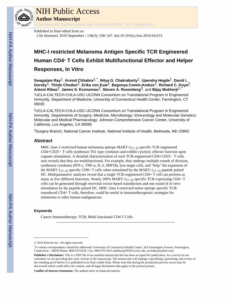
MHC-I restricted Melanoma Antigen Specific TCR Engineered Human CD4 + T Cells Exhibit Multifunctional Effector and Helper Responses, In Vitro Swagatam Ray 1 , Arvind Chhabra 1,* , Nitya G. Chakraborty 1 , Upendra Hegde 1 , David I. Dorsky 1 , Thinle Chodon 2 , Erika von Euw 2 , Begonya Comin-Anduix 2 , Richard C. Koya 2 , Antoni Ribas 2 , James S. Economou 2 , Steven A. Rosenberg 3 , and Bijay Mukherji 1,* 1 UCLA-CALTECH-CHLA-USC-UCONN Consortium on Translational Program in Engineered Immunity. Department of Medicine, University of Connecticut Health Center, Farmington, CT 06030 2 UCLA-CALTECH-CHLA-USC-UCONN Consortium on Translational Program in Engineered Immunity. Departments of Surgery; Medicine; Microbiology; Immunology and Molecular Genetics; Molecular and Medical Pharmacology; Johnson Comprehensive Cancer Center, University of California, Los Angeles, CA 90095 3 Surgery Branch, National Cancer Institute, National Institute of Health, Bethesda, MD 20892 Abstract MHC class 1-restricted human melanoma epitope MART-1 27–35 specific TCR engineered CD4+CD25− T cells synthesize Th1 type cytokines and exhibit cytolytic effector function upon cognate stimulation. A detailed characterization of such TCR-engineered CD4+CD25− T cells now reveals that they are multifunctional. For example, they undergo multiple rounds of division, synthesize cytokines (IFN-γ, TNF-α, IL-2, MIP1ß), lyse target cells, and “help” the expansion of the MART-1 27–35 specific CD8+ T cells when stimulated by the MART-1 27–35 peptide pulsed DC. Multiparametric analyses reveal that a single TCR-engineered CD4+ T cell can perform as many as five different functions. Nearly 100% MART-1 27–35 specific TCR expressing CD4+ T cells can be generated through retroviral vector-based transduction and one round of in vitro stimulation by the peptide pulsed DC. MHC class I-restricted tumor epitope specific TCR- transduced CD4+ T cells, therefore, could be useful in immunotherapeutic strategies for melanoma or other human malignancies. Keywords Cancer Immunotherapy; TCR; Multi functional CD4 T Cells © 2010 Elsevier Inc. All rights reserved * To whom correspondence should be addressed: University of Connecticut Health Center, 263 Farmington Avenue, Farmington, Connecticut – 06030 Phone: 860-679-4236 ; Fax: 860-679-1823 [email protected], [email protected]. Publisher's Disclaimer: This is a PDF file of an unedited manuscript that has been accepted for publication. As a service to our customers we are providing this early version of the manuscript. The manuscript will undergo copyediting, typesetting, and review of the resulting proof before it is published in its final citable form. Please note that during the production process errors may be discovered which could affect the content, and all legal disclaimers that apply to the journal pertain. Conflict of Interest Statement: The authors have no financial interest. NIH Public Access Author Manuscript Clin Immunol. Author manuscript; available in PMC 2011 September 1. Published in final edited form as: Clin Immunol. 2010 September ; 136(3): 338–347. doi:10.1016/j.clim.2010.04.013. NIH-PA Author Manuscript NIH-PA Author Manuscript NIH-PA Author Manuscript
Transcript of MHC-I-restricted melanoma antigen specific TCR-engineered human CD4+ T cells exhibit multifunctional...
MHC-I restricted Melanoma Antigen Specific TCR EngineeredHuman CD4+ T Cells Exhibit Multifunctional Effector and HelperResponses, In Vitro
Swagatam Ray1, Arvind Chhabra1,*, Nitya G. Chakraborty1, Upendra Hegde1, David I.Dorsky1, Thinle Chodon2, Erika von Euw2, Begonya Comin-Anduix2, Richard C. Koya2,Antoni Ribas2, James S. Economou2, Steven A. Rosenberg3, and Bijay Mukherji1,*1UCLA-CALTECH-CHLA-USC-UCONN Consortium on Translational Program in EngineeredImmunity. Department of Medicine, University of Connecticut Health Center, Farmington, CT060302UCLA-CALTECH-CHLA-USC-UCONN Consortium on Translational Program in EngineeredImmunity. Departments of Surgery; Medicine; Microbiology; Immunology and Molecular Genetics;Molecular and Medical Pharmacology; Johnson Comprehensive Cancer Center, University ofCalifornia, Los Angeles, CA 900953Surgery Branch, National Cancer Institute, National Institute of Health, Bethesda, MD 20892
AbstractMHC class 1-restricted human melanoma epitope MART-127–35 specific TCR engineeredCD4+CD25− T cells synthesize Th1 type cytokines and exhibit cytolytic effector function uponcognate stimulation. A detailed characterization of such TCR-engineered CD4+CD25− T cellsnow reveals that they are multifunctional. For example, they undergo multiple rounds of division,synthesize cytokines (IFN-γ, TNF-α, IL-2, MIP1ß), lyse target cells, and “help” the expansion ofthe MART-127–35 specific CD8+ T cells when stimulated by the MART-127–35 peptide pulsedDC. Multiparametric analyses reveal that a single TCR-engineered CD4+ T cell can perform asmany as five different functions. Nearly 100% MART-127–35 specific TCR expressing CD4+ Tcells can be generated through retroviral vector-based transduction and one round of in vitrostimulation by the peptide pulsed DC. MHC class I-restricted tumor epitope specific TCR-transduced CD4+ T cells, therefore, could be useful in immunotherapeutic strategies formelanoma or other human malignancies.
KeywordsCancer Immunotherapy; TCR; Multi functional CD4 T Cells
© 2010 Elsevier Inc. All rights reserved*To whom correspondence should be addressed: University of Connecticut Health Center, 263 Farmington Avenue, Farmington,Connecticut – 06030 Phone: 860-679-4236 ; Fax: 860-679-1823 [email protected], [email protected]'s Disclaimer: This is a PDF file of an unedited manuscript that has been accepted for publication. As a service to ourcustomers we are providing this early version of the manuscript. The manuscript will undergo copyediting, typesetting, and review ofthe resulting proof before it is published in its final citable form. Please note that during the production process errors may bediscovered which could affect the content, and all legal disclaimers that apply to the journal pertain.Conflict of Interest Statement: The authors have no financial interest.
NIH Public AccessAuthor ManuscriptClin Immunol. Author manuscript; available in PMC 2011 September 1.
Published in final edited form as:Clin Immunol. 2010 September ; 136(3): 338–347. doi:10.1016/j.clim.2010.04.013.
NIH
-PA Author Manuscript
NIH
-PA Author Manuscript
NIH
-PA Author Manuscript
IntroductionA role for CD4+ T cells in immune response in tumor immunity, in general, and in tumorimmunotherapy, in particular, is widely acknowledged [1;2;3]. However, CD4+ T cellsrecognize epitopes on MHC class II molecules and most solid tumor cells do not expressMHC class II molecules. These facts impose considerable difficulty in developing a way toincorporate them in therapeutic designs. As such, how to engage CD4+ T cells in tumorimmunotherapy has become a critical strategic issue. Lately, the use of CD4+ T cellstransduced to express the α/β chains of a relevant MHC class I-restricted tumor epitopespecific TCR has emerged as a mechanism to achieve that goal [4;5;6;7;8]. That such MHCclass-I restricted epitope specific TCR-engineered CD4+ T cells recognize the epitope onMHC class-I molecules with or without the requirement of co-receptor (i.e., CD8 molecules)engagement and exhibit effector function has been described by several groups in animalmodels [4;5] as well as in human systems [9;10;11;12]. We have shown that humanCD4+CD25− T cells transduced to express the α/β TCR chains specific for the Melan-A/MART-127–35 epitope, express type I cytokines and exhibit cytolytic function in a co-receptor-independent fashion [12]. Considering the potential of such tumor epitope specificTCR-engineered CD4+ T cells in human tumor immunotherapy, we undertook an extendedexamination of their biology. Here we show that the MHC class I-restricted MART-127–35epitope specific TCR-transduced CD4+CD25− T cells undergo multiple rounds of divisionand exhibit multifunctional effector function (synthesize IFN-γ, TNF-α, IL-2, mobilize lyticgranules and exhibit cytolytic effector function against melanoma targets) in a cognatemanner without requiring co-receptor-mediated additional signal. They also amplify theexpansion of the MART-127–35 epitope specific CD8+ T cells in an epitope specific CTLgeneration assay, in vitro.
Materials and MethodsStudy population, cell lines and reagents
The study population consisted of HLA-A2-positive healthy donors. The study wasapproved by Institutional Review Board and written consents were taken from allparticipants. Separation of CD4+CD25−, CD8+ T cells and culture conditions weredescribed previously [12]. T2 cells- a lymphoblastoid cell line with mutated TAP, was a giftfrom Peter Cresswell, Department of Immunobiology, Yale School of Medicine, NewHaven, Connecticut and the MART-1 negative melanoma line A375 cells engineered toexpress the MART-1 protein (A375-M) have been described before [13]. The melanoma celllines PT-M and M-202 were established from two HLA-A2.1 positive melanoma patients.MART-127–35 (M1) and MAGE-3271–279 (M3) peptides were purchased from NeoMPS(USA).
Retroviral vector constructionMART-127–35 epitope specific DMF5 TCR was isolated from a high avidity tumorinfiltrating lymphocyte (TIL) clone has been previously described [10]. The PG13packaging cell line [14] to produce DMF5 retrovirus was cultured in DMEM (Hyclone,USA) supplemented with 10% FBS. The cultures were grown to 70% confluence. Freshmedium was added and the supernatant containing the virus was harvested 16 h later.
Generation of MART-1 TCR transduced CD4+CD25− and CD8+ T cellsCD4+CD25− and CD8+ T cells were activated by plate-bound anti-CD3 (5μg) and anti-CD28 (1μg/ml) antibodies in presence of 100U/ml IL-2. After 48hr, the cells were infectedwith DMF5 TCR retrovirus containing supernatant in the presence of Retronectin (Takara,Japan) as per manufacturer protocol. 48hr after infection cells were stained with MART-1
Ray et al. Page 2
Clin Immunol. Author manuscript; available in PMC 2011 September 1.
NIH
-PA Author Manuscript
NIH
-PA Author Manuscript
NIH
-PA Author Manuscript

specific tetramer (Beckman Coulter, USA) and analyzed by flow cytometry in FACScalibur(BD Biosciences, USA). The transduced cells were rested in culture for 7–10 days withmedium changes and used for functional analyses or used after being frozen in FBS with10% DMSO. The viability of thawed cells was always in excess of 90% and no significantdifference in functional profile was observed with the frozen cells.
CFSE labeling and proliferation assaysCD4+CD25− and CD8+ T cells transduced with MART-1 specific DMF5-TCR werelabeled with 1mM CFSE (Invitrogen, USA) in PBS according to published protocol [15].Autologous DC were matured in LPS as published [12;13] and pulsed with 50mM ofpeptide. The CFSE labeled CD4F5 or CD8F5 cells were added to the peptide pulsed DC ortumor cell lines in 1:10 (Target:T cell) ratio. After 4 days, cells were analysed by flowcytometry.
Cytokine secretion assayT2 cells were pulsed with different dilutions of peptide in complete medium for 30min. 2 ×10⁴ peptide pulsed T2 were incubated overnight (approximately 16h) with 2 × 10⁵ CD4F5 orCD8F5 T cells in 500μl final volume in each well of a 48 well plate. The supernatants werecollected and cytokines (IL-2, IFNγ, TNFα, IL-10, IL-4 and TGF-β) were measured inELISA using an ELISA kit (R&D Systems, USA).
Intracellular cytokine stainingReagents for flow cytometric analysis (anti-CD107a-FITC, anti-IL-2-PerCP-Cy5.5, anti-IFNγ-V450, anti-TNFα-PE-Cy7, anti-MIP-1β-PE, anti-CD3-APC) were purchased from BDBiosciences (USA) and used as directed by the manufacturer. FoxP3 was stained using anti-FoxP3-APC and FoxP3 staining buffer from Miltenyei Biotec Inc (CA, USA) according tomanufacturer protocol. For intracellular cytokine assays, 5 × 10⁵ T cells were incubated with5 × 104 T2 pulsed with peptides. After 6 hr of incubation, the cells were stained andanalyzed with LSR-II flow-cytometer (BD Biosciences). The data were analyzed withFlowJo (TreeStar, USA) and polyfunctionality was assessed with PESTLE and SPICEsoftwares (provided by Mario Roederer, NIH, Bethesda, MD).
Cytotoxicity assay1 × 103 melanoma target cells were labeled with 51Cr and cytotoxicity was examined by 4hr 51Cr release assay [16] in presence of 50 fold excess K-562 as cold target competitors.
Co-culture to assess putative helper function of the TCR-engineered CD4+ T cellsPreviously published MART-127–35 specific in vitro CTL generation protocol [16] was usedas the basic CTL generation assay to assess helper function of the MART-127–35 specificTCR transduced CD4+CD25− T cells. Briefly, co-cultures were set up with freshly isolatedCD8+ T cell and DMF5 TCR engineered CD4+CD25− T cells (with mock transducedCD4+ T cells as control) against the MART-127–35 peptide loaded matured autologous DC.After 8–10 days, the numbers of MART-127–35 epitope specific CD8+ T cells weredetermined by tetramer staining.
ResultsMART-127–35 epitope specific TCR-engineered CD4+ T cells proliferate upon cognatestimulation
It is now quite clear that α/β TCRs that are restricted to MHC class I determinants, whenexpressed on to CD4+ T cells, are functional -- i.e., they send productive signal
Ray et al. Page 3
Clin Immunol. Author manuscript; available in PMC 2011 September 1.
NIH
-PA Author Manuscript
NIH
-PA Author Manuscript
NIH
-PA Author Manuscript
[9;12;17;18]. We have also previously shown that human CD4+CD25− T cells engineeredto express the MHC class-I-restricted MART-127–35 epitope specific TCRs synthesize type 1cytokines and exhibit cytolytic function [11;12]. Although MHC class I-restricted epitopespecific TCRs work on CD4+ T cells, such MHC class I-restricted TCR-engineered CD4+ Tcells are yet to be fully characterized especially in the context of their potential usefulness inhuman tumor immunotherapy. Using a different set of MART-127–35 epitope specific TCR,DMF5, with improved transduction efficiency [10], we undertook a more detailedcharacterization of the MART-127–35 epitope specific TCR-engineered CD4+CD25− Tcells, in vitro. We first examined the robustness of the transduction and the efficiency ingenerating large numbers of MHC class-1 restricted melanoma epitope specific TCR-expressing CD4+CD25− T cells. As shown in Fig.1, a large fraction of CD4+CD25− T cellscould be transduced with the DMF5 TCR retroviral vector to express the MART-127–35epitope specific TCR and a substantially larger fraction expressing the MART-127–35epitope specific TCR could be obtained after a single in vitro stimulation with theMART-127–35 peptide-loaded DC. A nearly homogenous population of MART-127–35epitope specific TCR expressing populations could be obtained after a second stimulation(data not shown). Fig. 2A shows the proliferative potential of the TCR transducedCD4+CD25− T cells in comparison with similarly engineered CD8+ T cells (Fig. 2B)assessed in CFSE dilution assay. As shown, the TCR-engineered CD4+ as well as CD8+ Tcells exhibit multiple rounds of division when they encounter the epitope on autologous DC(Figs. 2A & 2B). Of considerable interest, they also undergo multiple rounds of divisionwhen stimulated by melanoma cells (Fig. 2C).
MART-127–35> epitope specific TCR-engineered CD4+ T cells are multifunctionalWe then carried out a more extended functional characterization of DMF5 TCR-engineeredCD4+CD25− T cells and found that these TCR-engineered CD4+ T cells are multifunctional(Figs. 3 & 4). Figure 3A shows the cytokine synthetic ability (composite data) of the DMF5transduced CD4+ and CD8+ T cells from 5 different donors. As shown, they synthesizeIFN-γ, TNF-α, IL-2, MIP-1β. They also expose CD107a (Figs. 3B & 4A) upon cognatestimulation and exhibit cytolytic function (Fig. 4B). Of interest, intracytoplasmic stainingrevealed that a significant fraction of them exhibit more than one function – a sizeablefraction exhibiting multiple cytokine synthesis as well as exposing CD107a (Fig. 3B & C).Importantly, our analysis showed that a cell that makes IL-2 can also synthesize TNF-α andthat both IL-2- and IFN-γ-secreting cells expose CD107a, i.e., LAMP (Fig. 3B). Of furtherinterest, the DMF5 TCR-engineered CD4+ T cells do not express FoxP3 and TGF-β, evenwhen stimulated by the appropriate ligand (Fig 3D). The cytolytic function of the TCR-engineered CD4+CD25− T cells is not precisely comparable to that of the CD8+ T cellsagainst all target cells in chromium release assay (Fig 4B). Nonetheless when taken withtheir ability to expose CD107a (Fig 4A), as well as to lyse melanoma cells (Fig. 4B), thecytolytic function of the TCR-engineered CD4+ T cells is a distinct bonus. Given that themelanoma epitope specific TCR-engineered CD4+ T cells did not express CD8 co-receptorsand as CD8 co-receptor expression by TCR engineered CD4+ T cells have been shown toenhance effector function of MHC class-I TCR-engineered CD4+ T cells [11;19], it ispossible to argue that they might be made far better lytic effector cells if they were made toco-express CD8 molecules. Co-expressing CD8 molecules has not been a particularlydifficult task [11].
MART-127–35 epitope specific TCR-engineered CD4+ T cells provide “help” during theactivation/expansion of CD8+ T cells bearing endogenous MART-127–35 epitope specificTCR, in vitro
Given that CD4+ T cells provide important helper functions to CD8+ T cells in cell-mediated immune responses, we examined if the MART-127–35 epitope specific TCR
Ray et al. Page 4
Clin Immunol. Author manuscript; available in PMC 2011 September 1.
NIH
-PA Author Manuscript
NIH
-PA Author Manuscript
NIH
-PA Author Manuscript
expressing CD4+CD25− T cells could “help” the CD8+ T cells in CTL generation/expansion process. In our years of experience with in vitro CTL generation studies in thehuman melanoma model (i.e., co-culturing CD8+ T cells with peptide loaded matured DC),we have seldom found optimum functional activation and good expansion of the epitopespecific CD8+ T cells in the absence of exogenous cytokines such as IL-2 or IL-15.Accordingly, we addressed whether or not the CD4+CD25− T cells could help the CTLactivation/expansion process (CTL burst) in our in vitro CTL generation co-cultures in theabsence of exogenous IL-2. Figure 5A shows the result of a representative co-cultureexperiment demonstrating that the expansion of the epitope specific CD8+ T cell populationwas substantially amplified by the DMF5 epitope specific TCR expressing CD4+ T cells,when they were stimulated by the MART-127–35 peptide in the assay. Figure 5B shows thecomposite data from 5 separate experiments. The DMF5-transduced CD4+ T cells from 5HLA-A2 positive donors were co-cultured with the untransduced CD8+ T cells againstpeptide pulsed DC. The enhancements of MART-127–35 peptide specific CD8+ T cellexpansion (fold expansion of MART-127–35 tetramer positive populations), if any, was thendetermined by flowcytometry. As can be seen, the number of MART-127–35 epitope specificCD8+ T cells were considerably higher at all three CD4: CD8 ratios and that the increaseswere observed only when the co-cultures were performed with the MART-127–35 peptide.No enhancement was observed with the mock-transduced CD4+ T cells or with the DMF5-transduced CD4+ T cells in co-culture with CD8+ T cells against the HLA-A2 bindingcontrol MAGE-3271–279 peptide, M3. Of interest, they expanded the relevant TCRexpressing CD8+ T cells in the CTL generation cultures without any exogenous cytokinesupplementation. While it is possible that IL-2 synthesized by the CD4+ T cells provided thehelp, additional work will be needed to determine the underlying mechanism(s) behind thehelper effect of the TCR-transduced CD4+ T cells.
DiscussionActive specific immunotherapy have produced remarkable clinical responses in somemelanoma patients at times [20;21] and adoptive cell therapy with tumor-reactive tumorinfiltrating lymphocytes (TIL) have produced more impressive results in selected patientswith metastatic melanoma [22]. Melanoma reactive TIL, however, can be generated in abouthalf of melanoma patients and TIL can only rarely be obtained from patients with otherhistologic cancer types. New strategies are needed to improve the outcome of these types oftherapeutic approaches. Given that CD4+ T cells do positively influence the priming phaseof CD8+ CTL response generation as well as facilitate the CTL memory generation process[23;24;25], it is widely acknowledged that among other strategies that could be useful toimprove the results of tumor immunotherapy, figuring out a way that would simultaneouslyengage CD8+ as well as CD4+ T cells would be very helpful. Additionally, although theneed for “antigen specific” CD4+ T cells as “helper” cells is not yet clearly established, it isalso believed that a strategy that would engage CD4+ helper cells and CD8+ CTLrecognizing a relevant tumor epitope is likely to be highly effective. In this context, the datapresented here are noteworthy.
When collectively taken in the context to the question of how to engage cognate CD4+ Tcells -- along with CD8+ T effector cells -- in tumor immunotherapy, several interestingpoints emerge from the data. First, our data clearly show that a large number of melanomaepitope specific TCR-engineered CD4+CD25− T cells can be obtained through viral vector-based transduction and in vitro stimulation and that these MHC class I-restricted TCRtransduced CD4+CD25− T cells exhibit essentially all the effector functions that arenormally expected from CD8+ T cells. Additionally, the data show that they are truly“multifunctional” – i.e., they synthesize multiple cytokines, they are cytolytic, and theyprovide “help” during CTL burst in vitro CTL activation/expansion protocol. Given that the
Ray et al. Page 5
Clin Immunol. Author manuscript; available in PMC 2011 September 1.
NIH
-PA Author Manuscript
NIH
-PA Author Manuscript
NIH
-PA Author Manuscript
value of multifunctional effector function [26] is increasingly apparent in HIV immunity[27] as well as in tumor immunity [28], the multifunctional nature of the MHC class I-restricted TCR-engineered CD4+CD25– T cells would be useful in tumor immunotherapy.In this context, the robust proliferative potential of the MART-127–35 epitope specific TCR-engineered CD4+ T cells after encountering the epitope on DC or after encountering targetcells suggests that they could not only be made to proliferate through traditional DC andpeptide-based stimulation – a widely used practice in active specific immunization research- they could also be driven to undergo expansion at tumor sites – a highly desirable goal.Tumor antigen-driven proliferation and functions of CD4+ T cells at tumor sites could havemany positive effects. It should be, however, acknowledged that although our past study[12] have shown and present observations also show that the TCR-engineered CD4+ T cellsexhibit Th1 type phenotype, that CD4+ T cells engineered to express such MHC class I-restricted TCR (at least, a fraction of them) may have T regulatory (Treg) activities undercertain conditions. Additional work will be needed to determine condition(s) that wouldconsistently generate Th1 type CD4+ effector T cells and condition(s) that could lead to thegeneration of epitope specific Treg cells through TCR transgenesis of CD4+ T cells.
Our data showing that they amplify the burst size of the CD8+ CTL add another dimensionto their functional repertoire. Given that a role for CD4+ T cells in the CTL priming and inCTL memory generation is well established [23;24;25], the potential usefulness of suchMHC class I-restricted epitope specific CD4+ T cells in active specific immunization can beeasily envisioned. We have not addressed if the MART-127–35 epitope specific TCR-engineered CD4+ T cells could be made to transition into CD4+ memory T cells themselvesor if they could also “help” CD8+ T cells to become memory CTL, the possibility exists thatunder appropriate conditions (in vitro and/or in vivo) they could serve such purposes. Whileadditional work will be needed to figure out conditions that would make such MHC class I-restricted and epitope specific TCR-engineered CD4+ T cells to serve such purposes, thesuccessful use of this strategy for the induction of T cell memory response in an animaltumor model [4] and the relationship between multifunctional effector response and clinicalbenefit observed in a recent peptide and CTLA-4 antagonist-based immunization trial [28]strongly argue for moving these types of MHC class-1-restricted tumor epitope specificTCR-engineered CD4+ T cells to the clinic. Although our previous study [12] and this studyhave been carried out with a single (but two different) set of tumor epitope specific TCRs,the remarkably concordant results do not seem to be a reflection of a particular set of TCR.Of note, MHC class-I-restricted α/β TCRs specific for other human tumor-associatedepitopes have also been found to function when grafted onto CD4+ T cells [9;12;17;18].
The concept of adoptive immunotherapy with TCR-gene transduced T cells has moved toclinical trial [8]. While it is too early to assess its overall effectiveness, this type of cancerimmunotherapeutic approach would have limitations. Virtually all the well recognizedreasons underlying failures of T cell-based immunotherapy [29] are likely to frustrateadoptive immunotherapy with TCR-transduced T cells. Nonetheless, it should be pointedout, that most approaches to adoptive immunotherapy or active specific immunotherapy forthat matter have, so far, been based on strategies designed to harnessing the effectorfunctions of CD8+ CTL as most human cancer cells express only MHC class I molecule-associated epitopes. Two groups of investigators have recently shown that MHC class II-restricted epitope specific TCR-engineered CD4+ T cells exhibit superb anti-tumor effectorresponses including cytolytic function and that they could be activated and expandedthrough immunization, in vivo, in animal models [30;31]. The data presented here show apotentially novel way to directing MHC class I associated antigen specific CD4+CD25– Tcells – as effector cells and as “helper” cells --to human tumors that do not express MHCclass II molecules [32;33]. Accordingly, these types of tumor epitope specific and MHCclass I-restricted CD4+CD25− T cells -adoptively transferred in patients followed by
Ray et al. Page 6
Clin Immunol. Author manuscript; available in PMC 2011 September 1.
NIH
-PA Author Manuscript
NIH
-PA Author Manuscript
NIH
-PA Author Manuscript
immunization (to expand both CD4+ T cells and native CD8+ T cells bearing endogenousTCR for the given epitope) or adoptively transferred with similar TCR engineered CD8+ Tcells followed by immunization – could be novel and valuable strategies in active specific oradoptive T cell-based tumor immunotherapy.
AcknowledgmentsWe thank Dr Mario Roederer (Vaccine Research Center, NIAID, NIH, Bethesda, MD) for providingmultifunctional FACS analysis software.
The work was supported by PHS grants CA 83130 (BM), CA 88059 (BM), CA 129816 (JSE) and grants from theDowling Foundation (BM), Samuel Waxman Cancer Research Foundation, W.M. Keck Foundation, Joy and JerryMonkarsh Fund (JSE for UCLA-CALTECH-CHLA-USC-UCONN Consortium on Translational Program inEngineered Immunity), Breast Cancer Alliance, Connecticut (AC) and MO 1RR06192 from GCRC, UCHC.
References[1]. Pardoll DM, Topalian SL. The role of CD4+ T cell responses in antitumor immunity. Curr Opin
Immunol 1998;10:588–94. [PubMed: 9794842][2]. Toes RE, Ossendorp F, Offringa R, Melief CJ. CD4 T cells and their role in antitumor immune
responses. J Exp Med 1999;189:753–6. [PubMed: 10049938][3]. Ostrand-Rosenberg S. CD4+ T lymphocytes: a critical component of antitumor immunity. Cancer
Invest 2005;23:413–9. [PubMed: 16193641][4]. Morris EC, Tsallios A, Bendle GM, Xue SA, Stauss HJ. A critical role of T cell antigen receptor-
transduced MHC class I-restricted helper T cells in tumor protection. Proc Natl Acad Sci U S A2005;102:7934–9. [PubMed: 15908507]
[5]. Kessels HW, Schepers K, van den Boom MD, Topham DJ, Schumacher TN. Generation of T cellhelp through a MHC class I-restricted TCR. J Immunol 2006;177:976–82. [PubMed: 16818753]
[6]. Clay TM, Custer MC, Sachs J, Hwu P, Rosenberg SA, Nishimura MI. Efficient transfer of a tumorantigen-reactive TCR to human peripheral blood lymphocytes confers anti-tumor reactivity. JImmunol 1999;163:507–13. [PubMed: 10384155]
[7]. Morgan RA, Dudley ME, Yu YY, Zheng Z, Robbins PF, Theoret MR, Wunderlich JR, HughesMS, Restifo NP, Rosenberg SA. High efficiency TCR gene transfer into primary humanlymphocytes affords avid recognition of melanoma tumor antigen glycoprotein 100 and does notalter the recognition of autologous melanoma antigens. J Immunol 2003;171:3287–95. [PubMed:12960359]
[8]. Morgan RA, Dudley ME, Wunderlich JR, Hughes MS, Yang JC, Sherry RM, Royal RE, TopalianSL, Kammula US, Restifo NP, Zheng Z, Nahvi A, de Vries CR, Rogers-Freezer LJ, MavroukakisSA, Rosenberg SA. Cancer regression in patients after transfer of genetically engineeredlymphocytes. Science 2006;314:126–9. [PubMed: 16946036]
[9]. Roszkowski JJ, Lyons GE, Kast WM, Yee C, Van Besien K, Nishimura MI. Simultaneousgeneration of CD8+ and CD4+ melanoma-reactive T cells by retroviral-mediated transfer of asingle T-cell receptor. Cancer Res 2005;65:1570–6. [PubMed: 15735047]
[10]. Johnson LA, Heemskerk B, Powell DJ Jr. Cohen CJ, Morgan RA, Dudley ME, Robbins PF,Rosenberg SA. Gene transfer of tumor-reactive TCR confers both high avidity and tumorreactivity to nonreactive peripheral blood mononuclear cells and tumor-infiltrating lymphocytes.J Immunol 2006;177:6548–59. [PubMed: 17056587]
[11]. Willemsen RA, Sebestyen Z, Ronteltap C, Berrevoets C, Drexhage J, Debets R. CD8 alphacoreceptor to improve TCR gene transfer to treat melanoma: down-regulation of tumor-specificproduction of IL-4, IL-5, and IL-10. J Immunol 2006;177:991–8. [PubMed: 16818755]
[12]. Chhabra A, Yang L, Wang P, Comin-Anduix B, Das R, Chakraborty NG, Ray S, Mehrotra S,Yang H, Hardee CL, Hollis R, Dorsky DI, Koya R, Kohn DB, Ribas A, Economou JS, BaltimoreD, Mukherji B. CD4+CD25- T Cells Transduced to Express MHC Class I-Restricted Epitope-Specific TCR Synthesize Th1 Cytokines and Exhibit MHC Class I-Restricted Cytolytic EffectorFunction in a Human Melanoma Model. J Immunol 2008;181:1063–70. [PubMed: 18606658]
Ray et al. Page 7
Clin Immunol. Author manuscript; available in PMC 2011 September 1.
NIH
-PA Author Manuscript
NIH
-PA Author Manuscript
NIH
-PA Author Manuscript
[13]. Chhabra A, Mehrotra S, Chakraborty NG, Mukherji B, Dorsky DI. Cross-presentation of ahuman tumor antigen delivered to dendritic cells by HSV VP22-mediated protein translocation.Eur J Immunol 2004;34:2824–33. [PubMed: 15368298]
[14]. Johnson LA, Morgan RA, Dudley ME, Cassard L, Yang JC, Hughes MS, Kammula US, RoyalRE, Sherry RM, Wunderlich JR, Lee CC, Restifo NP, Schwarz SL, Cogdill AP, Bishop RJ, KimH, Brewer CC, Rudy SF, Vanwaes C, Davis JL, Mathur A, Ripley RT, Nathan DA, LaurencotCM, Rosenberg SA. Gene therapy with human and mouse T cell receptors mediates cancerregression and targets normal tissues expressing cognate antigen. Blood. 2009
[15]. Lyons AB. Analysing cell division in vivo and in vitro using flow cytometric measurement ofCFSE dye dilution. J Immunol Methods 2000;243:147–54. [PubMed: 10986412]
[16]. Mehrotra S, Stevens R, Zengou R, Chakraborty NG, Butterfield LH, Economou JS, Dorsky DI,Mukherji B. Regulation of melanoma epitope-specific cytolytic T lymphocyte response byimmature and activated dendritic cells, in vitro. Cancer Res 2003;63:5607–14. [PubMed:14500402]
[17]. Tsuji T, Yasukawa M, Matsuzaki J, Ohkuri T, Chamoto K, Wakita D, Azuma T, Niiya H,Miyoshi H, Kuzushima K, Oka Y, Sugiyama H, Ikeda H, Nishimura T. Generation of tumor-specific, HLA class I-restricted human Th1 and Tc1 cells by cell engineering with tumor peptide-specific T-cell receptor genes. Blood 2005;106:470–6. [PubMed: 15790789]
[18]. Willemsen R, Ronteltap C, Heuveling M, Debets R, Bolhuis R. Redirecting human CD4+ Tlymphocytes to the MHC class I-restricted melanoma antigen MAGE-A1 by TCR alphabeta genetransfer requires CD8alpha. Gene Ther 2005;12:140–6. [PubMed: 15496961]
[19]. Lyons GE, Moore T, Brasic N, Li M, Roszkowski JJ, Nishimura MI. Influence of human CD8 onantigen recognition by T-cell receptor-transduced cells. Cancer Res 2006;66:11455–61.[PubMed: 17145893]
[20]. Marchand M, Weynants P, Rankin E, Arienti F, Belli F, Parmiani G, Cascinelli N, Bourlond A,Vanwijck R, Humblet Y, et al. Tumor regression responses in melanoma patients treated with apeptide encoded by gene MAGE-3. Int J Cancer 1995;63:883–5. [PubMed: 8847150]
[21]. Nestle FO, Alijagic S, Gilliet M, Sun Y, Grabbe S, Dummer R, Burg G, Schadendorf D.Vaccination of melanoma patients with peptide- or tumor lysate-pulsed dendritic cells. Nat Med1998;4:328–32. [PubMed: 9500607]
[22]. Dudley ME, Yang JC, Sherry R, Hughes MS, Royal R, Kammula U, Robbins PF, Huang J, CitrinDE, Leitman SF, Wunderlich J, Restifo NP, Thomasian A, Downey SG, Smith FO, Klapper J,Morton K, Laurencot C, White DE, Rosenberg SA. Adoptive cell therapy for patients withmetastatic melanoma: evaluation of intensive myeloablative chemoradiation preparativeregimens. J Clin Oncol 2008;26:5233–9. [PubMed: 18809613]
[23]. Keene JA, Forman J. Helper activity is required for the in vivo generation of cytotoxic Tlymphocytes. J Exp Med 1982;155:768–82. [PubMed: 6801178]
[24]. Janssen EM, Lemmens EE, Wolfe T, Christen U, von Herrath MG, Schoenberger SP. CD4+ Tcells are required for secondary expansion and memory in CD8+ T lymphocytes. Nature2003;421:852–6. [PubMed: 12594515]
[25]. Bevan MJ. Helping the CD8(+) T-cell response. Nat Rev Immunol 2004;4:595–602. [PubMed:15286726]
[26]. Seder RA, Darrah PA, Roederer M. T-cell quality in memory and protection: implications forvaccine design. Nat Rev Immunol 2008;8:247–58. [PubMed: 18323851]
[27]. Duvall MG, Precopio ML, Ambrozak DA, Jaye A, McMichael AJ, Whittle HC, Roederer M,Rowland-Jones SL, Koup RA. Polyfunctional T cell responses are a hallmark of HIV-2 infection.Eur J Immunol 2008;38:350–63. [PubMed: 18200635]
[28]. Yuan J, Gnjatic S, Li H, Powel S, Gallardo HF, Ritter E, Ku GY, Jungbluth AA, Segal NH,Rasalan TS, Manukian G, Xu Y, Roman RA, Terzulli SL, Heywood M, Pogoriler E, Ritter G,Old LJ, Allison JP, Wolchok JD. CTLA-4 blockade enhances polyfunctional NY-ESO-1 specificT cell responses in metastatic melanoma patients with clinical benefit. Proc Natl Acad Sci U S A2008;105:20410–5. [PubMed: 19074257]
[29]. Campoli M, Ferrone S. T-cell-based immunotherapy of melanoma: what have we learned andhow can we improve? Expert Rev Vaccines 2004;3:171–87. [PubMed: 15056043]
Ray et al. Page 8
Clin Immunol. Author manuscript; available in PMC 2011 September 1.
NIH
-PA Author Manuscript
NIH
-PA Author Manuscript
NIH
-PA Author Manuscript
[30]. Xie Y, Akpinarli A, Maris C, Hipkiss EL, Lane M, Kwon EK, Muranski P, Restifo NP, AntonyPA. Naive tumor-specific CD4+ T cells differentiated in vivo eradicate established melanoma. JExp Med 207:651–67. [PubMed: 20156973]
[31]. Quezada SA, Simpson TR, Peggs KS, Merghoub T, Vider J, Fan X, Blasberg R, Yagita H,Muranski P, Antony PA, Restifo NP, Allison JP. Tumor-reactive CD4+ T cells develop cytotoxicactivity and eradicate large established melanoma after transfer into lymphopenic hosts. J ExpMed 207:637–50. [PubMed: 20156971]
[32]. Ray S, Chhabra A, Mehrotra S, Chakraborty NG, Ribas A, Economou J, Mukherji B. Obstaclesto and opportunities for more effective peptide-based therapeutic immunization in humanmelanoma. Clin Dermatol 2009;27:603–13. [PubMed: 19880048]
[33]. Chhabra A. MHC class I TCR engineered anti-tumor CD4 T cells: implications for cancerimmunotherapy. Endocr Metab Immune Disord Drug Targets 2009;9:344–52. [PubMed:19807670]
Ray et al. Page 9
Clin Immunol. Author manuscript; available in PMC 2011 September 1.
NIH
-PA Author Manuscript
NIH
-PA Author Manuscript
NIH
-PA Author Manuscript
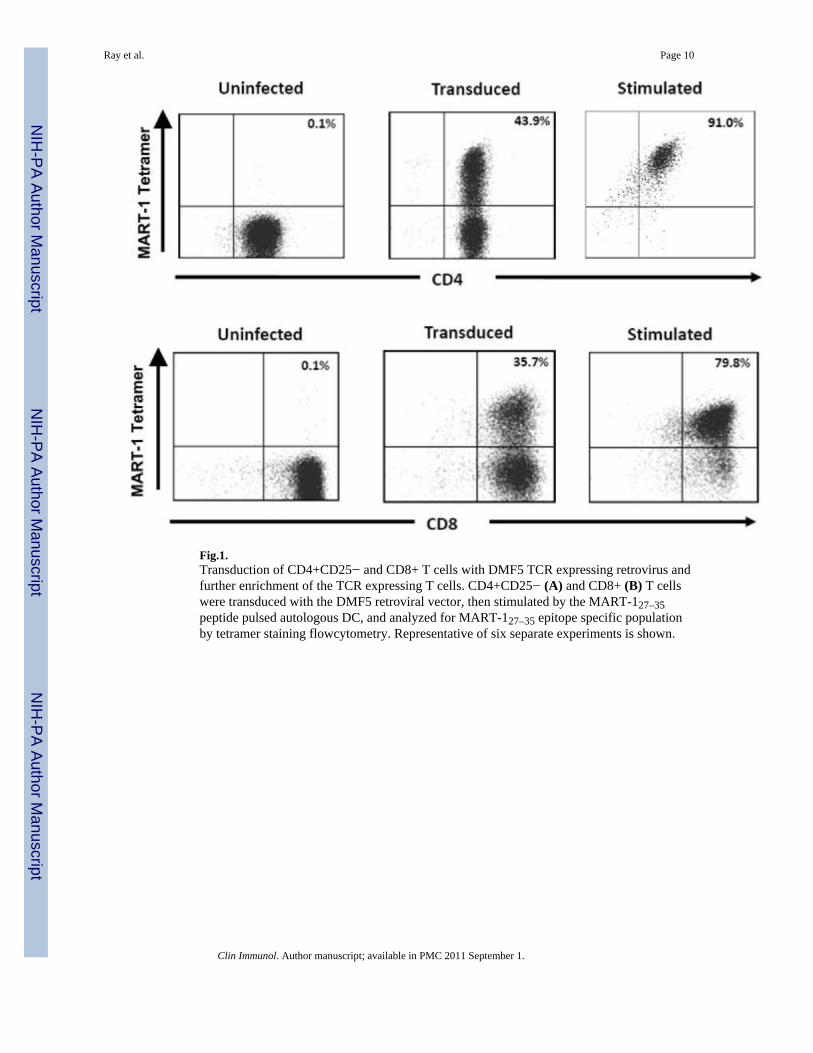
Fig.1.Transduction of CD4+CD25− and CD8+ T cells with DMF5 TCR expressing retrovirus andfurther enrichment of the TCR expressing T cells. CD4+CD25− (A) and CD8+ (B) T cellswere transduced with the DMF5 retroviral vector, then stimulated by the MART-127–35peptide pulsed autologous DC, and analyzed for MART-127–35 epitope specific populationby tetramer staining flowcytometry. Representative of six separate experiments is shown.
Ray et al. Page 10
Clin Immunol. Author manuscript; available in PMC 2011 September 1.
NIH
-PA Author Manuscript
NIH
-PA Author Manuscript
NIH
-PA Author Manuscript
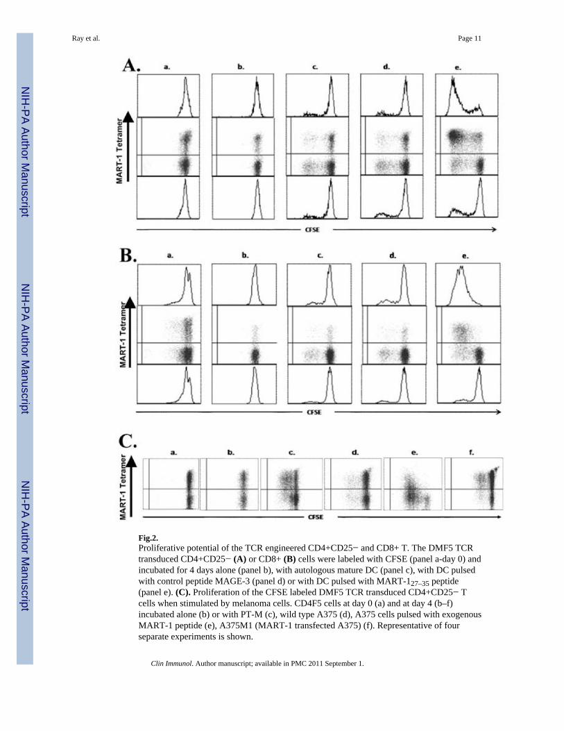
Fig.2.Proliferative potential of the TCR engineered CD4+CD25− and CD8+ T. The DMF5 TCRtransduced CD4+CD25− (A) or CD8+ (B) cells were labeled with CFSE (panel a-day 0) andincubated for 4 days alone (panel b), with autologous mature DC (panel c), with DC pulsedwith control peptide MAGE-3 (panel d) or with DC pulsed with MART-127–35 peptide(panel e). (C). Proliferation of the CFSE labeled DMF5 TCR transduced CD4+CD25− Tcells when stimulated by melanoma cells. CD4F5 cells at day 0 (a) and at day 4 (b–f)incubated alone (b) or with PT-M (c), wild type A375 (d), A375 cells pulsed with exogenousMART-1 peptide (e), A375M1 (MART-1 transfected A375) (f). Representative of fourseparate experiments is shown.
Ray et al. Page 11
Clin Immunol. Author manuscript; available in PMC 2011 September 1.
NIH
-PA Author Manuscript
NIH
-PA Author Manuscript
NIH
-PA Author Manuscript
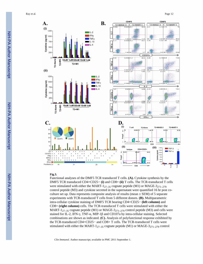
Fig.3.Functional analyses of the DMF5 TCR transduced T cells. (A). Cytokine synthesis by theDMF5 TCR transduced CD4+CD25− (i) and CD8+ (ii) T cells. The TCR-transduced T cellswere stimulated with either the MART-127–35 cognate peptide (M1) or MAGE-3271–279control peptide (M3) and cytokine secreted in the supernatant were quantified 16 hr post co-culture set up. Data represents composite analysis of results (mean ± SEM) of 5 separateexperiments with TCR-transduced T cells from 5 different donors. (B). Multiparametricintra-cellular cytokine staining of DMF5 TCR bearing CD4+CD25− (left column) andCD8+ (right column) cells. The TCR-transduced T cells were stimulated with either theMART-127–35 cognate peptide (M1) or MAGE-3271–279 control peptide (M3) and cells werestained for IL-2, IFN-γ, TNF-α, MIP-1β and CD107a by intra-cellular staining. Selectedcombinations are shown as indicated. (C). Analysis of polyfunctional response exhibited bythe TCR-transduced CD4+CD25− and CD8+ T cells. The TCR-transduced T cells werestimulated with either the MART-127–35 cognate peptide (M1) or MAGE-3271–279 control
Ray et al. Page 12
Clin Immunol. Author manuscript; available in PMC 2011 September 1.
NIH
-PA Author Manuscript
NIH
-PA Author Manuscript
NIH
-PA Author Manuscript
peptide (M3) and analyzed for multiple functional parameters shown on the X-axis byflowcytometry. The data were analyzed with FlowJo (TreeStar, USA) and polyfunctionalitywas assessed with PESTLE and SPICE softwares (provided by Mario Roederer, NIH,Bethesda, MD). Each slice of the pie chart shows fraction of total responsive cells werepositive for given number of functions (color coded groups below the bar plot). (D). (i)CD4+CD25− TCR transduced T cells were stimulated with MART-127–35 cognate peptide(M1) or MAGE-3271–279 control peptide (M3) and cells were stained with anti-FoxP3-APCby intracellular staining. (ii) Freshly isolated human peripheral blood CD4+CD25+ T cellswere stained for FoxP3 as a positive control. (iii) TCR transduced CD4+CD25− T cellswere stimulated for 16hr with either T2 alone, M3 or M1 pulsed T2 and supernatant weretested for TGF-β and TNF-α by ELISA. Representative of three separate experiments isshown.
Ray et al. Page 13
Clin Immunol. Author manuscript; available in PMC 2011 September 1.
NIH
-PA Author Manuscript
NIH
-PA Author Manuscript
NIH
-PA Author Manuscript
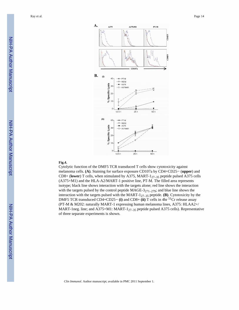
Fig.4.Cytolytic function of the DMF5 TCR transduced T cells show cytotoxicity againstmelanoma cells. (A). Staining for surface exposure CD107a by CD4+CD25− (upper) andCD8+ (lower) T cells, when stimulated by A375, MART-127–35 peptide pulsed A375 cells(A375+M1) and the HLA-A2/MART-1 positive line, PT-M. The filled area representsisotype; black line shows interaction with the targets alone; red line shows the interactionwith the targets pulsed by the control peptide MAGE-3271–279; and blue line shows theinteraction with the targets pulsed with the MART-127–35 peptide. (B). Cytotoxicity by theDMF5 TCR transduced CD4+CD25− (i) and CD8+ (ii) T cells in 4hr 51Cr release assay(PT-M & M202: naturally MART-1 expressing human melanoma lines, A375: HLAA2+/MART-1neg. line; and A375+M1: MART-127–35 peptide pulsed A375 cells). Representativeof three separate experiments is shown.
Ray et al. Page 14
Clin Immunol. Author manuscript; available in PMC 2011 September 1.
NIH
-PA Author Manuscript
NIH
-PA Author Manuscript
NIH
-PA Author Manuscript
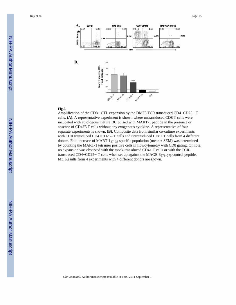
Fig.5.Amplification of the CD8+ CTL expansion by the DMF5 TCR transduced CD4+CD25− Tcells. (A). A representative experiment is shown where untransduced CD8 T cells wereincubated with autologous mature DC pulsed with MART-1 peptide in the presence orabsence of CD4F5 T cells without any exogenous cytokine. A representative of fourseparate experiments is shown. (B). Composite data from similar co-culture experimentswith TCR transduced CD4+CD25– T cells and untransduced CD8+ T cells from 4 differentdonors. Fold increase of MART-127–35 specific population (mean ± SEM) was determinedby counting the MART-1 tetramer positive cells in flowcytometry with CD8 gating. Of note,no expansion was observed with the mock-transduced CD4+ T cells or with the TCR-transduced CD4+CD25− T cells when set up against the MAGE-3271–279 control peptide,M3. Results from 4 experiments with 4 different donors are shown.
Ray et al. Page 15
Clin Immunol. Author manuscript; available in PMC 2011 September 1.
NIH
-PA Author Manuscript
NIH
-PA Author Manuscript
NIH
-PA Author Manuscript