Interaction Between Cytokines and Oxidative Stress in Acute Pancreatitis
Upload
independentCategory
view
2download
0
Immunity
Article
T Helper 2 Cytokines Inhibit Autophagic Controlof Intracellular Mycobacterium tuberculosisJames Harris,1,4,5 Sergio A. De Haro,1,5 Sharon S. Master,1 Joseph Keane,2 Esteban A. Roberts,1
Monica Delgado,1 and Vojo Deretic1,3,*1Department of Molecular Genetics and Microbiology, University of New Mexico Health Sciences Center,
University of New Mexico School of Medicine, 915 Camino de Salud NE, Albuquerque, NM 87131, USA2St. James’s Hospital and Trinity College Dublin, Dublin 8, Ireland3Department of Cell Biology and Physiology, University of New Mexico School of Medicine, Albuquerque, NM 87131, USA4Present address: Trinity Centre for Health Sciences, Trinity College Dublin and St. James’s Hospital, Dublin 8, Ireland.5These authors contributed equally to this work.*Correspondence: [email protected]
DOI 10.1016/j.immuni.2007.07.022
SUMMARY
Autophagy is a recently recognized immuneeffector mechanism against intracellular patho-gens. The role of autophagy in innate immunityhas been well established, but the extent of itsregulation by the adaptive immune responseis less well understood. The T helper 1 (Th1)cell cytokine IFN-g induces autophagy in mac-rophages to eliminate Mycobacterium tubercu-losis. Here, we report that Th2 cytokines affectautophagy in macrophages and their ability tocontrol intracellular M. tuberculosis. IL-4 andIL-13 abrogated autophagy and autophagy-mediated killing of intracellular mycobacteriain murine and human macrophages. Inhibitionof starvation-induced autophagy by IL-4 andIL-13 was dependent on Akt signaling, whereasthe inhibition of IFN-g-induced autophagy wasAkt independent and signal transducer andactivator of transcription 6 (STAT6) dependent.These findings establish a mechanism throughwhich Th1-Th2 polarization differentially affectsthe immune control of intracellular pathogens.
INTRODUCTION
Autophagy is a fundamental homeostatic mechanism in
which cells sequester discrete portions of the cytoplasm
into an autophagosome, a specialized vacuole with
a double membrane, which in turn delivers them to ly-
sosomes for degradation (Levine, 2005; Shintani and
Klionsky, 2004). This process removes damaged or sur-
plus organelles such as leaky mitochondria and excess
peroxisomes and, by degrading long-lived cytoplasmic
macromolecules during periods of starvation, promotes
cell survival (Kuma et al., 2004). Autophagy has recently
been shown to play a role in innate immunity against
intracellular pathogens, including Epstein-Barr Virus
Im
(Paludan et al., 2005), Shigella flexneri (Ogawa et al.,
2005), Salmonella typhimurium (Birmingham et al.,
2006), Toxoplasma gondii (Ling et al., 2006), and Myco-
bacterium tuberculosis (Gutierrez et al., 2004). In addition,
autophagy has been implicated in adaptive immunity,
playing a role in endogenous antigen presentation (Deng-
jel et al., 2005; Deretic, 2005; Paludan et al., 2005; Schmid
et al., 2006b).
Autophagy can be induced pharmacologically with ra-
pamycin, which inhibits TOR, a conserved Ser and Thr
kinase that regulates cell growth and metabolism in re-
sponse to growth factors, energy inputs, and nutritional
demands (Wullschleger et al., 2006). TOR integrates vari-
ous inputs; its activation stimulates anabolic processes
and biomass production, whereas its inhibition enhances
catabolic processes, including autophagy. A classical ex-
ample of this system in action is demonstrated by amino
acid starvation, which leads to inhibition of TOR and in-
duction of autophagy. Conversely, TOR can be activated
by growth factors via the Akt (also known as PKB) path-
way, resulting in the inhibition of autophagy (Wullschleger
et al., 2006). In some cases, withdrawal of growth factors
is sufficient for induction of autophagy, even in the pres-
ence of adequate nutrients. For example, removal of IL-3
from cultures of an IL-3-dependent hemopoietic cell line
that also lacks the apoptotic regulators Bax and Bak has
been shown to induce autophagy, and if left unchecked,
the cells eventually die (Lum et al., 2005). However, the
cells die more rapidly when autophagy is blocked, sug-
gesting that autophagy is a survival mechanism in these
cells (Lum et al., 2005). Autophagy can be modulated by
cytokines and other immunological signals (Andrade
et al., 2006; Djavaheri-Mergny et al., 2006; Singh et al.,
2006). For example, TNF-a can induce autophagy in
Ewing sarcoma cells in the absence of NF-kB activation
(Djavaheri-Mergny et al., 2006), whereas in macrophages
and other cells, IFN-g, a classical T helper 1 (Th1) cell
cytokine and a critical antituberculosis immune mediator,
induces or augments autophagy (Gutierrez et al., 2004; In-
bal et al., 2002; Pyo et al., 2005). Moreover, the protective
role of IFN-g against mycobacteria has been associated
with autophagy (Gutierrez et al., 2004; Singh et al.,
munity 27, 505–517, September 2007 ª2007 Elsevier Inc. 505
Immunity
Th2 Inhibition of Autophagy as an Immune Effector
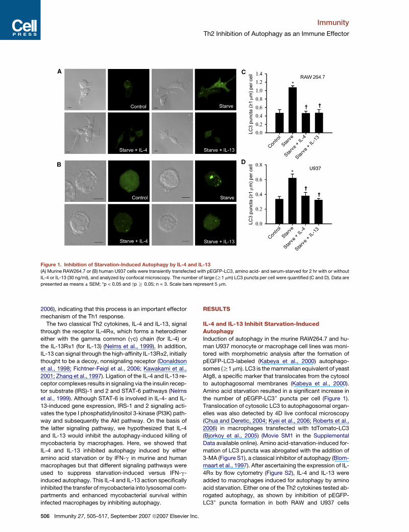
Figure 1. Inhibition of Starvation-Induced Autophagy by IL-4 and IL-13
(A) Murine RAW264.7 or (B) human U937 cells were transiently transfected with pEGFP-LC3, amino acid- and serum-starved for 2 hr with or without
IL-4 or IL-13 (30 ng/ml), and analyzed by confocal microscopy. The number of large (R1 mm) LC3 puncta per cell were quantified (C and D). Data are
presented as means ± SEM; *p < 0.05 and yp R 0.05; n = 3. Scale bars represent 5 mm.
2006), indicating that this process is an important effector
mechanism of the Th1 response.
The two classical Th2 cytokines, IL-4 and IL-13, signal
through the receptor IL-4Ra, which forms a heterodimer
either with the gamma common (gc) chain (for IL-4) or
the IL-13Ra1 (for IL-13) (Nelms et al., 1999). In addition,
IL-13 can signal through the high-affinity IL-13Ra2, initially
thought to be a decoy, nonsignaling receptor (Donaldson
et al., 1998; Fichtner-Feigl et al., 2006; Kawakami et al.,
2001; Zhang et al., 1997). Ligation of the IL-4 and IL-13 re-
ceptor complexes results in signaling via the insulin recep-
tor substrate (IRS)-1 and 2 and STAT-6 pathways (Nelms
et al., 1999). Although STAT-6 is involved in IL-4- and IL-
13-induced gene expression, IRS-1 and 2 signaling acti-
vates the type I phosphatidylinositol 3-kinase (PI3K) path-
way and subsequently the Akt pathway. On the basis of
the latter signaling pathway, we hypothesized that IL-4
and IL-13 would inhibit the autophagy-induced killing of
mycobacteria by macrophages. Here, we showed that
IL-4 and IL-13 inhibited autophagy induced by either
amino acid starvation or by IFN-g in murine and human
macrophages but that different signaling pathways were
used to suppress starvation-induced versus IFN-g-
induced autophagy. This IL-4 and IL-13 action specifically
inhibited the transfer of mycobacteria into lysosomal com-
partments and enhanced mycobacterial survival within
infected macrophages by inhibiting autophagy.
506 Immunity 27, 505–517, September 2007 ª2007 Elsevier Inc
RESULTS
IL-4 and IL-13 Inhibit Starvation-InducedAutophagyInduction of autophagy in the murine RAW264.7 and hu-
man U937 monocyte or macrophage cell lines was moni-
tored with morphometric analysis after the formation of
pEGFP-LC3-labeled (Kabeya et al., 2000) autophago-
somes (R1 mm). LC3 is the mammalian equivalent of yeast
Atg8, a specific marker that translocates from the cytosol
to autophagosomal membranes (Kabeya et al., 2000).
Amino acid starvation resulted in a significant increase in
the number of pEGFP-LC3+ puncta per cell (Figure 1).
Translocation of cytosolic LC3 to autophagosomal organ-
elles was also detected by 4D live confocal microscopy
(Chua and Deretic, 2004; Kyei et al., 2006; Roberts et al.,
2006) in macrophages transfected with tdTomato-LC3
(Bjorkoy et al., 2005) (Movie SM1 in the Supplemental
Data available online). Amino acid-starvation-induced for-
mation of LC3 puncta was abrogated with the addition of
3-MA (Figure S1), a classical inhibitor of autophagy (Blom-
maart et al., 1997). After ascertaining the expression of IL-
4Ra by flow cytometry (Figure S2), IL-4 and IL-13 were
added to macrophages induced for autophagy by amino
acid starvation. Either one of the Th2 cytokines tested ab-
rogated autophagy, as shown by inhibition of pEGFP-
LC3+ puncta formation in both RAW and U937 cells
.
Immunity
Th2 Inhibition of Autophagy as an Immune Effector
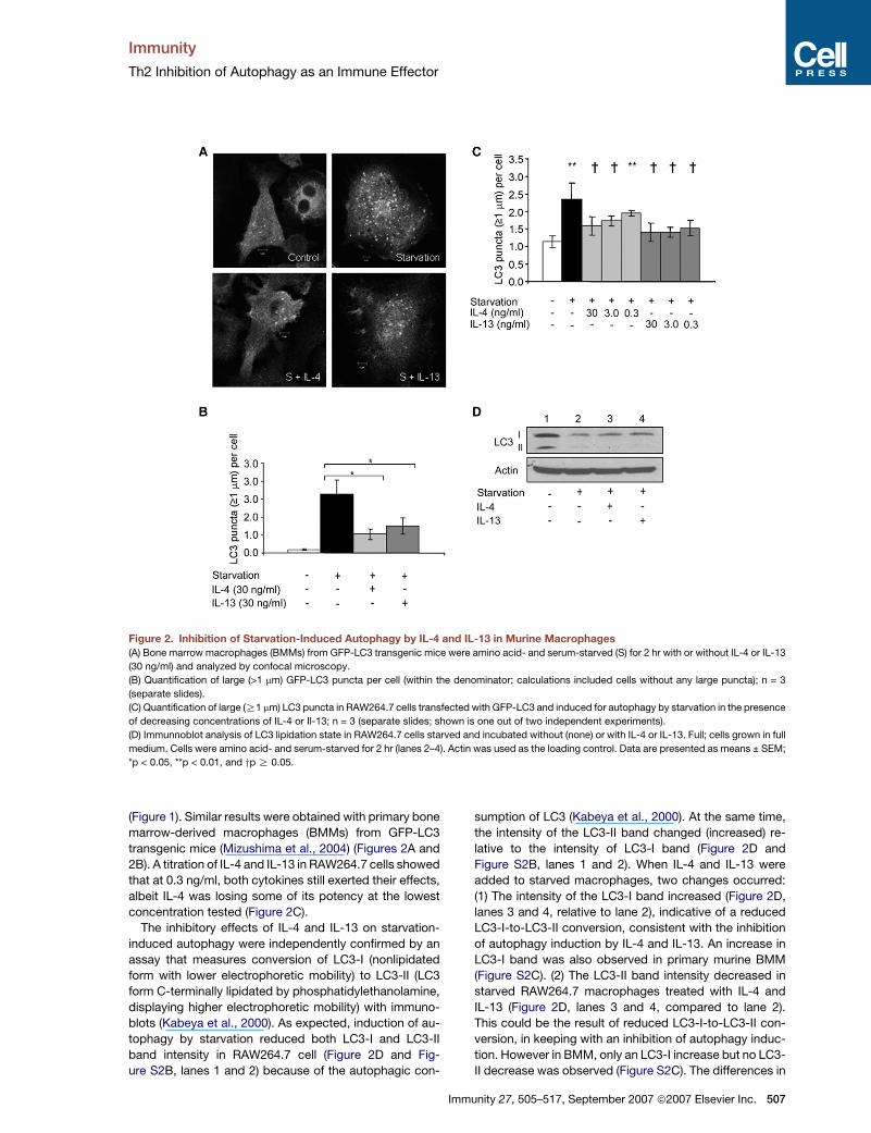
Figure 2. Inhibition of Starvation-Induced Autophagy by IL-4 and IL-13 in Murine Macrophages
(A) Bone marrow macrophages (BMMs) from GFP-LC3 transgenic mice were amino acid- and serum-starved (S) for 2 hr with or without IL-4 or IL-13
(30 ng/ml) and analyzed by confocal microscopy.
(B) Quantification of large (>1 mm) GFP-LC3 puncta per cell (within the denominator; calculations included cells without any large puncta); n = 3
(separate slides).
(C) Quantification of large (R1 mm) LC3 puncta in RAW264.7 cells transfected with GFP-LC3 and induced for autophagy by starvation in the presence
of decreasing concentrations of IL-4 or Il-13; n = 3 (separate slides; shown is one out of two independent experiments).
(D) Immunnoblot analysis of LC3 lipidation state in RAW264.7 cells starved and incubated without (none) or with IL-4 or IL-13. Full; cells grown in full
medium. Cells were amino acid- and serum-starved for 2 hr (lanes 2–4). Actin was used as the loading control. Data are presented as means ± SEM;
*p < 0.05, **p < 0.01, and yp R 0.05.
(Figure 1). Similar results were obtained with primary bone
marrow-derived macrophages (BMMs) from GFP-LC3
transgenic mice (Mizushima et al., 2004) (Figures 2A and
2B). A titration of IL-4 and IL-13 in RAW264.7 cells showed
that at 0.3 ng/ml, both cytokines still exerted their effects,
albeit IL-4 was losing some of its potency at the lowest
concentration tested (Figure 2C).
The inhibitory effects of IL-4 and IL-13 on starvation-
induced autophagy were independently confirmed by an
assay that measures conversion of LC3-I (nonlipidated
form with lower electrophoretic mobility) to LC3-II (LC3
form C-terminally lipidated by phosphatidylethanolamine,
displaying higher electrophoretic mobility) with immuno-
blots (Kabeya et al., 2000). As expected, induction of au-
tophagy by starvation reduced both LC3-I and LC3-II
band intensity in RAW264.7 cell (Figure 2D and Fig-
ure S2B, lanes 1 and 2) because of the autophagic con-
Im
sumption of LC3 (Kabeya et al., 2000). At the same time,
the intensity of the LC3-II band changed (increased) re-
lative to the intensity of LC3-I band (Figure 2D and
Figure S2B, lanes 1 and 2). When IL-4 and IL-13 were
added to starved macrophages, two changes occurred:
(1) The intensity of the LC3-I band increased (Figure 2D,
lanes 3 and 4, relative to lane 2), indicative of a reduced
LC3-I-to-LC3-II conversion, consistent with the inhibition
of autophagy induction by IL-4 and IL-13. An increase in
LC3-I band was also observed in primary murine BMM
(Figure S2C). (2) The LC3-II band intensity decreased in
starved RAW264.7 macrophages treated with IL-4 and
IL-13 (Figure 2D, lanes 3 and 4, compared to lane 2).
This could be the result of reduced LC3-I-to-LC3-II con-
version, in keeping with an inhibition of autophagy induc-
tion. However in BMM, only an LC3-I increase but no LC3-
II decrease was observed (Figure S2C). The differences in
munity 27, 505–517, September 2007 ª2007 Elsevier Inc. 507

Immunity
Th2 Inhibition of Autophagy as an Immune Effector
Figure 3. Inhibition of IFN-g-Induced Autophagy by IL-4 and IL-13
(A) Murine RAW264.7 or (B) human U937 cells were transiently transfected with pEGFP-LC3 and treated with 200 U/ml IFN-g or IFN-g in combination
with IL-4 or IL-13 (30 ng/ml) for 24 hr and were analyzed by confocal microscopy. Large (R1 mm) LC3 puncta per cell were quantified (C and D). Data
are presented as means ± SEM; *p < 0.05, **p < 0.01, yp R 0.05; n = 3. Scale bars represent 5 mm.
LC3-II band intensity between RAW364.7 and BMM may
be the net result of differences in the rates of LC3-I-to-
LC3-II conversion versus LC3-II depletion through degra-
dation in autolysosomes. This was substantiated with
lysosomal and autolysosomal protease inhibitors
(Figure S2, lanes 3 and 4, compared to lane 2), which, as
expected, increased LC3-II amounts by blocking its deg-
radation both in the presence or absence of IL-4. Consis-
tent with the conclusion that Th2 cytokines inhibit LC3-I-
to-LC3-II conversion, IL-4 presence increased LC3-I
band intensity (Figure S2B, lane 4 compared to lane 3).
Because the presence of IL-4 did not diminish LC3-II
band levels relative to protease inhibitors alone
(Figure S2, lane 4 versus lane 3), as one might expect
from lower LC3-I-to-LC3-II conversion, Th2 cytokines
may have an additional effect on the fate of LC3-II by par-
tially inhibiting its delivery to or its degradation in autolyso-
somes. This conclusion is consistent with the apparent
preservation of the LC3-II band in BMM (Figure S2C). Of
note is that IL-4 and IL-13 did not change expression of
the murine LC3 gene MAP1LC3b under starvation condi-
tions (Figure S2D). In conclusion, IL-4 and IL-13 inhibit
LC3-I-to-LC3-II conversion and initiation of autophagy
but may have additional effects on the autophagic path-
way; these effects become apparent when different cells
are examined.
508 Immunity 27, 505–517, September 2007 ª2007 Elsevier Inc
IL-4 and IL-13 Inhibit IFN-g-Induced Formationof AutophagosomesTreatment of macrophages with IFN-g promotes the for-
mation of autophagosomes (Gutierrez et al., 2004; Singh
et al., 2006). We tested whether this effect could be in-
hibited by treating RAW and U937 cells with IFN-g in com-
bination with either IL-4 or IL-13. In both cell types, IL-4
and IL-13 significantly reduced the number of IFN-g-
induced pEGFP-LC3+ puncta per cell (Figure 3). In
addition, we found that IFN-g treatment increased the per-
centage of RAW cells with large vacuoles that stained
positively for monodansylcadaverine (MDC), another
marker for autophagic vacuoles (Biederbick et al., 1995),
and this effect was inhibited by IL-4 or IL-13 (Figures
S3A and S3B). By using the MDC assay, we confirmed
the effects of IL-4 and IL-13 in primary human, peripheral
blood monocyte-derived macrophages (MDMs) and
found, in titration experiments, a similar concentration-
dependence pattern to that seen in murine macrophages
(Figure S3C).
IL-4 and IL-13 Inhibit Autophagy-Dependent BCGPhagolysosome MaturationMycobacterium tuberculosis normally resides in phago-
somes that do not acquire phagolysosomal properties,
such as lumenal acidification and the presence of
.

Immunity
Th2 Inhibition of Autophagy as an Immune Effector
Figure 4. Inhibition of Starvation- and
IFN-g-Induced BCG Phagosome Matura-
tion by IL-4 and IL-13
(A) Confocal images of murine RAW264.7 mac-
rophages infected with GFP-BCG for 2 hr.
Cells were amino acid- and serum-starved for
2 hr (during infection) or treated with IFN-g
(200 U/ml) for 24 hr prior to infection, with or
without IL-4 or IL-13 (30 ng/ml). After infection,
cells were fixed and stained for CD63. The per-
centage of CD63+ BCG phagosomes was
quantified (B and C). The percentage of Lyso-
tracker red (LT)+ BCG phagosomes was re-
corded in PMA-differentiated human THP-1
monocytes after (D) starvation or (E) IFN-g
(200 U/ml) treatment with or without IL-4 or
IL-13 (30 ng/ml). Representative images are
shown in Figure S6. Data are presented as
means ± SEM; *p < 0.05 and yp R 0.05; n = 3.
lysosomal hydrolases (Vergne et al., 2004). Induction of
autophagy has been shown to promote the transfer of
mycobacteria into degradative autolysosomal organelles
(Gutierrez et al., 2004). To investigate whether IL-4 and
IL-13 counteract this effect, we examined the maturation
of M. tuberculosis variant bovis BCG (BCG) phagosomes
by monitoring the late endosomal or lysosomal marker
CD63. Induction of autophagy by starvation in BCG-
infected RAW cells significantly increased colocalization
of GFP-BCG with CD63 (Figures 4A and 4B). The effect
of starvation-induced autophagy on BCG phagosome
maturation was inhibited by the addition of either IL-4 or
IL-13 (Figures 4A and 4B). Both IL-4 and IL-13 also in-
hibited IFN-g-induced phagosome maturation in BCG-
infected cells (Figures 4A and 4C). Starvation increased
phagosome maturation in U937 cells, an effect that was
inhibited with either IL-4 or IL-13 (Figure S4). For unknown
Im
reasons, IFN-g did not significantly increase BCG phago-
some maturation in U937 cells (data not shown). Instead,
we tested PMA-differentiated human THP-1 cells, by us-
ing the acidotropic dye LysoTracker Red (LT) to visualize
lysosomal compartments. As with RAW cells, phagosome
maturation was increased by either starvation or IFN-g;
this was inhibited by treatment with either IL-4 or IL-13
(Figures 4D and 4E and Figure S5).
Phagosome maturation was autophagy dependent, as
demonstrated by siRNA knockdown of Beclin 1, a critical
mammalian autophagy factor (Liang et al., 1999) (Figures
5A–5C). Although RAW cells transfected with scrambled
siRNA showed an increase in BCG phagosome matura-
tion in response to starvation (Figures 5A and 5D) or
IFN-g (Figures 5B and 5D), cells treated with Beclin 1
siRNA did not, demonstrating that these responses are
dependent on functional autophagic machinery. In the
munity 27, 505–517, September 2007 ª2007 Elsevier Inc. 509

Immunity
Th2 Inhibition of Autophagy as an Immune Effector
Figure 5. Starvation- and IFN-g-Induced BCG Phagosome Maturation Is Autophagy Dependent
(A) Quantitative analysis of CD63+ BCG phagosomes that were in murine RAW264.7 macrophages transiently transfected with either nontargeting
(scrambled) siRNA or Beclin 1 siRNA and that were amino acid- and serum-starved for 2 hr.
(B) CD63+ BCG phagosomes in RAW264.7 cells transiently transfected with scrambled or Beclin 1 siRNA and treated with IFN-g (200 U/ml) for 24 hr.
Data are presented as means ± SEM; *p < 0.05, **p < 0.01, and yp R 0.05; n = 3 for (A) and n = 6 for (B).
(C) Immunnoblot confirmation of Beclin 1 knockdown by siRNA.
(D) Immunofluorescence panels of BCG colocalization with the late endosomal marker CD63, exemplifying the data in (A) and (B). CD63 is shown in
green; BCG is shown in red.
presence of full-nutrient media, treatment of RAW macro-
phages with IL-4 or IL-13 had no effect on the colocaliza-
tion of CD63 with either live or heat-killed BCG phago-
somes (Figure S6). Thus, these Th2 cytokines do not
inhibit mycobacterial phagosome maturation under nor-
mal conditions but rather specifically inhibit autophagy-
dependent maturation.
IL-4 and IL-13 Inhibit Autophagy-DependentKilling of Intracellular MycobacteriaTo determine whether IL-4 and IL-13 affect autophagic
elimination of mycobacteria (Gutierrez et al., 2004), we in-
fected RAW cells with BCG and induced autophagy by
amino acid starvation for 2 hr. Induction of autophagy de-
creased BCG survival in RAW cells (Figure 6A). Addition of
IL-4 or IL-13 to the starvation media abrogated this effect
(Figure 6A). Similar results were obtained when survival of
virulent M. tuberculosis H37Rv was tested with starvation
or IFN-g used as autophagy agonists (Figures 6B and 6C).
Figure 6D shows that these relationships hold true in pri-
mary macrophages because IL-4, used as an example,
abrogated killing of virulent M. tuberculosis H37Rv by star-
vation-induced autophagy in murine BMMs. These find-
ings demonstrate that IL-4 or IL-13 inhibit autophagy-
induced killing of mycobacteria by macrophages.
510 Immunity 27, 505–517, September 2007 ª2007 Elsevier Inc.
IL-4 and IL-13 Inhibit Autophagic PhagosomeMaturation in Primary Human CellsTo confirm that IL-4 and IL-13 inhibit autophagy-induced
effects on mycobacterial phagosomes in human primary
macrophages, we tested whether IL-4 and IL-13 influ-
enced autophagy-induced mycobacterial phagosome
maturation. Human peripheral blood monocyte-derived
macrophages (MDMs) were infected with BCG and in-
duced for autophagy by starvation. The effect of starva-
tion-induced autophagy on mycobacterial phagosome
maturation was decreased by the addition of either IL-4
or IL-13 (Figures 6E and 6F). Similarly, both IL-4 and IL-
13 had an inhibitory effect on IFN-g-induced mycobacte-
rial phagosome maturation (Figures 6E and 6F). These
results validate in human primary macrophages the con-
clusion that Th2 cytokines counteract autophagy in its
ability to deliver mycobacteria into the phagolysosome.
Inhibition of Starvation-Induced Autophagy by IL-4and IL-13 Is Akt DependentTo determine the signaling involved in the actions of IL-4
and IL-13 on autophagy in macrophages, we examined
whether the Akt pathway, known to activate TOR and in-
hibit autophagy (Wullschleger et al., 2006), was involved.
In both RAW (Figure 7A) and U937 (Figure S7A) macro-
phages, IL-4 and IL-13 increased phosphorylation of Akt

Immunity
Th2 Inhibition of Autophagy as an Immune Effector
Figure 6. IL-4 and IL-13 Inhibit Autophagy-Dependent Killing of M. tuberculosis in Murine Macrophages and Counteract Auto-
phagy-Induced Phagosome Maturation in Primary Human Macrophages
(A) Murine RAW264.7 macrophages were infected with BCG for 1 hr and were amino acid- and serum-starved with or without IL-4 or IL-13 (30 ng/ml)
for 2 hr. Cells were washed and lysed for viability determination and survival expressed as a percentage of the control.
(B) Murine RAW264.7 macrophages were infected with virulent M. tuberculosis H37Rv for 1 hr and were amino acid- and serum-starved with or with-
out IL-4 or IL-13 (30 ng/ml) for 2 hr. Cells were washed and lysed for viability determination.
(C) Murine RAW264.7 macrophages were either untreated or treated with 200 u/ml of m-IFN-g for 24 hr prior to infection with M. tuberculosis H37Rv
for 1 hr with or without IL-4 or IL-13 (30 ng/ml) for 2 hr. Cells were washed and lysed for viability (cfu) determination. Data are presented as means ±
SEM, *p < 0.05, **p < 0.01, and yp R 0.05; n = 3.
(D) IL-4 inhibits autophagy-dependent killing of M. tuberculosis H37Rv in primary murine macrophages. Murine bone marrow macrophages were
infected with virulent M. tuberculosis (strain H37Rv) for 1 hr and were amino acid- and serum-starved with or without IL-4 (30 ng/ml) for 2 hr. Myco-
bacterial viability (colony counts) is expressed as a percentage of the control.
(E and F) Primary human macrophages derived from peripheral blood monocytes were infected with BCG. Transfer of mycobacteria to phagolyso-
some dependent on autophagy was scored with the acidotropic dye LysoTracker. Autophagy was induced for 2 hr by starvation or by treatment with
200 u/ml human IFN-g. Cells were incubated in the absence or presence of 30 ng/ml of human IL-4 or IL-13, as indicated. (F) shows quantification of
experiments shown in (E). Data are presented as means ± SEM; *p < 0.05 and yp R 0.05; n = 3 (three independent donors).
under starvation conditions (Figure S7A, top two panels
and graph). We next tested the activity of TOR, a key neg-
ative regulator of autophagy (when TOR is active, autoph-
agy is inhibited) on the basis of reports that TOR is acti-
vated by Akt (Wullschleger et al., 2006). We investigated
TOR activation by monitoring phosphorylation of p70 S6
kinase (S6k) (Figure S7A, bottom two panels and graph)
and eukaryotic initiation factor 4E (eIF4E)-binding protein
1 (4E-BP1) (Figures 7B–7E and Figure S7), both commonly
used as indicators of TOR activation state (Wullschleger
et al., 2006). Treatment of U937 cells with IL-4 or IL-13
increased phosphorylation of S6k (Figure S7A). In
Im
RAW264.7 macrophages, IL-4 and IL-13 treatment in-
creased phosphorylation of 4E-BP1 under starvation con-
ditions in a dose-dependent manner (Figures S7B and
S7C). A finer-resolution analysis of 4E-BP1 phosphoryla-
tion was carried out next. TOR induces 4E-BP1 inactiva-
tion via multiple hierarchical phosphorylations (Gingras
et al., 2001). A fine-resolution immunoblot analysis can
be used for revealing 4E-BP1 hypophosphorylated a and
b forms, and hyperphosphorylated g form, resolved in
the order of their decreasing electrophoretic mobility.
Active Akt elicits 4E-BP1(g) hyperphosphorylation through
TOR activity (Gingras et al., 2001). Our analyses in primary
munity 27, 505–517, September 2007 ª2007 Elsevier Inc. 511

Immunity
Th2 Inhibition of Autophagy as an Immune Effector
Figure 7. Role of Akt and STAT6 in Suppression by IL-4 and IL-13 of Starvation- and IFN-g-Induced Autophagy
(A) Immunoblot analysis of Akt phosphorylation in RAW264.7 macrophages amino acid- and serum starved for 2 hr with or without IL-4 or IL-13.
(B) Immunoblot analysis of 4E-BP1 hierarchical phosphorylation in murine bone marrow macrophages (BMMs) starved and treated with IL-4 for 40,
60, and 120 min.
(C) Graph shows the ratio of p-4E-BP1(g)/p-4E-BP1(b).
(D) Immunoblot analysis of 4E-BP1 in murine bone marrow macrophages (BMMs).
512 Immunity 27, 505–517, September 2007 ª2007 Elsevier Inc.

Immunity
Th2 Inhibition of Autophagy as an Immune Effector
murine BMMs and in human MDM revealed that IL-4 and
IL-13 treatment specifically increased phosphorylation of
4E-BP1(g) under starvation conditions (Figures 7B–7E).
Furthermore, a time course of 4E-BP1 showed that IL-4
exerted its effects by increasing 4E-BP1(g) hyperphos-
phorylation at 2 hr but had no discernible effects at earlier
time points (Figures 7B and 7C), consistent with other ef-
fects measured in our study. These results demonstrate
that IL-4 and IL-13 activate the Akt pathway in macro-
phages, resulting in the activation of TOR and inhibition
of starvation-induced autophagy.
To establish the role of Akt in the IL-4- and IL-13-
induced inhibition of autophagy in macrophages, we
knocked down Akt1 and Akt2 with siRNA in vitro
(Figure S8A). Compared with the control samples (scram-
bled siRNA), knockdown of Akt1 and Akt2 in RAW macro-
phages increased autophagy, monitored by LC3+ vacuole
formation (Figure 7F and Figure S8B), indicating that basal
levels of autophagy are controlled, at least in part, by
baseline Akt activation. Double knockdown of Akt1 and
Akt2 in RAW cells abrogated the effects of IL-4 and IL-
13 on starvation-induced autophagy (Figure 7F and
Figure S8B). In BCG-infected RAW cells, knockdown of
Akt1 and Akt2 also inhibited the effects of IL-4 and IL-13
on starvation-induced transfer of BCG to autophago-
somes (Figure 7G and Figure S8C).
Inhibition of IFN-g-Induced Autophagy Is STAT6DependentIn contrast to the effects with IL-4 and IL-13 on starvation-
induced autophagy, a double knockdown of Akt1 and
Akt2 with siRNA had no effect on the inhibition of IFN-g-
induced phagosome maturation by IL-4 and IL-13
(Figure 7H), suggesting that the suppressive effects of
these Th2 cytokines on the activation by IFN-g involved al-
ternative or additional pathways, independent of Akt sig-
naling. Akt3 was detectable only at very low amounts in
these cells, and its expression was not affected by knock-
down of Akt1 and Akt2 (data not shown). We therefore
tested whether the inhibitory effect of IL-4 and IL-13 on
IFN-g-induced autophagy was regulated via a different
pathway. IL-4 and IL-13 receptors, in addition to signaling
via the Akt pathway, are more commonly known for their
activation of STAT6 (Nelms et al., 1999). Thus, we tested
whether STAT6 was required for Th2 cytokine inhibition
Im
of autophagy activation by IFN-g. When STAT6 was
knocked down (Figure S9), IL-13 could no longer suppress
IFN-g-induced autophagosome formation in macro-
phages (Figures 7I–7L). Collectively, our data on IL-4
and IL-13 inhibition of starvation- and IFN-g-induced au-
tophagy demonstrate that Th2 cytokines can employ ei-
ther of the signaling pathways associated with ligation of
the IL-4 and IL-13 receptors to exert this effect. Suppres-
sion of starvation-induced autophagy occurs through Akt
signaling, whereas STAT6 is needed for suppression of
IFN-g-induced autophagy.
DISCUSSION
Recent studies have demonstrated that autophagy repre-
sents a mechanism for eliminating intracellular pathogens
(Deretic, 2005; Schmid et al., 2006a; Levine and Deretic,
2007), as shown in several bacterial, viral, and protozoan
systems (Birmingham et al., 2006; Gutierrez et al., 2004;
Ling et al., 2006; Ogawa et al., 2005; Orvedahl et al., 2007;
Paludan et al., 2005; Singh et al., 2006). Induction of au-
tophagy by amino acid starvation, rapamycin treatment,
or macrophage activation with IFN-g leads to an increase
in mycobacterial phagolysosome maturation. This in-
crease is concomitant with a decrease in the intracellular
survival of the bacilli (Singh et al., 2006), and such a sur-
vival decrease is independent of apoptosis (Gutierrez
et al., 2004), another process previously implicated in the
elimination of M. tuberculosis (Fratazzi et al., 1999). Here,
we have shown that autophagy-induced killing of myco-
bacteria by murine and human macrophages is inhibited
by the Th2 cytokines IL-4 and IL-13. Both cytokines coun-
teract the autophagic transfer of mycobacteria from their
normally immature phagosomes with early endosomal
characteristics (Vergne et al., 2004) to autolysosomes,
thus preventing the elimination of intracellular bacilli by
autophagy.
We have shown that the effect of IL-4 and IL-13 on star-
vation-induced autophagy is dependent on signaling via
the Akt pathway, which activates TOR. Activation of TOR,
downstream of Akt, inhibits autophagy. As expected, IL-4
and IL-13 could not influence rapamycin-induced auto-
phagy (data not shown) because rapamycin acts directly
on TOR, bypassing Akt signaling. Notably, the mechanism
of IL-4 and IL-13 inhibition of IFN-g-induced autophagy is
(E) Immunnoblot analysis of human monocyte-derived macrophages (MDMs) starved for 2 hr with or without IL-4 or IL-13. Graphs in (D) and (E) show
(n = 2; one out of two experiments with similar results) the intensity ratios of p-4E-BP1(g) to p-4E-BP1(b) bands.
(F) Quantification of large (R1 mm) pEGFP-LC3 puncta per cell in RAW 264.7 macrophages transiently transfected with pEGFP-LC3 and either scram-
bled siRNA or Akt 1 + Akt 2 siRNA and starved for 2 hr with or without IL-4 or IL-13.
(G) Quantitative analysis of CD63+ BCG phagosomes in RAW264.7 cells transiently transfected with scrambled siRNA or Akt 1 + Akt 2 siRNA and
infected with BCG for 2 hr. Infected cells were amino acid- and serum-starved with or without IL-4 or IL-13.
(H) Percentage of CD63+ BCG phagosomes in RAW264.7 cells transiently transfected with scrambled siRNA or Akt1 + Akt 2 siRNA, treated with IFN-g
with or without IL-4 or IL-13 for 24 hr, and infected with BCG for 2 hr. Representative images from these experiments and immunoblot-blot confir-
mation of Akt 1 and Akt 2 knockdown are shown in Figure S8.
(I–L) Th2 inhibition of IFNg-dependent autophagy is STAT6 dependent: (I and J) display confocal-microscopy images and quantification of GFP-LC3
puncta formation in response to IFN-g, with or without IL-13, in cells treated with control (csramble) siRNA. (K–L) display images and quantification of
GFP-LC3 puncta formation in response to IFN-g, with or without IL-13, in cells treated with control STAT6 siRNA. Immunoblot analysis of STAT6
knockdown is given in Figure S9. Data in all graphs (when error bars are shown) are presented as means ± SEM; n R 3; *p < 0.05, **p < 0.01, and
yp R 0.05.
munity 27, 505–517, September 2007 ª2007 Elsevier Inc. 513

Immunity
Th2 Inhibition of Autophagy as an Immune Effector
Akt independent. This finding was surprising given the es-
tablished role of Akt in inhibiting autophagy in general (Pe-
tiot et al., 2000) and given the inhibitory effects of Akt in
starvation-induced autophagy in macrophages demon-
strated here for IL-4 and IL-13 and in nonphagocytic cells
elsewhere for IL-13 (Arico et al., 2001). To explain these
observations, we have now uncovered a previously un-
known signaling relationship; STAT6 modulates the Th2
antiautophagic function when macrophages are stimu-
lated for autophagy by IFN-g. This places STAT6 within
the ranks of the immunologically relevant regulators of
autophagy. Most importantly, it provides an explanation
for how Th2 cytokines counter autophagy-dependent pro-
tection against intracellular pathogens afforded by Th1 re-
sponses. The effects of STAT6 on autophagy downstream
of IL-4 and IL-13 action may involve a number of path-
ways, one of which could be linked to the intriguing ability
of STAT6 to increase the Bcl-2 and Bcl-XL antiapoptotic
proteins (Wurster et al., 2002). Bcl-2 and Bcl-XL are now
also recognized as being antiautophagic effector proteins
(Maiuri et al., 2007; Pattingre et al., 2005). Bcl-2 and Bcl-XL
interact directly (Pattingre et al., 2005) through the recently
recognized BH3 domain (Maiuri et al., 2007) within Beclin 1,
the key activator of autophagy. Because Bcl-2 and Bcl-XL
association with Beclin 1 blocks autophagy induction
(Maiuri et al., 2007), and the rapid induction of STAT6
downstream of IL-4 and IL-13 stimulation may increase
Bcl-XL availability (Wurster et al., 2002), the IL-4-IL-13-
STAT6-dependent inhibition of autophagy observed in our
work could reflect these recently uncovered relationships.
In this work, we did not specifically study alternative
activation of macrophages by IL-4 and IL-13 (Gordon,
2003) but instead focused on the effects of short-term ex-
posure (2 hr) or treatment in combination with IFN-g.
Treatment of macrophages with IL-4 or IL-13 for 6 days
has been shown to enhance fluid-phase pinocytosis and
mannose receptor-mediated endocytosis through activa-
tion of phosphatidylinositol 3 kinase; both cytokines in-
crease tubular-vesicle formation at pericentriolar sites un-
der the plasma membrane, concurrent with decreased
particle sorting to the lysosomes (Montaner et al., 1999).
Although these previous observations could explain the
inhibitory effects on autophagolysosome formation, IL-4
and IL-13 had no effect on BCG phagosome maturation
in the absence of starvation- or IFN-g-induced autophagy,
demonstrating that the effects observed in our experi-
ments are autophagy specific. A study on the effects of al-
ternative activation of macrophages with Th2 cytokines, in
the context of autophagy, may be difficult to address
because prolonged autophagy can lead to type-II-
programmed cell death, and most cells will downregulate
this process to ensure their survival (Pattingre et al., 2005).
Nevertheless, alternative activation of macrophages with
IL-4 in vitro delays and inhibits antibacterial responses
to intracellular M. tuberculosis (Kahnert et al., 2006) by
an unknown mechanism that may be related to the pro-
cesses reported here.
The concentrations of IL-4 and IL-13 used in our titration
experiments fall well within the in vivo and ex vivo tissue
514 Immunity 27, 505–517, September 2007 ª2007 Elsevier Inc
levels of these cytokines under various conditions (Aspord
et al., 2007; Huang et al., 1995; Zhu et al., 1999), including
studies with mycobacterial antigens (Chensue et al.,
1997). Because local concentrations in the vicinity of T
cells secreting IL-4 or IL-13 in vivo can be much higher,
our range included 10-fold increments. Although lympho-
cytes in the lungs of patients with pulmonary tuberculosis
typically have a Th1 phenotype, secreting IFN-g and IL-12
(Mazzarella et al., 2003; Taha et al., 1997), little is known of
the phenotype or cytokine-secretion profiles of infected
macrophages and dendritic cells within the granuloma.
However in vitro studies, in human peripheral blood mono-
cyte-derived macrophages have demonstrated that viru-
lent strains of tuberculosis preferentially upregulate Th2
cytokines (IL-4, IL-5, IL-10, and IL-13), whereas nonviru-
lent strains induce Th1 cytokines and chemokines (Free-
man et al., 2006; Manca et al., 2004; Sun et al., 2006).
Moreover, Th2 lymphocyte subsets have been observed
in lung tissue from patients with cavitary tuberculosis,
compared with Th1 subsets in noncavitary disease (Maz-
zarella et al., 2003). This correlates with higher production
of IL-4 in the periphery of patients with cavitary disease
(van Crevel et al., 2000). Thus, it is possible that in an en-
vironment predominantly influenced by Th1 cytokines,
paracrine, and autocrine secretion of IL-4 and IL-13 could
specifically impair the response of infected macrophages
to IFN-g. Moreover, the peripheral response of patients
with active tuberculosis disease may not necessarily mir-
ror the response in the lungs. Up to 15% of patients with
active pulmonary tuberculosis display specific anergy to
tuberculin (Bloom and Small, 1998), and this has been
linked with increased ratios of IL-4- and IL-10-positive
lymphocytes and decreased ratios of IL-12 (Baliko et al.,
1998). Some authors have reported a Th1 response in
the periphery of patients with mild pulmonary tuberculosis
and Th2 responses in anergic patients and those with
more severe disease (Boussiotis et al., 2000; Dlugovitzky
et al., 1997). Clinical observations in tuberculin-reactive
patients have also demonstrated that Th1 responses, al-
though high in the granuloma, are often paradoxically de-
pressed in peripheral blood lymphocytes in response to
mycobacterial antigens (Jo et al., 2003). In this context,
it would be interesting to determine whether the cytokine
profile in the periphery could influence the autophagic sta-
tus of newly recruited monocytes in the granuloma. Thus,
the balance of Th1 and Th2 cytokines is clearly critical in
the host response to M. tuberculosis. On the basis of the
Th1 autophagy-promoting and Th2 autophagy-dampen-
ing roles, we can now shed more light on this dichotomy.
In summary, we have shown that inhibition of autophagy
by IL-4 and IL-13 impairs the ability of macrophages to kill
intracellular mycobacteria. The Th2 cytokines inhibit phys-
iologically and immunologically induced autophagy in
murine and human monocyte and macrophage cells.
Moreover, these cytokines inhibit autophagy-dependent
maturation of the mycobacterial phagosome and subse-
quent killing of mycobacteria. In addition to furthering
our understanding of the modulation of autophagy in mac-
rophages, these data present evidence for a novel role of
.

Immunity
Th2 Inhibition of Autophagy as an Immune Effector
Th1-Th2 polarization, modulating autophagy as an im-
mune effector mechanism in opposing ways. Our studies
offer an explanation as to why Th2 cytokines are incom-
patible with protection against certain intracellular patho-
gens, including M. tuberculosis. By antagonizing autoph-
agy, IL-4 and IL-13 inhibit Th1-dependent protection
against mycobacteria. Because Th1 and Th2 cytokines
show antagonistic effects on autophagy as an immune ef-
fector, the regulation of autophagy may represent a critical
aspect of Th1-Th2 polarization in the host response to in-
tracellular pathogens.
EXPERIMENTAL PROCEDURES
Antibodies and Reagents
Unless otherwise stated, reagents were from Sigma. Recombinant
murine and human IL-4 and IL-13 were purchased from R & D Sys-
tems. Rabbit polyclonal antibody against CD63 was from Santa Cruz
Biotechnology, rabbit polyclonal antibody against LC3 was from T.
Ueno and E. Kominami. pEGFP-LC3 was from T. Yoshimori and
GFP-LC3 transgenic mice were from N. Mizushima. Tdtomato-LC3
was from G. Bjorkoy.
Cells and Bacterial Cultures
Murine RAW264.7 macrophages were cultured in Dulbecco’s modified
Eagle’s medium (DMEM, Invitrogen) supplemented with 10% fetal bo-
vine serum (FBS) and L-glutamine (full-nutrient medium). The human
monoblastic cell lines, U937 and THP-1, were maintained in RPMI-
1640 (Invitrogen) with 10% FBS, L-glutamine, and HEPES. Before
use, U937 and THP-1 cells were differentiated with PMA (100 nM) for
24–72 hr. Mycobacterium bovis BCG was grown in Middlebrook 7H9
broth with 0.5% Tween, 0.2% glycerol, and albumin-dextrose-
catalase supplement (BD Diagnostics, Franklin Lakes, NJ, USA). Pri-
mary human monocyte-derived macrophages (MDMs) were isolated
and cultured from buffy coats by density gradient centrifugation on
Ficoll-Paque Plus (GE Healthcare). PBMCs were allowed to adhere
to gelatin-coated plates for at least 1 hr, washed so that nonadherent
cells could be removed, and cultured in RPMI-1640 with 10% human
human AB serum for 24 hr. After this time, cells were lifted, counted,
and plated on tissue-culture plastic or glass coverslips for 5–7 days.
Bone marrow-derived macrophages (BMMs) were isolated and cul-
tured as described.
Flow Cytometry
After blocking with seroblock (AbD Serotec), RAW264.7 cells were
stained with rat anti-mouse CD124 (IL-4Ra)-PE (BD) for 30 min on
ice. U937 and THP-1 cells were blocked with human serum (10%)
and stained first with anti-human CD124 for 30 min on ice, then with
anti-mouse IgG1-PE (BD) for 30 min on ice. The cells were analyzed
on a BD FACScaliber, and data were processed with CellQuest
software.
Induction of Autophagy
Autophagy was induced either by amino acid starvation, in which cells
were incubated for 2 hr in Earle’s balanced salt solution (EBSS) or by
treatment with IFN-g (200 U/ml) for 24 hr. (Gutierrez et al., 2004).
Macrophage Transfection
RAW264.7 and U937 cells were transfected by nucleoporation as pre-
viously described (Chua and Deretic, 2004). In brief, cells were har-
vested after 2–3 days in culture and resuspended in 100 ml of the
appropriate electroporation buffer (Amaxa Biosystems) with 5–10 mg
plasmid DNA or 1.5 mg siGENOME SMARTpool siRNA or siCONTROL
nontargeting siRNA (Dharmacon) and nucleofected with Amaxa Nucle-
ofector apparatus. After electroporation, cells were cultured in full-
nutrient medium for 24 hr before use.
Fluorescence Confocal Microscopy
Cells were cultured on glass coverslips, fixed in 2% paraformaldehyde
for 20 min at room temperature, permeabilized, and blocked in PBS
with 0.5% Tween 20, 1% BSA, and 2% goat serum for 30 min at
room temperature. Cells were incubated with primary antibody for
1 hr and then with secondary antibody for an additional 1 hr. Alterna-
tively, cells were incubated with Lysotracker Red DND-99 (LT, Invitro-
gen) for 2 hr prior to and during incubation with mycobacteria. For
labeling of acidic, lipid-rich vacuoles, cells were incubated with mono-
dansylcadaverine (MDC, 50 mM) for 15 min prior to fixing. Bone marrow
macrophages expressing GFP-LC3 were fixed with 2% paraformalde-
hyde for 10 min, permeabilized with 0.2% saponin for 5 min, blocked
for 30 min, and incubated with rabbit polyclonal antibodies against
GFP (Abcam) overnight for visualization of the GFP-LC3 fusion protein
(GFP fluorescence in BMMs isolated from these transgenic mice is
sporadic, and GFP antibody is routinely used for visualization of GFP-
LC3.). Slides were incubated with a secondary anti-mouse fluorescein
isothiocyanate-conjugated antibody. Coverslips were mounted onto
glass slides with Permafluor Aqueous mounting medium (Thermo
Scientific, Waltham, MA, USA) and analyzed on a Zeiss LSM510
META laser-scanning confocal microscope.
Immunoblotting
Cells were washed in PBS and lysed with buffer containing 10 mM Tris
HCl, 150 mM NaCl, 0.5% deoxycholate, 2 mM EDTA, 2% NP-40, 1 mM
PMSF, and protease inhibitor cocktail (Roche Applied Science). A total
of 50 mg of protein was loaded and separated on a 12% or 15% SDS-
polyacrylamide gel (BioRad) and transferred to nitrocellulose. The
membrane was blocked in 5% milk or 3% BSA in PBS/Tween 20
(0.1%) and probed with antibodies overnight at 4�C. After washing
with PBS/Tween, the blot was probed with HRP-conjugated second-
ary antibody for 1 hr at room temperature. Staining was revealed
with SuperSignal West Dura Extended Duration Substrate (Pierce).
Phagolysosome Maturation and Mycobacterial-Survival
assays
For phagocytosis of mycobacteria, macrophages were incubated with
Texas red-labeled BCG, GFP-BCG, or Mycobacterium tuberculosis
strain H37Rv for 15–30 min, washed, and incubated for an additional
2 hr, in the presence of starvation media in the presence or absence
of cytokines as indicated. After staining the fixed cells for the lyso-
somal marker CD63 (LAMP3), confocal microscopy was used so that
the percentage of CD63-positive mycobacterial phagosomes from at
least 100 cells could be recorded. For mycobacterial-survival assays,
RAW cells or MDM were infected with BCG or H37Rv for 1 hr, washed,
and incubated for an additional 2 hr with starvation media and cyto-
kines as indicated. Cells were washed with PBS and lysed with distilled
water. Serial dilutions of lysates were made, and 5 ml aliquots were in-
oculated on Middlebrook 7H10 agar plates supplemented with oleic
acid–albumin–dextrose–catalase (BD). Plates were sealed and incu-
bated for 2 weeks at 37�C, and colonies were counted from dilutions
yielding 10–50 visible colonies. Data are expressed as colony-forming
units per ml (cfu/ml).
Statistical Analysis
Data are presented as means ± SEM (approximately three indepen-
dent experiments); p values (Student’s t test; two-tailed) are relative
to the control, unless otherwise specified.
Supplemental Data
Nine figures and one movie are available at http://www.immunity.com/
cgi/content/full/27/3/505/DC1/.
ACKNOWLEDGMENTS
We thank G. Bjorkoy for tdTomato-LC3, T. Ueno for LC3 antibody, T.
Yoshimori for GFP-LC3, N. Mizushima for GFP-LC3 transgenic mice,
and M. Mudd, A. Davis, and G. Kyei for preparation of primary
Immunity 27, 505–517, September 2007 ª2007 Elsevier Inc. 515

Immunity
Th2 Inhibition of Autophagy as an Immune Effector
macrophages. This work was supported by grant AI069345 and in part
by grants AI045148 and AI042999 from the National Institutes of Health
(NIH). S. De Haro was supported by AI045148S1 supplement award
from NIH. E.A. Roberts was a Heiser Foundation Postdoctoral Fellow
in Tuberculosis and Leprosy Research.
Received: January 10, 2007
Revised: June 20, 2007
Accepted: July 25, 2007
Published online: September 20, 2007
REFERENCES
Andrade, R.M., Wessendarp, M., Gubbels, M.J., Striepen, B., and
Subauste, C.S. (2006). CD40 induces macrophage anti-Toxoplasma
gondii activity by triggering autophagy-dependent fusion of patho-
gen-containing vacuoles and lysosomes. J. Clin. Invest. 116, 2366–
2377.
Arico, S., Petiot, A., Bauvy, C., Dubbelhuis, P.F., Meijer, A.J.,
Codogno, P., and Ogier-Denis, E. (2001). The tumor suppressor
PTEN positively regulates macroautophagy by inhibiting the phospha-
tidylinositol 3-kinase/protein kinase B pathway. J. Biol. Chem. 276,
35243–35246.
Aspord, C., Pedroza-Gonzalez, A., Gallegos, M., Tindle, S., Burton,
E.C., Su, D., Marches, F., Banchereau, J., and Palucka, A.K. (2007).
Breast cancer instructs dendritic cells to prime interleukin 13-secreting
CD4+ T cells that facilitate tumor development. J. Exp. Med. 204,
1037–1047.
Baliko, Z., Szereday, L., and Szekeres-Bartho, J. (1998). Th2 biased
immune response in cases with active Mycobacterium tuberculosis
infection and tuberculin anergy. FEMS Immunol. Med. Microbiol. 22,
199–204.
Biederbick, A., Kern, H.F., and Elsasser, H.P. (1995). Monodansylca-
daverine (MDC) is a specific in vivo marker for autophagic vacuoles.
Eur. J. Cell Biol. 66, 3–14.
Birmingham, C.L., Smith, A.C., Bakowski, M.A., Yoshimori, T., and
Brumell, J.H. (2006). Autophagy controls salmonella infection in
response to damage to the salmonella-containing vacuole. J. Biol.
Chem. 281, 11374–11383.
Bjorkoy, G., Lamark, T., Brech, A., Outzen, H., Perander, M., Overvatn,
A., Stenmark, H., and Johansen, T. (2005). p62/SQSTM1 forms protein
aggregates degraded by autophagy and has a protective effect on
huntingtin-induced cell death. J. Cell Biol. 171, 603–614.
Blommaart, E.F., Krause, U., Schellens, J.P., Vreeling-Sindelarova, H.,
and Meijer, A.J. (1997). The phosphatidylinositol 3-kinase inhibitors
wortmannin and LY294002 inhibit autophagy in isolated rat hepato-
cytes. Eur. J. Biochem. 243, 240–246.
Bloom, B.R., and Small, P.M. (1998). The evolving relation between
humans and Mycobacterium tuberculosis. N. Engl. J. Med. 338,
677–678.
Boussiotis, V.A., Tsai, E.Y., Yunis, E.J., Thim, S., Delgado, J.C.,
Dascher, C.C., Berezovskaya, A., Rousset, D., Reynes, J.M., and
Goldfeld, A.E. (2000). IL-10-producing T cells suppress immune
responses in anergic tuberculosis patients. J. Clin. Invest. 105,
1317–1325.
Chensue, S.W., Warmington, K., Ruth, J.H., Lukacs, N., and Kunkel,
S.L. (1997). Mycobacterial and schistosomal antigen-elicited granu-
loma formation in IFN-gamma and IL-4 knockout mice: Analysis of lo-
cal and regional cytokine and chemokine networks. J. Immunol. 159,
3565–3573.
Chua, J., and Deretic, V. (2004). Mycobacterium tuberculosis repro-
grams waves of phosphatidylinositol 3-phosphate on phagosomal
organelles. J. Biol. Chem. 279, 36982–36992.
Dengjel, J., Schoor, O., Fischer, R., Reich, M., Kraus, M., Muller, M.,
Kreymborg, K., Altenberend, F., Brandenburg, J., Kalbacher, H.,
516 Immunity 27, 505–517, September 2007 ª2007 Elsevier Inc
et al. (2005). Autophagy promotes MHC class II presentation of pep-
tides from intracellular source proteins. Proc. Natl. Acad. Sci. USA
102, 7922–7927.
Deretic, V. (2005). Autophagy in innate and adaptive immunity. Trends
Immunol. 26, 523–528.
Djavaheri-Mergny, M., Amelotti, M., Mathieu, J., Besancon, F., Bauvy,
C., Souquere, S., Pierron, G., and Codogno, P. (2006). NF-kappaB
activation represses tumor necrosis factor-alpha-induced autophagy.
J. Biol. Chem. 281, 30373–30382.
Dlugovitzky, D., Torres-Morales, A., Rateni, L., Farroni, M.A.,
Largacha, C., Molteni, O., and Bottasso, O. (1997). Circulating profile
of Th1 and Th2 cytokines in tuberculosis patients with different de-
grees of pulmonary involvement. FEMS Immunol. Med. Microbiol.
18, 203–207.
Donaldson, D.D., Whitters, M.J., Fitz, L.J., Neben, T.Y., Finnerty, H.,
Henderson, S.L., O’Hara, R.M., Jr., Beier, D.R., Turner, K.J., Wood,
C.R., and Collins, M. (1998). The murine IL-13 receptor alpha 2: Molec-
ular cloning, characterization, and comparison with murine IL-13
receptor alpha 1. J. Immunol. 161, 2317–2324.
Fichtner-Feigl, S., Strober, W., Kawakami, K., Puri, R.K., and Kitani, A.
(2006). IL-13 signaling through the IL-13alpha2 receptor is involved in
induction of TGF-beta1 production and fibrosis. Nat. Med. 12, 99–106.
Fratazzi, C., Arbeit, R.D., Carini, C., Balcewicz-Sablinska, M.K.,
Keane, J., Kornfeld, H., and Remold, H.G. (1999). Macrophage
apoptosis in mycobacterial infections. J. Leukoc. Biol. 66, 763–764.
Freeman, S., Post, F.A., Bekker, L.G., Harbacheuski, R., Steyn, L.M.,
Ryffel, B., Connell, N.D., Kreiswirth, B.N., and Kaplan, G. (2006). Myco-
bacterium tuberculosis H37Ra and H37Rv differential growth and
cytokine/chemokine induction in murine macrophages in vitro. J. Inter-
feron Cytokine Res. 26, 27–33.
Gingras, A.C., Raught, B., Gygi, S.P., Niedzwiecka, A., Miron, M., Bur-
ley, S.K., Polakiewicz, R.D., Wyslouch-Cieszynska, A., Aebersold, R.,
and Sonenberg, N. (2001). Hierarchical phosphorylation of the transla-
tion inhibitor 4E–BP1. Genes Dev. 15, 2852–2864.
Gordon, S. (2003). Alternative activation of macrophages. Nat. Rev.
Immunol. 3, 23–35.
Gutierrez, M.G., Master, S.S., Singh, S.B., Taylor, G.A., Colombo, M.I.,
and Deretic, V. (2004). Autophagy is a defense mechanism inhibiting
BCG and Mycobacterium tuberculosis survival in infected macro-
phages. Cell 119, 753–766.
Huang, S.K., Xiao, H.Q., Kleine-Tebbe, J., Paciotti, G., Marsh, D.G.,
Lichtenstein, L.M., and Liu, M.C. (1995). IL-13 expression at the sites
of allergen challenge in patients with asthma. J. Immunol. 155,
2688–2694.
Inbal, B., Bialik, S., Sabanay, I., Shani, G., and Kimchi, A. (2002). DAP
kinase and DRP-1 mediate membrane blebbing and the formation of
autophagic vesicles during programmed cell death. J. Cell Biol. 157,
455–468.
Jo, E.K., Park, J.K., and Dockrell, H.M. (2003). Dynamics of cytokine
generation in patients with active pulmonary tuberculosis. Curr.
Opin. Infect. Dis. 16, 205–210.
Kabeya, Y., Mizushima, N., Ueno, T., Yamamoto, A., Kirisako, T.,
Noda, T., Kominami, E., Ohsumi, Y., and Yoshimori, T. (2000). LC3,
a mammalian homologue of yeast Apg8p, is localized in autophago-
some membranes after processing. EMBO J. 19, 5720–5728.
Kahnert, A., Seiler, P., Stein, M., Bandermann, S., Hahnke, K., Mollen-
kopf, H., and Kaufmann, S.H. (2006). Alternative activation deprives
macrophages of a coordinated defense program to Mycobacterium
tuberculosis. Eur. J. Immunol. 36, 631–647.
Kawakami, K., Taguchi, J., Murata, T., and Puri, R.K. (2001). The inter-
leukin-13 receptor alpha2 chain: An essential component for binding
and internalization but not for interleukin-13-induced signal transduc-
tion through the STAT6 pathway. Blood 97, 2673–2679.
.
Immunity
Th2 Inhibition of Autophagy as an Immune Effector
Kuma, A., Hatano, M., Matsui, M., Yamamoto, A., Nakaya, H., Yoshi-
mori, T., Ohsumi, Y., Tokuhisa, T., and Mizushima, N. (2004). The
role of autophagy during the early neonatal starvation period. Nature
432, 1032–1036.
Kyei, G.B., Vergne, I., Chua, J., Roberts, E., Harris, J., Junutula, J.R.,
and Deretic, V. (2006). Rab14 is critical for maintenance of Mycobac-
terium tuberculosis phagosome maturation arrest. EMBO J. 25,
5250–5259.
Levine, B. (2005). Eating oneself and uninvited guests: Autophagy-
related pathways in cellular defense. Cell 120, 159–162.
Levine, B., and Deretic, V. (2007). Unveiling the roles of autophagy in
innate and adaptive immunity. Nat. Rev. Immunol., in press. Published
online September 3, 2007. 10.1038/nri2161.
Liang, X.H., Jackson, S., Seaman, M., Brown, K., Kempkes, B.,
Hibshoosh, H., and Levine, B. (1999). Induction of autophagy and inhi-
bition of tumorigenesis by beclin 1. Nature 402, 672–676.
Ling, Y.M., Shaw, M.H., Ayala, C., Coppens, I., Taylor, G.A., Ferguson,
D.J., and Yap, G.S. (2006). Vacuolar and plasma membrane stripping
and autophagic elimination of Toxoplasma gondii in primed effector
macrophages. J. Exp. Med. 203, 2063–2071.
Lum, J.J., Bauer, D.E., Kong, M., Harris, M.H., Li, C., Lindsten, T., and
Thompson, C.B. (2005). Growth factor regulation of autophagy and cell
survival in the absence of apoptosis. Cell 120, 237–248.
Maiuri, M.C., Le Toumelin, G., Criollo, A., Rain, J.C., Gautier, F., Juin,
P., Tasdemir, E., Pierron, G., Troulinaki, K., Tavernarakis, N., et al.
(2007). Functional and physical interaction between Bcl-X(L) and
a BH3-like domain in Beclin-1. EMBO J. 26, 2527–2539.
Manca, C., Reed, M.B., Freeman, S., Mathema, B., Kreiswirth, B.,
Barry, C.E., 3rd, and Kaplan, G. (2004). Differential monocyte activa-
tion underlies strain-specific Mycobacterium tuberculosis pathogene-
sis. Infect. Immun. 72, 5511–5514.
Mazzarella, G., Bianco, A., Perna, F., D’Auria, D., Grella, E., Moscar-
iello, E., and Sanduzzi, A. (2003). T lymphocyte phenotypic profile in
lung segments affected by cavitary and non-cavitary tuberculosis.
Clin. Exp. Immunol. 132, 283–288.
Mizushima, N., Yamamoto, A., Matsui, M., Yoshimori, T., and Ohsumi,
Y. (2004). In vivo analysis of autophagy in response to nutrient starva-
tion using transgenic mice expressing a fluorescent autophagosome
marker. Mol. Biol. Cell 15, 1101–1111.
Montaner, L.J., da Silva, R.P., Sun, J., Sutterwala, S., Hollinshead, M.,
Vaux, D., and Gordon, S. (1999). Type 1 and type 2 cytokine regulation
of macrophage endocytosis: Differential activation by IL-4/IL-13 as
opposed to IFN-gamma or IL-10. J. Immunol. 162, 4606–4613.
Nelms, K., Keegan, A.D., Zamorano, J., Ryan, J.J., and Paul, W.E.
(1999). The IL-4 receptor: Signaling mechanisms and biologic
functions. Annu. Rev. Immunol. 17, 701–738.
Ogawa, M., Yoshimori, T., Suzuki, T., Sagara, H., Mizushima, N., and
Sasakawa, C. (2005). Escape of intracellular Shigella from autophagy.
Science 307, 727–731.
Orvedahl, A., Alexander, D., Talloczy, Z., Sun, Q., Wei, Y., Zhang, W.,
Burns, D., Leib, D., and Levine, B. (2007). HSV-1 ICP34.5 confers
neurovirulence by targeting the Beclin 1 autophagy protein. Cell Host
Microbe 1, 23–35.
Paludan, C., Schmid, D., Landthaler, M., Vockerodt, M., Kube, D.,
Tuschl, T., and Munz, C. (2005). Endogenous MHC class II processing
of a viral nuclear antigen after autophagy. Science 307, 593–596.
Pattingre, S., Tassa, A., Qu, X., Garuti, R., Liang, X.H., Mizushima, N.,
Packer, M., Schneider, M.D., and Levine, B. (2005). Bcl-2 antiapop-
totic proteins inhibit Beclin 1-dependent autophagy. Cell 122, 927–
939.
Petiot, A., Ogier-Denis, E., Blommaart, E.F., Meijer, A.J., and Co-
dogno, P. (2000). Distinct classes of phosphatidylinositol 30-kinases
are involved in signaling pathways that control macroautophagy in
HT-29 cells. J. Biol. Chem. 275, 992–998.
Imm
Pyo, J.O., Jang, M.H., Kwon, Y.K., Lee, H.J., Jun, J.I., Woo, H.N., Cho,
D.H., Choi, B., Lee, H., Kim, J.H., et al. (2005). Essential roles of Atg5
and FADD in autophagic cell death: Dissection of autophagic cell
death into vacuole formation and cell death. J. Biol. Chem. 280,
20722–20729.
Roberts, E.A., Chua, J., Kyei, G.B., and Deretic, V. (2006). Higher order
Rab programming in phagolysosome biogenesis. J. Cell Biol. 174,
923–929.
Schmid, D., Dengjel, J., Schoor, O., Stevanovic, S., and Munz, C.
(2006a). Autophagy in innate and adaptive immunity against intracellu-
lar pathogens. J. Mol. Med. 84, 194–202.
Schmid, D., Pypaert, M., and Munz, C. (2006b). Antigen-loading
compartments for major histocompatibility complex class II molecules
Continuously receive input from autophagosomes. Immunity 26, 79–
92.
Shintani, T., and Klionsky, D.J. (2004). Autophagy in health and
disease: A double-edged sword. Science 306, 990–995.
Singh, S.B., Davis, A.S., Taylor, G.A., and Deretic, V. (2006). Human
IRGM induces autophagy to eliminate intracellular mycobacteria.
Science 313, 1438–1441.
Sun, Y.J., Lim, T.K., Ong, A.K., Ho, B.C., Seah, G.T., and Paton, N.I.
(2006). Tuberculosis associated with Mycobacterium tuberculosis
Beijing and non-Beijing genotypes: A clinical and immunological com-
parison. BMC Infect. Dis. 6, 105.
Taha, R.A., Kotsimbos, T.C., Song, Y.L., Menzies, D., and Hamid, Q.
(1997). IFN-gamma and IL-12 are increased in active compared with
inactive tuberculosis. Am. J. Respir. Crit. Care Med. 155, 1135–1139.
van Crevel, R., Karyadi, E., Preyers, F., Leenders, M., Kullberg, B.J.,
Nelwan, R.H., and van der Meer, J.W. (2000). Increased production
of interleukin 4 by CD4+ and CD8+ T cells from patients with tubercu-
losis is related to the presence of pulmonary cavities. J. Infect. Dis.
181, 1194–1197.
Vergne, I., Chua, J., Singh, S.B., and Deretic, V. (2004). Cell biology of
mycobacterium tuberculosis phagosome. Annu. Rev. Cell Dev. Biol.
20, 367–394.
Wullschleger, S., Loewith, R., and Hall, M.N. (2006). TOR signaling in
growth and metabolism. Cell 124, 471–484.
Wurster, A.L., Rodgers, V.L., White, M.F., Rothstein, T.L., and Grusby,
M.J. (2002). Interleukin-4-mediated protection of primary B cells from
apoptosis through Stat6-dependent up-regulation of Bcl-xL. J. Biol.
Chem. 277, 27169–27175.
Zhang, J.G., Hilton, D.J., Willson, T.A., McFarlane, C., Roberts, B.A.,
Moritz, R.L., Simpson, R.J., Alexander, W.S., Metcalf, D., and Nicola,
N.A. (1997). Identification, purification, and characterization of a solu-
ble interleukin (IL)-13-binding protein. Evidence that it is distinct from
the cloned Il-13 receptor and Il-4 receptor alpha-chains. J. Biol.
Chem. 272, 9474–9480.
Zhu, Z., Homer, R.J., Wang, Z., Chen, Q., Geba, G.P., Wang, J., Zhang,
Y., and Elias, J.A. (1999). Pulmonary expression of interleukin-13
causes inflammation, mucus hypersecretion, subepithelial fibrosis,
physiologic abnormalities, and eotaxin production. J. Clin. Invest.
103, 779–788.
Note Added in Proof
The following errors as they appeared in the original publication of this
paper (on September 20, 2007) have now been corrected online: (1)
Figure S2 did not contain panel D; (2) Figures S5, S6, and S7 were
not in order and were incorrectly cited in the main text (detailed below
in points 3–6); (3) Figure S5 as it was published originally online should
have been Figure S7; (4) Figure S6 should have been Figure S5; (5) Fig-
ure S7 should have been Figure S6; (6) on page 511 of the original main
text, all citations for Figure S7 were incorrectly cited as Figure S5; (7)
and in Figure 3 of the main text, panels A and B were incorrectly
labeled-all instances of the label ‘‘Starve’’ should have read ‘‘IFN-g.’’
See the Correction in the October 26th issue for further details.
unity 27, 505–517, September 2007 ª2007 Elsevier Inc. 517
Copyright © 2022 FDOKUMEN