Interaction Between Cytokines and Oxidative Stress in Acute Pancreatitis
14
Current Medicinal Chemistry, 2006, 13, ????-???? 1 0929-8673/06 $50.00+.00 © 2006 Bentham Science Publishers Ltd. Interaction Between Cytokines and Oxidative Stress in Acute Pancreatitis Javier Pereda 1 , Luis Sabater 2 , Luis Aparisi 3 , Javier Escobar 1 , Juan Sandoval 4 , José Viña 1 , Gerardo López-Rodas 4 and Juan Sastre *,1 1 Department of Physiology, School of Medicine, University of Valencia; 2 Department of Surgery and 3 Laboratory of Pancreatic Function, Clinic Hospital of Valencia; 4 Department of Biochemistry & Molecular Biology, University of Valencia, Spain Abstract: Acute pancreatitis is an inflammation initially localized in the pancreatic gland which may lead to local and systemic complications. The development of severe acute pancreatitis is mediated by pathophysiological mechanisms involved in the systemic inflammatory response, cytokines and oxidative stress being their components of major importance. Nevertheless, it is still unknown why an episode of acute pancreatitis remains mild or progresses to a severe form. Activated leukocytes are the main source of cytokines. Interleukin 1ß and tumor necrosis factor alpha (TNF-α) initiate and propagate almost all the consequences of the systemic inflammatory response syndrome, leading to amplification of the inflammatory response. It is noteworthy that the systemic inflammatory response is restrained and the rate of mortality decreased in acute pancreatitis when TNF-α is blocked with specific antibodies or in knock-out mice deficient in its receptors. A synergy between pro-inflammatory cytokines and oxidative stress occurs in the development of the inflammatory response in acute pancreatitis. Pro-inflammatory cytokines and oxidative stress trigger common signal transduction pathways that lead to amplification of the inflammatory cascade, mainly through activation of mitogen-activated protein kinases (MAPK) and nuclear factor kappaB (NF-κ B). Furthermore, pro- inflammatory cytokines, particularly TNF-α , and oxidative stress promote each other generating a vicious circle in acute pancreatitis. This cross-talk that arises between pro-inflammatory cytokines and oxidative stress greatly contributes to amplification of the uncontrolled inflammatory cascade through MAPK and NF-κB. Keywords: Tumor necrosis factor α, interleukin 1β, platelet activating factor, oxygen free radicals, glutathione, lipid peroxidation, xanthine oxidase, nitric oxide. 1. ACUTE PANCREATITIS Acute pancreatitis (AP) is an initially localized inflammation of the pancreatic gland which may lead to local and systemic complications. The incidence of AP in the European Union and USA varies widely from 5 to 30 cases/100 000 /year, depending on the country, the year and the type of study [1, 2, 3, 4]. At present, the mortality due to AP is around 10 %, but this percentage increases up to 20-30 % in the severe forms of the disease due to multiple organ failure [5, 6]. Progress in intensive care units, antibiotic therapy and surgery all has contributed to diminish mortality and morbidity rates in severe AP in the past decades [7, 8]. However, no specific effective treatment has been reported so far in the clinical trials. The precise mechanisms by which the etiological factors induce an attack of acute pancreatitis (AP) are still unclear, but when initiated, common inflammatory and repair pathways seem to be involved. It is also unknown why an episode of acute pancreatitis (AP) remains mild or progresses to a severe form. The development of severe AP is mediated by pathophysiological mechanisms involved in the systemic inflammatory response, cytokines and oxidative stress being their components of major importance [9]. The pathophysiological mechanisms responsible for systemic inflammation seem to be different from those *Address correspondence to this author at the Department of Physiology, University of Valencia, Avda. Blasco Ibañez 15, 46010 Valencia, Spain; Tel: 34-96-3864646; Fax: 34-96-3864642; E-mail: [email protected] involved in the local pancreatic damage. TNF-α and IL-1β decisively contribute together with platelet activating factor [10], oxygen free radicals [9,11], phospholipase A2 [12] and the complement system [13] to trigger the systemic response in acute pancreatitis, which causes multiple organ failure and death. Accordingly, inhibition of TNF-α or IL-1β restrains the systemic inflammatory response and reduces death in AP. On the other hand, proteolytic enzymes and recruited leukocytes seem to be the major responsible agents for cell necrosis in the pancreas. Acinar cell necrosis, when extensive and specially when infected, certainly favors mortality in AP [14]. Acute pancreatitis should be considered as another pathological condition which together with sepsis, trauma, burns, and surgery, may lead to the systemic inflammatory response syndrome (SIRS) [15]. In all these inflammatory diseases cytokines and oxygen free radicals certainly play a key role as initiators, enhancers and damaging agents [16]. Main differences between acute pancreatitis and other inflammatory disease may rely on the role of acinar cell necrosis to determine severity if extensive or infected and on the alterations of splanchnic circulation that favor bacterial translocation. Pancreatic digestive enzymes contribute at an early stage to necrosis of acinar cells and consequently to the inflammation of the pancreas. Nevertheless, they are not responsible for the conversion of a local inflammatory process into a systemic inflammatory response. Cytokines and oxidative stress seems to be the major actors for development of SIRS.
-
Upload
independent -
Category
Documents
-
view
4 -
download
0
Transcript of Interaction Between Cytokines and Oxidative Stress in Acute Pancreatitis

Current Medicinal Chemistry, 2006, 13, ????-???? 1
0929-8673/06 $50.00+.00 © 2006 Bentham Science Publishers Ltd.
Interaction Between Cytokines and Oxidative Stress in Acute Pancreatitis
Javier Pereda1, Luis Sabater2, Luis Aparisi3, Javier Escobar1, Juan Sandoval4, José Viña1,Gerardo López-Rodas4 and Juan Sastre*,1
1Department of Physiology, School of Medicine, University of Valencia; 2Department of Surgery and 3Laboratoryof Pancreatic Function, Clinic Hospital of Valencia; 4Department of Biochemistry & Molecular Biology,University of Valencia, Spain
Abstract: Acute pancreatitis is an inflammation initially localized in the pancreatic gland which may lead tolocal and systemic complications. The development of severe acute pancreatitis is mediated bypathophysiological mechanisms involved in the systemic inflammatory response, cytokines and oxidativestress being their components of major importance. Nevertheless, it is still unknown why an episode of acutepancreatitis remains mild or progresses to a severe form. Activated leukocytes are the main source of cytokines.Interleukin 1ß and tumor necrosis factor alpha (TNF-α ) initiate and propagate almost all the consequences ofthe systemic inflammatory response syndrome, leading to amplification of the inflammatory response. It isnoteworthy that the systemic inflammatory response is restrained and the rate of mortality decreased in acutepancreatitis when TNF-α is blocked with specific antibodies or in knock-out mice deficient in its receptors. Asynergy between pro-inflammatory cytokines and oxidative stress occurs in the development of theinflammatory response in acute pancreatitis. Pro-inflammatory cytokines and oxidative stress trigger commonsignal transduction pathways that lead to amplification of the inflammatory cascade, mainly through activationof mitogen-activated protein kinases (MAPK) and nuclear factor kappaB (NF-κ B). Furthermore, pro-inflammatory cytokines, particularly TNF-α , and oxidative stress promote each other generating a viciouscircle in acute pancreatitis. This cross-talk that arises between pro-inflammatory cytokines and oxidative stressgreatly contributes to amplification of the uncontrolled inflammatory cascade through MAPK and NF-κB.
Keywords: Tumor necrosis factor α , interleukin 1β, platelet activating factor, oxygen free radicals, glutathione, lipidperoxidation, xanthine oxidase, nitric oxide.
1. ACUTE PANCREATITIS
Acute pancreatitis (AP) is an initially localizedinflammation of the pancreatic gland which may lead tolocal and systemic complications. The incidence of AP inthe European Union and USA varies widely from 5 to 30cases/100 000 /year, depending on the country, the year andthe type of study [1, 2, 3, 4]. At present, the mortality dueto AP is around 10 %, but this percentage increases up to20-30 % in the severe forms of the disease due to multipleorgan failure [5, 6]. Progress in intensive care units,antibiotic therapy and surgery all has contributed todiminish mortality and morbidity rates in severe AP in thepast decades [7, 8]. However, no specific effective treatmenthas been reported so far in the clinical trials.
The precise mechanisms by which the etiological factorsinduce an attack of acute pancreatitis (AP) are still unclear,but when initiated, common inflammatory and repairpathways seem to be involved. It is also unknown why anepisode of acute pancreatitis (AP) remains mild or progressesto a severe form. The development of severe AP is mediatedby pathophysiological mechanisms involved in the systemicinflammatory response, cytokines and oxidative stress beingtheir components of major importance [9].
The pathophysiological mechanisms responsible forsystemic inflammation seem to be different from those
*Address correspondence to this author at the Department of Physiology,University of Valencia, Avda. Blasco Ibañez 15, 46010 Valencia, Spain;Tel: 34-96-3864646; Fax: 34-96-3864642; E-mail: [email protected]
involved in the local pancreatic damage. TNF-α and IL-1βdecisively contribute together with platelet activating factor[10], oxygen free radicals [9,11], phospholipase A2 [12] andthe complement system [13] to trigger the systemic responsein acute pancreatitis, which causes multiple organ failure anddeath. Accordingly, inhibition of TNF-α or IL-1β restrainsthe systemic inflammatory response and reduces death inAP. On the other hand, proteolytic enzymes and recruitedleukocytes seem to be the major responsible agents for cellnecrosis in the pancreas. Acinar cell necrosis, when extensiveand specially when infected, certainly favors mortality in AP[14].
Acute pancreatitis should be considered as anotherpathological condition which together with sepsis, trauma,burns, and surgery, may lead to the systemic inflammatoryresponse syndrome (SIRS) [15]. In all these inflammatorydiseases cytokines and oxygen free radicals certainly play akey role as initiators, enhancers and damaging agents [16].Main differences between acute pancreatitis and otherinflammatory disease may rely on the role of acinar cellnecrosis to determine severity if extensive or infected and onthe alterations of splanchnic circulation that favor bacterialtranslocation. Pancreatic digestive enzymes contribute at anearly stage to necrosis of acinar cells and consequently to theinflammation of the pancreas. Nevertheless, they are notresponsible for the conversion of a local inflammatoryprocess into a systemic inflammatory response. Cytokinesand oxidative stress seems to be the major actors fordevelopment of SIRS.

2 Current Medicinal Chemistry, 2006, Vol. 13, No. 20 Pereda et al.
2. ROLE OF CYTOKINES AND OTHER INFLAM-MATORY MEDIATORS IN THE INFLAMMATORYRESPONSE IN ACUTE PANCREATITIS
Cytokines are low molecular weight soluble proteinsproduced during stress or injury in numerous cell types asmeans of cell-to-cell communication [17, 18]. Activatedleukocytes are the main source of cytokines, which areconsequently essential components of the inflammatorycascade. The primary members of the cytokine inflammatoryfamily, particularly interleukin 1ß (IL-1ß) and tumornecrosis factor alpha (TNF-α), induce their own expressionas well as expression of other cytokines, leading toamplification of the inflammatory response [18]. Thesecytokines initiate and propagate almost all the consequencesof the systemic inflammatory response syndrome [18, 19].
Acute pancreatitis is characterized initially by interstitialedema together with migration of macrophages andneutrophils towards the pancreatic tissue [20, 21]. Leukocyterecruitment within the inflamed pancreas begins with rollingand adhesion of the circulating leukocytes to theendothelium. This process is carried out via adhesionmolecules such as intercellular adhesion molecule-1 (ICAM-1,) which is markedly up-regulated during inflammation[22]. Activated macrophages release pro-inflammatorycytokines, such as IL-1, IL-6 and TNF-α, in response to thelocal damage of the pancreas [5]. Hence, one of the earlyevents in the evolution of AP is the release of thesecytokines from the pancreatic tissue. In fact, serum levels ofIL-1, IL-6 and TNF-α are increased in experimental acutepancreatitis, and these levels correlate with the degree ofpancreatic inflammation [23-25]. Monocytes from patientswith systemic complications in acute pancreatitis exhibit anincreased production of TNF-α, IL-6 and IL-8 in comparisonwith those from patients without them [26]. Similar resultswere found regarding the release of these cytokines byperipheral blood mononuclear cells from patients with severeAP [27].
Despite all the evidence implicating pro-inflammatorycytokines in the progression of AP, no clinical trialspertaining to cytokine modulation show clear beneficialeffects [7]. The discovery of specific inhibitors of pro-inflammatory cytokines may allow the development of aneffective anti-inflammatory therapy [6,8].
2.1. TNF-αααα
TNF-α is a key pro-inflammatory cytokine that causesvasodilation, enhances microvascular permeability, activatesleukocytes, and induces the release of other cytokines andthe expression of cellular adhesion molecules, whichpropagate the inflammatory cascade. Different groups haveevidenced that serum TNF-α levels rise after induction ofsevere AP, even without an increase in serum endotoxins[24, 28]. TNF-α production is closely associated withinduction of the expression of related genes, such as thoseencoding for TNF receptors [18]. Furthermore, soluble TNF-α receptors (sTNF-αR-I and sTNF-αR-II) are released inserum in the early stages of the disease concomitantly withthe increase of TNF-α , [27, 29]. Serum TNF-α is mainlybound to specific proteins, probably to its own soluble
receptors, and induces the shedding of sTNF-αR-I [29]. Thepresence of such receptors could interfere with themeasurement of serum TNF-α levels [30]. Hence, serumlevels of total TNF-α (free plus bound to receptor) should bemeasured instead of free TNF-α levels to follow theevolution of TNF-α production [30]. In addition, plasmaconcentrations of soluble TNF-α receptors have beencorrelated with the development of multiple organ failure,since they were significantly higher in patients with thissevere response than in those without it [31].
TNF-α is released from different tissues in the course ofacute pancreatitis. There is an induction of its mRNA andprotein in pancreas [24, 32]. Although acinar cells mayproduce TNF-α, leukocytes from the inflammatory infiltratewithin the pancreatic tissue are considered as thepredominant source [18]. A few hours after the increase inTNF-α expression in pancreas, an induction of its mRNAand protein also occurs in lung, liver and spleen [32, 33].Extrapancreatic production significantly contributes to thesystemic levels of this cytokine, since the Kupffer cellblockade by gadolinium chloride reduces serum TNF-αlevels in hemorrhagic pancreatitis in rats [34]. It is worthnoting that the concentration of TNF-α within the pancreatictissue is several orders of magnitude higher than that inserum and therefore may be toxic to cells [18]. Inaccordance, Grewal et al. [28] found higher TNF-α levels inthe portal vein than in the systemic circulation. Resident andrecruited macrophages seem to be the major source of thegreat amounts of TNF-α present in the pancreas.
TNF-α may contribute to the severity of pancreatitisthrough the induction of apoptosis in acinar cells [18].Indeed, TNF-α induces apoptosis in acinar cells atconcentrations found in pancreas during pancreatitis andknockout animals devoid of TNF receptors show reducedapoptosis during AP [32, 35]. Anti-TNF-α antibodiesimproved the survival and restrained the increase in amylasein taurocholate-or bilis-induced AP [23, 36]. We found thatinhibition of TNF-α production by pentoxifylline markedlydiminished leukocyte infiltrate, edema and glutathionedepletion in pancreas as well as reduced serum lipase activityafter cerulein-induced pancreatitis in rats [37]. In knockoutmice deficient in TNF- α receptors, the rate of mortality dueto necrotizing AP decreased because the systemic responsewas restrained, although there was no reduction in theseverity of the pancreatic damage [38].
2.2. IL-1ß
The production of IL-1ß in AP is both pancreatic andextrapancreatic. As with TNF-α , a few hours after theincrease in the expression of IL-1ß in pancreas, both itsmRNA and protein are induced in lungs and liver [32]. In asimilar fashion to TNF-α, leukocytes from the inflammatoryinfiltrate within the pancreatic tissue, but not acinar cells,appear to be the predominant source of IL-1ß [18]. IL-1production is closely associated with induction of othergenes within the same gene-family, such as that encoding forinterleukin 1ß-converting enzyme (ICE), which is necessaryfor cleavage of pro-IL-1 protein into its active form [18]. IL-1 exhibits similar actions to TNF-α . Thus, it induces therelease of other cytokines, such as IL-2 by T-helper

Cytokines and Oxidative Stress in Pancreatitis Current Medicinal Chemistry, 2006 Vol. 13, No. 20 3
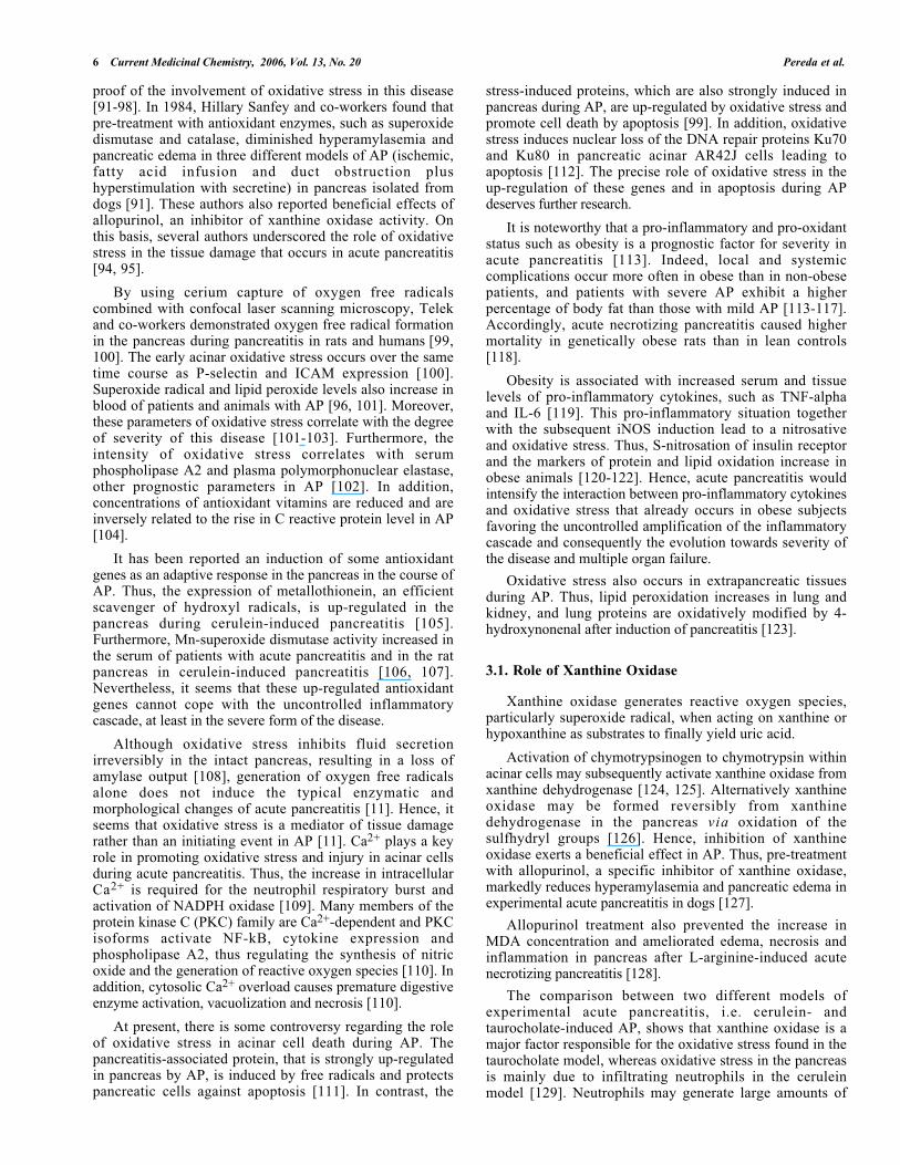
Fig. (1). Pathophysiological mechanisms of the systemic inflammatory response associated with acute pancreatitis. Modified fromGómez-Cambronero et al. 2002. Abbreviations: IL- 1β : interleukin 1β; Il-10: interleukin 10; PAF: platelet activating factor; PAP-I:pancreatitis-associated protein I; ROS : Reactive oxygen species; TNF-α : tumor necrosis factor α; XO : xanthine oxidase.
lymphocytes, and cellular adhesion molecules, which extendthe inflammatory response [5]. The relevance of IL-1ß inacute pancreatitis is currently established by the followingfindings:
-IL-1 RA, the receptor antagonist of IL-1, blocks theaction of this cytokine and diminishes the injury indistant organs in necrotizing acute pancreatitis [39,40].
-When necrotizing pancreatitis is induced in knockoutmice deficient in IL-1 receptors, the systemicresponse is restrained and consequently there is adecrease in mortality, although there is no reductionin severity of pancreatic damage [38].
-IL-1ß appears to mediate neutrophil infiltration andthe progression of acinar cell death by necrosis [41].Indeed, inhibition of ICE dramatically decreased IL-

4 Current Medicinal Chemistry, 2006, Vol. 13, No. 20 Pereda et al.
1ß protein expression and subsequent neutrophilinfiltration in the pancreas after induction of severeAP, reducing the extent of acinar cell necrosis [41].Furthermore, inhibition of ICE decreased the deathrate, acinar cell necrosis, IL-1ß and TNF-α levels andmyeloperoxidase activity after the induction ofpancreatitis by taurocholate in rats [42].
Although IL-1 RA may attenuate the severity ofexperimental AP, the IL-1beta:IL-1RA ratio has been foundto be lower in patients with severe acute pancreatitis than inthose with mild disease [25, 43]. Powell and co-workershave even suggested that IL-1 RA may be an early marker ofseverity [43].
Finally, it has been suggested that both IL-1 and TNF-αare pivotal in the propagation of the systemic inflammatoryresponse and the subsequent organ failure due to acutepancreatitis. This hypothesis is supported on the findingsthat a marked attenuation of severity in AP is observed whenthe receptors of IL-1 and TNF-α are blockaded and thatmortality is also dramatically reduced in IL-1 and TNF-αknockout mice after AP [44-46].
Therefore, clinical and experimental evidences show thatpro-inflammatory cytokines, especially TNF-α and IL-1ß,are essential to understand why pancreatic damage becomes asystemic disease. Nevertheless, most randomized trialscarried out so far to assess the beneficial effects of anti-cytokine therapies have yielded negative results on theoutcome, although the markers of inflammation werereduced. In our opinion, this does not mean that cytokinesdo not play a significant role in the development of AP,rather it suggests that a combined treatment providingsimultaneous blockade of different cytokines and other pro-inflammatory pathways, such as oxidative stress, is needed.
2.3. IL-6 and IL-8
Many other cytokines are involved in the systemicinflammatory response in AP. In fact, it is believed that theseverity of pancreatitis results from the ratio between pro-inflammatory and anti-inflammatory mediators. IL-6 is theprimary inducer of the acute phase protein response [47].Serum levels of IL-6 correlate with the severity of thedisease in patients with acute pancreatitis [26, 48, 49].Moreover, IL-6 increases earlier and may be a more sensitivemarker of severity than reactive protein C [49]. IL- 8 is apro-inflammatory cytokine released by activatedmacrophages or endothelial cells that is involved inneutrophil activation and degranulation [5]. IL-8 mediatesthe release of enzymes, such as elastase, from neutrophilsthat may cause tissue damage when overproduced [50]. Therelationship between severity of pancreatitis and serum IL-8levels is similar to that of IL-6 [51, 52]. In accordance withthis, peak concentrations of serum IL-8 rise in severe AP[53]. At present, both IL-6 and IL-8 are considered markersof severity of AP, although they are not the driving force forinitiation and propagation of the systemic inflammatoryresponse [18].
2.4. Platelet Activating Factor
Another mediator linked to the systemic inflammatoryresponse in AP is platelet activating factor (PAF), a lipid
that functions as a pro-inflammatory cytokine, since itinduces platelet activation and aggregation, neutrophil andmonocyte activation, chemotaxis, vasodilatation as well asan increase in vascular permeability [18,54]. PAF is releasedfrom activated leukocytes, platelets and endothelial cells[54]. The regulation of PAF production is closely related toIL-1 and TNF-α , as inhibition of production of either ofthese cytokines attenuates PAF production, whereasinhibition of PAF attenuates IL-1 and TNF-α production[18]. PAF has also been involved in the pathophysiology ofacute pancreatitis [55, 56]. An increase in PAF in thepancreatic tissue and in plasma in the course of experimentalAP has been observed [55, 57]. Moreover, a significant roleof PAF in AP is supported by the finding that PAFinjection in a pancreatic artery causes AP in rabbits [58].Furthermore, PAF antagonists exhibit beneficial effects inexperimental AP: diminishing plasma IL-1 levels andpermeability of pancreatic capillary endothelium [59] andreducing the severity of systemic inflammation [60].
2.5. IL-10 and PAP
Recently, much attention has been focused on the keyrole of the anti-inflammatory response mediated byInterleukin 10 (IL-10) and pancreatitis-associated protein I(PAP-I) in the evolution of acute pancreatitis.
IL-10 is a potent anti-inflammatory cytokine withimmunosuppressive and anti-inflammatory activities [61].Indeed, it inhibits the production of IL-1ß and TNF-α. IL-10plasma levels were highest on the day of admission forhospitalization in patients with AP and stayed high in severecases of AP, but decreased rapidly during the following daysin mild cases of AP [62]. Interestingly, an increase in IL-10levels relative to IL-6 or IL-8 is associated with improvedclinical outcome [63]. Accordingly, neutralization ofendogenous IL-10 by specific antibodies increased theseverity of pancreatitis and associated lung injury as well asTNF-α expression in experimental AP [64]. Consequently,IL-10 might play a crucial role determining the evolution ofAP towards the severe or to the mild form of the disease,and this prompted its use in the treatment of AP. Thus, up-regulation of IL-10 expression by gene therapy oradministration of IL-10 decreases the severity and mortalityin different models of AP [65-69].
A relationship between IL-10 and oxidative stress mayexist. The elevated IL-10 levels correlated with enhancedlipid peroxidation in patients with severe acute pancreatitis[70]. On the other hand, antioxidants may modulate the ratioof anti-inflammatory to pro-inflammatory cytokines inexperimental AP. Thus, administration of the antioxidant N-acetyl cysteine to rats with AP diminished the pancreaticinjury, enhanced the ability of acinar cells to produce IL-10at early stages and increased the ratio IL-10/IL-6 [71, 72].
Pancreatitis-associated protein I (PAP I) was initiallycharacterized as a protein overexpressed in acute pancreatitisand it is involved in the regulation of inflammation [73].Functional similarities to IL-10 suggest that PAP I couldplay a role similar to this anti-inflammatory cytokine inepithelial cells. PAP I inhibits the inflammatory response byblocking NF-kappaB activation [73]. Accordingly, PAPprevents TNF-alpha induced NF-kappaB activation and

Cytokines and Oxidative Stress in Pancreatitis Current Medicinal Chemistry, 2006 Vol. 13, No. 20 5
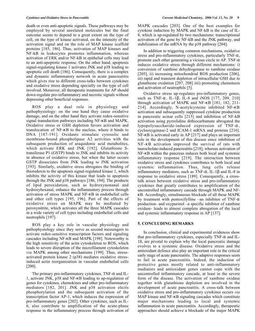
Fig. (2). Interaction between cytokines and oxidative stress in the inflammatory response in acute pancreatitis. Abbreviations used:IL- 1β : interleukin 1β; β; Il-10: interleukin 10; MnSOD: Mn-superoxide dismutase; PAP-I: pancreatitis-associated protein I; TNF-α :tumor necrosis factor.
down-regulates cytokine production and adhesion moleculeexpression [74, 75]. Hence, it may represent an importantanti-inflammatory mechanism in AP. Indeed, inhibition ofPAP expression by antisense oligodeoxyribonucleotidessignificantly worsened edema and fat necrosis and elevatedleukocyte infiltration in AP [76]. Consequently, PAP seemsto exert significant protection against pancreatic injury inAP.
2.6. Growth Factors and Heat Shock Proteins
Growth factors, such as IGF-1 and HGF, and heat shockproteins, such as Hsp27, Hsp60 and Hsp70, are up-regulatedin the pancreas during AP as protective mechanisms inresponse to acinar cell injury [77-82].
Thus, the protective effects of thermal-stress induced Hspagainst pancreatitis and particularly against trypsinogenactivation were reversed by blockade of Hsp70 with anHsp70-antisense oligonucleotide [80, 83]. Moreover, there isspontaneous activation of pancreatic trypsinogen in Hsp70.1knockout mice [84]. The induction of Hsp60 followingwater immersion stress also ameliorated cerulein-inducedpancreatitis in rats [85]. More recently, it has been reportedthat overexpression of Hsp27, a regulator of actinpolymerization, preserved the F-actin microfilaments,reduced the severity and protected against the systemicinflammatory response in cerulein-induced AP [86].
On the other hand, growth factors mediate at least part ofthe anti-inflammatory defence in the course of AP. Indeed,
HGF attenuates pancreatic damage in cerulein-inducedpancreatitis in rats at least in part by increasing theproduction of IL-10 [81]. Administration of IGF-1 alsoenhanced IL-10 production and reduced pancreatic damage incerulein-induced pancreatitis [87].
3. ROLE OF OXIDATIVE STRESS IN THEINFLAMMATORY RESPONSE IN ACUTEPANCREATITIS
Oxidative stress is defined as a disturbance of the balancebetween pro-oxidants and antioxidants in favor of the former[88]. Free radicals, which are any species that contains oneor more unpaired electrons, are considered the mostimportant pro-oxidant molecules because of their reactivity.The term “Reactive Oxygen Species” (ROS) is used toinclude all oxygen-derived toxic molecules, mostly freeradicals. ROS also act as inflammatory mediators throughinduction of leukocyte activation, migration, and adhesionas well as enhancing the expression of other inflammatorymediators, such as cellular adhesion molecules [89]. Indeed,antioxidants suppress nuclear factor kappaB (NF-κB )activation in vitro, a key regulator of cytokine induction,and inhibit cytokine production [90]. On the other hand, theactivation of leukocytes contributes to oxidative stress byproducing large amounts of ROS through NADPH oxidase.
During the last two decades, the beneficial effect ofantioxidants in the treatment of AP has provided an indirect
6 Current Medicinal Chemistry, 2006, Vol. 13, No. 20 Pereda et al.
proof of the involvement of oxidative stress in this disease[91-98]. In 1984, Hillary Sanfey and co-workers found thatpre-treatment with antioxidant enzymes, such as superoxidedismutase and catalase, diminished hyperamylasemia andpancreatic edema in three different models of AP (ischemic,fatty acid infusion and duct obstruction plushyperstimulation with secretine) in pancreas isolated fromdogs [91]. These authors also reported beneficial effects ofallopurinol, an inhibitor of xanthine oxidase activity. Onthis basis, several authors underscored the role of oxidativestress in the tissue damage that occurs in acute pancreatitis[94, 95].
By using cerium capture of oxygen free radicalscombined with confocal laser scanning microscopy, Telekand co-workers demonstrated oxygen free radical formationin the pancreas during pancreatitis in rats and humans [99,100]. The early acinar oxidative stress occurs over the sametime course as P-selectin and ICAM expression [100].Superoxide radical and lipid peroxide levels also increase inblood of patients and animals with AP [96, 101]. Moreover,these parameters of oxidative stress correlate with the degreeof severity of this disease [101-103]. Furthermore, theintensity of oxidative stress correlates with serumphospholipase A2 and plasma polymorphonuclear elastase,other prognostic parameters in AP [102]. In addition,concentrations of antioxidant vitamins are reduced and areinversely related to the rise in C reactive protein level in AP[104].
It has been reported an induction of some antioxidantgenes as an adaptive response in the pancreas in the course ofAP. Thus, the expression of metallothionein, an efficientscavenger of hydroxyl radicals, is up-regulated in thepancreas during cerulein-induced pancreatitis [105].Furthermore, Mn-superoxide dismutase activity increased inthe serum of patients with acute pancreatitis and in the ratpancreas in cerulein-induced pancreatitis [106, 107].Nevertheless, it seems that these up-regulated antioxidantgenes cannot cope with the uncontrolled inflammatorycascade, at least in the severe form of the disease.
Although oxidative stress inhibits fluid secretionirreversibly in the intact pancreas, resulting in a loss ofamylase output [108], generation of oxygen free radicalsalone does not induce the typical enzymatic andmorphological changes of acute pancreatitis [11]. Hence, itseems that oxidative stress is a mediator of tissue damagerather than an initiating event in AP [11]. Ca2+ plays a keyrole in promoting oxidative stress and injury in acinar cellsduring acute pancreatitis. Thus, the increase in intracellularCa2+ is required for the neutrophil respiratory burst andactivation of NADPH oxidase [109]. Many members of theprotein kinase C (PKC) family are Ca2+-dependent and PKCisoforms activate NF-kB, cytokine expression andphospholipase A2, thus regulating the synthesis of nitricoxide and the generation of reactive oxygen species [110]. Inaddition, cytosolic Ca2+ overload causes premature digestiveenzyme activation, vacuolization and necrosis [110].
At present, there is some controversy regarding the roleof oxidative stress in acinar cell death during AP. Thepancreatitis-associated protein, that is strongly up-regulatedin pancreas by AP, is induced by free radicals and protectspancreatic cells against apoptosis [111]. In contrast, the
stress-induced proteins, which are also strongly induced inpancreas during AP, are up-regulated by oxidative stress andpromote cell death by apoptosis [99]. In addition, oxidativestress induces nuclear loss of the DNA repair proteins Ku70and Ku80 in pancreatic acinar AR42J cells leading toapoptosis [112]. The precise role of oxidative stress in theup-regulation of these genes and in apoptosis during APdeserves further research.
It is noteworthy that a pro-inflammatory and pro-oxidantstatus such as obesity is a prognostic factor for severity inacute pancreatitis [113]. Indeed, local and systemiccomplications occur more often in obese than in non-obesepatients, and patients with severe AP exhibit a higherpercentage of body fat than those with mild AP [113-117].Accordingly, acute necrotizing pancreatitis caused highermortality in genetically obese rats than in lean controls[118].
Obesity is associated with increased serum and tissuelevels of pro-inflammatory cytokines, such as TNF-alphaand IL-6 [119]. This pro-inflammatory situation togetherwith the subsequent iNOS induction lead to a nitrosativeand oxidative stress. Thus, S-nitrosation of insulin receptorand the markers of protein and lipid oxidation increase inobese animals [120-122]. Hence, acute pancreatitis wouldintensify the interaction between pro-inflammatory cytokinesand oxidative stress that already occurs in obese subjectsfavoring the uncontrolled amplification of the inflammatorycascade and consequently the evolution towards severity ofthe disease and multiple organ failure.
Oxidative stress also occurs in extrapancreatic tissuesduring AP. Thus, lipid peroxidation increases in lung andkidney, and lung proteins are oxidatively modified by 4-hydroxynonenal after induction of pancreatitis [123].
3.1. Role of Xanthine Oxidase
Xanthine oxidase generates reactive oxygen species,particularly superoxide radical, when acting on xanthine orhypoxanthine as substrates to finally yield uric acid.
Activation of chymotrypsinogen to chymotrypsin withinacinar cells may subsequently activate xanthine oxidase fromxanthine dehydrogenase [124, 125]. Alternatively xanthineoxidase may be formed reversibly from xanthinedehydrogenase in the pancreas via oxidation of thesulfhydryl groups [126]. Hence, inhibition of xanthineoxidase exerts a beneficial effect in AP. Thus, pre-treatmentwith allopurinol, a specific inhibitor of xanthine oxidase,markedly reduces hyperamylasemia and pancreatic edema inexperimental acute pancreatitis in dogs [127].
Allopurinol treatment also prevented the increase inMDA concentration and ameliorated edema, necrosis andinflammation in pancreas after L-arginine-induced acutenecrotizing pancreatitis [128].
The comparison between two different models ofexperimental acute pancreatitis, i.e. cerulein- andtaurocholate-induced AP, shows that xanthine oxidase is amajor factor responsible for the oxidative stress found in thetaurocholate model, whereas oxidative stress in the pancreasis mainly due to infiltrating neutrophils in the ceruleinmodel [129]. Neutrophils may generate large amounts of

Cytokines and Oxidative Stress in Pancreatitis Current Medicinal Chemistry, 2006 Vol. 13, No. 20 7
superoxide radical and hydrogen peroxide by NADPHoxidase activation.
Xanthine Oxidase may contribute to the local pancreaticdamage. Thus, the superoxide generated by xanthine oxidasecaused injury in pancreatic acinar cells in vitro [130] andisolated zymogen granules were specially susceptible to thisoxidative damage [131]. In addition, the lipid compositionof pancreatic membranes changes during experimental acutepancreatitis, increasing the susceptibility of these membranesto lipid peroxidation induced by xanthine oxidase [132].
Closa and co-workers demonstrated that xanthine oxidaseis involved not only in pancreatic damage but also in thelung inflammation associated with experimental acutepancreatitis [133]. During the early stages of acutepancreatitis, α-amylase is absorbed from ascites fluid andonce in the circulation promotes the release of XO bybreaking down the binding of XDH/XO to glycoproteins ofthe endothelial cells. Proteolytic enzymes convert XDH intoits oxidase form contributing to the increase in circulatingXO that has been involved in the triggering of the systemicinflammatory process. Accordingly, the increased expressionof P-selectin, one of the major mediators of neutrophilinfiltration, in the lung appears to be triggered by amechanism dependent on free radicals generated by xanthineoxidase and on the availability of oxygen in the alveolar airspace [22, 134-136]. We have recently found that inhibitingxanthine oxidase using oxypurinol partially prevented therise in lung myeloperoxidase (MPO) activity, but combinedwith pentoxifylline, an inhibitor of TNF-α production,prevented almost completely both the increase in MPOactivity and leukocyte infiltration [137]. Xanthine oxidaseand TNF-α actions are not confined to system sites andendothelium since simultaneous administration ofpentoxifylline and oxypurinol also prevented to a greatextent the inflammatory infiltrate and the interstitial edemain the pancreas [137]. Hence, this combination lead to asignificant blockade of the inflammatory cascade in thepancreatic parenchyma and in the lung.
The beneficial effects of allopurinol in experimental APprompted several authors to perform clinical trials. Thus,recently the efficacy of allopurinol in the prevention of post-ERCP pancreatitis has been assessed in different clinicaltrials, but they give contradictory results. In a prospectiverandomized double-blind trial, Katsinelos et al. [138] havereported that pre-treatment with high dose allopurinoldecreased the frequency of post-ERCP pancreatitis. However,Mosler et al. [139] have found that prophylactic allopurinoltreatment did not affect the frequency or severity of post-ERCP pancreatitis.
3.2. Role of NADPH Oxidase
The burst of ROS generated during phagocytosis viaNADPH oxidase activation [140, 141] contributes topancreatic damage in AP. Thus, it has been reported that thecerulein-induced trypsin activation in the pancreas isattenuated in mice deficient in NADPH oxidase [142].Hence, neutrophils infiltrating the pancreas contribute tointrapancreatic activation of digestive enzymes via NADPHoxidase [142]. NADPH oxidase activation may also trigger
acinar cell death in AP since it causes ROS-inducedapoptosis in pancreatic acinar AR42J cells stimulated withcerulein [143].
3.3. Role of Glutathione
Reduced glutathione (GSH) is the most abundant non-protein thiol and a major antioxidant in mammalian cells.Glutathione plays an important role in the antioxidantdefense in acinar cells [144]. Glutathione depletion andincreased lipid peroxidation are found in the pancreatic tissueduring the initial phase of AP [93, 94]. Treatment withglutathione monoethyl ester increased pancreatic glutathionelevels and diminished the pancreatic damage induced bycerulein in mice [145]. Furthermore, pretreatment with N-acetylcysteine, a glutathione precursor, decreased the severityof acute pancreatitis in mice, since it diminished pancreaticlevels of conjugated dienes and IL-6, the severity of lunginjury and mortality [146]. On the contrary, inhibition ofglutathione synthesis with L-buthionine-(S,R)-sulfoximineled to more pancreatic necrosis and reduced survival in ratswith AP [147]. Interestingly, NF-κ B knockout miceexhibited increased glutathione levels and reducedhistological alterations, suggesting an interaction betweenNF-κB and glutathione in the pancreas [148]. It remains tobe proved whether lipid peroxidation and glutathionedepletion are the cause or consequence of AP [11].
We have found that glutathione depletion, but notglutathione oxidation, occurs initially in the pancreas in AP[37]. Therefore, detoxification of reactive oxygen speciesassociated with the inflammatory process does not appear tobe the major cause for the early glutathione depletion.Alternatively, depletion of pancreatic glutathione may be duein part to the activation of proenzymes, since it is knownthat activated proteases such as carboxypeptidase can cleaveGSH [149]. In fact, trypsinogen activation is accompaniedby glutathione depletion in experimental acute pancreatitis[144]. So far there are no new insights on this issue andfurther research is needed to elucidate the precise mechanismresponsible for glutathione depletion in acute pancreatitis.
Glutathione plays a role in acinar stimulus-secretioncoupling [150], and in the maintenance of the cytoskeleton[151] and appropriate protein folding in the endoplasmaticreticulum [152]. Thus, a depletion of intracellularglutathione may contribute to impaired zymogen granuletransport (secretory block) and to the premature activation ofpancreatic proenzymes [153, 154].
Mitochondrial damage is closely associated with severeglutathione depletion [149]. Mitochondria cannot synthesizeglutathione as they lack γ-glutamylcysteine synthetase orglutathione synthetase activities [149], and thus obtainglutathione by transport from the cytosol. Marked depletionof mitochondrial glutathione causes mitochondrial damage,which is completely prevented by administration ofglutathione monoesters [149]. Depletion of pancreaticglutathione may be responsible for the mitochondrialdamage associated with acute pancreatitis. In fact, we havefound that partial restoration of intracellular glutathionelevels following pentoxifylline treatment prevented damageto pancreatic mitochondria [37].

8 Current Medicinal Chemistry, 2006, Vol. 13, No. 20 Pereda et al.
3.4. Role of Nitrogen Reactive Species
Endothelial nitric oxide synthase (eNOS) generates nitricoxide (NO) as a potent vasodilator to maintain vascularhomeostasis, whereas inducible nitric oxide synthase (iNOS)produce large amounts of NO upon inflammation [155-158].Nitrogen reactive species, such as nitric oxide andparticularly the most harmful peroxynitrite, may generate anitrosative stress associated with the inflammatory process.NO reacts with superoxide radical (O2-.) to yieldperoxynitrite, a powerful oxidant which causes nitrosylationof protein thiols and nitration of aromatic amino acids [159].On the other hand, NO may modulate the inflammatoryresponse by inhibiting leukocyte activation, adhesion andmigration [160] and endogenous NO may even play aprotective role against oxidative damage to subcellularfractions [107]. Inducible nitric oxide synthase (iNOS) isstrongly up-regulated in pancreas and serum NO metabolites- such as NO2
-/NO3- and S-nitrosothiols - increase in the
course of AP [37, 97, 161-163]. Furthermore, peroxynitriteproduction rises in pancreas during AP, since there is anincrease of 3-nitrotyrosine formation in proteins [41]. TheNO production mediated by iNOS expression seems to bestrongly modulated by oxygen free radicals and neutrophilsin AP, since an anti-ICAM-1 antibody or superoxidedismutase plus catalase reduce iNOS expression in thepancreas during AP [41].
The role of nitric oxide in the pathogenesis of acutepancreatitis remains controversial, with some studiessuggesting that NO potentiates pancreatic oxidative stressand damage [164, 165], whereas others report that NOdiminishes oxidative damage to subcellular fractions andameliorates pancreatic dysfunction by enhancing pancreaticblood flow and/or secretion in response to endothelium-derived NO [107, 166-168]. This controversy also arisesregarding the role of iNOS, since one study reports enhancedsusceptibility to cerulein-induced pancreatitis in iNOS-deficient mice [169], whereas in another study iNOS-deficient mice exhibited resistance to cerulein-induced AP[170].
eNOS seems to play a significant role in the initiation ofAP since eNOS deletion, but not nNOS or iNOS deletion,increased pancreatic trypsin activity and serum lipase in theinitiation phase of cerulein-induced AP [171]. eNOS-derivedNO is likely to exert protective effects by promotingvasodilatation and increasing the blood flow in thepancreatic parenchyma. Moreover, constitutively producednitric oxide released by endothelial cells may inhibit therolling and adhesion of leucocytes in the microcirculation[172]. It has been reported that NO contributes to thepancreatic edema. It also causes nitrosative stress bygenerating peroxynitrite which induces protein nitration andnitrosylation. Nevertheless, nitrosative stress does not seemto be critical for acinar cell damage since no correlation hasbeen found between 3-nitrotyrosine formation, serumnitrite/nitrate levels and cell damage in the pancreas.Regarding the action(s) of NO on leukocytes, on the onehand it modulates leukocyte recruitment in tissues [172], buton the other hand when overproduced it may reduce theirhalf-life by inducing nitrosative stress.
Generation of large amounts of NO may be deleteriousfor the pancreas, since an excessive dose of L-arginine, a
substrate for NO synthase, induces acute necrotizingpancreatitis in rats [165]. NO leads to pancreatic edema inthis experimental model by increasing the vascularpermeability and protein extravasation [173]. Nevertheless,NO does not seem to contribute significantly to acinar celldamage in AP [97,161]. Indeed, no correlation has beenfound between 3-nitrotyrosine formation, serumnitrite/nitrate levels and acinar cell damage [97]. However,NO may play a significant role in the systemic responseassociated with AP. Thus, the AP-associated increase inserum phospholipase activity is involved in the activation ofalveolar macrophages, which produces a large amount ofnitric oxide contributing to the pulmonar damage [174-176].Moreover, inhibition of p38 mitogen activated kinasedecreases the pulmonary injury through attenuatedproduction of TNF-α and NO, suggesting a primary role forthese mediators in pancreatitis-induced respiratory distresssyndrome [177]. It is worth noting that total urinary nitriteexcretion is increased in patients with severe AP.Furthermore, patients with higher serum nitric oxide levelsexhibit a higher risk of sepsis and mortality [178]. Theseauthors have suggested that increase in serum NO may be aconsequence of endotoxin mediated up-regulation of iNOSactivity [179].
These results altogether demonstrate that oxidative stressand the antioxidant defence play an important role in thecritical early stage of the pathogenesis of AP and alsosuggest that not only activation of xanthine oxidase but alsoglutathione depletion are key mechanisms involved in thedevelopment of this disease. An important step in thisresearch should be to understand the interaction betweenoxidative stress, particularly oxygen free radicals, andcytokines which contribute to the uncontrolled inflammatorycascade in the systemic response associated with AP.
4. INTERACTION BETWEEN CYTOKINES ANDOXIDATIVE STRESS IN ACUTE PANCREATITIS.ROLE OF MAP KINASES AND NF-κκκκB
At present, there are experimental evidences of a synergybetween oxidative stress and pro-inflammatory cytokines inthe development of the inflammatory response in AP.Recently, we have shown that simultaneous inhibition ofTNF-α production and xanthine oxidase activity blocks to agreat extent the local and systemic inflammatory response inAP [137]. Oxidative stress and pro-inflammatory cytokinestrigger common signal transduction pathways that lead toamplification of the inflammatory cascade, mainly throughactivation of mitogen-activated protein kinases (MAPK) andnuclear factor kappaB (NF-kB) [180-182]. The MAPK are alarge family of serine/threonine kinases involved in cellproliferation, differentiation and death. Three major MAPKactivating cascades have been identified so far in mammaliancells: extracellular signal regulated kinases (ERK), c-jun N-terminal kinases (JNK) and p38 kinase [183]. The ERKsfunction in the control of cell division and consequently areassociated with proliferation and differentiation, whereas theJNKs and p38 are critical regulators of transcription involvedin the cellular response to pro-inflammatory cytokines andstress [183, 184].
MAP kinases and NF-kB are involved in a variety ofprocesses that include inflammation, cell proliferation, cell
Cytokines and Oxidative Stress in Pancreatitis Current Medicinal Chemistry, 2006 Vol. 13, No. 20 9
death or even anti-apoptotic signals. These pathways may beemployed by several unrelated molecules but the finaloutcome seems to depend to a great extent on the type ofcell, on the type of kinase activated, on the intensity of theactivation signal and on the role of MAP kinase scaffoldproteins [185, 186]. Thus, activation of MAP kinases andNF-kB in leukocytes promotes inflammation, whereasactivation of ERK and/or NF-kB in epithelial cells may leadto an anti-apoptotic response. On the other hand, apoptosissignal-regulating kinase 1 activates JNK and p38 leading toapoptotic cell death [186]. Consequently, there is a complexand dynamic inflammatory network in acute pancreatitiswhich gives rise to different cross-talks between cytokinesand oxidative stress depending specially on the type of cellinvolved. Moreover, all therapeutic treatments for AP shoulddown-regulate pro-inflammatory and necrotic signals withoutdepressing other beneficial responses.
ROS play a dual role in physiology andpathophysiology: on the one hand they cause oxidativedamage, and on the other hand they activate redox-sensitivesignal transduction pathways including NF-κB and MAPK.Oxidative stress or GSH depletion favors activation andtranslocation of NF-κB to the nucleus, where it binds toDNA [187-191]. Oxidants stimulate cytosolic andmembrane-bound phospholipase A2 activities with thesubsequent production of araquidonic acid metabolites,which activate ERK and JNK [192]. Glutathione S-transferase P1 (GSTP) binds to JNK and inhibits its activityin absence of oxidative stress, but when the latter occursGSTP dissociates from JNK leading to JNK activation[193]. Similarly, oxidative stress abrogates the binding ofthioredoxin to the apoptosis signal-regulated kinase 1, whichinhibits the activity of this kinase that leads to apoptosisthrough the JNK and p38 pathways [186, 194]. The productsof lipid peroxidation, such as hydroxynonanal andhydroxyhexanal, enhance the inflammatory process throughactivation of stress MAPK and NF-κB in endothelial cellsand other cell types [195, 196]. Part of the effects ofoxidative stress on MAPK may be mediated byperoxynitrite, which activates all the three MAPK cascadesin a wide variety of cell types including endothelial cells andneutrophils [197].
ROS play a key role in vascular physiology andpathophysiology since they serve as second messengers toactivate redox-sensitive transcription factors and signalingcascades including NF-κB and MAPK [198]. Noteworthy isthe high sensitivity of the actin cytoskeleton to ROS, whichleads to severe disruption of the microfilament cytoskeletonvia MAPK among other mechanisms [199]. Thus, stress-activated protein kinase 2 (p38) mediates oxidative stress-induced actin reorganization in vascular endothelial cells[200].
The primary pro-inflammatory cytokines, TNF-α and IL-1, activate JNK, p38 and NF-κB leading to up-regulation ofgenes for cytokines, chemokines and other pro-inflammatorymediators [182, 201]. JNK and p38 activation elicitsphosphorylation and the subsequent activation of thetranscription factor AP-1, which induces the expression ofpro-inflammatory genes [202]. Other cytokines, such as IL-6, also contribute to amplification of the acute-phaseresponse in the inflammatory process through activation of
MAPK cascades [203]. One of the best examples forcytokine induction by MAPK and NF-kB is the case of IL-8, which is up-regulated by two mechanisms: transcriptionalactivation of the gene by NF-kB and the JNK pathway, andstabilization of the mRNA by the p38 pathway [204].
In addition to triggering common mechanisms, oxidativestress and pro-inflammatory cytokines, particularly TNF-α,promote each other generating a vicious circle in AP. TNF-αinduces oxidative stress through different mechanisms: i)conversion of xanthine dehydrogenase to xanthine oxidase[205], ii) increasing mitochondrial ROS production [206],iii) rapid and transient depletion of intracellular GSH due toglutathione oxidation [207, 208] iiii) promoting chemotaxisand activation of neutrophils [5].
Oxidative stress up-regulates pro-inflammatory genes,such as TNF-α , IL-1β, IL-8 and iNOS [177, 209, 210]through activation of MAPK and NF-κB [181, 182, 211-214]. Accordingly, N-acetylcysteine inhibited NF-κBactivation and subsequently suppressed cytokine productionin pancreatic acinar cells [215] and inhibition of NF-kBactivation using pyrrolidine dithiocarbamate abrogated thelypopolysaccharide-induced expression of TNF-α ,cyclooxygenase-2 and ICAM-1 mRNA and proteins [216].NF-κB is activated early in AP [217] and plays an importantrole in the development of this disease since inhibition ofNF-κ B activation improved the survival of rats withtaurocholate-induced pancreatitis [218], whereas activation ofNF-κB within the pancreas induces both local and systemicinflammatory response [219]. The interaction betweenoxidative stress and cytokines contributes to both local andsystemic inflammation. Thus, lung cells releaseinflammatory mediators, such as TNF-α, IL-1β and IL-8 inresponse to oxidative stress [189]. Consequently, a cross-talk arises between oxidative stress and pro-inflammatorycytokines that greatly contributes to amplification of theuncontrolled inflammatory cascade through MAPK and NF-κB. Accordingly, simultaneous blockade of all these MAPKby treatment with pentoxifylline –an inhibitor of TNF-αproduction- and oxypurinol –a specific inhibitor of xanthineoxidase- leads to almost complete abrogation of the localand systemic inflammatory response in AP [137].
5. CONCLUDING REMARKS
In conclusion, clinical and experimental evidences showthat pro-inflammatory cytokines, especially TNF-α and IL-1ß, are pivotal to explain why the local pancreatic damageevolves to a systemic disease. Oxidative stress and theantioxidant defence also play an important role in the criticalearly stage of acute pancreatitis. The adaptive responses seemto fail in acute pancreatitis. Indeed, the induction ofprotective genes mostly related to anti-inflammatorymediators and antioxidant genes cannot cope with theuncontrolled inflammatory cascade, at least in the severeform of the disease. The activation of xanthine oxidasetogether with glutathione depletion are involved in thedevelopment of acute pancreatitis. A cross-talk betweenoxidative stress and pro-inflammatory cytokines occurs viaMAP kinase and NF-κB signaling cascades which constitutemajor mechanisms leading to local and systemicinflammation in acute pancreatitis. Accordingly, therapeuticapproaches should achieve a blockade of the major MAPK

10 Current Medicinal Chemistry, 2006, Vol. 13, No. 20 Pereda et al.
together with NF-κB to abrogate the inflammatory responsein acute pancreatitis.
ACKNOWLEDGEMENTS
The authors acknowledge the financial support obtainedby grants from Ministerio de Ciencia y TecnologíaSAF2003-05865 to J. S. and BFU2004-03616 to G. L-R.
REFERENCES
[1] Lankisch, P.G.; Assmus, C.; Maisonneuve, P.; Lowenfels, A.B.Pancreatology, 2002, 2, 469-477.
[2] Carballo, F.; Mateos, J. In Tratado de páncreas exocrine;Navarro, S.; Pérez-Mateo, M.; Guarner, L., Ed.; J&C EdicionesMédicas S.L.: Barcelona, 2002, pp. 118 – 132.
[3] Go, V.LW. In Acute Pancreatitis: Diagnosis and therapy;Bradley, E..L., Ed.; Raven Press : New York, 1994; IIIª ed, pp.235-239.
[4] Goldacre, M.J.; Roberts, S.E. BMJ, 2004, 328, 1466-1469.[5] Kusske, A.M.; Rongione, A.J.; Reber, H.A. Gastroenterology,
1996, 110, 639-642.[6] Bhatia, M.; Neoptolemos, J.P.; Slavin, J. Curr. Opin. Investig.
Drugs, 2001, 2, 496-501.[7] Yamauchi, J.; Shibuya, K.; Sunamura, M.; Arai, K.; Shimamura,
H.; Motoi, F.; Takeda, K.; Matsuno, S.J. Hepatobiliary Pancreat.Surg., 2001, 8, 195-203.
[8] Norton, I.D.; Clain, J.E. Drugs, 2001, 61, 1581.[9] Gómez-Cambronero, L.G.; Sabater, L.; Pereda, J.; Cassinello, N.;
Camps, B.; Viña, J.; Sastre, J. Curr. Drug Targets Inflamm.Allergy, 2002, 1, 393-403.
[10] Hofbauer, B.; Saluja, A.K.; Bhatia, M.; Frossard, J.L.; Lee, H.S.;Bhagat, L.; Steer, M.L. Gastroenterology, 1998, 115, 1238-1247.
[11] Rau, B.; Poch, B.; Gansauge, F.; Bauer, A.; Nussler, A.K.;Nevalainen, T.; Schoenberg, M.H.; Beger, H.G. Ann. Surg., 2000,231, 352-360.
[12] Edelson, J.D.; Vadas, P.; Villar, J.; Mullen, J.B.; Pruzanski, W.Am. Rev. Resp. Dis., 1991, 143, 1102-1109.
[13] Whicher, J.T.; Barnes, M.P.; Brown, A.; Cooper, M.J.; Read, R.;Walters, G.; Williamson, R.C. Gut, 1982, 23, 944-950.
[14] Buchler, M.W.; Gloor, B.; Muller, C.A.; Friess, H.; Seiler, C.A.;Uhl, W. Ann Surg., 2000, 232, 627-629.
[15] Nyström, P.O. J. Antimicrob. Chemoth., 1998, 41 (Suppl. A), 1-7.[16] Closa, D.; Folch-Puy, E. IUBMB Life., 2004, 56, 185-191.[17] Dinarello, C.A. FASEB J., 1994, 8, 1314-1325.[18] Norman, J. Am. J. Surg., 1998, 175, 76-83.[19] Dinarello, C.A.; Gelfand, J.A.; Wolff, S.M. JAMA, 1993 ,
269,1829-1835.[20] Bettinger, J.R.; Grendell, J.H. Pancreas, 1991, 6 (suppl. 1), 2-6.[21] Lerch, M.M.; Adler, G. Int. J. Pancreatol., 1994 , 15, 159-70.[22] Folch, E.; Salas, A.; Panes, J.; Gelpi, E.; Rosello-Catafau, J.;
Anderson, D.C.; Navarro, S.; Pique, J.M.; Fernandez-Cruz, L.;Closa, D. Ann. Surg., 1999, 230, 792-799.
[23] Grewal, H.P.; Mohey el Din, A.; Gaber, L.; Kotb, M.; Gaber,A.O. Am. J. Surg., 1994, 167, 214-219.
[24] Norman, J.G.; Fink, G.W.; Franz, M.G. Arch. Surg., 1995, 130,966-970.
[25] Mayer, J.; Rau, B.; Gansauge, F.; Beger, H.G. Gut, 2000, 47, 546-52.
[26] McKay, C.; Gallagher, G.; Brooks, B.; Imrie, C.W.; Baxter, J.N.Br. J. Surg., 1996, 83, 919-923.
[27] De Beaux, A.C.; Fearon, K.C.H. Scand. J. Gastroenterol., 1996,31 (Suppl. 219), 43-46.
[28] Grewal, H.P.; Kotb, M.; El Din, A.M.; Omán, M.; Salem, A.;Gaber, L.; Gaber, A.O. Surgery, 1994, 115, 213-221.
[29] Granell, S.; Pereda, J.; Gómez-Cambronero, L.G.; Cassinello, N.;Sabater, L.; Closa, D.; Sastre, J. Cytokine, 2004, 25, 187-191.
[30] Folch, E.; Serrano, A.; Sabater, L.; Gelpi, E.; Rosello-Catafau, J.;Closa, D. Crit. Care Med., 2001, 29, 1023-1026.
[31] Hirota, M.; Nozawa, F.; Okabe, A.; Shibata, M.; Beppu, T.;Shimada, S.; Egami, H.; Yamaguchi,Y.; Ikei, S.; Okajima, T.;Okamoto, K.; Ogawa, M. Pancreas, 2000, 21, 141-146.
[32] Norman, J.G.; Fink, G.W.; Denham, W.; Yang, J.; Carter, G.;Sexton, C.; Flakner, J.; Gower, W.R.; Franz, M.G. Dig. Dis. Sci.,1997, 42, 1783-1788.
[33] Hughes, C.B.; Henry, J.; Kotb, M.; Lobaschevsky, A.; Sabek, O.;Gaber, A.O. J. Surg. Res., 1995, 59, 687-693.
[34] Gloor, B.; Blinman, T.A.; Rigberg, D.A.; Todd, K.E.; Lane, J.S.;Hines, O.J.; Reber, H.A. Pancreas, 2000, 21, 414-420.
[35] Gukovskaya, A.; Perkins, P.; Zaninovic.; Sandoval, D.;Rutherford, R.; Fitzsimmons,T.; Pandol, S.J.; Poucell- Hatton, S.Gastroenterology, 1996, 110, 876-884.
[36] Hughes, C.B.; Grewal, H.P.; Gaber, L.W.; Kotb, M.; MoheyEl-din, A.B.; Mann, L.; Gaber, A.O. Am. J. Surg., 1996, 171, 274-280.
[37] Gómez-Cambronero, L.G.; Camps, B.; García de la Asunción, J.;Cerdá, M. ; Pallardó, F.V.; Sweiry, J.H.; Mann, G.E.; Viña, J.;Sastre, J. J. Pharmacol. Exp. Ther., 2000, 293, 670-676.
[38] Denham, W.; Yang, J.; Fink, G.; Denham, D.; Carter, G.; Ward,K.; Norman, J. Gastroenterology, 1997, 113, 1741-1746.
[39] Tanaka, N.; Murata, A.; Uda, K.; Toda, H.; Kato, T.; Hayashida,H.; Matsuura, N.; Mori, T. Crit. Care Med., 1995, 23, 901-908.
[40] Norman, J.G.; Franz, M.G.; Fink, G.S.; Messina, J.; Fabri, P.J.;Gower, W.R.; Carey, L.C. Ann. Surg., 1995 b, 221, 625-634.
[41] Rau, B.; Paszkowski, A.; Lillich, S.; Baumgart, K.; Moller, P.;Beger, H.G. Lab. Invest., 2001, 81, 1001-13.
[42] Paszkowski, A.S.; Rau, B.; Mayer, J.M.; Moller, P.; Beger, H.G.Ann. Surg., 2002, 235, 68-76.
[43] Powell, J.J.; Fearon, K.C.; Siriwardena, A.K.; Ross, J.A. Surgery,2001, 129, 633-40.
[44] Pastor, C.M.; Frossard, J.L. FASEB J., 2001, 15, 893-897.[45] Pastor, C.M.; Matthay, M.A.; Frossard, J.L. New Insights Chest,
2003, 124, 2341-2351.[46] Zyromski, N.; Murr, M.M. Surgery, 2003, 133, 235-237.[47] Biffl, W.L.; Moore, E.E.; Moore, F.A.; Peterson, V.M. Ann. Surg.,
1996, 224, 647-664.[48] Leser, H.G.; Gross, V.; Scheibenbogen, C.; Heinisch, A.; Salm, R.;
Lausen, M.; Ruckauer, K.; Andreesen, R.; Farthmann, E.H.;Scholmerich, J. Gastroenterology, 1991, 101, 782-785.
[49] Viedma, J.A.; Pérez-Mateo, M.; Domínguez, J.E.; Carballo, F.Gut, 1992, 33, 1264-1267.
[50] Baggiolini, M.; Loetscher, P.; Moser, B. Int. J.Immunopharmacol., 1995, 7, 103-108.
[51] Gross, V.; Andreesen, R.; Leser, H.G. ; Ceska, M.; Liehl, E.;Lausen, M.; Farthmann, E.H.; Scholmerich, J. Eur. J. Clin. Invest.,1992, 22, 200-203.
[52] Pezzilli, R.; Billi, P.; Miniero, R.; Fiocchi, M.; Cappelletti O.;Morselli-Labate, A.M.; Barakat, B.; Sprovieri, G.; Miglioli, M.Dig. Dis. Sci., 1995, 40, 2341-2348.
[53] Rau, B.; Steinbach, G.; Gansauge, F.; Mayer, J.M.; Grunert, A.;Beger, H.G. Gut, 1997, 41, 832-840.
[54] McKay, C.J.; Curran, F.; Sharples, C.; Baxter, J.N.; Imrie, C.W.Br. J. Surg., 1997, 84, 1239-1243.
[55] Konturek, S.J.; Dembinski, A.; Konturek, P.J.; Warzecha, Z.;Jaworek, J.; Gustaw, P.; Tomaszewska, R.; Stachura J. Gut, 1992,33, 1268-1274.
[56] Kingsnorth, A.N. Scand. J. Gastroenterol., 1996, 31, S28-S31.[57] Ais, J.G.; Novo, C.; Gomez-Garre, M.; Lopez-Farre, A.; Lopez-
Novoa, J.M.; Romeo, J.M. Rev. Esp. Enf. Digest., 1992, 81, 25-28.[58] Emanuelli, G.; Montrucchio, G.; Gaia, E.; Dughera, L.; Corvetti,
G.; Gubetta, L. Am. J. Pathol., 1989, 134, 315-326.[59] Wang, X.; Sun, Z.; Borjesson, A.; Haraldsen, P.; Aldman, M.;
Deng, X.; Leveau, P.; Andersson, R. Int. J. Pancreatol., 1999, 25,44-52.
[60] Lane, J.S.; Todd, K.E.; Gloor, B.; Chandler, C.F.; Kau, A.W.;Ashley, S.W.; Reber, H.A.; McFadden, D.W. J. Surg. Res., 2001,99, 365-370.
[61] Dumot, J.A.; Conwell, D.L.; Zuccaro, G. Jr; Vargo, J.J.; Shay,S.S.; Easley, K.A.; Ponsky, J.L. Am. J. Gastroenterol., 2001, 96,2098-102.
[62] Berney, T.; Gasche, Y.; Robert, J.; Jenny, A.; Mensi, N.; Grau, G.;Vermeulen, B.; Morel, P. Pancreas, 1999, 18, 371-7.
[63] Simovic, M.O.; Bonham, M.J.; Abu-Zidan, F.M.; Windsor, J.A.Crit. Care Med., 1999, 27, 2662-5.
[64] Van Laethem, J.L.; Eskinazi, R.; Louis, H.; Rickaert, F.;Robberecht, P.; Deviere, J. Gut, 1998, 43, 408-13.
[65] Van Laethem, J.L.; Marchant, A.; Delvaux, A.; Goldman, M.;Robberecht, P.; Velu, T.; Deviere, J. Gastroenterology, 1995, 108,1917-1922.
Cytokines and Oxidative Stress in Pancreatitis Current Medicinal Chemistry, 2006 Vol. 13, No. 20 11
[66] Kusske, A.M.; Rongione, A.J.; Ashley, S.W.; McFadden, D.W.;Reber, H.A. Surgery, 1996, 120, 284-8.
[67] Rongione, A.J.; Kusske, A.M.; Kwan, K.; Ashley, S.W.; Reber,H.A.; McFadden, D.W. Gastroenterology, 1997, 112, 960-967.
[68] Denham, W.; Denham, D.; Yang, J.; Carter, G.; MacKay, S.;Moldawer, L.L.; Carey, L.C.; Norman, J. Ann. Surg., 1998, 227,812-20.
[69] Chen, Z.Q.; Tang, Y.Q.; Zhang, Y.; Jiang, Z.H.; Mao, E.Q.; Zou,W.G.; Lei, R.Q.; Han, T.Q.; Zhang, S.D. World J. Gastroenterol.,2004, 10, 3021-5.
[70] Wereszczynska-Siemiatkowska, U.; Dabrowski, A.;Siemiatkowski, A.; Mroczko, B.; Laszewicz, W.; Gabryelewicz,A. Pancreas, 2003, 26, 144-52.
[71] Ramudo, L.; Manso, M.A.; Vicente, S.; De Dios, I. Cytokine,2005, 32, 125-31.
[72] Shi, C.; Zhao, X.; Wang, X.; Andersson, R. Scand. J.Gastroenterol., 2005, 40, 103-8.
[73] Folch-Puy, E.; Granell, S.; Dagorn, J.C.; Iovanna, J.L.; Closa, D. J.Immunol., 2006, 176, 3774-3779.
[74] Vasseur, S.; Folch-Puy, E.; Hlouschek, V.; Garcia, S.; Fiedler, F.;Lerch, M.M.; Dagorn, J.C.; Closa, D.; Iovanna, J.L. J. Biol.Chem., 2004, 279, 7199-207.
[75] Gironella, M.; Iovanna, J.L.; Sans, M.; Gil, F.; Penalva, M.; Closa.D.; Miquel, R.; Pique, J.M.; Panes, J. Gut, 2005, 54, 1244-53.
[76] Zhang, H.; Kandil, E.; Lin, Y.Y.; Levi, G.; Zenilman, M.E. Scand.J. Gastroenterol., 2004, 39, 870-81.
[77] Weber, C.K.; Gress, T.; Muller-Pillasch, F.; Lerch, M.M.;Weidenbach, H.; Adler, G. Pancreas, 1995, 10, 360-367.
[78] Wagner, A.C.; Weber, H.; Jonas, L.; Nizze, H.; Strowski, M.;Fiedler, F.; Printz, H.; Steffen, H.; Goke, B. Gastroenterology,1996, 111, 1333-1342.
[79] Menke, A.; Yamaguchi, H.; Giehl, K.; Adler, G. Pancreas, 1999,18, 28-33.
[80] Bhagat, L.; Singh, V.P.; Hietaranta, A.J.; Agrawal, S.; Steer, M.L.;Saluja, A.K. J. Cin. Invest., 2000, 106, 81-89.
[81] Warzecha, Z.; Dembinski, A.; Konturek, P.C.; Ceranowicz, P.;Konturek, S.J.; Tomaszewska, R.; Schuppan, D.; Stachura, J.;Nakamura, T. Eur. J. Pharmacol., 2001, 430, 113-121.
[82] Rakonczay, Z. Jr.; Takacs, T.; Boros, I.; Lonovics, J. J. Cell.Physiol., 2003, 195, 383-391.
[83] Bhagat, L.; Singh, V.P.; Song, A.M.; van Acker, G.J.; Agrawal, S.;Steer, M.L.; Saluja, A.K. Gastroenterology, 2002, 122, 156-165.
[84] Hwang, J.H.; Ryu, J.K.; Yoon, Y.B.; Lee, K.H.; Park, Y.S.; Kim,J.W.; Kim, N; Lee, D.H.; Jeong, J.B.; Seo, J.S.; Kim; Y.T.Pancreas, 2005, 31, 332-336.
[85] Lee, H.S.; Bhagat, L.; Frossard, J.L., Hietaranta, A.; Singh, V.P.;Steer, M.L.; Saluja, A.K. Gastroenterology, 2000, 119, 220-229.
[86] Kubisch, C.; DiMagno, M.J.; Tietz, A.B.; Welsh, M.J.; Ernst, S.A.;Brandt-Nedelev, B.; Diebold, J.; Wagner, A.C.; Goke, B.;Williams, J.A., Schafer, C. Gastroenterology, 2004, 127, 275-286.
[87] Warzecha, Z.; Dembinski, A.; Ceranowicz, P.; Konturek, S.J.;Tomaszewska, R.; Stachura, J.; Konturek, P.C. J. Physiol.Pharmacol., 2003, 54, 575-590.
[88] Sies, H. Angewandte Chem., 1986, 25, 1058-1071.[89] Telek, G.; Ducroc, R.; Scoazec, J.Y.; Pasquier, C.; Feldmann, G.;
Roze, C. J. Surg. Res., 2001, 96, 56-67.[90] Blanchard, J.A.2nd.; Barve, S.; Joshi-Barve, S.; Talwalker, R.;
Gates, L.K. Jr. 2. Dig. Dis. Sci., 2001, 46, 2768-2772.[91] Sanfey, H.; Bulkley, G.B.; Cameron, J.L. Ann. Surg., 1984, 200,
405-413.[92] Guice, K.S.; Miller, D.E.; Ofanan, R.T.; Towsend, C.M.;
Thompson, J.C. Am. J. Surg., 1986, 151, 163-169.[93] Schoenberg, M.H.; Büchler, M.; Beger, H.G. Free. Radic. Biol.
Med., 1992, 12, 515-522.[94] Sweiry, J.H.; Mann, G.E. Scand. J. Gastroenterol., 1996, 31
(Suppl. 219), 10-15.[95] Dabrowski, A.; Konturek, S.J.; Konturek, J.W.; Gabryelewicz, A.
Eur. J. Pharmacol., 1999, 377, 1-11.[96] Schulz, H.U.; Niederau, C.; Klonowsk-Stumpe, H.; Halangk, W.;
Lüthen, R.; Lippert, H. Hepatogastroenterology, 1999, 46, 2736-2750.
[97] Rau, B.; Bauer, A.; Wang, A.; Gansauge, F.; Weidenbach, H.;Nevalainen, T.; Poch, B.; Beger, H.G.; Nussler, A.K. Ann. Surg.,2001, 233, 195-203.
[98] Fu, K.; Tomita, T.; Sarras, M.P. Jr; De Lisle, R.C.; Andrews, G.K.Pancreas, 1998, 17, 238-246.
[99] Tomasini, R.; Samir, A.A.; Vaccaro, M.I.; Pebusque, M.J.;Dagorn, J.C.; Iovanna, JL.; Dusetti, N.J. J. Biol. Chem., 2001, 276,44185-44192.
[100] Telek, G.; Regoly-Merei, J.; Kovacs, G.C.; Simon, L.; Nagy, Z.;Hamar, J.; Jakab, F. Hepatogastroenterology, 2001, 48, 1252-1258.
[101] Tsai, K.; Wang, S.S.; Chen, T.S.; Kong, C.W.; Chang, F.Y.; Lee,S.D.; Lu, F.J. Gut, 1998, 42, 850-855.
[102] Wereszczynska-Siemiatkowska, Dabrowski, A.; Jedynak, M.;Gabryelewicz, A. Pancreas, 1998, 17, 163-168.
[103] Abu-Zidan, F.M.; Bonham, M.J.; Windsor, J.A. Br. J. Surg., 2000,87, 1019-23.
[104] Curran, F.J.; Sattar, N.; Talwar, D.; Baxter, J.N.; Imrie, C.W. Br.J. Surg., 2000, 87, 301-305.
[105] Fu, K.; Sarras, M.P. Jr.; De Lisle, R.C.; Andrews, G.K. Am JPhysiol., 1997, 273 (3Pt 1), G696-G705.
[106] Simovic, S.O.; Bonham, M.J.D.; Abu-Zidan, F.M.; Windsor, J.A.Pancreas, 1997, 15, 78-82.
[107] Sanchez-Bernal, C.; Garcia-Morales, O.H.; Dominguez, C.;Martin-Gallan, P.; Calvo, J.J.; Ferreira, L.; Perez-Gonzalez, N.Páncreas, 2004, 28, e9-e15.
[108] Sweiry, J.H.; Shibuya, I.; Asada, N.; Niwa, K.; Doolabh, K.;Habara, Y.; Kanno, T.; Mann, G.E. Biochim. Biophys. Acta, 1999,1454, 19-30.
[109] Kim-Park, W.K.; Moore, M.A.; Hakki, Z.W.; Kowolik, M.J. Ann.N.Y. Acad. Sci., 1997, 832, 394-404.
[110] Petersen, O.H.; Sutton, R. Trends Pharmacol. Sci., 2006, 27, 113-120.
[111] Ortiz, E.M.; Dusetti, N.J.; Vasseur, S.; Malka, D.; Bodeker, H.;Dagorn, J.C.; Iovanna, J.L. Gastroenterology, 1998, 114, 808-816.
[112] Song, J.Y.; Lim, J.W.; Kim, H.; Morio, T.; Kim, K.H. J. Biol.Chem., 2003, 278, 36676-36687.
[113] Martínez, J.; Sánchez-Payá, J.; Palazón, J.M.; Suazo-Barahona, J.;Robles-Díaz, G.; Pérez-Mateo, M. Pancreatology, 2004, 4, 42-8.
[114] Porter, K.A.; Banks, P.A. Int. J. Pancreatol., 1991, 10, 247-52.[115] Funnell, I.C.; Bornman, P.C.; Weakley, S.P.; Terblanche, J.;
Marks, I.N. Br. J. Surg., 1993, 80, 484-6.[116] Martínez, J.; Sánchez-Payá, J.; Palazón, J.M.; Aparicio, J.R.; Picó,
A.; Pérez-Mateo, M. Pancreas, 1999, 15-20.[117] Johnson, C.D.; Toh, S.K.; Campbell, M.J. Pancreatology, 2004, 4,
1-6.[118] Segersvard, R.; Sylvan, M.; Herrington, M.; Larsson, J.; Permert,
J. Scand. J. Gastroenterol., 2001, 36, 658-63.[119] Perrault, M.; Marette, A. Nat. Med., 2001, 7, 1138-43.[120] Beltowski, J.; Wojcicka, G.; Gorny, D.; Marciniak, A. J. Physiol.
Pharmacol., 2000, 51, 883-96.[121] Asayama, K.; Nekane, T.; Dobashi, K.; Kodera, K.; Hayashibe,
H.; Uchida, N.; Nakazawa, S. Free Radic. Res., 2001, 34, 337-47.[122] Carvalho-Filho, M.A.; Ueno, M.; Hirabara, S.M.; Seabra, A.B.;
Carvalheira, J.B.; de Oliveira, M.G.; Velloso, L.A.; Curi, R.; Saad,M.J. Diabetes, 2005, 54, 959-67.
[123] Gilgenast, O.; Brandt-Nedelev, B.; Wiswedel, I.; Lippert, H.;Halangk, W.; Reinheckel, T. Dig. Dis. Sci., 2001, 46, 932-937.
[124] Butelli, M.G.; Lorenzoni, S.; Stirpe, F. Biochem. J., 1973, 131,191-198.
[125] Sanfey, H.; Targarona, E.M.; Fernández-Cruz, L. Gastroenterol.Hepatol., 1991, 14, 172-180.
[126] Nordback, I.H.; Cameron, J.L. Surgery, 1993, 113, 90-97.[127] Sanfey, H.; Bulkley, G.B.; Cameron, J.L. Ann. Surg., 1985, 201,
633-639.[128] Czako, L.; Takacs, T.; Varga, I.S.; Hai, D.Q.; Tiszlavicz, L.;
Hegyi, P.; Mandi, Y.; Matkovics, B.; Lonovics, J. J. Physiol. Paris,2000, 94, 43-50.
[129] Closa, D.; Bulbena, O.; Hotter, G.; Rosello-Catafau, J.;Fernandez-Cruz, L.; Gelpi, E. Arch. Int. Physiol. Biochim.Biophys., 1994, 102, 167-170.
[130] Tamura, K.; Manabe, T.; Imanishi, K.; Nishikawa, H.; Ohshio, G.;Tobe, T. Hepatogastroenterology, 1992, 39, 536-539.
[131] Schulz, H.U.; Niederau, C. Hepatogastroenterology, 1994, 41,309-312.
[132] Ferreira, L.; Perez-Gonzalez, N.; Llanillo, M.; Calvo, J.J.;Sanchez-Bernal, C. Lipids, 2002, 37, 167-171.
[133] Folch, E.; Gelpi, E.; Rosello-Catafau, J.; Closa, D. Dig. Dis. Sci.,1998, 43, 2405-2410.
[134] Folch, E.; Prats, N.; Hotter, G.; Lopez, S.; Gelpi, E.; Rosello-Catafau, J.; Closa, D. Dig. Dis. Sci., 2000, 45, 1535-1544.

12 Current Medicinal Chemistry, 2006, Vol. 13, No. 20 Pereda et al.
[135] Granell, S.; Bulbena, O.; Genesca, M.; Sabater, L.; Sastre, J.;Gelpi, E.; Closa, D. BMC Gastroenterol., 2004, 4, 1-10.
[136] Granell, S.; Serrano-Mollar, A.; Folch-Puy, E.; Navajas, D.; Farre,R.; Bulbena, O.; Closa, D. Free Radic. Biol. Med., 2004, 37, 1640-1647.
[137] Pereda, J.; Sabater, L.; Casinello, N.; Gómez-Cambronero, L.;Closa, D.; Folch E.; Aparisi, L.; Calvete, J.; Lledó, S.; Viña,J;Sastre, J. Ann. Surg., 2004, 240, 108-116.
[138] Katsinelos, P.; Kountouras, J.; Chatzis, J.; Christodoulou, K.;Paroutoglou, G.; Mimidis, K.; Beltsis, A.; Zavos, C., Gastrointest.Endosc., 2005, 61, 407-415.
[139] Mosler, P.; Sherman, S.; Marks, J.; Watkins, J.L.; Geenen, J,E.;Jamidar, P.; Fogel, E.L.; Lazzell-Pannell, L.; Temkit, M.;Tarnasky, P.; Block, K.P.; Frakes, J.T.; Aziz, A.A.; Malik, P.;Nickl, N.; Slivka, A.; Goff, J.; Lehman G.A. Gastrointest. Endosc.,2005, 62, 245-50.
[140] Donko, A.; Peterfi, Z.; Sum, A.; Leto, T.; Geiszt, M. Philos. Trans.R. Soc. Lond B Biol. Sci., 2005, 360, 2301-2308.
[141] Sheppard, F.R.; Kelher, M.R.; Moore, E.E.; McLaughlin N.J.;Banerjee, A.; Silliman, C.C. J. Leukoc. Biol., 2005, 78, 1025-1042.
[142] Gukovskaya, A.S.; Vaquero, E.; Zaninovic, V.; Gorelick, F.S.;Lusis, A.J.; Brennan, M.L.; Holland, S.; Pandol, S.J.Gastroenterology, 2002, 122, 974-984.
[143] Yu, J.H.; Lim, J.W.; Kim, K.H.; Morio, T.; Kim, H. Free Radic.Biol. Med., 2005, 39, 590-602.
[144] Lüthen, R.; Grendell, J.H.; Niederau, C.; Haussinger, D. Int. J.Pancreatol., 1998, 24, 193-202.
[145] Neuschwander-Tetri, B.A.; Ferrell, L.D.; Sukhabote, R.J.;Grendell, J.H. J. Clin. Invest., 1992, 89, 109-116.
[146] Demols, A.; Van Laethem, J.L.; Quertinmont, E.; Legros, F.;Louis, H.; Le Moine, O.; Deviere, J. Pancreas, 2000, 20, 161-169.
[147] Alsfasser, G.; Gock, M.; Herzog, L.; Gebhard, M.M.; Herfarth,C.; Klar, E.; Schmidt, J. Dig. Dis. Sci., 2002, 47, 1793-1799.
[148] Altavilla, D.; Famulari, C.; Passaniti, M.; Galeano, M.; Macri, A.;Seminara, P.; Minutoli, L.; Marini, H.; Calo, M.; Venuti, F.S.;Esposito, M.; Squadrito, F. Lab. Invest., 2003, 83, 1723-1732.
[149] Meister, A. Pharmacol. Ther., 1991, 51, 155-194.[150] Stenson, W.F.; Lobos, E.; Wedner, H.J. Am. J. Physiol., 1983, 244,
273-277.[151] Jewell, S.A.; Bellomo, G.; Thor, H.; Orrenius, S. Sci. Wash. DC.,
1982, 217, 1257-1259.[152] Scheele, G.; Jakoby, R. J. Biol. Chem., 1982, 257, 12277-12282.[153] Lüthen, R.; Niederau, C.; Groendel, J.H. Am. J. Physiol., 1995,
268, G592-G604.[154] Yu, H.; Klonowski-Stumpe, H.; Lüthen, R. Pancreas, 2002, 24,
53-62.[155] Moncada, S.; Palmer, R.M.; Higgs, E.A. Pharmacol. Rev., 1991,
43, 109-142.[156] Ignarro, L.J.; Cirino, G.; Casini, A.; Napoli, C. J. Cardiovasc.
Pharmacol., 1999, 34, 879-886.[157] Ignarro, L.J. Curr. Top. Med. Chem., 2005, 5, 595.[158] Moncada, S.; Higgs, E.A. Br. J. Pharmacol., 2006, 147 (Suppl 1),
S193-S201.[159] Beckman, J.S.; Koppenol, W.H. Am. J. Physiol., 1996, 271 (Pt1),
1424-1437.[160] Kubes, P.; Suzuki, M.; Granger. D.N. Proc. Natl. Acad. Sci. USA,
1991, 88, 4651-4655.[161] Closa, D.; Hotter, G.; Prats N.; Bulbena, O.; Rosello-Catafau, J.;
Fernandez-Cruz, L.; Gelpi, E. Inflammation, 1994b, 18, 469-480.[162] Al-Mufti, R.A.; Williamson, R.C.; Mathie, R.T. Gut, 1998, 43, 564-
570.[163] Chen, H.M.; Shyr, M.H.; Lau, Y.T.; Hwang, T.L.; Chen, M.F.
Shock, 1998, 10, 218-222.[164] Dabrowski, A.; Gabryelewicz, A. Scand. J. Gastroenterol., 1994,
29, 943-948.[165] Tani, S.; Itoh, H.; Okabayashi, Y.; Nakamura, T.; Fujii, M.;
Fujisawa, T.; Koide, M.; Otsuki, M. Dig. Dis. Sci., 1990, 35, 367-374.
[166] Gukovskaya, A.; Pandol, S. Am. J. Physiol., 1994, 266 (3Pt1), 350-356.
[167] Holst, J.J.; Rasmussen, T.N.; Schmidt, P. Am. J. Physiol., 1994, 266(2Pt1), G206-G213.
[168] Satoh, A.; Shimosegawa, T.; Abe, T.; Kikuchi, Y.; Abe, R.;Koizumi, M.; Toyota, T. Pancreas, 1994, 9, 574-579.
[169] Qui, B.; Mei, Q.B.; Ma, J.J.; Korsten, M.A. Pancreas, 2001, 23,89-93.
[170] Cuzzocrea, S.; Mazzon, E.; Dugo, L.; Serraino, I.; Centorrino, T.;Ciccolo, A.; Van de Loo, F.A.; Britti, D.; Caputi, A.P.;Thiemermann, C. Shock, 2002, 17, 416-422.
[171] DiMagno, M.J.; Williams, J.A.; Hao, Y.; Ernst, S.A.; Owyang, C.Am. J. Physiol. Gastrointest. Liver Physiol., 2004, 287, 80-87.
[172] Hickey, M.J. Clin. Sci., 2001, 100, 1-12.[173] Takacs, T.; Czako, L.; Morschl, E.; Laszlo, F.; Tiszlavicz, L.;
Rakonczay, Z. Jr.; Lonovics, J. Pancreas, 2002, 25, 277-282.[174] Edelson, J.D.; Vadas, P.; Villar, J.; Mullen, J.B.M. Prizanski W.
Am. Rev. Respir. Dis., 1991, 143, 1102-1109.[175] Closa, D.; Sabater, L.; Fernández-Cruz, L.; Prats, N.; Gelpí, E.;
Roselló-Catafau, J. Ann. Surg., 1999, 229, 230-36.[176] Tsukahara, Y.; Morisaki, T.; Horita, Y.; Torisu, M.; Tanaka, M.
Ann. Surg., 1999, 229, 385-392.[177] Denham, W.; Yang, J.; Wang, H.; Botchkina, G.; Tracey, K.J.;
Norman, J. Crit. Care Med., 2000, 28, 2567-2572.[178] Mettu, S.R.; Wig, J.D.; Khullar, M.; Singh, G.; Gupta, R.
Pancreatology, 2003, 3,506-513.[179] Rahman, I. J. Biochem. Mol. Biol., 2003, 36, 95-109.[180] Raingeaud, J.; Gupta, S.; Rogers, J.S.; Dickens, M.; Han, J.;
Ulevitch, R.J.; Davis, R.J. J. Biol. Chem., 1995, 270, 7420-7426.[181] Wang, X.; Martindale, J.L.; Liu, Y.; Holbrook, N.J. Biochem. J.,
1998, 333, 291-300.[182] Rahman, I.; MacNee, W. Free Radic. Biol. Med., 2000, 28, 1405-
1420.[183] Torres, M.; Forman, H.J. Biofactors, 2003, 17, 287-296.[184] Johnson, G.L.; Lapadat, R. Science, 2002, 298, 1911-1912.[185] Morrison, D.K.; Davis, R.J. Annu. Rev. Cell. Dev. Biol., 2003, 19,
91-118.[186] Sumbayev, V.V.; Yasinska, I.M. Arch. Biochem. Biophys., 2005,
436, 406-412.[187] Ginn-Pease, M.E.; Whisler, R.L. Biochem. Biophys. Res.
Commun., 1996, 226, 695-702.[188] Bowie, A.G.; Moynagh, P.N.; O´Neill, L.A. J. Biol. Chem., 1997,
272, 25941-25950.[189] Rahman, I.; MacNee, W. Thorax, 1998, 53, 601-612.[190] Janssen-Heininger, Y.M.W.; Macara, I.; Mossman, B.T. Am. J.
Respir. Cell. Mol. Biol., 1999, 20, 942-952.[191] Oliva, M.R., Iradi, A., Garrido, F., Ramos, M., Oltra, A.M.,
Muñiz, P., Sáez, G.T. Free Radic. Res., 2001, 35, 119-128.[192] Chakraborti, S.; Chakraborti, T. Cell Signal. 1998, 10, 675-683.[193] Adler, V.; Yin, Z.; Fuchs, S.Y.; Benezra, M.; Rosario, L.; Tew,
K.D.; Pincus, M.R.; Sardana, M. et al. EMBO J., 1999, 18, 1321-1334.
[194] Saitoh, M.; Nishitoh.; Fujii, M.; Takeda, K.; Tobiume, K.; Sawada,Y.; Kawabata, M.; Miyazono, K. EMBO J., 1998, 17, 2596-2606.
[195] Rahman, S.H.; Ammori, B.J.; Larvin, M.; McMahon, M.J. Gut,2003, 52, 270-274.
[196] Je, J.H.; Lee, J.Y.; Jung, K.J.; Sung, B.; Go, E.K.; Yu, B.P.; Chung,H.Y. FEBS lett., 2004, 566, 183-189.
[197] Klotz, L.O.; Schoroeder, P.; Sies H. Free Radic. Biol. Med., 2002,33, 737-743.
[198] Griendling, K.K.; Sorescu, D.; Lassegue, B.; Ushio-Fukai M.Aterioscler. Thromb. Vasc. Biol., 2000, 20, 2175-2183.
[199] Dalle-Donne, I.; Rossi, R.; Milzane, A.; Di Simplicio P.; Colombo,R. Free Radic. Biol. Med., 2001, 15, 31, 1624-1632.
[200] Landry, J.; Huot, J. Biochem. Soc. Symp., 1999, 64, 79-89.[201] Garg, A.K.; Aggarwal, B.B. Mol. Inmunol., 2002, 39, 509-517.[202] Wilhelm, D.; Bender, K.; Knebel, A.; Angel, P. Mol. Cell. Biol.,
1997, 17, 4792-4800.[203] Heinrich, P.C.; Behrmann, I.; Haan, S.; Hermans, H.M.; Muller-
Newen, G.; Schaper, F. Biochem. J., 2003, 374 (Pt1), 1-20.[204] Hoffmann, E.; Dittrich-Breiholz, O.; Holtmann, H.; Kracht, M. J.
Leukoc. Biol., 2002, 72, 847-855.[205] Friedl, H.P.; Till, G.O.; Ryan, U.S.; Ward, P.A. FASEB J., 1989, 3,
2512-2518.[206] Adamson, G.M.; Billings, R.E. Arch. Biochem. Biophys., 1992,
294, 223-229.[207] Phelps, D.T.; Ferro, T.J.; Higgins, P.J.; Shankar, R.; Parker, D.M.;
Johnson, A. Am. J. Physiol., 1995, 29, L551-L559.[208] Rahman, I.; Antonicelli, F.; MacNee, W. J. Biol. Chem., 1999,
274, 5088-5096.[209] Yang, J.; Murphy, C.; Denham, W.; Botchkina, G; Tracey, K.J.;
Norman, J. Surgery, 1999, 126, 216-222.[210] Blinman, T.A.; Gukovsky, I.; Mouria, M.; Zaninovic, V ;
Livingston, E.; Pandol, S.J.; Gukovskaya, A.S. Am. J. Physiol.,2000, 279, C1993-C2003.

Cytokines and Oxidative Stress in Pancreatitis Current Medicinal Chemistry, 2006 Vol. 13, No. 20 13
[211] Mendelson, K.G.; Contois, L.R.; Tevosian, S.G.; Davis, R.J.;Paulson, K.E. Proc. Natl. Acad. Sci. USA, 1996, 93, 12908-12913.
[212] Aikawa, R.; Komuro, I.; Yamazaki, T.; Zou, Y.; Kudoh, S.;Tanaka, M.; Shiojima, I.; Hiroi, Y.; Yazaki, Y. J. Clin. Invest.,1997, 100, 1813-1821.
[213] Uchida, K.; Shiraishi, M.; Naito, Y.; Torii, Y.; Nakamura, Y.;Osawa, T. J. Biol. Chem., 1999, 274, 2234-2242.
[214] Kurata S. J. Biol. Chem., 2000, 275, 23413-23416.
[215] Kim, H.; Seo, J.Y.; Roh, K.H.; Lim, J.W.; Kim, K.H. Free Radic.Biol. Med., 2000, 29, 674-683.
[216] Liu, S.F.; Ye, X.; Malik, A.B. Circulation, 1999, 100, 1330-1337.[217] Gukovsky, I.; Gukovskaya, A.S.; Blinman, T.A.; Zaninovic, V.;
Pandol, S.J. Am. J. Physiol., 1998, 275, G1402-G1414.[218] Satoh, A.; Shimosegawa, T.; Fujita, M.; Kimura, K.; Masamune,
A.; Koizumi, M.; Toyota, T. Gut, 1999, 44, 253-258.[219] Chen, X.; Ji, B.; Han, B.; Ernst, S.A.; Simeone, D.; Logsdon, C.D.
Gastroenterology, 2002, 122, 448-457.
Received: November 28, 2005 Revised: May 29, 2006 Accepted: May 30, 2006
Free Articles. Search For Content On ArticleCube, Your Source For Free Articles!
PDF to Word Converter Convert PDF Files into MS Word Easy, Fast, Exact. Free download.
Interaction Between Cytokines and Oxidative Stress in Acute Pancreatitis
Authors: Pereda, Javier1; Sabater, Luis1; Aparisi, Luis1; Escobar, Javier1; Sandoval, Juan1; Vina, Jose1; Lopez-Rodas, Gerardo1; Sastre, Juan1
Source: Current Medicinal Chemistry, Volume 13, Number 23, October 2006, pp. 2775-2787(13) Publisher: Bentham Science Publishers
Key: - Free content - New Content - Subscribed Content - Free Trial Content
Abstract:
Acute pancreatitis is an inflammation initially localized in the pancreatic gland which may lead to local and systemic complications. The development of severe acute pancreatitis is mediated by pathophysiological mechanisms involved in the systemic inflammatory response, cytokines and oxidative stress being their components of major importance. Nevertheless, it is still unknown why an episode of acute pancreatitis remains mild or progresses to a severe form. Activated leukocytes are the main source of cytokines. Interleukin 1β and tumor necrosis factor alpha (TNF-α) initiate and propagate almost all the consequences of the systemic inflammatory response syndrome, leading to amplification of the inflammatory response. It is noteworthy that the systemic inflammatory response is restrained and the rate of mortality decreased in acute pancreatitis when TNF-α is blocked with specific antibodies or in knock-out mice deficient in its receptors. A synergy between pro-inflammatory cytokines and oxidative stress occurs in the development of the inflammatory response in acute pancreatitis. Pro-inflammatory cytokines and oxidative stress trigger common signal transduction pathways that lead to amplification of the inflammatory cascade, mainly through activation of mitogen-activated protein kinases (MAPK) and nuclear factor kappaB (NF-κB). Furthermore, proinflammatory cytokines, particularly TNF-α, and oxidative stress promote each other generating a vicious circle in acute pancreatitis. This cross-talk that arises between pro-inflammatory cytokines and oxidative stress greatly contributes to amplification of the uncontrolled inflammatory cascade through MAPK and NF-κB.
Keywords: Tumor necrosis factor α; interleukin 1β; platelet activating factor; oxygen free radicals; glutathione; lipid peroxidation; xanthine oxidase; nitric oxide Document Type: Research article DOI: 10.2174/092986706778522011
< previous article | next article > | View Table of Contents
Página 1 de 2IngentaConnect Interaction Between Cytokines and Oxidative Stress in Acute Pancr...
09/05/2007http://www.ingentaconnect.com/content/ben/cmc/2006/00000013/00000023/art00005...