Nutrition Support in Severe Acute Pancreatitis
16
Nutrition Support in Severe Acute Pancreatitis Stephen J.D. O’Keefe, MD, MSc * , Sumit Sharma, MD Division of Gastroenterology, University of Pittsburgh School of Medicine, 200 Lothrop Street, M2 C Wing PUH, Pittsburgh, PA 15213, USA K nowledge of the pathophysiologic response is crucial to understand why feeding a patient who has acute pancreatitis is so difficult. The recogni- tion that acute pancreatitis is initiated by the intracellular activation of proteolytic enzymes, such as trypsin, and that food stimulates the further pro- duction of trypsinogen, led to the avoidance of oral or enteral feeding. How- ever, although long-term fasting ‘‘rested’’ the pancreas, it led to high rates of body nitrogen and protein loss, which was shown to increase mortality. The advent of total parenteral nutrition (TPN) enabled ‘‘pancreatic rest’’ to be maintained while preventing nitrogen loss, but increased inflammatory and in- fectious complications by its adverse effects on gut function and innate immu- nity, and also by increasing hyperglycemia and septicemicias. Despite the known pancreatic stimulatory effects of proximal enteral feeding, metanalysis of all the randomized controlled trials performed to date has shown that enteral feeding is superior to TPN, particularly with regard to reducing inflammatory and infectious complications. These results are best explained by the positive effects of enteral feeding on gut function and immunity, and by the avoidance of TPN complications. The results of two recent studies indicate that nasogas- tric (NG) feeding is feasible, but there is concern that the mortality rates were unacceptably high and that NG feeding will be ineffective in patients who have compression of the upper gastrointestinal tract by pancreatic inflammatory masses. Although technically more difficult, the endoscopic positioning of feed- ing tubes beyond the compression and sites of pancreatic stimulation (eg, mid- jejunal) has the potential to increase the delivery of nutrients and suppress acute pancreatitis. There is an urgent need for sufficiently powered randomized controlled trial studies to identify what is the optimal feeding method and whether it can prevent multiple organ failure and improve patient outcome and survival. Nutritional support can improve the outcome from severe acute pancreatitis in two ways: first by providing the building blocks for tissue repair and recov- ery, and second, by modulating the inflammatory response and preventing *Corresponding author. E-mail address: [email protected] (S.J.D. O’Keefe). 0889-8553/07/$ – see front matter ª 2007 Elsevier Inc. All rights reserved. doi:10.1016/j.gtc.2007.03.009 gastro.theclinics.com Gastroenterol Clin N Am 36 (2007) 297–312 GASTROENTEROLOGY CLINICS OF NORTH AMERICA
-
Upload
independent -
Category
Documents
-
view
3 -
download
0
Transcript of Nutrition Support in Severe Acute Pancreatitis
Gastroenterol Clin N Am 36 (2007) 297–312
GASTROENTEROLOGY CLINICSOF NORTH AMERICA
Nutrition Support in Severe AcutePancreatitis
Stephen J.D. O’Keefe, MD, MSc*, Sumit Sharma, MDDivision of Gastroenterology, University of Pittsburgh School of Medicine,200 Lothrop Street, M2 C Wing PUH, Pittsburgh, PA 15213, USA
Knowledge of the pathophysiologic response is crucial to understand whyfeeding a patient who has acute pancreatitis is so difficult. The recogni-tion that acute pancreatitis is initiated by the intracellular activation of
proteolytic enzymes, such as trypsin, and that food stimulates the further pro-duction of trypsinogen, led to the avoidance of oral or enteral feeding. How-ever, although long-term fasting ‘‘rested’’ the pancreas, it led to high rates ofbody nitrogen and protein loss, which was shown to increase mortality. Theadvent of total parenteral nutrition (TPN) enabled ‘‘pancreatic rest’’ to bemaintained while preventing nitrogen loss, but increased inflammatory and in-fectious complications by its adverse effects on gut function and innate immu-nity, and also by increasing hyperglycemia and septicemicias. Despite theknown pancreatic stimulatory effects of proximal enteral feeding, metanalysisof all the randomized controlled trials performed to date has shown that enteralfeeding is superior to TPN, particularly with regard to reducing inflammatoryand infectious complications. These results are best explained by the positiveeffects of enteral feeding on gut function and immunity, and by the avoidanceof TPN complications. The results of two recent studies indicate that nasogas-tric (NG) feeding is feasible, but there is concern that the mortality rates wereunacceptably high and that NG feeding will be ineffective in patients who havecompression of the upper gastrointestinal tract by pancreatic inflammatorymasses. Although technically more difficult, the endoscopic positioning of feed-ing tubes beyond the compression and sites of pancreatic stimulation (eg, mid-jejunal) has the potential to increase the delivery of nutrients and suppressacute pancreatitis. There is an urgent need for sufficiently powered randomizedcontrolled trial studies to identify what is the optimal feeding method andwhether it can prevent multiple organ failure and improve patient outcomeand survival.
Nutritional support can improve the outcome from severe acute pancreatitisin two ways: first by providing the building blocks for tissue repair and recov-ery, and second, by modulating the inflammatory response and preventing
*Corresponding author. E-mail address: [email protected] (S.J.D. O’Keefe).
0889-8553/07/$ – see front matter ª 2007 Elsevier Inc. All rights reserved.doi:10.1016/j.gtc.2007.03.009 gastro.theclinics.com
298 O’KEEFE & SHARMA
organ failure, both of which are responsible for most of the morbidity and mor-tality associated with the disease. This review discusses the evidence on whichthese statements are based.
NUTRITIONAL SUPPORT FOR TISSUE REPAIR AND RECOVERYEarlier studies concluded that acute pancreatitis was one of the most hypermet-abolic and catabolic of disease conditions [1]. Furthermore, studies showed thatthe higher the rate of catabolic nitrogen loss, the higher the mortality [2]. In theauthors’ recent studies, based on enteral and intravenous infusions of isotopelabeled amino acids, they have shown that the splanchnic bed takes the bruntof the catabolic response, which is expected from the known pathophysiologyof the condition [3]. However, only patients who have severe and necrotizingdisease had accelerated amino acid oxidative losses, indicating that patientswho have the milder forms of the disease may be managed conservatively with-out nutritional intervention during the first week of illness [4]. In contrast, pa-tients who have initially severe disease, or those who have high predictiveseverity scores, should be immediately started on nutritional support, becauseprotein stores would be consumed within the week if nutrient stores were notsupplemented.
NUTRITIONAL MODULATION OF THE INFLAMMATORYRESPONSEThe excess mortality of severe acute pancreatitis is associated with the conse-quences of pancreatic injury rather than the pancreatic damage itself. In con-trast to patients who have mild disease, those who have severe pancreatitisdevelop a systemic inflammatory response characterized by a flood of proin-flammatory cytokines, which impairs respiratory, renal, and intestinal functionresulting in multiple organ failure (Fig. 1) [5,6]. This process has been exten-sively studied in animal models. The initiating factor in the inflammatory cas-cade is accepted to be an increase in intracellular calcium flux with prematureactivation of trypsinogen within the pancreas, leading to intracellular proteoly-sis or ‘‘autophagia’’ [7,8].
Fig. 1 helps illustrate some of what is known to happen in the evolution ofmultiple organ failure, and emphasizes the key role of the intestine in its genesisand how feeding may influence its development. Intracellular injury results ingeneration of a cascade of proinflammatory cytokines such as IL-1b, TNF al-pha, IL-17, IL-18 by way of activation of periacinar myofibrocytic NF kappaB, and MAP kinase [9–11]. This in turn stimulates the release of IL- 6 andthe cytoattraction of neutrophils, which in turn leads to further cytokine gener-ation. The intense inflammatory response with endothelin-A activation resultsin the arterial constriction with resultant apoptosis and necrosis, affecting notonly the pancreas, but also the intestine [12,13]. Splanchnic and whole bodyprotein catabolism is accelerated [14]. If the inflammation were containedwithin the pancreatic bed, the disease process would be far less serious. Unfor-tunately, the cytokines are released into the circulation, and a secondary

299NUTRITION SUPPORT IN SEVERE ACUTE PANCREATITIS
response commences approximately 48 hours later, which leads to the genera-tion of PG-2, thromboxane, LTB-4, and oxygen-derived free radicals within thebronchial and intestinal mucosa leading to cytotoxic injury [15]. The situationis compounded further by the release of proteolytic enzymes, such as trypsin,elastase, phospholipase, and caspase l into the circulation, which leads to ampli-fication of cell injury within the lung (‘‘2nd hit’’) and gastrointestinal tract lead-ing to acute respiratory distress syndrome (ARDS), intestinal ischemia,bacterial translocation, and the well-recognized systemic inflammatory re-sponse syndrome (SIRS) [16–20]. It is these complications that account forthe high mortality rates, which can approach 30% to 50%, in severe necrotizingpancreatitis.
Despite the accumulating knowledge of the mechanisms involved, therehave been no major breakthroughs in treatment. Most of the improvementin mortality can be attributed to better supportive management in the ICU.One of the key ‘‘supportive’’ measures is nutritional support. Well known isthe potential for oral feeding to exacerbate the disease, as we know oral feedingstimulates the synthesis of trypsinogen and therefore adds ‘‘fuel to the fire’’(see Fig. 1). Less known is the potential for enteral feeding to suppress the sys-temic inflammatory response. There is abundant experimental evidence thatmuch of the severe systemic inflammatory response is generated or amplifiedwithin the gut lumen and mucosa. Two key pathways are indicated on Fig. 1,namely the prevention of intestinal ischemia, and the prevention of luminal sta-sis and bacterial overgrowth. Intestinal ischemia is a common event in patients
Fig. 1. Generation of the SIRS and acute respiratory distress syndrome by acute pancreatitisand where enteral feeding may influence it.

300 O’KEEFE & SHARMA
who have severe acute pancreatitis and can be so severe as to produce gan-grene and perforation [21]. Experimental studies suggest that the ischemia issecondary to vasoconstrictors released from the injured pancreas such as endo-thelin [13]. Intestinal ischemia in turn leads to mucosal leakage and the risk ofbacterial organism and endotoxin translocation. For example, Rahman andcolleagues [22] demonstrated in patients who have severe acute pancreatitisthat the urinary excretion of intestinal fatty acid binding protein, an acceptedmeasure of intestinal ischemia, correlated positively with severity of disease,mucosal permeability (urinary polyethylene glycol 400:3500), C-reactive pro-tein, and with endotoxin levels. Enteral nutrition plays a key role in preventingthis cycle of events because it is the most potent stimulator of blood flow,through stimulation of the release of trophic gut peptides and incretins, suchas GLP-1 and 2 [23,24], and of its content of arginine, the natural precursorfor nitric oxide [24]. Experimental studies in the rat acute pancreatitis modelhave shown that nitric oxide antagonizes endothelin-A and that endothelin-Areceptor blockade abolished the acute pancreatitis–associated capillary constric-tion and attenuated the inflammation-associated leukocytic response and pan-creatic injury [25].
NUTRITION AND ITS EFFECT ON GASTROINTESTINALIMMUNITYThere have been previous clinical and laboratory studies that have providedthe evidence that the enteral processing of nutrients induce effects that affectthe metabolic response to septic insults and improve host defenses. Gut barrierfunction related to mucosal immunity and intestinal priming of neutrophilsseems interrelated and influenced by the route and type of nutrition. Severalstudies have shown a significantly lower incidence of pneumonia and a reduc-tion in the intra-abdominal abscesses and sepsis rate in patients who have mod-erate to severe injury when fed by direct small bowel feeding formula diet [26].Studies of diets enriched in omega-3 fatty acids, arginine, and nucleotides and/or glutamine have shown a significant reduction in the septic complications anddevelopments of multiorgan dysfunction syndrome in patients who have mod-erate to severe intra-abdominal injuries [27]. Peyer’s patches found in the smallintestine are the principal site responsible for sensitization and maintenance ofmucosal immunity [28]. Multiple extrinsic mechanisms such as the mucouscoat, gylcocalyx, microbial flora, peristalsis, proteolytic secretions and othermucosal factors (lactoferrin, peroxidases), and secretory IgA assist in maintain-ing the state of immunity. T and B cells interact in a specific way under the in-fluence of cytokines to induce mucosal immunity. Native B cells mature and/orproliferate within the mesenteric lymph nodes and are transformed to plasmacells, and T cells produce either Th-2 type IgA–stimulating (IL-4, IL-5, IL-6, IL-10, and IL-13) or Th-1 type IgA-inhibiting (IFN gamma, TNF- beta, IL-2)cytokines [29]. Clinical and experimental evidence support the importance ofenteral stimulation on the gut associated lymphoid tissue (GALT) and its

301NUTRITION SUPPORT IN SEVERE ACUTE PANCREATITIS
nutritional manipulation [30]. The type and route of nutrition by affecting thecytokine levels and expression, circulating IgA levels, and expression of muco-sal immunity reinforce the common mucosal hypothesis. Studies in animalmodels have shown that with 3 days of parenteral feeding, the percentage ofCD4 and CD8 cells within the Peyer’s patches remained stable in contrastto a significant reduction in the lamina propria, with a change in CD4:CD8ratio from 2:1 to 1:1, suggestive of significant shifts in cell and cytokine profiles[31]. Furthermore, animal studies have shown that after 5 days of parenteralfeeding, levels of IFN-gamma, IL-5, and IL-6 remained stable, but levels ofIL-4 and IL-10 mRNA and IgA dropped by approximately 50% in laminapropria GALT cells [32]. Also there is evidence of a loss in respiratory mucosalimmunity when the gastrointestinal tract is not stimulated with enteral feeding[33]. Mucosal addressin cellular adhesion molecule-1 (MAdCAM-1) in Peyer’spatches is the gateway molecule for cellular migration into the mucosal im-mune system. Lack of enteral feeding during parenteral nutrition rapidly de-creases Peyer’s patches MAdCAM-1, leading to drops in mucosal T and Bcells and intestinal and respiratory IgA. Studies looking at the level of MAd-CAM-1 using dual monoclonal antibody techniques found similar changesto those identified in the mucosa [34]. The critical importance of luminal nu-trients in inducing these changes has been shown by comparative enteraland parenteral feeding studies, whereby the same quantity of nutrients givenparenterally may even have the opposite effect of promoting Th-1 responses[31–33]. One exception may be the ability of intravenous glutamine supple-mentation to elevate IgA levels and reduce bacterial translocation in experi-mental animals [35], but these findings have not been reproduced inhumans. The finding that the administration of bombesin to animals withTPN-induced GALT atrophy stimulated recovery of GALT cell populationand returned intestinal and respiratory IgA levels to normal without enteralfeeding [36] suggests that the beneficial effects of enteral feeding is mediatedby neuropeptide (gastrin-releasing peptide, neurotensin, cholecystokinin, andgastrin) release. The two-hit hypothesis, which results after a second insultsuch as tissue injury, causes the primed cells (first hit) to react with in an in-creased response and causes an increased endothelial injury within the targetorgans such as the lungs [37].
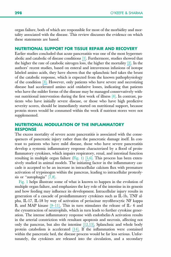
In summary, in patients who are at risk of developing septic complications,enteral feeding has a strong potential to limit the rate of development of mul-tiple organ dysfunction. Route and type of nutrition has effects in modulatingthe immune response by protecting from intraluminal bacteria and toxic prod-ucts. Changes in cytokine levels and expression influence the vascular endo-thelial response to septic conditions. Extensive experimental evidencesuggests that this is related to maintenance of the mucosal-associated lymphoidtissue, which provides immunologic protection for both the gastrointestinaland respiratory tracts against microbial flora and infectious pathogens andsupports a communication between GI and mucosal surfaces throughout thebody (Fig. 2) [38].
302 O’KEEFE & SHARMA
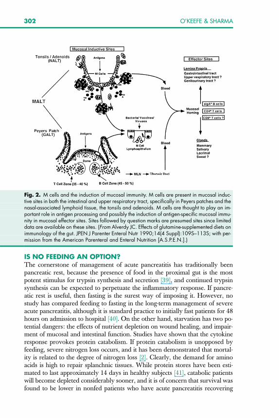
IS NO FEEDING AN OPTION?The cornerstone of management of acute pancreatitis has traditionally beenpancreatic rest, because the presence of food in the proximal gut is the mostpotent stimulus for trypsin synthesis and secretion [39], and continued trypsinsynthesis can be expected to perpetuate the inflammatory response. If pancre-atic rest is useful, then fasting is the surest way of imposing it. However, nostudy has compared feeding to fasting in the long-term management of severeacute pancreatitis, although it is standard practice to initially fast patients for 48hours on admission to hospital [40]. On the other hand, starvation has two po-tential dangers: the effects of nutrient depletion on wound healing, and impair-ment of mucosal and intestinal function. Studies have shown that the cytokineresponse provokes protein catabolism. If protein catabolism is unopposed byfeeding, severe nitrogen loss occurs, and it has been demonstrated that mortal-ity is related to the degree of nitrogen loss [2]. Clearly, the demand for aminoacids is high to repair splanchnic tissues. While protein stores have been esti-mated to last approximately 14 days in healthy subjects [41], catabolic patientswill become depleted considerably sooner, and it is of concern that survival wasfound to be lower in nonfed patients who have acute pancreatitis recovering
Fig. 2. M cells and the induction of mucosal immunity. M cells are present in mucosal induc-tive sites in both the intestinal and upper respiratory tract, specifically in Peyers patches and thenasal-associated lymphoid tissue, the tonsils and adenoids. M cells are thought to play an im-portant role in antigen processing and possibly the induction of antigen-specific mucosal immu-nity in mucosal effector sites. Sites followed by question marks are presumed sites since limiteddata are available on these sites. (From Alverdy JC. Effects of glutamine-supplemented diets onimmunology of the gut. JPEN J Parenter Enteral Nutr 1990;14(4 Suppl):109S–113S; with per-mission from the American Parenteral and Enteral Nutrition [A.S.P.E.N.].)

303NUTRITION SUPPORT IN SEVERE ACUTE PANCREATITIS
from pancreatic surgery [42]. A further concern is that starvation results in dis-use atrophy of the mucosa, stasis, bacterial overgrowth and increased perme-ability, and increased risk of infection from enteric organisms, thusexacerbating the patholologic changes illustrated in Fig. 1.
THE PROBLEM WITH TOTAL PARENTERAL NUTRITION ANDBOWEL RESTThe development of TPN was heralded as a potential major breakthrough be-cause, for the first time, feeding could be maintained while maintaining pancre-atic rest. The authors have shown in human studies that TPN is efficacious inmeeting nutritional requirements without stimulating the pancreas [38,43]. Un-fortunately its use was also associated with an alarming increase in metabolic(namely, hyperglycemia) and septic complications. Indeed, one study showedthat TPN use worsened outcome in patients who had mild acute pancreatitiswhen compared with no feeding at all [43]. The explanation is complex. First,TPN and bowel rest probably exacerbate the intestinal and distant organ dys-function that characterizes acute pancreatitis. Kudsk’s studies in rats showedthat TPN and bowel rest result in atrophy of the mucosa and the gut immunesystem, with suppression of Th-2 responses and activation of adhesion mole-cules, which leads to neutrophil priming and migration to distant targets,such as the lung, producing a first-hit phenomenon [31–33]. This is remarkablysimilar to what is found in the acute pancreatitis associated gut injury shown onFig. 1. Other studies have shown that IL-6 is the mediator of gut barrier dys-function [44], and acute pancreatitis and TPN induce intestinal IL-6 production[45,46]. The use of TPN (ie, failure to feed patients enterally) further reducesmotility and blood flow, increases the risk of small bowel bacterial overgrowthwith antegrade colonization with colonic organisms, and increases mucosal per-meability, thus exacerbating the pathophysiologic response to acute pancreatitis[47,48]. This may be of critical importance, because the organisms most com-monly responsible for pancreatic infections are of colonic origin [49], and endo-toxemia commonly accompanies severe disease [50]. Second, the presence ofa central vein catheter in TPN-fed patients provides an open conduit for nos-ocomial infections. Third, intravenous feeding invariably results in hyperglyce-mia in patients who have severe acute pancreatitis, because (1) the glycemiceffect of glucose administered parenterally is greater than if it is given enterally[29], (2) acute pancreatitis impairs pancreatic endocrine function resulting ina relative insulin deficiency [51], and (3) the acute inflammatory responseand secretion of counter-regulatory hormones increases endogenous glucoseproduction and creates insulin resistance [52]. Recent studies have demon-strated that hyperglycemia worsens outcome in any form of critical illness[53]. In hyperglycemia, leukocyte function is impaired and intestinal motilityis reduced leading to increased infection risk from enteric pathogens [54]. Con-sequently, it is likely that the potential benefits of TPN on resting the pancreasare overshadowed by its detrimental effects on intestinal function and mucosalintegrity, and by its septic and metabolic complications.

304 O’KEEFE & SHARMA
WHY ENTERAL FEEDING IS SUPERIOR TO TOTAL PARENTERALNUTRITION AND BOWEL RESTThe complications associated with TPN have led the authors and others to ex-plore the use of specialized forms of enteral feeding. In the five major random-ized comparative studies published thus far [39,55–58], and their metanalysis[59], enteral feeding has been shown consistently to be superior to TPN withregard to cost, complications, and outcome. Marik and Zaluga [59] have re-cently published a metanalysis, which searched the National Library of Medi-cine’s Medline database, Embase, and the Cochrane Database of SystematicReviews looking for randomized controlled clinical trials comparing enteralto parenteral feeding in acute pancreatitis conducted between 1966 and 2004.Although the search identified 117 studies, only 6 fulfilled the criteria for inclu-sion, with a total population of 263 patients who had acute pancreatitis. Thisincluded 4 of the studies previously reviewed in more detail, namely Abou-Assi, Kalfarentzos, McClave, and Windsor. The authors noted that 4 of the6 had low Jahad scores less than 3, indicating poor quality, and that therewas considerable heterogeneity in patient selection and study design, limitingthe power of their conclusions. Fig. 3 illustrates the chief conclusion that enteralfeeding was associated with a lower incidence of infections (relative risk [RR]0.45, P < .0004). This almost chiefly reflects the avoidance of TPN sepsis, al-though the suppression of enteral bacterial overgrowth may also have beena factor. In addition, they found that enteral feeding was associated with re-duced hospital stay (mean reduction 2.9 days, P < .001), and reduced needfor surgical interventions (RR 0.48, P ¼ .05).
How can these observations be reconciled with the pathophysiology of acutepancreatitis previously outlined, whereby enteral feeding may exacerbate theinitiating factors in the inflammatory cascade? It is not because any of the spe-cialized forms of feeding used in these studies avoided pancreatic stimulation.
Fig. 3. Random effects model of relative risk (95% confidence interval) of infections associ-ated with enteral feeding compared with parenteral nutrition. (From Marik PE, Zaloga GP.Meta-analysis of parenteral nutrition versus enteral nutrition in patients with acute pancreatitis.Br Med J 2004;328(7453):1407; with permission.)
305NUTRITION SUPPORT IN SEVERE ACUTE PANCREATITIS
The authors’ studies in healthy volunteers show that all these modes of enteralfeeding stimulate the pancreas [38,51,60]. Another suggestion, based on exper-imental studies, was that the pancreas becomes unresponsive in acute pancre-atitis. Again, the authors’ investigations have shown that although thesecretory response to feeding in acute pancreatitis is reduced, trypsin synthesiscontinues [3] and can be stimulated [60]. The predominant difference seems tobe the ability of enteral feeding to reduce systemic inflammation, as previouslydiscussed, and infective complications. In a more recent prospective non-randomized study of 87 patients who had necrotizing pancreatitis, Modenaand colleagues [61] found that jejunal feeding without antibiotics reduced pan-creatic infections from 74% with TPN and antibiotics to only 20%, multiple or-gan failure (MOF) from 85% to 51%, and death from 35% to 5% (P < .001).Another consistent finding has been the ability of enteral feeding to reduce hy-perglycemia, and, as Van den Berge and colleagues [53] have shown, hypergly-cemia is associated with adverse outcome in all forms of critical illness. Clinicalevidence for the importance of tight glucose control with insulin infusions hasemerged from a recent report from Sweden whereby 48 patients were random-ized to early enteral nutrition versus TPN [62]. Although hyperglycemia wasmore common in TPN-fed patients, insulin infusions were started when levelsincreased over 16 mmol/l (ie, 280 mg/dL). Unlike any other study reported todate, total complications were more common in the enteral feeding group, al-though most consisted on atelectasis and pleural effusions within the first 3 day-s—the significance of which remains questionable.
In general, these findings lead to the conclusion that the superiority of enteralfeeding over TPN was accounted for not by any beneficial effect of enteralfeeding on the pancreas, but by the greater efficiency of enteral feeding in pre-serving intestinal function and splanchnic metabolism, and by the avoidance ofTPN-associated complications.
IS PANCREATIC REST IMPORTANT?In clinical practice, it is possible to feed and maintain pancreatic rest by usingeither TPN, or by feeding enterally 40 to 60 cm past the ligament of Treitz[63,64]. The fact that proximal enteral feeding has proven superior to TPNdose not mean that pancreatic rest is unimportant, because, as previously dis-cussed, the explanation probably involves the avoidance of the septic and met-abolic side effects of TPN and the greater ability of enteral feeding to supportintestinal and splanchnic function. To date, no study has compared stimulatoryto nonstimulatory enteral (or feeding > 40 cm down the jejunum) feeding. Therecent publication of two randomized controlled trials demonstrating that NGfeeding was as effective as ‘‘nasojejunal’’ feeding in the management of patientswho have severe acute pancreatitis seemed, at first glance, to suggest that pan-creatic rest is unimportant. Unfortunately, the trials compared like with like asboth forms of feeding were stimulatory to the pancreas. As discussed in the au-thors’ review [65], Eatock and colleagues’ [58] study compared NG feeding toa mixture of nasoduodenal and nasojejunal feeding, and Kumar and colleagues’

306 O’KEEFE & SHARMA
[66] study compared NG against nasoduodenal—despite the fact that they titledthe article ‘‘NG versus Nasojejunal’’! Consequently, neither study fed farenough down the jejunum to avoid pancreatic stimulation. Furthermore, itcould be argued that both were bad rather than good because the mortalityin both arms was unacceptably high at 25% to 30%.
NASOGASTRIC VERSUS NASOJEJUNAL FEEDINGDespite the reservations most of us initially had, nasogastric feeding is technicallyfeasible in acute pancreatitis as illustrated by recent publication of the Eatock andcolleagues [58] and Kumar and colleagues [66] studies. The question remains,however, whether it is an effective form of nutritional support. We know thatgastric emptying is impaired in critically ill patients, and better powered and con-trolled studies have been performed in nonpancreatic ICU ventilated patientswhich have clearly shown that (1) the efficiency of true jejunal feeding was high-er than NG feeding in supplying approximately 20% more calories and proteinand (2) that the incidence of pneumonias was higher with NG feeding [67]. It ispossible that these differences will be amplified in patients who have severe com-plicated acute pancreatitis because of the added problem of gastroduodenalcompression due to the adjacent pancreatic inflammatory mass. In keepingwith this concern, the authors note that the Kumar study had to supplement pa-tients in the first week with intravenous feeding, and, even then, there was de-terioration in nutritional status. Nonetheless, both studies reported thatoutcome was similar in NG-fed patients to postpyloric-fed patients, leading totheir conclusion that NG feeding should be the preferred method, because itis easier to place, quicker to start, and less expensive. This has made it an attrac-tive alternative to the more complex jejunal tube placement, which is dependenton radiologic or endoscopic placement skills, which may be limited or unavail-able in some hospitals. The problem is that small scale clinical trials such as thesecan lead to erroneous conclusions, and sufficiently powered clinical trials haveto be performed before standards of practice are changed.
CAN ENTERAL FEEDING BE GIVEN WITHOUT STIMULATINGTHE PANCREAS?A recent publication of the authors’ findings [63] has supported the results of anearlier study from Holland [64], that it is possible to use enteral feeding withoutstimulating the pancreas, if the infusion is delivered 40 to 60 cm down thejejunum. This makes physiologic sense, because there has to be a null pointin between the duodenum, where feeding stimulates through cholinergic re-flexes and cholecystokinin release, and the ileum where feeding inhibits byway of the ileal brake mechanism [68]. The authors have shown that this dis-tance is easy to achieve in clinical practice with transnasal endoscopic place-ment of tube systems that are commercially available [69,70]. There isextensive literature that the presence of nutrients (in particular long chain fattyacids [71,72], carbohydrate [72], and amino acids [73]) in the ileum suppressespancreatic secretion in humans, principally through release of GLP-I and

307NUTRITION SUPPORT IN SEVERE ACUTE PANCREATITIS
peptide-YY (PYY) from ileal L-cells and neurotensin from N-cells [74]. Activa-tion of the ileal brake in this way may have the additional benefits by suppress-ing acute pancreatitis and inhibiting gastric acid secretion [74] and the need forNG decompression. Fascinating recent studies in animal models of lethal nec-rotizing pancreatitis have shown that PYY infusions given either prophylacti-cally or therapeutically reduce the histologic evidence of pancreatitis and theIL-6 systemic cytokine secretion, preventing death [75,76].
DOES AVOIDANCE OF PANCREATIC STIMULATION IMPROVEOUTCOME?No study has prospectively investigated this question. However, we know thattrypsinogen activation within the pancreas is the initiating factor, or ‘‘trigger,’’in the disease, leading to the SIRS in patients who have the severe form of thedisease. A recent prospective study from Scotland, designed specifically to eval-uate the therapeutic use of Lexipafant, a platelet-activating antagonist, exam-ined the relationship between SIRS and mortality in the subgroup that hadsevere disease (n ¼ 121) and found that the incidence of SIRS on admission,at 24 hours and 48 hours, as well as the persistence of SIRS, was correlatedwith survival as shown in Table 2 in Fig. 4 [77]. The authors also examinedthe relationship between MOF and death risk, and showed that transientMOF (ie, MOF that resolved with the first week) was associated with no mor-tality, but ‘‘permanent’’ or progressive MOF was associated with a mortalityrate of 55%. More recently, a full evaluation of all the patients who had severedisease enrolled in the UK Lexipafant study (n ¼ 290) was reported confirmingthe predictive value of permanent MOF on mortality [78]. In addition, they ex-amined the relationship between MOF and local complications, such as pancre-atic necrosis and infection. They found that there was a significant association,such that 52% of patients who had permanent MOF developed local
Fig. 4. The dynamic nature of early MOF determines outcome in acute pancreatitis. (FromButer A, Imrie CW, Carter CR, et al. Dynamic nature of early organ dysfunction determinesoutcome in acute pancreatitis. Br J Surg 2002;89:300; with permission.)

308 O’KEEFE & SHARMA
complications, as opposed to none who had no organ failure and 26% who hadtransient MOF (P < .001). These studies suggest that there is a ‘‘window of op-portunity’’ in the first week of hospitalization to reduce mortality if we can ap-ply measures to suppress the development of SIRS, and thus MOF. Enteralfeeding has the potential to do this by maintaining gut function and suppressingthe inflammatory response. It remains to be seen whether distal feeding addsbenefit by avoiding exacerbation of pancreatic inflammation and by stimulatingthe PYY release.
Effective deliveryOvercomes obstructionNo pancreatic stimulation
Distal Jejunal Nasogastric
Ineffective deliveryStimulates pancreasGastroesophagealreflux
Only experts can place
Anyone can place
Quicker to startInexpensive
Fig. 5. Nasogastric versus distal jejunal feeding.
Box 1: The pros and cons of various nutritional approaches
1. The proinflammatory cytokine ‘‘storm’’ activates proteolysis, resulting in pro-tein catabolism, increasing the demand for protein for tissue repair, eitherfrom body stores or the diet.
2. Starvation produces pancreatic rest and prevents further trypsinogen activa-tion within the inflammatory mass, but deprives the intestine of luminal nutri-tion, thus exacerbating the mucosal damage initiated by the acutepancreatitis cytokine response. Furthermore, it results in severe negative nitro-gen balance and protein deficiency due to unopposed protein catabolism,thus impairing tissue repair.
3. TPN also rests the pancreas but aggravates the intestinal injury in the sameway described for starvation. It does, however, stem the loss of body protein.
4. Enteral Feeding, as currently practiced, aggravates the pancreatic injury bystimulating trypsinogen production, but counteracts the intestinal injury by pro-viding luminal nutrition and suppressing the systemic inflammatory responsewhile, at the same time, supporting splanchnic metabolism and recovery.

309NUTRITION SUPPORT IN SEVERE ACUTE PANCREATITIS
SUMMARYKnowledge of the pathophysiologic response is crucial to understand why feed-ing a patient who has acute pancreatitis is so incredibly difficult. The pros andcons (Fig. 5) of the various nutritional approaches advocated, namely, fasting,TPN, and enteral feeding, are summarized in Box 1.
Strong circumstantial evidence has been presented that shows that enteralfeeding has the potential to reduce the inflammatory response and the subse-quent development of the complications that lead to mortality from the disease.Although the authors’ center favors distal jejunal feeding, there are pros andcons to both distal jejunal and NG feeding, as summarized. As suggested inHeyland’s metanalysis [79], the ultimate balance can only be determined bythe conduct of a sufficiently powered multicenter clinical randomized compar-ative trial incorporating patients at risk of developing the complications associ-ated with high mortality rates, and whose illness is likely to be protracted,making nutritional support essential for survival.
References[1] Shaw JH, Wolfe RR. Glucose, fatty acid, and urea kinetics in patients with severe pancrea-
titis. The response to substrate infusion and total parenteral nutrition. Ann Surg 1986;204:665–72.
[2] Sitzmann J, Steinborn P, Zinner M, et al. Total parenteral nutrition and alternate energysubstrates in treatment of severe acute pancreatitis. Surg Gynecol Obstet 1989;168:311–6.
[3] O’Keefe SJD, Lee RB, Clore J. Enteral feeding during acute pancreatitis is associated withpreserved trypsin synthesis and increased splanchnic protein metabolism. Gastroenterol-ogy 2004;126(4):A–91.
[4] Stephen JD, O’Keefe, Neeraj Kaushik, Barbara Stoll, et al. The impact of disease severity onprotein metabolism in acute pancreatitis. Gastroenterology 2007;132(4):A62.
[5] Makhija R, Kingsnorth AN. Cytokine storm in acute pancreatitis. J Hepatobiliary PancreatSurg 2002;9(4):401–10.
[6] Bhatia M, Wong FL, Cao Y, et al. Pathophysiology of acute pancreatitis [review]. Pancrea-tology 2005;5(2–3):132–44 [Epub 2005 Apr 21].
[7] Steer ML. The early intracellular cell events which occurred during acute pancreatitis. Pan-creas 1988;17(1):31–7.
[8] Leach SD, Modlin IM, Scheele GA, et al. Intracellular activation of digestive zymogens in ratpancreatic acini. J Clin Invest 1991;87:362–6.
[9] Shi C, Zhao X, Wang X, et al. Role of nuclear factor-kappa B, reactive oxygen species andcellular signaling in the early phase of acute pancreatitis. Scand J Gastroenterol2005;40(1):103–8.
[10] Liu HS, Pan CE, Liu QG, et al. Effect of NF-kappaB and p38 MAPK in activated monocytes/macrophages on pro-inflammatory cytokines of rats with acute pancreatitis. World J Gastro-enterol 2003;9(11):2513–8.
[11] Vaquero E, Gukovsky I, Zaninovic V, et al. Localized pancreatic NF-kappaB activation andinflammatory response in taurocholate-induced pancreatitis. Am J Physiol Gastrointest LiverPhysiol 2001;280(6):G1197–208.
[12] Leindler L, Morschl E, Laszlo F, et al. Importance of cytokines, nitric oxide, and apoptosisin the pathological process of necrotizing pancreatitis in rats. Pancreas 2004;29(2):157–61.
[13] Inoue K, Hirota M, Kimura Y, et al. Further evidence for endothelin as an important mediatorof pancreatic and intestinal ischemia in severe acute pancreatitis. Pancreas 2003;26(3):218–23.

310 O’KEEFE & SHARMA
[14] Fong YM, Marano MA, Moldawer LL, et al. The acute splanchnic and peripheral tissue met-abolic response to endotoxin in humans. J Clin Invest 1990;85(6):1896–904.
[15] Ammori BJ. Role of the gut in the course of severe acute pancreatitis [review]. Pancreas2003;26(2):122–9.
[16] Shi C, Andersson R, Zhao X, et al. Potential role of reactive oxygen species in pancreatitis-associated multiple organ dysfunction. Pancreatology 2005;5(4–5):492–500.
[17] Zhao X, Dib M, Andersson E, et al. Alterations of adhesion molecule expression and inflam-matory mediators in acute lung injury induced by septic and non-septic challenges. Lung2005;183(2):87–100.
[18] Jaffray C, Yang J, Carter G, et al. Pancreatic elastase activates pulmonary nuclear factorkappa B and inhibitory kappa B, mimicking pancreatitis-associated adult respiratory dis-tress syndrome. Surgery 2000;128(2):225–31.
[19] Hietaranta A, Mustonen H, Puolakkainen P, et al. Proinflammatory effects of pancreatic elas-tase are mediated through TLR4 and NF-kappaB. Biochem Biophys Res Commun2004;323(1):192–6.
[20] Bhatia M. Inflammatory response on the pancreatic acinar cell injury. Scand J Surg2005;94(2):97–102.
[21] Hirota M, Inoue K, Kimura Y, et al. Non-occlusive mesenteric ischemia and its associatedintestinal gangrene in acute pancreatitis. Pancreatology 2003;3(4):316–22.
[22] Rahman SH, Ammori BJ, Holmfield J, et al. Intestinal hypoperfusion contributes to gut barrierfailure in severe acute pancreatitis. J Gastrointest Surg 2003;7(1):26–35 [discussion:35–6].
[23] Dahly EM, Gillingham MB, Guo Z, et al. Role of luminal nutrients and endogenous GLP-2 inintestinal adaptation to mid-small bowel resection. Am J Physiol Gastrointest Liver Physiol2003;284(4):G670–82.
[24] Murphy MS. Growth factors and the gastrointestinal tract [review]. Nutrition 1998;14(10):771–4.
[25] Plusczyk T, Witzel B, Menger MD, et al. ETA and ETB receptor function in pancreatitis-asso-ciated microcirculatory failure, inflammation, and parenchymal injury. Am J Physiol Gastro-intest Liver Physiol 2003;285(1):G145–53.
[26] Moore FA, Feliciano DV, Andrassy RJ, et al. Early enteral feeding, compared with paren-teral, reduces postoperative septic complications. The results of a meta-analysis. AnnSurg 1992;216(2):172–83.
[27] Moore FA, Moore EE, Kudsk KA, et al. Clinical benefits of an immune-enhancing diet forearly postinjury enteral feeding. J Trauma 1994;37(4):607–15.
[28] Owen RL. Sequential uptake of horseradish peroxidase by lymphoid follicle epithelium ofPeyer’s patches in the normal unobstructed mouse intestine: an ultrastructural study. Gastro-enterology 1977;72(3):440–51.
[29] Lebman DA, Coffman RL. Cytokines in the mucosal immune system. In: Ogra PL, Lamm ME,McGhee JR, et al, editors. Handbook of mucosal immunology. San Diego (CA): Academic;1994. p. 243–9.
[30] El Kaissouni J, Bene MC, Thionnois S, et al. Maturation of B cells in the lamina propriaof human gut and bronchi in the first months of human life. Dev Immunol 1998;5(3):153–9.
[31] King BK, Li J, Kudsk KA. A temporal study of TPN-induced changes in gut-associated lym-phoid tissue and mucosal immunity. Arch Surg 1997;132(12):1303–9.
[32] Wu Y, Kudsk KA, DeWitt RC, et al. Route and type of nutrition influence IgA-mediating intes-tinal cytokines. Ann Surg 1999;229(5):662–7 [discussion: 667–8].
[33] King BK, Kudsk KA, Li J, et al. Route and type of nutrition influence mucosal immunity to bac-terial pneumonia. Ann Surg 1999;229(2):272–8.
[34] Fukatsu K, Zarzaur BL, Johnson C, et al. MAdCAM-1 expression in Peyer patches: a mech-anism controlling gut associated lymphoid tissue (GALT). Surg Forum 2000;51:211–4.

311NUTRITION SUPPORT IN SEVERE ACUTE PANCREATITIS
[35] Burke DJ, Alverdy JC, Aoys E, et al. Glutamine-supplemented total parenteral nutrition im-proves gut immune function. Arch Surg 1989;124(12):1396–9.
[36] DeWitt RC, Wu Y, Renegar KB, et al. Bombesin recovers gut-associated lymphoid tissue andpreserves immunity to bacterial pneumonia in mice receiving total parenteral nutrition. AnnSurg 2000;231(1):1–8.
[37] Moore FA, Moore EE, Sauaia A. Postinjury multiple-organ failure. In: Mattox KL,Feliciano DV, Moore EE, editors. Trauma. 4th edition. New York: McGraw-Hill; 1999.p. 1427–60.
[38] van Ginkel FW, Nguyen HH, McGhee JR. Synopsis. Vaccines for mucosal immunity tocombat emerging infectious diseases. Available at: www.cdc.gov/ncidod/eid/vol6no2/vanginkle.htm. Accessed May 3, 2007.
[39] O’Keefe SJD, Lee RB, Li J, et al. Trypsin and splanchnic protein turnover during feeding andfasting in human subjects. Am J Physiol Gastrointest Liver Physiol 2006;290(2):G213–21.
[40] Abou-Assi S, Craig K, O’Keefe SJD. Hypocaloric jejunal feeding is better than total paren-teral nutrition in acute pancreatitis: results of a randomized comparative study. Am J Gastro-enterol 2002;97(9):2255–62.
[41] Cahill GJ Jr, Owen OE, Morgan AP. The consumption of fuels during prolonged starvation.Adv Enzyme Regul 1968;6:143–50.
[42] Austrums E, Pupelis G, Snippe K. Postoperative enteral stimulation by gut feeding improvesoutcomes in severe acute pancreatitis. Nutrition 2003;19(6):487–91.
[43] Sax HC, Warner BW, Talamini MA, et al. Early total parenteral nutrition in acute pancrea-titis: lack of beneficial effects. Am J Surg 1987;153:117–24.
[44] Fink MP. Gastrointestinal mucosal injury in experimental models of shock, trauma and sep-sis. Crit Care Med 1991;19:627–41.
[45] Kudsk KA, Minard G, Wojtysiac SL, et al. Visceral protein response to enteral versus paren-teral nutrition and sepsis in patients with trauma. Surgery 1994;116:516–23.
[46] Pooran N, Indaram A, Singh P, et al. Cytokines (IL-6, IL-8, TNF): early and reliable predictorsof severe acute pancreatitis. J Clin Gastroenterol 2003;37(3):263–6.
[47] Fong YM, Marano MA, Barber A, et al. Total parenteral nutrition and bowel rest modify themetabolic response to endotoxin in humans. Ann Surg 1989;210(4):449–56.
[48] Fukatsu K, Zarzaur BL, Johnson CD, et al. Lack of enteral feeding increases expression ofE-selectin after LPS challenge. J Surg Res 2001;97(1):41–8.
[49] Beger HG, Bittner R, Block S, et al. Bacterial contamination of pancreatic necrosis. A pro-spective clinical study. Gastroenterology 1986;91(2):433–8.
[50] Gray KD, Simovic MO, Chapman WC, et al. Endotoxin potentiates lung injury in cerulein-induced pancreatitis. Am J Surg 2003;186(5):526–30.
[51] O’Keefe SJD, Lee RB, Anderson FP, et al. The physiological effects of enteral and parenteralfeeding on pancreatic enzyme secretion in humans. Am J Physiol 2003;284:27–36.
[52] O’Keefe SJD, Moldawer LL, Young VR, et al. The influence of intravenous nutrition on proteindynamics following surgery. Metabolism 1981;30(12):1150–8.
[53] Van den Berghe G, Wouters P, Weekers F, et al. Intensive insulin therapy in critically ill pa-tients. N Engl J Med 2001;345:1359–67.
[54] Van den Berghe G. How does blood glucose control with insulin save lives in intensive care?J Clin Invest 2004;114(9):1187–95.
[55] McClave SA, Greene LM, Snider HL, et al. Comparison of the safety of early enteral vs par-enteral in mild acute pancreatitis. JPEN J Parenter Enteral Nutr 1997;21:14–20.
[56] Windsor AC, Kanwar S, Li AG, et al. Compared with parenteral nutrition, enteral feedingattenuates the acute phase response, and improves disease severity in acute pancreatitis.Gut 1998;42:431–5.
[57] Kalfarentzos F, Kehagias J, Mead M, et al. Enteral nutrition is superior to parenteral nutritionin severe acute pancreatitis. Results of a randomized perspective trial. Br J Surg1997;84(12):1665–9.

312 O’KEEFE & SHARMA
[58] Eatock FC, Chong P, Menezes N, et al. A randomized study of early nasogastric versus na-sojejunal feeding in severe acute pancreatitis. Am J Gastroenterol 2005;100:432–9.
[59] Marik PE, Zaloga GP. Meta-analysis of parenteral nutrition versus enteral nutrition in patientswith acute pancreatitis. Br Med J 2004;328(7453):1407–13.
[60] O’Keefe SJ, Broderick T, Turner M, et al. Nutrition in the management of necrotizing pancre-atitis. Clin Gastroenterol Hepatol 2003;1:315–21.
[61] Modena LT, Cevasco LB, Basto CA, et al. Total enteral nutrition as prophylactic therapy forpancreatic necrosis infection in severe acute pancreatitis. Pancreatology 2006;6:58–64.
[62] Eckerwall GE, Axelsson JB, Andersson RG. Early nasogastric feeding in predicted severeacute pancreatitis. Ann Surg 2006;244(6):959–67.
[63] Kaushik N, O’Keefe SJ. Nutritional support in acute pancreatitis. Curr Gastroenterol Rep2004;6(4):320–6.
[64] Vu MK, Van Der Veek P, Frolich M, et al. Does jejunal feeding activate exocrine pancreaticsecretion? Eur J Clin Invest 1999;29(12):1053–9.
[65] O’Keefe SJD, McClave SA. Feeding the injured pancreas. Gastroenterology2005;129(30):1129–30.
[66] Kumar A, Singh N, Prakash S, et al. Early enteral nutrition in severe acute pancreatitis: a pro-spective randomized controlled trial comparing nasojejunal and nasogastric routes. J ClinGastroenterol 2006;40:431–4.
[67] Heyland DK, Drover JW, MacDonald S, et al. Effect of postpyloric feeding on gastroesoph-ageal regurgitation and pulmonary microaspiration: results of a randomized controlledtrial. Crit Care Med 2001;29(8):1495–501.
[68] Van Citters GW, Lin HC. The ileal brake: a fifteen-year progress report. Curr GastroenterolRep 1999;1:404–9.
[69] O’Keefe SJD. What’s the best approach to early feeding in patients with acute pancreatitis?Point-counterpoint: jejunal feeding is the best approach: AGA Perspectives. 2006;2(2):17–9.
[70] O’Keefe SJD, Foody W, Gill S. Transnasal endoscopic placement of feeding tubes in the in-tensive care unit. J Parenter Enteral Nutr 2003;27(5):349–54.
[71] Layer P, Peschel S, Schlesinger T, et al. Human pancreatic secretion and intestinal motility:effects of ileal nutrient perfusion. Am J Physiol Gastrointest Liver Physiol 1990;285(21):G196–201.
[72] Keller J, Holst JJ, Layer P. Inhibition of human pancreatic and biliary output but not intestinalmotility by physiological intraileal lipid loads. Am J Physiol Gastrointest Liver Physiol2006;290(4):G704–9.
[73] Niebergall-Roth E, Teyssen S, Niebel W, et al. Pancreatic secretory response to intrailealamino acids. Int J Pancreatol 2000;28(2):83–90.
[74] Naruse S, Kitagawa M, Ishiguro H, et al. Feedback regulation of pancreatic secretion bypeptide YY. Peptides 2002;23:359–65.
[75] Grise KR, McFadden DW. Peptide YY improves local and systemic parameters and preventsdeath in lethal necrotizing pancreatitis. Pancreas 2002;24(1):90–5.
[76] Robinson K, Vona-Davis L, Riggs D, et al. Peptide YYattenuates STAT1 and STAT3 activationinduced by TNF-alpha in acinar cell line AR42J. J Am Coll Surg 2006;202(5):788–96.
[77] Buter A, Imrie CW, Carter CR, et al. Dynamic nature of early organ dysfunction determinesoutcome in acute pancreatitis. Br J Surg 2002;89(3):298–302.
[78] Johnson CD, Abu-Hilal M. Persistent organ failure during the first week as a marker of fataloutcome in acute pancreatitis. Gut 2004;53(9):1340–4.
[79] Heyland DK, Drover JW, Dhaliwal R, et al. Optimizing the benefits and minimizing the risksof enteral nutrition in the critically-ill: role of small bowel feeding. JPEN J Parenter EnteralNutr 2002;26:S51–7.