Identification of Inflammatory and Regulatory Cytokines IL-1 ...
23
Citation: Vitenberga-Verza, Z.; Pilmane, M.; Šerstn , ova, K.; Melderis, I.; Gontar, L.; Kocha´ nski, M.; Drutowska, A.; Maróti, G.; Prieto-Simón, B. Identification of Inflammatory and Regulatory Cytokines IL-1α-, IL-4-, IL-6-, IL-12-, IL-13-, IL-17A-, TNF-α-, and IFN-γ-Producing Cells in the Milk of Dairy Cows with Subclinical and Clinical Mastitis. Pathogens 2022, 11, 372. https://doi.org/10.3390/ pathogens11030372 Academic Editor: Maria Filippa Addis Received: 9 February 2022 Accepted: 15 March 2022 Published: 17 March 2022 Publisher’s Note: MDPI stays neutral with regard to jurisdictional claims in published maps and institutional affil- iations. Copyright: © 2022 by the authors. Licensee MDPI, Basel, Switzerland. This article is an open access article distributed under the terms and conditions of the Creative Commons Attribution (CC BY) license (https:// creativecommons.org/licenses/by/ 4.0/). pathogens Article Identification of Inflammatory and Regulatory Cytokines IL-1α-, IL-4-, IL-6-, IL-12-, IL-13-, IL-17A-, TNF-α-, and IFN-γ-Producing Cells in the Milk of Dairy Cows with Subclinical and Clinical Mastitis Zane Vitenberga-Verza 1, * ,M¯ ara Pilmane 1 , Ksenija Šerstn , ova 1 , Ivars Melderis 1 , Lukasz Gontar 2 , Maksymilian Kocha ´ nski 2 , And ˙ zelika Drutowska 2 , Gergely Maróti 3,4 and Beatriz Prieto-Simón 5,6 1 The Institute of Anatomy and Anthropology, R¯ ıga Stradin , š University, 1010 R¯ ıga, Latvia; [email protected] (M.P.); [email protected] (K.Š.); [email protected] (I.M.) 2 Research and Innovation Centre Pro-Akademia, 95-050 KonstantynówLódzki, Poland; [email protected] (L.G.); [email protected] (M.K.); [email protected] (A.D.) 3 Seqomics Biotechnology Ltd., 6782 Morahalom, Hungary; [email protected] 4 Biological Research Center, Plant Biology Institute, 6726 Szeged, Hungary 5 Department of Electronic Engineering, Universitat Rovira i Virgili, 43007 Tarragona, Spain; [email protected] 6 ICREA, 08010 Barcelona, Spain * Correspondence: [email protected]; Tel.: +371-67320862 Abstract: In naturally occurring bovine mastitis, effects of infection depend on the host inflammatory response, including the effects of secreted cytokines. Knowledge about the inflammatory and regulatory cytokines in milk cells of free-stall barn dairy cows and in naturally occurring mastitis is lacking as most studies focus on induced mastitis. Hereby, the aim of the study was to determine inflammatory and regulatory cytokines in the milk of dairy cows with subclinical and clinical mastitis. The following examinations of milk samples were performed: differential counting of somatic cells (SCC), bacteriological examination, and immunocytochemical analysis. Mean SCC increased in subclinical and clinical mastitis cases. The number of pathogenic mastitis-causing bacteria on plates increased in subclinical mastitis cases but decreased in clinical mastitis. The inflammatory and regulatory markers in the milk cells of healthy cows showed the highest mean cell numbers (%). In mastitis cases, immunoreactivity was more pronounced for IL-4, IL-6, IL-12, IL-13, IL-17A, TNF-α, and IFN-γ. Data about subclinical and clinical mastitis demonstrate inflammatory responses to intramammary infection driven by IL-1α, IL-4, and IL-17A. Moreover, the host defense response in mastitis is characterized by continuation or resolution of initial inflammation. IL-12 and INF-γ immunoreactivity was recognized to differ mastitis cases from the relative health status. Keywords: mastitis; bovine; cytokines; inflammation; immunohistochemistry; milk; diagnosis 1. Introduction Bovine mastitis is extremely important due to its economic, social, and environmental impact. This infectious disease is not limited to animal health and welfare but can also question potential endangerment of dairy consumers. The economic burden of bovine mastitis on the dairy sector is huge due to production losses and processing [1]. In addition, mastitis of dairy cows contributes significantly to increasing antimicrobial resistance at the forefront of the most pressing global health challenges suggesting misdiagnosis and reduction in antibiotic usage [2–5]. So far, delayed and poor intervention of bovine mas- titis is mainly due to significant limitations in the methods currently used for diagnosis, including their expenses [6]. On-farm diagnosis of mastitis mainly relies on somatic cell Pathogens 2022, 11, 372. https://doi.org/10.3390/pathogens11030372 https://www.mdpi.com/journal/pathogens
-
Upload
khangminh22 -
Category
Documents
-
view
0 -
download
0
Transcript of Identification of Inflammatory and Regulatory Cytokines IL-1 ...

�����������������
Citation: Vitenberga-Verza, Z.;
Pilmane, M.; Šerstn, ova, K.; Melderis,
I.; Gontar, Ł.; Kochanski, M.;
Drutowska, A.; Maróti, G.;
Prieto-Simón, B. Identification of
Inflammatory and Regulatory
Cytokines IL-1α-, IL-4-, IL-6-, IL-12-,
IL-13-, IL-17A-, TNF-α-, and
IFN-γ-Producing Cells in the Milk of
Dairy Cows with Subclinical and
Clinical Mastitis. Pathogens 2022, 11,
372. https://doi.org/10.3390/
pathogens11030372
Academic Editor: Maria
Filippa Addis
Received: 9 February 2022
Accepted: 15 March 2022
Published: 17 March 2022
Publisher’s Note: MDPI stays neutral
with regard to jurisdictional claims in
published maps and institutional affil-
iations.
Copyright: © 2022 by the authors.
Licensee MDPI, Basel, Switzerland.
This article is an open access article
distributed under the terms and
conditions of the Creative Commons
Attribution (CC BY) license (https://
creativecommons.org/licenses/by/
4.0/).
pathogens
Article
Identification of Inflammatory and Regulatory CytokinesIL-1α-, IL-4-, IL-6-, IL-12-, IL-13-, IL-17A-, TNF-α-, andIFN-γ-Producing Cells in the Milk of Dairy Cows withSubclinical and Clinical MastitisZane Vitenberga-Verza 1,* , Mara Pilmane 1 , Ksenija Šerstn, ova 1, Ivars Melderis 1, Łukasz Gontar 2,Maksymilian Kochanski 2 , Andzelika Drutowska 2, Gergely Maróti 3,4 and Beatriz Prieto-Simón 5,6
1 The Institute of Anatomy and Anthropology, Rıga Stradin, š University, 1010 Rıga, Latvia;[email protected] (M.P.); [email protected] (K.Š.); [email protected] (I.M.)
2 Research and Innovation Centre Pro-Akademia, 95-050 Konstantynów Łódzki, Poland;[email protected] (Ł.G.); [email protected] (M.K.);[email protected] (A.D.)
3 Seqomics Biotechnology Ltd., 6782 Morahalom, Hungary; [email protected] Biological Research Center, Plant Biology Institute, 6726 Szeged, Hungary5 Department of Electronic Engineering, Universitat Rovira i Virgili, 43007 Tarragona, Spain;
[email protected] ICREA, 08010 Barcelona, Spain* Correspondence: [email protected]; Tel.: +371-67320862
Abstract: In naturally occurring bovine mastitis, effects of infection depend on the host inflammatoryresponse, including the effects of secreted cytokines. Knowledge about the inflammatory andregulatory cytokines in milk cells of free-stall barn dairy cows and in naturally occurring mastitis islacking as most studies focus on induced mastitis. Hereby, the aim of the study was to determineinflammatory and regulatory cytokines in the milk of dairy cows with subclinical and clinical mastitis.The following examinations of milk samples were performed: differential counting of somatic cells(SCC), bacteriological examination, and immunocytochemical analysis. Mean SCC increased insubclinical and clinical mastitis cases. The number of pathogenic mastitis-causing bacteria on platesincreased in subclinical mastitis cases but decreased in clinical mastitis. The inflammatory andregulatory markers in the milk cells of healthy cows showed the highest mean cell numbers (%). Inmastitis cases, immunoreactivity was more pronounced for IL-4, IL-6, IL-12, IL-13, IL-17A, TNF-α,and IFN-γ. Data about subclinical and clinical mastitis demonstrate inflammatory responses tointramammary infection driven by IL-1α, IL-4, and IL-17A. Moreover, the host defense responsein mastitis is characterized by continuation or resolution of initial inflammation. IL-12 and INF-γimmunoreactivity was recognized to differ mastitis cases from the relative health status.
Keywords: mastitis; bovine; cytokines; inflammation; immunohistochemistry; milk; diagnosis
1. Introduction
Bovine mastitis is extremely important due to its economic, social, and environmentalimpact. This infectious disease is not limited to animal health and welfare but can alsoquestion potential endangerment of dairy consumers. The economic burden of bovinemastitis on the dairy sector is huge due to production losses and processing [1]. In addition,mastitis of dairy cows contributes significantly to increasing antimicrobial resistance atthe forefront of the most pressing global health challenges suggesting misdiagnosis andreduction in antibiotic usage [2–5]. So far, delayed and poor intervention of bovine mas-titis is mainly due to significant limitations in the methods currently used for diagnosis,including their expenses [6]. On-farm diagnosis of mastitis mainly relies on somatic cell
Pathogens 2022, 11, 372. https://doi.org/10.3390/pathogens11030372 https://www.mdpi.com/journal/pathogens

Pathogens 2022, 11, 372 2 of 23
count, although this method depends on multiple factors such as the type of causativepathogen and inner factors [7]. The study builds upon new knowledge acquired on hostresponse-derived biomarkers, such as proinflammatory cytokines, identified as prospectivemastitis biomarkers to report disease status and etiology. Leveraging the presence of thesebiomarkers in milk, a "liquid biopsy" approach could be pursued to ensure non-invasive,stress-free, and prompt diagnosis of naturally occurring bovine mastitis.
Mastitis is mainly a bacterial infection; however, it is also caused by algae, viruses,fungi, or improper milking procedures [8–13]. Prototheca spp. is the third most commonmastitis pathogen after Streptococcus and Staphylococcus spp. [14]. Mastitis can manifest intoclinical or subclinical conditions. Recent evidence suggests that effects of infection dependon the host response in the early stages of the disease, including the effects of secretedcytokines [15–17]. Cytokines are small molecule proteins that play an important role in cell-to-cell communication. There is a myriad of processes that are stimulated or inhibited bycytokines, such as cell differentiation, proliferation, remodeling, degeneration, regeneration,and even cell death. Both experimentally induced or naturally occurring mastitis results inan increase in the somatic cell count (SCC) and levels of produced cytokines (interleukin(IL) IL-1, IL-2, IL-4, IL-5, IL-6, IL-8, IL-10, and IL-12) in milk [18]. Similarly, also biochemicalanalysis of milk provides plenty of diagnostic options in search of mastitis markers [19].
The interleukin (IL)-1 (IL-1) family is one of the most widely represented groups ofcytokines [20]. IL-1 family cytokines work in relation to the tissue immunocompetentcells via several signaling pathways [21], performing mostly inflammatory, autoimmune,fibrosis-initiating, and mitogenic functions [22]. In experimental acute bovine mastitis,interleukin-1 alpha (IL-1α) demonstrates local modulation of such first-line defense media-tors such as prostaglandin (PG-F2α) and leukotriene (LT-B4), suggesting its role in earlyinflammation [23].
The main producers of IL-4 in bovine mammary glands are T and B lymphocytes,eosinophils and basophils, mast cells, plasma cells [24,25], as well as epithelial cells, whichtogether form the basis of type 2 immunity [26]. Furthermore, IL-4 regulates innate im-munity and houses an inhibitory effect on interferon (IFN)-γ in dairy cows [25]. However,associated with mastitis, both IL-4 expression [27] and levels in milk [28] were decreased,suggesting further studies.
IL-6 is a pleiotropic cytokine that has plenty of different functions such as the hostorganism defense by performing immune response, hematopoiesis, and regulation ofinflammation [29,30]. In bovine mastitis, IL-6 is considered as an early but nonspecificindicator of various inflammation states, especially in subclinical mastitis [28]. Contrary,IL-6 was not significantly different between healthy and subclinical mastitis-affected cowsquestioning its association with other factors [31].
IL-12 stimulates IFN-γ production and NK cell proliferation, cytotoxicity, and inducesNK cells to produce cytokines. In addition, IL-12 promotes the proliferation and activa-tion of T cells and provides immune cell differentiation [32]. IL-12 has been studied toincrease in the milk of cows with naturally occurring mastitis, suggesting its role in innateimmunity [33].
Mast cells and basophils as key effector cells of innate immunity participate in allergicproinflammatory responses by secreting soluble factors of type I cytokine IL-13 [34]. IL-13in bovine udder, milk, and association with mastitis has been poorly studied.
IL-17A is a proinflammatory cytokine that has an important role in host defenseagainst different microbial and non-microbial pathogens. IL-17A is produced by multiplecell types of both the adaptive and innate immune systems [35–38]. In induced bovinemastitis, IL-17A mediates host defense-pathogen interactions during mastitis [39,40].
Tumor necrosis factor (TNF)-α is a proinflammatory cytokine mainly produced bymacrophages. Depending on the localization of its release and the receptor it will bind to, itcan perform different functions, such as stimulating the synthesis of other cytokines andcausing inflammatory reactions, controlling vital processes of the cell, and maintainingtissue homeostasis [29,41–44]. IFN-γ is a cytokine secreted by different cells of the innate

Pathogens 2022, 11, 372 3 of 23
and adaptive immunity, as well as antigen-presenting cells having the features of protectiveimmune responses and elimination of pathogens [45–48]. Altogether with other selectedcytokines, TNF-α and IFN-γmultifunctionality in bovine subclinical and clinical mastitismust be studied.
In the bovine mammary gland, mRNA and relative protein expression levels indi-cate the up-regulation of proinflammatory cytokines (e.g., TNF-α and IL-6) in infectedtissues rather than non-infected tissues. Ignited proinflammatory pathways implicate theunderlying regulatory pathways for proper immune function in mammary glands [49].Various molecules released by the host in the early stages of infection have already beenidentified as biomarkers for early diagnosis. Interestingly, their expression has previouslybeen reported to be related to the causative agent [17]. Altogether, studies on the cytokinesin bovine milk promote a better understanding of the role of these mediators in the patho-genesis of mastitis. This knowledge suggests potential therapeutic applications and theidentification of diagnostic and prognostic biomarkers.
Hereby, the aim of the study was to determine inflammatory and regulatory cy-tokines IL-1α, IL-4, IL-6, IL-12, IL-13, IL-17A, TNF-α, and IFN-γ in the milk of dairy cowswith subclinical and clinical mastitis, and to differentiate the changes of their expressionthroughout three consecutive days after the settlement of the diagnosis of subclinical orclinical mastitis. Knowledge about the inflammatory and regulatory cytokines in milkcells of free-stall housed dairy cows and in naturally occurring mastitis is lacking as moststudies focus on pathogen-induced mastitis. In naturally occurring mastitis, the study ofcytokines in milk is a potential tool for the early and timely diagnosis and in prospectivepathogenesis-based treatment.
2. Results2.1. Evaluation of Milk Quality
In healthy cows, the mean SCC log10 values were 4.65 (0.13) cells on day 1, 4.44 (0.26)cells on day 2, and 4.45 (0.14) cells on day 3 (Figure 1). In subclinical mastitis-affected cows,mean log10 (SD) SCC numbers were 5.37 (0.34) cells on day 1, then increased to 5.90 (0.34)cells on day 2, but then decreased to 5.77 (0.52) cells on day 3. In the clinical mastitis group,SCC mean log10 SCC values were increasingly rising from 6.04 (0.37) cells on day 1 to 6.35(0.33) cells on day 2 and to 6.43 (0.10) cells on day 3.
Pathogens 2022, 11, x FOR PEER REVIEW 3 of 24
it can perform different functions, such as stimulating the synthesis of other cytokines and causing inflammatory reactions, controlling vital processes of the cell, and maintaining tissue homeostasis [29,41–44]. IFN-γ is a cytokine secreted by different cells of the innate and adaptive immunity, as well as antigen-presenting cells having the features of protec-tive immune responses and elimination of pathogens [45–48]. Altogether with other se-lected cytokines, TNF-α and IFN-γ multifunctionality in bovine subclinical and clinical mastitis must be studied.
In the bovine mammary gland, mRNA and relative protein expression levels indicate the up-regulation of proinflammatory cytokines (e.g., TNF-α and IL-6) in infected tissues rather than non-infected tissues. Ignited proinflammatory pathways implicate the under-lying regulatory pathways for proper immune function in mammary glands [49]. Various molecules released by the host in the early stages of infection have already been identified as biomarkers for early diagnosis. Interestingly, their expression has previously been re-ported to be related to the causative agent [17]. Altogether, studies on the cytokines in bovine milk promote a better understanding of the role of these mediators in the patho-genesis of mastitis. This knowledge suggests potential therapeutic applications and the identification of diagnostic and prognostic biomarkers.
Hereby, the aim of the study was to determine inflammatory and regulatory cyto-kines IL-1α, IL-4, IL-6, IL-12, IL-13, IL-17A, TNF-α, and IFN-γ in the milk of dairy cows with subclinical and clinical mastitis, and to differentiate the changes of their expression throughout three consecutive days after the settlement of the diagnosis of subclinical or clinical mastitis. Knowledge about the inflammatory and regulatory cytokines in milk cells of free-stall housed dairy cows and in naturally occurring mastitis is lacking as most studies focus on pathogen-induced mastitis. In naturally occurring mastitis, the study of cytokines in milk is a potential tool for the early and timely diagnosis and in prospective pathogenesis-based treatment.
2. Results 2.1. Evaluation of Milk Quality
In healthy cows, the mean SCC log10 values were 4.65 (0.13) cells on day 1, 4.44 (0.26) cells on day 2, and 4.45 (0.14) cells on day 3 (Figure 1). In subclinical mastitis-affected cows, mean log10 (SD) SCC numbers were 5.37 (0.34) cells on day 1, then increased to 5.90 (0.34) cells on day 2, but then decreased to 5.77 (0.52) cells on day 3. In the clinical mastitis group, SCC mean log10 SCC values were increasingly rising from 6.04 (0.37) cells on day 1 to 6.35 (0.33) cells on day 2 and to 6.43 (0.10) cells on day 3.
Figure 1. The graphical appearance of mean log10 SCC in the milk of healthy cows (green line) and cows with subclinical (blue line) and clinical mastitis (red line). Error bars: +/- 2 SD.
Figure 1. The graphical appearance of mean log10 SCC in the milk of healthy cows (green line) andcows with subclinical (blue line) and clinical mastitis (red line). Error bars: +/−2 SD.
Statistical analysis determined that mean log10 value of SCC did not statisticallydiffer over day 1 to 3 in healthy animals (p = 0.076), in subclinical mastitis-affected cows(p = 0.074), and in clinical mastitis-affected cows (p = 0.07). The main effect of statusindicated a statistically significant difference in mean log10 SCC counts between studygroups (F(2, 12) = 78.308, p < 0.001, partial η2 = 0.929). Mixed two-way ANOVA determined

Pathogens 2022, 11, 372 4 of 23
a statistically significant two-way interaction of status and time on SCC mean log10 value(F(4, 24) = 3.491, p = 0.022, partial η2 = 0.368). Mean log10 SCC values were statisticallysignificantly greater in clinical mastitis group than in healthy cows (p < 0.001) and insubclinical mastitis-affected mastitis cows (p = 0.004), and also statistically significantlygreater in subclinical mastitis group than in healthy cows (p < 0.001).
2.2. Bacteriological Examination
Microbiological examination evaluated the logarithmic values of the total numberof pathogenic mastitis-causing bacteria (log CFU/mL) or the absence of bacterial growth(bacterial clearance).
In subclinical mastitis-affected cows, mean TBC log CFU/mL increased from a valueof 2.80 (0.47) on day 4 to a value of 3.47 (0.46) on day 5, to a value of 3.53 (0.43) on day 6(Figure 2). In milk of clinical mastitis-affected cows, mean TBC log CFU/mL values were3.95 (0.66) on day 4 and 3.95 (0.67) on day 5, but then decreased to a value of 3.59 (0.75)on day 6.
Pathogens 2022, 11, x FOR PEER REVIEW 4 of 24
Statistical analysis determined that mean log10 value of SCC did not statistically dif-fer over day 1 to 3 in healthy animals (p = 0.076), in subclinical mastitis-affected cows (p = 0.074), and in clinical mastitis-affected cows (p = 0.07). The main effect of status indicated a statistically significant difference in mean log10 SCC counts between study groups (F(2, 12) = 78.308, p < 0.001, partial η2 = 0.929). Mixed two-way ANOVA determined a statisti-cally significant two-way interaction of status and time on SCC mean log10 value (F(4, 24) = 3.491, p = 0.022, partial η2 = 0.368). Mean log10 SCC values were statistically significantly greater in clinical mastitis group than in healthy cows (p < 0.001) and in subclinical masti-tis-affected mastitis cows (p = 0.004), and also statistically significantly greater in subclin-ical mastitis group than in healthy cows (p < 0.001).
2.2. Bacteriological Examination Microbiological examination evaluated the logarithmic values of the total number of
pathogenic mastitis-causing bacteria (log CFU/mL) or the absence of bacterial growth (bacterial clearance).
In subclinical mastitis-affected cows, mean TBC log CFU/mL increased from a value of 2.80 (0.47) on day 4 to a value of 3.47 (0.46) on day 5, to a value of 3.53 (0.43) on day 6 (Figure 2). In milk of clinical mastitis-affected cows, mean TBC log CFU/mL values were 3.95 (0.66) on day 4 and 3.95 (0.67) on day 5, but then decreased to a value of 3.59 (0.75) on day 6.
Figure 2. The graphical appearance of mean TBC (log CFU/mL) in the milk of cows with subclinical (blue line) and clinical mastitis (red line). Error bars: +/- 2 SD.
In subclinical mastitis, a statistically significant overall difference was evaluated be-tween days 4 to 6 (F(2, 8) = 4.6, p = 0.047), the population effect size partial omega-squared ω2 = 0.4; sphericity assumed); however, post-hoc analysis with a Bonferroni adjustment revealed no statistically significant difference between days (p > 0.05). Mixed two-way ANOVA determined no statistically significant two-way interaction of status and time on TBC (p = 0.101) in the milk of both subclinical and clinical mastitis-affected cows. In clinical mastitis-affected cows, mean log10 values of TBC did not statistically significantly differ over time (p = 0.524).
2.3. Identification of Cytokines Producing Cells in Milk of Dairy Cows In healthy animals, IL-1α immunoreactive cell numbers indicated 1/3 of intensively
stained (++) cells and 2/3 of weakly stained (+) cells. Mean (%) IL-1α immunoreactive cell counts in healthy cows decreased from 56.0 cells on day 4 to 49.6 cells on day 5 and to 46.2 cells on day 6. Overall, IL-1α-positive cell counts insignificantly decreased, whereas neg-ative cell counts increased. Immunoreactivity for IL-4 in healthy cows remained stable for
Figure 2. The graphical appearance of mean TBC (log CFU/mL) in the milk of cows with subclinical(blue line) and clinical mastitis (red line). Error bars: +/−2 SD.
In subclinical mastitis, a statistically significant overall difference was evaluatedbetween days 4 to 6 (F(2, 8) = 4.6, p = 0.047), the population effect size partial omega-squared ω2 = 0.4; sphericity assumed); however, post-hoc analysis with a Bonferroniadjustment revealed no statistically significant difference between days (p > 0.05). Mixedtwo-way ANOVA determined no statistically significant two-way interaction of status andtime on TBC (p = 0.101) in the milk of both subclinical and clinical mastitis-affected cows. Inclinical mastitis-affected cows, mean log10 values of TBC did not statistically significantlydiffer over time (p = 0.524).
2.3. Identification of Cytokines Producing Cells in Milk of Dairy Cows
In healthy animals, IL-1α immunoreactive cell numbers indicated 1/3 of intensivelystained (++) cells and 2/3 of weakly stained (+) cells. Mean (%) IL-1α immunoreactivecell counts in healthy cows decreased from 56.0 cells on day 4 to 49.6 cells on day 5 and to46.2 cells on day 6. Overall, IL-1α-positive cell counts insignificantly decreased, whereasnegative cell counts increased. Immunoreactivity for IL-4 in healthy cows remained stablefor most cells being positive; in addition, most of the positive cells were stained intensively(++). These data showed a mean (%) of 96.0 cells on day 4, 98.0 cells on day 5, and 97.2 cellson day 6. Further, IL-6 immunoreactivity in milk cells of healthy cows decreased fromday 4 to 6 by mean values (%) of positive cell counts 35.8 on day 4, 33.4 on day 5, and32.6 on day 6; intensively and weakly stained immunoreactive cells were roughly 1:1. Themean IL-12 immunoreactive cell counts (%) in healthy cows also decreased from 57.2 onday 4 to 53.6 on day 5, and to 51.2 on day 6; in addition, intensively and weakly stained
Pathogens 2022, 11, 372 5 of 23
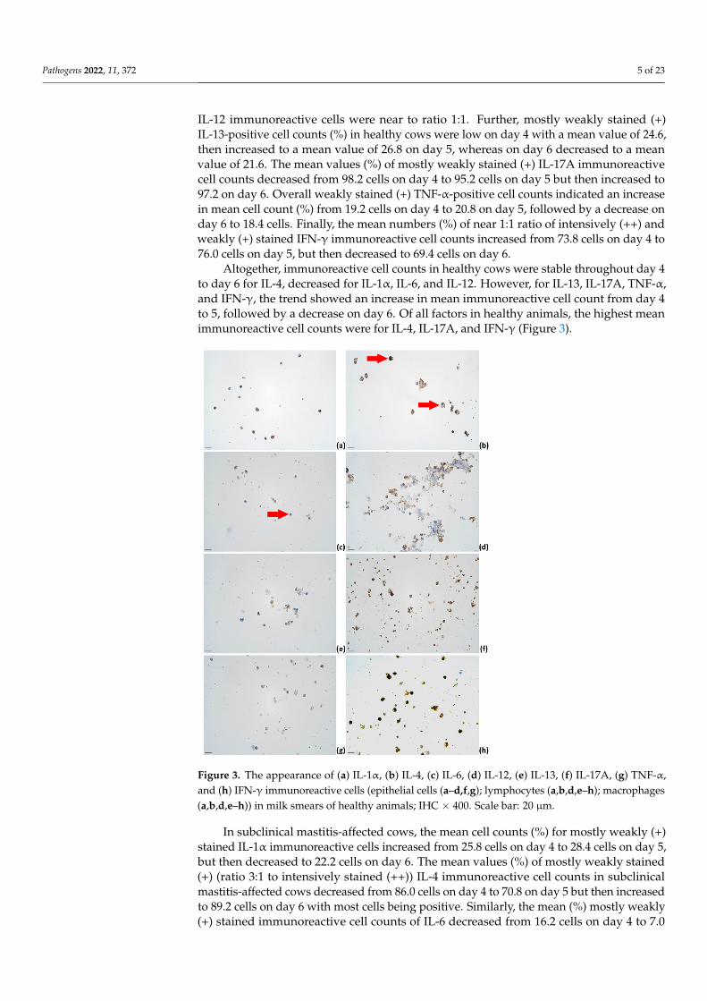
IL-12 immunoreactive cells were near to ratio 1:1. Further, mostly weakly stained (+)IL-13-positive cell counts (%) in healthy cows were low on day 4 with a mean value of 24.6,then increased to a mean value of 26.8 on day 5, whereas on day 6 decreased to a meanvalue of 21.6. The mean values (%) of mostly weakly stained (+) IL-17A immunoreactivecell counts decreased from 98.2 cells on day 4 to 95.2 cells on day 5 but then increased to97.2 on day 6. Overall weakly stained (+) TNF-α-positive cell counts indicated an increasein mean cell count (%) from 19.2 cells on day 4 to 20.8 on day 5, followed by a decrease onday 6 to 18.4 cells. Finally, the mean numbers (%) of near 1:1 ratio of intensively (++) andweakly (+) stained IFN-γ immunoreactive cell counts increased from 73.8 cells on day 4 to76.0 cells on day 5, but then decreased to 69.4 cells on day 6.
Altogether, immunoreactive cell counts in healthy cows were stable throughout day 4to day 6 for IL-4, decreased for IL-1α, IL-6, and IL-12. However, for IL-13, IL-17A, TNF-α,and IFN-γ, the trend showed an increase in mean immunoreactive cell count from day 4to 5, followed by a decrease on day 6. Of all factors in healthy animals, the highest meanimmunoreactive cell counts were for IL-4, IL-17A, and IFN-γ (Figure 3).
Pathogens 2022, 11, x FOR PEER REVIEW 5 of 24
most cells being positive; in addition, most of the positive cells were stained intensively (++). These data showed a mean (%) of 96.0 cells on day 4, 98.0 cells on day 5, and 97.2 cells on day 6. Further, IL-6 immunoreactivity in milk cells of healthy cows decreased from day 4 to 6 by mean values (%) of positive cell counts 35.8 on day 4, 33.4 on day 5, and 32.6 on day 6; intensively and weakly stained immunoreactive cells were roughly 1:1. The mean IL-12 immunoreactive cell counts (%) in healthy cows also decreased from 57.2 on day 4 to 53.6 on day 5, and to 51.2 on day 6; in addition, intensively and weakly stained IL-12 immunoreactive cells were near to ratio 1:1. Further, mostly weakly stained (+) IL-13-positive cell counts (%) in healthy cows were low on day 4 with a mean value of 24.6, then increased to a mean value of 26.8 on day 5, whereas on day 6 decreased to a mean value of 21.6. The mean values (%) of mostly weakly stained (+) IL-17A immunoreactive cell counts decreased from 98.2 cells on day 4 to 95.2 cells on day 5 but then increased to 97.2 on day 6. Overall weakly stained (+) TNF-α-positive cell counts indicated an increase in mean cell count (%) from 19.2 cells on day 4 to 20.8 on day 5, followed by a decrease on day 6 to 18.4 cells. Finally, the mean numbers (%) of near 1:1 ratio of intensively (++) and weakly (+) stained IFN-γ immunoreactive cell counts increased from 73.8 cells on day 4 to 76.0 cells on day 5, but then decreased to 69.4 cells on day 6.
Altogether, immunoreactive cell counts in healthy cows were stable throughout day 4 to day 6 for IL-4, decreased for IL-1α, IL-6, and IL-12. However, for IL-13, IL-17A, TNF-α, and IFN-γ, the trend showed an increase in mean immunoreactive cell count from day 4 to 5, followed by a decrease on day 6. Of all factors in healthy animals, the highest mean immunoreactive cell counts were for IL-4, IL-17A, and IFN-γ (Figure 3).
Figure 3. The appearance of (a) IL-1α, (b) IL-4, (c) IL-6, (d) IL-12, (e) IL-13, (f) IL-17A, (g) TNF-α, and (h) IFN-γ immunoreactive cells (epithelial cells (a–d,f,g); lymphocytes (a,b,d,e–h); macrophages (a,b,d,e–h)) in milk smears of healthy animals; IHC × 400. Scale bar: 20 μm.
Figure 3. The appearance of (a) IL-1α, (b) IL-4, (c) IL-6, (d) IL-12, (e) IL-13, (f) IL-17A, (g) TNF-α,and (h) IFN-γ immunoreactive cells (epithelial cells (a–d,f,g); lymphocytes (a,b,d,e–h); macrophages(a,b,d,e–h)) in milk smears of healthy animals; IHC × 400. Scale bar: 20 µm.
In subclinical mastitis-affected cows, the mean cell counts (%) for mostly weakly (+)stained IL-1α immunoreactive cells increased from 25.8 cells on day 4 to 28.4 cells on day 5,but then decreased to 22.2 cells on day 6. The mean values (%) of mostly weakly stained(+) (ratio 3:1 to intensively stained (++)) IL-4 immunoreactive cell counts in subclinicalmastitis-affected cows decreased from 86.0 cells on day 4 to 70.8 on day 5 but then increasedto 89.2 cells on day 6 with most cells being positive. Similarly, the mean (%) mostly weakly(+) stained immunoreactive cell counts of IL-6 decreased from 16.2 cells on day 4 to 7.0

Pathogens 2022, 11, 372 6 of 23
cells on day 5 but then increased to 10.6 cells on day 6. In subclinical mastitis-affectedcows, the mean 1:1 intensively (++) and weakly (+) stained immunoreactive cell counts(%) of IL-12 increased from 25.6 cells on day 4 to 28.4 cells on day 5 to 36.0 cells on day 6.For IL-13, the immunoreactive cells counts were low and showed a minor increase from1.2 cells on day 4 to 2.0 cells on day 5 to 3.0 cells on day 6. For IL-17A immunoreactivityin milk mostly weakly (+) stained cells of subclinical mastitis cows, the mean positivecell counts (%) increased from 84.2 cells on day 4 to 84.8 on day 5 to 91.6 on day 6, withmost cells being positive for IL-17A. In addition, the mean (%) only weakly stained (+)immunoreactive cell counts for TNF-α increased from 1.6 cells on day 4 to 3.0 cells on day5, to 5.8 cells on day 6, although they were very low. For IFN-γ in subclinical mastitis cows,the mean immunoreactive cell counts (%) increased from 28.4 cells on day 4 to 39.2 cellson day 5, to 55.2 cells on day 6, and the ratio of intensively (++) to weakly (+) stained cellschanged from 1:1 to 1:3.
Overall, immunoreactive cell counts in cows affected by subclinical mastitis increasedfor IL-12, IL-13, IL-17A, TNF-α, IFN-γ, and IL-4. The mean number of IL-6 immunoreactivecells decreased on day 5 but then increased on day 6. Lastly, the mean numbers of IL-1αimmunoreactive cell counts increased on day 5 but decreased on day 6 (Figure 4). Insubclinical mastitis-affected cows, the highest means of immunoreactive cell counts wereobserved for IL-4 and IL-17A.
Pathogens 2022, 11, x FOR PEER REVIEW 7 of 24
Figure 4. The appearance of (a) IL-1α, (b) IL-4, (c) IL-6, (d) IL-12, (e) IL-13, (f) IL-17A, (g) TNF-α, and (h) IFN-γ immunoreactive cells (epithelial cells (f,g); lymphocytes (a,b,d,f,h); macrophages (a,b,d,f); neutrophils (b,d,f)) in milk smears of subclinical mastitis-affected animals; IHC × 400. Scale bar: 20 μm.
In clinical mastitis-affected cows, the mean (%) IL-1α mostly weakly (+) stained im-munoreactive cell counts increased from 20.8 cells on day 4 to a peak value of 43.4 cells on day 5, but then decreased to 26.0 cells on day 6. For IL-4, the mean (%) mostly weakly stained (+) immunoreactive cell counts in the clinical mastitis group increased from 68.6 cells on day 4 to 85.4 cells on day 5 to 88.0 cells on day 6. Similarly, the mean (%) IL-6 mostly weakly (+) stained immunoreactive cell counts in clinical mastitis-affected cows increased from 9.6 cells on day 4 to 16.2 cells on day 5 to 17.2 cells on day 6. In addition, mean values (%) of IL-12 mostly weakly (+) positive cell numbers increased throughout day 4 to day 6 from 6.8 cells on day 4 to 10.4 cells on day 5 to 11.6 cells on day 6. Mean IL-13 immunoreactive cell counts (%) and overall immunoreactivity for IL-13 in clinical mas-titis-affected cows were almost none. In clinical mastitis-affected cows, the mean values (%) of predominant IL-17A weakly stained (+) immunoreactive cell counts increased from 79.6 cells on day 4 to 85.4 cells on day 5 to 88.6 cells on day 6. The mean values (%) of TNF-α weakly stained (+) immunoreactive cell counts in clinical mastitis-affected cows de-creased from 3.0 cells on day 4 to 2.4 cells on day 5 to 1.4 cells on day 6. Finally, mean IFN-γ mostly weakly stained (+) immunoreactive cell counts (%) increased from 3.2 cells on day 4 to 2.4 cells on day 5 to 11.8 cells on day 6.
Figure 4. The appearance of (a) IL-1α, (b) IL-4, (c) IL-6, (d) IL-12, (e) IL-13, (f) IL-17A, (g) TNF-α,and (h) IFN-γ immunoreactive cells (epithelial cells (f,g); lymphocytes (a,b,d,f,h); macrophages(a,b,d,f); neutrophils (b,d,f)) in milk smears of subclinical mastitis-affected animals; IHC × 400.Scale bar: 20 µm.
In clinical mastitis-affected cows, the mean (%) IL-1αmostly weakly (+) stained im-munoreactive cell counts increased from 20.8 cells on day 4 to a peak value of 43.4 cells
Pathogens 2022, 11, 372 7 of 23
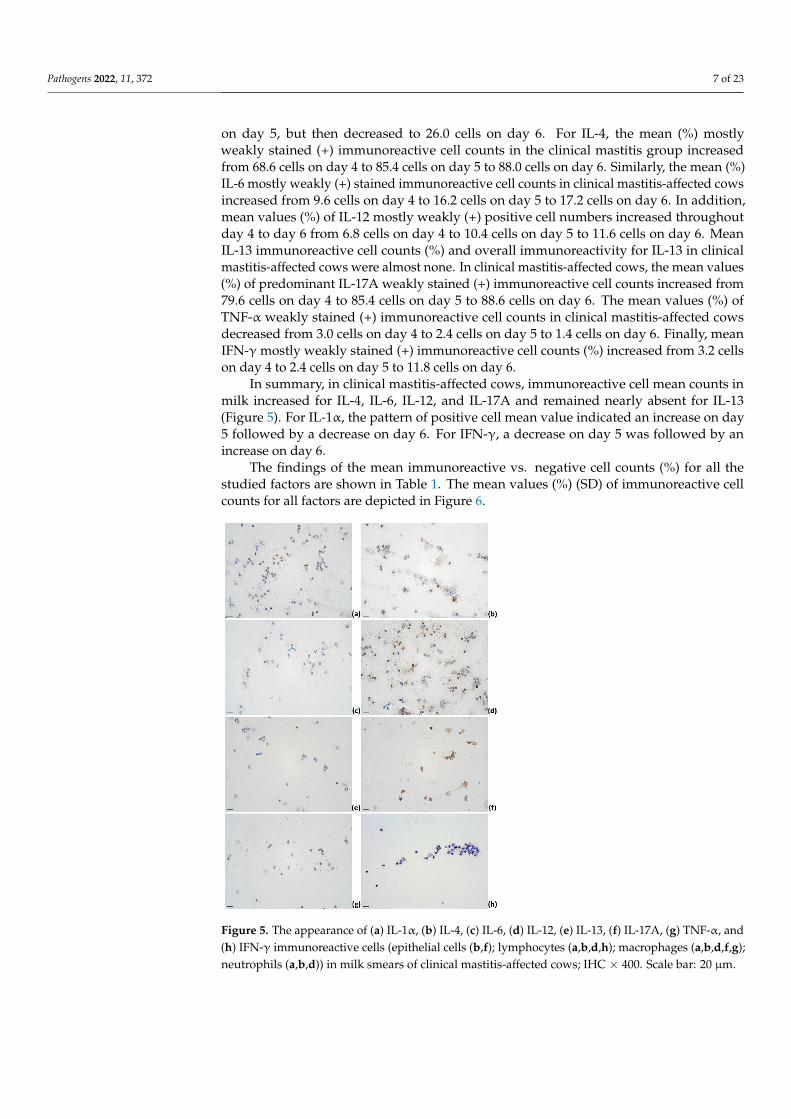
on day 5, but then decreased to 26.0 cells on day 6. For IL-4, the mean (%) mostlyweakly stained (+) immunoreactive cell counts in the clinical mastitis group increasedfrom 68.6 cells on day 4 to 85.4 cells on day 5 to 88.0 cells on day 6. Similarly, the mean (%)IL-6 mostly weakly (+) stained immunoreactive cell counts in clinical mastitis-affected cowsincreased from 9.6 cells on day 4 to 16.2 cells on day 5 to 17.2 cells on day 6. In addition,mean values (%) of IL-12 mostly weakly (+) positive cell numbers increased throughoutday 4 to day 6 from 6.8 cells on day 4 to 10.4 cells on day 5 to 11.6 cells on day 6. MeanIL-13 immunoreactive cell counts (%) and overall immunoreactivity for IL-13 in clinicalmastitis-affected cows were almost none. In clinical mastitis-affected cows, the mean values(%) of predominant IL-17A weakly stained (+) immunoreactive cell counts increased from79.6 cells on day 4 to 85.4 cells on day 5 to 88.6 cells on day 6. The mean values (%) ofTNF-α weakly stained (+) immunoreactive cell counts in clinical mastitis-affected cowsdecreased from 3.0 cells on day 4 to 2.4 cells on day 5 to 1.4 cells on day 6. Finally, meanIFN-γmostly weakly stained (+) immunoreactive cell counts (%) increased from 3.2 cellson day 4 to 2.4 cells on day 5 to 11.8 cells on day 6.
In summary, in clinical mastitis-affected cows, immunoreactive cell mean counts inmilk increased for IL-4, IL-6, IL-12, and IL-17A and remained nearly absent for IL-13(Figure 5). For IL-1α, the pattern of positive cell mean value indicated an increase on day5 followed by a decrease on day 6. For IFN-γ, a decrease on day 5 was followed by anincrease on day 6.
The findings of the mean immunoreactive vs. negative cell counts (%) for all thestudied factors are shown in Table 1. The mean values (%) (SD) of immunoreactive cellcounts for all factors are depicted in Figure 6.
Pathogens 2022, 11, x FOR PEER REVIEW 8 of 24
In summary, in clinical mastitis-affected cows, immunoreactive cell mean counts in milk increased for IL-4, IL-6, IL-12, and IL-17A and remained nearly absent for IL-13 (Fig-ure 5). For IL-1α, the pattern of positive cell mean value indicated an increase on day 5 followed by a decrease on day 6. For IFN-γ, a decrease on day 5 was followed by an in-crease on day 6.
Figure 5. The appearance of (a) IL-1α, (b) IL-4, (c) IL-6, (d) IL-12, (e) IL-13, (f) IL-17A, (g) TNF-α, and (h) IFN-γ immunoreactive cells (epithelial cells (b,f); lymphocytes (a,b,d,h); macrophages (a,b,d,f,g); neutrophils (a,b,d)) in milk smears of clinical mastitis-affected cows; IHC × 400. Scale bar: 20 μm.
The findings of the mean immunoreactive vs. negative cell counts (%) for all the stud-ied factors are shown in Table 1. The mean values (%) (SD) of immunoreactive cell counts for all factors are depicted in Figure 6.
Figure 5. The appearance of (a) IL-1α, (b) IL-4, (c) IL-6, (d) IL-12, (e) IL-13, (f) IL-17A, (g) TNF-α, and(h) IFN-γ immunoreactive cells (epithelial cells (b,f); lymphocytes (a,b,d,h); macrophages (a,b,d,f,g);neutrophils (a,b,d)) in milk smears of clinical mastitis-affected cows; IHC × 400. Scale bar: 20 µm.

Pathogens 2022, 11, 372 8 of 23
Table 1. The appearance and mean numbers of IL-1α, IL-4, IL-6, IL-12, IL-13, IL-17A, TNF-α, and IFN-γ immunoreactive cells in healthy cows and cows withsubclinical and clinical mastitis.
IL-1α IL-4 IL-6 IL-12
Day 4 Day 5 Day 6 Day 4 Day 5 Day 6 Day 4 Day 5 Day 6 Day 4 Day 5 Day 6++ + 0 ++ + 0 ++ + 0 ++ + 0 ++ + 0 ++ + 0 ++ + 0 ++ + 0 ++ + 0 ++ + 0 ++ + 0 ++ + 0
HealthyMean (%) 15 41 44 15 34 51 14 33 54 51 45 4 64 34 2 54 43 3 18 18 64 18 16 67 16 17 67 29 28 43 28 26 46 21 30 49
+/− 56 */44 49/51 47/54 96/4 98/2 97/3 36/64 34/67 33/67 57/43 54/46 51/49Subclinical mastitis
Mean (%) 6 20 74 8 21 71 5 17 78 24 64 12 15 56 29 31 55 14 5 11 84 3 4 93 1 9 89 10 16 74 12 16 72 14 22 64+/− 26/74 29/71 22/78 88/12 71 */29 86/14 16/84 7/93 10/89 26/74 28/72 36/64
Clinical mastitisMean (%) 1 20 79 6 37 57 3 23 74 19 56 31 17 69 15 23 65 12 1 9 90 6 10 84 2 15 83 2 5 93 3 7 90 2 10 88
+/− 21/79 43/57 26/74 75/31 86/15 88/12 10/90 16/84 17/83 7/93 10/90 12/88
IL-13 IL-17A TNF-α IFN-γ
Day 4 Day 5 Day 6 Day 4 Day 5 Day 6 Day 4 Day 5 Day 6 Day 4 Day 5 Day 6++ + 0 ++ + 0 ++ + 0 ++ + 0 ++ + 0 ++ + 0 ++ + 0 ++ + 0 ++ + 0 ++ + 0 ++ + 0 ++ + 0
HealthyMean (%) 4 20 76 4 23 73 3 19 79 17 81 2 21 74 5 23 74 3 1 18 81 2 19 79 2 16 82 42 31 26 42 43 24 35 35 30
+/− 24/76 27/73 22/79 98 * /2 95/5 97/3 19/81 21/79 18/82 73/26 76/24 70/30Subclinical mastitis
Mean (%) 0 1 99 0 2 98 0 3 97 10 74 16 11 74 15 17 75 8 0 2 98 0 3 97 0 6 94 12 16 72 16 23 60 14 42 49+/− 1/99 2/98 3/97 84/16 85/15 92/8 2/98 3/97 6/94 28/72 40/60 26/49
Clinical mastitisMean (%) 0 0 100 0 0 100 0 0 100 3 77 19 5 81 14 7 82 11 0 3 97 0 2 98 0 1 99 1 4 95 1 2 98 4 8 88
+/− 0/100 0/100 0/100 80/19 86/14 89/11 3/97 2/98 1/99 5/95 3/98 12/88Mean—mean value (%) of intensively (++), weakly (+) and negatively (0) stained immunoreactive cells, numbers are indicated with zero decimals; +/−—immunoreactive cells (both values of intensively (++) and weakly (+)stained immunoreactive cells) (+) vs. negative cells (no immunoreactivity) (−); numbers are indicated with zero decimals. Abbreviations in table: *—statistically significant difference (p < 0.05); IL-1α, IL-4, IL-6, IL-12, IL-13,IL-17A—interleukins (IL)-1α, -4, -6, -12, -13, -17A; TNF-α—tumor necrosis factor-alpha; IFN-γ—interferon-gamma.

Pathogens 2022, 11, 372 9 of 23
Pathogens 2022, 11, x FOR PEER REVIEW 10 of 24
Figure 6. The graphical appearance of mean immunoreactive cell numbers (%) in healthy cows (green line) and cows with subclinical (blue line) and clinical mastitis (red line): (a) IL-1α; (b) IL-4; (c) IL-6; (d) IL-12; (e) IL-13; (f) IL-17A; (g) TNF-α; (h) IFN-γ. Together mean values (%) (SD) of in-tensively (++) and weakly (+) stained immunoreactive cells are demonstrated. Abbreviations in
Figure 6. The graphical appearance of mean immunoreactive cell numbers (%) in healthy cows (greenline) and cows with subclinical (blue line) and clinical mastitis (red line): (a) IL-1α; (b) IL-4; (c) IL-6;(d) IL-12; (e) IL-13; (f) IL-17A; (g) TNF-α; (h) IFN-γ. Together mean values (%) (SD) of intensively(++) and weakly (+) stained immunoreactive cells are demonstrated. Abbreviations in figure: IL-1α,IL-4, IL-6, IL-12, IL-13, IL-17A—interleukins (IL)-1α, -4, -6, -12, -13, -17A; TNF-α—tumor necrosisfactor-alpha; IFN-γ—interferon-gamma. Error bars: +/− 1 SD; +/− 2 SD.

Pathogens 2022, 11, 372 10 of 23
2.4. Statistical Analysis
A one-way repeated-measures ANOVA determined that mean numbers (%) of IL-1α(p = 0.253), IL-4 (p = 0.728), IL-6 (p = 0.892), IL-12 (p = 0.614), IL-13 (p = 0.442), TNF-α(p = 0.842), and IFN-γ (p = 0.586) immunoreactive cell counts in healthy cows did not showa statistically significant difference over time. However, the mean numbers (%) of IL-17Aimmunoreactive cells indicated statistically significant differences between the days 4 to6 (F(2, 8) = 10.0, p = 0.007, the population effect size partial omega-squared ω2 = 0.62;sphericity assumed). In particular, post-hoc analysis with a Bonferroni adjustment revealedthe mean value of IL-17A immunoreactive cell count was significantly higher on day 4 vs.day 5 and 6 (by 2.0 (95% CI, 0.9 to 3.11) cells, p = 0.003, partial η2 = 0.909).
For cows with subclinical mastitis, a one-way repeated-measures ANOVA determinedno statistically significant difference of mean immunoreactive cell count numbers (%) overtime for all factors IL-1α (p = 0.294), IL-4 (p = 0.085), IL-6 (p = 0.081), IL-12 (p = 0.494), IL-13(p = 0.147), IL-17A (p = 0.084), TNF-α (p = 0.294), and IFN-γ (p = 0.156).
Similarly, in clinical mastitis-affected cows, no statistically significant difference ofmean immunoreactive cell count numbers (%) over time was observed for IL-1α (p = 0.068,ε = 0.531), IL-4 (p = 0.125), IL-6 (p = 0.65), IL-12 (p = 0.557), IL-17A (p = 0.111), TNF-α(p = 0.374), and IFN-γ (p = 0.255, ε = 0.51). IL-13 immunoreactivity in clinical mastitis wasalmost undetectable.
Further analysis to determine the main group effect showed no statistically signifi-cant difference in mean (%) IL-1α (p = 0.071) and IL-6 (p = 0.073) immunoreactive countsbetween study groups. However, the main status effect showed a statistically signifi-cant difference in mean immunoreactive cell counts (%) between studied groups for IL-4(F(2, 12) = 88.044, p < 0.001, partial η2 = 0.936), IL-12 (F(2, 12) = 8.95, p = 0.004, partialη2 = 0.599), IL-13 (F(2, 12) = 32.447, p < 0.001, partial η2 = 0.844), IL-1A (F(2, 12) = 6.025,p = 0.015, partial η2 = 0.501), TNF-α (F(2, 12) = 27.523, p < 0.001, partial η2 = 0.821), andIFN-γ (F(2, 12) = 21.78, p < 0.001, partial η2 = 0.784).
On day 4 and 6, but not on day 5, mean IL-1α immunoreactive cell counts (%) ofhealthy animals almost doubled the values found in subclinical mastitis and clinical mastitis-affected animals. Mixed two-way ANOVA determined a statistically significant two-wayinteraction of status and time on IL-1α immunoreactive cell count (F(4, 24) = 3.633, p = 0.019,partial η2 = 0.377; sphericity assumed). Mean IL-1α immunoreactive cell count (%) wasstatistically different between groups on day 4 (F(2, 12) = 9.58, p = 0.003, partial η2 = 0.615),but not on day 5 (p = 0.312) and day 6 (p = 0.107). The mean IL-1α immunoreactive cellcount in healthy cows on day 4 was statistically significantly greater than in subclinicalmastitis- (p = 0.012) and in clinical mastitis-affected cows (p = 0.004), but not on day 5(p = 0.312) and day 6 (p = 0.107).
Altogether, most cells in the milk of studied animals were immunoreactive for IL-4;however, a statistically significant difference was observed between status and time onthe mean values (F(4, 24) = 3.198, p = 0.031, partial η2 = 0.348; sphericity assumed). Themean value (%) of IL-4 immunoreactive cell numbers on day 4 was statistically significantlylower in clinical mastitis-affected cows vs. healthy cows (p = 0.006). On day 5, a mean value(%) of IL-4 immunoreactive cell count was statistically significantly lower in subclinicalmastitis-affected cows than in healthy cows (p < 0.001) and clinical mastitis-affected cows(p = 0.026).
For mean (%) IL-6 (p = 0.896), IL-12 (p = 0.852), IL-13 (p = 0.909), IL-17A (p = 0.063), TNF-α (p = 0.759), and IFN-γ (p = 0.184) immunoreactive cell counts, there was no statisticallysignificant interactions found between day and status.
The summary of findings to test the significance of the time point and status onimmunoreactive cell counts (%) is shown in Table 2.

Pathogens 2022, 11, 372 11 of 23
Table 2. Statistically significant interactions of time and status on mean numbers (%) of IL-1α, IL-4,IL-6, IL-12, IL-13, IL-17A, TNF-α, and IFN-γ immunoreactive cells in the healthy cows and animalswith subclinical and clinical mastitis: a summary.
Cytokine
p-ValueD
S S × DHealthy Subclinical
MastitisClinicalMastitis
IL-1α 0.253 0.294 0.068 0.071 0.019 *IL-4 0.728 0.085 0.125 <0.001 * 0.031 *IL-6 0.892 0.081 0.65 0.073 0.896IL-12 0.614 0.494 0.557 0.004 * 0.852IL-13 0.442 0.147 0.111 <0.001 * 0.909
IL-17A 0.007 * 0.084 0.374 0.015 * 0.063TNF-α 0.842 0.294 0.255 <0.001 * 0.759IFN-γ 0.586 0.156 0.178 <0.001 * 0.184
D—day (4, 5 and 6); S—status (healthy cows, subclinical mastitis-affected cows, clinical mastitis-affected cows);IL-1α, IL-4, IL-6, IL-12, IL-13, IL-17A—interleukins (IL)-1α, -4, -6, -12, -13, -17A; TNF-α—tumor necrosis factor-alpha; IFN-γ—interferon-gamma; *—statistically significant difference (p < 0.05).
3. Discussion
Milk SCC is a valuable, easy-to-perform tool in the bovine mastitis diagnostic ap-proach. Combined with clinical objectives, SCC numbers determine subclinical and clinicalmastitis. Although low numbers of SCCs in milk are associated with relative health, moredetailed observation by differential cell counts showed still existing inflammatory reactionssuggesting persistent activity by host defense immunity [50]. This might indicate the hostdefense reactivity due to the continuous exposure to pathogenic microorganisms in the barnconditions. Reported experimental studies confirm prominent SCC early increase in milkafter the intended exposure to mastitis-associated antigens [51], while in vivo studied SCCthreshold for subclinical mastitis is also recognized [52]. Previous studies of cows in thedry-off period showed a maximum SCC increase of 12 h after experimental inoculation andcontinuous increase up to 7 days [53]. On-farm diagnosis of mastitis mainly relies on SCC,although this method depends on multiple factors such as the type of causative pathogen.In subclinical mastitis, SCC greatly differs from the microbiological culture and leukocytedifferential studies, suggesting SCC alone might not provide as detailed information ofmammary quarter status as leukocyte differential analysis or all together [54]. Laboratory-based diagnosis of mastitis via cell culture and/or molecular analysis effectively identifiesbacterial pathogens, but its wide use by farmers is prevented due to the time and/or costinvolved. Moreover, SCC alone has been overall criticized for not being sensitive enough towork as a screening tool in subclinical mastitis diagnosis, but it works in the context of otherparameters [55,56]. Altogether, microbiological analysis, variety of clinical symptoms, andthe search for precise diagnostic approaches request the implementation of multiple param-eters or even an algorithm. A knowledge acquired on host response-derived biomarkers inmilk, such as proinflammatory cytokines, could potentially support the identification ofperspective mastitis biomarkers to report disease status and etiology.
The trend in the presence and findings of inflammatory and regulatory markers inthe milk cells of healthy cows showed the highest mean cell numbers (%) compared tosubclinical and clinical mastitis-affected cows. If we look at the SCC and proinflammatorycytokines as indicators of early inflammation such as TNF-α, they reach a peak in 1 to 12 hfollowed by a gradual drop [51], suggesting our findings of inflammatory and regulatorymarkers on day 4 to 6 in the milk of subclinical and clinical mastitis correspond to theresolution of initial inflammation. During bovine mastitis, CD4+ T lymphocytes, activatedupon antigen recognition, dominate. They also activate macrophages through the produc-tion of cytokines. Depending on the type of cytokines produced, the Th cells are dividedinto types Th1 or Th2 [57]. Th1 cells are characterized by the production of IFN-γ andIL-2, whereas Th2 cells produced IL-4, IL-5, IL-6, IL-10, and IL-13 [58]. Importantly, IL-10

Pathogens 2022, 11, 372 12 of 23
inhibits IFN-γ and IL-2 via Th1 lymphocytes; IL-4 and IL-5—via Th2 lymphocytes; IL-1,IL-6, IL-8, IL-12, and TNF-α—via macrophages; IFN-γ and TNF-α—via NK cells [59,60]. Inmastitis, IL-10 production highly increases, reaching superior relative numbers over otherfactors [27]. This may suggest a possible IL-10 anti-inflammatory effect on the reducednumbers of milk cells containing inflammatory and regulatory factors and must be thesubject of future research.
Initially, IL-1 was associated with leukocytes and the ability to induce fever in ani-mals [61,62]. Subsequent studies have also described IL-1 family cytokines in relation tothe functions of immunocompetent tissue cells such as T cells, macrophages, endothelialand epithelial cells. The effect of IL-1 on immunity is indirect; it works through severalsignaling pathways involving other messenger molecules [21]. In the mammary gland, IL-1is produced by macrophages, lymphocytes, monocytes, endothelial cells, and fibroblasts.In addition, IL-1 stimulates its own production, as well as that of IL-6, IL-8, IL-12, andTNF-α [25]. In our study, the IL-1α linear pattern through day 4 to 6 in healthy cows wassimilar to IL-6, IL-12, and like IL-13, TNF-α, and INF-γ by the drop on day 6, however,having no association with other factors in the cases of subclinical and clinical mastitis. Thispossibly suggests the key regulation by other proinflammatory cytokines in mastitis condi-tions. Studies evidence bovine mammary epithelial cells challenged with E. coli-derivedlipopolysaccharides showed an increase in mRNA expression of IL-1α similar to that foundin mastitis-affected cows [63]. When challenged with E. coli, S. aureus, or S. agalactiae, goatmammary epithelial cells also showed increased expression of IL-1α [64]. In epithelial cellsof bovine mammary glands, IL-1α effects were associated with the inflammatory responseto induced mastitis [65]. In both statuses of subclinical and clinical mastitis, IL-1α meannumbers (%) were statistically significantly lower than in healthy cows, suggesting itsrole in bovine mammary gland immunity and early inflammation. However, a peak inIL-1α immunoreactivity was noted on day 5 of clinical mastitis-affected cows suggestingits inflammatory role in the support and maintenance of inflammation.
In the bovine milk of healthy cows, the expression of regulatory cytokines IL-4, IL-10,and IFN-γ has been previously reported [66]. In our study, almost all cells in milk samplesof healthy cows were indeed positive for IL-4, underpinning a high immunoreactivity.Both IL-4 and IL-13 in the association of differentiation of T cells into Th-2 cells mayhave an immunoregulatory role in the bovine mammary gland [67]. However, mean IL-13 immunoreactive cell numbers (%) were low in healthy animals and almost absent inanimals affected by mastitis. Both mean numbers (%) of IL-4 and IL-13 immunoreactivecells did statistically significantly differ by status, but IL-4 also differed both by statusand day, promoting the profound stability of IL-4 both in healthy and disease-affectedanimals. Activation of bovine tissue macrophages by IL-4/IL-13 induces a cell phenotypethat is not anti-inflammatory itself but comprises a diverse phenotype that contributes tothe tissue repair and could trigger a proinflammatory reaction. Bovine monocyte-derivedmacrophages suggested IL-4/IL-13 priming in macrophage polarization to wound-healingphenotype [68]. In bovine endothelial cells, both IL-4 and IL-13 protect the endothelialsurface against inflammatory mediator-induced procoagulant changes [69]. This might becrucial to hold readiness for both inflammation risk and tissue damage in healthy cowsby both IL-4 and IL-13, but only by IL-4 in mastitis-affected cows. The concentration ofIL-4 was significantly lower in the milk of cows with Staphylococcal subclinical mastitiscompared to control animals [28], possibly supporting our findings by causative explanationin subclinical and clinical mastitis.
Contrary to our findings, IL-6 levels in whey of mastitis-affected cows [70] and subclin-ical mastitis group cows [28] were shown to be higher than in samples from healthy cows. Inaddition, the detection of IL-6 concentration in milk samples of subclinical mastitis-affectedcows predicted inflammation more precisely and earlier than SCC [71]. In our study, meanIL-6-containing cell numbers (%) did not statistically significantly differ by status or bothstatus and day (time). Up to the knowledge, Klebsiella pneumoniae promotes the productionof the inflammatory cytokine genes IL-6, IL-8, IL-1β, and TNF-α in mastitis already after

Pathogens 2022, 11, 372 13 of 23
6 h [72]. However, the IL-6 gene was among those genes that were not differentially ex-pressed in healthy vs. mastitis-affected cows suggesting they are not suitable markers orindicators of mastitis [27]. In addition, there was no difference in milk IL-6 between healthyand naturally occurring subclinical mastitis cases [31]. The discrepancy in the findings ofIL-6 might substantiate its proinflammatory role in the early inflammation phase. Severalexperimental studies have shown the importance of inflammatory factors in an early inflam-matory event. Within a few hours after infusion of E. coli lipopolysaccharide, high amountsof IL-1, IL-6, and TNF-α are found in milk that further promote IL-1-induced neutrophilmigration [73]. Once bovine mammary epithelial cells recognize microbial components,they can initiate an innate immune response by secreting proinflammatory cytokines (IL-1β,TNF-α, IL-6, IL-8) and antimicrobial molecules (cathelicidin, defensins) [74,75].
In the environment of the mammary gland, IL-12 is known as a mediator betweeninnate and adaptive immunity by the regulation of T lymphocytes. It also works asa growth factor for active NK cells and supports their cytotoxicity [25]. In our study,we found IL-12 immunoreactivity to be the highest in healthy cows compared to lowernumbers in subclinical mastitis and the lowest in clinical mastitis. Moreover, IL-12 didnot change much through days 4 to 6. Within the relative expression patterns of bovinemilk cytokines, IL-12 was found to be significantly higher in late lactation compared to itscorresponding level in mid-lactation. Possibly, an increase in IL-12 in the late lactation couldindicate the shift toward a more pronounced Th1 immunity [76]. Altogether, elevation ofIL-12 may indicate its importance in enhancing the innate immunity responses in bovineudder [76], supporting our results in healthy cows. The cytokine transcriptional patternof S. aureus-associated post-infection IL-12 level presented a significant elevation in 24 hfollowed by a sharp decrease in 32 h [77]. Similar findings were observed in milk cells ofdairy cows experimentally challenged with either E. coli or S. aureus [78]. However, whenchallenged with Pseudomonas aeruginosa, the maximum peak level was observed after 32 hand remained elevated only for 24 h [79]. In response to Mycoplasma bovis intramammaryinfection, milk IL-12 concentrations increased only within 78 h of infection and remainedelevated throughout the study [80]. In Klebsiella pneumoniae-challenged mastitis, IL-12 levelspeaked at 48 h [81]. Furthermore, the differences in milk IL-12 are associated with lowerlevels in mild vs. moderate/severe clinical mastitis, Gram-positive vs. Gram-negativemicroorganisms, early group vs. mid and late days in milk production, and spring vs. otherseasons [33], indicating a high heterogeneity of IL-12 peak in mastitis. In our study, thenumbers suggested significant differences between the study groups by status, consideringIL-12 to be an informative diagnostic tool for mastitis.
For IL-17A, most milk cells were found to be immunoreactive in all studied groupsby status. IL-17A is a proinflammatory cytokine with host defense properties againstmicroorganisms both in innate and adaptive immunity and a protective role [82]. After cowimmunization with ovalbumin, IL-17A was present in the milk of all the high-respondercows right after milk leukocytosis developed [83]. An increase in IL-17A in response toovalbumin or its combination with E. coli was also studied [84]. In immunized cows withE. coli, an increase in TNF-α, IL-6, IL-10, and IL-17A was detected. IL-17A was found inthe milk of all cows at the onset of inflammation, and concentrations remained elevatedafter the acute phase [85]. Therefore, as this occurrence is still high and with increasingpattern in subclinical and clinical mastitis cases of our study, IL-17A significance in thecontext of acute-phase cytokine might propose not only its continuous role in early defensein healthy animals but also inflammation resolution in mastitis. Certainly, the expression ofthe IL-17A gene is induced in udder tissues of cows experimentally infected with E. coli,suggesting IL-17A could play an important role in mediating host-pathogen interactionsduring mastitis [39]. In dairy cattle challenged with S. uberis, the increase in IL-17A levelsmatched with inversion of the pre-challenge CD4(+)-to-CD8(+) T lymphocyte ratio, thatfollowed by normalization of this ratio altogether with respective IL-17A levels, suggestingthat IL-17A may be involved in the resolution of intramammary infection [86].

Pathogens 2022, 11, 372 14 of 23
TNF-α is an acute-phase cytokine produced during the early stages of infection [87,88].In our study, we found low numbers of TNF-α-containing immunoreactive cells in milksamples of all studied cases. TNF-α could potentially be associated with early initiativeactivity; however, this role has been questioned. In serum and whey taken from naturallyoccurring mastitis-affected animals, significantly higher concentrations of IFN-γ and TNF-αwere found. As cows recovered, IFN-γ and TNF-α concentrations reduced significantly [89].Therefore, TNF-α significance may exceed the proinflammatory role. However, in bothserum and milk of mid-lactation cows with feed restriction influences, TNF-α concentra-tions showed only modest increases during endotoxin infusion [90]. When investigatingthe effect of intramammarily infused lipopolysaccharide on the acute-phase reaction inearly and in late lactation, no detectable levels of TNF-α were found in serum, while levelsof both increased in milk. Peak concentration was higher in early lactation than in latelactation [91]. In an experiment of bovine mastitis induced by endotoxin or E. coli, nosignificant differences between sample types and over time for TNF-α concentrations inmilk were detected; however, an increase in TNF-αwas observed in afferent lymph [92].TNF-α immunoreactivity was modest and even weak in our cases, suggesting this factordoes not exceed its rather baseline expression and stable presence. Moreover, its function-ality is not associated with cell quantity. Lastly, in response to M. bovis intramammaryinfection over a period of several days, initial elevations of TNF-α, IL-1β, and IFN-γ wereobserved between 90 and 102 h post infection [80], suggesting the late onset of these factorsdepend on causative pathogens. This was also studied in means of endotoxin amountenough to initiate pronounced cell recruitment but insufficient to induce a marked TNF-αsecretion [88]. No significant change in gene expression of IL-2, IL-4, IL-6, IL-8, IL-10, IFN-γ,and TNF-α by real-time PCR in milk among animals without mastitis vs. animals withmastitis was observed [27].
Lastly, IFN-γ indicated a prominent difference between study groups, with most cellsbeing immunoreactive for IFN-γ in healthy animals, increasing moderate numbers of IFN-γimmunoreactive cells in subclinical mastitis cases. However, only a minor number of suchcells were found in clinical mastitis. It is known that IFN-γ activates the acquired immuneresponse and T lymphocytes and IL-12 production [93]. The last must be recognized inthe context of our findings that marked distinctively similar levels of IL-12 in each statusgroup. Moreover, IFN-γ immunoreactivity also indicated diagnostic potential in mastitisvs. healthy cases. However, IFN-γ significance might be addressed to innate host defensesignaling as milk IFN-γ immunoreactivity in our clinical mastitis-affected cows was low.Similarly, the mRNA of IL-2 and IFN-γ was not expressed in milk cells isolated fromdiseased cows before treatment [94]. In bovine mastitis induced by endotoxin or E. coli,IFN-γ concentration was too low to be detected by ELISA [92]. Interestingly, IL-4 maintainsan antagonistic action to IFN-γ [27]. Therefore IFN-γ immunoreactive mean numberswere lower in subclinical mastitis and almost absent in clinical mastitis, suggesting IL-4antagonist activity over INF-γ in mastitis conditions. IL-4 immunoreactivity was prominentin our study.
This study aimed to determine inflammatory and regulatory cytokines in the milk ofdairy cows and to differentiate the changes in their expression. Notably, the immunore-activity for all factors was quite stable throughout the time period of three consecutivedays after the settlement of the diagnosis and division of study groups of subclinical orclinical mastitis. Understandably, in naturally occurring mastitis, the study of early inflam-matory onset is limited to the time spent on the diagnosis to enroll these cases in the study.Hereby, knowledge, at the cellular and molecular level, of the immune response at relativehealth and during the infection is fundamental to the early and timely diagnosis and inprospective pathogenesis-based treatment.
Nonetheless, this study has limitations to discuss. The study emphasizes the necessityto extend the time period corresponding to the prolonged acute phase up to 7 to 10 days.The evaluation of cytokines in this time period would express the dynamics of the cytokineappearance in milk. Furthermore, additional to the already studied cytokines, including

Pathogens 2022, 11, 372 15 of 23
IL-2, IL-8, IL-10, IL-13, nuclear factor kappa B, beta defensins, transforming growth factorbeta 1, and cathelicidin LL37, altogether would explain their relationships in detailedinterrelations by correlation analysis to substantiate the knowledge on the broad cytokinepleiad participating in both host immune defense and mastitis inflammation.
4. Materials and Methods4.1. Study Animals and Ethical Statements
The research was performed in the herd of 79 cows (Holstein Friesian cattle), whichincluded 41 adult cows (of those, 35 were milking cows) and 38 heifers. Based on theveterinary observations as well as screening of somatic cell count in milk performed overthree consecutive days, we selected 15 milking cows and allocated them into three studygroups: 5 healthy animals, 5 animals with subclinical mastitis, and 5 animals with clinicalmastitis (selection criteria are described in Section 4.3). In healthy and subclinical mastitis-affected groups, the first-, the second-, and the third-parity cows were included. In theclinical mastitis group, the second-, fourth-, and fifth-parity cows were included. Meanparity in each group was 1.6, 2, and 3, respectively, while mean days in milk in each groupwere 181.6, 149.8, and 256, respectively. Due to previous mastitis infections, three animalshad a history of prior veterinary interventions: (1) one cow in the subclinical mastitisgroup had antibiotic treatment that finished one week before the sampling start; (2) onecow in the clinical mastitis group had non-antibiotic treatment applied (anti-inflammatoryointment) that finished three days before the sampling start; and (3) one cow in the clinicalmastitis group had antibiotic treatment that finished one month before the sampling start.Since antibiotic and anti-inflammatory interventions were finished several days beforethe study start, milk samples from animals subject to those interventions were includedin the analyses.
All animals were kept in a free-stall barn located in northern Poland, without access toenclosure or pasture. The cows were milked twice a day and fed two times daily, and waterwas available ad libitum. The feed included silage, corn silage, and grain by-products,which were provided as separate components.
The study aims to perform an analysis of cow’s milk. The research involving animalsuse has the clear scientific purpose of increasing their welfare, which justifies their engage-ment in the research project. The research involved techniques administered in a way thatdid not cause pain, suffering, distress, or damage to the body of participating animals. Inthis sense and in view of the national regulations of the partner in charge of validation (RICPro-Akademia, Poland), the study did not involve experiments on animals as defined inthe Law of 15th January 2015 on the protection of animals used for scientific or educationalpurposes (Journal of Laws of 2015 pos. 266 with further amendments) [95]. Animal studieswere carried out humanely according to national and international Animal Care and UseCommittee protocols. The study involved obtaining milk samples by standard milkingprocedure without affecting the cow’s routine.
4.2. Sample Collection
The following sample collection was conducted. On days 1 to 3, milk samples werecollected for the milk quality examination (somatic cell count screening) from all milkingcows (35 animals, of each 4 quarters). Further, on days 4 to 6, milk samples from selected15 cows were collected for the milk quality analysis, bacteriological examination, and milksediment preparations. Samples were collected into sterile containers and immediatelytransported to the laboratory. Before sample collection, udders were cleaned, dried, anddisinfected. The foremilk was discarded before taking the sample. Sample analysis wasconducted at RIC Pro-Akademia, Poland. No veterinary treatments (neither antibiotic noranti-inflammatory) were applied to the animals over the sample collection period.
The following analytical procedures of milk samples were performed: differentialcounting of somatic cells, bacteriological examination, and immunocytochemical analysis.

Pathogens 2022, 11, 372 16 of 23
4.3. Differential Counting of Somatic Cells
Somatic cell count (SCC) is an essential indicator of bovine udder inflammation, whichhighly correlates with the presence of a mammary infection [96]. SCC is a well-establishedmeasurement for identifying animals with suspected mammary gland infection and in-flammation [97]. SCC investigation was performed by fluorescent automatic cell counterLactoscan SCC (Milkotronic Ltd., Stara Zagora, Bulgaria). On days 1 to 3, the research cov-ered all milking cows (n = 35, of each 4 quarters) to identify relevant cases of subclinical andclinical mastitis. The animals were assigned to the three study groups using the followingcriteria, based on the average number of somatic cells in milk and observation of ud-ders/mammary glands, in line with the mastitis severity scoring provided by the scientificannals of the Polish Society of Animal Production (Polskie Towarzystwo Zootechniczne,PZH) [98]: (1) healthy group—SCC under 200,000 cells/mL milk, udders/mammary glandnot showing redness, swelling, or pain—equivalent to PZH severity score of I or II; (2)subclinical mastitis group—SCC between 200,000 and 500,000 cells/mL milk, no signs ofudder/mammary gland inflammation—equivalent to PZH severity score of III or IV; (3)clinical mastitis group—SCC above 500,000 cells/mL, udders/mammary gland showingredness, swelling, or pain—equivalent to PZH severity score higher than IV. No exclusioncriteria for each group were applied.
The further study analyses were aimed to investigate selected udder quarters thatwere affected by mastitis. In eight cows, one-quarter were affected by mastitis. In two cowswith clinical mastitis, out of two mastitis-affected quarters, one quarter was selected by thehighest SCC for further analyses.
4.4. Bacteriological Examination
Microbiological analysis was performed according to ISO 4833:2013 standard on days4 to 6 by the detection of total bacteria count on PCA (Plate Count Agar) from the milksamples of selected quarters (N = 10 on each day) from all cows. For the total bacteriacount (TBC) on PCA, samples, and a series of 10-fold dilutions plated on PCA were set toobtain 15 TBC results (logarithmic (log) CFU/mL). Before plating, samples were 10-timesdiluted in 0.9% saline solution. Plating was performed using automatic plater Easy Spiral(Interscience, Saint-Nom-la-Bretèche, France).
4.5. Immunocytochemistry
In total, 135 samples of milk sediments were prepared at RIC Pro-Akademia, Poland,for further investigation. These were constituent of 3 repetitions (day 4, day 5, and day 6)with the collection of 100 mL of milk from the 15 cows of a selected quarter of each. Milksamples of 100 mL were obtained directly after the milking. In total, 300 mL of milkwas prepared for centrifugation. Milk sediments were prepared by centrifugation of milksamples at 2000 (1000 to 3000) RPM for 3 min. The supernatant was carefully removedand discarded. Samples were again centrifuged at 2000 (1000 to 3000) RPM for 3 min. Thesupernatant was carefully removed and discarded. Samples were again centrifuged at 2000(1000 to 3000) RPM for 3 min. The samples were centrifuged using Frontier 5000 Multi Pro(Model FC5816R, OHAUS, Parsippany, NJ, USA) laboratory centrifuge, equipped with aswing rotor (Model OH_30314828, OHAUS, Parsippany, NJ, USA). In total, 45 Eppendorftubes of 5 mL were prepared with 2 mL of milk sample (sediment) and 2 mL of Tyrode’ssolution buffer from each day of sample collection round. Samples were stored at −20 ◦C.The delivery of samples was carried out with dried ice.
Preparation and staining of milk smears were performed at the Laboratory of Mor-phology, Institute of Anatomy and Anthropology, Riga Stradins University. A modifiedbiotin-streptavidin immunocytochemical method was performed to examine cytokinesIL-1α, IL-4, IL-6, IL-12, IL-13, IL-17A, TNF-α, and IFN-γ in prepared milk smears [99,100].
For the cytological smear, fixation with the Stefanini (Zamboni) solution was per-formed [101]. Stefanini tissue fixative was prepared using paraformaldehyde (20 g), 150 mLpicric acid (0.2% concentration), 425 mL Sorensen buffer (pH 7.2), and 425 mL distilled

Pathogens 2022, 11, 372 17 of 23
water. Paraformaldehyde was dissolved in distilled water. The resulting mixture was thenplaced in buffer and was added with filtered picric acid. Milk sediments were proceededfor a wash in TRIS buffer twice for 5 min. Samples were then placed in 3% hydrogenperoxide (H2O2) solution for 30 min at 4 ◦C. Washing with TRIS buffer was performedrepeatedly for 5 min each time. Milk sediment samples were incubated with a HiDefDetectionTM reaction amplifier (code 954D-31, Cell MarqueTM, Rocklin, CA, USA) for thedetection of antibodies acquired from mouse or rabbit. Incubation with HiDef DetectionTMhorseradish peroxidase (HRP) polymer detector (code-954D-32, Cell MarqueTM, Rocklin,CA, USA) was managed. These procedures were substantially followed by washing thesamples in a wash buffer and then processed with 3,3’-diaminobenzidine (DAB) SubstrateKit (code-957D-30, Cell MarqueTM, Rocklin, CA, USA) to obtain immunoreactive structurestaining in brown color. To avoid the artifact presence, cells of smears were also stainedby hematoxylin (code 05-M06002, Mayer’s, Bio Optica Milano S.p.A., Milano, Italy). Thefollowing antibodies were used for the immunocytochemical staining of milk smears: IL-1α(code ab7632, dilution 1:100, rabbit, Abcam, Cambridge, UK), IL-4 (code orb10908, dilution1:100, rabbit, Biorbyt Ltd., Cambridge, UK), IL-6 (code sc-28343, dilution 1:50, mouse, SantaCruz Biotechnology Inc., Santa Cruz, CA, USA), IL-12 (code orb10894, dilution 1:100, rabbit,Biorbyt Ltd., Cambridge, UK), IL-13 (code orb10895, dilution 1:200, rabbit, Biorbyt Ltd.,Cambridge, UK), IL-17A (code ab79056, dilution 1:200, rabbit, Abcam, Cambridge, UK),TNF-α (code ab6671, dilution 1:100, rabbit, Abcam, Cambridge, UK), and IFN-γ (codeab218426, dilution 1:100, mouse, Abcam, Cambridge, UK).
Milk sediment samples were examined by using bright-field microscopy with a LeicaDC 300F camera microscope (Leica DM500RB, Leica Biosystems Richmond, Buffalo Grove,IL, USA) for cytological analysis and photography. Acquired images were analyzed usingImage-Pro Plus 6.0 software (Media Cybernetics, Rockville, MD, USA).
4.6. Data Statistical Analysis
In the data statistical analysis, SCC absolute values were transformed to logarithmic(log) 10 values (log10).
Immunoreactive cells were assessed by the semiquantitative counting method usingthe following labeling system set on the intensity: “−“—no immunoreactivity (Figure 3C;red arrow), “+”—weak immunoreactivity (Figure 3B; bottom red arrow), and “++”—intensive immunoreactivity (Figure 3B; top red arrow) [100]. Direct cell counts of im-munoreactive and negative cells were estimated in absolute numbers [102]. The presenteddata are mean values (%) (immunocytochemistry) or mean values (SCC; TBC) (standarddeviation (SD), multiplier) unless stated otherwise.
For the data analysis, the selected 15 cows were grouped by the status of healthy,subclinical mastitis-affected, and clinical mastitis-affected, with 5 subjects in each. ANOVAstatistical tests with post-hoc tests were performed to test the significance of the timepoint and status of cows on immunoreactive cell counts. The dependent variable wasimmunoreactive cell count, whereas independent variables were the time period of day4 to 6 (D) and the status of cows (S) with the division of study subjects into healthycows, subclinical mastitis-affected cows, and clinical mastitis-affected cows. One-wayrepeated-measures ANOVA was performed to determine the interactions of time pointsto immunoreactive cell count in each status separately. Mixed two-way ANOVA wasperformed to determine the interactions of both independent variables on the dependentvariable where subjects of each status are different. Here, within-subjects factors are timepoints, whereas subjects are separated into different groups based on between-subjectsfactor of status. Before the ANOVA tests, statistical test assumptions were tested. Outliersas assessed by examination of studentized residuals for values greater than ±3 were dealtwith regarding the results and conclusions of statistical tests performed with/without theoutlier. If the results and conclusions did not differ sufficiently, outliers were kept in thedata, whereas outliers affecting the assumptions were dropped. Furthermore, Shapiro–Wilktest of normality, Levene’s test for homogeneity of variances, as well as Mauchly’s test of

Pathogens 2022, 11, 372 18 of 23
sphericity were performed accordingly. In a one-way repeated-measures ANOVA, multiplepaired-samples t-tests with a Bonferroni adjustment for multiple comparisons were run forpost-hoc tests. In mixed two-way ANOVA, post-hoc multiple comparisons for observedmeans were performed with a Tukey test for equal variances assumed, but Games–Howelltest for equal variances was not assumed. Statistical simple main effects were examined ifthe interaction was statistically significant, but the main effects if the interaction was notstatistically significant.
The statistical analysis was performed using the statistical program SPSS Statistics,version 26.0 (IBM Company, Armonk, NY, USA). In all the statistical analyses, two-tailedp-values < 0.05 were considered statistically significant.
5. Conclusions
In healthy cows, high numbers of immunoreactive cells substantiate host immunityunder continuous pathogen exposure. Consequently, both local cellular and humoralimmune responses play a role in defense responses, where pronounced findings of IL-1α,IL-4, IL-6, IL-12, IL-17A, and INF-γ highlight these suggestions in terms of relative health.
Together, data about naturally occurring subclinical and clinical mastitis demonstrateinflammatory responses to intramammary infection driven by IL-1α, IL-4, IL-12, IL-17A,and IFN-γ in subclinical mastitis, and IL-1α, IL-4, IL-6, and IL-17A in clinical mastitis.Moreover, host defense response in mastitis is characterized by the continuation or res-olution of initial inflammation as no prominent increase or peak was evaluated in theimmunoreactivity of proinflammatory factors. The latter approach may be substantiatedby the protection of the udder, reducing transiently the risk of infection and lowering theseverity of eventually occurring mastitis as microbiological examination noted a decreasein total bacterial count.
The understanding of the immune mechanisms involved in the mammary glanddefense against invading microbiological pathogens may lead to the development of im-proved diagnostic approach and treatment, in particular, using cytokines to build diagnostictools and design immunomodulatory strategies for the control of bovine mastitis. In thismatter, IL-12 and INF-γ immunoreactivity was recognized to differ mastitis cases from therelative health status.
Author Contributions: Conceptualization, B.P.-S.; methodology, B.P.-S., M.P.; software, all; validation,all; formal analysis, Z.V.-V., M.P., K.Š., I.M., Ł.G., M.K. and A.D.; investigation, Z.V.-V., M.P., K.Š.,I.M., Ł.G., M.K. and A.D.; resources, Z.V.-V., M.P., K.Š., I.M., Ł.G., M.K. and A.D.; data curation,Z.V.-V., M.P., K.Š., I.M., Ł.G., M.K. and A.D.; writing, original draft preparation, Z.V.-V.; writing,review and editing, all; visualization, immunocytochemistry, Z.V.-V., K.Š., M.P. and I.M.; visualization,statistical analysis, Z.V.-V.; supervision, B.P.-S. and M.P.; project administration, B.P.-S., M.P., A.D.and G.M.; funding acquisition, B.P.-S. All authors have read and agreed to the published version ofthe manuscript.
Funding: The project is implemented under ERA-NET ICRAD (International Coordination of Re-search on Infectious Diseases) and co-financing of Spanish State Research Agency, Hungarian NationalFood Chain Safety Office, Polish National Centre for Research and Development, and Latvian StateEducation Development Agency. This project has received funding from the European Union’sHorizon 2020 research and innovation program under grant agreement no. 862605.
Institutional Review Board Statement: The study aims to perform an analysis of cow’s milk. Theresearch involving animals use has the clear scientific purpose of increasing their welfare, whichjustifies their engagement in the research project. The research involved techniques administered ina way that did not cause pain, suffering, distress, or damage to the body of participating animals.In this sense and in view of the national regulations of the partner in charge of validation (RIC Pro-Akademia, Poland), the study did not involve experiments on animals as defined in the Law of 15thJanuary 2015 on the protection of animals used for scientific or educational purposes (Journal of Lawsof 2015 pos. 266 with further amendments). Animal studies were carried out humanely according tonational and international Animal Care and Use Committee protocols. The study involved obtaining

Pathogens 2022, 11, 372 19 of 23
milk samples by standard milking procedure without affecting the cow’s routine. Therefore, noethical committee’s consent was required.
Informed Consent Statement: Not applicable.
Data Availability Statement: The data sets used and/or analyzed during the current study areavailable from the corresponding author on request.
Acknowledgments: We would like to acknowledge the support from Mirosław and Janina Gontar,the farm owners, who take care of the animals and have assisted us in collecting milk samples andperforming our research. We would like to extend our gratitude to Laboratory Assistant NatalijaMoroza for milk smear sample technique preparation and processing.
Conflicts of Interest: The authors declare no conflict of interest.
References1. Krishnamoorthy, P.; Suresh, K.P.; Jayamma, K.S.; Shome, B.R.; Patil, S.S.; Amachawadi, R.G. An Understanding of the Global
Status of Major Bacterial Pathogens of Milk Concerning Bovine Mastitis: A Systematic Review and Meta-Analysis (Scientometrics).Pathogens 2021, 10, 545. [CrossRef] [PubMed]
2. Pedersen, R.R.; Krömker, V.; Bjarnsholt, T.; Dahl-Pedersen, K.; Buhl, R.; Jørgensen, E. Biofilm Research in Bovine Mastitis. Front.Vet. Sci. 2021, 8, 656810. [CrossRef] [PubMed]
3. Boireau, C.; Cazeau, G.; Jarrige, N.; Calavas, D.; Madec, J.-Y.; Leblond, A.; Haenni, M.; Gay, É. Antimicrobial Resistance in BacteriaIsolated from Mastitis in Dairy Cattle in France, 2006–2016. J. Dairy Sci. 2018, 101, 9451–9462. [CrossRef]
4. Krömker, V.; Leimbach, S. Mastitis Treatment—Reduction in Antibiotic Usage in Dairy Cows. Reprod. Domest. Anim. 2017, 52,21–29. [CrossRef] [PubMed]
5. Garcia, S.N.; Osburn, B.I.; Cullor, J.S. A One Health Perspective on Dairy Production and Dairy Food Safety. One Health 2019,7, 100086. [CrossRef] [PubMed]
6. Ashraf, A.; Imran, M. Diagnosis of Bovine Mastitis: From Laboratory to Farm. Trop. Anim. Health Prod. 2018, 50, 1193–1202.[CrossRef] [PubMed]
7. Dos Reis, C.B.M.; Barreiro, J.R.; Moreno, J.F.G.; Porcionato, M.A.F.; Santos, M.V. Evaluation of Somatic Cell Count Thresholds toDetect Subclinical Mastitis in Gyr Cows. J. Dairy Sci. 2011, 94, 4406–4412. [CrossRef] [PubMed]
8. Krukowski, H.; Lassa, H.; Zastempowska, E.; Smulski, S.; Bis-Wencel, H. Etiological Agents of Bovine Mastitis in Poland. Med.Weter. 2020, 76, 221–225. [CrossRef]
9. Malinowski, E.; Lassa, H.; Kłlossowska, A.; Smulski, S.; Markiewicz, H.; Kaczmarowski, M. Etiological Agents of Dairy Cows’Mastitis in Western Part of Poland. Pol. J. Vet. Sci. 2006, 9, 191–194. [PubMed]
10. Sztachanska, M.; Baranski, W.; Janowski, T.; Pogorzelska, J.; Zdunczyk, S. Prevalence and Etiological Agents of SubclinicalMastitis at the End of Lactation in Nine Dairy Herds in North-East Poland. Pol. J. Vet. Sci. 2016, 19, 119–124. [CrossRef] [PubMed]
11. Kaczorek-Łukowska, E.; Małaczewska, J.; Wójcik, R.; Naumowicz, K.; Blank, A.; Siwicki, A.K. Streptococci as the New DominantAetiological Factors of Mastitis in Dairy Cows in North-Eastern Poland: Analysis of the Results Obtained in 2013–2019. Ir. Vet. J.2021, 74, 2. [CrossRef] [PubMed]
12. Jagielski, T.; Roeske, K.; Bakuła, Z.; Piech, T.; Wlazło, Ł.; Bochniarz, M.; Woch, P.; Krukowski, H. A Survey on the Incidence ofPrototheca Mastitis in Dairy Herds in Lublin Province, Poland. J. Dairy Sci. 2019, 102, 619–628. [CrossRef] [PubMed]
13. Watts, J.L. Etiological Agents of Bovine Mastitis. Vet. Microbiol. 1988, 16, 41–66. [CrossRef]14. Jagielski, T.; Krukowski, H.; Bochniarz, M.; Piech, T.; Roeske, K.; Bakuła, Z.; Wlazło, Ł.; Woch, P. Prevalence of Prototheca Spp. on
Dairy Farms in Poland—A Cross-Country Study. Microb. Biotechnol. 2019, 12, 556–566. [CrossRef] [PubMed]15. Alluwaimi, A.M. The Cytokines of Bovine Mammary Gland: Prospects for Diagnosis and Therapy. Res. Vet. Sci. 2004, 77, 211–222.
[CrossRef] [PubMed]16. Shah, K.N.; Valand, P.; Nauriyal, D.S.; Joshi, C.G. Immunomodulation of IL-1, IL-6 and IL-8 Cytokines by Prosopis Juliflora
Alkaloids during Bovine Sub-Clinical Mastitis. 3 Biotech 2018, 8, 409. [CrossRef] [PubMed]17. Murphy, M.P.; Niedziela, D.A.; Leonard, F.C.; Keane, O.M. The in Vitro Host Cell Immune Response to Bovine-Adapted
Staphylococcus Aureus Varies According to Bacterial Lineage. Sci. Rep. 2019, 9, 6134. [CrossRef] [PubMed]18. Bannerman, D.D. Pathogen-Dependent Induction of Cytokines and Other Soluble Inflammatory Mediators during Intramammary
Infection of Dairy Cows. J. Anim. Sci. 2009, 87, 10–25. [CrossRef] [PubMed]19. Bochniarz, M.; Piech, T.; Kocki, T.; Iskra, M.; Krukowski, H.; Jagielski, T. Tryptophan, Kynurenine and Kynurenic Acid
Concentrations in Milk and Serum of Dairy Cows with Prototheca Mastitis. Animals 2021, 11, 3608. [CrossRef]20. Garlanda, C.; Dinarello, C.A.; Mantovani, A. The Interleukin-1 Family: Back to the Future. Immunity 2013, 39, 1003–1018.
[CrossRef]21. Dinarello, C.A. Immunological and Inflammatory Functions of the Interleukin-1 Family. Annu. Rev. Immunol. 2009, 27, 519–550.
[CrossRef] [PubMed]22. Garlanda, C.; Mantovani, A. Ligands and Receptors of the Interleukin-1 Family in Immunity and Disease. Front. Immunol. 2013,
4, 396. [CrossRef] [PubMed]

Pathogens 2022, 11, 372 20 of 23
23. Piotrowska-Tomala, K.K.; Siemieniuch, M.J.; Szóstek, A.Z.; Korzekwa, A.J.; Woclawek-Potocka, I.; Galváo, A.M.; Okuda, K.;Skarzynski, D.J. Lipopolysaccharides, Cytokines, and Nitric Oxide Affect Secretion of Prostaglandins and Leukotrienes by BovineMammary Gland Epithelial Cells. Domest. Anim. Endocrinol. 2012, 43, 278–288. [CrossRef] [PubMed]
24. Karo-Atar, D.; Bitton, A.; Benhar, I.; Munitz, A. Therapeutic Targeting of the Interleukin-4/Interleukin-13 Signaling Pathway: InAllergy and Beyond. BioDrugs 2018, 32, 201–220. [CrossRef] [PubMed]
25. Ezzat Alnakip, M.; Quintela-Baluja, M.; Böhme, K.; Fernández-No, I.; Caamaño-Antelo, S.; Calo-Mata, P.; Barros-Velázquez, J.The Immunology of Mammary Gland of Dairy Ruminants between Healthy and Inflammatory Conditions. J. Vet. Med. 2014,2014, 659801. [CrossRef] [PubMed]
26. Heeb, L.E.M.; Egholm, C.; Impellizzieri, D.; Ridder, F.; Boyman, O. Regulation of Neutrophils in Type 2 Immune Responses. Curr.Opin. Immunol. 2018, 54, 115–122. [CrossRef]
27. Fonseca, I.; Silva, P.V.; Lange, C.C.; Guimarães, M.F.M.; Weller, M.M.D.C.A.; Sousa, K.R.S.; Lopes, P.S.; Guimarães, J.D.; Guimarães,S.E.F. Expression Profile of Genes Associated with Mastitis in Dairy Cattle. Genet. Mol. Biol. 2009, 32, 776–781. [CrossRef]
28. Bochniarz, M.; Zdzisinska, B.; Wawron, W.; Szczubiał, M.; Dabrowski, R. Milk and Serum IL-4, IL-6, IL-10, and Amyloid AConcentrations in Cows with Subclinical Mastitis Caused by Coagulase-Negative Staphylococci. J. Dairy Sci. 2017, 100, 9674–9680.[CrossRef] [PubMed]
29. Sellati, T.J.; Sahay, B. Cells of Innate Immunity: Mechanisms of Activation. In Pathobiology of Human Disease; McManus, L.M.,Mitchell, R.N., Eds.; Academic Press: San Diego, CA, USA, 2014; pp. 258–274. ISBN 978-0-12-386457-4.
30. Kaur, S.; Bansal, Y.; Kumar, R.; Bansal, G. A Panoramic Review of IL-6: Structure, Pathophysiological Roles and Inhibitors. Bioorg.Med. Chem. 2020, 28, 115327. [CrossRef]
31. Shaheen, T.; Bilal Ahmad, S.; Rehman, M.U.; Muzamil, S.; Razak Bhat, R.; Hussain, I.; Bashir, N.; Mir, M.U.R.; Paray, B.A.;Dawood, M.A.O. Investigations on Cytokines and Proteins in Lactating Cows with and without Naturally Occurring Mastitis.J. King Saud Univ. Sci. 2020, 32, 2863–2867. [CrossRef]
32. Commins, S.P.; Borish, L.; Steinke, J.W. Immunologic Messenger Molecules: Cytokines, Interferons, and Chemokines. J. AllergyClin. Immunol. 2010, 125, 53–72. [CrossRef] [PubMed]
33. Wenz, J.R.; Fox, L.K.; Muller, F.J.; Rinaldi, M.; Zeng, R.; Bannerman, D.D. Factors Associated with Concentrations of SelectCytokine and Acute Phase Proteins in Dairy Cows with Naturally Occurring Clinical Mastitis. J. Dairy Sci. 2010, 93, 2458–2470.[CrossRef] [PubMed]
34. Junttila, I.S.; Watson, C.; Kummola, L.; Chen, X.; Hu-Li, J.; Guo, L.; Yagi, R.; Paul, W.E. Efficient Cytokine-Induced IL-13Production by Mast Cells Requires Both IL-33 and IL-3. J. Allergy Clin. Immunol. 2013, 132, 704–712. [CrossRef] [PubMed]
35. Cua, D.J.; Tato, C.M. Innate IL-17-Producing Cells: The Sentinels of the Immune System. Nat. Rev. Immunol. 2010, 10, 479–489.[CrossRef] [PubMed]
36. Dungan, L.S.; Mills, K.H.G. Caspase-1-Processed IL-1 Family Cytokines Play a Vital Role in Driving Innate IL-17. Cytokine 2011,56, 126–132. [CrossRef]
37. Kim, B.-S.; Park, Y.-J.; Chung, Y. Targeting IL-17 in Autoimmunity and Inflammation. Arch. Pharm. Res. 2016, 39, 1537–1547.[CrossRef] [PubMed]
38. Chen, K.; Kolls, J.K. Interluekin-17A (IL17A). Gene 2017, 614, 8–14. [CrossRef] [PubMed]39. Roussel, P.; Cunha, P.; Porcherie, A.; Petzl, W.; Gilbert, F.B.; Riollet, C.; Zerbe, H.; Rainard, P.; Germon, P. Investigating the
Contribution of IL-17A and IL-17F to the Host Response during Escherichia Coli Mastitis. Vet. Res. 2015, 46, 56. [CrossRef][PubMed]
40. Porcherie, A.; Gilbert, F.B.; Germon, P.; Cunha, P.; Trotereau, A.; Rossignol, C.; Winter, N.; Berthon, P.; Rainard, P. IL-17A Is anImportant Effector of the Immune Response of the Mammary Gland to Escherichia Coli Infection. J. Immunol. 2016, 196, 803–812.[CrossRef] [PubMed]
41. Chu, W.-M. Tumor Necrosis Factor. Cancer Lett. 2013, 328, 222–225. [CrossRef] [PubMed]42. Kalliolias, G.D.; Ivashkiv, L.B. TNF Biology, Pathogenic Mechanisms and Emerging Therapeutic Strategies. Nat. Rev. Rheumatol.
2016, 12, 49–62. [CrossRef]43. Ahmad, S.; Azid, N.A.; Boer, J.C.; Lim, J.; Chen, X.; Plebanski, M.; Mohamud, R. The Key Role of TNF-TNFR2 Interactions in the
Modulation of Allergic Inflammation: A Review. Front. Immunol. 2018, 9, 2572. [CrossRef] [PubMed]44. Holbrook, J.; Lara-Reyna, S.; Jarosz-Griffiths, H.; McDermott, M. Tumour Necrosis Factor Signalling in Health and Disease.
F1000Research 2019, 8. [CrossRef] [PubMed]45. Schroder, K.; Hertzog, P.J.; Ravasi, T.; Hume, D.A. Interferon-Gamma: An Overview of Signals, Mechanisms and Functions.
J. Leukoc. Biol. 2004, 75, 163–189. [CrossRef] [PubMed]46. Ivashkiv, L.B. IFNγ: Signalling, Epigenetics and Roles in Immunity, Metabolism, Disease and Cancer Immunotherapy. Nat. Rev.
Immunol. 2018, 18, 545–558. [CrossRef] [PubMed]47. Kak, G.; Raza, M.; Tiwari, B.K. Interferon-Gamma (IFN-γ): Exploring Its Implications in Infectious Diseases. Biomol. Concepts
2018, 9, 64–79. [CrossRef] [PubMed]48. Jorgovanovic, D.; Song, M.; Wang, L.; Zhang, Y. Roles of IFN-γ in Tumor Progression and Regression: A Review. Biomark. Res.
2020, 8, 49. [CrossRef]

Pathogens 2022, 11, 372 21 of 23
49. Akhtar, M.; Guo, S.; Guo, Y.; Zahoor, A.; Shaukat, A.; Chen, Y.; Umar, T.; Deng, P.G.; Guo, M. Upregulated-Gene Expression ofpro-Inflammatory Cytokines (TNF-α, IL-1β and IL-6) via TLRs Following NF-KB and MAPKs in Bovine Mastitis. Acta Trop. 2020,207, 105458. [CrossRef] [PubMed]
50. Schwarz, D.; Diesterbeck, U.S.; König, S.; Brügemann, K.; Schlez, K.; Zschöck, M.; Wolter, W.; Czerny, C.-P. Flow CytometricDifferential Cell Counts in Milk for the Evaluation of Inflammatory Reactions in Clinically Healthy and Subclinically InfectedBovine Mammary Glands. J. Dairy Sci. 2011, 94, 5033–5044. [CrossRef] [PubMed]
51. Wellnitz, O.; Baumert, A.; Saudenowa, M.; Bruckmaier, R.M. Immune Response of Bovine Milk Somatic Cells to Endotoxin inHealthy Quarters with Normal and Very Low Cell Counts. J. Dairy Res. 2010, 77, 452–459. [CrossRef]
52. Sumon, S.M.M.R.; Parvin, M.S.; Ehsan, M.A.; Islam, M.T. Relationship between Somatic Cell Counts and Subclinical Mastitis inLactating Dairy Cows. Vet. World 2020, 13, 1709–1713. [CrossRef] [PubMed]
53. Pellegrino, M.; Berardo, N.; Giraudo, J.; Nader-Macías, M.E.F.; Bogni, C. Bovine Mastitis Prevention: Humoral and CellularResponse of Dairy Cows Inoculated with Lactic Acid Bacteria at the Dry-off Period. Benef. Microbes 2017, 8, 589–596. [CrossRef][PubMed]
54. Gonçalves, J.L.; Lyman, R.L.; Hockett, M.; Rodriguez, R.; Dos Santos, M.V.; Anderson, K.L. Using Milk Leukocyte Differentials forDiagnosis of Subclinical Bovine Mastitis. J. Dairy Res. 2017, 84, 309–317. [CrossRef] [PubMed]
55. Middleton, J.R.; Hardin, D.; Steevens, B.; Randle, R.; Tyler, J.W. Use of Somatic Cell Counts and California Mastitis Test Resultsfrom Individual Quarter Milk Samples to Detect Subclinical Intramammary Infection in Dairy Cattle from a Herd with a HighBulk Tank Somatic Cell Count. J. Am. Vet. Med. Assoc. 2004, 224, 419–423. [CrossRef] [PubMed]
56. Furukawa, Y.; Hayashi, T.; Mizuta, M.; Ebara, S.; Kiku, Y.; Ozawa, T.; Matsubara, T.; Ito, I.; Kitamura, D.; Mizuta, R. IncreasedConcentration of High-Mobility Group Box 1 Protein in Milk Is Related to the Severity of Bovine Mastitis. Vet. Res. Commun.2011, 35, 47–54. [CrossRef] [PubMed]
57. Brown, W.C.; Rice-Ficht, A.C.; Estes, D.M. Bovine Type 1 and Type 2 Responses. Vet. Immunol. Immunopathol. 1998, 63, 45–55.[CrossRef]
58. Pellegrino, M.; Rodriguez, N.; Vivas, A.; Giraudo, J.; Bogni, C. Staphylococcus Aureus Avirulent Mutant Vaccine Induces Humoraland Cellular Immune Responses on Pregnant Heifers. Vaccine 2016, 34, 3356–3362. [CrossRef]
59. Commins, S.; Steinke, J.W.; Borish, L. The Extended IL-10 Superfamily: IL-10, IL-19, IL-20, IL-22, IL-24, IL-26, IL-28, and IL-29.J. Allergy Clin. Immunol. 2008, 121, 1108–1111. [CrossRef]
60. Taylor, A.; Akdis, M.; Joss, A.; Akkoç, T.; Wenig, R.; Colonna, M.; Daigle, I.; Flory, E.; Blaser, K.; Akdis, C.A. IL-10 Inhibits CD28and ICOS Costimulations of T Cells via Src Homology 2 Domain-Containing Protein Tyrosine Phosphatase 1. J. Allergy Clin.Immunol. 2007, 120, 76–83. [CrossRef]
61. Atkins, E. Fever: The Old and the New. J. Infect. Dis. 1984, 149, 339–348. [CrossRef]62. Beeson, P.B. Temperature-Elevating Effect of a Substance Obtained from Polymorphonuclear Leucocytes. J. Clin. Investig. 1948,
27, 524. [PubMed]63. Fukuyama, K.; Islam, M.A.; Takagi, M.; Ikeda-Ohtsubo, W.; Kurata, S.; Aso, H.; Vignolo, G.; Villena, J.; Kitazawa, H. Evaluation of
the Immunomodulatory Ability of Lactic Acid Bacteria Isolated from Feedlot Cattle Against Mastitis Using a Bovine MammaryEpithelial Cells In Vitro Assay. Pathogens 2020, 9, 410. [CrossRef]
64. Ru, K.; Su, F.; Zheng, Y.; Zhang, Y.; Luo, Y.; Guo, Z.; He, X.; Liu, X.; Zhang, J.; Liu, J.; et al. Inducible Expression of EnhancedGreen Fluorescent Protein by Interleukin-1α, Interleukin-1β and Toll-like Receptor 2 Promoters in Goat Mammary EpithelialCells in Response to Bacterial Challenges. Vet. J. 2015, 203, 85–91. [CrossRef]
65. Piotrowska-Tomala, K.K.; Bah, M.M.; Jankowska, K.; Lukasik, K.; Warmowski, P.; Galvao, A.M.; Skarzynski, D.J. Lipopolysac-charides, Cytokines, and Nitric Oxide Affect Secretion of Prostaglandins and Leukotrienes by Bovine Mammary Gland duringExperimentally Induced Mastitis in Vivo and in Vitro. Domest. Anim. Endocrinol. 2015, 52, 90–99. [CrossRef] [PubMed]
66. Peli, A.; Scagliarini, A.; Britti, D.; Boari, A. Detection of Proinflammatory and Regulatory Cytokines in Bovine Milk Using RT-PCR.Vet. Res. Commun. 2003, 27 (Suppl. 1), 779–781. [CrossRef] [PubMed]
67. Taylor, B.C.; Dellinger, J.D.; Cullor, J.S.; Stott, J.L. Bovine Milk Lymphocytes Display the Phenotype of Memory T Cells and ArePredominantly CD8+. Cell. Immunol. 1994, 156, 245–253. [CrossRef] [PubMed]
68. Düvel, A.; Frank, C.; Schnapper, A.; Schuberth, H.-J.; Sipka, A. Classically or Alternatively Activated Bovine Monocyte-DerivedMacrophages in Vitro Do Not Resemble CD163/Calprotectin Biased Macrophage Populations in the Teat. Innate Immun. 2012, 18,886–896. [CrossRef]
69. Herbert, J.M.; Savi, P.; Laplace, M.C.; Lalé, A.; Dol, F.; Dumas, A.; Labit, C.; Minty, A. IL-4 and IL-13 Exhibit Comparable Abilitiesto Reduce Pyrogen-Induced Expression of Procoagulant Activity in Endothelial Cells and Monocytes. FEBS Lett. 1993, 328,268–270. [CrossRef]
70. Hagiwara, K.; Kataoka, S.; Yamanaka, H.; Kirisawa, R.; Iwai, H. Detection of Cytokines in Bovine Colostrum. Vet. Immunol.Immunopathol. 2000, 76, 183–190. [CrossRef]
71. Sakemi, Y.; Tamura, Y.; Hagiwara, K. Interleukin-6 in Quarter Milk as a Further Prediction Marker for Bovine Subclinical Mastitis.J. Dairy Res. 2011, 78, 118–121. [CrossRef]
72. Cheng, J.; Zhang, J.; Han, B.; Barkema, H.W.; Cobo, E.R.; Kastelic, J.P.; Zhou, M.; Shi, Y.; Wang, J.; Yang, R.; et al. KlebsiellaPneumoniae Isolated from Bovine Mastitis Is Cytopathogenic for Bovine Mammary Epithelial Cells. J. Dairy Sci. 2020, 103,3493–3504. [CrossRef] [PubMed]

Pathogens 2022, 11, 372 22 of 23
73. Riollet, C.; Rainard, P.; Poutrel, B. Cells and Cytokines in Inflammatory Secretions of Bovine Mammary Gland. In Biology of theMammary Gland; Mol, J.A., Clegg, R.A., Eds.; Advances in Experimental Medicine and Biology; Springer: Boston, MA, USA, 2002;pp. 247–258. ISBN 978-0-306-46832-2.
74. Boudjellab, N.; Chan-Tang, H.S.; Zhao, X. Bovine Interleukin-1 Expression by Cultured Mammary Epithelial Cells (MAC-T) andIts Involvement in the Release of MAC-T Derived Interleukin-8. Comp. Biochem. Physiol. A Mol. Integr. Physiol. 2000, 127, 191–199.[CrossRef]
75. Bronzo, V.; Lopreiato, V.; Riva, F.; Amadori, M.; Curone, G.; Addis, M.F.; Cremonesi, P.; Moroni, P.; Trevisi, E.; Castiglioni, B. TheRole of Innate Immune Response and Microbiome in Resilience of Dairy Cattle to Disease: The Mastitis Model. Animals 2020,10, 1397. [CrossRef]
76. Alluwaimi, A.M.; Cullor, J.S. Cytokines Gene Expression Patterns of Bovine Milk During Middle and Late Stages of Lactation.J. Vet. Med. Ser. B 2002, 49, 105–110. [CrossRef]
77. Alluwaimi, A.M.; Leutenegger, C.M.; Farver, T.B.; Rossitto, P.V.; Smith, W.L.; Cullor, J.S. The Cytokine Markers in StaphylococcusAureus Mastitis of Bovine Mammary Gland. J. Vet. Med. Ser. B 2003, 50, 105–111. [CrossRef] [PubMed]
78. Lee, J.-W.; Bannerman, D.D.; Paape, M.J.; Huang, M.-K.; Zhao, X. Characterization of Cytokine Expression in Milk Somatic Cellsduring Intramammary Infections with Escherichia Coli or Staphylococcus Aureus by Real-Time PCR. Vet. Res. 2006, 37, 219–229.[CrossRef] [PubMed]
79. Bannerman, D.D.; Chockalingam, A.; Paape, M.J.; Hope, J.C. The Bovine Innate Immune Response during Experimentally-InducedPseudomonas Aeruginosa Mastitis. Vet. Immunol. Immunopathol. 2005, 107, 201–215. [CrossRef] [PubMed]
80. Kauf, A.C.W.; Rosenbusch, R.F.; Paape, M.J.; Bannerman, D.D. Innate Immune Response to Intramammary Mycoplasma BovisInfection. J. Dairy Sci. 2007, 90, 3336–3348. [CrossRef]
81. Bannerman, D.D.; Paape, M.J.; Hare, W.R.; Hope, J.C. Characterization of the Bovine Innate Immune Response to IntramammaryInfection with Klebsiella Pneumoniae. J. Dairy Sci. 2004, 87, 2420–2432. [CrossRef]
82. Rainard, P.; Cunha, P.; Martins, R.P.; Gilbert, F.B.; Germon, P.; Foucras, G. Type 3 Immunity: A Perspective for the Defense of theMammary Gland against Infections. Vet. Res. 2020, 51, 129. [CrossRef]
83. Rainard, P.; Cunha, P.; Ledresseur, M.; Staub, C.; Touzé, J.-L.; Kempf, F.; Gilbert, F.B.; Foucras, G. Antigen-Specific MammaryInflammation Depends on the Production of IL-17A and IFN-γ by Bovine CD4+ T Lymphocytes. PLoS ONE 2015, 10, e0137755.[CrossRef] [PubMed]
84. Rainard, P.; Cunha, P.; Gilbert, F.B. Innate and Adaptive Immunity Synergize to Trigger Inflammation in the Mammary Gland.PLoS ONE 2016, 11, e0154172. [CrossRef] [PubMed]
85. Herry, V.; Gitton, C.; Tabouret, G.; Répérant, M.; Forge, L.; Tasca, C.; Gilbert, F.B.; Guitton, E.; Barc, C.; Staub, C.; et al. LocalImmunization Impacts the Response of Dairy Cows to Escherichia Coli Mastitis. Sci. Rep. 2017, 7, 3441. [CrossRef] [PubMed]
86. Tassi, R.; McNeilly, T.N.; Fitzpatrick, J.L.; Fontaine, M.C.; Reddick, D.; Ramage, C.; Lutton, M.; Schukken, Y.H.; Zadoks, R.N.Strain-Specific Pathogenicity of Putative Host-Adapted and Nonadapted Strains of Streptococcus Uberis in Dairy Cattle. J. DairySci. 2013, 96, 5129–5145. [CrossRef] [PubMed]
87. Sordillo, L.M.; Shafer-Weaver, K.; DeRosa, D. Immunobiology of the Mammary Gland. J. Dairy Sci. 1997, 80, 1851–1865. [CrossRef]88. Rainard, P.; Paape, M. Sensitization of the Bovine Mammary Gland to Escherichia Coli Endotoxin. Vet. Res. 1997, 28, 231–238.
[PubMed]89. Hisaeda, K.; Hagiwara, K.; Eguchi, J.; Yamanaka, H.; Kirisawa, R.; Iwai, H. Interferon-Gamma and Tumor Necrosis Factor-Alpha
Levels in Sera and Whey of Cattle with Naturally Occurring Coliform Mastitis. J. Vet. Med. Sci. 2001, 63, 1009–1011. [CrossRef][PubMed]
90. Perkins, K.H.; VandeHaar, M.J.; Burton, J.L.; Liesman, J.S.; Erskine, R.J.; Elsasser, T.H. Clinical Responses to IntramammaryEndotoxin Infusion in Dairy Cows Subjected to Feed Restriction. J. Dairy Sci. 2002, 85, 1724–1731. [CrossRef]
91. Lehtolainen, T.; Røntved, C.; Pyörälä, S. Serum Amyloid A and TNF Alpha in Serum and Milk during Experimental EndotoxinMastitis. Vet. Res. 2004, 35, 651–659. [CrossRef]
92. Persson Waller, K.; Colditz, I.G.; Lun, S.; Ostensson, K. Cytokines in Mammary Lymph and Milk during Endotoxin-InducedBovine Mastitis. Res. Vet. Sci. 2003, 74, 31–36. [CrossRef]
93. Oviedo-Boyso, J.; Valdez-Alarcón, J.J.; Cajero-Juárez, M.; Ochoa-Zarzosa, A.; López-Meza, J.E.; Bravo-Patiño, A.; Baizabal-Aguirre,V.M. Innate Immune Response of Bovine Mammary Gland to Pathogenic Bacteria Responsible for Mastitis. J. Infect. 2007, 54,399–409. [CrossRef] [PubMed]
94. De, U.K.; Mukherjee, R. Expression of Cytokines and Respiratory Burst Activity of Milk Cells in Response to Azadirachta Indicaduring Bovine Mastitis. Trop. Anim. Health Prod. 2009, 41, 189–197. [CrossRef] [PubMed]
95. Rakoczy, B. Act on the Protection of Animals Used for Scientific or Educational Purposes—Legal Regulation Review. 2016.Available online: https://doi.org/10.12775/PYEL.2015.004 (accessed on 29 November 2021).
96. Rainard, P.; Foucras, G.; Boichard, D.; Rupp, R. Invited Review: Low Milk Somatic Cell Count and Susceptibility to Mastitis.J. Dairy Sci. 2018, 101, 6703–6714. [CrossRef] [PubMed]
97. Costa, A.; Neglia, G.; Campanile, G.; Marchi, M.D. Milk Somatic Cell Count and Its Relationship with Milk Yield and QualityTraits in Italian Water Buffaloes. J. Dairy Sci. 2020, 103, 5485–5494. [CrossRef] [PubMed]
98. Jakiel, M.; Jesiołkiewicz, E.; Ptak, E. Zaleznosc miedzy zawartoscia komórek somatycznych a cechami wydajnosci mlecznej wmleku krów rasy PHF odmiany czarno-białej. Rocz. Nauk. Pol. Tow. Zootech. 2011, 7, 9.
Pathogens 2022, 11, 372 23 of 23
99. Hsu, S.M.; Raine, L.; Fanger, H. The Use of Antiavidin Antibody and Avidin-Biotin-Peroxidase Complex in ImmunoperoxidaseTechnics. Am. J. Clin. Pathol. 1981, 75, 816–821. [CrossRef]
100. Gulbe, G.; Pilmane, M.; Saulıte, V.; Donin, a, S.; Jermolajevs, J.; Peškova, L.; Valdovska, A. Cells and Cytokines in Milk ofSubclinically Infected Bovine Mammary Glands after the Use of Immunomodulatory Composition GLP 810. Mediat. Inflamm.2020, 2020, 8238029. [CrossRef]
101. Stefanini, M.; De Martino, C.; Zamboni, L. Fixation of Ejaculated Spermatozoa for Electron Microscopy. Nature 1967, 216, 173–174.[CrossRef]
102. Boutinaud, M.; Rulquin, H.; Keisler, D.H.; Djiane, J.; Jammes, H. Use of Somatic Cells from Goat Milk for Dynamic Studies ofGene Expression in the Mammary Gland. J. Anim. Sci. 2002, 80, 1258–1269. [CrossRef]