A new player in cartilage homeostasis: adiponectin induces nitric oxide synthase type II and...
9
A new player in cartilage homeostasis: adiponectin induces nitric oxide synthase type II and pro-inflammatory cytokines in chondrocytes 1 R. Lago B.S., Pre-doctoral Fellowy, R. Gomez B.S., Pre-doctoral Fellowy, M. Otero Ph.D., Post-doctoral Fellowy, F. Lago Ph.D., Laboratory Headz, R. Gallego Ph.D., Professorx, C. Dieguez M.D., Ph.D., Head Professork, J. J. Gomez-Reino M.D., Ph.D., Professory and O. Gualillo Ph.D., Pharm.D., Laboratory Heady* y Santiago University Clinical Hospital, Research Laboratory 4, NEIRID (NeuroEndocrine Interactions in Rheumatology and Inflammatory Diseases) Laboratory, Santiago de Compostela, Spain z Santiago University Clinical Hospital, Research Laboratory 1, Laboratory of Molecular and Cellular Cardiology, Santiago de Compostela, Spain x University of Santiago de Compostela, Department of Morphological Sciences, School of Medicine, Santiago de Compostela, Spain k University of Santiago de Compostela, Department of Physiology, School of Medicine, Santiago de Compostela, Spain Summary Objective: Recent studies revealed a close connection between adipose tissue, adipokines and articular degenerative inflammatory diseases such as rheumatoid arthritis (RA) and osteoarthritis (OA). The goal of this work was to investigate the activity of adiponectin in human and murine chondrocytes and to study its functional role in the modulation of nitric oxide synthase type II (NOS2). For completeness, interleukin (IL)-6, IL-1b, matrix metalloproteinase (MMP)-2, MMP-3, MMP-9, tissue inhibitor of metalloproteinase (TIMP)-1, prostaglandin E2 (PGE2), leu- kotriene B4 (LTB4), tumor necrosis factor alpha (TNF)-a and monocyte chemoattractant protein-1 (MCP-1) accumulation have been evaluated in adiponectin-stimulated chondrocytes cell culture supernatants. Methods: Murine ATDC5 cell line, C28/I2, C20A4, TC28a2 human immortalized chondrocytes, and human cultured chondrocytes were used. Nitrite accumulation was determined by Griess reaction. Adiponectin receptors (AdipoRs) expression was evaluated by immunofluorescence microscopy and confirmed by reverse transcriptase-polymerase chain reaction. NOS2 expression was evaluated by Western blot analysis whereas cytokines, prostanoids and metalloproteinases production was evaluated by specific enzyme-linked immunosorbent assays. Results: Human and murine chondrocytes express functional AdipoRs. Adiponectin induces NOS2. This effect is inhibited by aminoguanidine, dexamethasone and by a selective inhibitor of phosphatidylinositol 3-kinase. In addition, adiponectin is able to increase IL-6, MMP-3, MMP-9 and MCP-1 by murine cultured chondrocytes whereas it was unable to modulate TNF-a, IL-1b, MMP-2, TIMP-1, PGE2 and LTB4 release. Conclusions: These results bind more closely the interactions between fat-derived adipokines and articular inflammatory diseases, and suggest that adiponectin is a novel key element in the maintenance of cartilage homeostasis which might be considered as a potential therapeutical target in joint degenerative diseases. ª 2008 Osteoarthritis Research Society International. Published by Elsevier Ltd. All rights reserved. Key words: Adipokines, Adiponectin, Nitric oxide synthase, PI3-Kinase, Chondrocytes. Introduction The concept of white adipose tissue (WAT) as a passive tissue in charge of maintaining only whole body energy reserve has been radically modified after the discovery of leptin and it’s wide ranging physiological activity in 1994 1 . So, WAT is emerging as a dynamic sharer in the regulation of physiopathological processes including immunity and inflammation and it is no longer viewed as a simply storage depot for body energy 2 . WAT has been described to produce and secrete a pleth- ora of peptides that participates in diverse metabolic pro- cesses via endocrine, paracrine, autocrine and juxtacrine models of action. In addition, some of these factors have been acknowledged as pro-inflammatory mediators with cytokine-like activity, now called adipokines 3 . The grade of present knowledge is that adipose tissue is a critical 1 The research described in this article has been supported by the Spanish Ministry of Health, Fondo de Investigacion Sanitaria, Insti- tuto de Salud Carlos III (FIS PI05/0525, PI030115, and PI050419), and Xunta de Galicia. OG and FL are recipients of a contract under the ‘‘Programme of Stabilization of Researchers’’ co-funded by the Instituto de Salud Carlos III and SERGAS (BOE no. 23 27/01/2006 and no. 20 23/01/2007). MO is a recipient of a postdoctoral fellow- ship funded by Caixa Galicia Fundation, his present address is: Hospital for Special Surgery, Caspary Research Building, New York City, USA. RL is a recipient of a predoctoral fellowship funded by the Instituto de Salud Carlos III. RG is a recipient of a project- associated fellowship from the Instituto de Salud Carlos III (FIS 05/0525). *Address correspondence and reprint requests to: Dr Oreste Gualillo, Ph.D., Pharm.D., Santiago University Clinical Hospital, Research Laboratory 4, NEIRID Laboratory, Research Area, Travesı ´a Choupana sin nu ´ mero, 15706 Santiago de Compostela, Spain. Tel/Fax: 34-981-950-905; E-mail: [email protected] Received 7 May 2007; revision accepted 22 December 2007. Osteoarthritis and Cartilage (2008) 16, 1101e1109 ª 2008 Osteoarthritis Research Society International. Published by Elsevier Ltd. All rights reserved. doi:10.1016/j.joca.2007.12.008 International Cartilage Repair Society 1101
-
Upload
independent -
Category
Documents
-
view
7 -
download
0
Transcript of A new player in cartilage homeostasis: adiponectin induces nitric oxide synthase type II and...
Osteoarthritis and Cartilage (2008) 16, 1101e1109
ª 2008 Osteoarthritis Research Society International. Published by Elsevier Ltd. All rights reserved.doi:10.1016/j.joca.2007.12.008
InternationalCartilageRepairSociety
A new player in cartilage homeostasis: adiponectin induces nitric oxidesynthase type II and pro-inflammatory cytokines in chondrocytes1
R. Lago B.S., Pre-doctoral Fellowy, R. Gomez B.S., Pre-doctoral Fellowy,M. Otero Ph.D., Post-doctoral Fellowy, F. Lago Ph.D., Laboratory Headz,R. Gallego Ph.D., Professorx, C. Dieguez M.D., Ph.D., Head Professork,J. J. Gomez-Reino M.D., Ph.D., Professory and O. Gualillo Ph.D., Pharm.D., Laboratory Heady*ySantiago University Clinical Hospital, Research Laboratory 4, NEIRID (NeuroEndocrine Interactionsin Rheumatology and Inflammatory Diseases) Laboratory, Santiago de Compostela, SpainzSantiago University Clinical Hospital, Research Laboratory 1, Laboratory of Molecular andCellular Cardiology, Santiago de Compostela, SpainxUniversity of Santiago de Compostela, Department of Morphological Sciences, School of Medicine,Santiago de Compostela, SpainkUniversity of Santiago de Compostela, Department of Physiology, School of Medicine,Santiago de Compostela, Spain
Summary
Objective: Recent studies revealed a close connection between adipose tissue, adipokines and articular degenerative inflammatory diseasessuch as rheumatoid arthritis (RA) and osteoarthritis (OA). The goal of this work was to investigate the activity of adiponectin in human andmurine chondrocytes and to study its functional role in the modulation of nitric oxide synthase type II (NOS2). For completeness, interleukin(IL)-6, IL-1b, matrix metalloproteinase (MMP)-2, MMP-3, MMP-9, tissue inhibitor of metalloproteinase (TIMP)-1, prostaglandin E2 (PGE2), leu-kotriene B4 (LTB4), tumor necrosis factor alpha (TNF)-a and monocyte chemoattractant protein-1 (MCP-1) accumulation have been evaluatedin adiponectin-stimulated chondrocytes cell culture supernatants.
Methods: Murine ATDC5 cell line, C28/I2, C20A4, TC28a2 human immortalized chondrocytes, and human cultured chondrocytes were used.Nitrite accumulation was determined by Griess reaction. Adiponectin receptors (AdipoRs) expression was evaluated by immunofluorescencemicroscopy and confirmed by reverse transcriptase-polymerase chain reaction. NOS2 expression was evaluated by Western blot analysiswhereas cytokines, prostanoids and metalloproteinases production was evaluated by specific enzyme-linked immunosorbent assays.
Results: Human and murine chondrocytes express functional AdipoRs. Adiponectin induces NOS2. This effect is inhibited by aminoguanidine,dexamethasone and by a selective inhibitor of phosphatidylinositol 3-kinase. In addition, adiponectin is able to increase IL-6, MMP-3, MMP-9and MCP-1 by murine cultured chondrocytes whereas it was unable to modulate TNF-a, IL-1b, MMP-2, TIMP-1, PGE2 and LTB4 release.
Conclusions: These results bind more closely the interactions between fat-derived adipokines and articular inflammatory diseases, andsuggest that adiponectin is a novel key element in the maintenance of cartilage homeostasis which might be considered as a potentialtherapeutical target in joint degenerative diseases.ª 2008 Osteoarthritis Research Society International. Published by Elsevier Ltd. All rights reserved.
Key words: Adipokines, Adiponectin, Nitric oxide synthase, PI3-Kinase, Chondrocytes.
1The research described in this article has been supported by theSpanish Ministry of Health, Fondo de Investigacion Sanitaria, Insti-tuto de Salud Carlos III (FIS PI05/0525, PI030115, and PI050419),and Xunta de Galicia. OG and FL are recipients of a contract underthe ‘‘Programme of Stabilization of Researchers’’ co-funded by theInstituto de Salud Carlos III and SERGAS (BOE no. 23 27/01/2006and no. 20 23/01/2007). MO is a recipient of a postdoctoral fellow-ship funded by Caixa Galicia Fundation, his present address is:Hospital for Special Surgery, Caspary Research Building, NewYork City, USA. RL is a recipient of a predoctoral fellowship fundedby the Instituto de Salud Carlos III. RG is a recipient of a project-associated fellowship from the Instituto de Salud Carlos III (FIS05/0525).
*Address correspondence and reprint requests to: Dr OresteGualillo, Ph.D., Pharm.D., Santiago University Clinical Hospital,Research Laboratory 4, NEIRID Laboratory, Research Area,Travesıa Choupana sin numero, 15706 Santiago de Compostela,Spain. Tel/Fax: 34-981-950-905; E-mail: [email protected]
Received 7 May 2007; revision accepted 22 December 2007.
1101
Introduction
The concept of white adipose tissue (WAT) as a passivetissue in charge of maintaining only whole body energyreserve has been radically modified after the discovery ofleptin and it’s wide ranging physiological activity in 19941.So, WAT is emerging as a dynamic sharer in the regulationof physiopathological processes including immunity andinflammation and it is no longer viewed as a simply storagedepot for body energy2.
WAT has been described to produce and secrete a pleth-ora of peptides that participates in diverse metabolic pro-cesses via endocrine, paracrine, autocrine and juxtacrinemodels of action. In addition, some of these factors havebeen acknowledged as pro-inflammatory mediators withcytokine-like activity, now called adipokines3. The gradeof present knowledge is that adipose tissue is a critical
1102 R. Lago et al.: Adiponectin effect on NOS type II in chondrocytes
component in modulating the crosstalk among several tis-sues and organs including adrenal, central and peripheralnervous system, and immune system.
Thus, WAT is now considered as a source of pro-inflammatory peptides that significantly contributes to theso-called ‘‘low grade inflammatory state’’ noted particu-larly in subjects with obesity and metabolic syndrome4.This cluster of metabolic abnormalities is characterizedby insulin resistance, dyslipidemia, alteration of coagula-tion, and it is associated with increased risk of cancer,type II diabetes, cardiovascular complications and autoim-mune inflammatory diseases. It is dutiful to underline thatWAT also produces, likely as an adaptive response, anti-inflammatory factors such as interleukin (IL)-1 receptorantagonist (IL1-RA) and IL-10, circulating levels of bothare also elevated in obese individuals5.
The basis for obesity as pro-inflammatory state is thatseveral markers of inflammation, including pro-inflammatorycytokines and acute phase proteins, are elevated in obesesubjects6. Apparently simple, the physiology of adipocyteshas been ignored along decades, but several evidences re-vealed to be extremely complex and finely tuned. Followingthe seminal discovery of leptin, many research efforts havebeen devoted to the identification of other proteic factorsspecifically secreted by adipocytes, and, in the last 10 yearsmore than 50 factors have been characterized and consti-tuted the so-called ‘‘adipokinome’’.
Therefore, it is of particular interest that recent studieshave revealed multiple links among adipose tissue, adipo-kines, and inflammatory joint diseases. For instance, pro-in-flammatory cytokines have been described to be producedby the infrapatellar fat pad of osteoarthritic subjects7. More-over, studies aimed to determine adipokine levels in inflam-matory articular diseases have demonstrated high levels ofadiponectin and resistin in the synovial fluid of patients withosteoarthritis (OA) and rheumatoid arthritis (RA)8.
Adiponectin, also called GBP-28, apM1, Acrp 30 or Adi-poQ, is a 244 aa adipose tissue specific protein that hasstructural homology to collagen VIII and X and complementfactor C1q9,10. Thus, it belongs to the C1q-tumor necrosisfactor (TNF) super-family, whose members are thought tobe derived from a common ancestral molecule and to sharecommon ‘‘pro-inflammatory’’ properties11. Adiponectin cir-culates in the blood in large amounts and constitutes about0.01% of total plasma proteins. Adiponectin is present inserum as oligomeric isoforms constituted prevalently by tri-mers, hexamers but also by high molecular weight isoforms(12e18 mer)12.
Adiponectin has been investigated primarily in the contextof energetic substrate metabolism (fatty acids and glu-cose)13 and for its role on the cardiovascular system,wherein it has been suggested to act as anti-atheroscleroticfactor and to exert, in general, anti-inflammatory propertiesat the endothelial level14,15. At cartilage level, Chen et al.suggested that adiponectin is involved in the modulation ofcartilage destruction in chondrocytes by up-regulatingtissue inhibitor of metalloproteinase (TIMP)-2 and down-reg-ulating IL-1b-induced matrix metalloproteinase (MMP)-1316.
In contrast, our findings of elevated adiponectin levels inRA patients17, confirmed also by other authors8, whichdemonstrated elevated adiponectin levels in synovial fluidof RA patients suggested a pro-inflammatory role for thisadipokine. In addition, recent reports, demonstrated thatadiponectin is expressed in synovial fibroblasts18, and thatadiponectin enhances IL-6 production via a complex path-way involving specific adiponectin receptors (AdipoRs)and intracellular intermediates18,19.
Given the fact that other members of the C1q-TNF super-family are involved in articular degenerative diseases path-ophysiology, and considering that other adipokines, such asleptin, have been described to act as pro-inflammatoryfactor at joint level20e22, it was conceivable to hypothesizethat other adipokines, and particularly adiponectin, mightplay a distinct pathogenic role in inflammatory joint diseasesby acting at the unique cellular component of articularcartilage: the chondrocyte.
Therefore, in this study we investigated the functional roleof adiponectin as modulator of inflammatory response byusing as molecular target the nitric oxide synthase type II(NOS2). In addition, other relevant factors involved in theinflammatory response in chondrocytes upon adiponectinstimulation have been evaluated.
Methods
REAGENTS
All culture reagents were purchased from SIGMA (St Louis, MO, USA),except Dulbecco’s modified Eagle’s medium (DMEM)/Ham’s F12 mediumand Trypsineethylenediaminetetraacetic acid (EDTA) that was from Lonza(Lonza Group Ltd, Basel, Switzerland). Reverse transcriptase-polymerasechain reaction (RT-PCR) products were supplied by Invitrogen (Life Technol-ogies, Carlsbad, CA, USA) and Stratagene (Cedar Creeck, TX, USA) andfull-length adiponectin by Biocat (BioVision Research Products, MountainView, CA, USA).
CELL CULTURE AND TREATMENTS
The clonal chondrogenic cell line ATDC5 was chosen for these studiesbecause it has been shown to be a useful in vitro model for examining themultistep differentiation of chondrocytes. Undifferentiated ATDC5 cells prolif-erate rapidly until they reach confluence, at which point they undergo growtharrest. When treated with insulin, transferrin and sodium selenite, confluentATDC5 cells re-enter a proliferative phase and form cartilaginous matrixnodules (mature chondrocytes). As differentiation progresses, these cellsundergo a late differentiation phase, becoming hypertrophic, calcifying chon-drocytes that synthesize type X collagen and osteopontin e a marker ofterminal chondrocyte differentiation23. ATDC5 cells were a kind gift fromDr Agamemnon E Grigoriadis (Department of Craniofacial Development,King’s College, London Guy’s Hospital, London, UK). Unless otherwisespecified, cells were cultured in DMEM/Ham’s F12 medium supplementedwith 5% fetal bovine serum (FBS), 10 mg/ml human transferrin, 3�10�8 mol/l sodium selenite and antibiotics (50 U/ml penicillin and 50 mg/mlstreptomycin). ATDC5 cells were used in both undifferentiated and differen-tiated stage as they have shown a similar behavior in all differentiationstages, at least in term of nitric oxide (NO) type II activation20. The immortal-ized human juvenile costal chondrocytes cell lines TC28a2, C20/A4, andC28/I2 were cultured in DMEM/F12 as described previously24 supplementedwith 10% FBS, L-glutamine, and antibiotics. Human normal articular cartilagesamples (a kind gift of Mary B. Goldring, Hospital for Special Surgery, NYC,USA) were obtained from knee joints of patients undergoing leg amputationsfrom above the knee because of peripheral vascular disease. (Permissionfrom the local ethical committee was granted.) None of the patients hada clinical history of arthritis or any other pathology affecting the cartilage,and the specimens appeared normal on morphological examination (nochange in color and no fibrillation). Articular osteoarthritic cartilage sampleswere obtained from femoral condyles of patients with clinical history of OAundergoing leg amputations as above described. Samples were takenfrom the main defective area of maximal load. Patients with RA (or other au-toimmune disease), infection-induced OA and posttraumatic OA have notbeen considered for cartilage sample harvesting. For chondrocyte isolation,aseptically dissected cartilage was subjected to sequential digestion withpronase (catalog number 165921; Roche Molecular Biochemicals, Indianap-olis, IN, USA) and collagenase P (catalog number 1213873; Roche Molecu-lar Biochemicals) at a final concentration of 1 mg/ml in DMEM/F12 plus 10%FBS and sterilized by filtration, in accordance with the manufacturer’s in-structions. Cartilage specimens were finely diced in phosphate-buffered sa-line (PBS), and after removing PBS diced tissue was incubated for 30 minwith pronase in a shaking water bath at 37�C. Pronase was subsequently re-moved from the digestion flask and the cartilage pieces were washed withPBS. After removal of PBS, digestion was continued with addition of collage-nase P; this was done over 6e8 h in a shaking water bath at 37�C. The re-sulting cell suspension was filtered through a 40-mm nylon cell strainer (BDBiosciences Europe, Erembodegem, Belgium) in order to remove debris.Cells were centrifuged and washed twice with PBS, counted and plated in

1103Osteoarthritis and Cartilage Vol. 16, No. 9
24-well tissue culture plates for chondrocyte culture. Cells were sub-culturedfrom a chondrocyte primary culture to obtain a sufficient number of cells andused between the first and second passages to warrant chondrocytephenotype.
For NO accumulation studies, enzyme-linked immunosorbent assay(ELISA), and Western Blot (WB) analysis, cells were seeded in P24 multiwellplates up to complete adhesion (85e90% confluence) and starved overnight inserum free conditions (for WB analysis cells were seeded in P6 multiwell platesas above described). Cells were treated with adiponectin (0.1 and 1 mg/ml) inthe presence of polymixin B (10 mg/ml) in order to neutralize the eventual pres-ence of endotoxin (which is<0.1 ng/mg of recombinant adiponectin, as statedby provider’s analysis certificate). Different specific pharmacological inhibitorswere added 1 h before stimulation: aminoguanidine (1 mM) for NOS2 activityblockade, LY294002 (10 mM) for phosphatidylinositol 3-kinase (PI3-K) inhibi-tion and dexamethasone (10 mM) to inhibit NOS2 de novo synthesis. All treat-ments were performed at least in three independent experiments.
IMMUNOFLUORESCENCE MICROSCOPY
Cell slide preparation
Cells were seeded on glass microslides and unless otherwise specified,all cell lines were fixed in 90% ethanol for 10 min and mounted on Dakoadhesion microslides (DakoCytomation, Carpinteria, CA, USA).
Primary antibodies and procedure
Rabbit anti-adiponectin receptor 1 (AdipoR1) and anti-adiponectin recep-tor 2 (AdipoR2) (Phoenix Pharmaceuticals, Inc, Belmont, CA, USA) diluted1:100, for 1 h at room temperature; mouse anti-collagen II (Chemicon,Temecula, CA, USA) diluted 1:20, overnight at 4�C. Collagen II epitope re-trieval was done by microwave oven in 0.05 M Tris EDTA buffer pH 9(20 min at 700 W) and proteinase K (40 ml in 2 ml 0.05 M Tris HCl pH7.5, Dako) 5 min at 37�C. In the single immunofluorescence, primary anti-bodies were incubated as above described. Detection system used in thiscase was an anti-rabbit antibody (Ab) conjugated with Alexa 488 (1:200,for 1 h at room temperature; Molecular Probes, Carlsbad, CA, USA) for Adi-poR1 and AdipoR2; anti-mouse Ab conjugated with Cyanine 3 (Sigma, StLouis, MO, USA) 1:100 for 1 h for collagen II. For double immunofluores-cence the protocol was as follows: (1) epitope retrieval: 20 min microwaveoven in Tris EDTA buffer, pH 9 and proteinase K (5 min at 37�C); (2) firstprimary Ab mouse anti-collagen II 1:20 overnight at RT; (3) anti-mouse Cy-anine 3 (1:100, 1 h); (4) second primary Ab: AdipoR1 or AdipoR2; (5) andanti-rabbit conjugated with Alexa 488 (1:200, 1 h). Between steps, slideswere washed twice for 5 min with tris buffered saline (TBS) (Tris 20 mM,NaCl 137 mM) and after step 5 in distilled water. At the end, slides weremounted with Immuno-Fluore mounting medium (ICN, Aurora, OH, USA).The sections were observed and photographed by using a Provis AX70 mi-croscope (Olympus Corp., Tokyo, Japan).
RNA ISOLATION AND RT-PCR
RNA was isolated from cell culture by TRIzol LS, according to the manu-facturer’s instructions (Gibco BRL Life Technologies, Grand Island, NY,USA) as previously described21. Briefly, total RNA (2e5 mg) was used to per-form RT-PCR. Complementary DNA (cDNA) was synthesized using 200units of Moloney murine leukemia virus (MMLV) RT (Gibco BRL) and 6 mlof deoxynucleotide triphosphate (dNTP) mix (10 mM of each dNTP), 6 ml offirst-strand buffer (250 mM Tris HCl, pH 8.3, 375 mM KCl, 15 mM MgCl2;Gibco BRL), 1.5 ml of 50 mM MgCl2, 0.17 ml of random hexamer solution(3 mg/ml; Gibco BRL), and 0.25 ml of RNAseOut (recombinant ribonucleaseinhibitor, 40 units/ml; Gibco BRL), in a total volume of 30 ml. Reaction mix-tures were incubated at 37�C for 50 min and at 42�C for 15 min. The RT
TablePrimer sequences and amplification
Amplification conditions Mouse AdipoR1 Mouse AdipoR2 Huma
Annealing temperature ((C) 60 60
Product size 132 72Number of cycles 40 40
Accession number BC014875 XM_132831 NM_Forward 470e491 564e586 110Accession number BC014875 XM_132831 NM_Reverse 592e602 614e635 115
GAPDH, Glyceraldehyde 3-phosphate dehydrogenase.
reaction was terminated by heating at 95�C for 5 min, and the mixture wassubsequently quick-chilled on ice. Three microliters of the RT reaction mix-ture was used for PCR amplification. The amplification conditions were asfollows: 5 ml of PCR buffer (200 mM Tris HCl, pH 8.4, and 500 mM KCl;Gibco BRL), 1.5 ml of 50 mM MgCl2, 4 ml of dNTP mix, 150 ng of upstreamprimer, 150 ng of downstream primer and 1.25 units of Taq DNA polymerase(Gibco BRL). Amplification conditions were as shown in Table I.
Negative controls consisted of omitting the RT reaction mixture for eachsample and amplifying samples of RT reaction mixture without MMLV. To ex-clude a competitive amplification of genomic DNA, RT-PCR was performedon mouse genomic DNA. The identity of the amplimer was confirmed by per-forming RT-PCR together with positive controls. PCR products were sepa-rated on 1.5% agarose gel, stained with ethidium bromide, examined withultraviolet light, and visualized with a Typhoon 9410 documentation system(Amersham Pharmacia Biotech, Little Chalfont, UK).
WB
Human cultured chondrocytes and ATDC5 murine chondrogenic cellswere seeded as above described. Cells were stimulated with adiponectinat different doses (0.1 and 1 mg/ml) and different times (5, 10, 30, 120 minfor 50AMP activated protein kinase (AMPK) activation studies, and 48 h forNOS type II and PI3-K studies). In some experiments, lipopolysaccharide(LPS) (500 ng/ml), was used as positive control. After pertinent stimulations,cells were rapidly washed with ice cold PBS and scraped in Radio ImmunoPrecipitation Assay (RIPA) lysis buffer. Lysed cells were centrifuged at13,000g for 15 min. Lysates from control or stimulated cells were collectedand separated by SDS-PAGE on a 10% polyacrylamide gel. Proteins weresubsequently transferred to a polyvinylidene difluoride transfer membrane(Hybond TM-P; Amersham International, Little Chalfont, UK).
Blots were incubated with the pertinent primary Ab: (1) anti-NOS2 diluted1:1000 (Upstate, Lake Placid, NY, USA), (2) AMPK diluted 1:1000, (3) Phos-pho-AMPK diluted 1:1000 (two and three from Cell Signalling Technology,Inc, Dambers, MA, USA), (4) anti-b-actin diluted 1:2500 (Sigma, St Louis,MO, USA) and (5) anti-PI3-K diluted 1:1000 (Upstate, Lake Placid, NY,USA). Immunoblots were visualized using ECLPlus detection Kit (GE Health-care, Little Chalfont, UK) using anti-rabbit or anti-mouse horseradish perox-idase labeled secondary Ab. Images were acquired with an EC3 ImagingSystem (UVP, Upland, CA, USA) and densitometred using the ImageMasterTotalLab tool (GE Healthcare, Little Chalfont, UK).
NITRITE ASSAY
Nitrite accumulation was measured in the culture medium by Griess reac-tion as previously described20e22.
ELISAS
MMP-2 (DMP200), MMP-3 (MMP300), MMP-9 (MMP900), and TIMP-1(MTM100) ELISA kits were purchased from R&D Systems Europe, Abing-don, UK. IL-1b (EMIL1B), IL-6 (EM2IL6), monocyte chemoattractant protein(MCP)-1 (EMMCP1), TNF-a (EMTNFA), prostaglandin E2 (PGE2)(EHPGE2) and leukotriene B4 (LTB4) (EHLTB4) were purchased fromPierce Biotechnology, Rockford, USA. Assays were performed accordingto manufacturer’s protocol. Range, sensitivity and inter-assay precisionswere as described by manufacturer’s technical sheets.
STATISTICAL ANALYSIS
All results are expressed as mean� S.E.M. of at least three independentexperiments, each with at least three independent observations. Statistical
Iconditions for PCR reaction
n AdipoR1 Human AdipoR2 Mouse GAPDH Human GAPDH
62 62 Same conditionsof target
Same conditionsof target
71 76 376 37635 35 Same conditions
of targetSame conditionsof target
015999 NM_024551 M32599 NM_0020460e1121 1119e1140 531e554 593e616015999 NM_024551 M32599 NM_002046
1e1170 1177e1194 884e907 946e969
1104 R. Lago et al.: Adiponectin effect on NOS type II in chondrocytes
analysis was performed by analysis of variance (ANOVA) followed by theStudenteNewmaneKeuls test and Bonferroni or Dunnet multiple comparisontest. A P value less than 0.05 was considered as significant.
Results
ADIPONECTIN RECEPTORS EXPRESSION
AdipoR1 and AdipoR2 immunofluorescence
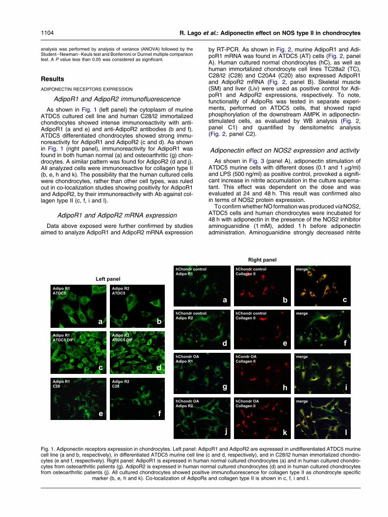
As shown in Fig. 1 (left panel) the cytoplasm of murineATDC5 cultured cell line and human C28/I2 immortalizedchondrocytes showed intense immunoreactivity with anti-AdipoR1 (a and e) and anti-AdipoR2 antibodies (b and f).ATDC5 differentiated chondrocytes showed strong immu-noreactivity for AdipoR1 and AdipoR2 (c and d). As shownin Fig. 1 (right panel), immunoreactivity for AdipoR1 wasfound in both human normal (a) and osteoarthritic (g) chon-drocytes. A similar pattern was found for AdipoR2 (d and j).All analyzed cells were immunoreactive for collagen type II(b, e, h and k). The possibility that the human cultured cellswere chondrocytes, rather than other cell types, was ruledout in co-localization studies showing positivity for AdipoR1and AdipoR2, by their immunoreactivity with Ab against col-lagen type II (c, f, i and l).
AdipoR1 and AdipoR2 mRNA expression
Data above exposed were further confirmed by studiesaimed to analyze AdipoR1 and AdipoR2 mRNA expression
Left panel
Adipo R1
C28
Adipo R2
C28
Adipo R1
ATDC5
Adipo R2
ATDC5
Adipo R1
ATDC5 DIF
Adipo R2
ATDC5 DIF
a b
c
e
d
f
hChondr OA
Adipo R1
hChondr OA
Adipo R2
hChondr control
Adipo R1
hChondr control
Adipo R2
Fig. 1. Adiponectin receptors expression in chondrocytes. Left panel: Adipcell line (a and b, respectively), in differentiated ATDC5 murine cell line (cytes (e and f, respectively). Right panel: AdipoR1 is expressed in humancytes from osteoarthritic patients (g). AdipoR2 is expressed in human norfrom osteoarthritic patients (j). All cultured chondrocytes showed positive
marker (b, e, h and k). Co-localization of AdipoRs
by RT-PCR. As shown in Fig. 2, murine AdipoR1 and Adi-poR1 mRNA was found in ATDC5 (AT) cells (Fig. 2, panelA). Human cultured normal chondrocytes (hC), as well ashuman immortalized chondrocyte cell lines TC28a2 (TC),C28/I2 (C28) and C20A4 (C20) also expressed AdipoR1and AdipoR2 mRNA (Fig. 2, panel B). Skeletal muscle(SM) and liver (Liv) were used as positive control for Adi-poR1 and AdipoR2 expressions, respectively. To note,functionality of AdipoRs was tested in separate experi-ments, performed on ATDC5 cells, that showed rapidphosphorylation of the downstream AMPK in adiponectin-stimulated cells, as evaluated by WB analysis (Fig. 2,panel C1) and quantified by densitometric analysis(Fig. 2, panel C2).
Adiponectin effect on NOS2 expression and activity
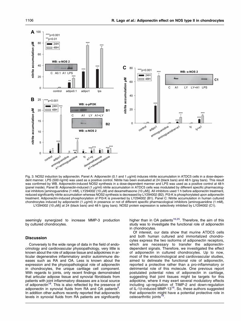
As shown in Fig. 3 (panel A), adiponectin stimulation ofATDC5 murine cells with different doses (0.1 and 1 mg/ml)and LPS (500 ng/ml) as positive control, provoked a signifi-cant increase in nitrite accumulation in the culture superna-tant. This effect was dependent on the dose and wasevaluated at 24 and 48 h. This result was confirmed alsoin terms of NOS2 protein expression.
To confirm whether NO formation was produced via NOS2,ATDC5 cells and human chondrocytes were incubated for48 h with adiponectin in the presence of the NOS2 inhibitoraminoguanidine (1 mM), added 1 h before adiponectinadministration. Aminoguanidine strongly decreased nitrite
Right panel
hCondr OA
Collagen II
hChondr OA
Collagen II
hChondr control
Collagen II
hChondr control
Collagen II
merge
merge
merge
merge
a b
d
c
g
e f
h i
lkj
oR1 and AdipoR2 are expressed in undifferentiated ATDC5 murinec and d, respectively), and in C28/I2 human immortalized chondro-
normal cultured chondrocytes (a) and in human cultured chondro-mal cultured chondrocytes (d) and in human cultured chondrocytes
immunofluorescence for collagen type II as chondrocyte specificand collagen type II is shown in c, f, i and l.

MWM SM
AR1
132bp
GAPDH
376bp
MWM SM C20 Liv PCR- C20 SM PCR-
AR1
70bp
GAPDH
376bp
A
B
C
5 10 30 120
% o
f p
ho
sp
ho
rilatio
n
over u
nstim
ulated
cells
0
20
40
60
80 ** **p<0.01
P-AMPK
AMPK
0
- +++Adiponectin
FL 1ug/mL
C1
C2
AR2
72bp
LivATSMAT AT Liv
+
minutes12030105
AR2
75bp
LivhCTCC28hCTCC28PCR-hCTCC28C20
Fig. 2. AdipoRs expression and functionality in chondrocytes. Panel A: AdipoR1 and AdipoR2 mRNA expression in ATDC5 (AT) cells deter-mined by RT-PCR. SM and Liv were used as positive controls. Panel B: AdipoR1 and AdipoR2 mRNA expression in C20A4 (C20), C28/I2(C28), TC28a2 (TC) and human cultured chondrocytes (hC), determined by RT-PCR. SM and Liv were used as positive controls. Panel C:WB for phosphorylated AMPK. Phosphorylated AMPK was identified by phospho-AMPKa Ab Thr172 (Cell Signalling catalog number2531, 62 kDa) in lysates coming from ATDC5 cells stimulated with adiponectin (1 mg/ml). Phosphorylation levels were evaluated, in time-dependent experiments, and compared to total AMPK (AMPKa Ab, Cell Signalling catalog number 2532, 62 kDa). Blot is representative ofat least four independent experiments (C1). Densitometric analysis of WB experiments is reported in panel C2. Data are represented as per-
cent� S.E.M. over basal AMPK phosphorylation in unstimulated cells.
1105Osteoarthritis and Cartilage Vol. 16, No. 9
accumulation in the culture supernatant of ATDC5 cells(Fig. 3, panel B) and fully inhibited NO accumulation inhuman cultured chondrocytes (Fig. 3, panel C). Tonote, pre-treatment with dexamethasone (10 mM), aclassic de novo NOS2 inhibitor, also blocked nitrite accu-mulation in adiponectineATDC5 stimulated cells (Fig. 3,panel B).
PI3-K inhibition blocks adiponectin-induced NO produc-tion and NOS2 protein expression.
We also investigated the role played by PI3-K in nitriteaccumulation and NOS2 expression evoked by adiponec-tin in murine and human chondrocytes by using LY294002(10 mM). The dose of LY294002 was selected according toprevious published literature20e22. As shown in Fig. 3(panel B1), PI3-K is phosphorylated after chondrocytetreatment with recombinant adiponectin. Pre-treatmentwith LY294002 specifically blocked adiponectin-inducednitrite accumulation in, respectively, murine and humancultured chondrocytes (Fig. 3, panel B and C). This resultwas confirmed also in terms of NOS2 expression sincecell pre-treatment with LY294002 significantly decreasesNOS2 protein expression (Fig. 3, panel B2 and C1)
and completely inhibited PI3-K phosphorylation (Fig. 3,panel B1).
Adiponectin effect on cytokines, metalloproteinasesand prostanoids
As shown in Fig. 4, adiponectin was able to significantlyincrease IL-6, MCP-1, MMP-3 and MMP-9 in the superna-tant of adiponectin-stimulated ATDC5 cells for 48 h(Fig. 4, panels AeF, respectively), whereas was unable tomodulate IL-1b, MMP-2 (Fig. 4, panels C and D), TIMP-1,LTB4, PGE2 and TNF-a [Fig. 5(AeD), respectively]. Inhibi-tion of PI3-K pathway by LY294002 strongly blunted adipo-nectin induction of IL-6 and MMP-9 (Fig. 4, panels A and F).In addition, LY294002 decreased, per se, MMP-2 basalspontaneous accumulation (Fig. 4, panel D). Intriguingly,this PI3-K pharmacological blocker was able to partially in-hibit adiponectin-induced MCP-1 when a minimal dose ofadiponectin was used whereas was unable to counteracthigher adiponectin doses (Fig. 4, panel B). A similar patternwas observed for MMP-3 (Fig. 4, panel E), where sur-prisingly, co-stimulation with LY294002 and adiponectin
control adipo0.1 adipo1 lps
nitrite
a
cc
um
ula
tio
n μ
Mn
itrite
a
cc
um
ula
tio
n μ
M
nitrite
a
cc
um
ula
tio
n μ
M
0
20
40
60
80
100
24H48H
*****
***
***
***
******
******
***p<0.001**p<0.01
24H48H
24H48H
***p<0.001C A0.1 A1 LPS
WB: α-NOS 2
WB: α-NOS 2
WB: α-NOS 2
controladipo1
aminoguanidine
a1+aminoguanidine lya1+ly
controladipo1
aminoguanidine
aminoguanidine+a1 ly
dexamethasone
a1+dexamethasone0
5
10
15
20
25 ***p<0.001
C
A
B
C
ly+a10
20
40
60
80
***
***
***
******
***
LPS
WB: α-pPI3K
B1
B2
C1
A1+LYLYA1
C LYA1
***
*** ****** ***
LY+A1
Fig. 3. NOS2 induction by adiponectin. Panel A: Adiponectin (0.1 and 1 mg/ml) induces nitrite accumulation in ATDC5 cells in a dose-depen-dent manner. LPS (500 ng/ml) was used as a positive control. Nitrite has been evaluated at 24 (black bars) and 48 h (gray bars). This resultwas confirmed by WB. Adiponectin-induced NOS2 synthesis in a dose-dependent manner and LPS was used as a positive control at 48 h(panel inside). Panel B: Adiponectin-induced (1 mg/ml) nitrite accumulation in ATDC5 cells was modulated by different specific pharmacolog-ical inhibitors [aminoguanidine (1 mM), LY294002 (10 mM) and dexamethasone (10 mM)]. All inhibitors used 1 h before adiponectin treatment,reduced significantly nitrite accumulation whereas NOS2 synthesis is decreased by LY294002 (B2). PI3-K is phosphorylated upon adiponectintreatment. Adiponectin-induced phosphorylation of PI3-K is prevented by LY294002 (B1). Panel C: Nitrite accumulation in human culturedchondrocytes induced by adiponectin (1 mg/ml) in presence or not of different specific pharmacological inhibitors [aminoguanidine (1 mM),
LY294002 (10 mM)] at 24 (black bars) and 48 h (gray bars). NOS2 protein expression is selectively inhibited by LY294002 (C1).
1106 R. Lago et al.: Adiponectin effect on NOS type II in chondrocytes
seemingly synergized to increase MMP-3 productionby cultured chondrocytes.
Discussion
Conversely to the wide range of data in the field of endo-crinology and cardiovascular physiopathology, very little isknown about the role of adipose tissue and adipokines in ar-ticular degenerative inflammatory and/or autoimmune dis-eases such as RA and OA. Less is known about theexpression and the physiopathological role of adiponectinin chondrocytes, the unique cartilage cell component.With regards to joints, only recent findings demonstratedthat articular adipose tissue and synovial fibroblasts frompatients with joint inflammatory diseases are a local sourceof adiponectin18. This is also reflected by the presence ofadiponectin in synovial fluids from RA and OA patients8.In addition other authors recently reported that adiponectinlevels in synovial fluids from RA patients are significantly
higher than in OA patients16,25. Therefore, the aim of thisstudy was to investigate the functional role of adiponectinin chondrocytes.
Of interest, our data show that murine ATDC5 cellsand both human cultured and immortalized chondro-cytes express the two isoforms of adiponectin receptors,which are necessary to transfer the adiponectin-dependent signals. Therefore, we investigated the effectof adiponectin in cultured chondrocytes. Up to now,most of the endocrinological and cardiovascular studies,aimed to delineate the functional role of adiponectin,reported a protective rather than a pro-inflammatory ordetrimental role of this molecule. One previous reportpostulated potential roles of adiponectin in cartilage,suggesting that joint tissues might be targets for thisadipokine, where it may exert several modulatory effectsincluding up-regulation of TIMP-2 and down-regulationof IL-1b-induced MMP-1316. So, these authors suggestedthat adiponectin might have a potential protective role inosteoarthritic joints16.

control
adiponectin 0.1
adiponectin 1 LY
a 0.1+LYa 1+LY
control
adiponectin 0.1
adiponectin 1 LY
a 0.1+LYa 1+LY
control
adiponectin 0.1
adiponectin 1 LY
a 0.1+LYa 1+LY
control
adiponectin 0.1
adiponectin 1 LY
IL
-6
(n
g/m
l)
0,0
0,5
1,0
1,5
2,0
2,5
3,0
3,5
***p<0.001 vs control### p<0.001 vs Adiponectin 1
***p<0.001 vs control### p<0.001 vs Adiponectin 0.1
***
###
MC
P-1
(n
g/m
l)
*** ***
###
LY+ a 0.1Ly+ a 1
control
adiponectin 0.1
adiponectin 1 LY
LY+ a 0.1Ly+ a 1
control
adiponectin 0.1
adiponectin 1 LY
LY+ a 0.1Ly+ a 1
IL
-1
b (p
g/m
L)
0,00,20,40,60,81,01,21,41,61,8
MM
P-2
(p
g/m
L)
0
10
20
30
40
50
****
*p<0.05 vs adiponectin 0.1***p<0.001 vs adiponectin 1
MM
P-3
(n
g/m
L)
0,0
0,5
1,0
1,5
2,0
2,5
0,0
0,5
1,0
1,5
2,0
2,5
*** ***
**p<0.05 vs adiponectin 1***p<0.001 vs control
MM
P-9
%
o
ve
r
un
stim
ula
te
d c
on
tro
l
0
50
100
150
200
250
300
* *
** **
*p<0.05 vs control**p<0.01 vs adiponectin
A C
D FE
B
Fig. 4. Adiponectin induces pro-inflammatory cytokines. Adiponectin (0.1 and 1 mg/ml) induces a significant increase in IL-6, MCP-1, MMP-3,and MMP-9 production by murine ATDC5 cells in a dose-dependent manner (panels AeF). This effect is strongly decreased by the PI3-Kinhibitor LY294002 (10 mM) for IL-6, and MMP-9 (panels A and F). LY294002 partially attenuated adiponectin-induced MCP-1 (B). Adiponectin
is unable to modulate IL-1b, MMP-2 levels (panels C and D).
1107Osteoarthritis and Cartilage Vol. 16, No. 9
By contrast, our study demonstrates, for the first time,that adiponectin might also exert significant pro-inflamma-tory effects in chondrocytes by inducing the expression ofNOS2 and also by strongly stimulating IL-6, MMP-3,MMP-9 and MCP-1 release.
NO, produced in great amounts by the NOS2 induciblesynthase, controls a variety of cartilage functions, includingloss of chondrocyte phenotype, chondrocyte apoptosis, andextracellular matrix degradation26. In vitro, human articularcartilage is able to produce large amounts of NO27, whichcan be enhanced by pro-inflammatory cytokines. In addi-tion, NO production can be significantly increased by thepresence of leptin, as shown in our previous works20e22.The ability of chondrocytes to respond to adiponectin, by in-creasing specifically the expression and activity of NOS2,appears to be highly selective. Indeed, its induction isblocked by aminoguanidine, a well known NOS2 activity in-hibitor, and also by dexamethasone (an inhibitor of NOS2de novo synthesis). Our data regarding adiponectin-induced factors production by chondrocytes are partially inagreement with data published by Chen et al., at least forthose regarding MCP-1 and TIMP-116. In contrast to datareported by these authors, adiponectin, in our hands, wasable to strongly induce IL-6 and MMP-3. This discrepancymight be due to the different adiponectin doses used inour experimental set (0.1 and 1 mg/ml, which are in thephysiological range of adiponectin levels), in comparisonto the dose used by Chen et al. (30 mg/ml). Another possi-bility might be represented by the different technologiesused in the measurement of these parameters.
The ability to respond to adiponectin is not restricted tochondrocytes. Indeed, other cell types such as adipocytes,
synovial fibroblasts, and normal fibroblasts exhibit thepotential to secrete IL-6 upon stimulation with adiponectinin vitro18,19. To note, all the above mentioned cells are ofmesenchymal origin. Thus, it is conceivable that the abilityto mount a pro-inflammatory response when stimulated byadiponectin could be a common characteristic of mesen-chymal-derived cells.
To address the question which molecular mechanismmight mediate the induction of NOS2 and cytokines produc-tion in response to adiponectin, we examined a key pathwayof intracellular signaling. Actually, previous studies address-ing intracellular effects of adiponectin in energy substratemetabolism, as well as in cardiovascular diseases, revealedthat one of the intermediates upon adiponectin binding to itsrespective receptors is the PI3-K28,29. To note, PI3-K hasbeen previously described as a relevant intracellularsignaling molecule involved also in the NOS2 induction inchondrocytes elicited by several pro-inflammatory cytokinesincluding leptin5. Consistent with this hypothesis, wedemonstrated that PI3-K pharmacological blockade byLY294002 resulted in a marked reduction of adiponectin-induced NOS2 expression, as well as in a decrease of adi-ponectin-induced IL-6 and MMP-9 production. Intriguingly,LY294002 was able to partially attenuate adiponectin-induced MCP-1 and MMP-3, suggesting that other alternateand/or convergent signaling pathways, rather than PI3-K,are at play. Finally, LY294002 was able to strongly de-crease basal MMP-2 production in chondrocytes, per se.Very recently, it has been reported that LY294002 signifi-cantly reduces adiponectin-induced nuclear factor kappaB (NFkB) activity in monocytic cells U937 where adiponectinprovoked, among other actions, the induction of MCP-130.

LT
B4 (p
g/m
l)
0
10
20
30
40
50
60
PG
E2 (p
g/m
l)
0
10
20
30
40
50
60
70
TN
F-α
(p
g/m
l)
0
10
20
30
40
50
60
B
C
control
adiponectin 0.1
adiponectin 1
control
adiponectin 0.1
adiponectin 1
control
adiponectin 0.1
adiponectin 1
control
adiponectin 0.1
adiponectin 1
TIM
P-1 (p
g/m
L)
0
1000
2000
3000
4000
5000A
D
Fig. 5. Adiponectin does not modulate prostanoids, TIMP-1 or TNF-a. Adiponectin (0.1 and 1 mg/ml) does not induce any significant modula-tion in TIMP-1 (A), LTB4 (B), PGE2 (C) and TNF-a (D).
1108 R. Lago et al.: Adiponectin effect on NOS type II in chondrocytes
Finally, recent data by Tang et al., indicated that adiponec-tin-induced IL-6 production in synoviocytes occurred via p38and NFkB activation19. To note, the activation of this nucleartranscription factor is a key element for the triggering ofNOS2 in several cell lineages31.
In conclusion, these data show for the first time that adipo-nectin is a frank modulator of the inflammatory response inchondrocytes. Upon these cells, adiponectin is able to inducekey mediators of cartilage degeneration such as NO, IL-6,MCP-1 and metalloproteinases such as MMP-3 and MMP-9, whose induction appears to be modulated by at leastone limited regulating mechanism represented by PI3-K.
Conflict of interest
The authors have no conflict of interest.
References
1. Zhang Y, Proenca R, Maffei M, Barone M, Leopold L, Friedman JM.Positional cloning of the mouse obese gene and its human homo-logue. Nature 1994;372:425e32.
2. Otero M, Lago R, Lago R, Casanueva FF, Dieguez C, Gomez-Reino JJ,et al. Leptin, from fat to inflammation: old questions and new insights.FEBS Lett 2005;579:295e301.
3. Trayhurn P, Bing C, Wood IS. Adipose tissue and adipokines e energyregulation from the human perspective. J Nutr 2006;136:1935Se9S.
4. Trayhurn P, Wood IS. Signalling role of adipose tissue: adipokines andinflammation in obesity. Biochem Soc Trans 2005;33:1078e81.
5. Lago F, Dieguez C, Gomez-Reino J, Gualillo O. The emerging role ofadipokines as mediators of inflammation and immune responses.Cytokine Growth Factor Rev 2007;18:313e25.
6. Lau DC, Dhillon B, Yan H, Szmitko PE, Verma S. Adipokines: molecularlink between obesity and atherosclerosis. Am J Physiol Heart CircPhysiol 2005;288:H2031e41.
7. Presle N, Pottie P, Dumond H, Guillaume C, Lapicque F, Pallu S,et al. Differential distribution of adipokines between serum and sy-novial fluid in patients with osteoarthritis. Contribution of joint tis-sues to their articular production. Osteoarthritis Cartilage 2006;14:690e5.
8. Schaffler A, Ehling A, Neumann E, Herfarth H, Tarner I, Scholmerich J,et al. Adipocytokines in synovial fluid. JAMA 2003;290:1709e10.
9. Scherer PE, Williams S, Fogliano M, Baldini G, Lodish HF. A novel se-rum protein similar to C1q, produced exclusively in adipocytes. J BiolChem 1995;270:26746e9.
10. Hu E, Liang P, Spiegelman BM. AdipoQ is a novel adipose-specific genedysregulated in obesity. J Biol Chem 1996;271:10697e703.
11. Shapiro L, Scherer PE. The crystal structure of a complement 1q familyprotein suggests an evolutionary link to tumor necrosis factor. CurrBiol 1998;8:335e8.
12. Trujillo ME, Scherer PE. Adiponectin e journey from an adipocyte secre-tory protein to biomarker of the metabolic syndrome. J Intern Med2005;257:167e75.
13. Yamauchi T, Kamon J, Minokoshi Y, Ito Y, Waki H, Uchida S, et al.Adiponectin stimulates glucose utilization and fatty-acid oxidation byactivating AMP-activated protein kinase. Nat Med 2002;8:1288e95.
14. Ouchi N, Shibata R, Walsh K. Cardioprotection by adiponectin. TrendsCardiovasc Med 2006;16:141e6.
15. Gualillo O, Gonzalez-Juanatey JR, Lago F. The emerging role of adipo-kines as mediators of cardiovascular diseases: physiological and clin-ical perspectives. Trends Cardiovasc Med 2007;17:277e85.
16. Chen TS, Chen L, Hsieh MS, Chang CP, Chou DT, Tsai SH. Evidencefor a protective role for adiponectin in osteoarthritis. Biochim BiophysActa 2006;1762:711e8.
17. Otero M, Lago R, Gomez R, Lago F, Dieguez C, Gomez-Reino JJ, et al.Changes in fat-derived hormones plasma concentrations: adiponec-tin, leptin, resistin, and visfatin in rheumatoid arthritis subjects. AnnRheum Dis 2006;65:1198e201.
18. Ehling A, Schaffler A, Herfarth H, Tarner IH, Anders S, Distler O, et al.The potential of adiponectin in driving arthritis. J Immunol 2006;176:4468e78.

1109Osteoarthritis and Cartilage Vol. 16, No. 9
19. Tang CH, Chiu YC, Tan TW, Yang RS, Fu WM. Adiponectin enhancesIL-6 production in human synovial fibroblast via an adipoR1 receptor,AMPK, p38, and NF-kB pathway. J Immunol 2007;179:5483e92.
20. Otero M, Lago R, Lago F, Gomez-Reino JJ, Gualillo O. Signalling path-way involved in nitric oxide synthase type II activation in chondro-cytes: synergistic effect of leptin with interleukin-1. Arthritis ResTher 2005;7:R581e91.
21. Otero M, Gomez-Reino JJ, Gualillo O. Synergistic induction of nitricoxide synthase type II: in vitro effect of leptin and interferon-gammain human chondrocytes and ATDC5 chondrogenic cells. ArthritisRheum 2003;48:404e9.
22. Otero M, Lago R, Gomez R, Lago F, Gomez-Reino JJ, Gualillo O. Phos-phatidylinositol 3-kinase, MEK-1 and p38 mediate leptin/interferon-gamma synergistic NOS type II induction in chondrocytes. Life Sci2007;doi:10.1016/j.lfs.2007.09.007
23. Thomas DP, Sunters A, Gentry A, Grigoriadis AE. Inhibition of chondro-cyte differentiation in vitro by constitutive and inducible overexpressionof the c-fos proto-oncogene. J Cell Sci 2000 Feb;113(Pt 3):439e50.
24. Goldring MB. Immortalization of human articular chondrocytes for gener-ation of stable, differentiated cell lines. Methods Mol Med 2004;100:23e36.
25. Senolt L, Pavelka K, Housa D, Haluzık M. Increased adiponectin is neg-atively linked to the local inflammatory process in patients with rheu-matoid arthritis. Cytokine 2006;35:247e52.
26. Goldring MB, Berenbaum F. The regulation of chondrocyte function byproinflammatory mediators: prostaglandins and nitric oxide. ClinOrthop Relat Res 2004;427:S37e46.
27. Vuolteenaho K, Moilanen T, Al-Saffar N, Knowles RG, Moilanen E. Reg-ulation of the nitric oxide production resulting from the glucocorticoid-insensitive expression of NOS type II in human osteoarthritic cartilage.Osteoarthritis Cartilage 2001;9:597e605.
28. Kobashi C, Urakaze M, Kishida M, kibayashi E, Kobayashi H, Kihara S,et al. Adiponectin inhibits endothelial synthesis of interleukin-8. CircRes 2005;97:1245e52.
29. Maeda N, Shimomura I, Kishida K, Nishizawa H, Matsuda M,Nagaretani H, et al. Diet-induced insulin resistance in mice lackingadiponectin/ARCP30. Nat Med 2002;8:731e7.
30. Haugen F, Drevon CA. Activation of nuclear factor-kappaB by high mo-lecular weight and globular adiponectin. Endocrinology 2007;148:5478e86.
31. D’Acquisto F, May MJ, Ghosh S. Inhibition of NFkB: an emerging themein anti-inflammatory therapies. Mol Interv 2002;2:22e35.