Perceived Occupational Stressors and the Health Software Professionals in Bengaluru, India
www.elsevier.com/locate/pnpbp
Progress in Neuro-Psychopharmacology & B
Review article
Cytokines, stressors, and clinical depression:
Augmented adaptation responses underlie depression pathogenesis
Donn A. Simmonsa, Patricia A. Broderickb,c,d,e,f,*
aDepartment of Psychology, Emory University, Atlanta, GA, USAbDepartment of Physiology and Pharmacology, CUNY Medical School, Convent Avenue and West 138th St., New York, NY 10031, USA
cDepartment of Biology, CUNY Graduate School, NY, USAdDepartment of Psychology, CUNY Graduate School, NY, USA
eMARC and MBRS Programs, City College of New York, NY, USAfDepartment of Neurology, NYU School of Medicine, NYU Comprehensive Epilepsy Center, NY, USA
Accepted 1 March 2005
Available online 31 May 2005
Abstract
By influencing the central nervous system, cytokines, which regulate immune function innately and adaptively, may play a key
role in mediating depression-like neuro-behavioral changes. However, the similarity between cytokine and stressor-effects in animal
models raises a question about the degree to which behavioral and neurochemical outcomes of cytokine challenge represent
depressive disorder per se. The present review attempts to illustrate the degree of overlap between cytokines and stressors with
respect to their effects on neurochemistry and behavior in animal models. The review also shows how short-term effects of cytokine
exposure in typical animals may be discerned from characteristics that might otherwise be described as depression-like. By comparing
outcomes of immune challenge in typical rodent strains (e.g., Sprague–Dawley [SD], Wistar) and an accepted animal model of
depression (e.g., Fawn Hooded [FH] rodent strain), differences between short-term effects of cytokines and depression-like
characteristics in rodents are demonstrated. Additionally, because it is known that preexisting vulnerability to depression may affect
outcomes of immune challenge, we further compare immunological, biochemical and behavioral effects of cytokines between SD and
FH rodent strains. Interestingly, the acute neurochemical and behavioral effects of the cytokine interleukin 1a (IL-1a) reveal stressor-
like responses during behavioral habituation in both strains, though this appears to a stronger degree in FH animals. Further, the
subacute response to IL-1a vastly differed between strains, indicating differences in adaptive mechanisms. Thus, stressor-like effects
of immune challenge, particularly in FH animals, provide validation for recent ‘‘cross-sensitization’’ models of depression pathogenesis
that incorporate immune factors.
D 2005 Elsevier Inc. All rights reserved.
Keywords: Ambulations; Behavior; Concanavalin-A; Cytokines; Depression; Exploration; Fawn Hooded rats; Fine movements; Habituation; Hippocampus;
Immune system; Interleukin; Locomotion; Microvoltammetry; Mitogen response; Nanotechnology; Neurochemistry; Neuromolecular imaging; Norepinephrine;
Open-field behavior; Physiological stress; Psychological stress; Serotonin; Sprague–Dawley rats; Voltammetry
0278-5846/$ - see front matter D 2005 Elsevier Inc. All rights reserved.
doi:10.1016/j.pnpbp.2005.03.009
Abbreviations: FH, Fawn Hooded; HPC, hippocampus; HPA, hypothalamus-pituitary-adrenal axis; 5-HIAA, 5-hydroxyindoleacetic acid; IL-1a
interleukin-1 alpha; IL-1h, interleukin-1 beta; IL-1ra, interleukin-1 receptor antagonist; IL-2, interleukin-2; IL-6, interleukin-6; IL-10, interleukin-10; LPS
lipopolysaccharide; LC, locus coeruleus; NE-ergic, noradrenergic; NE, norepinephrine; NMI, neuromolecular imaging; PFC, prefrontal cortex; 5-HT
serotonin; 5-HT-ergic, serotonergic; SD, Sprague–Dawley; TNFa, tumor necrosis factor alpha.
* Corresponding author. Department of Physiology and Pharmacology, CUNY Medical School, Convent Avenue and West 138th St., New York, NY 10031
USA. Tel.: +1 212 650 5479; fax: +1 212 650 7754.
E-mail address: [email protected] (P.A. Broderick).
,
,
,
,
iological Psychiatry 29 (2005) 793 – 807
D.A. Simmons, P.A. Broderick / Progress in Neuro-Psychopharmacology & Biological Psychiatry 29 (2005) 793–807794
Contents
. . . . . . 794
. . . . . . 795
. . . . . . 796
. . . . . . 796
. . . . . . 796
. . . . . . 797
. . . . . . 798
. . . . . . 799
. . . . . . 799
. . . . . . 799
. . . . . . 800
. . . . . . 801
. . . . . . 801
1. Introduction. . . . . . . . . . . . . . . . . . . . . . . . . . . . . . . . . . . . . . . . . . . . . . . . . . . . .
2. Presence of cytokines in the central nervous system . . . . . . . . . . . . . . . . . . . . . . . . . . . . . . . .
3. The Fawn-Hooded animal model of depression . . . . . . . . . . . . . . . . . . . . . . . . . . . . . . . . . .
4. Central and behavioral effects of cytokines and stressors in normal, ‘‘non-depressed’’ rat strains . . . . . . . . .
4.1. Effects of cytokines and stressors on 5-HT release in HPC . . . . . . . . . . . . . . . . . . . . . . . . .
4.2. Effects of cytokines and stressors on NE release in HPC . . . . . . . . . . . . . . . . . . . . . . . . . .
4.3. Effects of cytokines and stressors on locomotor activity . . . . . . . . . . . . . . . . . . . . . . . . . .
5. Effects of IL-1a injection in the FH model of depression; comparison with the SD strain . . . . . . . . . . . .
5.1. Pre-existing vulnerability affects outcome of immune challenge . . . . . . . . . . . . . . . . . . . . . .
5.2. Effects of IL-1a exposure on 5-HT release in HPC: comparison of FH and SD animals . . . . . . . . . .
5.3. Effects of IL-1a exposure on NE release in HPC: comparison of FH and SD animals . . . . . . . . . . .
5.4. Effects of IL-1a exposure on open-field behavior: comparison of FH and SD animals. . . . . . . . . . .
5.5. Effects of IL-1a exposure on immune cell mitogenic responses: comparison of FH and SD animals . . .
6. Conclusions . . . . . . . . . . . . . . . . . . . . . . . . . . . . . . . . . . . . . . . . . . . . . . . . . . . .
. . . . . . 802Acknowledgements . . . . . . . . . . . . . . . . . . . . . . . . . . . . . . . . . . . . . . . . . . . . . . . . . . . . . . . . . 802
References . . . . . . . . . . . . . . . . . . . . . . . . . . . . . . . . . . . . . . . . . . . . . . . . . . . . . . . . . . . . . . 803
1. Introduction
Mounting evidence that immune activity influences
central nervous function supports the position that cyto-
kines (e.g., IL-1, IL-2, IL-6, TNFa, etc.), perhaps as much
as neurotransmitters, play a role in regulating cognition,
affect, and behavior. In addition, central output can impact
immune function (Fig. 1). In experimental animals, the
behavioral (e.g., ‘‘sickness behavior’’, Maier and Watkins,
1998; Konsman et al., 2002) and physiological (e.g.,
hypothalamic-pituitary axis [HPA] activation, Besedovsky
et al., 1986; Ando and Dunn, 1999) effects of acute
immune challenge share such similarity to major depressive
disorder in humans that communication between the
immune, nervous and endocrine systems has become a
major focus of affective disorders research in general. For
these reasons, immune dysfunction involving excessive
macrophage derived cytokine production has in the past
Cellular ImmuneChanges
N
EnS
Immune System
Fig. 1. Immune tissues are receptive to classic neurotransmitters and hormones; ner
is the basis of much behavioral/neuroendocrine change associated with immune
central nervous function.
been proposed a fundamental aspect of depression patho-
genesis (Smith, 1991), and more currently, a role for
cytokines in mediating depression-like neuro-behavioral
changes in various somatic disease states is widely
recognized (Dantzer, 2004).
Additional evidence from animal studies, however,
provides reason to believe the relevant behavioral and
physiological effects of cytokines are more comparable
with the effects of stressors than with depression per se,
given similarities in neuroendocrine changes that stressors
and cytokines both can produce (Anisman et al., 2002a,b).
Moreover, neurochemical effects of cytokines overlap to
such a degree with those elicited by psychological (i.e.,
involving higher-order, sensory/cognitive processing)
stressors that the brain appears to interpret and react to
each as if they were the same (Anisman and Merali,
1999). Stressors clearly play a role in depression,
particularly in situations where duration of exposure to
Coordination Of
Behavioral Responses
HPA Reactivity
ervous&
docrineystems
vous and endocrine tissues are receptive to immune factors. Such interaction
system activity, and physiological/immune system change associated with
Behavioral Responses
HPA Reactivity
Cytokines
Hippocampus
Stressors
D.A. Simmons, P.A. Broderick / Progress in Neuro-Psychopharmacology & Biological Psychiatry 29 (2005) 793–807 795
them is chronic, and/or where physiological responses to
them are disproportionately large (Chrousos and Gold,
1992, 1998; Claes, 2004). Taking into consideration the
degree of overlap between effects of stressors and
cytokines, the possibility that both similarly provoke
depression is a compelling one. In line with this thinking,
analogous to the manner in which regimens of chronic
stress produce a depression-like syndrome in experimental
animals (Willner, 1984; Willner et al., 1987, 1996),
chronic administration of cytokines in a therapeutic
context is well documented to produce a depression-like
syndrome in humans (Capuron and Dantzer, 2003;
Schaefer et al., 2003). Further, a number of studies have
reported that a single stressor or immune challenge can
result in lasting changes in vulnerability to affective
illnesses by augmenting responses to subsequent insult
(Anisman and Merali, 2003). In sum, like stressors,
immune challenge appears capable of provoking a
depression-like syndrome over a similar time course, by
affecting the same systems, and by proactively influencing
vulnerability to subsequent insult.
Nevertheless, there is clearly more to learn in terms of
just how similar cytokines are to stressors in these
respects, and furthermore, how short-term effects of
cytokine exposure may be discerned from depression-like
characteristics observed in experimental animal models. To
address these issues, the body of this review will provide
a summary of the literature on some of the short-term
effects of acute immune challenge and stressors in rodents,
along with some attention to specific mechanisms by
which these effects take place. In addition, we will
compare outcomes of immune challenge in typical rodent
strains (e.g., Sprague–Dawley [SD], Wistar) against what
is known in the context of clinical depression and
depression models, including measures reported for the
Fawn Hooded (FH) rat strain, an accepted animal model
of clinical depression. Emphasis will be placed on
comparative studies of biochemistry, behavior, and
immune function between two genetically diverse animal
models, i.e., one that is normal (SD) and one that is
biochemically and immunologically compromised (FH).
The purpose of this review is to illustrate how outcomes
of immune challenge may differ depending on preexisting
vulnerability to depression.
Fig. 2. The hippocampus (HPC) plays a central role in sensory
information processing, coordination of behavioral responses, and
regulation of HPA reactivity. Behavioral and physiological outcomes of
stressor or cytokine challenge are very similar, and likely are mediated to
some degree via the HPC. Underlying differences in HPC neurochemistry
(e.g., 5-HT, NE) are revealed between animals that differ in vulnerability
to depression. Pre-existing differences between animals (e.g., HPC
neurochemistry), whether in-born, or resulting from previous exposure
to stressor or cytokine challenge, may underlie differential reactivity of
HPA output and behavior.
2. Presence of cytokines in the central nervous system
Many reports have confirmed the presence of cytokines
and their receptors in several brain areas (e.g., hypothal-
amus, hippocampus [HPC], prefrontal cortex [PFC], and
brainstem; Farrar et al., 1987; Katsuura et al., 1988; Araujo
et al., 1989; Koenig, 1991; Lapchak, 1992; Schobitz et al.,
1992; Ban et al., 1993; Haour et al., 1995), where
concentrations are reported to fluctuate in response to a
variety of insults (e.g., concussive brain injury, cerebral
ischemia, seizure, infection or endotoxin challenge, psycho-
logical stress; Licinio and Wong, 1999; Rothwell, 1999;
Plata-Salaman, 2000; Rothwell and Luheshi, 2000; Reich-
lin, 2001). In situations where central function is affected,
cytokines can be produced endogenously within the brain
(Rothwell, 1999), or can gain access to the brain via
circumventricular organs (Banks, 1999), by crossing the
blood–brain barrier by altering cerebrovascular permeabil-
ity (Ellison et al., 1990; Banks and Kastin, 1992), or else by
recruiting peripheral immune cells into the brain, which may
then express cytokines in turn (Proescholdt et al., 2002). In
addition to direct influence on central targets, cytokines can
also affect central function indirectly by stimulating
peripheral vagal afferents (Maier et al., 1998; MohanKumar
et al., 2000).
In later sections we will focus on studies that have
reported the manner in which stressors and cytokines affect
monoamines, serotonin (5-HT) and norepinephrine (NE), in
the HPC. Given the central role HPC plays in sensory
information processing, coordination of behavioral
responses, and regulating HPA reactivity to stressors
(e.g., Young et al., 1991; Herman et al., 2003), we feel
the region serves well as an anatomical locus for examining
possible cytokine-linked depression- and/or stress-like
changes (Fig. 2).
D.A. Simmons, P.A. Broderick / Progress in Neuro-Psychopharmacology & Biological Psychiatry 29 (2005) 793–807796
3. The Fawn-Hooded animal model of depression
The inbred FH rodent strain is an animal model of
depression due most prominently to central and peripheral
5-HT abnormalities; 5-HT abnormalities are frequently
associated with depressive disorders (Maes and Meltzer,
1995). Specifically, decreased concentrations of 5-HT in
blood platelets (Tschopp and Weiss, 1974), as well as
decreased 5-HT and 5-hydroxyindoleacetic acid (5HIAA)
concentrations in brain loci for cell body synthesis of 5-HT
are found in these animals (Aulakh et al., 1994). Additional
depression-like markers in the FH strain that involve central
5-HT include reduction in 5-HT agonist labeling in HPC
and PFC (Hulihan-Giblin et al., 1992), abnormalities in 5-
HT1A receptor binding (Hulihan-Giblin et al., 1993; Chen
and Lawrence, 2000), and blunted responses to centrally
acting 5-HT agonists (Aulakh et al., 1988a,b; Wang et al.,
1988). Other pharmacological evidence is based on NE
dysfunction and provides support for the FH strain as an
animal model of depression because, in human studies,
depressed patients exhibit NE alterations; antidepressant
medications are often designed to compensate for NE-ergic
deficiencies (Baldessarini, 1989; Blier, 2001; Morrow,
2001). A specific parallel between clinically depressed
patients (Charney et al., 1982; Lesch et al., 1990) and the
FH strain (Aulakh et al., 1992) that involves NE trans-
mission occurs in blunted growth hormone response to
clonidine compared with control groups of nondepressed
patients and normal animals, respectively. Important new
evidence for NE deficiency in CA1 region of HPC in freely
moving and behaving FH rats comes from neuromolecular
imaging (NMI) studies using BRODERICK PROBE\sensors; the data show that basal NE release in FH animals
are deficient by two-fold compared with normal, non-
depressed SD rats (Broderick and Hope, unpublished data).
Additional abnormalities in the FH strain related to
depression include exaggerated immobility in the forced
swim test (Overstreet and Rezvani, 1996) and elevated
serum corticosterone (Aulakh et al., 1993), both of which
are reversible via chronic antidepressant treatment (Rezvani
and Overstreet, 1992; Aulakh et al., 1993).
In addition to central markers of depression, FH animals
display differences in immune function compared to normal
(e.g., SD) animals. We compared flow cytometric enumer-
ation results of T-helper (CD4+) and T-suppressor (CD8+)
splenocyte frequencies in SD and FH rats and the studies
revealed a two-fold higher frequency of CD8+ in FH as
compared with the SD strain. This shift in the frequency of
CD8+ cells in the FH animals may well reflect an immune
system which exhibits an incompatibility with normal
homeostatic function (Coico and Broderick, unpublished
data). It is interesting that such an incompatibility may also
occur in HIV patients (Leserman et al., 2002) as well as in
the elderly depressed (Castle et al., 1995). Given the caveat
that immune function may differ between rodents and
humans, in human depressed patients CD8+ frequency has
been reported to be lower, i.e., in depressed patients, the
ratio of T-helper to T-suppressor lymphocyte population
(CD4+/CD8+ ratio) is elevated with graded depression
severity (Charles et al., 1992). Finally, there were no
significant differences in T and B cell proliferative mitogen
responses using untreated SD versus FH splenocytes
cultured with concanavalin-A (Con A) or LPS. However,
there remains the stipulation that such responses, as studied,
tested only lymphoid functionality and did not test the
capacity of either animal strain to display T cell function-
ality as regards cytokine production and/or B cell function-
ality as regards antibody production (Coico and Broderick,
unpublished data). These differences in immune function
are most relevant since immune dysfunction has been
hypothesized to bear fundamentally on central and behav-
ioral manifestations of depression (Smith, 1991) albeit
depression-associated immune modifications may be sec-
ondary to depression-associated neuroendocrine changes
(Tecoma and Huey, 1985). In another rodent model of
depression (8 weeks of a chronic mild stress regimen, e.g.,
Willner, 1984; Willner et al., 1987, 1996), enhancement of
IL-1 and IL-2 production by splenocytes, and enhanced
Con-A mitogen responses were observed; importantly, these
changes were reversible via antidepressant treatment
(Kubera et al., 1996). More work is needed in which
additional immune markers (e.g., aspects of cytokine
function) implicated in clinical depression are examined in
FH animals. Nonetheless, FH animals provide a viable
experimental model in which to examine aspects of clinical
depression.
4. Central and behavioral effects of cytokines and stressors
in normal, ‘‘non-depressed’’ rat strains
4.1. Effects of cytokines and stressors on 5-HT release in
HPC
There is considerable evidence that immune related
factors affect the central transmission of 5-HT. It is
important to point out, though, that effects among individual
cytokines can differ depending on dose administered and
functional outcome measured (Petitto et al., 1997; Poll-
macher et al., 2002). Also, while non-specific immune
activation via administration of bacterial endotoxin (e.g.,
lipopolysaccharade, LPS) is reported to enhance central 5-
HT in rats (Lavicky and Dunn, 1995; Linthorst et al., 1995,
1996; Linthorst and Reul, 1998; Connor et al., 1999), the
effects of cytokines are often receptor specific.
Receptor specific effects of cytokines are clearly present
in the neurochemical changes they elicit within discrete
regions of the brain; again, for present purposes attention
will be focused on studies examining HPC. For example, in
results published by Pauli et al. (1998), intracerebroven-
tricular injection of IL-2 enhanced HPC 5-HT in Wistar rats,
whereas injection of TNFa yielded no effect. Pauli and
D.A. Simmons, P.A. Broderick / Progress in Neuro-Psychopharmacology & Biological Psychiatry 29 (2005) 793–807 797
colleagues further reported that effects of IL-2 injection
come indirectly through IL-1 mechanisms, as pretreatment
with a receptor antagonist for IL-1 (i.e., IL-1ra) prevented or
attenuated the effect of IL-2 on HPC 5-HT. Pretreatment
with IL-1ra was also found to attenuate induction of HPC 5-
HT following peripheral injection of LPS (Linthorst et al.,
1995). Other studies have confirmed the ability of IL-1 to
increase HPC 5-HT in Wistar and SD strains (Linthorst et
al., 1994, 1997; Merali et al., 1997; Broderick, 2002).
Data from several reports suggest that the effects of
immune challenge on central 5-HT are mediated via the 5-
HT2 receptor subtype. As well, other centrally mediated
effects of IL-1 or LPS injection in experimental animals
(e.g., defensive rage, anorexia, REM sleep change) are 5-
HT2 mediated (Imeri et al., 1999; Hassanain et al., 2003a;
von Meyenburg et al., 2003). Consistent with the hypothesis
that IL-1 induction of 5-HT is mediated via the 5-HT2
receptor subtype, 5-HT2 receptors act in opposition to 5-
HT1A autoreceptors which, in addition to suppressing IL-1
elicited defensive rage and anorexia, diminish 5-HT trans-
mission (Blanchard et al., 1993; Saphier et al., 1995;
Cologer-Clifford et al., 1997; El-Haj et al., 2002; Hassanain
et al., 2003b). Data from additional reports suggest that
cytokines elicit these effects by acting directly on 5-HT
terminals. Studies that have examined immediate early gene
expression in response to peripheral LPS or IL-1 challenge
report no detectable increase in c-fos mRNA in raphe nuclei
(Elmquist et al., 1993; Wan et al., 1993; Brady et al., 1994;
Ericsson et al., 1994; Sagar et al., 1995). More evidence of
action at 5-HT terminals is provided by findings that intra-
HPC infusion of IL-1 results in local increases in
extracellular 5-HT (Linthorst et al., 1994). Taken together,
the effects of immune challenge, particularly IL-1, on
central 5-HT appear to be mediated through the 5-HT2
receptor subtype within 5-HT terminal regions.
Similar to the impact of immune cytokine challenge,
microdialysis studies have shown acute stressor exposure
also results in increased HPC 5-HT (Kalen et al., 1989; Pei
et al., 1990; Kawahara et al., 1993; Thorre et al., 1997;
Konstandi et al., 2000). An additional similarity to
cytokines lies in the mechanism by which stressor effects
are at least partially mediated. Specifically, stressor elicited
behavioral changes (e.g., anorexia) appear to be mediated by
5-HT2 receptors (Grignaschi et al., 1993; Papp et al., 2003),
and are countered by the 5-HT1A receptor subtype (Grigna-
schi et al., 1993; Shimizu et al., 2000). However, unlike
immune challenge, acute stressors are demonstrated to
increase the activity of raphe neurons (Bliss et al., 1972;
Kennett and Joseph, 1981).
Both stressor and cytokine elicited increases in HPC 5-
HT are in line with a role for this biogenic amine in
neutralizing the central impact of destabilizing events
(Kennett and Joseph, 1981; Kennett et al., 1985; Smelik,
1987; Cassano and D’mello, 2001; Lowry, 2002). Given
the role of HPC in sensory information processing and
regulating physiological reactivity to stressors (Herman et
al., 2003), normal 5-HT function in this brain region would
appear crucial for maintaining central stability in the
presence of insult, whether of psychological or of systemic
(e.g., immune) origin. Overlooking any possible differ-
ences in magnitude of effects elicited by stressor versus
cytokine exposure, there is little to suggest that cytokines
are unlike psychological stressors in terms of their impact
on HPC 5-HT in normal animals. Finally, the general effect
of acute cytokine challenge in normal animals, which is an
apparent increase in HPC 5-HT, is not what we might
expect as a depression-like outcome because anti-depres-
sant medications often enhance 5-HT transmission (Meltzer
and Lowy, 1987; Curzon, 1988; Price et al., 2001).
Indications of reduced 5-HT in severely depressed patients
(Asberg et al., 1976) and in the FH animal model of
depression (Tschopp and Weiss, 1974; Aulakh et al., 1994)
reinforce this idea further.
4.2. Effects of cytokines and stressors on NE release in HPC
The central impact of immune challenge is also
demonstrated by alterations in NE observed within a
number of brain structures. Most frequently, studies have
focused on the hypothalamus and have shown variable
effects of immune challenge on NE release, including
marked increases (Shintani et al., 1993, 1995; Connor et
al., 1998; Linthorst and Reul, 1998), increased turnover
(Zalcman et al., 1994), results that depend on the
hypothalamic sub-region examined (MohanKumar et al.,
1998; Brebner et al., 2000), or no effect at all (Merali et al.,
1997). Fewer studies have examined the impact of immune
challenge on HPC NE, and as reported for the hypothal-
amus, outcomes are somewhat conflicting. Linthorst and
Reul (1998) demonstrated a modest increase in NE efflux
within HPC elicited by peripheral injection of LPS to Wistar
rats, whereas Merali et al. (1997) found no effect of IL-1h in
SD rats, and Broderick (2002) observed a modest but
significant IL-1a induced suppression in HPC NE in SD
rats. Inconsistency of effects across studies may be
accounted for by cytokine receptor specificity described
earlier, and also by differences in route of administration;
IL-2 was found to increase HPC NE upon intracerebroven-
tricular injection (Pauli et al., 1998), while systemic
administration had no effect (Lacosta et al., 2000).
Regarding the mechanism by which immune challenge
affects central NE, LPS is observed to induce c-fos
activation in locus coeruleus (LC) (Elmquist et al., 1993;
Wan et al., 1993; Sagar et al., 1995), which indicates a direct
impact on NE cell bodies, though this is not found for all
cytokines tested (e.g., IL-1h,Brady et al., 1994; Ericsson et
al., 1994). Alternatively, the impact of cytokines on central
NE may come about via indirect means. Suppression of
HPC NE is noted to occur simultaneously with increased
HPC 5-HT in the same animals following IL-1a injection
(Broderick, 2002). It seems plausible that NE undergoes a
compensatory reaction to 5-HT. Consistent with this
D.A. Simmons, P.A. Broderick / Progress in Neuro-Psychopharmacology & Biological Psychiatry 29 (2005) 793–807798
hypothesis, NE/5-HT interactions are documented to bear an
inverse relationship, evidenced by data showing unit activity
within LC is suppressed by 5-HT innervation (Gorea and
Adrien, 1988), unit activity within raphe nucleus is sup-
pressed by treatments that enhance NE release (Baraban and
Aghajanian, 1980), and by data showing drug treatments
that decrease HPC NE efflux simultaneously increase 5-HT
release in the same region (Broderick, 1997).
Acute stressor exposure increases activity in LC neurons
(Abercrombie and Jacobs, 1988; Valentino and Wehby,
1988; Valentino et al., 1998), and elevates HPC NE
measured by microdialysis (Abercrombie and Jacobs,
1988; Kalen et al., 1989; Zhang et al., 1995). Elevated
NE has been implicated in producing an enhanced state of
arousal (Anisman and Zacharko, 1990), which is of adaptive
significance where increased vigilance might be necessary
for an animal to escape potentially life-threatening (i.e.,
stressful) situations. Considering that behavioral and/or
physiological response to cytokines and infection often
include increased sleep (Krueger and Toth, 1994; Krueger
and Majde, 1995) which is of adaptive significance in its
own right, one would suspect concurrent increases in central
NE would be less consistently reported across brain regions
implicated in behavioral vigilance, unlike reactions to
psychologically stressful stimuli. This appears to be the
case, at least for suppressed HPC NE observed following
IL-1a injection in SD rats (Broderick, 2002). Rather, NE
suppression appears relatively depression-like in parallel
with lower basal HPC NE observed in the FH depression
model versus SD animals (Broderick and Hope, unpub-
lished data). However, the IL-1a induced suppression
observed in SD animals being far less, at 25% below
baseline, than the two-fold difference from baseline
described between SD and FH strains, it remains to be
determined whether suppression of this relative magnitude
is appropriately characterized as a depression-like change in
normal animals.
In any event, it is difficult to draw general conclusions
with respect to the impact of immune cytokine challenge on
HPC NE given the degree of inconsistency among studies,
in addition to the likelihood that multiple factors (e.g.,
receptor specificity, route of administration, administration
of cytokines during habituation or conversely during
exploration) contribute to experimental outcomes. It is
apparent, though, that the stressor effect on NE is more
consistent across studies than is the cytokine effect on NE.
4.3. Effects of cytokines and stressors on locomotor activity
Suppression of spontaneous locomotor activity is a
hallmark indication of behavioral depression in rodent
models (Willner, 1984), and a frequently reported behav-
ioral outcome of LPS challenge (Plata-Salaman and
Borkoski, 1993; Kozak et al., 1994, 1995; Huang et al.,
1999; Tollner et al., 2000; Engeland et al., 2001). The effect
of LPS may be mediated via IL-1 and TNFa, which also
suppress locomotor activity when administered separately
(Bianchi et al., 1992; Lacosta et al., 1998, 1999; Morgan et
al., 2004). Consistent with these reports, IL-10 pretreatment
blocks LPS stimulated production of IL-1 and TNFa from
macrophages, and also suppresses locomotor effects of LPS
challenge (de Waal Malefyt et al., 1991; Fiorentino et al.,
1991; Dinarello, 1993; Moore et al., 1993). As for central
effects described earlier, the impact of different cytokines on
locomotor activity can vary. Whereas TNFa reduced
spontaneous locomotor behavior as described by Bianchi
et al. (1992), IL-1a had no effect within the same series of
experiments, and Broderick (2002) reported IL-1a produced
a tendency to increase locomotor activity in SD animals
when measured during behavioral habituation.
As well as receptor specificity, other factors weigh in to
account for variable locomotor effects of acute cytokine
challenge. Pauli et al. (1998) reports differential locomotor
effects of IL-2 and TNFa that depend on circadian cycle. In
this work, locomotor activity was unaffected by IL-2 or
TNFa during the light phase, but was suppressed during
the dark phase of the circadian cycle. Others that have
noted circadian effects also report an impact of sex
differences on cytokine-linked changes in locomotor
behavior (Engeland et al., 2003; Franklin et al., 2003).
Disparity in effects reported between cytokines, or between
studies for a given cytokine may further be accounted for
by differences in behavioral paradigm, i.e., environmental
familiarity may play a role. More specifically, behavioral
effects of cytokine injection appear to vary depending on
whether an animal is undergoing active exploration of a
relatively unfamiliar environment, or is exhibiting a
reduced level of overt responsiveness within an environ-
ment with which it has previously become familiar. In work
by Broderick (2002) wherein cytokines produced a
tendency to increase locomotor activity, SD animals
received IL-1a injection after spending 2 h in the testing
environment, during the habituation period, whereas
behavioral suppression was observed following cytokine
injection when activity values were collected immediately
upon animals being newly placed in an unfamiliar environ-
ment (e.g., Lacosta et al., 1998). This ‘‘environmental
familiarity’’ effect appears critical to the outcome; it is
consistent with behavioral studies from other laboratories
as well (Svensson et al., 1986).
Stressor affects on locomotor activity also vary, but this
appears to be contingent on the type of stressor to which
animals are exposed. In general, suppressed locomotor
activity is a frequently reported result of exposure to
physical stressors (e.g., footshock; Katz, 1981; Van den
Berg et al., 1998). In contrast, psychological stressors yield
the opposite effect, an increase in locomotor activity (Van
den Berg et al., 1998). Differential aspects of physical and
psychological stressors are not unique in terms of affects
on locomotor activity, but are also reported for impact on
acquisition of cocaine and morphine self-administration
(Kuzmin et al., 1996; Ramsey and Van Ree, 1993),
D.A. Simmons, P.A. Broderick / Progress in Neuro-Psychopharmacology & Biological Psychiatry 29 (2005) 793–807 799
development of saccharine preference in tests measuring
anhedonic effects of stressors (Pijlman et al., 2003), and
for neurochemical changes in the nucleus accumbens
(Marinelli et al., 2004). Herman and Cullinan (1997)
proposed that differences between physical and psycho-
logical stressors may be accounted for by the manner in
which they are processed. Specifically, physical stressors
are suggested to gain relatively direct access to centers
involved in triggering HPA outflow, whereas psychological
stressors are more likely channeled through limbic (e.g.,
HPC) circuits. It is interesting then that some cytokines
(e.g., IL-1a) are capable of exerting direct actions on
aspects of limbic circuitry in addition to HPA outflow.
Thus, classifying behavioral outcomes of immune chal-
lenge may not be as simple as making distinctions between
physical and psychological stressors. Furthermore, the
circumstances under which behavioral measures take place
must be carefully considered before drawing conclusions
in this regard.
5. Effects of IL-1A injection in the FHmodel of depression;
comparison with the SD strain
5.1. Pre-existing vulnerability affects outcome of immune
challenge
In summary of evidence reviewed thus far, in normal
experimental animals, the neurochemical effects of acute
cytokine challenge share much in common with those of
stressors; in particular, the 5-HT-ergic effects of cytokines
and stressors are remarkably similar. Fewer clear parallels
exist between acute cytokine effects in normal animals
and clinical depression, with the noted exception of
suppressed HPC NE after cytokine challenge in exper-
imental paradigms.
It is noteworthy that in normal animals, protracted
effects of stressor (Bartanusz et al., 1993; Van Dijken et
al., 1993; Johnson et al., 2004) or cytokine exposure
(Tilders and Schmidt, 1998; Hayley et al., 2001; Anisman
et al., 2003; Schmidt et al., 2003) are observed as cross-
sensitization of systems in which a single prior exposure
results in augmented reactions to subsequent insult. In this
manner, psychological or systemic (i.e., immune) chal-
lenges may promote enhanced vulnerability to clinical
depression, even in previously uncompromised animals.
These sensitization effects of cytokines and stressors, and
their possible relation to clinical depression pathogenesis
have been well described in recent reviews (Anisman and
Merali, 2003). But, preexisting vulnerability too affects
outcome of acute immune challenge and there are little or
no reports directly comparing an absence of preexisting
vulnerability and a presence of preexisting vulnerability in
terms of stress and/or clinical depression, although
previous reports have indicated that age and past psychi-
atric history seem determinants of depression-like changes
over the course of chronic cytokine therapy regimens
(Valentine et al., 1998; Capuron and Ravaud, 1999). For
this reason, we will now focus attention on experimental
paradigms which demonstrate dramatic differences in the
acute and subacute effects of cytokine challenge between
normal (SD) animals and animals that have vulnerabilities
associated with depression (FH).
Briefly, the effects of systemically administered IL-1a
were observed in freely moving and behaving SD and FH
animals. Within each animal, HPC (CA1 region) 5-HT and
NE release were detected within seconds, separately and
selectively, in real time, with BRODERICK PROBE\ nano-
and microsensors, using neuromolecular imaging (NMI) and
a semidifferential voltammetric circuit (national and interna-
tional patents: 1989, 1995, 1999 issued, 2002, 2004,
pending). Uniquely, at the same time, locomotor and
stereotypic behaviors were monitored with infrared photo-
cell beams, thus providing an accurate and close cause-and-
effect relationship between monoamines and behavior.
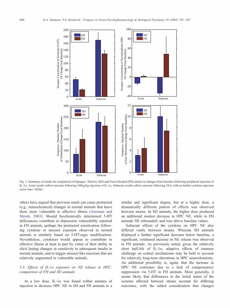
Results of these experiments are summarized in Fig. 3.
5.2. Effects of IL-1a exposure on 5-HT release in HPC:
comparison of FH and SD animals
Within minutes following peripheral acute injection, IL-
1a significantly increased HPC 5-HT in both strains,
though this occurred to a significantly lesser degree in FH
animals. Notwithstanding previously mentioned deficien-
cies in the FH strain, the 5-HT biochemical pathway is
apparently still capable of producing 5-HT, and further, of
mounting a significant reaction to immune challenge not
unlike central 5-HT reactions to stressors. Yet, the HPC 5-
HT response to IL-1a in the FH strain appears blunted
relative to normal animals.
Subacute studies were also performed in both strains, 24
h later, with no further injection of cytokine and the
dramatic differences in HPC 5-HT continued, within and
between strains. It is interesting, given the relatively short
half-life of the cytokine (Ramanathan, 1996), that the effect
of injection on central 5-HT continued well beyond the
completion of IL-1a metabolism. This may indicate lasting,
perhaps adaptive effects of immune challenge upon central
mechanisms implicated in stress and depression. Continued
deficiency in HPC 5-HT release in FH animals is likely
associated with 5-HT deficiencies previously noted.
Given evidence that 5-HT modulates central reactions to
stressors (Kennett and Joseph, 1981; Kennett et al., 1985;
Smelik, 1987; Cassano and D’mello, 2001; Lowry, 2002), it
seems reasonable to surmise HPC 5-HT deficiencies in the
FH strain may be permissive of downstream physiological
responses to insult, as noted in depression (e.g., loss of HPA
feedback inhibition: Young et al., 1991). That is, because of
5-HT deficiencies, perhaps FH animals have a lower
threshold for the initiation of activity in neuroendocrine
and/or automonic systems that promote changes relevant to
affective disorders like depression. As mentioned earlier,
0
20
40
60
80
100
120
140
160
180
200
-40
-20
0
20
40
60
80
100
0
50
100
150
200
250
300
0
2
4
6
8
10
12
Syna
ptic
Con
cent
ratio
n of
Ser
oton
in (
5-H
T)
(% C
hang
e Fr
om B
asel
ine)
Freq
uenc
y: A
bsol
ute
Num
ber
of A
mbu
latio
ns (
Cha
nge
From
Bas
elin
e)
Syna
ptic
Con
cent
ratio
n of
Nor
epin
ephr
ine
(NE
)(%
Cha
nge
From
Bas
elin
e)Fr
eque
ncy:
Abs
olut
e N
umbe
r of
Fin
e M
ovem
ents
(C
hang
e Fr
om B
asel
ine)
Acute Subacute Acute Subacute
Acute Subacute Acute Subacute
SDFH
SDFH
SDFH
SDFH
Fig. 3. Summary of results for comparison of Sprague–Dawley (SD) and Fawn Hooded (FH) strains in changes from baseline following peripheral injection of
IL-1a. Acute results reflect outcome following 100ng/kg injection of IL-1a. Subacute results reflect outcome following 24 h, with no further cytokine injection
(error bars=SEM).
D.A. Simmons, P.A. Broderick / Progress in Neuro-Psychopharmacology & Biological Psychiatry 29 (2005) 793–807800
others have argued that previous insult can cause protracted
(e.g., neurochemical) changes in normal animals that leave
them more vulnerable to affective illness (Anisman and
Merali, 2003). Should biochemically determined 5-HT
deficiencies contribute to depression vulnerability reported
in FH animals, perhaps the protracted sensitization follow-
ing cytokine or stressor exposure observed in normal
animals is similarly based on 5-HT-ergic modifications.
Nevertheless, cytokines would appear to contribute to
affective illness at least in part by virtue of their ability to
elicit lasting changes in sensitivity to subsequent insults in
normal animals, and to trigger stressor-like reactions that are
relatively augmented in vulnerable animals.
5.3. Effects of IL-1a exposure on NE release in HPC:
comparison of FH and SD animals
At a low dose, IL-1a was found within minutes of
injection to decrease HPC NE in SD and FH animals to a
similar and significant degree, but at a higher dose, a
dramatically different pattern of effects was observed
between strains. In SD animals, the higher dose produced
an additional modest decrease in HPC NE, while in FH
animals NE rebounded, and rose above baseline values.
Subacute effects of the cytokine on HPC NE also
differed vastly between strains. Whereas SD animals
displayed a further significant decrease below baseline, a
significant, continued increase in NE release was observed
in FH animals. As previously noted, given the relatively
short half-life of IL-1a, adaptive effects of immune
challenge on central mechanisms may be held to account
for relatively long-term alterations in HPC neurochemistry.
An additional possibility is, again, that the increase in
HPC NE continues due to a lack of compensatory
suppression via 5-HT in FH animals. More generally, it
seems likely that differences in the initial status of the
systems affected between strains account for differing
outcomes, with the added consideration that changes

D.A. Simmons, P.A. Broderick / Progress in Neuro-Psychopharmacology & Biological Psychiatry 29 (2005) 793–807 801
elicited by cytokines are rather labile, prone to vary in
form and magnitude over time (Schmidt et al., 1995;
2003). Regardless, measures of HPC NE in SD and FH
animals provide further indication of lasting, perhaps
adaptive effects of immune challenge upon central
mechanisms implicated in stress and depression.
The difference between strains for HPC NE found at the
higher IL-1a dose provides further indication that FH
animals are especially prone to physiological changes
typically associated with stressors (e.g., elevated HPC
NE, Abercrombie and Jacobs, 1988; Kalen et al., 1989;
Zhang et al., 1995). What is more, measures of HPC NE in
the FH strain are reminiscent of changes elicited by chronic
stressor regimens that produce depression in normal
animals. Analogous to the effect seen following IL-1a
challenge in the FH strain, SD animals previously exposed
to chronic cold water immersion display significantly
elevated and prolonged HPC NE release in response to a
novel stressor, compared to reactions observed in naı̈ve rats
(Nisenbaum et al., 1991; Nisenbaum and Abercrombie,
1992). Similar ‘‘stress-induced sensitization’’ is noted for
PFC NE (Gresch et al., 1994; Finlay et al., 1997), as well as
for systems mediating other responses that are relevant to
stress and depression (e.g., HPA outflow, Bartanusz et al.,
1993; Van Dijken et al., 1993). With respect to the inverse
regulatory relationship suggested earlier between 5-HT and
NE, it is possible blunted 5-HT in FH animals permits a
relatively large NE response to insult of psychological or
immune origin. In sum, central NE reaction to IL-1a
injection in FH animals appears to be rather stressor-like
and to reflect an exaggerated sensitivity to insult. This
further illustrates how outcomes of immune challenge can
vary depending on varying degree of preexisting vulner-
ability across animal models.
5.4. Effects of IL-1a exposure on open-field behavior:
comparison of FH and SD animals
The effects of IL-1a observed in movement behaviors
(e.g., ambulation, stereotypic sniffing and grooming) were
episodic in both strains; this was more readily apparent in
FH animals. There was an apparent tendency toward an
increase in locomotor activity in both strains following acute
injection. Interestingly, locomotor counts were higher in FH
versus SD animals, though this may have resulted in
association with the episodic nature of the behavioral
outcome. Stereotypic sniffing and grooming behaviors also
increased in both strains. Again, there was a tendency for
stereotypic sniffing and grooming to be increased in FH
versus SD animals.
The subacute effects of IL-1a injection were observed as
significant increases in locomotor activity in both strains
versus baseline; no significant difference was observed
between strains. Stereotypic sniffing and grooming was
again increased in SD and FH animals versus baseline,
though there was no significant difference between strains.
Described earlier, a tendency toward increased behav-
ioral responsiveness within a previously habituated to
environment may indicate stressor associated mechanisms
are in play (e.g., increased vigilance). With respect to the
effects of injection, the difference between strains in the
acute study provides an additional example of how animals
with preexisting biochemical vulnerabilities associated with
depression express augmented stressor-like reactions to
immune challenge. The subacute results are more difficult
to resolve, though again this may be accounted for by
potential differences in the initial status of systems affected
in each strain, the labile nature of cytokine effects over time,
and the episodic nature of these behavioral responses
following injection. Nevertheless, it appears most likely
that differences in reactions between strains to IL-1a
injection indicate FH animals are more sensitive in terms
of stressor-like outcome.
5.5. Effects of IL-1a exposure on immune cell mitogenic
responses: comparison of FH and SD animals
In addition to relatively direct central and/or behavioral
effects, stressor- or depression-like outcomes of a given
insult may occur indirectly via a cascade of cellular immune
events (e.g., stress associated increases in T-cell mitogen
responsiveness and cytokine release: Kubera et al., 1996).
For this reason, it was of interest to examine effects of
cytokine injection on aspects of immune function between
rodent strains. In our laboratories we examined T cell
proliferative mitogen responses in spleen cells from rats
injected with IL-1a. There was a profound difference
between strains in the initial acute response in that measures
obtained for the FH strain were significantly higher (two-
fold) than those observed from SD animals. Although no
change occurred in the B cell subset, a significant change in
the T cell mitogen response indicates in the FH strain that
there may be an incurred hypersensitivity following
cytokine challenge, i.e., an augmented responsiveness to
cytokine challenge in the FH strain compared with the SD
strain. The finding suggests that the IL-1a mediated cascade
of immune events is potentiated in FH versus SD animals,
wherein the magnitude of inflammation may be relatively
pronounced (Coico and Broderick, unpublished data).
Therefore, regarding the differential effects of IL-1a on
mitogen responses between SD and FH animals, it would
appear that in addition to augmented behavioral and
neurochemical stressor-like effects noted in FH animals,
there is evidence to suggest augmented responsiveness to
IL-1a in aspects of cellular immunity.
The subacute effects of IL-1a were less dramatic, though
there was still a tendency for T cell mitogen responses to be
elevated in FH versus SD animals. The difference in
magnitude between acute and subacute effects may corre-
spond with the short half-life of the cytokine. As well,
differences between acute and subacute effects may be
accounted for by the lability of cytokine effects over time, as

D.A. Simmons, P.A. Broderick / Progress in Neuro-Psychopharmacology & Biological Psychiatry 29 (2005) 793–807802
observed for behavioral and neurochemical measures
described earlier.
6. Conclusions
Evidence reviewed here suggests that cytokines, much
like psychological stressors, are capable of provoking
physiological changes implicated in depression, and of
promoting greater vulnerability to depression that becomes
manifest upon subsequent exposure to insult. Furthermore,
evidence that FH animals, like normal animals previously
exposed to stressor or cytokine challenge, show augmented
behavioral and neurochemical reactions to IL-1a injection
provides greater validation for models of depression
pathogenesis that are based on the protracted effects of
acute psychological or systemic insult (Fig. 4). The
additional finding, that IL-1a pretreatment enhanced
mitogen responses in FH more so than in SD animals,
provides evidence to suggest immune function is compro-
mised during depression. However, there remain questions
as to whether basal immune differences in FH animals are
purely of collateral nature should depression be driven
fundamentally by abnormal neuroendocrine function, or
whether, to any degree, immune dysfunction contributes
more fundamentally to observed neuroendocrine changes
(i.e., via excessive release of cytokines). Regarding the
short-term impact of immune challenge, despite any degree
of overlap between cytokine effects and basal measures
Fig. 4. Given the central role the hippocampus (HPC) plays in sensory informat
reactivity to stressors, abnormal behavioral and physiological outcomes of stresso
neurochemical abnormalities are present. Underlying differences in HPC ne
vulnerability to depression (e.g., SD vs. FH) may contribute to differential HPA o
CD4/CD8 ratio) which may be affected secondarily. Differences between SD
reactions to IL-1a injection.
noted for clinical depression and depression models
described presently, the changes elicited do not seem a
sufficient characterization of depressive disorder per se.
More work is needed where effects of relatively chronic
(i.e., >7 days) regimens of cytokine exposure are
implemented in experimental animals, given that in natural
situations where cytokine levels are altered, the changes
are rather persistent by comparison to most experimental
models previously reported. Furthermore, the changes
observed in humans over the course of cytokine immuno-
therapy may be unique to that clinical situation, and effects
of relatively short-term cytokine treatment in experimental
models, too, may be limited in interpretation.
Acknowledgements
This work was supported by an Aaron Diamond
Foundation Award to Professors Broderick and Coico,
CUNY Medical School. Partial support was provided by
an NIH/NIGMS Award (SO6 GM08168), and a Profes-
sional Staff Congress/The City University of New York
Award (RF 64282-00 33) to Professor Broderick. Partial
support was also provided by an NIH/RCMA Award to
Professor Coico. This review was inspired by a project
written by D.A. Simmons as a partial requirement for
graduation from the Emory University Department of
Psychology doctoral program in Neuroscience and Animal
Behavior.
ion processing, coordination of behavioral responses, and regulating HPA
r or cytokine challenge likely correspond in an animal within which HPC
urochemistry (e.g., 5-HT, NE) revealed between strains that differ in
utflow and behavior, as well as other concomitants (e.g., mitogen activity,
and FH animals are evident in neurochemical, behavioral, and immune

D.A. Simmons, P.A. Broderick / Progress in Neuro-Psychopharmacology & Biological Psychiatry 29 (2005) 793–807 803
References
Abercrombie, E.D., Jacobs, B.L., 1988. Systemic naloxone administration
potentiates locus coeruleus noradrenergic neuronal activity under stress-
ful but not non-stressful conditions. Brain Res. 441 (1–2), 362–366.
Ando, T., Dunn, A.J., 1999. Mouse tumor necrosis factor-alpha increases
brain tryptophan concentrations and norepinephrine metabolism while
activating the HPA axis in mice. Neuroimmunomodulation 6 (5),
319–329.
Anisman, H., Merali, Z., 1999. Anhedonic and anxiogenic effects of
cytokine exposure. Adv. Exp. Med. Biol. 461, 199–233.
Anisman, H., Merali, Z., 2003. Cytokines, stress and depressive illness:
brain-immune interactions. Ann. Med. 35, 2–11.
Anisman, H., Zacharko, R.M., 1990. Multiple neurochemical and behav-
ioral consequences of stressors: implications for depression. Pharmacol.
Ther. 46 (1), 119–136.
Anisman, H., Hayley, S., Turrin, N., Merali, Z., 2002a. Cytokines as a
stressor: implications for depressive illness. Int. J. Neuropsychophar-
macol. 5 (4), 357–373.
Anisman, H., Kokkinidis, L., Merali, Z., 2002b. Further evidence for the
depressive effects of cytokines: anhedonia and neurochemical changes.
Brain Behav. Immun. 16 (5), 544–556.
Anisman, H., Turrin, N.P., Merali, Z., Hayley, S., 2003. Neurochemical
sensitization associated with systemic administration of tumor necrosis
factor-alpha: adjuvant action in combination with bovine serum
albumin. J. Neuroimmunol. 145 (1–2), 91–102.
Araujo, B.M., Lapchak, P.A., Collier, B., Quirion, R., 1989. Localization
of interleukin-2 immunoreactivity and interleukin-2 receptors in the
rat brain: interaction with the cholinergic system. Brain Res. 498,
257–266.
Asberg, M., Traskman, L., Thoren, P., 1976. 5-HIAA in the cerebrospinal
fluid. A biochemical suicide predictor? Arch. Gen. Psychiatry 33,
1193–1197.
Aulakh, C.S., Hill, J.L., Murphy, D.L., 1988. A comparison of feeding and
locomotion responses to serotonin agonists in three rat strains.
Pharmacol. Biochem. Behav. 31 (3), 567–571.
Aulakh, C.S., Wozniak, K.M., Hill, J.L., Devane, C.L., Tolliver, T.J.,
Murphy, D.L., 1988. Differential neuroendocrine response to the 5-HT
agonist m-CPP in Fawn-Hooded rats relative to Wistar and Sprague–
Dawley rats. Neuroendocrinology 48 (4), 401–406.
Aulakh, C.S., Hill, J.L., Lesch, K.P., Murphy, D.L., 1992. Functional
subsensitivity of 5-hydroxytryptamine1C or alpha 2 adrenergic hetero-
receptors mediating clonidine-induced growth hormone release in the
Fawn-Hooded rat strain relative to the Wistar rat strain. J. Pharmacol.
Exp. Ther. 262 (3), 1038–1043.
Aulakh, C.S., Hill, J.L., Murphy, J.L., 1993. Attenuation of hyper-
cortisolemia in fawn-hooded rats by antidepressant drugs. Eur. J.
Pharmacol. 240 (1), 85–88.
Aulakh, C.S., Tolliver, T., Wozniak, K.M., Hill, J.L., Murphy, D.L., 1994.
Functional and biochemical evidence for altered serotonergic function
in the Fawn-Hooded rat strain. Pharmacol. Biochem. Behav. 49 (3),
615–620.
Baldessarini, R.J., 1989. Current status of antidepressants: clinical
pharmacology and therapy. J. Clin. Psychiatry 50, 117–126.
Ban, E., Marquette, C., Sarrieau, A., Fitzpatrick, F., Fillion, G., Milon, G.,
Rostene, W., Haour, F., 1993. Regulation of interleukin-1 receptor
expression in mouse brain and pituitary by lipopolysaccharide and
glucocorticoids. Neuroendocrinology 58, 581–587.
Banks, W.A., 1999. Physiology and pathology of the blood–brain barrier:
implications for microbial pathogenesis, drug delivery and neuro-
degenerative disorders. J. NeuroVirol. 5, 538–555.
Banks, W.A., Kastin, A.J., 1992. The interleukins-1a, -1b, and -2 do not
acutely disrupt the murine blood–brain barrier. Int. J. Immunopharma-
col. 14, 629–636.
Baraban, J.M., Aghajanian, G.K., 1980. Suppression of firing activity of 5-
HT neurons in the dorsal raphe by alpha-adrenoceptor antagonists.
Neuropharmacology 19 (4), 355–363.
Bartanusz, V., Jezova, D., Bertini, L.T., Tilders, F.J.H., Aubry, J.M., Kiss,
J.Z., 1993. Stress-induced increase in vasopressin and corticotropin-
releasing factor expression in hypophysiotrophic paraventricular neu-
rons. Endocrinology 132, 895–902.
Besedovsky, H., del Rey, A., Sorkin, E., Dinarello, C.A., 1986. Immunor-
egulatory feedback between interleukin-1 and glucocorticoid hormones.
Science 233, 652–654.
Bianchi, M., Sacerdote, P., Ricciardi-Castagnoli, P., Mantegazza, P.,
Panerai, A.E., 1992. Central effects of tumor necrosis factor alpha and
interleukin-1 alpha on nociceptive thresholds and spontaneous locomo-
tor activity. Neurosci. Lett. 148 (1–2), 76–80.
Blanchard, R.J., Shepherd, J.K., Armstrong, J., Tsuda, S.F., Blanchard,
D.C., 1993. An ethopharmacological analysis of the behavioral effects
of 8-OH-DPAT. Psychopharmacology 112 (1), 55–63.
Blier, P., 2001. Norepinephrine and selective norepinephrine reuptake
inhibition in depression and mood disorders: their pivotal roles.
J. Psychiatry Neurosci. 26, S1–S2 (Suppl.)
Bliss, E.L., Thatcher, W., Ailion, J., 1972. Relationship of stress to
brain serotonin and 5-hydroxyindoleacetic acid. J. Psychiatr. Res. 9
(2), 71–80.
Brady, L.S., Lynn, A.B., Herkenham, M., Gottesfeld, Z., 1994. Systemic
interleukin-1 induces early and late patterns of c-fos mRNA expression
in brain. J. Neurosci. 14 (8), 4951–4964.
Brebner, K., Hayley, S., Zacharko, R., Merali, Z., Anisman, H., 2000.
Synergistic effects of interleukin-1 beta, interleukin-6, and tumor
necrosis factor: central monoamine, corticosterone and behavioral
variations. Neuropsychopharmacology 22 (6), 566–580.
Broderick, P.A., 1997. Alprazolam, diazepam, yohimbine, clonidine: in
vivo CA1 hippocampal norepinephrine and serotonin release profiles
under chloral hydrate anesthesia. Prog. Neuro-Psychopharmacol. Biol.
Psychiatry 21 (7), 1117–1140.
Broderick, P.A., 2002. Interleukin 1alpha alters hippocampal serotonin
and norepinephrine release during open-field behavior in Sprague–
Dawley animals: differences from the Fawn-Hooded animal model of
depression. Prog. Neuro-Psychopharmacol. Biol. Psychiatry 26 (7–8),
1355–1372.
Capuron, L., Dantzer, R., 2003. Cytokines and depression: the need for a
new paradigm. Brain Behav. Immun. 17, S119–S124.
Capuron, L., Ravaud, A., 1999. Prediction of the depressive effects of
interferon Alpha therapy by the patient’s initial affective state. New
Engl. J. Med. 340 (17), 1370.
Cassano Jr., W.J., D’mello, A.P., 2001. Acute stress-induced facili-
tation of the hypothalamic-pituitary-adrenal axis: evidence for the
roles of stressor duration and serotonin. Neuroendocrinology 74 (3),
167–177.
Castle, S., Wilkins, S., Heck, E., Tanzy, K., Fahey, J., 1995. Depression in
caregivers of demented patients is associated with altered immunity:
impaired proliferative capacity, increased CD8+, and a decline in
lymphocytes with surface signal transduction molecules (CD38+) and a
cytotoxicity marker (CD56+ CD8+). Clin. Exp. Immunol. 101 (3),
487–493.
Charles, G., Machowski, R., Brohee, D., Wilmotte, J., Kennes, B., 1992.
Lymphocyte subsets in major depressive patients. Neuropsychobiology
25, 94–98.
Charney, D.S., Heninger, G.R., Sternberg, D.E., 1982. Adrenergic receptor
sensitivity in depression: effects of clonidine in depressed patients and
healthy patients. Arch. Gen. Psychiatry 39 (3), 290–294.
Chen, F., Lawrence, A.J., 2000. 5-HT transporter sites and 5-HT1A and 5-
HT3 receptors in Fawn-Hooded rats: a quantitative autoradiography
study. Alcohol Clin. Exp. Res. 24 (7), 1093–1102.
Chrousos, G.P., Gold, P.W., 1992. The concepts of stress and stress system
disorders. JAMA 267 (9), 1244–1252.
Chrousos, G.P., Gold, P.W., 1998. A healthy body in a healthy mind-and
vice versa—the damaging power of uncontrollable stress. J. Clin.
Endocrinol. Metab. 83 (6), 1842–1845.
Claes, S.J., 2004. Corticotropin-releasing hormone (CRH) in psychiatry:
from stress to psychopathology. Ann. Med. 36 (1), 50–61.
D.A. Simmons, P.A. Broderick / Progress in Neuro-Psychopharmacology & Biological Psychiatry 29 (2005) 793–807804
Cologer-Clifford, A., Simon, N.G., Lu, S.F., Smoluk, S.A., 1997. Serotonin
agonist-induced decreases in intermale aggression are dependent on
brain region and receptor subtype. Pharmacol. Biochem. Behav. 58 (2),
425–430.
Connor, T.J., Song, C., Leonard, B.E., Merali, Z., Anisman, H., 1998. An
assessment of the effects of central interleukin-1 beta, -2, -6 and tumor
necrosis factor-alpha administration on some behavioral, neurochem-
ical, endocrine, and immune parameters in the rat. Neuroscience 84 (3),
923–933.
Connor, T.J., Song, C., Leonard, B.E., Anisman, H., Merali, Z., 1999.
Stressor-induced alterations in serotonergic activity in an animal model
of depression. NeuroReport 10 (3), 523–528.
Curzon, G., 1988. Serotonergic mechanisms of depression. Clin. Neuro-
pharmacol. 11 (Suppl. 2), S11–S20.
Dantzer, R., 2004. Innate immunity at the forefront of psychoneuroimmu-
nology. Brain Behav. Immun. 18 (1), 1–6.
de Waal Malefyt, R., Abrams, J., Bennett, B., Figdor, C.G., de Vries, J.E.,
1991. Interleukin 10(IL-10) inhibits cytokine synthesis by human
monocytes: an autoregulatory role of IL-10 produced by monocytes.
J. Exp. Med. 174 (5), 1209–1220.
Dinarello, C.A., 1993. Modalities for reducing interleukin 1 activity in
disease. Trends Pharmacol. Sci. 14 (5), 155–159.
El-Haj, T., Poole, S., Farthing, M.J., Ballinger, A.B., 2002. Anorexia in a rat
model of colitis: interaction of interleukin-1 and hypothalamic
serotonin. Brain Res. 927 (1), 1–7.
Ellison, M., Krieg, R.J., Povlishock, J.T., 1990. Differential central nervous
system responses following single and multiple recombinant IL-2
infusions. J. Neuroimmunol. 28, 249–260.
Elmquist, J.K., Ackermann, M.R., Register, K.B., Rimler, R.B., Ross, L.R.,
Jacobson, C.D., 1993. Induction of Fos-like immunoreactivity in the rat
brain following Pasteurella multocida endotoxin administration. Endo-
crinology 133 (6), 3054–3057.
Engeland, C.G., Nielsen, D.V., Kavaliers, M., Ossenkopp, K.P., 2001.
Locomotor activity changes following lipopolysaccharide treatment in
mice: a multivariate assessment of behavioral tolerance. Physiol. Behav.
72 (4), 481–491.
Engeland, C.G., Kavaliers, M., Ossenkopp, K.P., 2003. Sex differences in
the effects of muramyl dipeptide and lipopolysaccharide on locomotor
activity and the development of behavioral tolerance in rats. Pharmacol.
Biochem. Behav. 74 (2), 433–447.
Ericsson, A., Kovacs, K.J., Sawchenko, P.E., 1994. A functional anatomical
analysis of central pathways subserving the effects of interleukin-1 on
stress-related neuroendocrine neurons. J. Neurosci. 14 (2), 897–913.
Farrar, W.L., Kilian, P.L., Ruff, M., Hill, J.M., Pert, C.B., 1987.
Visualization and characterization of interleukin-1 receptors in brain.
J. Immunol. 139, 459–463.
Finlay, J.M., Jedema, H.P., Rabinovic, A.D., Mana, M.J., Zigmond, M.J.,
Sved, A.F., 1997. Impact of corticotropin-releasing hormone on
extracellular norepinephrine in prefrontal cortex after chronic cold
stress. J. Neurochem. 69 (1), 144–150.
Fiorentino, D.F., Zlotnik, A., Mosmann, T.R., Howard, M., O’Garra, A.,
1991. IL-10 inhibits cytokine production by activated macrophages.
J. Immunol. 147 (11), 3815–3822.
Franklin, A.E., Engeland, C.G., Kavaliers, M., Ossenkopp, K.P., 2003.
Lipopolysaccharide-induced hypoactivity and behavioral tolerance
development are modulated by the light–dark cycle in male and female
rats. Psychopharmacology 170 (4), 399–408.
Gorea, E., Adrien, J., 1988. Serotonergic regulation of noradrenergic
coerulean neurons: electrophysiological evidence for the involvement of
5-HT2 receptors. Eur. J. Pharmacol. 154 (3), 285–291.
Gresch, P.J., Sved, A.F., Zigmond, M.J., Finlay, J.M., 1994. Stress-induced
sensitization of dopamine and norepinephrine efflux in medial pre-
frontal cortex of the rat. J. Neurochem. 63 (2), 575–583.
Grignaschi, G., Mantelli, B., Samanin, R., 1993. The hypophagic effect of
restraint stress in rats can be mediated by 5-HT2 receptors in the
paraventricular nucleus of the hypothalamus. Neurosci. Lett. 152 (1–2),
103–106.
Haour, F., Marquette, C., Ban, E., Crumeyrolle-Arias, M., Rostene, W.,
Tsiang, H., Fillion, G., 1995. Receptors for interleukin-1 in the central
nervous and endocrine systems. Am. Endocrinol. 56, 173–179.
Hassanain, M., Zalcman, S., Bhatt, S., Siegel, A., 2003a. Interleukin-1 beta
in the hypothalamus potentiates feline defensive rage: role of serotonin-
2 receptors. Neuroscience 120 (1), 227–233.
Hassanain, M., Bhatt, S., Siegel, A., 2003b. Differential modulation of
feline defensive rage behavior in the medial hypothalamus by 5-HT1A
and 5-HT2 receptors. Brain Res. 981 (1–2), 201–209.
Hayley, S., Lacosta, S., Merali, Z., van Rooijen, N., Anisman, H., 2001.
Central monoamine and plasma corticosterone changes induced by a
bacterial endotoxin: sensitization and cross-sensitization effects. Eur. J.
Neurosci. 13 (6), 1155–1165.
Herman, J.P., Cullinan, W.E., 1997. Neurocircuitry of stress: central control
of the hypothalamo-pituitary-adrenocortical axis. Trends Neurosci. 20
(2), 78–84.
Herman, J.P., Figueiredo, H., Mueller, N.K., Ulrich Lai, Y., Ostrander,
M.M., Choi, D.C., Cullinan, W.E., 2003. Central mechanisms of
stress integration: hierarchical circuitry controlling hypothalamo-
pituitary-adrenocortical responsiveness. Front. Neuroendocrinol. 24
(3), 151–180.
Huang, Q.H., Hruby, V.J., Tatro, J.B., 1999. Role of central melanocortins
in endotoxin-induced anorexia. Am. J. Physiol. 276 (3,2), R671–R864.
Hulihan-Giblin, B.A., Park, Y.D., Aulakh, C.S., Goldman, D., 1992.
Regional analysis of 5-HT1A and 5-HT2 receptors in the Fawn-Hooded
rat. Neuropharmacology 31 (11), 1095–1100.
Hulihan-Giblin, B.A., Park, Y.D., Goldman, D., Aulakh, C.S., 1993.
Analysis of 5-HT1C receptor and the serotonin uptake site in Fawn-
Hooded rat brain. Eur. J. Pharmacol. 239 (1–3), 99–102.
Imeri, L., Mancia, M., Opp, M.R., 1999. Blockade of 5-hydroxytryptamine
(serotonin)-2 receptors alters interleukin-1-induced changes in rat sleep.
Neuroscience 92 (2), 745–749.
Johnson, J.D., O’Connor, K.A., Watkins, L.R., Maier, S.F., 2004. The role
of IL-1h in stress-induced sensitization of proinflammatory cytokine
and corticosterone responses. Neuroscience 127 (3), 569–577.
Kalen, P., Rosegren, E., Lindvall, O., Bjorklund, A., 1989. Hippocampal
noradrenaline and serotonin release over 24 hours as measured by the
dialysis technique in freely moving rats: correlation to behavioural
activity state, effect of handling and tail-pinch. Eur. J. Neurosci. 1,
181–188.
Katsuura, G., Gottschall, P.E., Arimura, A., 1988. Identification of a high-
affinity receptor for interleukin-1 beta in rat brain. Biochem. Biophys.
Res. Commun. 156, 61–67.
Katz, R.J., 1981. Animal models and human depressive disorders. Neuro-
sci. Biobehav. Rev. 5 (2), 231–246.
Kawahara, H., Yoshida, M., Yokoo, H., Nishi, M., Tanaka, M., 1993.
Psychological stress increases serotonin release in the rat amygdala and
prefrontal cortex assessed by in vivo microdialysis. Neurosci. Lett. 162
(1–2), 81–84.
Kennett, G.A., Joseph, M.H., 1981. The functional importance of increased
brain tryptophan in the serotonergic response to restraint stress.
Neuropharmacology 20 (1), 39–43.
Kennett, G.A., Dickinson, S.L., Curzon, G., 1985. Enhancement of some
5-HT-dependent behavioural responses following repeated immobili-
zation in rats. Brain Res. 330 (2), 253–263.
Koenig, J.I., 1991. Presence of cytokines in the hypothalamic-pituitary axis.
Prog. Neuroendocr. Immunol. 4, 143–153.
Konsman, J.P., Parnet, P., Dantzer, R., 2002. Cytokine-induced sick-
ness behaviour: mechanisms and implications. Trends Neurosci. 25,
154–159.
Konstandi, M., Johnson, E., Lang, M.A., Malamas, M., Marselos, M., 2000.
Noradrenaline, dopamine, serotonin: different effects of psychological
stress on brain biogenic amines in mice and rats. Pharmacol. Res. 41
(3), 341–346.
Kozak, W., Conn, C.A., Kluger, M.J., 1994. Lipopolysaccharide induces
fever and depresses locomotor activity in unrestrained mice. Am. J.
Physiol. 266 (1,2), R125–R135.
D.A. Simmons, P.A. Broderick / Progress in Neuro-Psychopharmacology & Biological Psychiatry 29 (2005) 793–807 805
Kozak, W., Zheng, H., Conn, C.A., Soszynski, D., van der Ploeg, L.H.,
Kluger, M.J., 1995. Thermal and behavioral effects of lipopolysac-
chaide and influenza in interleukin-1 beta-deficient mice. Am. J.
Physiol. 269 (5,2), R969–R977.
Krueger, J.M., Majde, J.A., 1995. Cytokines and sleep. Int. Arch. Allergy
Immunol. 106 (2), 97–100.
Krueger, J.M., Toth, L.A., 1994. Cytokines as regulators of sleep. Ann.
N.Y. Acad. Sci. 739, 299–310.
Kubera, M., Symbirtsev, A., Basta-Kaim, A., Borycz, J., Roman, A., Papp,
M., Claesson, M., 1996. Effect of chronic treatment with imipramine on
interleukin 1 and interleukin 2 production by splenocytes obtained from
rats subjected to a chronic mild stress model of depression. Pol. J.
Pharmacol. 48 (5), 503–506.
Kuzmin, A., Semenova, S., Zvartau, E.E., Van Ree, J.M., 1996. Enhance-
ment of morphine self-administration in drug naive, inbred strains of
mice by acute emotional stress. Eur. Neuropsychopharmacology 6 (1),
63–68.
Lacosta, S., Merali, Z., Anisman, H., 1998. Influence of interleukin-1beta
on exploratory behaviors, plasma ACTH, corticosterone, and central
biogenic amines in mice. Psychopharmacology 137 (4), 351–361.
Lacosta, S., Merali, Z., Anisman, H., 1999. Influence of acute and
repeated interleukin-2 administration on spatial learning, locomotor
activity, exploratory behaviors, and anxiety. Behav. Neurosci. 113 (5),
1030–1041.
Lacosta, S., Merali, Z., Anisman, H., 2000. Central monoamine alterations
associated with acute and repeated interleukin-2 administration. Neuro-
immunomodulation 9, 1–16.
Lapchak, P.A., 1992. A role for interleukin-2 in the regulation of
dopaminergic function. NeuroReport 3, 165–168.
Lavicky, J., Dunn, A.J., 1995. Endotoxin administration stimulates cerebral
catecholamine release in freely moving rats as assessed by micro-
dialysis. J. Neurosci. Res. 40 (3), 407–413.
Lesch, K.P., Laux, G., Mueller, T., 1990. A2-Adrenoceptor responsivity in
depression: effect of chronic treatment with moclobemide, a selective
MAO-A inhibitor, vs. maprotiline. J. Neural Transm. 32, 457–461.
Leserman, J., Petitto, J.M., Gu, H., Gaynes, B.N., Barroso, J., Golden, R.N.,
Perkins, D.O., Folds, J.D., Evans, D.L., 2002. Progression to AIDS, a
clinical AIDS condition and mortality: psychosocial and physiological
predictors. Psychol. Med. 32 (6), 1059–1073.
Licinio, J., Wong, M.L., 1999. Interleukin 1 receptor antagonist gene
expression in rat pituitary in the systemic inflammatory response
syndrome: pathophysiological implications. Mol. Psychiatry 2, 99–103.
Linthorst, A.C., Reul, J.M., 1998. Brain neurotransmission during
peripheral inflammation. Ann. N.Y. Acad. Sci. 840, 139–152.
Linthorst, A.C.E., Flachskamm, C., Holsboer, F., Reul, J.M.H.M., 1994.
Local administration of recombinant human interleukin-1 beta in the rat
hippocampus increases serotonergic neurotransmission, hypothalamic-
pituitary-adrenocortical axis activity, and body temperature. Endocri-
nology 135 (2), 520–532.
Linthorst, A.C.E., Flachskamm, C., Mhller-Preuss, P., Holsboer, F., Reul,
J.M.H.M., 1995. Effect of bacterial endotoxin and interleukin-1 beta on
hippocampal serotonergic neurotransmission, behavioral activity, and
free corticosterone levels: an in vivo microdialysis study. J. Neurosci.
15 (4), 2920–2934.
Linthorst, A.C.E., Flachskamm, C., Holsboer, F., Reul, J.M.H.M., 1996.
Activation of serotonergic and noradrenergic neurotransmission in the
rat hippocampus after peripheral administration of bacterial endotoxin:
involvement of the cyclo-oxygenase pathway. Neuroscience 72 (4),
989–997.
Linthorst, A.C., Flachskamm, C., Hopkins, S.J., Hoadley, M.E., Labeur,
M.S., Holsboer, F., Reul, J.M., 1997. Long-term intracerebroventricular
infusion of corticotropin-releasing hormone alters neuroendocrine,
neurochemical, autonomic, behavioral, and cytokine responses to a
systemic inflammatory challenge. J. Neurosci. 17 (11), 4448–4460.
Lowry, C.A., 2002. Functional subsets of serotonergic neurones: implica-
tions for control of the hypothalamic-pituitary-adrenal axis. J. Neuro-
endocrinol. 14 (11), 911–923.
Maes, M., Meltzer, H.Y., 1995. The serotonin hypothesis of major
depression. In: Bloom, F.E., Kupfer, D.J. (Eds.), Psychopharmacology:
the fourth generation of progress. Raven Press, New York, pp.
933–944.
Maier, S.F., Watkins, L.R., 1998. Cytokines for psychologists:
implications of bidirectional immune-to-brain communication for
understanding behavior, mood, and cognition. Psychol. Rev. 105 (1),
83–107.
Maier, S.F., Goehler, L.E., Fleshner, M., Watkins, L.R., 1998. The role of
the vagus nerve in cytokine-to-brain communication. Ann. N.Y. Acad.
Sci. 840, 289–300.
Marinelli, P.W., Quirion, R., Gianoulakis, C., 2004. An in vivo profile
of h-endorphin release in the arcuate nucleus and nucleus accum-
bens following exposure to stress or alcohol. Neuroscience 127 (3),
777–784.
Meltzer, H.Y., Lowy, M.T., 1987. The serotonin hypothesis of depression.
In: Meltzer, H.Y. (Ed.), Psychopharmacology, The Third Generation of
Progress. Raven Press, New York, pp. 513–526.
Merali, Z., Lacosta, S., Anisman, H., 1997. Effects of interleukin-1 beta and
mild stress on alterations of norepinephrine, dopamine and serotonin
neurotransmission: a regional microdialysis study. Brain Res. 761 (2),
225–235.
MohanKumar, S.M., MohanKumar, P.S., Quadri, S.K., 1998. Specificity of
interleukin-1 beta induced changes in monoamine concentrations in
hypothalamic nuclei: blockade by interleukin-1 receptor antagonist.
Brain Res. Bull. 47 (1), 29–34.
MohanKumar, S.M., MohanKumar, P.S., Quadri, S.K., 2000. Effects of
bacterial lipopolysaccharade on central monoamines and fever in the
rat: involvement of the vagus. Neurosci. Lett. 284 (3), 159–162.
Moore, K.W., O’Garra, A., de Waal Malefyt, R., Vieira, P., Mosmann, T.R.,
1993. Interleukin-10. Annu. Rev. Immunol. 11, 165–190.
Morgan, M.M., Clayton, C.C., Heinricher, M.M., 2004. Simultaneous
analysis of the time course for changes in core body temperature,
activity, and nociception following systemic administration of inter-
leukin-1beta in the rat. Brain Res. 996 (2), 187–192.
Morrow, T.J., 2001. The pharmaco-economics of renlafaxine in depression.
Am. J. Manag. Care 7 (Suppl. 11), S386–S392.
Nisenbaum, L.K., Abercrombie, E.D., 1992. Enhanced tyrosine hydrox-
ylation in hippocampus of chronically stressed rats upon exposure to a
novel stressor. J. Neurochem. 58 (1), 276–281.
Nisenbaum, L.K., Zigmond, M.J., Sved, A.F., Abercrombie, E.D., 1991.
Prior exposure to chronic stress results in enhanced synthesis and
release of hippocampal norepinephrine in response to a novel stressor. J.
Neurosci. 11 (5), 1478–1484.
Overstreet, D.H., Rezvani, A.H., 1996. Behavioral differences between two
inbred strains of Fawn-Hooded rats: a model of serotonin dysfunction.
Psychopharmacology 128 (3), 328–330.
Papp, M., Gruca, P., Boyer, P.A., Mocaer, E., 2003. Effect of agomelatine in
the chronic mild stress model of depression in the rat. Neuropsycho-
pharmacology 28 (4), 694–703.
Pauli, S., Linthorst, A.C.E., Reul, J.M.H.M., 1998. Tumour necrosis factor-
a and interleukin-2 differentially affect hippocampal serotonergic
neurotransmission, behavioural activity, body temperature and hypo-
thalamic-pituitary-adrenocortical axis activity in the rat. Eur. J. Neuro-
sci. 10 (3), 868–878.
Pei, Q., Zetterstrom, T., Fillenz, M., 1990. Tail pinch-induced
changes in the turnover and release of dopamine and 5-hydroxytrypt-
amine in different brain regions of the rat. Neuroscience 35 (1),
133–138.
Petitto, J.M., McCarthy, D.B., Rinker, C.M., Huang, Z., Getty, T., 1997.
Modulation of behavioral and neurochemical measures of forebrain
dopamine function in mice by species-specific interleukin-2. J. Neuro-
immunol. 73 (1–2), 183–190.
Pijlman, F.T., Wolterink, G., Van Ree, J.M., 2003. Physical and
emotional stress have differential effects on preference for saccha-
rine and open field behaviour in rats. Behav. Brain Res. 139 (1–2),
131–138.
D.A. Simmons, P.A. Broderick / Progress in Neuro-Psychopharmacology & Biological Psychiatry 29 (2005) 793–807806
Plata-Salaman, C.R., 2000. Central nervous system mechanisms
contributing to the cachexia-anorexia syndrome. Nutrition 16,
1009–1012.
Plata-Salaman, C.R., Borkoski, J.P., 1993. Centrally administered bacterial
lipopolysaccharide depresses feeing in rats. Pharmacol. Biochem.
Behav. 46 (4), 787–791.
Pollmacher, T., Haack, M., Schuld, A., Reichenberg, A., Yirmiya, R., 2002.
Low levels of circulating inflammatory cytokines—do they affect
human brain functions? Brain Behav. Immun. 16 (5), 525–532.
Price, L.H., Charney, D.S., Delgado, P.L., Heninger, G.R., 2001. Lithium
and serotonin function: implications for the serotonin hypothesis of
depression. Psychopharmacology (Berlin) 100, 3–12.
Proescholdt, M.G., Chakravarty, S., Foster, J.A., Foti, S.B., Briley, E.M.,
Herkenham, M., 2002. Intracerebroventricular but not intravenous
interleukin-1beta induces widespread vascular-mediated leukocyte
infiltration and immune signal mRNA expression followed by brain-
wide glial activation. Neuroscience 112 (3), 731–749.
Ramanathan, M., 1996. A pharmacokinetic approach for evaluating
cytokine binding macromolecules as antagonists. Pharm. Res. 13,
84–90.
Ramsey, N.F., Van Ree, J.M., 1993. Emotional but not physical stress
enhances intravenous cocaine self-administration in drug-naive rats.
Brain Res. 608 (2), 216–222.
Reichlin, S., 2001. Seccretion of neuromodulatory mediators from the
brain: an alternative pathway of neuroimmunomodulation. In: Ader, R.,
Felten, D.L., Cohen, N. (Eds.), Psychoneuroimmunology. Academic
Press, New York, pp. 499–516.
Rezvani, A.H., Overstreet, D.H., 1992. Effects of sertraline on ethanol
intake and model depressive behavior in Fawn-Hooded rats. Presented
at meeting of Research Society on Alcoholism, San Diego, CA, June.
Alcohol Clin. Exp. Res. 16, 394.
Rothwell, N.J., 1999. Annual review prize lecture cytokines—killers in the
brain? J. Physiol. 514, 3–17.
Rothwell, N.J., Luheshi, G.N., 2000. Interleukin 1 in the brain: biology,
pathology and therapeutic target. Trends Neurosci. 12, 618–625.
Sagar, S.M., Price, K.J., Kasting, N.W., Sharp, F.R., 1995. Anatomic
patterns of Fos immunostaining in rat brain following systemic
endotoxin administration. Brain Res. Bull. 36 (4), 381–392.
Saphier, D., Farrar, G.E., Welch, J.E., 1995. Differential inhibition of
stress-induced adrenocortical responses by 5-HT1A agonists and by
5-HT2 and 5-HT3 antagonists. Psychoneuroendocrinology 20 (3),
239–257.
Schaefer, M., Schwaiger, M., Pich, M., Lieb, K., Heinz, A., 2003.
Neurotransmitter changes by interferon-alpha and therapeutic implica-
tions. Pharmacopsychiatry 36 (S3), S203–S206.
Schmidt, E.D., Janszen, A.W., Wouterlood, F.G., Tilders, F.J., 1995.
Interleukin-1-induced long-lasting changes in hypothalamic cortico-
tropin-releasing hormone (CRH)—neurons and hyperresponsiveness
of the hypothalamus-pituitary-adrenal axis. J. Neurosci. 15 (11),
7417–7426.
Schmidt, E.D., Aguilera, G., Binnekade, R., Tilders, F.J., 2003. Single
administration of interleukin-1 increased corticotropin releasing hor-
mone and corticotropin releasing hormone-receptor mRNA in the
hypothalamic paraventricular nucleus which paralleled long-lasting
(weeks) sensitization to emotional stressors. Neuroscience 116 (1),
275–283.
Schobitz, B., De Kloet, E.R., Holsboer, F., 1992. Gene expression and
function of interleukin 1, interleukin 6 and tumor necrosis factor in the
brain. Prog. Neurobiol. 44, 397–432.
Shimizu, N., Hori, T., Ogino, C., Kawanishi, T., Hayashi, Y., 2000. The 5-
HT1A receptor agonist, 8-OH-DPAT, attenuates stress-induced anorexia
in conjunction with the suppression of hypothalamic serotonin release
in rats. Brain Res. 887 (1), 178–182.
Shintani, F., Kanba, S., Nakaki, T., Nibuya, M., Kinoshita, N., Suzuki, E.,
Yagi, G., Kato, R., Asai, M., 1993. Interleukin-1 beta augments release
of norepinephrine, dopamine, and serotonin in the rat anterior
hypothalamus. J. Neurosci. 13 (8), 3574–3581.
Shintani, F., Nakaki, T., Sato, K., Yagi, G., Shiozawa, M., Aiso, S., Kato,
R., Asai, M., 1995. Involvement of interleukin-1 in immobilization
stress-induced increase in plasma adrenocorticotropic hormone and in
release of hypothalamic monoamines in the rat. J. Neurosci. 15 (3,1),
1961–1970.
Smelik, P.G., 1987. Adaptation and brain function. Prog. Brain Res. 72,
3–9.
Smith, R.S., 1991. The macrophage theory of depression. Med. Hypotheses
35 (4), 298–306.
Svensson, K., Johansson, A.M., Magnusson, T., Carlsson, A., 1986. (+)-AJ
76 and (+)-UH-232: central stimulants acting as preferential dopamine
autoreceptor antagonists. Arch. Pharmacol. 334, 234–245.
Tecoma, E.S., Huey, L.Y., 1985. Psychic distress and the immune response.
Life Sci. 36 (19), 1799–1812.
Thorre, K., Chaouloff, F., Sarre, S., Meeusen, R., Ebinger, G., Michotte, Y.,
1997. Differential effects of restraint stress on hippocampal 5-HT
metabolism and extracellular levels of 5-HT in streptozotocin-diabetic
rats. Brain Res. 772 (1–2), 209–216.
Tilders, F.J., Schmidt, E.D., 1998. Interleukin-1-induced plasticity of
hypothalamic CRH neurons and long-term stress hyperresponsiveness.
Ann. N.Y. Acad. Sci. 840, 65–73.
Tollner, B., Roth, J., Storr, B., Martin, D., Voigt, K., Zeisberger, E.,
2000. The role of tumor necrosis factor (TNF) in the febrile and
metabolic responses of rats to intraperitoneal injection of a high
dose of lipopolysaccharide. Pflugers Arch./Eur. J. Physiol. 440 (6),
925–932.
Tschopp, T.B., Weiss, H.J., 1974. Decreased ATP, ADP and serotonin in
young platelets of Fawn-Hooded rats with storage pool disease.
Thromb. Diath. Haemorrh. 32, 670–677.
Valentine, A.D., Meyers, C.A., Kling, M.A., Richelson, E., Hauser, P.,
1998. Mood and cognitive side effects of interferon-alpha therapy.
Semin. Oncol. 25 (1 Suppl. 1), 39–47.
Valentino, R.J., Wehby, R.G., 1988. Corticotropin-releasing factor: evi-
dence for a neurotransmitter role in the locus ceruleus during
hemodynamic stress. Neuroendocrinology 48 (6), 674–677.
Valentino, R.J., Curtis, A.L., Page, M.E., Pavcovich, L.A., Florin-Lechner,
S.M., 1998. Activation of the locus ceruleus brain noradrenergic system
during stress: circuitry, consequences, and regulation. Adv. Pharmacol.
42, 781–784.
Van den Berg, C.L., Lamberts, R.R., Wolterink, G., Wiegant, V.M., Van
Ree, J.M., 1998. Emotional and footshock stimuli induce differential
long-lasting behavioural effects in rats; involvement of opioids. Brain
Res. 799 (1), 6–15.
Van Dijken, H.H., de Goeij, D.C., Sutanto, W., Mos, J., de Kloet, E.R.,
Tilders, F.J., 1993. Short inescapable stress produces long-lasting
changes in the brain-pituitary-adrenal axis of adult male rats. Neuro-
endocrinology 58 (1), 57–64.
von Meyenburg, C., Langhans, W., Hrupka, B.J., 2003. Evidence for a role
of the 5-HT2C receptor in central lipopolysaccharide-, interleukin-1
beta-, and leptin-induced anorexia. Pharmacol. Biochem. Behav. 74 (4),
1025–1031.
Wan, W., Janz, L., Vriend, C.Y., Sorensen, C.M., Greenberg, A.H.,
Nance, D.M., 1993. Differential induction of c-Fos immunoreactivity
in hypothalamus and brain stem nuclei following central and
peripheral administration of endotoxin. Brain Res. Bull. 32 (6),
581–587.
Wang, P., Aulakh, C.S., Hill, J.L., Murphy, D.L., 1988. Fawn-Hooded rats
are subsensitive to the food intake suppressant effects of 5-HT agonists.
Psychopharmacology 94 (4), 562–588.
Willner, P., 1984. The validity of animal models of depression. Psycho-
pharmacology 83 (1), 1–16.
Willner, P., Towell, A., Sampson, D., Sophokleous, S., Muscat, R., 1987.
Reduction of sucrose preference by chronic unpredictable mild stress,
and its restoration by a tricyclic antidepressant. Psychopharmacology 93
(3), 358–364.
Willner, P., Moreau, J.L., Nielsen, C.K., Papp, M., Sluzewska, A.,
1996. Decreased hedonic responsiveness following chronic mild
D.A. Simmons, P.A. Broderick / Progress in Neuro-Psychopharmacology & Biological Psychiatry 29 (2005) 793–807 807
stress is not secondary to loss of body weight. Physiol. Behav. 60
(1), 129–134.
Young, E.A., Haskett, R.F., Murphy-Weinberg, V., Watson, S.J., Akil, H.,
1991. Loss of glucocorticoid fast feedback in depression. Arch. Gen.
Psychiatry 48, 693–699.
Zalcman, S., Green-Johnson, J.M., Murray, L., Nance, D.M., Dyck, D.,
Anisman, H., Greenberg, A.H., 1994. Cytokine-specific central mono-
amine alterations induced by interleukin-1, -2, and -6. Brain Res. 643
(1–2), 40–49.
Zhang, X., Kindel, G.H., Wulfert, E., Hanin, I., 1995. Effects of
immobilization stress on hippocampal monoamine release: modification
by mivazerol, a new alpha 2-adrenoceptor agonist. Neuropharmacology
34 (12), 1661–1672.
Copyright © 2022 FDOKUMEN