Caesalpinia sappan extract inhibits IL1β-mediated overexpression of matrix metalloproteinases in...
12
RESEARCH PAPER Caesalpinia sappan extract inhibits IL1b-mediated overexpression of matrix metalloproteinases in human chondrocytes Stefan Toegel • Shengqian Q. Wu • Miguel Otero • Mary B. Goldring • Pimporn Leelapornpisid • Catharina Chiari • Alexander Kolb • Frank M. Unger • Reinhard Windhager • Helmut Viernstein Received: 30 June 2011 / Accepted: 28 July 2011 / Published online: 18 August 2011 Ó Springer-Verlag 2011 Abstract Exacerbated production of matrix metallopro- teinases (MMPs) is a key event in the progression of osteoarthritis (OA) and represents a promising target for the management of OA with nutraceuticals. In this study, we sought to determine the MMP-inhibitory activity of an ethanolic Caesalpinia sappan extract (CSE) in human OA chondrocytes. Thus, human articular chondrocytes isolated from OA cartilage and SW1353 chondrocytes were stim- ulated with Interleukin-1beta (IL1b), without or with pre- treatment with CSE. Following viability assays, the production of MMP-2 and MMP-13 was assessed using ELISA, whereas mRNA levels of MMP-1, MMP-2, MMP-3, MMP-7, MMP-8, MMP-9, MMP-13 and TIMP-1, TIMP-2, TIMP-3 were quantified using RT-qPCR assays. Chon- drocytes were co-transfected with a MMP-13 luciferase reporter construct and NF-kB p50 and p65 expression vectors in the presence or absence of CSE. In addition, the direct effect of CSE on the proteolytic activities of MMP-2 was evaluated using gelatin zymography. We found that CSE significantly suppressed IL1b-mediated upregulation of MMP-13 mRNA and protein levels via abrogation of the NF-kB(p65/p50)-driven MMP-13 promoter activation. We further observed that the levels of IL1b-induced MMP-1, MMP-3, MMP-7, and MMP-9 mRNA, but not TIMP mRNA levels, were down-regulated in chondrocytes in response to CSE. Zymographic results suggested that CSE did not directly interfere with the proteolytic activity of MMP-2. In summary, this study provides evidence for the MMP-inhibitory potential of CSE or CSE-derived com- pounds in human OA chondrocytes. The data indicate that the mechanism of this inhibition might, at least in part, involve targeting of NF-kB-mediated promoter activation. Keywords Chondrocytes Á Osteoarthritis Á Caesalpinia sappan Á Matrix metalloproteinase Á Tissue inhibitors of MMP Á NFkB Background Osteoarthritis (OA), a chronic and degenerative joint dis- ease, has the highest prevalence and economic impact among arthritic maladies. Besides mechanical and genetic factors, pro-inflammatory cytokines such as interleukin-1b (IL1b) or tumor necrosis factor alpha (TNFa) contribute to the impaired chondrocyte function and extracellular matrix (ECM) integrity in OA. Histological examination has demonstrated the localization of IL1b in the zones of Electronic supplementary material The online version of this article (doi:10.1007/s12263-011-0244-8) contains supplementary material, which is available to authorized users. S. Toegel (&) Á C. Chiari Á A. Kolb Á R. Windhager Department of Orthopaedics, Medical University Vienna, Waehringer Guertel 18-20, 1090 Vienna, Austria e-mail: [email protected] S. Toegel Á S. Q. Wu Á F. M. Unger Á H. Viernstein Department of Pharmaceutical Technology and Biopharmaceutics, University of Vienna, Vienna, Austria S. Toegel Á M. Otero Á M. B. Goldring Laboratory of Cartilage Biology, Research Division, Hospital for Special Surgery, Weill Cornell Medical College, New York, NY, USA S. Q. Wu Department of Pharmacology and Toxicology, University of Vienna, Vienna, Austria P. Leelapornpisid Department of Pharmaceutical Sciences, Faculty of Pharmacy, Chiang Mai University, Chiang Mai, Thailand 123 Genes Nutr (2012) 7:307–318 DOI 10.1007/s12263-011-0244-8
-
Upload
sacklerinstitute -
Category
Documents
-
view
0 -
download
0
Transcript of Caesalpinia sappan extract inhibits IL1β-mediated overexpression of matrix metalloproteinases in...

RESEARCH PAPER
Caesalpinia sappan extract inhibits IL1b-mediated overexpressionof matrix metalloproteinases in human chondrocytes
Stefan Toegel • Shengqian Q. Wu • Miguel Otero • Mary B. Goldring •
Pimporn Leelapornpisid • Catharina Chiari • Alexander Kolb •
Frank M. Unger • Reinhard Windhager • Helmut Viernstein
Received: 30 June 2011 / Accepted: 28 July 2011 / Published online: 18 August 2011
� Springer-Verlag 2011
Abstract Exacerbated production of matrix metallopro-
teinases (MMPs) is a key event in the progression of
osteoarthritis (OA) and represents a promising target for
the management of OA with nutraceuticals. In this study,
we sought to determine the MMP-inhibitory activity of an
ethanolic Caesalpinia sappan extract (CSE) in human OA
chondrocytes. Thus, human articular chondrocytes isolated
from OA cartilage and SW1353 chondrocytes were stim-
ulated with Interleukin-1beta (IL1b), without or with pre-
treatment with CSE. Following viability assays, the
production of MMP-2 and MMP-13 was assessed using
ELISA, whereas mRNA levels of MMP-1, MMP-2, MMP-3,
MMP-7, MMP-8, MMP-9, MMP-13 and TIMP-1, TIMP-2,
TIMP-3 were quantified using RT-qPCR assays. Chon-
drocytes were co-transfected with a MMP-13 luciferase
reporter construct and NF-kB p50 and p65 expression
vectors in the presence or absence of CSE. In addition, the
direct effect of CSE on the proteolytic activities of MMP-2
was evaluated using gelatin zymography. We found that
CSE significantly suppressed IL1b-mediated upregulation
of MMP-13 mRNA and protein levels via abrogation of the
NF-kB(p65/p50)-driven MMP-13 promoter activation. We
further observed that the levels of IL1b-induced MMP-1,
MMP-3, MMP-7, and MMP-9 mRNA, but not TIMP
mRNA levels, were down-regulated in chondrocytes in
response to CSE. Zymographic results suggested that CSE
did not directly interfere with the proteolytic activity of
MMP-2. In summary, this study provides evidence for the
MMP-inhibitory potential of CSE or CSE-derived com-
pounds in human OA chondrocytes. The data indicate that
the mechanism of this inhibition might, at least in part,
involve targeting of NF-kB-mediated promoter activation.
Keywords Chondrocytes � Osteoarthritis � Caesalpinia
sappan � Matrix metalloproteinase � Tissue inhibitors of
MMP � NFkB
Background
Osteoarthritis (OA), a chronic and degenerative joint dis-
ease, has the highest prevalence and economic impact
among arthritic maladies. Besides mechanical and genetic
factors, pro-inflammatory cytokines such as interleukin-1b(IL1b) or tumor necrosis factor alpha (TNFa) contribute to
the impaired chondrocyte function and extracellular matrix
(ECM) integrity in OA. Histological examination has
demonstrated the localization of IL1b in the zones of
Electronic supplementary material The online version of thisarticle (doi:10.1007/s12263-011-0244-8) contains supplementarymaterial, which is available to authorized users.
S. Toegel (&) � C. Chiari � A. Kolb � R. Windhager
Department of Orthopaedics, Medical University Vienna,
Waehringer Guertel 18-20, 1090 Vienna, Austria
e-mail: [email protected]
S. Toegel � S. Q. Wu � F. M. Unger � H. Viernstein
Department of Pharmaceutical Technology and
Biopharmaceutics, University of Vienna, Vienna, Austria
S. Toegel � M. Otero � M. B. Goldring
Laboratory of Cartilage Biology, Research Division,
Hospital for Special Surgery, Weill Cornell Medical College,
New York, NY, USA
S. Q. Wu
Department of Pharmacology and Toxicology,
University of Vienna, Vienna, Austria
P. Leelapornpisid
Department of Pharmaceutical Sciences, Faculty of Pharmacy,
Chiang Mai University, Chiang Mai, Thailand
123
Genes Nutr (2012) 7:307–318
DOI 10.1007/s12263-011-0244-8

degenerated OA cartilage (Saha et al. 1999) as well as in
the synovial membranes and synovial fluids of OA patients
(Smith et al. 1997; Kubota et al. 1997). At the cellular
level, chondrocytes respond to this cytokine via a cascade
of events leading to loss of balance between cartilage
anabolism and catabolism, associated with phenotypic
modulation and activation of the normally quiescent
chondrocytes (Goldring and Marcu 2009; Goldring et al.
2008). In OA cartilage, IL1b and TNFa co-localize with
increased levels of matrix metalloproteinases (MMPs)
(Tetlow et al. 2001), and IL1b was shown to upregulate
MMP expression levels in normal human chondrocytes,
OA chondrocytes, and chondrosarcoma cells (Tetlow et al.
2001; Fan et al. 2005; Mix et al. 2001).
According to their main substrate specificity, proteins of
the MMP family can be divided into collagenases (MMP-1,
MMP-8, MMP-13), gelatinases (MMP-2 and MMP-9),
stromelysins (MMP-3, MMP-10, MMP-11), and membrane
type I MMPs (MMP-14-17). Restriction of the proteolytic
activities of MMPs is mediated by endogenous tissue
inhibitors of MMPs (TIMPs) (Nagase and Woessner 1999).
MMPs play important roles in physiological processes such
as tissue repair, but also become part of destructive disease
processes due both to its overexpression and the excessive
activation of pro-MMPs into their active forms. Interest-
ingly, a recent study by Little et al. demonstrated that
MMP-13-knockout mice were less prone than wild-type
mice to develop tibial cartilage structural damage follow-
ing medial meniscal destabilization surgery in the knee
(Little et al. 2009). Taken together, MMPs might represent
promising targets for therapeutic intervention in OA.
To date, classic therapeutic approaches of OA are still
limited to exercise, palliative drugs or surgical interven-
tion. There is growing awareness, however, that nutritional
factors can positively influence the maintenance of bone
and joint health. Articular cartilage is critically dependent
upon the regular provision of nutrients, vitamins, and
essential trace elements (Goggs et al. 2005). Thus, dietary
supplementation programs and nutraceuticals might pro-
vide long-term benefits to patients with a chronic disease
such as OA. In general, the benefits offered by nutraceu-
ticals are based on the lack of adverse effects and the tar-
geting of multiple signaling pathways addressed by
nutritional compound mixtures such as plant extracts
(Bjorkman 1999). A comprehensive review on current
nutraceuticals for the management of OA has been pub-
lished elsewhere (Ameye and Chee 2006).
Caesalpinia sappan L. (Leguminosae) is distributed in
Southeast Asia, and its dried heartwood (also known as
Sappan wood) has been a traditional ingredient of food or
beverages such as Bir Pletok, a Batavian spice drink. The
extract was shown to lack toxic effects and to possess anti-
atherogenic potential in rats based on its constituent
hematein (Sireeratawong et al. 2010; Oh et al. 2001). In
traditional Chinese medicine, Caesalpinia sappan is known
to promote blood circulation and to possess analgesic or
anti-inflammatory properties (China Pharmacopoeia 2010).
In Japan, Sappan wood was listed in the 15th edition of the
Japanese Pharmacopoeia (2006). A range of constituents
including brazilin, hematoxylin, brazilein, photosappanin
derivatives, flavones, and other homoisoflavonoids has
been isolated from Caesalpinia sappan (Chen et al. 2008;
Washiyama et al. 2009; Xie et al. 2000). Extracts of
Caesalpinia sappan as well as some of their defined con-
stituents have been investigated as functional food,
revealing potential anti-inflammatory (Jeong et al. 2008),
vasorelaxing (Xie et al. 2000), immunosuppressive (Ye
et al. 2006), and hepatoprotective effects (Srilakshmi et al.
2010). At the time of finalizing this manuscript, Sun et al.
published a study demonstrating the efficacy of a Cae-
salpinia sappan extract in an animal model of rheumatoid
arthritis (Sun et al. 2011). Rats treated daily with 1.2, 2.4,
or 3.6 g/kg extract showed reduced signs of paw swelling
and arthritis severity as well as lower levels of proinflam-
matory mediators in the plasma as compared to untreated
animals. The mechanisms underlying these effects, how-
ever, are still not fully understood.
The present study was driven by the hypothesis that the
extract of Caesalpinia sappan (CSE) interferes with cata-
bolic processes in human OA chondrocytes. We cultured
human chondrocytes in the presence of IL1b and examined
the biological effects of CSE co-treatment on the expres-
sion and activity of major MMPs. The results presented
here demonstrate that CSE strongly inhibits chondrocyte
catabolism by suppressing IL1b-induced MMPs mRNA
and protein levels. We further present evidence that CSE
interferes with MMP-13 promoter activation mediated by
NF-kB subunits p50 and p65.
Materials and methods
Preparation and characterization of CSE
The heartwoods of Caesalpinia sappan were collected in
June 2006 in San-Pa-Thong district, Chiang Mai province,
Thailand, and identified by comparison with the voucher
specimen (No. 87-1631) at the Herbarium Section, North-
ern Research Center for Medicinal Plants, Faculty of
Pharmacy, Chiang Mai University, Thailand. Ethanolic
extraction was performed by continuously extracting 30 g
powdered heartwood in 350 ml 96% EtOH for 24 h using a
Soxhlet apparatus. Then, the liquid extract was evaporated
under reduced pressure to yield solid CSE. The composi-
tion of CSE has been described in detail previously (Chen
et al. 2008). For further characterization, 4 batches of CSE
308 Genes Nutr (2012) 7:307–318
123

were analyzed qualitatively using HPLC. Briefly, CSE
samples were dissolved in MeOH (1 mg/ml), filtrated
through a 0.2 lm sterile filter, and analyzed by HPLC
(ICS-3000, Dionex, USA) using a Nucleodur C-18 column
(Macherey–Nagel, D), a PDA-100 detector (Dionex) and a
solvent gradient of MeOH and bidistilled H2O (both con-
taining 2.5% acetic acid). At 280 nm, all 4 batches yielded
congruent chromatograms which were also comparable to
that of the standardized reference Natural Red 24 (MP
Biomedicals, USA, LOT #: R25179), a commercially
available Sappan wood extract (Supplementary Figure 1).
For cell treatment, CSE was dissolved in 96% EtOH
(10 mg/ml) and used in appropriate dilutions for the in
vitro assays. The activity of specific components was not
addressed in the current study.
Cell culture
Human articular cartilage was obtained from intact regions
of femoral condyles and tibial plateaus at the time of total
knee replacement surgery in OA patients with informed
consent and in accordance with the terms of the ethics
committee of the Medical University Vienna (EKNr.:
081/2005). Primary chondrocytes (PCs) were isolated
according to standard protocols (Toegel et al. 2009), see-
ded at 105 cells/cm2, and grown in an atmosphere of
humidified 5% CO2/95% air at 37�C using Dulbecco’s
Modified Eagle Medium (DMEM; Gibco, A) supplemented
with 10% fetal calf serum (FCS; Biochrom AG, D) and
gentamycin (complete medium). Only freshly isolated PCs
were used for all assays throughout the study. Human
SW1353 chondrosarcoma cells were seeded at 104 cells/
cm2 and maintained in complete medium, whereas
T/C28a2 immortalized chondrocytes were seeded at 104
cells/cm2 and cultured using DMEM/F12 containing 10%
FCS. Prior to cell treatment, chondrocytes were starved for
6 h in serum-free DMEM containing 2% insulin-transfer-
rin-selenium solution (Gibco, A) (ITS medium). Then,
cells were pretreated for 1 h with CSE or vehicle in ITS
medium followed by the addition of 10 ng/ml IL1b(Strathmann Biotec, D) and further incubation for the
indicated times.
Viability assay
Primary chondrocytes were seeded at 3 9 103 cells/well
into 96-well microplates (Iwaki, J), serum-starved, and
treated in quadruplicate with serial dilutions of CSE for
72 h. Cell proliferation was evaluated using the MTT-
based EZ4U cell proliferation assay (Biomedica, A) fol-
lowing the manufacturer’s instructions. The experiment
was repeated twice with cells from different donors.
Gelatin zymography
Serum-starved cells were cultured in the presence of 5 lg/
ml CSE for 48 h. Ten microliter of cell culture superna-
tant was mixed with 10 ll Sample buffer (0.5 M Tris–
HCl, 20% Glycerol, 10% SDS, 0.1% Bromophenol Blue)
and incubated for 10 min at room temperature. The
samples were loaded on gelatin (1 mg/ml)—containing
SDS–polyacrylamide gels for electrophoresis with con-
stant voltage (125 V). After running, the gel was incu-
bated in renaturation buffer (2.5% Triton X-100) with
gentle agitation for 30 min at room temperature. After-
ward, the gel was equilibrated for 30 min in developing
buffer (50 mM Tris-Base, 0.2 M NaCl, 5 mM CaCl2,
0.02% Brij35) and then incubated in fresh developing
buffer overnight at 37�C. The zymographic activities were
revealed by staining the gel with 0.2% Coomassie Blue.
Gels were destained with a destaining solution (7.5%
acetic acid in 20% ethanol). Areas of protease activity
appeared as clear bands against a dark blue background,
where the substrate digestion by the protease occurred.
Molecular weights were estimated by comparison with
prestained SDS–PAGE markers and a human pro-MMP-2
standard (AnaSpec, USA).
In a different approach, 0.1 lg/ml human pro-MMP-2
(AnaSpec, USA) were mixed with sample buffer and
subjected to gelatine zymography as described above.
Following electrophoresis, the gel was divided into equal
parts, and the individual strips were developed in different
developing buffers containing 5 lg/ml CSE, 10 lg/ml
CSE, or 1 mM EDTA as positive control, respectively
(Albini et al. 2001; Mazzoni et al. 2007).
MMP ELISA
Primary human chondrocytes (n = 3 donors) and SW1353
cells were cultured as described in 12-well plates and
pretreated with 5 lg/ml CSE followed by the addition of
10 ng/ml IL1b. After 48 h, the culture supernatants were
collected, centrifuged, and stored at -80�C. ELISA assays
for MMP-2 and MMP-13 were carried out following the
protocols provided by the manufacturer (RayBiotech,
USA).
Quantitative real-time RT-PCR
Primary chondrocytes and SW1353 cells were cultured as
described in 12-well plates and pretreated for 1 h with
5 lg/ml CSE followed by the addition of 10 ng/ml IL1b.
After 6 h, cell monolayers were washed with PBS and total
RNA was extracted using the NucleoSpin RNA II Kit
(Macherey–Nagel, D) according to the manufacturer’s
instructions. On-column DNase digestion was performed
Genes Nutr (2012) 7:307–318 309
123
for all samples during the isolation process. To quantify
and control for the integrity of the isolated RNA prepara-
tions, each sample was run on the Agilent 2100 Bioana-
lyzer Nano LabChip prior to reverse transcription of equal
RNA quantities into cDNA using the High Capacity cDNA
Reverse Transcription Kit (Applied Biosystems, A). RNA
integrity numbers (RINs) were found to range from 7.8 to
10 for all samples.
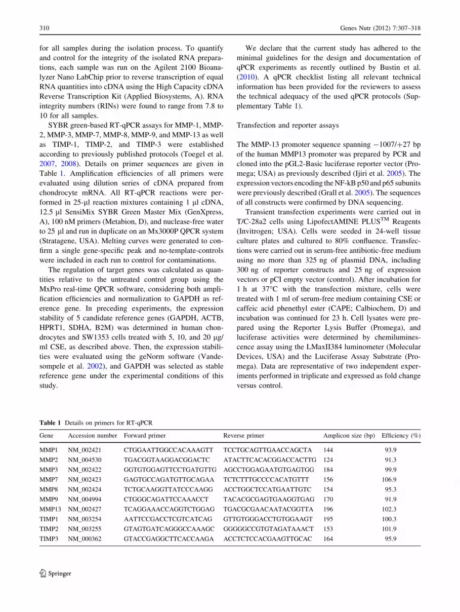
SYBR green-based RT-qPCR assays for MMP-1, MMP-
2, MMP-3, MMP-7, MMP-8, MMP-9, and MMP-13 as well
as TIMP-1, TIMP-2, and TIMP-3 were established
according to previously published protocols (Toegel et al.
2007, 2008). Details on primer sequences are given in
Table 1. Amplification efficiencies of all primers were
evaluated using dilution series of cDNA prepared from
chondrocyte mRNA. All RT-qPCR reactions were per-
formed in 25-ll reaction mixtures containing 1 ll cDNA,
12.5 ll SensiMix SYBR Green Master Mix (GenXpress,
A), 100 nM primers (Metabion, D), and nuclease-free water
to 25 ll and run in duplicate on an Mx3000P QPCR system
(Stratagene, USA). Melting curves were generated to con-
firm a single gene-specific peak and no-template-controls
were included in each run to control for contaminations.
The regulation of target genes was calculated as quan-
tities relative to the untreated control group using the
MxPro real-time QPCR software, considering both ampli-
fication efficiencies and normalization to GAPDH as ref-
erence gene. In preceding experiments, the expression
stability of 5 candidate reference genes (GAPDH, ACTB,
HPRT1, SDHA, B2M) was determined in human chon-
drocytes and SW1353 cells treated with 5, 10, and 20 lg/
ml CSE, as described above. Then, the expression stabili-
ties were evaluated using the geNorm software (Vande-
sompele et al. 2002), and GAPDH was selected as stable
reference gene under the experimental conditions of this
study.
We declare that the current study has adhered to the
minimal guidelines for the design and documentation of
qPCR experiments as recently outlined by Bustin et al.
(2010). A qPCR checklist listing all relevant technical
information has been provided for the reviewers to assess
the technical adequacy of the used qPCR protocols (Sup-
plementary Table 1).
Transfection and reporter assays
The MMP-13 promoter sequence spanning -1007/?27 bp
of the human MMP13 promoter was prepared by PCR and
cloned into the pGL2-Basic luciferase reporter vector (Pro-
mega; USA) as previously described (Ijiri et al. 2005). The
expression vectors encoding the NF-kB p50 and p65 subunits
were previously described (Grall et al. 2005). The sequences
of all constructs were confirmed by DNA sequencing.
Transient transfection experiments were carried out in
T/C-28a2 cells using LipofectAMINE PLUSTM Reagents
(Invitrogen; USA). Cells were seeded in 24-well tissue
culture plates and cultured to 80% confluence. Transfec-
tions were carried out in serum-free antibiotic-free medium
using no more than 325 ng of plasmid DNA, including
300 ng of reporter constructs and 25 ng of expression
vectors or pCI empty vector (control). After incubation for
1 h at 37�C with the transfection mixture, cells were
treated with 1 ml of serum-free medium containing CSE or
caffeic acid phenethyl ester (CAPE; Calbiochem, D) and
incubation was continued for 23 h. Cell lysates were pre-
pared using the Reporter Lysis Buffer (Promega), and
luciferase activities were determined by chemilumines-
cence assay using the LMaxII384 luminometer (Molecular
Devices, USA) and the Luciferase Assay Substrate (Pro-
mega). Data are representative of two independent exper-
iments performed in triplicate and expressed as fold change
versus control.
Table 1 Details on primers for RT-qPCR
Gene Accession number Forward primer Reverse primer Amplicon size (bp) Efficiency (%)
MMP1 NM_002421 CTGGAATTGGCCACAAAGTT TCCTGCAGTTGAACCAGCTA 144 93.9
MMP2 NM_004530 TGACGGTAAGGACGGACTC ATACTTCACACGGACCACTTG 124 91.3
MMP3 NM_002422 GGTGTGGAGTTCCTGATGTTG AGCCTGGAGAATGTGAGTGG 184 99.9
MMP7 NM_002423 GAGTGCCAGATGTTGCAGAA TCTCTTTGCCCCACATGTTT 156 106.9
MMP8 NM_002424 TCTGCAAGGTTATCCCAAGG ACCTGGCTCCATGAATTGTC 154 95.3
MMP9 NM_004994 CTGGGCAGATTCCAAACCT TACACGCGAGTGAAGGTGAG 170 91.9
MMP13 NM_002427 TCAGGAAACCAGGTCTGGAG TGACGCGAACAATACGGTTA 196 102.3
TIMP1 NM_003254 AATTCCGACCTCGTCATCAG GTTGTGGGACCTGTGGAAGT 195 100.3
TIMP2 NM_003255 GTAGTGATCAGGGCCAAAGC GGGGGCCGTGTAGATAAACT 153 101.9
TIMP3 NM_000362 GTACCGAGGCTTCACCAAGA ACCTCTCCACGAAGTTGCAC 164 95.9
310 Genes Nutr (2012) 7:307–318
123

Statistics
Data were exported to the GraphPad Prism statistics software
package (GraphPad Prism Software, USA). The Gaussian
distribution of the data was verified using the Kolmogorov–
Smirnov test. Statistics were performed using one-way
analysis of variance (ANOVA) with post hoc Tukey tests,
cross-comparing all study groups (95% confidence interval).
P-values \0.05 were considered significant.
Results
Cytotoxicity
Aiming to test the potential use of CSE as MMP inhibitor
in chondrocytes, we first evaluated the cytotoxic effects of
the extract on primary human chondrocytes using an MTT-
based viability assay. As shown in Fig. 1, CSE concen-
trations up to 20 lg/ml did not induce cytotoxicity in
chondrocytes, whereas 40 lg/ml CSE significantly reduced
viability (47.0 ± 2.2%). In the subsequent assays, there-
fore, we used the concentrations of CSE below 20 lg/ml.
CSE does not directly modify gelatinolytic activities
Previous work has suggested that MMP-2 is involved in
both the physiological collagen turnover in articular carti-
lage and the matrix degradation in OA cartilage (Duerr
et al. 2004). Using gelatin zymography, the present study
confirms that chondrocytes isolated from OA cartilage, as
well as SW1353 cells (data not shown), express functional
pro-MMP-2 (Fig. 2a). In contrast, bands corresponding to
MMP-9 activity were hardly detectable (data not shown),
reflecting the differences in MMP-2 and MMP-9 mRNA
levels (Table 2). The images also indicate that the activity
of MMP2 was not affected in primary chondrocytes treated
with 5 lg/ml CSE (Fig. 2a). Similarly, when added to the
buffer during zymography development, CSE did not
substantially affect the enzymatic activity of recombinant
MMP-2, whereas the addition of EDTA completely abro-
gated the gelatinolytic activity (Fig. 2b).
CSE inhibits IL1b -stimulated production of MMP-13
In order to investigate the effect of CSE on MMP-13 and
MMP-2 protein levels produced by human chondrocytes,
supernatants of treated cell cultures were collected and
subjected to ELISA. We observed that neither IL1b nor
CSE significantly modified the expression of MMP-2 in
primary or SW1353 cells (data not shown). In contrast,
MMP-13 levels were significantly up-regulated in both
SW1353 and primary cells after treatment with IL1b(Fig. 3). Pretreatment of SW1353 cells with 5 lg/ml CSE
significantly reduced IL1b-stimulated MMP-13 production
from 6.7 ± 1.6 to 2.5 ± 1.4 pg/ml, which was comparable
to the levels found in untreated cells (1.2 ± 0.5 pg/ml;
P [ 0.05). In primary chondrocytes, CSE significantly
reduced IL1b-stimulated MMP-13 levels from 16.3 ± 1.4
to 13.1 ± 2.7 pg/ml (P \ 0.05; n = 3 donors).
0 0.625 1.25 2.5 5 10 20 400
50
100
150
*
CSE (µg/ml)
viab
ility
(%
)
Fig. 1 Cell viability of primary chondrocytes treated with CSE.
Serum-starved primary chondrocytes were treated for 72 h with
varying concentrations of CSE (up to 40 lg/ml). Cell-viability was
evaluated using a MTT-based proliferation assay. The experiment
was repeated twice with cells from different donors, and each
experimental condition was set up in quadruplicate. Mean values and
standard deviations are given with respect to untreated cells (100%
viability). *indicates significant differences with respect to untreated
controls (P \ 0.05)
recomb.pro MMP-2
PC1 PC2
CSE (5 µg/ml) +— — — +
(A)
(B)recomb.
pro MMP-2
CSE
EDTA
10 µg/ml 5 µg/ml ——
— — — +
Fig. 2 CSE does not directly interfere with the gelatinolytic activity
of MMP-2. a Cell culture supernatants of primary chondrocytes
isolated from 2 patients (PC1 and PC2) were treated and subjected to
gelatine zymography as described in Materials and Methods. Lane 1shows the activity of a recombinant human pro-MMP-2. Lanes 2 and
4 show the supernatants of untreated chondrocytes, whereas lanes 3and 5 present the supernatants of the chondrocytes treated with 5 lg/
ml CSE, respectively. b Recombinant human pro-MMP-2 was
subjected to gelatine zymography and developed separately in
different developing buffers containing 5 lg/ml CSE, 10 lg/ml
CSE or 1 mM EDTA
Genes Nutr (2012) 7:307–318 311
123

CSE inhibits gene expression of MMPs,
but not of TIMPs
To evaluate the effects of CSE on MMP and TIMP
expression in human chondrocytes, mRNA levels of 7
MMPs (MMP-1, MMP-2, MMP-3, MMP-7, MMP-8,
MMP-9, MMP-13) and 3 TIMPs (TIMP-1, TIMP-2, TIMP-
3) were quantified using RT-qPCR (Table 2). In agreement
with previous reports (Fan et al. 2005), MMP-3 was the
most abundant (12.1 ± 12.2 molecules/molecules GAP-
DH) of the MMPs analyzed in human OA primary chon-
drocytes, with MMP-7 and MMP-8 showing the lower
expression levels. The expression levels of TIMP-1, TIMP-
2, and TIMP-3 were relatively higher compared with most
MMPs, with lower expression of TIMP-3 (Table 2).
Interestingly, the SW1353 cell line showed a different
MMP and TIMP expression profile as compared to primary
cells (Table 2), with MMP-2 being the most abundant (0.22
molecules/molecule GAPDH) and very low levels of
MMP-1, MMP-3, and MMP-13 mRNA.
IL1b up-regulated the expression of a number of MMPs in
SW1353 cells, including MMP-1 (Fig. 4a, 64.0 ± 3.1 fold),
MMP-3 (Fig. 4c, 27.4 ± 0.8 fold), MMP-7 (Fig. 4d, 3.2 ±
0.1 fold), MMP-8 (Fig. 4e, 6.3 ± 1.1 fold), MMP-9 (Fig. 4f,
1.8 ± 0.1 fold), and MMP-13 (Fig. 4g, 71.4 ± 1.1 fold).
Preincubation of SW1353 cells with CSE suppressed the
IL1b-induced mRNA levels of these MMPs in a dose-
dependent manner. Of note, 5 lg/ml CSE strongly inhibited
the expression of MMP-1 (Fig. 4a), MMP-3 (Fig. 4c), and
MMP-13 (Fig. 4g) mRNA as compared to IL1b-treated cells
(P \ 0.05), and 10 lg/ml CSE further down-regulated the
expression of these genes to levels comparable to control
cells (P [ 0.05). In addition, 5 lg/ml of CSE reduced the
IL-1b-induced MMP-7 (Fig. 4d), MMP-8 (Fig. 4e), and
MMP-9 (Fig. 4f) expression to basal levels. Regarding
MMP-2 gene expression, no alterations were observed in
SW1353 cells under the experimental conditions of this study
(Fig. 4b).
In keeping with those observations, the results obtained
with human OA primary chondrocytes (n = 3; PC1,2,3)
further confirmed those obtained in SW1353 cells, as shown
in Table 3. Namely, despite minor inter-individual differ-
ences, IL1b significantly increased mRNA levels of MMP-
1, MMP-3, MMP-7, MMP-9, and MMP-13 in all 3 cell
populations. At 5 lg/ml, CSE significantly suppressed the
IL1b-mediated upregulation of MMP-3, MMP-7, MMP-9,
and MMP-13 in all cell populations and that of MMP-1 in 2
out of 3 donors. Similar to the results in SW1353 cells,
Table 2 mRNA expression levels of MMPs and TIMPs in SW1353 cells and primary chondrocytes
MMP1 MMP2 MMP3 MMP7 MMP8 MMP9 MMP13 TIMP1 TIMP2 TIMP3
SW1353 0.001 0.22 0.004 0.0003 0.000004 0.0001 0.00003 0.06 0.13 0.01
PC1 0.21 0.01 26.0 0.0000003 0.00003 0.000837 0.031 0.51 0.16 0.05
PC2 0.29 0.09 3.4 0.0000367 0.00006 0.000015 0.023 0.67 2.38 0.36
PC3 0.25 0.01 7.0 0.0000437 0.00008 0.000005 0.018 0.73 0.04 0.01
Mean PC (n=3)
0.25 0.04 12.1 0.00003 0.00006 0.0003 0.024 0.64 0.86 0.14
SD 0.04 0.05 12.2 0.00002 0.00002 0.0005 0.007 0.11 1.32 0.19
mRNA levels were quantified using RT-qPCR and expressed as number of molecules per molecules of GAPDH. Articular chondrocytes were
isolated from cartilage specimens of 3 donors (PC1, PC2, PC3) and cultured and analyzed separately. Mean values and standard deviations (SD)
of the 3 primary cell populations are also presented
Fig. 3 CSE inhibits IL1b-stimulated MMP-13 production in human
chondrocytes. Primary chondrocytes (PC) were isolated from 3
donors and cultured and analyzed separately. Serum-starved SW1353
cells and PC were pretreated with 5 lg/ml CSE followed by the
coincubation with 10 ng/ml IL1b for 48 h. MMP-13 concentration
was determined in the cell culture supernatants using ELISA. Mean
values and standard deviations from 2 independent experiments
(SW1353) and from 3 donors (PC) are given. *indicates significant
differences with respect to cells treated with IL1b only (P \ 0.05).#indicates significant differences with respect to untreated controls
(P \ 0.05)
312 Genes Nutr (2012) 7:307–318
123

10 and 20 lg/ml CSE reduced the IL-1b-induced MMP
mRNA expression to basal levels. Expression of MMP-2
and MMP-8 were only slightly altered in the presence of
IL1b and CSE (P \ 0.05 in 1 out of 3 donors).
Since the balance between MMP and TIMP activities
determines cartilage degradation in OA [Nagase and Wo-
essner 1999), we examined the effect of 5 lg/ml CSE on the
gene expression of TIMP-1, TIMP-2, and TIMP-3 in IL1b-
treated chondrocytes (Fig. 5). In primary cells, neither IL1bnor 5 lg/ml CSE regulated the mRNA levels of any of the
TIMPs (Fig. 5a, b, c). In SW1353 cells, however, TIMP-1
and TIMP-3 mRNA levels were up-regulated by IL1b
2.1 ± 0.3 fold and 3.0 ± 0.1 fold, respectively. Treatment
with 5 lg/ml CSE reduced only TIMP-3 mRNA levels
significantly in SW1353 cells (Fig. 5d).
These results suggest that CSE is a potent inhibitor of
IL1b-mediated MMP transcription in human chondrocytes,
whereas it appears not to interfere with TIMP gene
expression.
CSE inhibits MMP-13 promoter activation
It has been shown that IL1b-induced MMP gene expression
is mediated by diverse signaling pathways and transcription
Fig. 4 CSE inhibits IL1b-
mediated MMP transcription in
SW1353 cells. Serum-starved
SW1353 cells were pretreated
with 5, 10, or 20 lg/ml of CSE
followed by the addition of
10 ng/ml IL1b. After 6 h of
incubation, total RNA was
isolated and mRNA levels of
a MMP-1, b MMP-2, c MMP-3,
d MMP-7, e MMP-8, f MMP-9,
and g MMP-13 were quantified
using RT-qPCR and expressed
as fold change with respect to
untreated controls. Mean values
and standard deviations are
given. *indicates significant
differences with respect to cells
treated with IL1b only
(P \ 0.05). #indicates
significant differences with
respect to untreated controls
(P \ 0.05)
Genes Nutr (2012) 7:307–318 313
123

factors, including NF-kB (Vincenti and Brinckerhoff
2002; Marcu et al. 2010). To further explore the mechanism
underlying suppression of MMP transcription by CSE,
human T/C-28a2 chondrocytes were co-transfected with a
MMP-13 reporter construct and p65/p50 expression vec-
tors in the presence or absence of CSE. As shown in
Fig. 6, p65/p50 overexpression transactivated the
pGL2B(-1007/?27)MMP-13 construct by 7.1 ± 1.5 fold,
and CSE reversed the p65/p50-driven MMP-13 promoter
transactivation in a concentration-dependent manner.
Whereas 5 lg/ml CSE significantly inhibited MMP-13
promoter activity by about 65%, 10 lg/ml CSE com-
pletely blocked the p65/p50-mediated promoter activation,
re-establishing levels comparable to those in the untreated
control (P [ 0.05).
For comparison, additional experiments using CAPE
were performed (Fig. 6). CAPE dose-dependently reduced
the p65/p50-dependent promoter activation at a concen-
tration of 0.5 lg/ml (P \ 0.05) and re-established control
levels at 2.5 lg/ml.
Discussion
The present study shows for the first time that CSE inhibits
MMP expression in human chondrocytes in vitro. As our
data demonstrate, pretreatment of primary chondrocytes
with CSE effectively suppressed the IL1b-mediated
upregulation of MMP-1, MMP-3, MMP-7, MMP-9, and
MMP-13 at the level of gene expression involving NFkB
signaling.
Since exacerbated production and activation of MMPs is
associated with rheumatoid arthritis and OA, MMPs rep-
resent promising pharmacological targets for the treatment
of arthritic diseases (Jacobsen et al. 2010). Strategies for
MMP inhibition include direct MMP inhibition (by TIMPs,
small molecule MMP inhibitors, or antibodies) and indirect
MMP inhibition targeting signaling pathways that lead to
MMP expression. Although considerable progress has been
made in the development of direct MMP inhibitors, severe
off-target effects, poor bioavailability and efficacy are
frequently associated with their use in clinical trials
Table 3 Regulation of MMP transcription by IL1b and CSE in primary chondrocytes
Ctrl IL-1b
?CSE 5 lg/ml ?CSE 10 lg/ml ?CSE 20 lg/ml
MMP-1 PC1 1.00 ± 0.10 5.76 ± 0.26* 1.83 ± 0.03** 0.91 ± 0.07** 0.78 ± 0.06**
PC2 1.00 ± 0.18 15.4 ± 0.01* 10.12 ± 0.45** 2.21 ± 0.22** 0.91 ± 0.10**
PC3 1.00 ± 0.15 4.24 ± 0.28* 3.63 ± 0.18 3.10 ± 0.06** 1.27 ± 0.09**
MMP-2 PC1 1.00 ± 0.06 1.46 ± 0.06* 1.05 ± 0.09** 0.97 ± 0.05** 0.83 ± 0.02**
PC2 1.00 ± 0.02 1.17 ± 0.05 1.08 ± 0.15 1.06 ± 0.13 0.82 ± 0.02
PC3 1.00 ± 0.09 1.24 ± 0.03 1.23 ± 0.04 1.55 ± 0.08** 1.06 ± 0.08
MMP-3 PC1 1.00 ± 0.01 3.16 ± 0.01* 1.35 ± 0.02** 1.08 ± 0.01** 0.84 ± 0.01**
PC2 1.00 ± 0.01 8.37 ± 0.25* 3.71 ± 0.09** 1.12 ± 0.01** 1.05 ± 0.04**
PC3 1.00 ± 0.01 4.74 ± 0.02* 3.12 ± 0.02** 3.48 ± 0.02** 1.15 ± 0.01**
MMP-7 PC1 1.00 ± 0.46 10.40 ± 1.06* 5.56 ± 0.92** 1.97 ± 1.60** 3.41 ± 0.02**
PC2 1.00 ± 0.47 8.19 ± 0.26* 2.41 ± 0.27** 3.15 ± 1.33** 2.14 ± 0.82**
PC3 1.00 ± 0.19 4.45 ± 0.17* 3.02 ± 0.12** 2.78 ± 0.06** 3.04 ± 0.11**
MMP-8 PC1 1.00 ± 0.57 1.29 ± 0.02 1.92 ± 0.22 0.65 ± 0.35 1.32 ± 0.58
PC2 1.00 ± 0.06 2.11 ± 0.33* 1.69 ± 0.35 1.02 ± 0.02** 0.57 ± 0.11**
PC3 1.00 ± 0.02 1.44 ± 0.13 1.14 ± 0.03 2.04 ± 0.26 0.89 ± 0.17
MMP-9 PC1 1.00 ± 0.01 1.89 ± 0.28* 1.11 ± 0.04** 0.85 ± 0.16** 1.03 ± 0.10**
PC2 1.00 ± 0.04 10.29 ± 1.28* 2.64 ± 0.36** 0.66 ± 0.16** 0.54 ± 0.06**
PC3 1.00 ± 0.37 7.65 ± 1.48* 3.76 ± 1.15** 1.22 ± 0.14** 2.15 ± 0.18**
MMP-13 PC1 1.00 ± 0.17 13.22 ± 0.07* 1.39 ± 0.03** 0.72 ± 0.04** 0.64 ± 0.05**
PC2 1.00 ± 0.01 10.42 ± 0.37* 2.71 ± 0.23** 1.11 ± 0.03** 0.85 ± 0.01**
PC3 1.00 ± 0.02 4.65 ± 0.40* 2.80 ± 0.24** 4.42 ± 0.54 1.20 ± 0.01**
Primary chondrocytes were isolated from 3 donors (PC1, PC2, PC3) and cultured and analyzed separately. Serum-starved cells were pretreated
with 5 to 20 lg/ml CSE followed by stimulation with 10 ng/ml IL1b for 6 h. mRNA levels of MMP-1, MMP-2, MMP-3, MMP-7, MMP-8,
MMP-9, MMP-13 were quantified using RT-qPCR and expressed as fold change with respect to untreated controls. Mean values and standard
deviations are given. * indicates significant differences with respect to untreated controls (P \ 0.05). ** indicates significant differences with
respect to cells treated with IL1b only (P \ 0.05)
314 Genes Nutr (2012) 7:307–318
123

(Clutterbuck et al. 2009). Thus, indirect MMP inhibition
using nutraceuticals or plant-derived phytopharmaceuticals
might provide a promising alternative to present thera-
peutic approaches. In this context, curcumin, resveratrol, or
green tea catechins have already been investigated
regarding their MMP-inhibitory actions (Shakibaei et al.
2007; Lee and Moon 2005; Annabi et al. 2007).
The results presented here suggest that Caesalpinia
sappan-derived phytochemicals might be developed as
nutraceuticals for the management of arthritic diseases. In
this context, the strong inhibitory action of CSE on the
IL1b-mediated overexpression of MMP-1, MMP-3, and
MMP-13 appears of particular interest. Exacerbated pro-
duction and activation of the collagenases, MMP-1 and
PC 1 PC 2 PC 30.0
0.5
1.0
1.5
2.0 (A)
*
#
Fo
ld c
han
ge
#
PC 1 PC 2 PC 30.0
0.5
1.0
1.5
2.0 (B)
Fo
ld c
han
ge
PC 1 PC 2 PC 30.0
0.5
1.0
1.5
2.0 (C)
Fo
ld c
han
ge
TIMP1 TIMP2 TIMP30
1
2
3
4 (D)
*
#
Fo
ld c
han
ge
# #
#
Fig. 5 Regulation of TIMP transcription by IL1b and CSE in
SW1353 cells and primary chondrocytes. a–c Primary chondrocytes
were isolated from 3 donors (PC1, PC2, PC3) and cultured and
analyzed separately. Serum-starved cells were pretreated with 5 lg/
ml CSE followed by the addition of 10 ng/ml IL1b. After 6 h of
incubation, the mRNA levels of TIMP-1 (a), TIMP-2 (b), and TIMP-3
(c) were quantified using RT-qPCR and expressed as fold change with
respect to untreated controls (white bars). Gray bars represent IL1b-
treated cells, whereas black bars represent cells pretreated with CSE.
d TIMP-1, TIMP-2, and TIMP-3 mRNA levels were quantified in
SW1353 cells under exposure to IL1b and CSE as described. Mean
values and standard deviations are given. *indicates significant
differences with respect to cells treated with IL1b only (P \ 0.05).#indicates significant differences with respect to untreated controls
(P \ 0.05)
Fig. 6 Effect of CSE or CAPE on MMP-13 promoter activity.
T/C28a2 chondrocytes, cotransfected with a MMP-13 luciferase
reporter construct and p50 and p65 expression vectors, were treated
with CAPE (gray bars) or CSE (black bars) in a dose-dependent
manner. Both CAPE and CES inhibited the NF-kB(p65/p50)-driven
MMP-13 promoter activation. #indicates significant differences with
respect to cells transfected with the MMP-13 reporter construct and
the pCI empty vector (P \ 0.05). *indicates significant differences
with respect to cells co-transfected with the MMP-13 promoter and
the p65/p50 expression vectors (P \ 0.05)
Genes Nutr (2012) 7:307–318 315
123

MMP-13, in articular chondrocytes are known to promote
the cleavage of collagen triple helices allowing further
degradation by other MMPs (Nagase and Woessner 1999).
Supporting the role of MMP-13 in connective tissue deg-
radation, Neuhold et al. (2001) reported that transgenic
mice overexpressing MMP-13 show signs of articular
cartilage degradation and exhibit joint pathology similar to
that found in OA. Our results confirm that IL1b represents
a potent inflammatory stimulus that leads to overexpression
of MMP-1, MMP-3, and MMP-13 mRNA in primary and
SW1353 chondrocytes. Interestingly, the upregulation of
these genes by IL1b was more pronounced in SW1353
cells than in primary OA chondrocytes; however, when
cultured in the presence of 5–20 lg/ml CSE, the effect of
IL1b was suppressed in both cell models and the expres-
sion of MMP-1, MMP-3, and MMP-13 mRNA was down-
regulated to levels found under control conditions. The
relevance of the RT-qPCR data was further verified by
ELISA, demonstrating reduced MMP-13 protein levels in
chondrocytes treated with CSE. In contrast, however, the
transcription of TIMPs was not affected by the presence of
CSE. Of note, CSE treatment did not totally block the
expression of MMPs but rather inhibited IL1b-induced
MMP overexpression. In this regard, CSE might be
advantageous over other MMP inhibitors, since total MMP
inhibition not only impairs physiological ECM remodeling,
but may also affect cellular signaling to which MMPs
contribute (Clutterbuck et al. 2009). In this context, it is
noteworthy that CSE did not directly influence the gela-
tinolytic activity of MMP-2 under the experimental con-
ditions of this study.
In a first attempt to elaborate the mechanism underly-
ing the MMP-inhibitory action of CSE, we focused on the
role of NF-kB in MMP-13 promoter activation. It is well
established that IL1b, once bound to its type 1 receptor,
activates NF-kB dimers by triggering phosphorylation and
subsequent degradation of the inhibitory kB proteins
(Daheshia and Yao 2008). The most prevalent activated
NF-kB dimer is the heterodimer RelA (p65)/p50, which
can bind to sites in NF-kB-dependent promoters regulat-
ing the transcription of response genes (Roman-Blas and
Jimenez 2006). Mengshol et al. have assessed the role of
NF-kB in MMP-13 transcription in chondrocytes and
found that, besides JNK and p38, NF-kB signaling is
required for the IL1b-mediated induction of MMP-13
(Mengshol et al. 2000). Although the promoter of MMP-
13 does not contain consensus NF-kB binding sites
(Burrage et al. 2006), the link between NF-kB activation
and MMP-13 induction has been further demonstrated in
a previous study on the effect of hyaluronan oligosac-
charides in articular chondrocytes (Ohno et al. 2006). We
show here that, indeed, NF-kB p65/p50 overexpression
leads to MMP-13 promoter transactivation and that CSE
suppresses the p65/p50-driven MMP-13 promoter activa-
tion, thereby suggesting that CSE’s MMP-inhibitory
actions are related to its blockade of the NF-kB signaling.
Furthermore, the comparison to CAPE, a chemically
defined specific inhibitor of NF-kB (Natarajan et al.
1996), revealed that CSE was only about 4 times less
active in our assay. Our results go along with previous
reports demonstrating that CSE and its constituents
interfere with NF-kB signaling in different cell types.
Namely, Bae et al. (2005) showed that brazilin, the main
component of CSE, inhibited the DNA binding activity of
NF-kB and AP-1 in LPS-stimulated mouse macrophages.
In addition, protosappanin A was shown to target the
NF-kB pathway in rat heart tissue (Wu et al. 2010).
Therefore, our results suggest that CSE contains one or
more active components that may act as potent inhibitors
of NF-kB in human chondrocytes.
To date, the bioavailability of specific components of
CSE has not been elaborated and it remains elusive whe-
ther the doses of CSE used in this study are representative
for physiologically active concentrations. Despite this lack
of specific knowledge, the efficacy of CSE in diverse in
vivo models supports the apparent bioavailability of CSE
components at therapeutically effective doses (Sun et al.
2011; Oh et al. 2001). Moreover, the present study dem-
onstrates that CSE affects human chondrocytes in vitro at
concentrations that are comparable to those of well-known
dietary antioxidants including ascorbic acid, curcumin, or
resveratrol (Csaki et al. 2009; Graeser et al. 2009). Similar
to curcumin and resveratrol, CSE appears to exert its
effects on chondrocytes via mechanisms targeting NFkB
signaling. Interestingly, recent data have explored the dif-
ferent molecular targets of curcumin and resveratrol as well
as their synergistic chondroprotective effects (Csaki et al.
2009). In this context, it might be of interest to further
investigate the synergism between CSE components and
other dietary antioxidants regarding the modulation of
biomarkers of inflammation in cultured chondrocytes.
Conclusion
In summary, this study suggests that CSE or CSE-derived
compounds may be beneficial as indirect MMP inhibitors
for the future management of degenerative or inflammatory
joint diseases. We present evidence that CSE effectively
suppresses IL1b-mediated overexpression of major MMPs
in human chondrocytes in vitro. More specifically, CSE
inhibits the de novo synthesis of MMP-1, MMP-3, MMP-7,
MMP-9, and MMP-13 at the gene expression level without
affecting the expression of TIMPs. Further, we showed that
the molecular mechanism underlying MMP-13 inhibition
by CSE involves, at least in part, the interference with the
316 Genes Nutr (2012) 7:307–318
123

NF-kB (p65/p50)-driven MMP-13 promoter transactiva-
tion. Future studies should therefore aim to define CSE’s
actions in the context of arthritic diseases and to further
delineate the molecular mechanisms evoked by CSE and its
specific constituents both in vitro and in vivo.
Acknowledgments This research was supported partially by
National Institutes of Health (NIH) grant R01-AG022021 (Mary B
Goldring), an Arthritis Foundation Postdoctoral Fellowship (Miguel
Otero), and the graduate program ‘‘Molecular Drug Targets’’ spon-
sored by the University of Vienna (Shengqian Q Wu).
Conflict of interest The authors declare that they have no conflict
of interest.
References
Albini A, Morini M, D’Agostini F, Ferrari N, Campelli F, Arena G,
Noonan DM, Pesce C, De Flora S (2001) Inhibition of
angiogenesis-driven Kaposi’s sarcoma tumor growth in nude
mice by oral N-acetylcysteine. Cancer Res 61:8171–8178
Ameye LG, Chee WSS (2006) Osteoarthritis and nutrition. From
nutraceuticals to functional foods: a systematic review of the
scientific evidence. Arthr Res Ther 8:R127
Annabi B, Currie JC, Moghrabi A, Beliveau R (2007) Inhibition of
HuR and MMP-9 expression in macrophage-differentiated HL-
60 myeloid leukemia cells by green tea polyphenol EGCg. Leuk
Res 31:1277–1284
Bae IK, Min HY, Han AR, Seo EK, Lee SK (2005) Suppression of
lipopolysaccharide-induced expression of inducible nitric oxide
synthase by brazilin in RAW 264.7 macrophage cells. Eur J
Pharmacol 513:237–242
Bjorkman DJ (1999) Current status of nonsteroidal anti-inflammatory
drug (NSAID) use in the United States: risk factors and
frequency of complications. Am J Med 107:3S–8S
Burrage PS, Mix KS, Brinckerhoff CE (2006) Matrix metallopro-
teinases: role in arthritis. Front Biosci 11:529–543
Bustin SA, Beaulieu JF, Huggett J, Jaggi R, Kibenge FSB, Olsvik PA,
Penning LC, Toegel S (2010) MIQE precis: practical implemen-
tation of minimum standard guidelines for fluorescence-based
quantitative real-time PCR experiments. BMC Mol Biol 11:74
Chen YP, Liu L, Zhou YH, Wen J, Jiang Y, Tu PF (2008) Chemical
constituents from Sappan Lignum. J Chin Pharm Sci 17:82–86
China pharmacopoeia commission (2010) The Chinese Pharmaco-
poeia. Medical science press, Beijing
Clutterbuck AL, Asplin KE, Harris P, Allaway D, Mobasheri A
(2009) Targeting matrix metalloproteinases in inflammatory
conditions. Curr Drug Targets 10:1245–1254
Csaki C, Mobasheri A, Shakibaei M (2009) Synergistic chondropro-
tective effects of curcumin and resveratrol in human articular
chondrocytes: inhibition of IL-1beta-induced NF-kappaB-med-
iated inflammation and apoptosis. Arthr Res Ther 11:R165
Daheshia M, Yao JQ (2008) The interleukin 1beta pathway in the
pathogenesis of osteoarthritis. J Rheumatol 35:2306–2312
Duerr S, Stremme S, Soeder S, Bau B, Aigner T (2004) MMP-2/
gelatinase A is a gene product of human adult articular
chondrocytes and is increased in osteoarthritic cartilage. Clin
Exp Rheumatol 22:603–608
Fan Z, Bau B, Yang H, Soeder S, Aigner T (2005) Freshly isolated
osteoarthritic chondrocytes are catabolically more active than
normal chondrocytes, but less responsive to catabolic stimulation
with interleukin-1b. Arthr Rheum 52:136–143
Goggs R, Vaughan-Thomas A, Clegg PD, Carter SD, Innes JF,
Mobasheri A, Shakibaei M, Schwab W, Bondy CA (2005)
Nutraceutical therapies for degenerative joint diseases: a critical
review. Crit Rev Food Sci Nutr 45:145–164
Goldring MB, Marcu KB (2009) Cartilage homeostasis in health and
rheumatic diseases. Arthr Res Ther 11:224
Goldring MB, Otero M, Tsuchimochi K, Ijiri K, Li Y (2008) Defining
the roles of inflammatory and anabolic cytokines in cartilage
metabolism. Ann Rheum Dis 67:75–82
Graeser AC, Giller K, Wiegand H, Barella L, Boesch Saadatmandi C,
Rimbach G (2009) Synergistic chondroprotective effect of alpha-
tocopherol, ascorbic acid, and selenium as well as glucosamine
and chondroitin on oxidant induced cell death and inhibition of
matrix metalloproteinase-3—studies in cultured chondrocytes.
Molecules 15:27–39
Grall FT, Prall WC, Wei W, Gu X, Cho JY, Choy BK, Zerbini LF,
Inan MS, Goldring SR, Gravallese EM, Goldring MB, Oettgen P,
Libermann TA (2005) The Ets transcription factor ESE-1
mediates induction of the COX-2 gene by LPS in monocytes.
FEBS J 272:1676–1687
Ijiri K, Zerbini LF, Peng H, Correa RG, Lu B, Walsh N, Zhao Y,
Taniguchi N, Huang XL, Otu H, Wang H, Wang JF, Komiya S,
Ducy P, Rahman MU, Flavell RA, Gravallese EM, Oettgen P,
Libermann TA, Goldring MB (2005) A novel role for GADD45-
beta as a mediator of MMP-13 gene expression during chondro-
cyte terminal differentiation. J Biol Chem 280:38544–38555
Jacobsen JA, Major Jourden JL, Miller MT, Cohen SM (2010) To
bind zinc or not to bind zinc: an examination of innovative
approaches to improved metalloproteinase inhibition. Biochim
Biophys Acta 1803:72–94
Jeong IY, Chang HJ, Park YD, Lee HJ, Choi DS, Byun MW, Kim YJ
(2008) Anti-inflammatory activity of an ethanol extract of
Caesalpinia sappan L. in LPS-induced RAW 264.7 cells. J Food
Sci Nutr 13:253–258
Kubota E, Imamura H, Kubota T, Shibata T, Murakami K (1997)
Interleukin 1 beta and stromelysin (MMP3) activity of synovial
fluid as possible markers of osteoarthritis in the temporoman-
dibular joint. J Oral Maxillofac Surg 55:20–27
Lee B, Moon SK (2005) Resveratrol inhibits TNF-alpha-induced
proliferation and matrix metalloproteinase expression in human
vascular smooth muscle cells. J Nutr 135:2767–2773
Little CB, Barai A, Burkhardt D, Smith SM, Fosang AJ, Werb Z,
Shah M, Thompson EW (2009) Matrix metalloproteinase
13-deficient mice are resistant to osteoarthritic cartilage erosion
but not chondrocyte hypertrophy or osteophyte development.
Arthr Rheum 60:3723–3733
Marcu KB, Otero M, Olivotto E, Borzi RM, Goldring MB (2010) NF-
kappaB signaling: multiple angles to target OA. Curr Drug
Targets 11:599–613
Mazzoni A, Mannello F, Tay FR, Tonti GA, Papa S, Mazzotti G, Di
Lenarda R, Pashley DH, Breschi L (2007) Zymographic analysis
and characterization of MMP-2 and -9 forms in human sound
dentin. J Dent Res 86:436–440
Mengshol JA, Vincenti MP, Coon CI, Barchowsky A, Brinckerhoff
CE (2000) Interleukin-1 induction of collagenase 3 (matrix
metalloproteinase 13) gene expression in chondrocytes requires
p38, c-Jun N-terminal kinase, and nuclear factor kappa B:
differential regulation of collagenase 1 and collagenase 3. Arthr
Rheum 43:801–811
Mix KS, Mengshol JA, Benbow U, Vincenti MP, Sporn MB,
Brinckerhoff CE (2001) A synthetic triterpenoid selectively
inhibits the induction of matrix metalloproteinases 1 and 13 by
inflammatory cytokines. Arthr Rheum 44:1096–1104
Genes Nutr (2012) 7:307–318 317
123

Nagase H, Woessner JF (1999) Matrix metalloproteinases. J Biol
Chem 274:21491–21494
Natarajan K, Singh S, Burke TR Jr, Grunberger D, Aggarwal BB
(1996) Caffeic acid phenethyl ester is a potent and specific
inhibitor of activation of nuclear transcription factor NF-kappa
B. Proc Natl Acad Sci USA 93:9090–9095
Neuhold LA, Killar L, Zhao W, Sung ML, Warner L, Kulik J, Turner
J, Wu W, Billinghurst C, Meijers T, Poole AR, Babij P,
DeGennaro LJ (2001) Postnatal expression in hyaline cartilage
of constitutively active human collagenase-3 (MMP-13) induces
osteoarthritis in mice. J Clin Invest 107:35–44
Oh GT, Choi JH, Hong JJ, Kim DY, Lee SB, Kim JR, Lee CH, Hyun
BH, Oh SR, Bok SH, Jeong TS (2001) Dietary hematein
ameliorates fatty streak lesions in the rabbit by the possible
mechanism of reducing VCAM-1 and MCP-1 expression.
Atherosclerosis 159:17–26
Ohno S, Im HJ, Knudson CB, Knudson W (2006) Hyaluronan
oligosaccharides induce matrix metalloproteinase 13 via tran-
scriptional activation of NFkappaB and p38 MAP kinase in
articular chondrocytes. J Biol Chem 281:17952–17960
Roman-Blas JA, Jimenez SA (2006) NF-kB as a potential therapeutic
target in osteoarthritis and rheumatoid arthritis. Osteoarthr Cartil
14:839–848
Saha N, Moldovan F, Tardif G, Pelletier JP, Cloutier JM, Martel-
Pelletier J (1999) Interleukin-1-b-converting enzyme/caspase-1
in human osteoarthritic tissues: localization and role in the
maturation of interleukin-1b and interleukin-18. Arthr Rheum
42:1577–1587
Shakibaei M, John T, Schulze-Tanzil G, Lehmann I, Mobasheri A
(2007) Suppression of NF-kappaB activation by curcumin leads
to inhibition of expression of cyclo-oxygenase-2 and matrix
metalloproteinase-9 in human articular chondrocytes: implica-
tions for the treatment of osteoarthritis. Biochem Pharmacol
73:1434–1445
Sireeratawong S, Piyabhan P, Singhalak T, Wongkrajang Y,
Temsiririrkkul R, Punsrirat J, Ruangwises N, Saraya S,
Lerdvuthisopon N, Jaijoy K (2010) Toxicity evaluation of
sappan wood extract in rats. J Med Assoc Thai 93:S50–S57
Smith MD, Triantafillou S, Parker A, Youssef PP, Coleman M
(1997) Synovial membrane inflammation and cytokine produc-
tion in patients with early osteoarthritis. J Rheumatol 24:365–
371
Srilakshmi VS, Vijayan P, Raj PV, Dhanaraj SA, Chandrashekhar HR
(2010) Hepatoprotective properties of Caesalpinia sappan Linn.
heartwood on carbon tetrachloride induced toxicity. Indian J Exp
Biol 48:905–910
Sun SQ, Wang YZ, Zhou YB (2011) Extract of the dried heartwood of
Caesalpinia sappan L. attenuates collagen-induced arthritis.
J Ethnopharmacol Article, in press
Tetlow LC, Adlam DJ, Woolley DE (2001) Matrix metalloproteinase
and proinflammatory cytokine production by chondrocytes of
human osteoarthritic cartilage: associations with degenerative
changes. Arthr Rheum 44:585–594
The Society of Japanese Pharmacopoeia (2006) The Japanese
Pharmacopoeia, 15th edn. Hirokawa Publishing, Tokyo
Toegel S, Huang W, Piana C, Unger FM, Wirth M, Goldring MB,
Gabor F, Viernstein H (2007) Selection of reliable reference
genes for qPCR studies on chondroprotective action. BMC Mol
Biol 8:13
Toegel S, Wu SQ, Piana C, Unger FM, Wirth M, Goldring MB, Gabor F,
Viernstein H (2008) Comparison between chondroprotective
effects of glucosamine, curcumin, and diacerein in IL-1b stimu-
lated C-28/I2 chondrocytes. Osteoarthr Cartil 16:1205–1212
Toegel S, Plattner VE, Wu SQ, Chiari C, Gabor F, Unger FM,
Goldring MB, Nehrer S, Viernstein H, Wirth M (2009) Lectin
binding patterns reflect the phenotypic status of in vitro
chondrocyte models. In Vitro Cell Dev Biol Anim 45:351–360
Vandesompele J, De Preter K, Pattyn F, Poppe B, Van Roy N, De
Paepe A, Speleman F (2002) Accurate normalization of real-time
quantitative RT-PCR data by geometric averaging of multiple
internal control genes. Genome Biol 18:3
Vincenti MP, Brinckerhoff CE (2002) Transcriptional regulation of
collagenase (MMP-1, MMP-13) genes in arthritis: integration of
complex signaling pathways for the recruitment of gene-specific
transcription factors. Arthr Res 4:157–164
Washiyama M, Sasaki Y, Hosokawa T, Nagumo S (2009) Anti-
inflammatory constituents of Sappan Lignum. Biol Pharm Bull
32:941–944
Wu J, Zhang M, Jia H, Huang X, Zhang Q, Hou J, Bo Y (2010)
Protosappanin A induces immunosuppression of rats heart
transplantation targeting T cells in grafts via NF-kappaB
pathway. Naunyn Schmied Arch Pharmacol 381:83–92
Xie YW, Ming DS, Xu HX, Dong H, But PP (2000) Vasorelaxing
effects of Caesalpinia sappan involvement of endogenous nitric
oxide. Life Sci 67:1913–1918
Ye M, Xie WD, Lei F, Meng Z, Zhao YN, Su H, Du LJ (2006)
Brazilein, an important immunosuppressive component from
Caesalpinia sappan L. Int Immunopharmacol 6:426–432
318 Genes Nutr (2012) 7:307–318
123