Tissue inhibitor of metalloproteinases-1 promotes liver fibrosis development in a transgenic mouse...
7
Tissue Inhibitor of Metalloproteinases-1 Promotes Liver Fibrosis Development in a Transgenic Mouse Model HITOSHI YOSHIJI, 1 SHIGEKI KURIYAMA, 1 YOJI MIYAMOTO, 1 UNNUR P. THORGEIRSSON, 2 DANIEL E. GOMEZ, 4 MITSUHIRO KAWATA, 1 JUNICHI YOSHII, 1 YASUHIDE IKENAKA, 1 RYUICHI NOGUCHI, 1 HIROHISA TSUJINOUE, 1 TOSHIYA NAKATANI, 1 SNORRI S. THORGEIRSSON, 3 AND HIROSHI FUKUI 1 Tissue inhibitor of metalloproteinases-1 (TIMP-1) has been shown to be increased in liver fibrosis development both in murine experimental models and human samples. However, the direct role of TIMP-1 during liver fibrosis de- velopment has not been defined. To address this issue, we developed transgenic mice overexpressing human TIMP-1 (hTIMP-1) in the liver under control of the albumin pro- moter/ enhancer. A model of CCl 4 –induced hepatic fibrosis was used to assess the extent of fibrosis development in TIMP-1 transgenic (TIMP-Tg) mice and control hybrid (Cont) mice. Without any treatment, overexpression of TIMP-1 itself did not induce liver fibrosis. There were no significant differences of pro-(a1)-collagen-I, (a2)-colla- gen-IV, and a-smooth muscle actin (a-SMA) mRNA expres- sion in the liver between TIMP-Tg and Cont-mice, suggest- ing that overexpression of TIMP-1 itself did not cause he- patic stellate cell (HSC) activation. After 4-week treatment with CCl 4 , however, densitometric analysis revealed that TIMP-Tg-mice had a seven-fold increase in liver fibrosis compared with the Cont-mice. The hepatic hydroxyproline content and serum hyaluronic acid were also significantly increased in TIMP-Tg-mice, whereas CCl 4 –induced liver dysfunction was not altered. An active form of matrix met- alloproteinases-2 (MMP-2) level in the liver of TIMP-Tg- mice was decreased relative to that in Cont-mice because of the transgenic TIMP-1. Immunohistochemical analysis re- vealed that collagen-I and collagen-IV accumulation was markedly increased in the liver of CCl 4 –treated TIMP-Tg- mice with a pattern similar to that of a-SMA positive cells. These results suggest that TIMP-1 does not by itself result in liver fibrosis, but strongly promotes liver fibrosis develop- ment. (HEPATOLOGY 2000;32:1248-1254.) Extensive remodeling of the extracellular matrix (ECM) has been shown during liver fibrosis development. Gross re- modeling of the ECM in the fibrotic liver is likely to be regu- lated by synthesis and enzymatic degradation of the ECM. 1-5 Matrix degradation is catalyzed by the activity of matrix me- talloproteinases (MMPs), which consist of collagenases, gela- tinases, stromelysins, and membrane type (MT)-MMPs. The activities of MMPs are inhibited by tissue inhibitors of metal- loproteinases (TIMPs) by the formation of noncovalent 1:1 complexes with the MMPs. Four members of the TIMP family have been characterized so far, designated as TIMP-1, TIMP-2, TIMP-3, and TIMP-4. TIMP-1 and TIMP-2 are capa- ble of inhibiting the activities of all known MMPs and, as such, play a key role in maintaining the balance between ECM de- position and degradation in different physiological processes, including liver fibrosis development. In the liver, TIMP-1 and TIMP-2 were identified, and it has been suggested that TIMP-1 plays a more important role in pathological processes of the liver than TIMP-2. 6-8 During the development of liver fibrosis, TIMP-1 expression in the liver and serum TIMP-1 level were markedly up-regulated both in humans and murine fibrosis models. 9-19 The serum levels of TIMP-1 were in- creased in patients with chronic active liver disease, correlat- ing with the histological degree of human liver fibrosis. 11 The liver TIMP-1 protein levels were also closely correlated with the histological degree of liver fibrosis. 10,11 In experimental models, it has been reported that TIMP-1 mRNA expression was increased after bile duct ligation and it was also shown to rise in acute CCl 4 –induced liver injury and remain elevated as the liver become fibrotic. 12 Moreover, in a rat model of revers- ible liver fibrosis, matrix remodeling and resolution of liver fibrosis were closely associated with a marked decrease in TIMP-1 expression. 17 Accordingly, it has been suggested that TIMP-1 plays an important role in liver fibrosis development. To date, however, a direct role of TIMP-1 in liver fibrogenesis has not yet been clarified. Although the dynamic alternations of MMP/TIMP balance should play a pivotal role in liver fi- brosis development, the direct effect of the alternation in vivo has not yet been clarified. It is difficult to elucidate these interactions in vivo with the conventional experimental method. To clarify these issues, we generated transgenic mice overexpressing human TIMP-1 (hTIMP-1) in the liver under control of the mouse albumin promoter/enhancer. With these mice, we assessed the effect of a high concentration of TIMP-1 in the liver microenvironment on liver fibrogenesis. In the present study, we examined the direct effect of over- expression of TIMP-1 on liver fibrosis development in a CCl 4 – Abbreviations: a-SMA, a-smooth muscle actin; ECM, extracellular matrix; HSC, he- patic stellate cell; TIMP-1, tissue inhibitor of metalloproteinases-1; MMP, matrix metal- loproteinases. From the 1 Third Department of Internal Medicine, Nara Medical University, Nara, Japan; 2 Tumor Biology and Carcinogenesis Section, Laboratory of Cellular Carcinogen- esis and Promotion, 3 Laboratory of Experimental Carcinogenesis, Division of Basic Sci- ence, National Cancer Institute, National Institutes of Health, Bethesda, MD; 4 Labora- tory of Molecular Oncology, Department of Science and Technology, Quilmes National University, Buenos Aires, Argentina. Received June 21, 2000; accepted September 28, 2000. Address reprint requests to: Hitoshi Yoshiji, M.D., Ph.D., Third Department of Inter- nal Medicine, Nara Medical University, Shijo-cho, Kashihara, Nara 634-8522, Japan. E-mail: [email protected]; fax: (81) 744-24-7122. Copyright © 2000 by the American Association for the Study of Liver Diseases. 0270-9139/00/3206-0010$3.00/0 doi:10.1053/jhep.2000.20521 1248
-
Upload
independent -
Category
Documents
-
view
5 -
download
0
Transcript of Tissue inhibitor of metalloproteinases-1 promotes liver fibrosis development in a transgenic mouse...

Tissue Inhibitor of Metalloproteinases-1 Promotes Liver FibrosisDevelopment in a Transgenic Mouse Model
HITOSHI YOSHIJI,1 SHIGEKI KURIYAMA,1 YOJI MIYAMOTO,1 UNNUR P. THORGEIRSSON,2 DANIEL E. GOMEZ,4 MITSUHIRO KAWATA,1
JUNICHI YOSHII,1 YASUHIDE IKENAKA,1 RYUICHI NOGUCHI,1 HIROHISA TSUJINOUE,1 TOSHIYA NAKATANI,1
SNORRI S. THORGEIRSSON,3 AND HIROSHI FUKUI1
Tissue inhibitor of metalloproteinases-1 (TIMP-1) hasbeen shown to be increased in liver fibrosis developmentboth in murine experimental models and human samples.However, the direct role of TIMP-1 during liver fibrosis de-velopment has not been defined. To address this issue, wedeveloped transgenic mice overexpressing human TIMP-1(hTIMP-1) in the liver under control of the albumin pro-moter/ enhancer. A model of CCl4–induced hepatic fibrosiswas used to assess the extent of fibrosis development inTIMP-1 transgenic (TIMP-Tg) mice and control hybrid(Cont) mice. Without any treatment, overexpression ofTIMP-1 itself did not induce liver fibrosis. There were nosignificant differences of pro-(a1)-collagen-I, (a2)-colla-gen-IV, and a-smooth muscle actin (a-SMA) mRNA expres-sion in the liver between TIMP-Tg and Cont-mice, suggest-ing that overexpression of TIMP-1 itself did not cause he-patic stellate cell (HSC) activation. After 4-week treatmentwith CCl4, however, densitometric analysis revealed thatTIMP-Tg-mice had a seven-fold increase in liver fibrosiscompared with the Cont-mice. The hepatic hydroxyprolinecontent and serum hyaluronic acid were also significantlyincreased in TIMP-Tg-mice, whereas CCl4–induced liverdysfunction was not altered. An active form of matrix met-alloproteinases-2 (MMP-2) level in the liver of TIMP-Tg-mice was decreased relative to that in Cont-mice because ofthe transgenic TIMP-1. Immunohistochemical analysis re-vealed that collagen-I and collagen-IV accumulation wasmarkedly increased in the liver of CCl4–treated TIMP-Tg-mice with a pattern similar to that of a-SMA positive cells.These results suggest that TIMP-1 does not by itself result inliver fibrosis, but strongly promotes liver fibrosis develop-ment. (HEPATOLOGY 2000;32:1248-1254.)
Extensive remodeling of the extracellular matrix (ECM)has been shown during liver fibrosis development. Gross re-modeling of the ECM in the fibrotic liver is likely to be regu-lated by synthesis and enzymatic degradation of the ECM.1-5
Matrix degradation is catalyzed by the activity of matrix me-talloproteinases (MMPs), which consist of collagenases, gela-tinases, stromelysins, and membrane type (MT)-MMPs. Theactivities of MMPs are inhibited by tissue inhibitors of metal-loproteinases (TIMPs) by the formation of noncovalent 1:1complexes with the MMPs. Four members of the TIMP familyhave been characterized so far, designated as TIMP-1,TIMP-2, TIMP-3, and TIMP-4. TIMP-1 and TIMP-2 are capa-ble of inhibiting the activities of all known MMPs and, as such,play a key role in maintaining the balance between ECM de-position and degradation in different physiological processes,including liver fibrosis development. In the liver, TIMP-1 andTIMP-2 were identified, and it has been suggested thatTIMP-1 plays a more important role in pathological processesof the liver than TIMP-2.6-8 During the development of liverfibrosis, TIMP-1 expression in the liver and serum TIMP-1level were markedly up-regulated both in humans and murinefibrosis models.9-19 The serum levels of TIMP-1 were in-creased in patients with chronic active liver disease, correlat-ing with the histological degree of human liver fibrosis.11 Theliver TIMP-1 protein levels were also closely correlated withthe histological degree of liver fibrosis.10,11 In experimentalmodels, it has been reported that TIMP-1 mRNA expressionwas increased after bile duct ligation and it was also shown torise in acute CCl4–induced liver injury and remain elevated asthe liver become fibrotic.12 Moreover, in a rat model of revers-ible liver fibrosis, matrix remodeling and resolution of liverfibrosis were closely associated with a marked decrease inTIMP-1 expression.17 Accordingly, it has been suggested thatTIMP-1 plays an important role in liver fibrosis development.To date, however, a direct role of TIMP-1 in liver fibrogenesishas not yet been clarified. Although the dynamic alternationsof MMP/TIMP balance should play a pivotal role in liver fi-brosis development, the direct effect of the alternation in vivohas not yet been clarified. It is difficult to elucidate theseinteractions in vivo with the conventional experimentalmethod. To clarify these issues, we generated transgenic miceoverexpressing human TIMP-1 (hTIMP-1) in the liver undercontrol of the mouse albumin promoter/enhancer. With thesemice, we assessed the effect of a high concentration of TIMP-1in the liver microenvironment on liver fibrogenesis.
In the present study, we examined the direct effect of over-expression of TIMP-1 on liver fibrosis development in a CCl4–
Abbreviations: a-SMA, a-smooth muscle actin; ECM, extracellular matrix; HSC, he-patic stellate cell; TIMP-1, tissue inhibitor of metalloproteinases-1; MMP, matrix metal-loproteinases.
From the 1Third Department of Internal Medicine, Nara Medical University, Nara,Japan; 2Tumor Biology and Carcinogenesis Section, Laboratory of Cellular Carcinogen-esis and Promotion, 3Laboratory of Experimental Carcinogenesis, Division of Basic Sci-ence, National Cancer Institute, National Institutes of Health, Bethesda, MD; 4Labora-tory of Molecular Oncology, Department of Science and Technology, Quilmes NationalUniversity, Buenos Aires, Argentina.
Received June 21, 2000; accepted September 28, 2000.Address reprint requests to: Hitoshi Yoshiji, M.D., Ph.D., Third Department of Inter-
nal Medicine, Nara Medical University, Shijo-cho, Kashihara, Nara 634-8522, Japan.E-mail: [email protected]; fax: (81) 744-24-7122.
Copyright © 2000 by the American Association for the Study of Liver Diseases.0270-9139/00/3206-0010$3.00/0doi:10.1053/jhep.2000.20521
1248

induced experimental model, using liver-targeted TIMP-1transgenic mice.
MATERIALS AND METHODS
Animals. The mouse albumin enhancer/promoter was used to tar-get liver expression of the human TIMP-1(hTIMP-1) gene in trans-genic mice as described previously.43 Human TIMP-1 cDNA wascloned into the EcoRI and NotI sites of the Pcl-neo cytomegalovirus-promoter–driven mammalian expression vector (Promega, Madi-son, WI) as described previously.20 The cytomegalovirus promoterwas deleted from the vector by restriction enzyme digestion withBglII and I-Ppo I and replaced with the mouse albumin promoter,containing the enhancer (located between 210.4 Kb and 28.5 Kb)and proximal promoter regions.21 The entire 3.7-Kb promoter trans-gene construct was removed from the vector with an HgaI restrictiondigest. This 3.7-Kb transgene was microinjected into the pronuclei ofone cell C57BL/6J-CBA hybrid mouse embryos with standard trans-genic technology.22 Of the 5 founders identified, a heterozygous linewas developed from the founder with the highest TIMP-1 expressionin the liver determined by Northern blot analysis. This line waschosen for the study, then bred to homozygosity. The hTIMP-1 pro-tein levels both in the serum and liver were measured by ELISA(Amercham Pharmacia Biotech, NJ) according to the manufacturer’sinstructions. This ELISA system reacts with human, but not mouse,TIMP-1. The MMP inhibitory activity of TIMP-1 in the liver of trans-genic and Cont-mice was assessed by reverse zymography.23,43
MMP-2 was used as a source of gelatinolytic activity at a concentra-tion of 160 ng/mL. Although it was not possible to differentiatebetween human and endogenous mouse TIMP-1 by this method, thenet MMP inhibitory activity, corresponding to TIMP-1 could be as-sessed semiquantitatively by densitometric analysis of the zymo-graphs.
All animal procedures were performed according to approved pro-tocols and in accordance with recommendations for the proper careand use of laboratory animals. Because hybrid mice with the samegenetic background have been used widely as a control group toexamine the effect of transgene in transgenic mice,22,24,25 we usedC57BL/6J-CBA hybrid mice as the control group in this study. In thefibrosis model, 6-week-old male TIMP-Tg mice and Cont-micegroup consisting of 7 mice in each group received intraperitoneal(IP) injection of CCl4 (1 mL/kg/BW) twice a week. Four weeks afterthe injection, the mice were killed and examined.
Histochemistry. Five-micrometer-thick sections of formalin-fixedand paraffin-embedded livers were processed routinely for hematox-ylin and eosin (H-E) and Azan-Mallory (A-M) staining. Immunohis-tochemical staining of collagen-I and IV (Chemicon, CA), anda-smooth muscle actin (a-SMA) (DAKO, Kyoto, Japan) was per-formed as previously described with paraffin-embedded sections.20
Quantitative analysis of fibrosis development and the immunoposi-tive cell area was carried out with the Fuji-BAS 2000 image analyzingsystem (Fuji Co., Tokyo, Japan) in 6 ocular fields (403 magnifica-tion) per specimen of 7 mice.
Serum Markers, Hepatic Hydroxyproline Content. At the end of theexperiment, serum samples from TIMP-Tg mice and Cont-mice wereassessed for total bilirubin and alanine transaminase (ALT) using theroutine laboratory methods. The serum hyaluronic acid was alsomeasured. The hepatic hydroxyproline content was determined asdescribed elsewhere.26 Briefly, liver specimens were weighed and 20mg of frozen samples were hydrolyzed in 6 M/L HCl in an autoclavefor 24 hours. After centrifugation, the supernatant was mixed with1% phenolphthalein and 8-N KOH to obtain a liquid at pH 7-8. Thissolution was stirred with KCl and borate buffer (pH 8.2) for 15minutes at room temperature and for another 15 minutes at 0°C,after chloramine T solution was added and stirred for 60 minutes at
FIG. 1. (A) Schema of the transgene construct of liver-targeted TIMP-1 transgenic mouse. Human TIMP-1 (hTIMP-1) cDNA was driven by the mousealbumin enhancer/promoter for liver-specific expression. Human b-globin intron and SV40 late poly A region were inserted into the transgene to stabilize theexpression. The 3.7 Kb construct was released from the expression vector by restriction digestion with HgaI as described in the section of Materials andMethods. (B) TIMP-1 protein expression in the liver and serum. The hTIMP-1 concentration in the liver and serum was 86.3 6 28.2 ng/mg protein and 486.6 6134.7 ng/mL, respectively at 6 weeks of age. At 30 weeks, high levels of TIMP-1 were still found both in liver and serum (92.6 6 27.8 ng/mg protein and 548.8 6163.6 ng/mL, respectively). L, liver; S, serum. (C) TIMP-1 biological activity in the liver. TIMP-1 activity corresponding to MMP-inhibitory activity wasmeasured by reverse zymography as described in Material and Methods. Optical densitometric analysis showed that mean TIMP-1 activity in the liver fromTIMP-Tg mice was 3.4-fold higher than that of the Cont-mice. Tg, TIMP-Tg mice; Cont, control hybrid mice. Data represent mean 6 SD. Each group consistedof five mice. **Statistically significant difference compared with the control group (P , .01).
HEPATOLOGY Vol. 32, No. 6, 2000 YOSHIJI ET AL. 1249

0°C. After the addition of 3.6 M/L sodium thiosulfate, the solutionwas incubated for 30 minutes at 120°C and stirred with toluene for20 minutes. Next, Ehrlich’s solution was added to the supernatantafter centrifugation at 2,000 rpm at 4°C and left for 30 minutes atroom temperature. Absorbance was measured at 560 nm. The hy-droxyproline content was expressed as mg/g wet liver.
MMP-2 and TIMP-1 Measurements. In each of the TIMP-Tg and Contgroups, liver lysate samples were obtained as previously described,20
and used for MMP-2 and TIMP-1 measurements. The MMP-2 levelwas measured by an ELISA detection kit (BIOTRAK: AmerchamPharmacia Biotech) according to the manufacturer’s instructions.This kit can measure both active and total (pro and active) form ofMMP-2 with or without p-aminophenylmercuric acetate (APMA)treatment for activation. We measured both forms in each sample intriplicate (n 5 6). The TIMP-1 activity was determined by reversezymography as described previously.23,43 TIMP-1 inhibitory activitywas normalized against a 22-kd band of MMP inhibition visible onreverse zymograms directly below TIMP-1, which corresponds toTIMP-4 activity.23,27
The mRNA Expression of Collagen and a-SMA in the Liver. For evalua-tion of pro-(a1)-collagen-I, (a2)-collagen-IV, and a-SMA mRNAexpression in the liver of TIMP-Tg and Cont-mice, we performed asemiquantitative RT-PCR analysis, using the corresponding genespecific primers.28,29 The whole livers from TIMP-Tg and Cont-micewith or without CCl4 treatment were collected and snap-frozen im-mediately. mRNA extraction and RT-PCR were performed as previ-ously described.30 Densitometric analysis was performed by measur-ing the absorbancy with a Fuji-BAS 2000 image analyzer normalizingwith glyceraldehyde-3-phosphate dehydrogenase (GAPDH).
Statistical Analysis. Kruskal-Wallis test followed by Mann-Whit-ney U test was used for statistical analysis. A P value of ,.05 wasconsidered to be significant.
RESULTS
Transgenic Mice Express TIMP-1 in the Liver. A transgenic con-struct consisting of hTIMP-1 cDNA under the control ofmouse albumin promoter was generated to specifically targetthe liver (Fig. 1A). Because TIMP-1 possesses the secretionsignal peptide sequence, high levels of TIMP-1 can be seen inthe liver and serum. The hTIMP-1 concentration in the liverranged from an average of 86.3 6 28.2 ng/mg protein at 6weeks to 92.6 6 27.8 ng/mg protein at 30 weeks of age (theplasma levels were 486.6 6 134.7 ng/mL at 6 weeks and548.8 6 163.6 ng/mL at 30 weeks, respectively: n 5 5; Fig.1B). The biological activity of TIMP-1 in the liver was mea-sured by reverse zymography as previously described.23,43
Densitometric analysis showed that the mean TIMP-1 activityin the liver of TIMP-Tg mice was 3.4-fold higher than that ofthe Cont-mice, suggesting that hTIMP-1 contributed to theMMP inhibitory activity in the transgenic mice (Fig. 1C). Noskeletal anomalies were observed in TIMP-1 transgenic miceand no macro- or microscopic abnormalities were detected inany of the major organs, including the liver. Furthermore,results of liver function tests in all the age groups were withinthe normal range, suggesting that transgenic TIMP-1 was nottoxic to the liver.43
Increased Liver Fibrosis in TIMP-Tg Mice. In CCl4–inducedliver fibrosis, Azan-Mallory staining revealed a marked in-crease in the extent of liver fibrosis in the TIMP-Tg micecompared with the Cont-mice. The Cont-mice only showedslight extension of fibrosis with 4-week treatment of CCl4. Onthe other hand, TIMP-Tg mice showed dense bridging fibrosisand nodule formation in the liver (TIMP-Tg and Cont-mice:Fig. 2A and B, respectively). Densitometric analysis showedthat the mean fibrotic areas in the livers of the TIMP-Tg micewere 7-fold larger than those of the Cont-mice (Table 1, P ,
.01). The serum concentrations of hyaluronic acid, which re-flect the extent of liver fibrosis was increased in TIMP-Tgmice, compared with Cont-mice (Table. 1). The liver hy-droxyproline content of TIMP-Tg mice was also significantlyhigher than that of the Cont-mice (P , .01), whereas theserum ALT and total bilirubin (T. Bil) levels of TIMP-Tg micewere not different from those of Cont-mice (Table 1). CCl4
intoxication per se did not alter the liver dysfunction either(ALT: TIMP-Tg; 474.3 6 41.1 U/L, Cont; 488 6 44.0 U/L, n 54). These results suggest that the increase in fibrosis develop-ment was not a secondary response to a transgenic TIMP-1mediated liver damage. Without CCl4 treatment, TIMP-Tgmice did not show an increase in any of these parameters. Thebody weights and liver weights did not show significant dif-ferences in the TIMP-Tg and Cont-mice. Because a high cir-culating TIMP-1 level was found in the serum, we examinedwhether the altered response to injury also occurred in othertissues. There were no significant histological differences be-tween TIMP-Tg and Cont-mice in the renal injury by cepha-losporin or in the lung injury by linoleylaninide. These resultsindicate that the altered response in this study was not due tothe nonspecific cytoprotective effect of TIMP-1 (data notshown).
MMP-2 Levels in Livers of CCl4–Treated TIMP-Tg Mice. Becauseit has been suggested that elevation of TIMP-1 expressionduring liver fibrosis development is closely related to MMPactivities, we assayed the active and total (pro and active)
FIG. 2. Photomicrographs of liver sections from TIMP-1 transgenic(TIMP-Tg) mice (A) and control (Cont) mice (B) following CCl4 treatmentfor 4 weeks. The liver in TIMP-Tg mice show nodule formation and thickfibrosis development, whereas the Cont-mice show slight extension of fibro-sis (Azan-Mallory stain, 403).
1250 YOSHIJI ET AL. HEPATOLOGY December 2000

MMP-2 protein levels in liver lysates by ELISA. In accordancewith a previous report,15 total and active MMP-2 levels wereincreased in the CCl4–treated fibrotic livers in both TIMP-Tgand Cont-mice compared with the untreated mice (data notshown). As shown in Fig. 3A, the total amount of MMP-2 inthe livers of TIMP-Tg mice was higher than that of Cont-mice(P , .05). On the contrary, the active form of MMP-2 in theliver of TIMP-Tg mice was significantly lower than that ofCont- mice (P , .01) by inhibition of transgenic TIMP-1. Themean MMP inhibitory activity in the CCl4–treated fibroticlivers, measured by reverse zymography was 3.2-fold higherin the TIMP-Tg mice than in the Cont-mice (Fig. 3B).
Immunohistochemistry. Immunohistochemical analysis wascarried out to examine the extent of collagen accumulation inthe livers of the TIMP-Tg and Cont-mice. These include col-lagen-I, which is the main interstitial type of collagen andcollagen-IV, the major structural component of the basementmembrane.1-5 The livers of TIMP-Tg mice showed more pro-nounced immunopositivities of both collagen-I and -IV (Fig.4A and C) than the Cont-mice (Fig. 4B and D). Because acti-vated HSC expresses the a-SMA during liver fibrosis develop-ment, we used serial immunostained sections to compare thelocalization of a-SMA with that of collagen-I and -IV. Wefound that a-SMA–positive cells had a distribution patternsimilar to that of collagen-I and IV (Fig. 4E; TIMP-Tg and 4F;Cont), which was along the sinusoidal lining (Fig. 4G). Den-sitometric analysis showed that mean accumulations of colla-gen-I and collagen-IV were 6.8- and 5.2-fold higher inTIMP-Tg mice than those of Cont-mice, respectively, and thedegree of augmentation of a-SMA was similar (7.1-fold) tothat of the collagens (P , .01) (Table 2).
Collagen-I, IV, and a-SMA mRNA Expression in the Liver ofTIMP-Tg Mice. To examine whether overexpression of TIMP-1altered pro-(a1)-collagen-I, (a2)-collagen-IV, and a-SMA
mRNA expression in the liver, we performed the semiquanti-tative RT-PCR analysis. Because the yield of HSC from CCl4–treated mice was notoriously low, we examined these geneexpressions in the whole liver. In accordance with the previ-ous reports,9-15 the pro-(a1)-collagen-I, (a2)-collagen-IV,and a-SMA mRNA expression were increased by CCl4 treat-ment. However, there was no significant differences in thesegenes expressions between TIMP-Tg and Cont-mice regard-less of CCl4 treatment (Fig. 5 and Table 3). These resultssuggested that overexpression of TIMP-1 itself caused neitherHSC activation nor increase in the collagen mRNA synthesis.
DISCUSSION
TIMP-1 expression has been shown to markedly increaseduring liver fibrosis development both in humans and in ex-perimental animal models,9-19 although the direct role ofTIMP-1 in its pathogenesis has not been elucidated. In thisstudy, we employed a liver-targeted TIMP-1 transgenic mousemodel. To the best of our knowledge, the transgenic mousemodel used in the present study was the first to specificallytarget TIMP-1 overexpression in the liver. With the use of thepowerful albumin promoter, a sustained production ofTIMP-1 transgene could be achieved in the liver. Although thetransgenic TIMP-1 in our model was not expressed by HSC,which is the main source of TIMP-1 in liver fibrogenesis,1,6,8,31
we could achieve a biological situation in which there was ahigh level of TIMP-1 in the liver microenvironment.
The ratio between MMP and TIMP expression has beenshown to play an important role in ECM remodeling.1-5 It hasbeen reported that during the course of fibrosis and cirrhosis,the interstitial collagenase (MMP-1) levels remained rela-tively unchanged although TIMP-1 expression was signifi-cantly increased.10,14 MMP-2, on the contrary, has beenshown to increase significantly during liver fibrosis both in
TABLE 1. Effect of TIMP-1 Overexpression on Various Markers in Animals Treated With CCl4
Fibrosis Area(mm2 Liver)
Hydroxyproline(mg/g Wet Liver)
Hyaluronic Acid(ng/mL) ALT (U/L)
T.Bil(mg/dL)
TIMP-Tg* 0.21 6 0.03† 278.4 6 86.3† 181.9 6 27.5† 260.7 6 45.7 1.4 6 0.2Cont* 0.03 6 0.01 42.4 6 14.1 33.5 6 6.2 247.7 6 37.1 1.4 6 0.3
* The results are expressed as means 6 SD (n 5 7) from TIMP-1 transgenic (TIMP-Tg) and control (Cont) mice.† Statistically significant difference as compared with the Cont-mice. (P , 0.01.)
FIG. 3. MMP-2 protein levelsand TIMP-1 activity in the liver ofTIMP-1 transgenic (TIMP-Tg) miceand control (Cont) mice. (A) Total(pro and active) MMP-2 levels werehigher in TIMP-Tg mice than in theCont-mice. On the other hand, thelevel of the active form of MMP-2protein in the liver of TIMP-Tg micewas significantly lower than that ofCont-mice. MMP-2 level was measuredby ELISA. (B) TIMP-1 activity in theliver measured by reverse zymography,was3.2-foldhigher in theCCl4 –treatedTIMP-Tg mice than in Cont-mice. Datarepresent means 6 SD. Each groupconsisted of five mice. *,**Statisticallysignificant difference compared withthe control group (P , .05 and P ,.01, respectively).Tg, TIMP-Tg mice;Cont., Control mice.
HEPATOLOGY Vol. 32, No. 6, 2000 YOSHIJI ET AL. 1251

human samples and in experimental models, and is involvedin ECM remodeling.13,15 We also found that the hepaticMMP-2 protein level was increased in the livers of bothTIMP-Tg and Cont-mice by CCl4 treatment. The total MMP-2level was higher in TIMP-Tg than in Cont-mice. The highertotal MMP-2 level in the TIMP-Tg livers may be a result of thebiological feedback homeostatic mechanism in response tothe transgenic TIMP-1. Alternatively, the increased number of
a-SMA–positive cells may provide an explanation for the in-crease in the total MMP-2 in TIMP-Tg mice. The active form,however, was decreased in the TIMP-Tg mice, and the netTIMP-1 activity in the liver was 3.2-fold higher in TIMP-Tgmice than Cont-mice because of the transgenic TIMP-1. Thisnet increase in MMP inhibitory activity should lead to a re-duction of matrix degradation in the liver, and consequentlycontribute to enhanced liver fibrosis development. However,
FIG. 4. Immunohistochemical analysis of colla-gen-I, collagen-IV, and a-SMA in CCl4–treatedliver. Extensive deposition of collagen-I and colla-gen-IV was observed in TIMP-Tg livers (A and C),whereas collagen-I and collagen-IV were rarely de-tected in the livers of the Cont-mice (B and D, re-spectively). Distribution of a-SMA positive cellswas similar to that of the collagen-I and -IV (TIMP-Tg: E; Cont: F). Magnifications, 403. (G) Localiza-tion of a-SMA in the CCl4–treated TIMP-Tg liver.Strong immunopositive staining was observedalong the sinusoidal wall. Magnification, 2003.
1252 YOSHIJI ET AL. HEPATOLOGY December 2000

it should be noted that the increase in MMP inhibition was notsufficient to explain the enhancement of TIMP-1-mediatedliver fibrosis, because the liver fibrogenesis was not inducedonly by TIMP-1 overexpression in the liver. We found thatoverexpression of TIMP-1 itself did not induce the HSC acti-vation. Another study has also showed that excessive TIMPproduction was not sufficient to cause ECM accumulation inthe rat model of bile duct ligation-induced hepatic fibrosis.9Once the liver fibrosis process was initiated in the liver byCCl4, TIMP-1 first revealed a strong fibrogenic response. Inother words, TIMP-1 seems not to be an initiator, but a strongpromoter of liver fibrosis development.
It has been reported that the activated MMP-2 is mitogenicfor HSC, and its activity is required for the proliferation ofHSC,32 and that TIMP-1 is a relatively poor inhibitor of MT1-MMP-mediated conversion of pro to active MMP-2.33 In thisstudy, the active form of MMP-2 was decreased in the liver ofTIMP-Tg mice than in Cont-mice. Taken together, a mecha-nism other than the direct enhancement of HSC proliferation
and activation is likely to contribute to TIMP-1-mediated liverfibrosis development.
TIMP-1 is now recognized as a multifunctional protein.TIMP-1 has been reported to stimulate steroidogenesis, in-hibit angiogenesis, and change cell morphology.4,33-37 Re-cently, TIMP-1 has been reported to inhibit apoptosis of sev-eral types of cells, which is independent of MMP inhibitoryactivity.38-40 It has been suggested that apoptosis of HSC andrapid decrease in TIMP-1 expression contribute to resolutionof the rat liver fibrosis model.17 In a preliminary study, wefound that regression of fibrosis after cessation of CCl4 treat-ment was significantly slower in TIMP-Tg mice than in Cont-mice. Furthermore, the addition of 500 ng/mL recombinanthTIMP-1 inhibited apoptosis of the isolated rat HSC in vitro(data not shown). It is possible that the high TIMP-1 activityin the liver microenvironment prevents apoptosis of HSC andthus contributes to the fibrogenic progress on liver injury.Alternatively, we previously reported that TIMP-1 transfec-tion of the rat mammary carcinoma cells led to increasedcollagen-IV and laminin accumulation.30 It has been shownthat TIMP-1 but not TIMP-2 can enter the nucleus of severaltypes of cells, and translocation of TIMP-1 into the nucleusmay further suggest that TIMP-1 can act as a transcriptionfactor.41,42 It is also possible that long-term exposure to highlevels of intranuclear TIMP-1 may somehow change the phe-notype of HSC, thereby leading to ECM accumulation in theliver. Further studies are required to elucidate the exact mech-anism.
In summary, we showed in the present study that hepato-cyte-targeted transgenic TIMP-1 expression does not by itselfresult in liver fibrosis, but on CCl4–mediated injury, markedlypromotes liver fibrosis.
REFERENCES
1. Olaso E, Friedman SL. Molecular regulation of hepatic fibrogenesis.J Hepatol 1998;29:836-847.
2. Arthur MJ. Degradation of matrix proteins in liver fibrosis. Pathol ResPract 1994;190:825-833.
3. Matrisian LM. Metalloproteinases and their inhibitors in matrix remod-eling. Trends Genet 1990;6:121-125.
4. Gomez DE, Alonso DF, Yoshiji H, Thorgeirsson UP. Tissue inhibitors ofmetalloproteinases: structure, regulation and biological functions. EurJ Cell Biol 1997;74:111-122.
5. Woessner JF, Jr. Matrix metalloproteinases and their inhibitors in con-nective tissue remodeling. Faseb J 1991;5:2145-2154.
6. Iredale JP. Tissue inhibitors of metalloproteinases in liver fibrosis. IntJ Biochem Cell Biol 1997;29:43-54.
7. Nakatsukasa H, Ashida K, Higashi T, Ohguchi S, Tsuboi S, Hino N,Nouso K, et al. Cellular distribution of transcripts for tissue inhibitor ofmetalloproteinases 1 and 2 in human hepatocellular carcinomas. HEPA-TOLOGY 1996;24:82-88.
8. Arthur MJ, Mann DA, Iredale JP. Tissue inhibitors of metalloproteinases,hepatic stellate cells and liver fibrosis. J Gastroenterol Hepatol 1998;13Suppl:S33-S38.
9. Kossakowska AE, Edwards DR, Lee SS, Urbanski LS, Stabbler AL, ZhangCL, Phillips BW, et al. Altered balance between matrix metalloprotein-
FIG. 5. The mRNA expression of pro-(a1)-collagen-I, (a2)-collagen-IV,and a-SMA in the liver of TIMP-1 transgenic (TIMP-Tg) and control (Cont)-mice. Overexpression of TIMP-1 did not increase these genes expressionsregardless of CCl4 treatment. Lane 1 and 3: TIMP-Tg mice without or withCCl4 treatment, respectively. Lane 2 and 4: Cont-mice without or with CCl4treatment, respectively. GAPDH expression was used as an internal control.
TABLE 2. Effect of TIMP-1 Overexpression on Collagen-I, Collagen-IV,and a-SMA Accumulation
Collagen-I Collagen-IV a-SMA
TIMP-Tg* 1.48 6 0.31† 2.26 6 0.28† 0.39 6 0.25†Cont* 0.22 6 0.04 0.43 6 0.11 0.19 6 0.05
* The results are relative optical density, and expressed as means 6 SD(n 5 7) from TIMP-1 transgenic (TIMP-Tg) and control (Cont) mice.
† Statistically significant difference as compared with the Cont-mice. (P ,0.01).
TABLE 3. Effect of TIMP-1 Overexpression on mRNA Expression ofCollagen-I, Collagen-IV, and a-SMA Accumulation
CCl4
Collagen-I Collagen-IV a-SMA
(2) (1) (2) (1) (2) (1)
TIMP-Tg* 1.08 3.41 0.36 1.10 0.27 1.35Cont 1.21 3.52 0.44 1.16 0.25 1.38
* Gene expression prosented after normalization with the glyceraldehide-3-phosphate dehydrogenase internal control.
HEPATOLOGY Vol. 32, No. 6, 2000 YOSHIJI ET AL. 1253
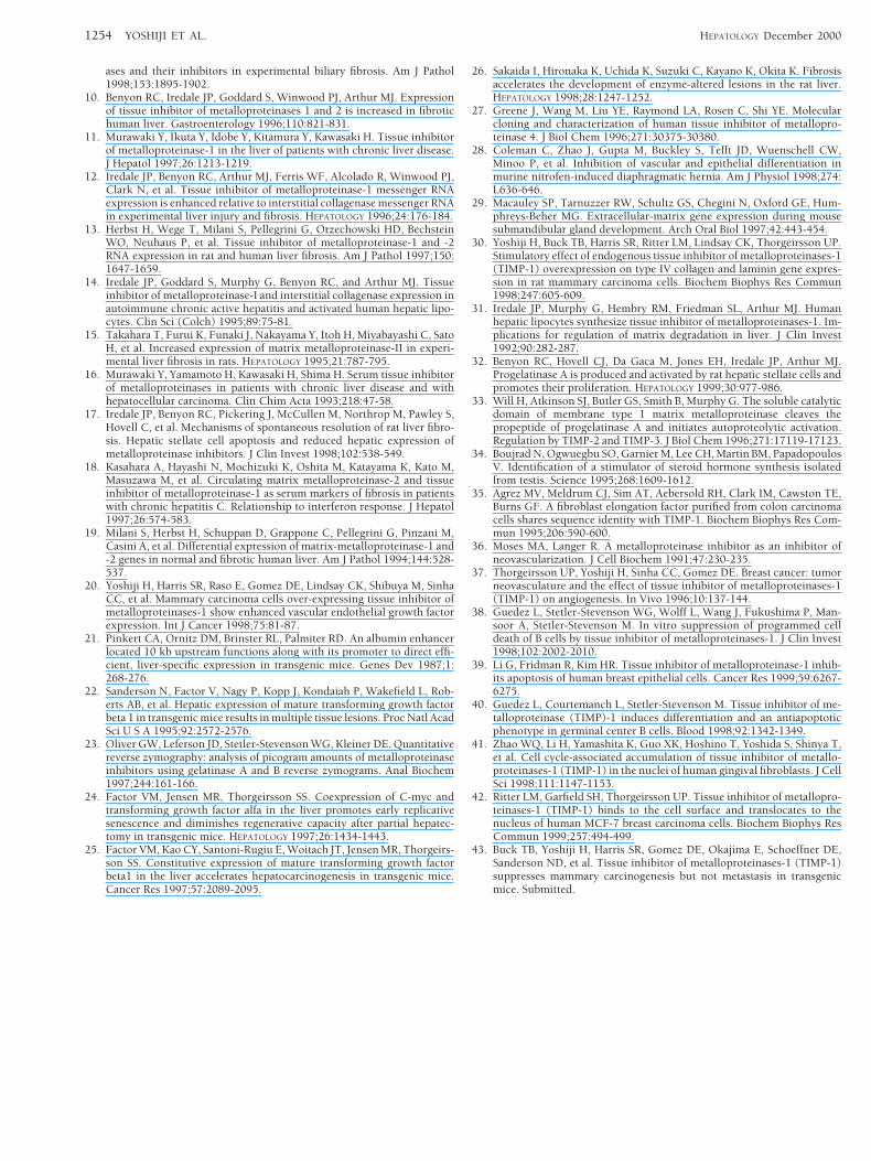
ases and their inhibitors in experimental biliary fibrosis. Am J Pathol1998;153:1895-1902.
10. Benyon RC, Iredale JP, Goddard S, Winwood PJ, Arthur MJ. Expressionof tissue inhibitor of metalloproteinases 1 and 2 is increased in fibrotichuman liver. Gastroenterology 1996;110:821-831.
11. Murawaki Y, Ikuta Y, Idobe Y, Kitamura Y, Kawasaki H. Tissue inhibitorof metalloproteinase-1 in the liver of patients with chronic liver disease.J Hepatol 1997;26:1213-1219.
12. Iredale JP, Benyon RC, Arthur MJ, Ferris WF, Alcolado R, Winwood PJ,Clark N, et al. Tissue inhibitor of metalloproteinase-1 messenger RNAexpression is enhanced relative to interstitial collagenase messenger RNAin experimental liver injury and fibrosis. HEPATOLOGY 1996;24:176-184.
13. Herbst H, Wege T, Milani S, Pellegrini G, Orzechowski HD, BechsteinWO, Neuhaus P, et al. Tissue inhibitor of metalloproteinase-1 and -2RNA expression in rat and human liver fibrosis. Am J Pathol 1997;150:1647-1659.
14. Iredale JP, Goddard S, Murphy G, Benyon RC, and Arthur MJ. Tissueinhibitor of metalloproteinase-I and interstitial collagenase expression inautoimmune chronic active hepatitis and activated human hepatic lipo-cytes. Clin Sci (Colch) 1995;89:75-81.
15. Takahara T, Furui K, Funaki J, Nakayama Y, Itoh H, Miyabayashi C, SatoH, et al. Increased expression of matrix metalloproteinase-II in experi-mental liver fibrosis in rats. HEPATOLOGY 1995;21:787-795.
16. Murawaki Y, Yamamoto H, Kawasaki H, Shima H. Serum tissue inhibitorof metalloproteinases in patients with chronic liver disease and withhepatocellular carcinoma. Clin Chim Acta 1993;218:47-58.
17. Iredale JP, Benyon RC, Pickering J, McCullen M, Northrop M, Pawley S,Hovell C, et al. Mechanisms of spontaneous resolution of rat liver fibro-sis. Hepatic stellate cell apoptosis and reduced hepatic expression ofmetalloproteinase inhibitors. J Clin Invest 1998;102:538-549.
18. Kasahara A, Hayashi N, Mochizuki K, Oshita M, Katayama K, Kato M,Masuzawa M, et al. Circulating matrix metalloproteinase-2 and tissueinhibitor of metalloproteinase-1 as serum markers of fibrosis in patientswith chronic hepatitis C. Relationship to interferon response. J Hepatol1997;26:574-583.
19. Milani S, Herbst H, Schuppan D, Grappone C, Pellegrini G, Pinzani M,Casini A, et al. Differential expression of matrix-metalloproteinase-1 and-2 genes in normal and fibrotic human liver. Am J Pathol 1994;144:528-537.
20. Yoshiji H, Harris SR, Raso E, Gomez DE, Lindsay CK, Shibuya M, SinhaCC, et al. Mammary carcinoma cells over-expressing tissue inhibitor ofmetalloproteinases-1 show enhanced vascular endothelial growth factorexpression. Int J Cancer 1998;75:81-87.
21. Pinkert CA, Ornitz DM, Brinster RL, Palmiter RD. An albumin enhancerlocated 10 kb upstream functions along with its promoter to direct effi-cient, liver-specific expression in transgenic mice. Genes Dev 1987;1:268-276.
22. Sanderson N, Factor V, Nagy P, Kopp J, Kondaiah P, Wakefield L, Rob-erts AB, et al. Hepatic expression of mature transforming growth factorbeta 1 in transgenic mice results in multiple tissue lesions. Proc Natl AcadSci U S A 1995;92:2572-2576.
23. Oliver GW, Leferson JD, Stetler-Stevenson WG, Kleiner DE. Quantitativereverse zymography: analysis of picogram amounts of metalloproteinaseinhibitors using gelatinase A and B reverse zymograms. Anal Biochem1997;244:161-166.
24. Factor VM, Jensen MR, Thorgeirsson SS. Coexpression of C-myc andtransforming growth factor alfa in the liver promotes early replicativesenescence and diminishes regenerative capacity after partial hepatec-tomy in transgenic mice. HEPATOLOGY 1997;26:1434-1443.
25. Factor VM, Kao CY, Santoni-Rugiu E, Woitach JT, Jensen MR, Thorgeirs-son SS. Constitutive expression of mature transforming growth factorbeta1 in the liver accelerates hepatocarcinogenesis in transgenic mice.Cancer Res 1997;57:2089-2095.
26. Sakaida I, Hironaka K, Uchida K, Suzuki C, Kayano K, Okita K. Fibrosisaccelerates the development of enzyme-altered lesions in the rat liver.HEPATOLOGY 1998;28:1247-1252.
27. Greene J, Wang M, Liu YE, Raymond LA, Rosen C, Shi YE. Molecularcloning and characterization of human tissue inhibitor of metallopro-teinase 4. J Biol Chem 1996;271:30375-30380.
28. Coleman C, Zhao J, Gupta M, Buckley S, Tefft JD, Wuenschell CW,Minoo P, et al. Inhibition of vascular and epithelial differentiation inmurine nitrofen-induced diaphragmatic hernia. Am J Physiol 1998;274:L636-646.
29. Macauley SP, Tarnuzzer RW, Schultz GS, Chegini N, Oxford GE, Hum-phreys-Beher MG. Extracellular-matrix gene expression during mousesubmandibular gland development. Arch Oral Biol 1997;42:443-454.
30. Yoshiji H, Buck TB, Harris SR, Ritter LM, Lindsay CK, Thorgeirsson UP.Stimulatory effect of endogenous tissue inhibitor of metalloproteinases-1(TIMP-1) overexpression on type IV collagen and laminin gene expres-sion in rat mammary carcinoma cells. Biochem Biophys Res Commun1998;247:605-609.
31. Iredale JP, Murphy G, Hembry RM, Friedman SL, Arthur MJ. Humanhepatic lipocytes synthesize tissue inhibitor of metalloproteinases-1. Im-plications for regulation of matrix degradation in liver. J Clin Invest1992;90:282-287.
32. Benyon RC, Hovell CJ, Da Gaca M, Jones EH, Iredale JP, Arthur MJ.Progelatinase A is produced and activated by rat hepatic stellate cells andpromotes their proliferation. HEPATOLOGY 1999;30:977-986.
33. Will H, Atkinson SJ, Butler GS, Smith B, Murphy G. The soluble catalyticdomain of membrane type 1 matrix metalloproteinase cleaves thepropeptide of progelatinase A and initiates autoproteolytic activation.Regulation by TIMP-2 and TIMP-3. J Biol Chem 1996;271:17119-17123.
34. Boujrad N, Ogwuegbu SO, Garnier M, Lee CH, Martin BM, PapadopoulosV. Identification of a stimulator of steroid hormone synthesis isolatedfrom testis. Science 1995;268:1609-1612.
35. Agrez MV, Meldrum CJ, Sim AT, Aebersold RH, Clark IM, Cawston TE,Burns GF. A fibroblast elongation factor purified from colon carcinomacells shares sequence identity with TIMP-1. Biochem Biophys Res Com-mun 1995;206:590-600.
36. Moses MA, Langer R. A metalloproteinase inhibitor as an inhibitor ofneovascularization. J Cell Biochem 1991;47:230-235.
37. Thorgeirsson UP, Yoshiji H, Sinha CC, Gomez DE. Breast cancer: tumorneovasculature and the effect of tissue inhibitor of metalloproteinases-1(TIMP-1) on angiogenesis. In Vivo 1996;10:137-144.
38. Guedez L, Stetler-Stevenson WG, Wolff L, Wang J, Fukushima P, Man-soor A, Stetler-Stevenson M. In vitro suppression of programmed celldeath of B cells by tissue inhibitor of metalloproteinases-1. J Clin Invest1998;102:2002-2010.
39. Li G, Fridman R, Kim HR. Tissue inhibitor of metalloproteinase-1 inhib-its apoptosis of human breast epithelial cells. Cancer Res 1999;59:6267-6275.
40. Guedez L, Courtemanch L, Stetler-Stevenson M. Tissue inhibitor of me-talloproteinase (TIMP)-1 induces differentiation and an antiapoptoticphenotype in germinal center B cells. Blood 1998;92:1342-1349.
41. Zhao WQ, Li H, Yamashita K, Guo XK, Hoshino T, Yoshida S, Shinya T,et al. Cell cycle-associated accumulation of tissue inhibitor of metallo-proteinases-1 (TIMP-1) in the nuclei of human gingival fibroblasts. J CellSci 1998;111:1147-1153.
42. Ritter LM, Garfield SH, Thorgeirsson UP. Tissue inhibitor of metallopro-teinases-1 (TIMP-1) binds to the cell surface and translocates to thenucleus of human MCF-7 breast carcinoma cells. Biochem Biophys ResCommun 1999;257:494-499.
43. Buck TB, Yoshiji H, Harris SR, Gomez DE, Okajima E, Schoeffner DE,Sanderson ND, et al. Tissue inhibitor of metalloproteinases-1 (TIMP-1)suppresses mammary carcinogenesis but not metastasis in transgenicmice. Submitted.
1254 YOSHIJI ET AL. HEPATOLOGY December 2000