Dynamic changes of matrix metalloproteinases and their tissue inhibitors in severe sepsis
Upload
khangminh22Category
view
3download
0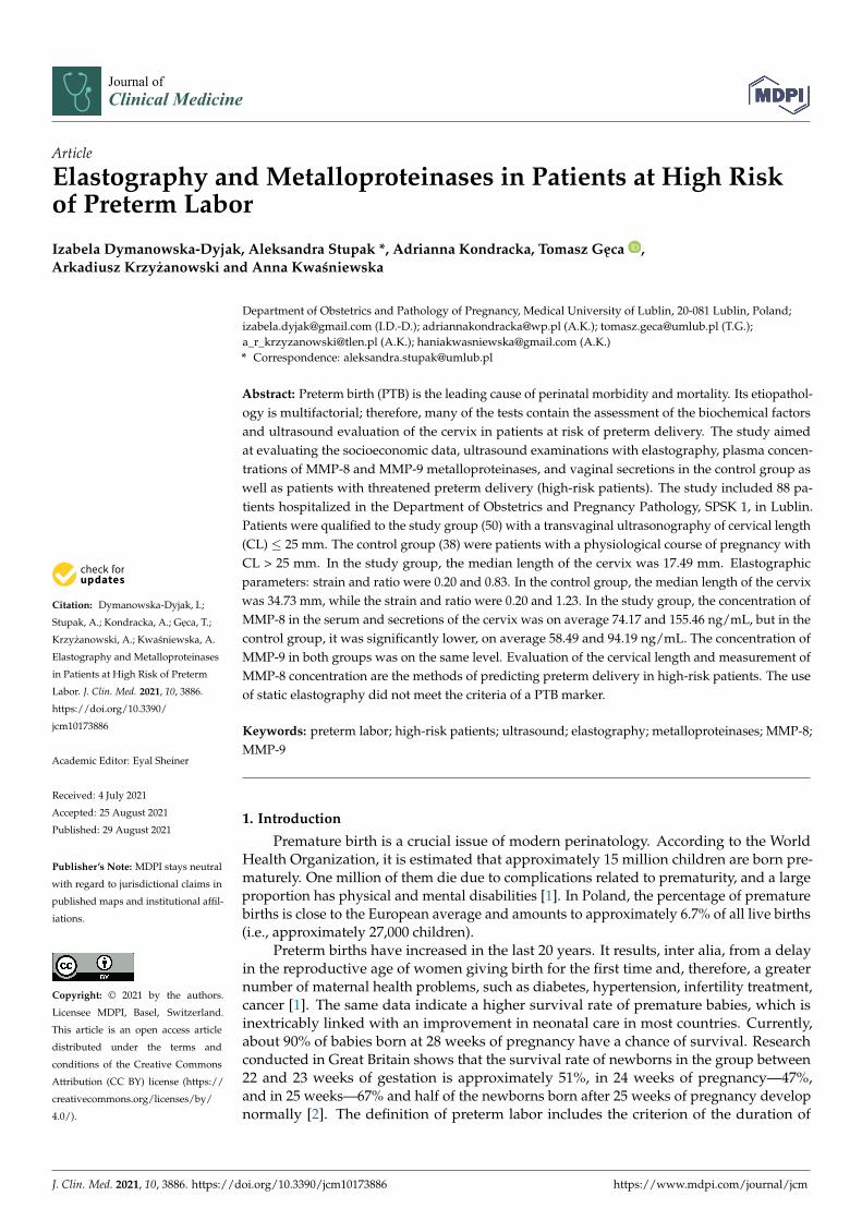
Journal of
Clinical Medicine
Article
Elastography and Metalloproteinases in Patients at High Riskof Preterm Labor
Izabela Dymanowska-Dyjak, Aleksandra Stupak *, Adrianna Kondracka, Tomasz Geca ,Arkadiusz Krzyzanowski and Anna Kwasniewska
�����������������
Citation: Dymanowska-Dyjak, I.;
Stupak, A.; Kondracka, A.; Geca, T.;
Krzyzanowski, A.; Kwasniewska, A.
Elastography and Metalloproteinases
in Patients at High Risk of Preterm
Labor. J. Clin. Med. 2021, 10, 3886.
https://doi.org/10.3390/
jcm10173886
Academic Editor: Eyal Sheiner
Received: 4 July 2021
Accepted: 25 August 2021
Published: 29 August 2021
Publisher’s Note: MDPI stays neutral
with regard to jurisdictional claims in
published maps and institutional affil-
iations.
Copyright: © 2021 by the authors.
Licensee MDPI, Basel, Switzerland.
This article is an open access article
distributed under the terms and
conditions of the Creative Commons
Attribution (CC BY) license (https://
creativecommons.org/licenses/by/
4.0/).
Department of Obstetrics and Pathology of Pregnancy, Medical University of Lublin, 20-081 Lublin, Poland;[email protected] (I.D.-D.); [email protected] (A.K.); [email protected] (T.G.);[email protected] (A.K.); [email protected] (A.K.)* Correspondence: [email protected]
Abstract: Preterm birth (PTB) is the leading cause of perinatal morbidity and mortality. Its etiopathol-ogy is multifactorial; therefore, many of the tests contain the assessment of the biochemical factorsand ultrasound evaluation of the cervix in patients at risk of preterm delivery. The study aimedat evaluating the socioeconomic data, ultrasound examinations with elastography, plasma concen-trations of MMP-8 and MMP-9 metalloproteinases, and vaginal secretions in the control group aswell as patients with threatened preterm delivery (high-risk patients). The study included 88 pa-tients hospitalized in the Department of Obstetrics and Pregnancy Pathology, SPSK 1, in Lublin.Patients were qualified to the study group (50) with a transvaginal ultrasonography of cervical length(CL) ≤ 25 mm. The control group (38) were patients with a physiological course of pregnancy withCL > 25 mm. In the study group, the median length of the cervix was 17.49 mm. Elastographicparameters: strain and ratio were 0.20 and 0.83. In the control group, the median length of the cervixwas 34.73 mm, while the strain and ratio were 0.20 and 1.23. In the study group, the concentration ofMMP-8 in the serum and secretions of the cervix was on average 74.17 and 155.46 ng/mL, but in thecontrol group, it was significantly lower, on average 58.49 and 94.19 ng/mL. The concentration ofMMP-9 in both groups was on the same level. Evaluation of the cervical length and measurement ofMMP-8 concentration are the methods of predicting preterm delivery in high-risk patients. The useof static elastography did not meet the criteria of a PTB marker.
Keywords: preterm labor; high-risk patients; ultrasound; elastography; metalloproteinases; MMP-8;MMP-9
1. Introduction
Premature birth is a crucial issue of modern perinatology. According to the WorldHealth Organization, it is estimated that approximately 15 million children are born pre-maturely. One million of them die due to complications related to prematurity, and a largeproportion has physical and mental disabilities [1]. In Poland, the percentage of prematurebirths is close to the European average and amounts to approximately 6.7% of all live births(i.e., approximately 27,000 children).
Preterm births have increased in the last 20 years. It results, inter alia, from a delayin the reproductive age of women giving birth for the first time and, therefore, a greaternumber of maternal health problems, such as diabetes, hypertension, infertility treatment,cancer [1]. The same data indicate a higher survival rate of premature babies, which isinextricably linked with an improvement in neonatal care in most countries. Currently,about 90% of babies born at 28 weeks of pregnancy have a chance of survival. Researchconducted in Great Britain shows that the survival rate of newborns in the group between22 and 23 weeks of gestation is approximately 51%, in 24 weeks of pregnancy—47%,and in 25 weeks—67% and half of the newborns born after 25 weeks of pregnancy developnormally [2]. The definition of preterm labor includes the criterion of the duration of
J. Clin. Med. 2021, 10, 3886. https://doi.org/10.3390/jcm10173886 https://www.mdpi.com/journal/jcm
J. Clin. Med. 2021, 10, 3886 2 of 22
pregnancy, and it is delivery after the 22nd week of pregnancy and before the 37th week ofpregnancy [1].
The greatest risk of complications concerns children born up to the 33rd week ofpregnancy. Therefore, it is important to identify risk factors for preterm labor, and thusqualify pregnant women to the group at risk of premature delivery [3]. These patients canbe divided into two groups:
• High-risk group (these are patients who have had a history of preterm labor inprevious pregnancies—about 15% of all preterm births).
• Low-risk group (85% of preterm deliveries without burdened medical history).
Preterm labor risk factors can be divided into maternal, environmental, occupational,and obstetric factors.
The maternal factors include:
• Low socioeconomic status;• Black race (compared to the white race) [4];• Low level of education;• Age below 18 and over 40 [5];• Free marital status;• Low maternal body weight before pregnancy (BMI below 19) [6];• Thyroid diseases and other maternal diseases, such as urinary tract infections and
periodontal diseases [7].
The environmental and professional factors include:
• Environment pollution;• Smoking and drugs addiction;• Stress (doubling the risk of preterm labor) [8];• Hard physical work, shift work, night work, and long-term standing [9].
The obstetric factors include:
• A history of preterm labor;• Multiple pregnancies;• Bleeding from the genital tract [10];• Intrauterine infection;• Endometriosis [11];• Hypertension and diabetes;• Incorrect amount of amniotic fluid;• Abdominal surgery in the second and third trimesters of pregnancy.
The etiology of preterm labor is multifactorial. About 50% of premature deliveries areassociated with spontaneous uterine contractions; in 20–30% of cases, it is the prematuretermination of pregnancy due to medical indications, while 20% of them are caused bythe preterm premature rupture of membranes (PPROM). Romero, in 2006, proposed theso-called “common way of delivery”, which involves the activation of multiple systemsthat lead to biochemical and anatomical changes aimed at the expulsion of the fetus andpostpartum [12]. According to the current state of knowledge, infections are the main causeof premature births. It is assumed that they are responsible for about 40% of prematurebirths. The pathogens identified in the amniotic fluid include both aerobic and anaerobicbacteria and viruses. The cytokines involved in the activation of the “common pathway”in preterm labor are IL-1 and TNF-alpha. It has been shown that the concentration ofIL-1 and IL-8 in the cervicovaginal secretion is significantly higher in women who gavebirth prematurely [13]. In addition, IL-1 stimulates the production of prostaglandinsby the temporal and amniotic fluid, thereby inducing uterine contractile activity. In thecase of TNF-alpha, there are reports that the concentration of this substance is muchhigher in patients whose preterm labor is caused by rupture of the membranes [14]. It isrelated to the stimulation of the production of metalloproteinases, mainly MMP-1, MMP-2,and MMP-9, which, etiopathogenetically, are associated with premature rupture of thefetal bladder and maturation of the cervix [15]. Metalloproteinases are enzymes that
J. Clin. Med. 2021, 10, 3886 3 of 22
catalyze the degradation processes of collagen types I, II, III, IV, VII, X, and elastin. Theiractivity in the membranes of the fetus is regulated by many different substances, includingtrypsin, elastase, and thrombin. Anti-inflammatory cytokines, such as IL-10, also playan important role in the mechanism that induces preterm labor and are involved in theimmune recognition and maintenance of pregnancy.
The causes of preterm labor also include cervical insufficiency, which we define aspainless opening and shortening of the cervix. The risk of premature childbirth is estimatedat 75% when the assessment of the length of the cervix is below 25 mm in pregnant womenbefore the 20th week of pregnancy [16]. There are also biochemical indicators of pretermlabor; these include fetal fibronectin.
Ultrasound is one of the best methods of assessing the length of the cervical canal.During ultrasound examination of the cervix, the following parameters should be takeninto account:
• Measurement of the length of the cervical canal;• Assessment of the shape of the internal os and the cervical canal;• Measurement of the internal os;• Measurement of the length of the invagination of the fetal bladder;• Cervical index;• Assessment of the posterior angle of the cervix.
The total length of the cervix is the distance between the inner and outer os. Its func-tional length is the distance between the lower pole of the amniotic sac, to the cervix,and the external os. In practice, functional length assessment is important for the pre-diction of preterm labor. We use the TYVU (Trust Your Vaginal Ultrasound) scheme toevaluate the shape of the internal os. Incorrect values of the index for patients from thehigher risk group significantly increase the real risk of preterm labor. According to the FetalMedicine Foundation (FMF), in order to correctly assess the length of the cervical canal,certain conditions must be met [17]. The elastography technique was first described in 1991by Ophir and his colleagues [18]. It is a method of imaging organs and tissues, assessingtheir stiffness and hardness. It determines the susceptibility of the examined tissue tocompression and mechanical decompression. This is possible thanks to digital ultrasoundand appropriate image processing obtained with a volumetric probe. Currently, the mostadvanced variant of this method is dynamic elastography (in other words, Shear WaveElastography). It is based on the objective assessment and measurement of the stiffnessof the tissue to obtain its numerical value expressed in kPa or m/s using the supersoniceffect in the Mach cone and Young’s modulus (on the extensibility and compressibility ofthe media) [19].
Elastography is a modern diagnostic method that is based on the fact that the hardnessof the tissue/organ changes significantly as a result of the disease process. There areattempts to predict preterm labor by assessment of placental flexibility. The rectus abdo-minis muscle and the subcutaneous tissue were used as reference points for the stiffnesscoefficients. In the case of the second factor, its value measured in the second trimester ofpregnancy can be effectively used as a marker of preterm labor [20]. Metalloproteinases(MMPs) are proteolytic enzymes that contain a zinc ion in the catalytic center. Both theMMP-8 and MMP-9 enzymes are of crucial importance during labor. MMP-9 has alsobeen identified in the amniotic fluid in patients at risk of preterm labor and in deliverypatients [21]. There are reports on the relationship between ultrasound examination andthe assessment of the concentration of MMP-9 as a marker of preterm labor within 7 daysfrom the examination. Most importantly, the concentration of MMP-9 in the maternalblood plasma remains stable during uncomplicated preterm pregnancy until the onsetof full-term labor. In active preterm labor, the concentration of MMP-9 is increased inthe amniotic fluid. MMP-9 is concentrated and activated in the amniotic fluid duringpregnancy complicated by premature rupture of the membranes. Moreover, in the case ofinflammation of the membranes and PROM, the expression of the gene encoding MMP-9is increased. Assuming a cut-off value of 15 mm for the measurement of the cervical
J. Clin. Med. 2021, 10, 3886 4 of 22
length and a proMMP-9 concentration of 67.157 ng/mL in plasma, we can achieve similarlynegative predictive values for preterm labor, approx. 96% for the length of the cervixand 96% for the concentration of pro-MMP-9, while for positive predictors, 69% and 60%respectively. Interestingly, when both prognostic factors were combined, the sensitivityand specificity of the test improved. However, in order to be able to use the combina-tion of both tests to predict premature labor, studies on a larger group of patients areneeded [15]. Increased concentration of MMP-8 is not only associated with the maturationof the cervix during labor but also in preterm labor, PPROM, and intrauterine infection.Increased concentrations of MMP-8 in the blood serum are associated with an increasein the number of leukocytes in vaginal swabs [22]. Neutrophils and the concentration ofMMP-8 in the amniotic fluid are believed to be an inflammatory response to ascendingvaginal infections [7,12,23–25].
As shown, preterm labor is a complex problem, and its diagnosis is one of the mostimportant obstetric issues.
2. The Aim of the Study
The aim of this study was a biochemical and ultrasound evaluation of the cervix inpatients at high-risk of preterm labor.
The aim of the work was achieved by assessing:
• Socioeconomic data;• Ultrasound examinations with elastography;• The concentration of MMP-8 and MMP-9 metalloproteinases in plasma and in vaginal
secretions in the group of patients at risk of preterm labor compared to the controlgroup of pregnant women with a physiological course of pregnancy.
3. Material and Methods
The clinical material of the study included 88 pregnant women hospitalized at theDepartment of Obstetrics and Pathology of Pregnancy of the Medical University of Lublin.Patients who met the criteria for high-risk premature labor between 25 and 38 weeksgestation qualified for the study group. The control group consisted of patients who didnot report premature delivery symptoms without concomitant diseases. After learningabout the purpose and method of conducting the research, all patients gave their informedand written consent to participate in this project. The consent for the research was issuedby the Bioethics Committee at the Medical University of Lublin (KE-0254/134/2009 andKE-0254/294/2017).
In patients hospitalized at the Department of Obstetrics and Pathology of Pregnancyat the Medical University of Lublin with symptoms of premature labor, the diagnosticstandard is an ultrasound examination, in which, in addition to the assessment of thefetal biometry and anatomy, the length of the cervix was determined. Blood samples werealso collected from the patients included in the study, and a swab was taken from theposterior vaginal fornix during the gynecological examination preceding the ultrasoundexamination. The VolusonTM E8 with Elastography Analysis mode was used for theultrasound examination. An endovaginal ultrasound was performed using an endoscopicprobe (VolusonTM E8, RIC5-9-D). During the examination, the patients were asked toassume the lithotomy position. An endovaginal probe was placed in the anterior vaginalfornix, and the bladder was identified as an orientation point. A standard sagittal imageof the cervix was then obtained, and the length of the cervix was measured. The probe,with elastography mode on, was used to produce up to five compression and decompres-sion cycles. After confirming the correct compression and manual decompression in theform of a green quality bar in the lower-left corner of the screen, a measurement was madewith each cycle lasting about 1 s. During each cycle, a tissue shift of approximately 1 cmwas achieved. On the images obtained in this way, two regions were selected: area A onthe upper cervical lip and area B on the bones of the fetal skull as the hardest referencepoint. Within these areas, circles of 5 mm in diameter were placed. From these circles,
J. Clin. Med. 2021, 10, 3886 5 of 22
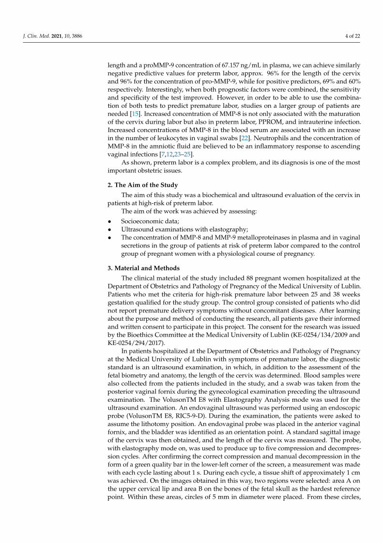
the Elastography Analysis program computed numerical values for the strain ratio andSR (SR–comparative tissue measurement). The value of the deformation factor meanscompression. The maximum value of compression in human tissue is 2%. The SR value,or comparative tissue measurement, indicates how much the tissue in the test area is harderor softer than the tissue in the reference test area. From these values, the means werecalculated and used for statistical calculations (Figure 1).
J. Clin. Med. 2021, 10, x FOR PEER REVIEW 5 of 22
each cycle lasting about 1 s. During each cycle, a tissue shift of approximately 1 cm was achieved. On the images obtained in this way, two regions were selected: area A on the upper cervical lip and area B on the bones of the fetal skull as the hardest reference point. Within these areas, circles of 5 mm in diameter were placed. From these circles, the Elas-tography Analysis program computed numerical values for the strain ratio and SR (SR–comparative tissue measurement). The value of the deformation factor means compres-sion. The maximum value of compression in human tissue is 2%. The SR value, or com-parative tissue measurement, indicates how much the tissue in the test area is harder or softer than the tissue in the reference test area. From these values, the means were calcu-lated and used for statistical calculations (Figure 1).
Figure. 1. Cervical elastographic evaluation in the Elastography Analysis program. -own source.
The transducer receives two sets of radiofrequency signals: before and after squeez-ing the cervix, and the amount of shift in the tissue is estimated from the waveform dif-ference. Tissue rate versus distance from the transducer is calculated for all image points. The rate of change values are known as strain values and are displayed in a variety of colors ranging from red to yellow, green to blue for soft and hard tissues. Tissues marked in red are considered to be the most flexible tissues, while tissues with the least elasticity are marked in blue (Figure 2).
Laboratory tests were performed concurrently with the ultrasound examinations. These studies used serum samples separated from peripheral blood and samples of cervi-cal secretions obtained from the simultaneous collection from a given patient.
Blood from the antecubital vein was collected in a volume of 9 mL into disposable S-Monovettes (Sarstedt, Germany) containing a blood clotting activator. The solidification process took place at room temperature and lasted approximately 30–40 min. The samples were then centrifuged in a centrifuge (Sigma 1-6P, Polygen) for 10 min at room tempera-ture and 3800 rpm. The serum obtained in this way was aliquoted 200 µL in Eppendorf tubes (Medlab Products) and stored at −75 °C (Platinum Angelantoni 500, Italy) until the measurements were made.
Figure 1. Cervical elastographic evaluation in the Elastography Analysis program.
The transducer receives two sets of radiofrequency signals: before and after squeezingthe cervix, and the amount of shift in the tissue is estimated from the waveform difference.Tissue rate versus distance from the transducer is calculated for all image points. The rateof change values are known as strain values and are displayed in a variety of colors rangingfrom red to yellow, green to blue for soft and hard tissues. Tissues marked in red areconsidered to be the most flexible tissues, while tissues with the least elasticity are markedin blue (Figure 2).
Laboratory tests were performed concurrently with the ultrasound examinations.These studies used serum samples separated from peripheral blood and samples of cervicalsecretions obtained from the simultaneous collection from a given patient.
Blood from the antecubital vein was collected in a volume of 9 mL into disposableS-Monovettes (Sarstedt, Germany) containing a blood clotting activator. The solidificationprocess took place at room temperature and lasted approximately 30–40 min. The sampleswere then centrifuged in a centrifuge (Sigma 1-6P, Polygen) for 10 min at room temperatureand 3800 rpm. The serum obtained in this way was aliquoted 200 µL in Eppendorftubes (Medlab Products) and stored at −75 ◦C (Platinum Angelantoni 500, Italy) until themeasurements were made.
Cervical discharge was collected with a sterile swab (Deltalab, Barcelona, Spain) whileexamined in a sterile speculum from the posterior vaginal fornix. If vaginal discharge couldnot be collected, a sample was collected from the cervix. A swab was inserted into theouter mouth of the cervix to a depth of 1–2 cm and then pressing the mucous membraneseveral times (for 10–15 s), which allowed for the absorption of the appropriate amount ofsecretion. The material collected in this way was placed in a sterile, tightly closed tube andsent to a laboratory.
J. Clin. Med. 2021, 10, 3886 6 of 22J. Clin. Med. 2021, 10, x FOR PEER REVIEW 6 of 22
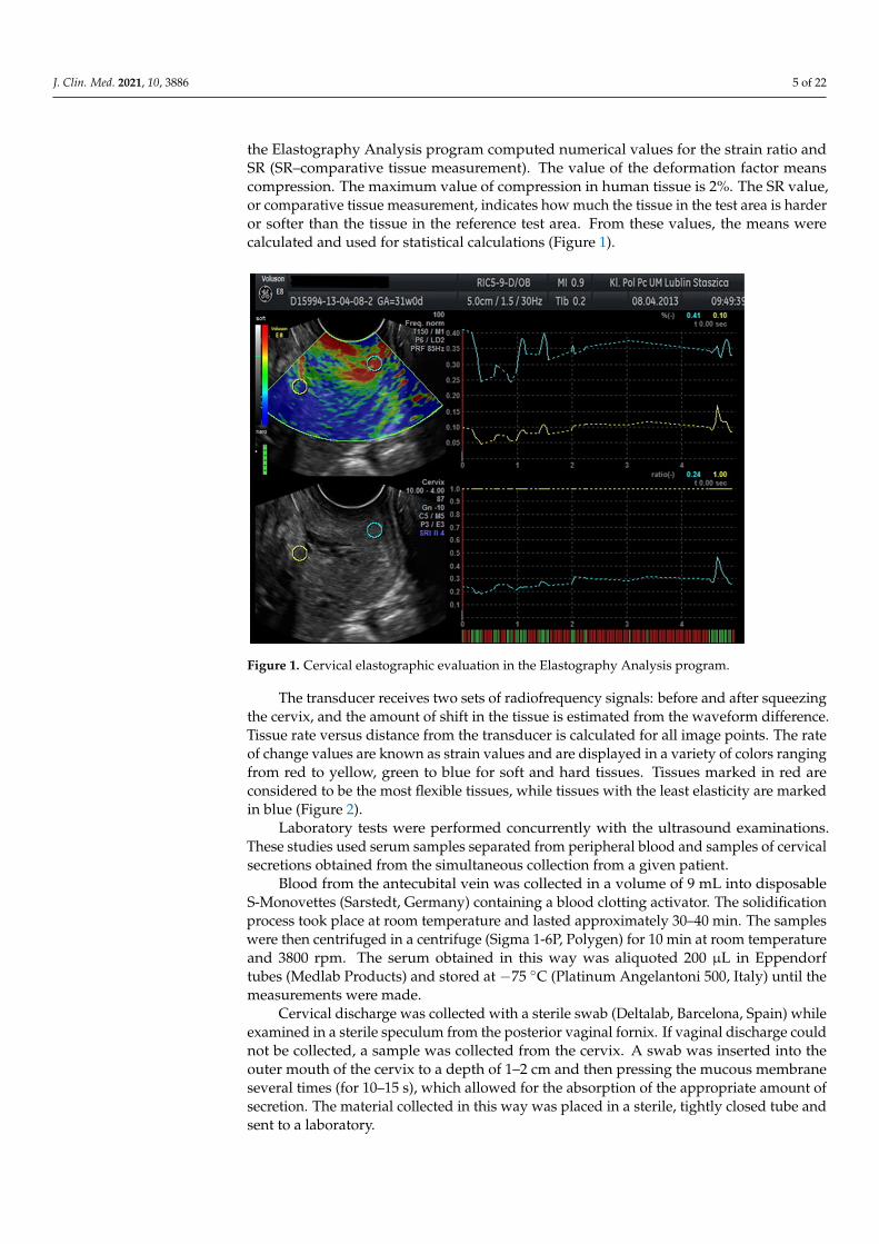
Figure 2. Cervical elastographic assessment. -own source Tissue elasticity distribution calculations in real-time and presented using a color scale—red (soft), blue (hard), and green (medium-hard).
Cervical discharge was collected with a sterile swab (Deltalab, Barcelona, Spain) while examined in a sterile speculum from the posterior vaginal fornix. If vaginal dis-charge could not be collected, a sample was collected from the cervix. A swab was inserted into the outer mouth of the cervix to a depth of 1–2 cm and then pressing the mucous membrane several times (for 10–15 s), which allowed for the absorption of the appropriate amount of secretion. The material collected in this way was placed in a sterile, tightly closed tube and sent to a laboratory.
In the next step, the swab with the sample was transferred to a tube containing 2 mL of PBS (Phosphate Buffered Saline (PAA Laboratories GmbH, Leonding, Austria) without calcium (Ca2+) and magnesium (Mg2+) ions). The secretion was extracted by vigorously rotating the swab inside the tube for about 10 s, and then the samples were centrifuged for 10 min at room temperature, at a speed of 3800 rpm. The supernatant thus obtained was collected from the sediment and aliquoted 200 µL. The material was stored in the same way as in the case of the serum.
Determination of the concentration of metalloproteinases 8 (MMP-8) and 9 (MMP-9) in the tested material was performed with the use of commercially available ELISA kits (Enzyme-linked Immunosorbent Assay) based on immunological reactions. All test steps were performed in accordance with the procedures recommended by the manufacturer of the assay kits. The following test was used to determine the concentration of MMP-8: Quantikine Human Total MMP-8 Immunoassay. For the quantitative determination of human active and pro-Matrix Metalloproteinase 8 (total MMP-8) concentrations in cell culture supernatants, serum, plasma, and saliva, Cat # DMP800 was used (R&D Systems Europe Ltd., Abingdon , UK). The mean analytical sensitivity of the assay was 0.02 ng/mL (0.01 to 0.06 ng/mL), and the measuring range was from 0 to 10 ng/mL (lowest concentra-tion to highest standard concentration).
The following assay was used to determine the level of MMP-9: Quantikine Human MMP-9 Immunoassay. For the quantitative determination of human active (82 kDa) and Pro- (92 kDa) Matrix Metalloproteinase 9 (MMP-9) concentrations in cell culture superna-tants, serum, plasma, saliva, and urine, Cat # DMP900 was used (R&D Systems Europe Ltd., Abingdon , UK).
Figure 2. Cervical elastographic assessment. Tissue elasticity distribution calculations in real-timeand presented using a color scale—red (soft), blue (hard), and green (medium-hard).
In the next step, the swab with the sample was transferred to a tube containing 2 mLof PBS (Phosphate Buffered Saline (PAA Laboratories GmbH, Leonding, Austria) withoutcalcium (Ca2+) and magnesium (Mg2+) ions). The secretion was extracted by vigorouslyrotating the swab inside the tube for about 10 s, and then the samples were centrifuged for10 min at room temperature, at a speed of 3800 rpm. The supernatant thus obtained wascollected from the sediment and aliquoted 200 µL. The material was stored in the sameway as in the case of the serum.
Determination of the concentration of metalloproteinases 8 (MMP-8) and 9 (MMP-9)in the tested material was performed with the use of commercially available ELISA kits(Enzyme-linked Immunosorbent Assay) based on immunological reactions. All test stepswere performed in accordance with the procedures recommended by the manufacturerof the assay kits. The following test was used to determine the concentration of MMP-8: Quantikine Human Total MMP-8 Immunoassay. For the quantitative determinationof human active and pro-Matrix Metalloproteinase 8 (total MMP-8) concentrations incell culture supernatants, serum, plasma, and saliva, Cat # DMP800 was used (R&DSystems Europe Ltd., Abingdon, UK). The mean analytical sensitivity of the assay was0.02 ng/mL (0.01 to 0.06 ng/mL), and the measuring range was from 0 to 10 ng/mL (lowestconcentration to highest standard concentration).
The following assay was used to determine the level of MMP-9: Quantikine HumanMMP-9 Immunoassay. For the quantitative determination of human active (82 kDa) andPro- (92 kDa) Matrix Metalloproteinase 9 (MMP-9) concentrations in cell culture super-natants, serum, plasma, saliva, and urine, Cat # DMP900 was used (R&D Systems EuropeLtd., Abingdon, UK).
The sensitivity of the assay, defined as the minimum detectable dose (MDD) of humanMMP-9, was less than 0.156 ng/mL. The measuring range was from 0 to 20 ng/mL (fromthe lowest concentration to the highest standard concentration).
A statistical analysis of clinical data was performed using: the arithmetic meanstandard deviation (SD), medians (ME) using the Shapiro–Wilk test, and the Kruskal-WallisH test. A comparison of the differences between the control group and the study group wascarried out with the Student’s t-test. For the variables tested, which did not show normaldistribution, non-parametric tests were used for further analysis. The concentrations of
J. Clin. Med. 2021, 10, 3886 7 of 22
MMP-8 and MMP-9 were compared using the U-Mann-Whitney test and the Pearson Chi2test. The relationship between individual substances and clinical data was carried outusing the Spearman correlation test. Statistical significance was set at p ≤ 0.05. Resultsstatistically insignificant were defined by the abbreviation “ns”. Statistical calculationswere based on Statistica 10 (StatSoft, Tulsa, OK, USA).
4. ResultsCharacteristics of the Study and Control Groups
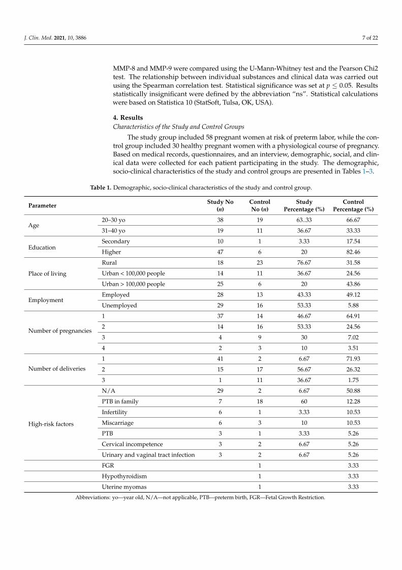
The study group included 58 pregnant women at risk of preterm labor, while the con-trol group included 30 healthy pregnant women with a physiological course of pregnancy.Based on medical records, questionnaires, and an interview, demographic, social, and clin-ical data were collected for each patient participating in the study. The demographic,socio-clinical characteristics of the study and control groups are presented in Tables 1–3.
Table 1. Demographic, socio-clinical characteristics of the study and control group.
Parameter Study No(n)
ControlNo (n)
StudyPercentage (%)
ControlPercentage (%)
Age20–30 yo 38 19 63..33 66.67
31–40 yo 19 11 36.67 33.33
EducationSecondary 10 1 3.33 17.54
Higher 47 6 20 82.46
Place of living
Rural 18 23 76.67 31.58
Urban < 100,000 people 14 11 36.67 24.56
Urban > 100,000 people 25 6 20 43.86
EmploymentEmployed 28 13 43.33 49.12
Unemployed 29 16 53.33 5.88
Number of pregnancies
1 37 14 46.67 64.91
2 14 16 53.33 24.56
3 4 9 30 7.02
4 2 3 10 3.51
Number of deliveries
1 41 2 6.67 71.93
2 15 17 56.67 26.32
3 1 11 36.67 1.75
High-risk factors
N/A 29 2 6.67 50.88
PTB in family 7 18 60 12.28
Infertility 6 1 3.33 10.53
Miscarriage 6 3 10 10.53
PTB 3 1 3.33 5.26
Cervical incompetence 3 2 6.67 5.26
Urinary and vaginal tract infection 3 2 6.67 5.26
FGR 1 3.33
Hypothyroidism 1 3.33
Uterine myomas 1 3.33
Abbreviations: yo—year old, N/A—not applicable, PTB—preterm birth, FGR—Fetal Growth Restriction.
J. Clin. Med. 2021, 10, 3886 8 of 22
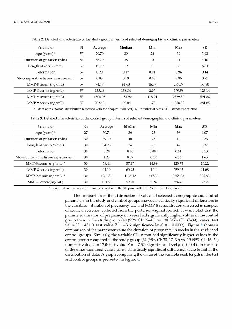
Table 2. Detailed characteristics of the study group in terms of selected demographic and clinical parameters.
Parameter N Average Median Min Max SD
Age (years) * 57 29.70 30 22 39 3.93
Duration of gestation (wks) 57 36.79 38 25 41 4.10
Length of cervix (mm) 57 17.49 19 2 30 6.34
Deformation 57 0.20 0.17 0.01 0.94 0.14
SR-comparative tissue measurement 57 0.83 0.59 0.03 3.86 0.77
MMP-8 serum (ng/mL) 57 74.17 61.63 16.59 287.77 51.50
MMP-8 cervix (ng/mL) 57 155.46 158.34 2.07 379.58 123.14
MMP-9 serum (ng/mL) 57 1308.98 1181.90 418.94 2569.52 591.88
MMP-9 cervix (ng/mL) 57 202.43 103.04 1.72 1258.57 281.85
*—data with a normal distribution (assessed with the Shapiro–Wilk test). N—number of cases, SD—standard deviation
Table 3. Detailed characteristics of the control group in terms of selected demographic and clinical parameters.
Parameter No Average Median Min Max SD
Age (years) * 27 30.74 30 25 39 4.07
Duration of gestation (wks) 30 39.10 40 28 41 2.26
Length of cervix * (mm) 30 34.73 34 25 46 6.37
Deformation 30 0.20 0.16 0.009 0.61 0.13
SR—comparative tissue measurement 30 1.23 0.57 0.17 6.56 1.65
MMP-8 serum (ng/mL) * 30 58.44 57.47 14.99 123.73 26.22
MMP-8 cervix (ng/mL) 30 94.19 60.95 1.14 259.02 91.08
MMP-9 serum (ng/mL) * 30 1261.56 1134.42 447.30 2258.83 505.83
MMP-9 cervix(ng/mL) 30 103.59 59.70 2.24 554.40 122.21
*—data with a normal distribution (assessed with the Shapiro–Wilk test). WKS—weeks gestation
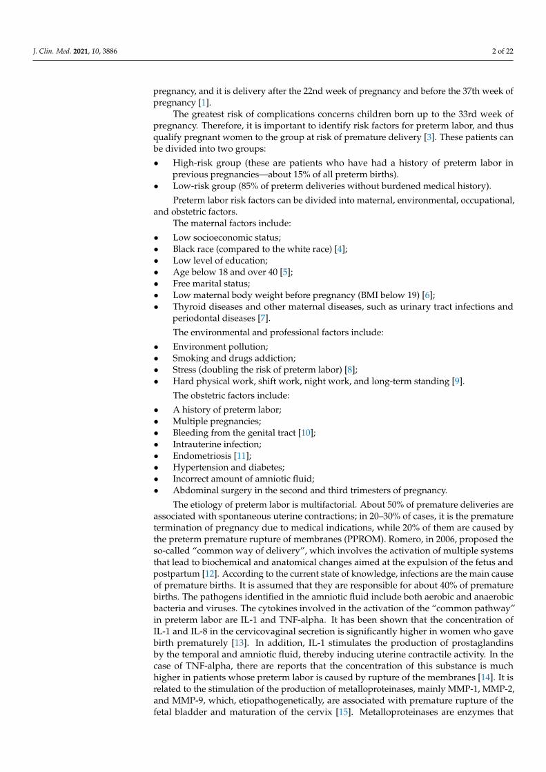
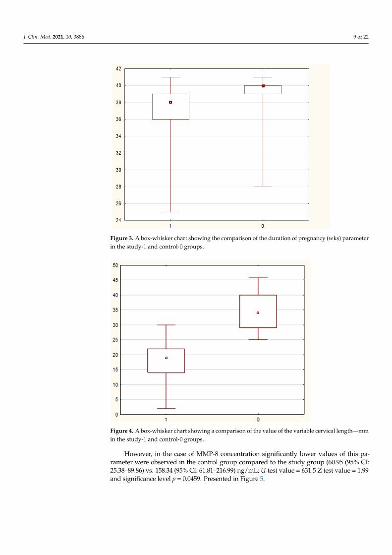
The comparison of the distribution of values of selected demographic and clinicalparameters in the study and control groups showed statistically significant differences inthe variables—duration of pregnancy, CL, and MMP-8 concentration (assessed in samplesof cervical secretion collected from the posterior vaginal fornix). It was noted that theparameter duration of pregnancy in weeks had significantly higher values in the controlgroup than in the study group (40 (95% CI: 39–40) vs. 38 (95% CI: 37–39) weeks; testvalue U = 451 0; test value Z = −3.6; significance level p = 0.0002). Figure 3 shows acomparison of the parameter value the duration of pregnancy in weeks in the study andcontrol groups. Similarly, the variable CL in mm had significantly higher values in thecontrol group compared to the study group (34 (95% CI: 30, 17–39) vs. 19 (95% CI: 16–21)mm; test value U = 12.0; test value Z = −7.52; significance level p < 0.0001). In the caseof the other examined variables, no statistically significant differences were found in thedistribution of data. A graph comparing the value of the variable neck length in the testand control groups is presented in Figure 4.
J. Clin. Med. 2021, 10, 3886 9 of 22J. Clin. Med. 2021, 10, x FOR PEER REVIEW 9 of 22
Figure 3. A box-whisker chart showing the comparison of the duration of pregnancy (wks) param-eter in the study-1 and control-0 groups.
Figure 4. A box-whisker chart showing a comparison of the value of the variable cervical length—mm in the study-1 and control-0 groups.
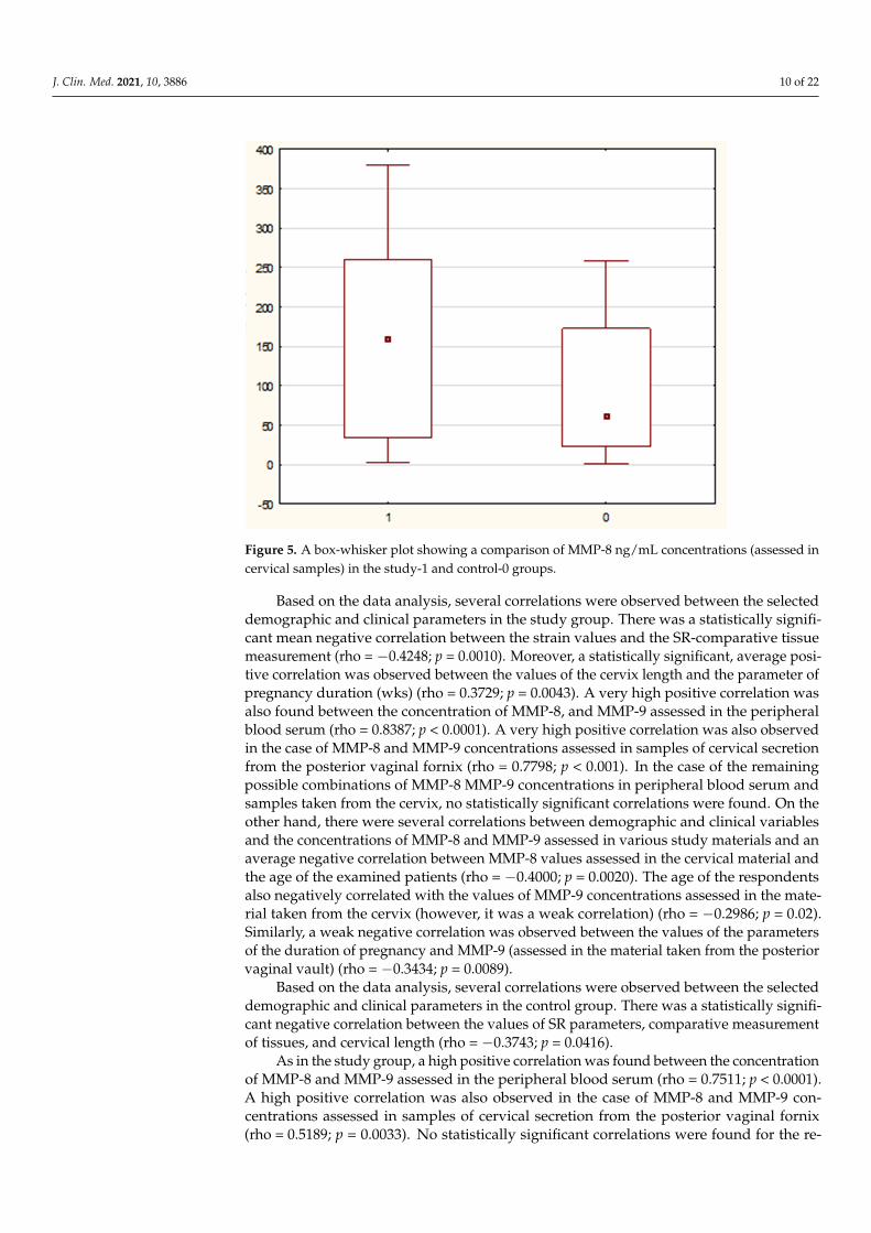
However, in the case of MMP-8 concentration significantly lower values of this pa-rameter were observed in the control group compared to the study group (60.95 (95% CI: 25.38–89.86) vs. 158.34 (95% CI: 61.81–216.99) ng/mL; U test value = 631.5 Z test value = 1.99 and significance level p = 0.0459. Presented in Figure 5.
Figure 3. A box-whisker chart showing the comparison of the duration of pregnancy (wks) parameterin the study-1 and control-0 groups.
J. Clin. Med. 2021, 10, x FOR PEER REVIEW 9 of 22
Figure 3. A box-whisker chart showing the comparison of the duration of pregnancy (wks) param-eter in the study-1 and control-0 groups.
Figure 4. A box-whisker chart showing a comparison of the value of the variable cervical length—mm in the study-1 and control-0 groups.
However, in the case of MMP-8 concentration significantly lower values of this pa-rameter were observed in the control group compared to the study group (60.95 (95% CI: 25.38–89.86) vs. 158.34 (95% CI: 61.81–216.99) ng/mL; U test value = 631.5 Z test value = 1.99 and significance level p = 0.0459. Presented in Figure 5.
Figure 4. A box-whisker chart showing a comparison of the value of the variable cervical length—mmin the study-1 and control-0 groups.
However, in the case of MMP-8 concentration significantly lower values of this pa-rameter were observed in the control group compared to the study group (60.95 (95% CI:25.38–89.86) vs. 158.34 (95% CI: 61.81–216.99) ng/mL; U test value = 631.5 Z test value = 1.99and significance level p = 0.0459. Presented in Figure 5.
J. Clin. Med. 2021, 10, 3886 10 of 22J. Clin. Med. 2021, 10, x FOR PEER REVIEW 10 of 22
Figure 5. A box-whisker plot showing a comparison of MMP-8 ng/mL concentrations (assessed in cervical samples) in the study-1 and control-0 groups.
Based on the data analysis, several correlations were observed between the selected demographic and clinical parameters in the study group. There was a statistically signifi-cant mean negative correlation between the strain values and the SR-comparative tissue measurement (rho = −0.4248; p = 0.0010). Moreover, a statistically significant, average pos-itive correlation was observed between the values of the cervix length and the parameter of pregnancy duration (wks) (rho = 0.3729; p = 0.0043). A very high positive correlation was also found between the concentration of MMP-8, and MMP-9 assessed in the periph-eral blood serum (rho = 0.8387; p < 0.0001). A very high positive correlation was also ob-served in the case of MMP-8 and MMP-9 concentrations assessed in samples of cervical secretion from the posterior vaginal fornix (rho = 0.7798; p < 0.001). In the case of the re-maining possible combinations of MMP-8 MMP-9 concentrations in peripheral blood se-rum and samples taken from the cervix, no statistically significant correlations were found. On the other hand, there were several correlations between demographic and clin-ical variables and the concentrations of MMP-8 and MMP-9 assessed in various study ma-terials and an average negative correlation between MMP-8 values assessed in the cervical material and the age of the examined patients (rho = −0.4000; p = 0.0020). The age of the respondents also negatively correlated with the values of MMP-9 concentrations assessed in the material taken from the cervix (however, it was a weak correlation) (rho = −0.2986; p = 0.02). Similarly, a weak negative correlation was observed between the values of the parameters of the duration of pregnancy and MMP-9 (assessed in the material taken from the posterior vaginal vault) (rho = −0.3434; p = 0.0089).
Based on the data analysis, several correlations were observed between the selected demographic and clinical parameters in the control group. There was a statistically signif-icant negative correlation between the values of SR parameters, comparative measure-ment of tissues, and cervical length (rho = −0.3743; p = 0.0416).
As in the study group, a high positive correlation was found between the concentra-tion of MMP-8 and MMP-9 assessed in the peripheral blood serum (rho = 0.7511; p < 0.0001). A high positive correlation was also observed in the case of MMP-8 and MMP-9 concentrations assessed in samples of cervical secretion from the posterior vaginal fornix
Figure 5. A box-whisker plot showing a comparison of MMP-8 ng/mL concentrations (assessed incervical samples) in the study-1 and control-0 groups.
Based on the data analysis, several correlations were observed between the selecteddemographic and clinical parameters in the study group. There was a statistically signifi-cant mean negative correlation between the strain values and the SR-comparative tissuemeasurement (rho = −0.4248; p = 0.0010). Moreover, a statistically significant, average posi-tive correlation was observed between the values of the cervix length and the parameter ofpregnancy duration (wks) (rho = 0.3729; p = 0.0043). A very high positive correlation wasalso found between the concentration of MMP-8, and MMP-9 assessed in the peripheralblood serum (rho = 0.8387; p < 0.0001). A very high positive correlation was also observedin the case of MMP-8 and MMP-9 concentrations assessed in samples of cervical secretionfrom the posterior vaginal fornix (rho = 0.7798; p < 0.001). In the case of the remainingpossible combinations of MMP-8 MMP-9 concentrations in peripheral blood serum andsamples taken from the cervix, no statistically significant correlations were found. On theother hand, there were several correlations between demographic and clinical variablesand the concentrations of MMP-8 and MMP-9 assessed in various study materials and anaverage negative correlation between MMP-8 values assessed in the cervical material andthe age of the examined patients (rho = −0.4000; p = 0.0020). The age of the respondentsalso negatively correlated with the values of MMP-9 concentrations assessed in the mate-rial taken from the cervix (however, it was a weak correlation) (rho = −0.2986; p = 0.02).Similarly, a weak negative correlation was observed between the values of the parametersof the duration of pregnancy and MMP-9 (assessed in the material taken from the posteriorvaginal vault) (rho = −0.3434; p = 0.0089).
Based on the data analysis, several correlations were observed between the selecteddemographic and clinical parameters in the control group. There was a statistically signifi-cant negative correlation between the values of SR parameters, comparative measurementof tissues, and cervical length (rho = −0.3743; p = 0.0416).
As in the study group, a high positive correlation was found between the concentrationof MMP-8 and MMP-9 assessed in the peripheral blood serum (rho = 0.7511; p < 0.0001).A high positive correlation was also observed in the case of MMP-8 and MMP-9 con-centrations assessed in samples of cervical secretion from the posterior vaginal fornix(rho = 0.5189; p = 0.0033). No statistically significant correlations were found for the re-
J. Clin. Med. 2021, 10, 3886 11 of 22
maining possible combinations of MMP-8 and MMP-9 concentrations in peripheral bloodserum and samples taken from the posterior vaginal fornix.
There were, however, two correlations between clinical variables and the values ofMMP-8 concentrations assessed in samples taken from the cervix.
Among other things, there was an average negative correlation between the MMP-8values assessed in the cervical material and the SR parameter—comparative tissue mea-surement in the control group (rho = −0.3737; p = 0.0419). There was also an averagenegative correlation between MMP-8 values assessed in the material collected from theposterior vaginal fornix and the parameter—duration of pregnancy in weeks (rho = 0.3623;p = 0.0491). Based on the analysis carried out with the use of ROC curves, it was foundthat among the studied demographic and clinical variables, only the length of the cervix(in mm) and the concentration of MMP-8 (in ng/mL), assessed in the cervical secretionobtained from the posterior vaginal fornix, were significantly high in the area under thecurve (AUC) value in the early detection of the possibility of preterm labor.
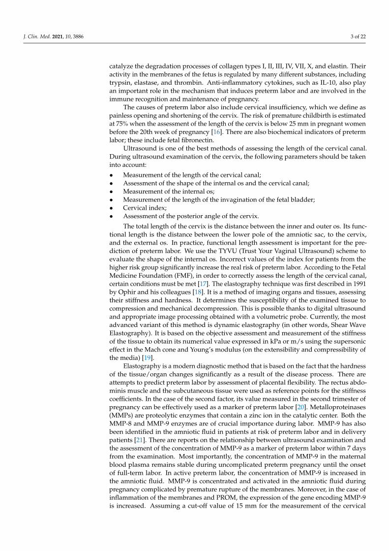
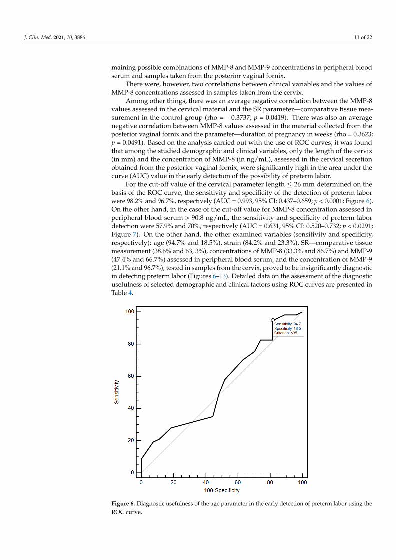
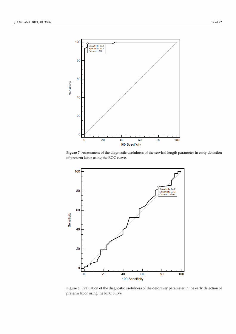
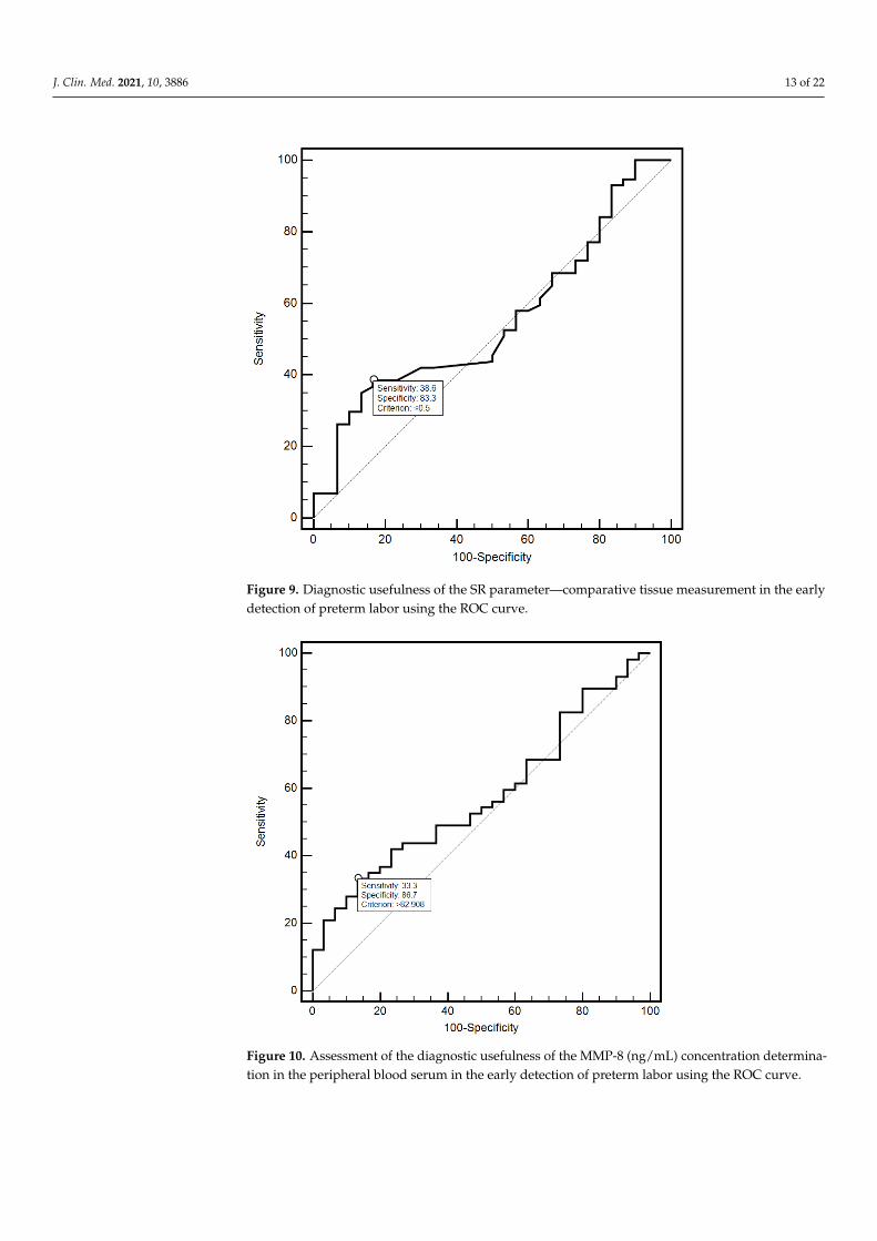
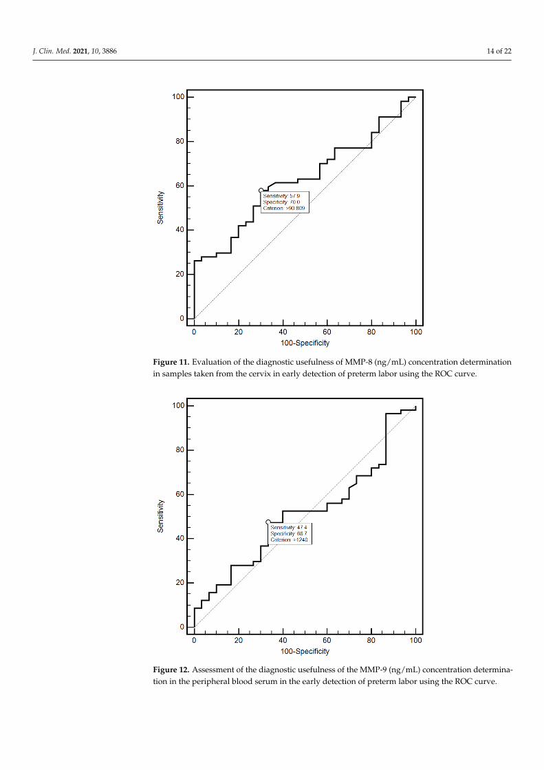
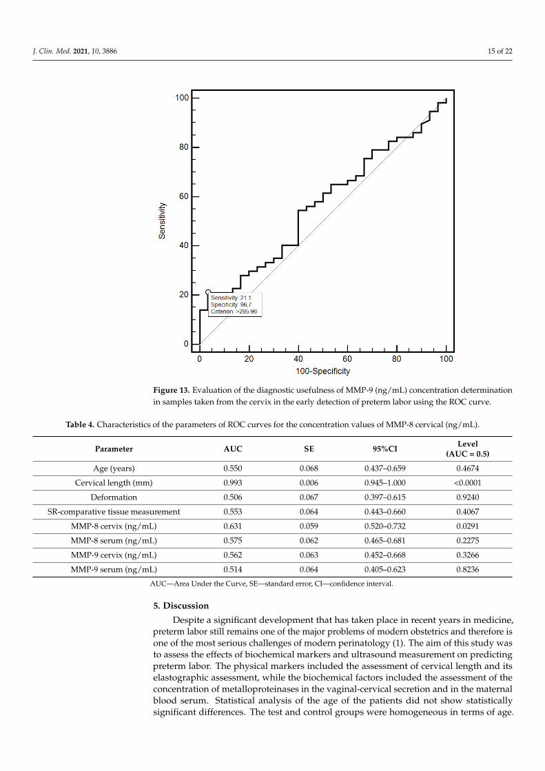
For the cut-off value of the cervical parameter length ≤ 26 mm determined on thebasis of the ROC curve, the sensitivity and specificity of the detection of preterm laborwere 98.2% and 96.7%, respectively (AUC = 0.993, 95% CI: 0.437–0.659; p < 0.0001; Figure 6).On the other hand, in the case of the cut-off value for MMP-8 concentration assessed inperipheral blood serum > 90.8 ng/mL, the sensitivity and specificity of preterm labordetection were 57.9% and 70%, respectively (AUC = 0.631, 95% CI: 0.520–0.732; p < 0.0291;Figure 7). On the other hand, the other examined variables (sensitivity and specificity,respectively): age (94.7% and 18.5%), strain (84.2% and 23.3%), SR—comparative tissuemeasurement (38.6% and 63, 3%), concentrations of MMP-8 (33.3% and 86.7%) and MMP-9(47.4% and 66.7%) assessed in peripheral blood serum, and the concentration of MMP-9(21.1% and 96.7%), tested in samples from the cervix, proved to be insignificantly diagnosticin detecting preterm labor (Figures 6–13). Detailed data on the assessment of the diagnosticusefulness of selected demographic and clinical factors using ROC curves are presented inTable 4.
1
6
7
Figure 6. Diagnostic usefulness of the age parameter in the early detection of preterm labor using theROC curve.
J. Clin. Med. 2021, 10, 3886 12 of 22
1
6
7
Figure 7. Assessment of the diagnostic usefulness of the cervical length parameter in early detectionof preterm labor using the ROC curve.
2
8
9
Figure 8. Evaluation of the diagnostic usefulness of the deformity parameter in the early detection ofpreterm labor using the ROC curve.
J. Clin. Med. 2021, 10, 3886 13 of 22
2
8
9 Figure 9. Diagnostic usefulness of the SR parameter—comparative tissue measurement in the earlydetection of preterm labor using the ROC curve.
3
10
11
Figure 10. Assessment of the diagnostic usefulness of the MMP-8 (ng/mL) concentration determina-tion in the peripheral blood serum in the early detection of preterm labor using the ROC curve.
J. Clin. Med. 2021, 10, 3886 14 of 22
3
10
11
Figure 11. Evaluation of the diagnostic usefulness of MMP-8 (ng/mL) concentration determinationin samples taken from the cervix in early detection of preterm labor using the ROC curve.
4
12
13
Figure 12. Assessment of the diagnostic usefulness of the MMP-9 (ng/mL) concentration determina-tion in the peripheral blood serum in the early detection of preterm labor using the ROC curve.
J. Clin. Med. 2021, 10, 3886 15 of 22
4
12
13 Figure 13. Evaluation of the diagnostic usefulness of MMP-9 (ng/mL) concentration determinationin samples taken from the cervix in the early detection of preterm labor using the ROC curve.
Table 4. Characteristics of the parameters of ROC curves for the concentration values of MMP-8 cervical (ng/mL).
Parameter AUC SE 95%CI Level(AUC = 0.5)
Age (years) 0.550 0.068 0.437–0.659 0.4674
Cervical length (mm) 0.993 0.006 0.945–1.000 <0.0001
Deformation 0.506 0.067 0.397–0.615 0.9240
SR-comparative tissue measurement 0.553 0.064 0.443–0.660 0.4067
MMP-8 cervix (ng/mL) 0.631 0.059 0.520–0.732 0.0291
MMP-8 serum (ng/mL) 0.575 0.062 0.465–0.681 0.2275
MMP-9 cervix (ng/mL) 0.562 0.063 0.452–0.668 0.3266
MMP-9 serum (ng/mL) 0.514 0.064 0.405–0.623 0.8236
AUC—Area Under the Curve, SE—standard error, CI—confidence interval.
5. Discussion
Despite a significant development that has taken place in recent years in medicine,preterm labor still remains one of the major problems of modern obstetrics and therefore isone of the most serious challenges of modern perinatology (1). The aim of this study wasto assess the effects of biochemical markers and ultrasound measurement on predictingpreterm labor. The physical markers included the assessment of cervical length and itselastographic assessment, while the biochemical factors included the assessment of theconcentration of metalloproteinases in the vaginal-cervical secretion and in the maternalblood serum. Statistical analysis of the age of the patients did not show statisticallysignificant differences. The test and control groups were homogeneous in terms of age.

J. Clin. Med. 2021, 10, 3886 16 of 22
In the study group, the mean age was 29.7 years. However, in the control group, the meanage was slightly higher, amounting to 30.7 years). The age of the patients had no significanteffect on the increased risk of preterm labor. Most of the scientific reports, analyzing thedata on patients with extreme age, confirm the fact that the age of the patients influencesthe risk of preterm labor [26,27]. In a study by Cooper et al., obstetric results were assessedin very young 15-year-old married women with secondary education, white race, undermedical care [26]. Similarly, Astolfi et al. Comparing the age of parents at 20–29 yearswith the age of the mother over 30 and the father over 40 showed that the influence ofpaternal age on the risk of preterm labor is smaller but significant if he is over 40 [28]. It isnoteworthy that the deformation parameter (cervical) was slightly higher in older patients,but this relationship did not become statistically significant.
In this research, the variable “duration of pregnancy” in the study and control groupwas compared with the variable “cervical length” assessed in the ultrasound examination.It was confirmed that the parameter “duration of pregnancy in weeks” had significantlyhigher values in the control group compared to the study group. It was shown that inpatients whose variable “cervical length” was less than or equal to 25 mm, the parameter“duration of pregnancy” in weeks had statistically significantly lower values. The obtainedresults lead to the conclusion that the CL expressed in millimeters is inversely proportionalto the risk of preterm labor. This is confirmed by the Berghella study, according to whichscreening the length of the cervix with transvaginal ultrasound is a good prognostic testfor the prediction of spontaneous preterm labor in single pregnancies with symptoms ofpreterm labor [29]. It has been shown that in patients at risk of preterm labor, for whomthe cervical length measurement is known, the incidence of spontaneous preterm labor islower, and the gestational age at delivery is higher. This is due to the possibility of usingappropriate clinical management. Asymptomatic women at risk should be screened at a2-week interval starting from 16 to 18 weeks, up to 24 weeks. CLs < 10th centile are at riskof PTB, especially with a decrease in CL after 16 weeks [30]
In our study, the results of the ultrasound assessment of the CL and an elastographicexamination were analyzed. The evaluation of the cervical length measurement in relationto other parameters is the subject of many studies comparing the palpation with ultra-sound examination of the cervix, especially in patients with premature contractions of theuterine muscle and preserved amniotic fluid [31–34]. Women between 24 and 34 weeksof pregnancy were assessed. On admission to the hospital, the cervix was assessed inrelation to the Bishop scale and the length of the cervix measured by ultrasound in all pa-tients. Statistical analysis showed a difference based on cervical evaluation methods. Moremedical information was provided by the results of ultrasound examinations. However,both methods had a similar predictive value [32]. In our study, the cervix was not assessedaccording to the Bishop score due to the subjectivity of this method, emphasized by otherauthors [33,34]. Similar results were obtained in the Pedretti screening [35]. It assessed thelength of the cervix in ultrasound in asymptomatic patients. It has been confirmed thatthis form of examination can be successfully used to identify asymptomatic patients withsingle pregnancies at risk of preterm labor.
The aim of this study was to confirm the hypothesis of the existence of a correlationbetween the so-called “Soft cervix” in the elastographic evaluation and its reduced length,and therefore the use of elastography as a marker of premature labor. One of the inspira-tions for this assumption was a study conducted by Thomas et al. in 2007 in non-pregnantwomen, who assessed the elasticity of neoplastic changes in the cervix and, for the firsttime, used the elastographic technique to assess the cervix [36]. Thomas et al. created acervical pattern. Measurements were made with a transvaginal probe with slight compres-sion of the cervix. The elastographic images were analyzed using a program that allowssemi-quantitative tissue stiffness analysis. Tissue elasticity distribution calculations wereperformed in real-time and presented using a color scale—red (soft), blue (hard), and green(medium-hard). Based on this study, it was determined that cervical neoplastic changescan be identified as less flexible structures within the cervix. The hypothesis of our research
J. Clin. Med. 2021, 10, 3886 17 of 22
was based on the assumption that there is a dependence of cervical elasticity on the extentof pregnancy. Unfortunately, the evaluation of the correlation of parameters such as strainand SR, i.e., a comparative measurement of the tissues determining the consistency of thecervix with the “duration of pregnancy”, proved to be statistically insignificant. There-fore, the above-mentioned parameters are not good markers of preterm labor. The resultson the basis of which this conclusion was drawn were calculated based on the analysisperformed with the use of ROC curves. Similar research results were obtained by Maureret al., who showed a very weak correlation between elastography and clinical features ofthe population, such as a history of preterm labor and cervical length less than 30 mm [37].Due to the non-standardized values of the force exerted by the probe on the cervix, onlyan assessment of the relative formability within each organ can be obtained. Therefore,this study does not provide information about the evolution of changes in the elasticity ofthe entire organ because it does not lead to the normalization of the strength as the colorscale signal depends on the imaged deformation, i.e., the susceptibility of the examinedtissues. Based on the tests performed with the Voluson E8 apparatus, using the RIC5-9-Dendovaginal probe with static elastography software, it was impossible to obtain the sametissue deformation forces during one ultrasound examination with the use of elastography,which conditioned the obtaining of comparable results, even when examined by the sameperson. Therefore, in the case of the calculated coefficients, mean values were taken intoaccount, which were included in the statistical calculations. Fruscalzo et al. proposedstandardization of the ROI area and developed guidelines for controlling the effect of aforce applied with a transvaginal probe [38,39]. The ROI was placed across the entirethickness of the cervical anterior lip to reduce the variation due to tissue heterogeneity andforce dispersion due to the distance from the transvaginal probe. Thus, it was possible toquantify cervical deformity on a continuous scale of values. In turn, Parra-Saavedra et al.calculated the cervical consistency index (CCI). In his study, he used the anteroposteriorthickness of the cervix (AP), measured before and after applying pressure to the cervix,respectively [40]. A significant relationship between CCI and the elastographic assessmentof the cervix was also confirmed in the study by Mazza et al. [34]. The above-mentionedauthors emphasize that the deformation coefficient depends on the distance between theROI frame and the transvaginal probe, i.e., the greater the elasticity of the test center,the less repeatable the test is. Therefore, in our study, the reference point of reference wasthe hard parts of the fetus (skull bones), considering them as reference areas in relation tothe cervix due to similar bone mineralization in a given week of pregnancy. The resultsof our own research showed an identical deformation coefficient for both groups. On theother hand, SR, i.e., the comparative tissue measurement, differed slightly between thestudied groups but did not reach the level of statistical significance. The obtained resultsdid not show a relationship between tissue elasticity and the duration of pregnancy anddid not confirm the hypothesis that the less elastic the cervical tissue, the lower the risk ofpreterm labor.
Promising research, opposed to our own research, was Nicolaides’s group’s 2012analysis. Its purpose was to confirm the objectivity of the elastographic examination.The repeatability of elastographic measurements was then assessed. Two investigatorsassessed the cervixes of the same pregnant women. No statistically significant differenceswere found in the elastographic measurements by both researchers, except for the area thatdirectly perceived the power of the transducer. On the basis of the conducted analysis,it was concluded that an objective assessment of the cervical elastogram is possible [33].It seems, however, that the elastographic evaluation of the neck has some limitations,as the studies conducted by the author did not confirm the conclusions resulting from theabove-mentioned analyzes. No statistically significant correlation was found between thevalue of the parameters assessed and the length of the cervix and the duration of pregnancy;therefore, the usefulness of elastography in a clinical study in predicting preterm labor wasnot demonstrated. Moreover, in the study group, a negative correlation between the valuesof deformation and the SR measurement of tissues was noted, and in the control group,
J. Clin. Med. 2021, 10, 3886 18 of 22
a negative correlation between the values of SR parameters—comparative measurement oftissues and the length of the cervix. This did not allow for a clear comparison of the twogroups in terms of the above-mentioned correlations and thus to draw conclusions aboutthe usefulness of this study in predicting preterm labor.
Further studies are needed to evaluate the suitability of this technique for clinicalapplication, such as predicting preterm labor or successful induction of labor. In our ownstudy, the elastographic evaluation used a numerical scale obtained in the ElastographyAnalysis program. However, elastographic maps in terms of color were not assessed, as wasdone, for instance by Wozniak et al. [41]. The above-mentioned researchers found thatelastographic evaluation of the internal cervix in 18–22 weeks of pregnancy in patients withshortened cervix may be useful in predicting premature labor. The hardness of the innerorifice of the cervix was assessed elastographically using a color scale: red (soft), yellow(medium-soft), blue (medium-hard), and purple (hard). In the case of visualization of twocolors around the inner mouth, the softer option was chosen. The following variableswere analyzed: the percentage of premature births in the different categories of internalorifice hardness and the sensitivity, specificity, negative and positive predictive valueof elastography in predicting preterm labor. The number of premature deliveries wassignificantly higher in the red group than in the blue and purple groups. The cut-off pointfor elastography suggests adopting both red and yellow colors as factors predisposingto preterm labor. The comparison of both methods (elastographic map and numericalscale) is not really possible. The parameters assessed in the own study (deformation andSR—comparative tissue measurement) in the study group showed a statistically significantaverage negative correlation with each other, which does not allow for drawing unequivocalconclusions. No results were obtained that would confirm the conclusions formulated bythe authors. However, it should be taken into account that the methods used by the authordiffered radically from the methods used by Wozniak et al., as can be seen, elastographyseems to be a promising diagnostic method.
Unfortunately, there is still no consensus on the optimal method for assessing thecervix. Virtually most of the previous studies, evaluating the use of various elastographymethods, gave many satisfactory results [42–44].
In this study, the relationship between the concentration of metalloproteinases inblood serum and vaginal-cervical secretions in pregnant women was analyzed. Moststudies analyze the concentration of MMP-8 and MMP-9 in the amniotic fluid in associationwith other fluid components and their correlation with preterm labor. The latest one aimedat investigating the association of MMP-1, MMP-8 and MMP-9 polymorphisms, and levelsof MMP-9 in preterm birth with positive results [45]. A study by Lee et al. found that amodel combining the concentration of various amniotic fluid proteins, including MMP-8and MMP-9, with clinical factors can improve the accuracy of preterm labor prediction [46].Moreover, the assessment of these correlations is more accurate than the assessment ofsingle biomarkers in women with cervical insufficiency. The concentration of MMP-8 in thecervical secretion was significantly higher in the study group as compared to the controlgroup. Therefore, it can be concluded that the measurement of MMP-8 concentration canbe one of the methods of predicting preterm labor, as was stated in research by Lee andPark [47]. Yoo et al. also assessed the concentration of metalloproteinases in the cervi-covaginal secretion. He showed that proteins involved in immune regulation, includingMMP-8 and MMP-9, alone or in combination with clinical risk factors, may be useful aspredictors of spontaneous preterm labor in women with cervical insufficiency or with anultrasound short cervix (≤25 mm) [48]. The combination of these markers and clinicalfactors significantly improves the predictability of preterm labor compared to the markersalone. The results obtained in our own study confirm the above-mentioned concept. A sta-tistically significant positive correlation was found between the cervical length values andthe duration of pregnancy, and it was also confirmed that the concentration of MMP-8 inthe uterine cervical discharge is significantly higher in the study group compared to the
J. Clin. Med. 2021, 10, 3886 19 of 22
control group. Therefore, it can be concluded that measuring the concentration of MMP-8together with the evaluation of the cervix may be useful markers of preterm labor.
Scientific studies have shown that in pregnancy, the concentration of matrix metallo-proteinases in the cervical mucus is statistically higher [49]. However, their physiologicaland pathophysiological significance is not fully understood and elucidated, although it hasbeen proven that the concentrations of MMP-8 and MMP-9 increase mainly in the distalpart of the mucous plug depending on the stages of pregnancy. It seems that it is related tothe defense against infectious agents acting mainly in this area of the cervix. In patientsdelivering prematurely, the concentrations of MMP-8, MMP-9, and IL-8 in the cervicalsecretion are several times higher compared to the concentrations of these substances in themucous plugs of patients delivering at term. As there are different molecular mechanismsunderlying preterm labor without and from damage to the membranes, the concentra-tion of MMP-8 in the cervical secretion may thus reflect the different functions of thisprotease. Therefore, the use of MMP-8 to differentiate the causes of preterm labor is notrecommended [50]. However, it does not change the fact that MMP-8 can be effectivelyused as a marker of premature labor.
In our study, the group of patients was quite heterogeneous in terms of the causeof preterm labor, and yet in all patients, the concentration of this metalloproteinase wassignificantly increased in the cervical secretion. On the other hand, the concentration ofmetalloproteinases in the peripheral blood serum of this dissertation is consistent with theresults obtained by other researchers and does not show significant differences betweenpatients with a physiological pregnancy and patients with a risk of premature delivery [51].In this study, the concentrations of MMP-9 in the blood serum and in the cervical secretionwere assessed. It would seem that the concentrations of MMP-9 should statistically signif-icantly differ between the study group and the control group. Unfortunately, the testedmaterial did not provide any results confirming this thesis. The concentration of MMP-9in the blood serum in patients in the study group did not differ significantly from theconcentration of MMP-9 in the control group, although the difference between the con-centration in the cervical secretion collected from the posterior vaginal fornix in the studygroup was slightly higher, but not statistically significant. Athayde et al. conducted astudy to determine whether the increased bioavailability of MMP-9 was associated withpreterm labor [52]. The results were as follows: spontaneous delivery at term was as-sociated with a statistically significant increase, and the concentration of MMP-9 in theamniotic fluid was statistically higher in the group of women with preterm labor comparedto the group of women with the risk of preterm delivery, who gave birth at the expecteddate of delivery. Moreover, MMP-9 concentrations did not change with the progressivegestational age. In conclusion, a significant increase in MMP-9 concentration is characteris-tic of PPROM (preterm premature rupture of membranes); therefore, MMP-9 cannot beconsidered a marker of preterm labor. This concept is also confirmed by the results of ourstudy. The study conducted by the authors showed differences in the concentrations ofboth metalloproteinases in the study group as compared to the control group; however,both groups were not analyzed in terms of the etiopathogenetic factor of preterm labor,i.e., infection. The concentrations of MMP-8 and MMP-9 in the cervical secretion werehigher in patients in the study group; the concentrations of MMP-8 were significantlyhigher while the concentrations of MMP-9 differed slightly. Similar results were obtainedby Myntti et al., but in his study, the cause of preterm labor was important [53]. This studyassessed the proteolytic biomarkers of the amniotic fluid that form the inflammatory cas-cade in response to microbial invasion of the amniotic cavity and intrauterine infectionin preterm labor with intact membranes. The research results obtained by the authorindicate that the concentrations of MMP-8, MMP-9 (and others) were higher in the caseof suspected infection compared to the control group and also in the case of intrauterineinfection. The tested biomarkers, including MMP-8 and MMP-9, had a sensitivity of 100%with thresholds based on the ROC curve. The analysis of the study group indicates that insome patients, the cause of preterm labor could be an intrauterine infection.
J. Clin. Med. 2021, 10, 3886 20 of 22
Metalloproteinases are involved in the pathogenesis of preterm labor but are notonly related to intrauterine birth. Therefore, an increase in their concentrations may be animportant predictive factor, especially in the case of MMP-8 not related to the infectiousnature of preterm labor. The results of the authors’ research indicate that the monitoring ofthe length of the cervix and the non-invasive assessment of biochemical markers of pretermlabor, including metalloproteinases, may allow for the personalization of pregnancies atrisk of preterm labor and, in the long term, also enable effective prevention of a number ofserious perinatal complications. Delivery and proper postnatal care in properly equippedperinatal centers, as well as timely implemented therapeutic interventions, can significantlyreduce the incidence of preterm labor and the effects of prematurity.
6. Conclusions
Assessment of the cervical length plays a key role in the ultrasound assessment ofcervical length and is one of the most important markers of preterm labor. The use of staticelastography does not meet the criterion of a good marker of premature labor in high-riskpatients. However, in the same group of patients, the MMP-8 concentration may be one ofthe methods of predicting preterm labor.
Author Contributions: Conceptualization, I.D.-D., A.K. (Arkadiusz Krzyzanowski) and A.K. (Alek-sandra Stupak); methodology, I.D.-D., A.K. (Aleksandra Stupak) and A.K. (Arkadiusz Krzyzanowski);software, T.G.; validation, I.D.-D., and A.S.; formal analysis, A.S.; investigation, I.D.-D., A.K. (Adri-anna Kondracka), T.G. and A.S.; resources, T.G., and A.K. (Anna Kwasniewska); data curation, I.D.-D.,A.K. (Adrianna Kondracka), T.G. and A.S.; writing—original draft preparation, I.D.-D. and A.S.;writing—review and editing, A.K. (Arkadiusz Krzyzanowski) and A.K. (Anna Kwasniewska); visual-ization, A.S. and T.G.; supervision, A.K. (Arkadiusz Krzyzanowski) and A.K. (Anna Kwasniewska);project administration, A.K. (Anna Kwasniewska); funding acquisition, A.K. (Anna Kwasniewska).All authors have read and agreed to the published version of the manuscript.
Funding: This work was supported by the Medical University of Lublin, Poland, under Grant DS 120.
Institutional Review Board Statement: The study was conducted according to the guidelines of theDeclaration of Helsinki, and approved by the Ethics Committee of Medical University of Lublin,Poland (KE-0254/134/2009 and KE-0254/294/2017).
Informed Consent Statement: Informed consent was obtained from all subjects involved in the study.
Conflicts of Interest: The authors declare no conflict of interest.
References1. Walani, S.R. Global burden of preterm birth. Int. J. Gynecol. Obstet. 2020, 150, 31–33. [CrossRef] [PubMed]2. Costeloe, K.; EPICure Study Group. EPICure: Facts and figures: Why preterm labour should be treated. BJOG 2006, 113, 10–12.
[CrossRef] [PubMed]3. The Fetal Medicine Foundation. 2017. Available online: https://fetalmedicine.org/fmf-certification/certificates-of-competence/
cervical-assessment-1 (accessed on 27 August 2021).4. Goldenberg, R.L.; Cliver, S.P.; Mulvihill, F.X.; Hickey, C.A.; Hoffman, H.J.; Klerman, L.V.; Johnson, M.J. Medical, psychosocial,
and behavioralrisk factors fpor low birth weight among black woman. Am. J Obstet. Gynekol. 1996, 175, 1317–1324. [CrossRef]5. Smith, L.K.; Draper, E.S.; Manktelow, B.N.; Dorling, J.S.; Field, D.J. Socioeonomic inequalities in very preterm birth rates. Arch.
Dis. Child. Fetal Neonatal Ed. 2007, 92, F11–F14. [CrossRef] [PubMed]6. Hendler, L.; Goldenberg, R.L.; Mercer, B.M.; Iams, J.D.; Meis, P.J.; Moawad, A.H.; MacPherson, C.A.; Caritis, S.N.; Miodovnik, M.;
Menard, K.M.; et al. The preterm prediction study: Association between maternal body mass index and spontaneous pretermbirth. Am. J. Obstet. Gynecol. 2005, 192, 882–886. [CrossRef] [PubMed]
7. Goldenberg, R.L.; Culhane, J.F.; Iams, J.D.; Romero, R. Epidemiology and causes of preterm birth. Lancet 2008, 371, 75–84.[CrossRef]
8. Copper, R.L.; Goldenberg, R.L.; Das, A.; Elder, N.; Swain, M.; Norman, G.; Ramsey, R.; Cotroneo, P.; Collins, B.A.; Johnson, F.;et al. The preterm prediction study: Maternal stress is associated with spontaneous preterm birth at less than thirty five weeksgestation. Am. J. Obstet. Gynecol. 1996, 175, 1286–1292. [CrossRef]
9. Mozurkewich, E.L.; Luke, B.; Avni, M.; Wolf, F.M. Working conditioned adverse pregnancy outcome: A meta analysis. Obstret.Gynecol. 2000, 95, 623–625.
J. Clin. Med. 2021, 10, 3886 21 of 22
10. Krupa, F.G.; Faltin, D.; Cecatti, J.G.; Surita, F.G.C.; Souza, J.P. Predictors of preterm birth. Int. J. Gynaecol. Obstet. 2006, 94, 5–11.[CrossRef]
11. Saraswat, L.; Ayansina, D.T.; Cooper, K.G.; Bhattacharya, S.; Miligkos, D.; Horne, A.W.; Bhattacharya, S. Pregnancy outcomes inwomen with endometriosis: A national record linkage study. BJOG 2017, 124, 444–452. [CrossRef]
12. Romero, R.; Espinoza, J.; Kusanovic, J.P.; Gotsch, F.; Hassan, S.; Erez, O.; Chaiworapongsa, T.; Mazor, M. The preterm parturitionsyndrome. BJOG 2006, 113, 17–42. [CrossRef] [PubMed]
13. Kedzierska-Markowicz, A.; Krekora, M.; Biesiada, L.; Głowacka, E.; Krasomski, G. Evaluation of the correlation between IL-1β,IL-8, IFN-γ cytokine concentration in cervico-vaginal fluid and the risk of preterm delivery. Ginekol. Pol. 2015, 86, 821–826.[CrossRef]
14. Kucukgul, S.; Ozkan, Z.S.; Yavuzkir, S.; Ilhan, N. Investigation of the maternal and cord plasma levels of IL-1 beta, TNF-alphaand VEGF in early membrane rupture. Matern. Fetal Neonatal Med. 2016, 29, 2157–2160. [CrossRef]
15. Vadillo-Ortega, F.; Estrada-Gutierrez, G. Role of matrix metalloproteinases in preterm labour. BJOG An Int. J. Obstet. Gynaecol.2005, 112 (Suppl. 1), 19–22. [CrossRef] [PubMed]
16. Campbell, S. Universal cervical-length screening and vaginal progesterone prevents early preterm births, reduces neonatalmorbidity and is cost saving: Doing nothing is no longer an option. Ultrasound Obstet. Gynecol. 2011, 38, 1–9. [CrossRef] [PubMed]
17. Kagan, K.O.; To, M.; Tsoi, E.; Nicolaides, K.H. Preterm birth: The value of sonographic measurement of cervical length. BJOG2006, 113 (Suppl. 3), 52–56. [CrossRef] [PubMed]
18. Ophir, J.; Cespedes, I.; Ponnekanti, H.; Yazdi, Y.; Li, X. Elastography: A quantitative method for imaging the elasticity of biologicaltissues. Ultrason. Imaging 1991, 13, 111–134. [CrossRef] [PubMed]
19. Forroro, L.; Selvatat, J.P.; Bonard, J.M.; Bacsa, R.; Thomson, N.H.; Garaj, S.; Thien-Nga, L.; Gaal, R.; Kulik, A.; Ruzicka, B.; et al.Electronic and Mechanical Properties of Carbon Nanotubes. Available online: https://link.springer.com/content/pdf/10.1007/0-306-47098-5_22.pdf (accessed on 1 January 2002).
20. Albayrak, E.; Dogru, H.Y.; Ozmen, Z.; Altunkas, A.; Kalayci, T.O.; Inci, M.F.; Server, S.; Sonmezgoz, F.; Aktas, F.; Demir, O. Isevaluation of placenta with real-time sonoelastography during the second trimester of pregnancy an effective method for theassessment of spontaneous preterm birth risk? Clin. Imaging 2016, 40, 926–930. [CrossRef]
21. Agrez, M.; Gu, X.; Giles, W. Matrix metalloproteinase 9 activity in urine of patients at risk for premature delivery. Am. J. Obstet.Gynecol. 1999, 181, 387–388. [CrossRef]
22. Balbin, M.; Fueyo, A.; Knauper, V.; Pendás, A.M.; López, J.M.; Jiménez, M.G.; Murphy, G.; López-Otín, C. Collagenase 2 (MMP-8)expression in murine tissue-remodeling processes. Analysis of its potential role in postpartum involution of the uterus. J. Biol.Chem. 1998, 273, 23959–23968. [CrossRef]
23. Iams, J.D. Prediction and early detection of preterm labour. Obstet. Gynecol. 2003, 101, 402–412. [PubMed]24. Mercer, B.M. Preterm premature rupture of the membranes. Obstet. Gynecol. 2003, 101, 178–193. [PubMed]25. Nien, J.K.; Yoon, B.H.; Espinoza, J.; Kusanovic, J.P.; Erez, O.; Soto, E.; Richani, K.; Gomez, R.; Hassan, S.; Mazor, M.; et al. A rapid
MMP-8 bedside test for the detection of intra-amniotic inflammation identifies patients at risk for imminent preterm delivery.Am. J. Obstet. Gynecol. 2006, 195, 1025–1030. [CrossRef]
26. Cooper, L.G.; Leland, N.L.; Alexander, G. Effect of maternal age on birth outcomes among young adolescents. Soc. Biol. 1995, 42,22–35. [CrossRef]
27. Reichman, N.E.; Pagnini, D.L. Maternal age and birth outcomes: Data from New Jersey. Fam. Plann. Perspect. 1997, 29, 268–272,295. [CrossRef] [PubMed]
28. Astolfi, P.; De Pasquale, A.; Zonta, L. Late childbearing and its impact on adverse pregnancy outcome: Stillbirth, preterm deliveryand low birth weight. Rev. Epidemiol. Sante Publique 2005, 53, 97–105. [CrossRef]
29. Berghella, V.; Palacio, M.; Ness, A.; Alfirevic, Z.; Nicolaides, K.H.; Saccone, G. Cervical length screening for prevention of pretermbirth in singleton pregnancy with threatened preterm labor: Systematic review and meta-analysis of randomized controlled trialsusing individual patient-level data. Ultrasound Obstet. Gynecol. 2017, 49, 322–329. [CrossRef]
30. Ville, Y.; Rozenberg, P. Predictors of preterm birth. Best Pract. Res. Clin. Obstet. Gynaecol. 2018, 52, 23–32. [CrossRef]31. Greco, E.; Gupta, R.; Syngelaki, A.; Poon, L.C.; Nicolaides, K.H. First-trimester screening for spontaneous preterm delivery with
maternal characteristics and cervical length. Fetal Diagn. Ther. 2012, 31, 154–161. [CrossRef]32. Andrade, K.C.; Bortoletto, T.G.; Almeida, C.M.; Daniel, R.A.; Avo, H.; Pacagnella, R.C.; Cecatti, J.G. Reference Ranges for
Ultrasonographic Measurements of the Uterine Cervix in Low-Risk Pregnant Women. Rev. Bras. Ginecol. Obstet. 2017, 39, 443–452.[CrossRef]
33. Sharvit, M.; Weiss, R.; Ganor Paz, Y.; Geffen, K.T.; Miller, N.D.; Biron-Shental, T. Vaginal examination vs. cervical length—Whichis superior in predicting preterm birth? J. Perinat. Med. 2017, 45, 977–983. [CrossRef]
34. Molina, F.S.; Gomez, L.F.; Florido, J.; Padilla, M.C.; Nicolaides, K.H. Quantification of cervical elastography: A reproducibilitystudy. Ultrasound Obstet. Gynecol. 2012, 39, 685–689. [CrossRef]
35. Mazza, E.; Parra-Saavedra, M.; Bajka, M.; Gratacos, E.; Nicolaides, K.; Deprest, J. In vivo assessment of the biomechanicalproperties of the uterine cervix in pregnancy. Prenat. Diagn. 2014, 34, 33–41. [CrossRef] [PubMed]
36. Pedretti, M.K.; Kazemier, B.M.; Dickinson, J.E.; Mol, B.W. Implementing universal cervical length screening in asymptomaticwomen with singleton pregnancies: Challenges and opportunities. J. Obstet. Gynaecol. 2017, 57, 221–227. [CrossRef] [PubMed]
J. Clin. Med. 2021, 10, 3886 22 of 22
37. Thomas, A.; Kummel, S.; Gemeinhardt, O.; Fischer, T. Real-time sonoelastography of the cervix: Tissue elasticity of the normaland abnormal cervix. Acad. Radiol. 2007, 14, 193–200. [CrossRef] [PubMed]
38. Maurer, M.M.; Badir, S.; Pensalfini, M.; Bajka, M.; Abitabile, P.; Zimmermann, R.; Mazza, E. Challenging the in vivo assessment ofbiomechanical properties of the uterine cervix: A critical analysis of ultrasound based quasi-static procedures. J. Biomech. 2015,48, 1541–1548. [CrossRef]
39. Fruscalzo, A.; Schmitz, R.; Klockenbusch, W.; Steinhard, J. Reliability of cervix elastography in late first and second trimester ofpregnancy. Ultraschall Med. 2012, 33, 1–7. [CrossRef] [PubMed]
40. Fruscalzo, A.; Schmitz, R. Quantitative cervical elastography in pregnancy. Ultra Obstet. Gynecol. 2012, 40, 612. [CrossRef]41. Parra-Saavedra, M.; Gomez, L.; Barrero, A.; Parra, G.; Vergara, F.; Navarro, E. Prediction of preterm birth using the cervical
consistency index. Ultra Obstet. Gynecol. 2011, 38, 44–51. [CrossRef] [PubMed]42. Wozniak, S.; Czuczwar, P.; Szkodziak, P.; Wrona, W.; Paszkowski, T. Elastography for predicting preterm delivery in patients with
short cervical length at 18–22 weeks of gestation: A prospective observational study. Ginekol. Pol. 2015, 86, 442–447. [CrossRef]43. Swiatkowska-Freund, M.; Preis, K. Cervical elastography during pregnancy: Clinical perspectives. Int. J. Womens Health 2017, 9,
245–254. [CrossRef] [PubMed]44. Pizzella, S.; El Helou, N.; Chubiz, J.; Wang, L.V.; Tuuli, M.G.; England, S.K.; Stout, M.J. Evolving cervical imaging technologies to
predict preterm birth. Semin. Immunopathol. 2020, 42, 385–396. [CrossRef] [PubMed]45. Pandey, M.; Awasthi, S. Role of MMP-1, MMP-8 and MMP-9 gene polymorphisms in preterm birth. J. Genet. 2020, 99, 2. [CrossRef]46. Lee, S.M.; Park, K.H.; Jung, E.Y.; Cho, S.H.; Ryu, A. Prediction of spontaneous preterm birth in women with cervical insufficiency:
Comprehensive analysis of multiple proteins in amniotic fluid. J. Obstet. Gynaecol. Res. 2016, 42, 776–783. [CrossRef] [PubMed]47. Park, J.W.; Park, K.H.; Jung, E.Y. Clinical significance of histologic chorioamnionitis with a negative amniotic fluid culture in
patients with preterm labor and premature membrane rupture. PLoS ONE 2017, 12, e0173312. [CrossRef]48. Yoo, H.N.; Park, K.H.; Jung, E.Y.; Kim, Y.M.; Kook, S.Y.; Jeon, S.J. Non-invasive prediction of preterm birth in women with cervical
insufficiency or an asymptomatic short cervix (≤25 mm) by measurement of biomarkers in the cervicovaginal fluid. PLoS ONE2017, 12, e0180878. Available online: http://journals.plos.org/plosone/article?id=10.1371/journal.pone.0180878 (accessed on10 July 2017).
49. Becher, N.; Hein, M.; Danielsen, C.C.; Uldbjerg, N. Matrix metalloproteinases in the cervical mucus plug in relation to gestationalage, plug compartment, and preterm labor. Reprod. Biol. Endocrinol. 2010, 8, 113. [CrossRef] [PubMed]
50. Rahkonen, L.; Rutanen, E.M.; Nuutila, M.; Sainio, S.; Sorsa, T.; Paavonen, J. Matrix metalloproteinase-8 in cervical fluid in earlyand mid pregnancy: Relation to spontaneouspreterm delivery. Prenat. Diagn. 2010, 30, 1079–1085. [CrossRef]
51. Kuc, P.; Lemancewicz, A.; Laudanski, P.; Kretowska, M.; Laudanski, T. Total matrix metalloproteinase-8 serum levels in patientslabouring preterm and patients with threatened preterm delivery. Folia Histochem. Cytobiol. 2010, 48, 366–370. [CrossRef]
52. Athayde, N.; Romero, R.; Gomez, R.; Maymon, E.; Pacora, P.; Mazor, M.; Yoon, B.H.; Fortunato, S.; Menon, R.; Ghezzi, F.; et al.Matrix metalloproteinaes-9 in preterm and term human parturition. J. Matern. Fetal Med. 1999, 8, 213–219.
53. Myntti, T.; Rahkonen, L.; Nupponen, I.; Pätäri-Sampo, A.; Tikkanen, M.; Sorsa, T.; Juhila, J.; Andersson, S.; Paavonen, J.;Stefanovic, V. Amniotic Fluid Infection in Preterm Pregnancies with Intact Membranes. Dis. Markers 2017, 2017, 1–9. [CrossRef][PubMed]
Copyright © 2022 FDOKUMEN