Immunolocalization of matrix metalloproteinases-2 and -9 during apical periodontitis development
9
This article appeared in a journal published by Elsevier. The attached copy is furnished to the author for internal non-commercial research and education use, including for instruction at the authors institution and sharing with colleagues. Other uses, including reproduction and distribution, or selling or licensing copies, or posting to personal, institutional or third party websites are prohibited. In most cases authors are permitted to post their version of the article (e.g. in Word or Tex form) to their personal website or institutional repository. Authors requiring further information regarding Elsevier’s archiving and manuscript policies are encouraged to visit: http://www.elsevier.com/copyright
Transcript of Immunolocalization of matrix metalloproteinases-2 and -9 during apical periodontitis development

This article appeared in a journal published by Elsevier. The attachedcopy is furnished to the author for internal non-commercial researchand education use, including for instruction at the authors institution
and sharing with colleagues.
Other uses, including reproduction and distribution, or selling orlicensing copies, or posting to personal, institutional or third party
websites are prohibited.
In most cases authors are permitted to post their version of thearticle (e.g. in Word or Tex form) to their personal website orinstitutional repository. Authors requiring further information
regarding Elsevier’s archiving and manuscript policies areencouraged to visit:
http://www.elsevier.com/copyright

Author's personal copy
Immunolocalization of matrix metalloproteinases-2and -9 during apical periodontitis development
Mauro V. Corotti a,h, Willian F. Zambuzzi b,h, Katiucia B.S. Paiva c,Renato Menezes d, Lidiane C. Pinto e, Vanessa S. Lara e, Jose M. Granjeiro f,g,*a Laboratory of Biochemistry, Department of Biological Sciences, University of Sao Paulo, Bauru, SP, Brazilb Laboratory of Bioassays and Signal Transduction, Department of Biochemistry, Institute of Biology, University of Campinas,
Campinas, SP, Brazilc Laboratory of Molecular Pathology, Department of Oral Pathology, Dental School, University of Sao Paulo, Sao Paulo, SP, BrazildDepartment of Oral Biology and Center for Craniofacial and Dental Genetics, School of Dental Medicine, University of Pittsburgh,
Pittsburgh, PA, USAe Laboratory of Pathology, Department of Stomatology, University of Sao Paulo, Bauru, SP, BrazilfDepartment of Cell and Molecular Biology, Institute of Biology, Fluminense Federal University, Niteroi, RJ, BrazilgCell and Molecular Biology Center (Nucell), University of Sao Paulo, Sao Paulo, SP, Brazil
a r c h i v e s o f o r a l b i o l o g y 5 4 ( 2 0 0 9 ) 7 6 4 – 7 7 1
a r t i c l e i n f o
Article history:
Accepted 17 April 2009
Keywords:
Apical periodontitis
Metalloproteinases
Extracellular matrix
Bone remodelling
a b s t r a c t
Objective: The objective of this study was to determine the expression of matrix metallo-
proteinase-2 (MMP-2) and -9 (MMP-9) during apical periodontitis development.
Methods: Using an experimental design of induced periapical lesions in rats and immuno-
histochemistry assay as investigative tool, the MMP-2 and MMP-9 expression and distribu-
tion were evaluated at 3, 7, 14, 21, 30, 60 and 90 days after coronary access and pulp exposure
of the first left mandibular molar to the oral environment. Two blind observers scored the
immunoreactivity. A semi-quantitative analysis was performed.
Results: Except at day 3, MMP-2 and MMP-9 immunostaining was observed in all experi-
mental periods. The MMP-2 ( p = 0.004) and MMP-9 (p = 0.005) immunostaining was higher in
the period between 7 and 21 days. They were mainly observed in cells surrounding the apical
foramen and adjacent periapical areas. Cells into the hypercementosis areas were strongly
stained while both osteoblasts and osteoclasts presented discrete staining along of this
study. No staining was observed on epithelial walls. At 30, 60 and 90 days, the subjacent
connective tissue presented intense MMP-2 and MMP-9 immunostaining in mononuclear
cells (suggestive of fibroblasts, macrophages, infiltrating neutrophils and lymphocytes).
Conclusion: The results observed in this study suggest that MMP-2 and MMP-9 play a critical
role in the development of inflammatory periapical lesions, probably involved in the
extracellular matrix (ECM) degradation during the initial phase of the lesion development.
# 2009 Elsevier Ltd. All rights reserved.
* Corresponding author at: Department of Cell and Molecular Biology, Institute of Biology, Fluminense Federal University, Outeiro de SaoJoao Baptista, s/n, Campus do Valonguinho, Zip Code: 24.020-150, Niteroi, RJ, Brazil. Tel.: +55 21 2629 2324; fax: +55 21 3701 1617.
E-mail address: [email protected] (J.M. Granjeiro).h These authors contributed equally to this study.
avai lable at www.sc iencedi rec t .com
journal homepage: www.intl.elsevierhealth.com/journals/arob
0003–9969/$ – see front matter # 2009 Elsevier Ltd. All rights reserved.doi:10.1016/j.archoralbio.2009.04.013
Author's personal copy
1. Introduction
Apical periodontitis is an inflammatory disorder of periradi-
cular tissues caused by persistent microbial infection within
the root canal system of the affected tooth.1 It is viewed as a
dynamic encounter between microbial factors and host
defenses at the interface between infected radicular pulp
and periodontal ligament that results in local inflammation,
resorption of hard tissues, destruction of other periapical
tissues, and eventual formation of various histopathological
categories of apical periodontitis, commonly referred to as
periapical lesions.2 As a result of bone and apical periodontal
extracellular matrix degradation, an apical granuloma or cyst
could be formed.2,3
Matrix metalloproteinases (MMPs), collectively called
matrixins, form a multi-gene family within the metallopro-
teinase class of endopeptidases that mediate the degradation
of practically all extracellular matrix (ECM) molecules,
including native and denatured collagen.4–7 Over the past
years, 24 different MMPs have been cloned, of which 23 are
found in humans.6–9 Matrix metalloproteinase (MMP)-2 and
MMP-9 are known to cleave specifically type 4 collagen, which
is a major structural component of basement membrane. This
is an early step of inflammation.
It has been proposed that MMPs play an important role in
the pulp and periapical tissue destruction during inflamma-
tion process.10–12 With the purpose of evaluating the tissue
levels of matrix metalloproteinase-2 and -9 and their
distribution in periapical lesions, a disease model in Wistar
rat molar was employed to better understand the molecular
and cellular mechanisms governing the onset and progression
of periapical lesions.
2. Material and methods
2.1. Animals and experimental group characterization
After proper IRB approval, obtained from Animal Research
Ethics Committee, Universityof Sao Paulo, BauruDentalSchool,
21 male Wistar rats (8-week old) were used in the study.
Periapical lesions were induced as previously described.13
All animals were maintained with water and food ad libitum,
throughout the entire experimental period.
2.2. Experimental induction of periapical lesions
At day 0, the animals were anesthetized (2% Xylazin-
Anasedan1 Vetbrands, Jacareı, SP, Brazil and 10% Ketamine-
Agener Uniao1, Uniao Quımica Farmaceutica Nacional S.A.,
Embu-Guacu, SP, Brazil) and mounted on a jaw retraction
board. Pulp exposure was made at the mesial fossa of left
mandibular first molars using # 1/4 round bur to the depth of
bur diameter. The exposed teeth were left open to the oral
environment (without any protection/cover) during the entire
experimental period, with the objective of contaminating the
root canal system and consequently inducing the periapical
lesion formation.13
The animals were randomly distributed in seven groups
(n = 3) and killed at 3, 7, 14, 21, 30, 60 and 90 days after pulp
exposure. Hereafter, the hemi-maxillae were collected and
fixed in phosphate-buffered formalin (10%) for 24 h, deminer-
alized in 0.05 M EDTA (pH 7.2), and dehydrated in an ethanol-
graded series, clarified in xylol. Paraffin wax-embedded
sections were processed for histological and immunohisto-
chemical analysis. Serial sections (5 mm of thickness) were cut
and mounted on Dako slides (Dako–cat. # S3003, Carpinteria,
CA, USA).
2.3. Immunohistochemical assay
Sections were submitted to the indirect immunoperoxidase
method.14–16 Briefly, the sections were deparaffinized through
immersion in xylene and ethanol followed by incubation with
3% hydrogen peroxide diluted in phosphate-buffered saline
(PBS; pH 7.2) for 40 min. Thereafter, the sections were
incubated with goat anti-MMP-2 (Santa Cruz Biotechnology,
sc-6838, Santa Cruz, CA, USA) and goat anti-MMP-9 (Santa
Cruz Biotechnology, sc-6840, Santa Cruz, CA, USA), diluted in
1% PBS-bovine serum albumin (BSA), at 4 8C overnight.
Following the incubation period, the sections were washed
in PBS with Triton X-100 p.a. (Merck, Darmstadt, Germany) and
incubated with biotinylated rabbit anti-goat IgG (Pierce,
Rockford, IL, USA) antibodies in 1% PBS-BSA for 60 min at
room temperature. Sections were washed in PBS with Triton
X-100 p.a. and incubated with avidin–biotin complex (Dako,
K0492, DAKO, Glostrup, Denmark) for 45 min at room
temperature. Further sections were incubated in a solution
with 3,3-diaminobenzidine plus H2O2 (DAKO, Glostrup, Den-
mark) for 5 min at room temperature. After washing with
distilled water, the slides were counterstained with Mayers’
haematoxylin. Breast cancer sections were used as positive
control for MMP-217 and MMP-9.18 Negative controls consisted
of sections in which primary antibodies were omitted and
replaced with non-immune murine serum (DAKO, Glostrup,
Denmark) or 1% PBS-BSA.
Immunoreactivity was evaluated by two blind observers,
who independently assessed the immunostaining by means of
light microscopy at 40� magnification high power field.
2.4. Semi-quantitative image analysis
Two calibrated investigators analyzed the intensity of the
immunostaining for MMP-9 and MMP-2 under 40�magnifica-
tion according to the following criteria: (0) no staining, (1) light
staining, (2) moderate staining and (3) intense staining. The
mean and standard deviation of the samples were analyzed by
using unpaired t-test with Welch correction (Instat, GraphPad,
Software, Oberlin, San Diego, CA, USA).
3. Results
In order to validate our results, positive and negative controls
were carried out during all experiments (Fig. 1).
Immunostaining for both MMP-2 and MMP-9 was observed
in almost all experimental periods, except on day 3 when the
lesion had not yet been established due to partial pulp vitality.
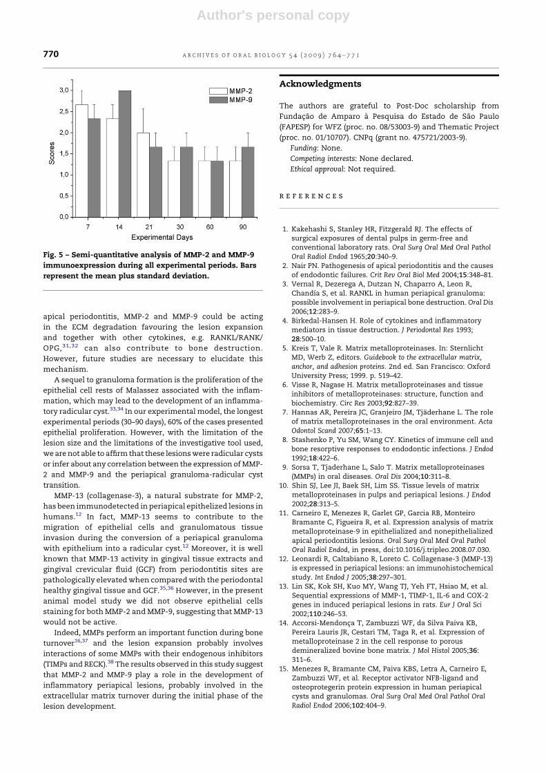
The MMP-2 and MMP-9 immunoexpression did not differ
significantly between 7–21 days (p > 0.05, unpaired t-test with
a r c h i v e s o f o r a l b i o l o g y 5 4 ( 2 0 0 9 ) 7 6 4 – 7 7 1 765

Author's personal copy
Welch correction) and 30–90 days (p > 0.05). However, the
MMP-2 and MMP-9 immunostaining at 7, 14 and 21 days
was higher than at 30, 60 and 90 days (p < 0.05, unpaired t-test
with Welch correction) (Fig. 5).
In the earliest periods, an acute inflammatory infiltrate
was predominant with a suggestive intense presence
of leukocytes, notably infiltrating neutrophils. The
immunostaining was intense for both MMP-2 and MMP-9
(Fig. 2A–D).
In the subsequent periods, the immunostaining was not
intense as previously described; it was dispersed and mainly
observed in isolated mononuclear cells.
After day 30, cells in the interface of the granulomatous
and exudative zones19 showed immunostaining for both
MMPs (Fig. 3A and B). A few cells presented immunostain-
ing in the fibrous zone, except for mononuclear cells
suggestive of fibroblasts, lymphocytes and macrophages
(Fig. 4A and B).
Fig. 1 – Serial sections of human breast cancer (hBC) biopsies were used as positive control for both MMPs. Letters A and C
represent MMP-2 and MMP-9 staining respectively (positive control). Negative controls consisted of sections of hBC in
which primary antibodies were omitted and replaced with non-immune murine serum (B) and (D). Letters (E) and (F)
summarize the negative control by using sections of 14th day of periapical lesion development. Magnification: 40T.
a r c h i v e s o f o r a l b i o l o g y 5 4 ( 2 0 0 9 ) 7 6 4 – 7 7 1766

Author's personal copy
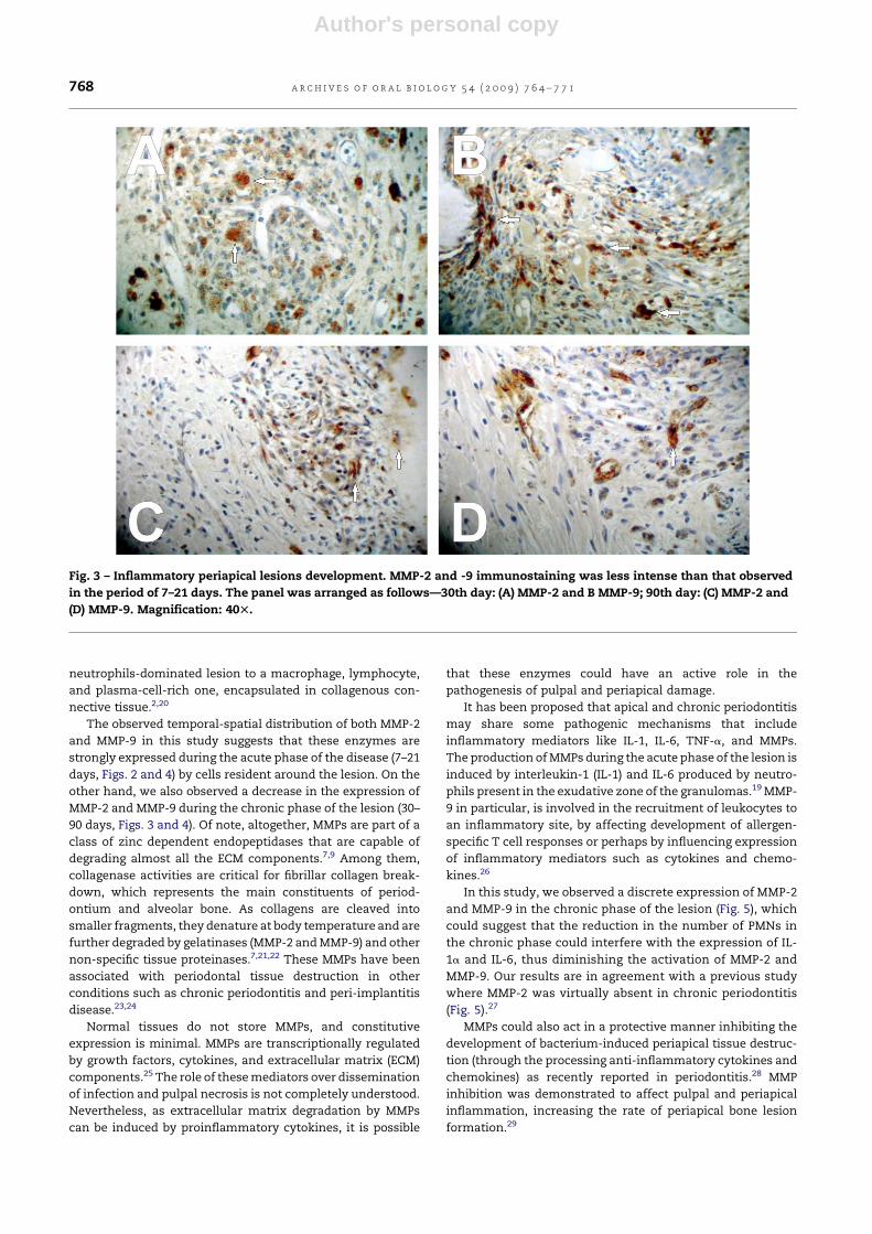
The periodontal ligament in the distant areas from the
lesion presented rare and smooth staining that became
intensified in the proximity of the exudative zone.19 Cemen-
toblast-like cells were noted strongly stained in hypercemen-
tous areas after 21 days (Fig. 4C).
Endothelial cells also demonstrated immunostaining for
both MMP-2 and MMP-9 in all periods (Fig. 4D). Sixty percent of
the cases assessed presented an epithelium after 30 days of
lesion development, however, no staining was found in any
period for both MMPs.
At the interface of the periapical lesion with the bone
tissue, rare cells suggestive of osteoblasts demonstrated
cytoplasmatic immunostaining (Fig. 4E). In addition, the most
of osteoclast-like cells presented weak cytoplasmatic staining
in all the experimental periods (Fig. 4F). On the other hand,
osteocyte did not present positive for both MMP-2 and MMP-9
staining in any of the assessed periods (Fig. 4F).
4. Discussion
The biological model used in this study is considered
satisfactory for observing the periapical lesions development
in all of its phases.13,20 Based on histological outcomes and
current literature, two distinct phases were observed: (1)
growth/acute phase (between 7 and 21 days), generally caused
by micro-organisms residing in or invading from the apical
root canal into the periapical tissues,2 and (2) a chronic phase
(period longer than 21 days), caused by a prolonged presence
of microbial irritants leading to a shift in the infiltrating
Fig. 2 – Inflammatory periapical lesions development. Both MMPs-2 and -9 were expressed by infiltrating mononuclear cells
surrounding the apical foramen and adjacent periapical areas. The panel was arranged as follows—7th day: (A) MMP-2 and
(B) MMP-9; 14th day: (C) MMP-2 and (D) MMP-9; 21st day: (E) MMP-2 and (F) MMP-9. Magnification: 40T.
a r c h i v e s o f o r a l b i o l o g y 5 4 ( 2 0 0 9 ) 7 6 4 – 7 7 1 767
Author's personal copy
neutrophils-dominated lesion to a macrophage, lymphocyte,
and plasma-cell-rich one, encapsulated in collagenous con-
nective tissue.2,20
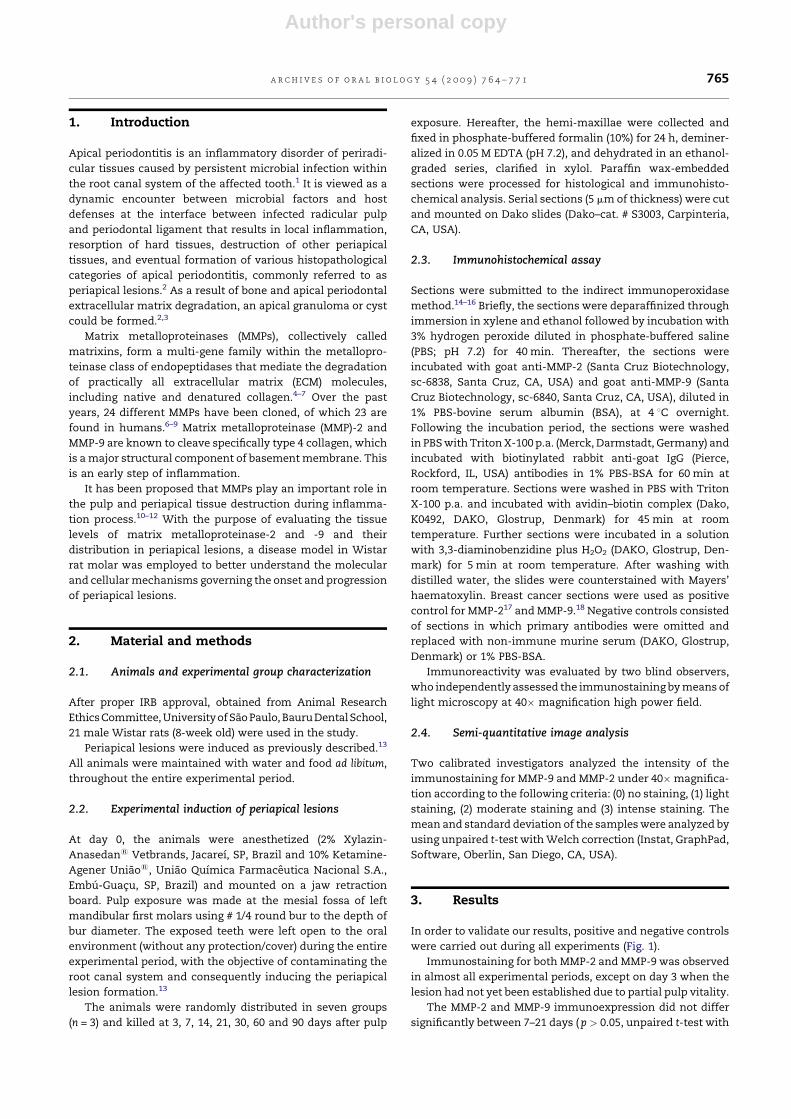
The observed temporal-spatial distribution of both MMP-2
and MMP-9 in this study suggests that these enzymes are
strongly expressed during the acute phase of the disease (7–21
days, Figs. 2 and 4) by cells resident around the lesion. On the
other hand, we also observed a decrease in the expression of
MMP-2 and MMP-9 during the chronic phase of the lesion (30–
90 days, Figs. 3 and 4). Of note, altogether, MMPs are part of a
class of zinc dependent endopeptidases that are capable of
degrading almost all the ECM components.7,9 Among them,
collagenase activities are critical for fibrillar collagen break-
down, which represents the main constituents of period-
ontium and alveolar bone. As collagens are cleaved into
smaller fragments, they denature at body temperature and are
further degraded by gelatinases (MMP-2 and MMP-9) and other
non-specific tissue proteinases.7,21,22 These MMPs have been
associated with periodontal tissue destruction in other
conditions such as chronic periodontitis and peri-implantitis
disease.23,24
Normal tissues do not store MMPs, and constitutive
expression is minimal. MMPs are transcriptionally regulated
by growth factors, cytokines, and extracellular matrix (ECM)
components.25 The role of these mediators over dissemination
of infection and pulpal necrosis is not completely understood.
Nevertheless, as extracellular matrix degradation by MMPs
can be induced by proinflammatory cytokines, it is possible
that these enzymes could have an active role in the
pathogenesis of pulpal and periapical damage.
It has been proposed that apical and chronic periodontitis
may share some pathogenic mechanisms that include
inflammatory mediators like IL-1, IL-6, TNF-a, and MMPs.
The production of MMPs during the acute phase of the lesion is
induced by interleukin-1 (IL-1) and IL-6 produced by neutro-
phils present in the exudative zone of the granulomas.19 MMP-
9 in particular, is involved in the recruitment of leukocytes to
an inflammatory site, by affecting development of allergen-
specific T cell responses or perhaps by influencing expression
of inflammatory mediators such as cytokines and chemo-
kines.26
In this study, we observed a discrete expression of MMP-2
and MMP-9 in the chronic phase of the lesion (Fig. 5), which
could suggest that the reduction in the number of PMNs in
the chronic phase could interfere with the expression of IL-
1a and IL-6, thus diminishing the activation of MMP-2 and
MMP-9. Our results are in agreement with a previous study
where MMP-2 was virtually absent in chronic periodontitis
(Fig. 5).27
MMPs could also act in a protective manner inhibiting the
development of bacterium-induced periapical tissue destruc-
tion (through the processing anti-inflammatory cytokines and
chemokines) as recently reported in periodontitis.28 MMP
inhibition was demonstrated to affect pulpal and periapical
inflammation, increasing the rate of periapical bone lesion
formation.29
Fig. 3 – Inflammatory periapical lesions development. MMP-2 and -9 immunostaining was less intense than that observed
in the period of 7–21 days. The panel was arranged as follows—30th day: (A) MMP-2 and B MMP-9; 90th day: (C) MMP-2 and
(D) MMP-9. Magnification: 40T.
a r c h i v e s o f o r a l b i o l o g y 5 4 ( 2 0 0 9 ) 7 6 4 – 7 7 1768
Author's personal copy
Tsubota et al. reported that MMP-8 expression in
osteoblasts, osteocytes, periodontal ligament cells, cemen-
toblasts, and cementocytes may be involved in remodelling
the periodontium during tooth eruption.30 A similar process
of periodontium remodelling was observed during periapi-
cal pathology installation and development. Here, period-
ontal ligament cells presented intense immunostaining for
both MMP-2 and MMP-9 when they are close to the lesion. In
addition, osteoblast- and osteoclast-like cells showed dis-
crete staining in all periods evaluated, while osteocytes
were not positive for MMP-2 and MMP-9 staining. These
events suggest that during the initial development of the
Fig. 4 – Some biological events found during inflammatory periapical lesions development. (A and B) Mononuclear cells
(fibroblast- and macrophage-like cells) at 21st day presenting MMP-2 immunostaining mainly localized in the connective
tissue (CT). The photomicrography (C) brings out the hypercementous areas (H) presenting cementoblast-like cells
immunopositive to MMP-9 (31st day). In all periods in this study, the presence of endothelial cells immunostained for both
enzymes was observed. Thus, (D) shows endothelial cells immunostained for MMP-9 (21st day). The (E) and (F) detail the
bone tissue and bring out some bone cells immunostained. In (E) osteoblast-like cells were immunopositive to MMP-9 (large
arrow, 21st day) and (F) immunostaining for both MMPs was not observed in osteocyte-like cell (arrows). Also in (F), note
that osteoclast-like cells were slightly immunostained (*, 21st day). Magnification: 40T.
a r c h i v e s o f o r a l b i o l o g y 5 4 ( 2 0 0 9 ) 7 6 4 – 7 7 1 769
Author's personal copy
apical periodontitis, MMP-2 and MMP-9 could be acting
in the ECM degradation favouring the lesion expansion
and together with other cytokines, e.g. RANKL/RANK/
OPG,31,32 can also contribute to bone destruction.
However, future studies are necessary to elucidate this
mechanism.
A sequel to granuloma formation is the proliferation of the
epithelial cell rests of Malassez associated with the inflam-
mation, which may lead to the development of an inflamma-
tory radicular cyst.33,34 In our experimental model, the longest
experimental periods (30–90 days), 60% of the cases presented
epithelial proliferation. However, with the limitation of the
lesion size and the limitations of the investigative tool used,
we are not able to affirm that these lesions were radicular cysts
or infer about any correlation between the expression of MMP-
2 and MMP-9 and the periapical granuloma-radicular cyst
transition.
MMP-13 (collagenase-3), a natural substrate for MMP-2,
has been immunodetected in periapical epithelized lesions in
humans.12 In fact, MMP-13 seems to contribute to the
migration of epithelial cells and granulomatous tissue
invasion during the conversion of a periapical granuloma
with epithelium into a radicular cyst.12 Moreover, it is well
known that MMP-13 activity in gingival tissue extracts and
gingival crevicular fluid (GCF) from periodontitis sites are
pathologically elevated when compared with the periodontal
healthy gingival tissue and GCF.35,36 However, in the present
animal model study we did not observe epithelial cells
staining for both MMP-2 and MMP-9, suggesting that MMP-13
would not be active.
Indeed, MMPs perform an important function during bone
turnover16,37 and the lesion expansion probably involves
interactions of some MMPs with their endogenous inhibitors
(TIMPs and RECK).38 The results observed in this study suggest
that MMP-2 and MMP-9 play a role in the development of
inflammatory periapical lesions, probably involved in the
extracellular matrix turnover during the initial phase of the
lesion development.
Acknowledgments
The authors are grateful to Post-Doc scholarship from
Fundacao de Amparo a Pesquisa do Estado de Sao Paulo
(FAPESP) for WFZ (proc. no. 08/53003-9) and Thematic Project
(proc. no. 01/10707). CNPq (grant no. 475721/2003-9).
Funding: None.
Competing interests: None declared.
Ethical approval: Not required.
r e f e r e n c e s
1. Kakehashi S, Stanley HR, Fitzgerald RJ. The effects ofsurgical exposures of dental pulps in germ-free andconventional laboratory rats. Oral Surg Oral Med Oral PatholOral Radiol Endod 1965;20:340–9.
2. Nair PN. Pathogenesis of apical periodontitis and the causesof endodontic failures. Crit Rev Oral Biol Med 2004;15:348–81.
3. Vernal R, Dezerega A, Dutzan N, Chaparro A, Leon R,Chandıa S, et al. RANKL in human periapical granuloma:possible involvement in periapical bone destruction. Oral Dis2006;12:283–9.
4. Birkedal-Hansen H. Role of cytokines and inflammatorymediators in tissue destruction. J Periodontal Res 1993;28:500–10.
5. Kreis T, Vale R. Matrix metalloproteinases. In: SternlichtMD, Werb Z, editors. Guidebook to the extracellular matrix,anchor, and adhesion proteins. 2nd ed. San Francisco: OxfordUniversity Press; 1999. p. 519–42.
6. Visse R, Nagase H. Matrix metalloproteinases and tissueinhibitors of metalloproteinases: structure, function andbiochemistry. Circ Res 2003;92:827–39.
7. Hannas AR, Pereira JC, Granjeiro JM, Tjaderhane L. The roleof matrix metalloproteinases in the oral environment. ActaOdontol Scand 2007;65:1–13.
8. Stashenko P, Yu SM, Wang CY. Kinetics of immune cell andbone resorptive responses to endodontic infections. J Endod1992;18:422–6.
9. Sorsa T, Tjaderhane L, Salo T. Matrix metalloproteinases(MMPs) in oral diseases. Oral Dis 2004;10:311–8.
10. Shin SJ, Lee JI, Baek SH, Lim SS. Tissue levels of matrixmetalloproteinases in pulps and periapical lesions. J Endod2002;28:313–5.
11. Carneiro E, Menezes R, Garlet GP, Garcia RB, MonteiroBramante C, Figueira R, et al. Expression analysis of matrixmetalloproteinase-9 in epithelialized and nonepithelializedapical periodontitis lesions. Oral Surg Oral Med Oral PatholOral Radiol Endod, in press, doi:10.1016/j.tripleo.2008.07.030.
12. Leonardi R, Caltabiano R, Loreto C. Collagenase-3 (MMP-13)is expressed in periapical lesions: an immunohistochemicalstudy. Int Endod J 2005;38:297–301.
13. Lin SK, Kok SH, Kuo MY, Wang TJ, Yeh FT, Hsiao M, et al.Sequential expressions of MMP-1, TIMP-1, IL-6 and COX-2genes in induced periapical lesions in rats. Eur J Oral Sci2002;110:246–53.
14. Accorsi-Mendonca T, Zambuzzi WF, da Silva Paiva KB,Pereira Lauris JR, Cestari TM, Taga R, et al. Expression ofmetalloproteinase 2 in the cell response to porousdemineralized bovine bone matrix. J Mol Histol 2005;36:311–6.
15. Menezes R, Bramante CM, Paiva KBS, Letra A, Carneiro E,Zambuzzi WF, et al. Receptor activator NFB-ligand andosteoprotegerin protein expression in human periapicalcysts and granulomas. Oral Surg Oral Med Oral Pathol OralRadiol Endod 2006;102:404–9.
Fig. 5 – Semi-quantitative analysis of MMP-2 and MMP-9
immunoexpression during all experimental periods. Bars
represent the mean plus standard deviation.
a r c h i v e s o f o r a l b i o l o g y 5 4 ( 2 0 0 9 ) 7 6 4 – 7 7 1770

Author's personal copy
16. Accorsi-Mendonca T, Paiva KB, Zambuzzi WF, Cestari TM,Lara VS, Taga R, et al. Expression of matrixmetalloproteinases-2 and -9 and RECK during alveolar boneregeneration in rat. J Mol Histol 2008;39:201–8.
17. Monteagudo C, Merino MJ, San-Juan J, Liotta LA, Stetler-Stevenson WG. Immunohistochemical distribution of typeIV collagenase in normal, benign, and malignant breasttissue. Am J Pathol 1990;136:585–92.
18. Ravindranath MH, Muthugounder S, Presser N,Viswanathan S. Anticancer therapeutic potential of soyisoflavone, genistein. Adv Exp Med Biol 2004;546:121–65.
19. Marton IJ, Kiss C. Protective and destructive immunereactions in apical periodontitis. Oral Microbiol Immunol2000;15:139–50.
20. Stashenko P, Wang CY, Tani-Ishii N, Yu SM. Pathogenesis ofinduced rat periapical lesions. Oral Surg Oral Med Oral Pathol1994;78:494–502.
21. Chung L, Dinakarpandian D, Yohida N, Lauer-Fields JL,Fields B, Visse R, et al. Collagenase unwinds triple-helicalcollagen prior to peptide bond hydrolysis. EMBO J2004;23:3020–30.
22. Pozo P, Valenzuela MA, Melej C, Zaldıvar M, Puente J,Gamonal J. Longitudinal analysis of metalloproteinases,TIMPs and clinical parameters in gingival crevicular fluidfrom periodontitis-affected patients. J Periodontal Res2005;40:199–207.
23. Ingman T, Tervahartiala T, Ding Y, Tsesche H, Haerian A,Kinane DF, et al. Matrix metalloproteinases and theirinhibitors in gingival crevicular fluid and saliva ofperiodontitis patients. J Clin Periodontol 1996;23:1127–32.
24. Puente X, Sanchez L, Overall C, Lopez-Otın C. Human andmouse proteases: a comparative genomic approach. Nat RevGenet 2003;4:544–56.
25. Shapiro SD, Senior RM. Matrix metalloproteinases. Matrixdegradation and more. Am J Respir Cell Mol Biol 1999;20:1100–2.
26. McMillan SJ, Kearley J, Campbell JD, Zhu XW, Larbi KY,Shipley JM, et al. Matrix metalloproteinase-9 deficiencyresults in enhanced allergen-induced airway inflammation.J Immunol 2004;172:2586–94.
27. Goncalves LD, Oliveira G, Hurtado PA, Feitosa A, Takiya CM,Granjeiro JM, et al. Expression of metalloproteinases andtheir tissue inhibitors in inflamed gingival biopsies. JPeriodontal Res 2008;43:570–7.
28. Kuula H, Salo T, Pirila E, Tuomainen AM, Jauhiainen M, UittoVJ, et al. Local and systemic responses in matrix
metalloproteinase 8-deficient mice during Porphyromonasgingivalis-induced periodontitis. Infect Immun 2009;77:850–9.
29. Tjaderhane L, Hotakainen T, Kinnunen S, Ahonen M, Salo T.The effect of chemical inhibition of matrixmetalloproteinases on the size of experimentally inducedapical periodontitis. Int Endod J 2007;40:282–9.
30. Tsubota M, Sasano Y, Takahashi I, Kagayama M, ShimauchiH. Expression of MMP-8 and MMP-13 mRNAs in ratperiodontium during tooth eruption. J Dent Res 2002;81:673–8.
31. Kawashima N, Suzuki N, Yang G, Ohi C, Okuhara S, Nakano-Kawanishi H, et al. Kinetics of RANKL, RANK and OPGexpressions in experimentally induced rat periapicallesions. Oral Surg Oral Med Oral Pathol Oral Radiol Endod2007;103:707–11.
32. Menezes R, Garlet TP, Letra A, Bramante CM, Campanelli AP,Figueira R, et al. Differential patterns of receptor activator ofnuclear factor kappa B ligand/osteoprotegerin expression inhuman periapical granulomas: possible association withprogressive or stable nature of the lesions. J Endod2008;34:932–8.
33. Gao Z, Mackenzie IC, Rittman BR, Korszun AK, Williams DM,Cruchley AT. Immunocytochemical examination ofimmune cells in periapical granulomata and odontogeniccysts. J Oral Pathol 1988;17:84–90.
34. Patas S, Nakou M, Rontogianni D. Inflammatory infiltrate ofchronic periradicular lesions: an immunohistochemicalstudy. Int Endod J 2003;36:464–71.
35. Hernandez M, Martınez B, Tejerina JM, Valenzuela MA,Gamonal J. MMP-13 and TIMP-1 determinations inprogressive chronic periodontitis. J Clin Periodontol2007;34:729–35.
36. Ilgenli T, Vardar-Sengul S, Gurkan A, Sorsa T, Stackelberg S,Kose T, et al. Gingival crevicular fluid matrixmetalloproteinase-13 levels and molecular formsin various types of periodontal diseases. Oral Dis 2006;12:573–9.
37. Zambuzzi WF, Yano CL, Cavagis ADM, Peppelenbosch MP,Granjeiro JM, Ferreira CV. Ascorbate-induced osteoblastdifferentiation recruits distinct MMP-inhibitors: RECK andTIMP-2. Mol Cell Biochem 2009;322:143–50.
38. Lin SK, Chiang CP, Hong CY, Lin CP, Lan WH, Hsieh CC, et al.Immunolocalization of interstitial collagenase (MMP-1) andtissue inhibitor of metalloproteinases-1 (TIMP-1) inradicular cysts. J Oral Pathol Med 1997;26:458–63.
a r c h i v e s o f o r a l b i o l o g y 5 4 ( 2 0 0 9 ) 7 6 4 – 7 7 1 771