Role for Periodontitis in the Progression of Lipid Deposition in an Animal Model
8
10.1128/IAI.71.10.6012-6018.2003. 2003, 71(10):6012. DOI: Infect. Immun. L. Stahl and Thomas E. Van Dyke Ashish Jain, Eraldo L. Batista Jr., Charles Serhan, Gregory Lipid Deposition in an Animal Model Role for Periodontitis in the Progression of http://iai.asm.org/content/71/10/6012 Updated information and services can be found at: These include: REFERENCES http://iai.asm.org/content/71/10/6012#ref-list-1 This article cites 21 articles, 6 of which can be accessed free at: CONTENT ALERTS more» articles cite this article), Receive: RSS Feeds, eTOCs, free email alerts (when new http://journals.asm.org/site/misc/reprints.xhtml Information about commercial reprint orders: http://journals.asm.org/site/subscriptions/ To subscribe to to another ASM Journal go to: on November 18, 2014 by guest http://iai.asm.org/ Downloaded from on November 18, 2014 by guest http://iai.asm.org/ Downloaded from
-
Upload
independent -
Category
Documents
-
view
0 -
download
0
Transcript of Role for Periodontitis in the Progression of Lipid Deposition in an Animal Model

10.1128/IAI.71.10.6012-6018.2003.
2003, 71(10):6012. DOI:Infect. Immun. L. Stahl and Thomas E. Van DykeAshish Jain, Eraldo L. Batista Jr., Charles Serhan, Gregory Lipid Deposition in an Animal ModelRole for Periodontitis in the Progression of
http://iai.asm.org/content/71/10/6012Updated information and services can be found at:
These include:
REFERENCEShttp://iai.asm.org/content/71/10/6012#ref-list-1This article cites 21 articles, 6 of which can be accessed free at:
CONTENT ALERTS more»articles cite this article),
Receive: RSS Feeds, eTOCs, free email alerts (when new
http://journals.asm.org/site/misc/reprints.xhtmlInformation about commercial reprint orders: http://journals.asm.org/site/subscriptions/To subscribe to to another ASM Journal go to:
on Novem
ber 18, 2014 by guesthttp://iai.asm
.org/D
ownloaded from
on N
ovember 18, 2014 by guest
http://iai.asm.org/
Dow
nloaded from

INFECTION AND IMMUNITY, Oct. 2003, p. 6012–6018 Vol. 71, No. 100019-9567/03/$08.00�0 DOI: 10.1128/IAI.71.10.60120–6018.2003Copyright © 2003, American Society for Microbiology. All Rights Reserved.
Role for Periodontitis in the Progression of Lipid Depositionin an Animal Model
Ashish Jain,1 Eraldo L. Batista, Jr.,1 Charles Serhan,2 Gregory L. Stahl,2and Thomas E. Van Dyke1*
Department of Periodontology and Oral Biology, Goldman School of Dental Medicine, Boston University,1 and Center forExperimental Therapeutics and Reperfusion Injury, Department of Anesthesiology, Preoperative and Pain Medicine,
Brigham and Women’s Hospital, Harvard Medical School,2 Boston, Massachusetts
Received 12 February 2003/Returned for modification 29 April 2003/Accepted 30 June 2003
Epidemiologic studies have implicated periodontitis as a risk factor for the development of cardiovasculardisease. However, no prospective studies investigating this potential relationship have been carried out. Age-and sex-matched New Zealand White rabbits were maintained on a diet consisting of 0.5% fat for 13 weeks toinduce the accumulation of lipid deposits in the aorta as a model for atherogenesis. One-half of the animalsreceived silk ligatures around their mandibular premolars followed by an application of a periodontal patho-gen, Porphyromonas gingivalis, to induce periodontitis. Animals were sacrificed after 14 weeks. Periodontaldisease severity was quantified radiographically, histologically, and by direct visualization of bone loss ondefleshed skulls. Lipid deposition was evaluated by computer-assisted morphometry in the aortas en face afterlipid deposits were stained with Sudan IV. Animals with experimentally induced periodontitis had moreextensive accumulations of lipids in the aorta than did nonperiodontitis animals (P < 0.05), and there was apositive correlation between the severity of periodontal disease and the extent of lipid deposition (r2 � 0.9501).The results provide direct evidence that periodontitis may be a risk factor and may contribute to the patho-genesis of atherosclerosis. The data support the concept that infections at remote locations can modulateatherosclerotic events distantly.
Atherosclerosis is a progressive disease process that involveslarge- to medium-sized muscular and large elastic arteries.Complications associated with atherosclerosis, including coro-nary thrombosis and myocardial infarction, contribute to over50% of the deaths related to this disease in the United States(13). There is general agreement implicating a role for innateimmune activation and inflammation in the pathogenesis ofatherosclerosis, thrombosis, and neointimal thickening afterarterial injury (18). While a role for inflammation is generallyaccepted, the initiating factors leading to the inflammatoryinjury remain speculative. Recent evidence has led to hypoth-eses implicating the involvement of one or more infectiousagents in the initiation of atherosclerosis, either through directdamage to the vessel wall or through nonspecific activation ofinnate immune pathways (7, 18, 19). A variety of pathogens,including Chlamydia pneumoniae, Helicobacter pylori, Cytomeg-alovirus, Herpes simplex virus, Streptococcus sanguinis, and Por-phyromonas gingivalis, have been associated with the pathogen-esis of atherosclerosis and have been detected in humanatheromas (4, 6).
Adult periodontitis is a chronic inflammatory disease result-ing from infections by specific microbial species that colonizethe oral cavity. The pathogens are mainly gram-negative an-aerobic species and include P. gingivalis, Bacteroides forsythus,and Treponema denticola. While these organisms clearly initi-ate disease, the pathogenesis of periodontitis is thought to be
mediated for the most part by host-mediated tissue injuryresulting from inflammation. As a result of periodontal infec-tion and inflammation, there is a loss of epithelial integritywithin the periodontal pocket, which results in bacteremiafollowing manipulation of the oral tissues. In a patient withperiodontitis, even daily routine procedures like tooth brush-ing and chewing have been shown to induce a transient bacte-remia (20). Thus, periodontitis is a chronic infection which canresult in repeated systemic exposure to gram-negative bacteriaand bacterial products.
Recent epidemiologic studies have implicated periodontitisas a risk factor for atheromatous changes in blood vessels andsubsequent vascular events (1). There are two prevailing hy-potheses for the relationship between periodontitis and car-diovascular disease. First, periodontal bacteria may have adirect effect on the vasculature. P. gingivalis is a common peri-odontal pathogen that has an interesting set of virulence fac-tors, including hemagglutination, trypsin-like proteases (gingi-pains), and the ability to invade epithelial and endothelial cells(5). It has been proposed that P. gingivalis invasion is an initi-ating event in the pathogenesis of atheromas, and P. gingivalisDNA has been detected in carotid atheromas (6). An alterna-tive hypothesis is that local inflammation causes an enhancedinflammatory response at distant sites without the spread ofthe infectious agent. In one study, P. gingivalis injected into amurine air pouch induced COX-2 mRNA expression in theheart and lungs 3 h later (16). These data suggest that a localor focal infection with this pathogen may have a systemicimpact on the status of the innate immune system. In a recentpaper (11), Li and coworkers demonstrated with an apoEknockout mouse model that bacteremia caused by intravenous
* Corresponding author. Mailing address: Goldman School of Den-tal Medicine, Department of Periodontology and Oral Biology, 100East Newton St., Rm G-05, Boston, MA 02118. Phone: (617) 638-4758.Fax: (617) 638-4799. E-mail: [email protected].
6012
on Novem
ber 18, 2014 by guesthttp://iai.asm
.org/D
ownloaded from

injections of P. gingivalis resulted in increased lipid depositionin major vessels. In our present study, we sought to evaluatethe effect of periodontitis on the cardiovascular outcome of ahigh-fat diet in a rabbit model to test the hypothesis thatperiodontitis will enhance lipid deposition in major vessels.
MATERIALS AND METHODS
Animal model. A total of 12 male New Zealand White rabbits (MilbrookBreeding Laboratories) matched for age and weight were used. The IACUCcommittee of Brigham and Women’s Hospital approved the animal protocol.The animals were individually housed with regular day and night light exposure(a cycle of 10 h of light and 14 h of darkness). All animals received water adlibitum and were fed a high-fat diet (0.5% [wt/wt] cholesterol) (Harlan Teklad).The animals were divided randomly into two groups of six animals each and wereanesthetized (40 mg of ketamine/kg of body weight and 9 mg of xylazine/kg ofbody weight). Animals in the test group (i.e., periodontal group) had a 3-0 silksuture placed around the second premolar of both mandibular quadrants. P.gingivalis (strain A7436) was grown as previously described (10, 22). Briefly, cellswere cultured on agar plates containing Trypticase soy agar supplemented with
0.5% (wt/vol) yeast extract, 5% defibrinated sheep red blood cells, 5 �g of heminper ml, and 1 �g of vitamin K per ml. Plates were incubated for 3 days at 37°Cin jars that were anaerobically maintained through palladium-catalyzed hydro-gen-carbon dioxide envelopes (GasPak Plus; Becton Dickinson MicrobiologySystems, Sparks, Md.). Colonies were randomly selected and anaerobically sub-cultured overnight at 37°C in Schaedler’s broth supplemented with vitamin K andhemin. Cell numbers were spectrophotometrically determined at 600 nm (11),and 109 CFU (0.8 optical density) was mixed with carboxymethycellulose to forma thick slurry which was applied topically to the ligated teeth. The control groupreceived carboxymethylcellulose slurry without P. gingivalis. For the first 7 weeksof the experiment, 109 CFU of the P. gingivalis slurry was applied topicallyaround the suture on Monday, Wednesday, and Friday, with the rabbits underanesthesia (isofluorane). At these times, the sutures were also checked, and lostor loose sutures were replaced. The control animals were anesthetized using thesame protocol and were given the carboxymethylcellulose slurry only, without P.gingivalis. Weekly measurements of body weight and standard hematologicalparameters, including clinical chemistry (complete blood count and total choles-terol levels), were recorded for all animals.
Specimen collection. After 14 weeks, the rabbits were euthanized by pento-barbital overdose and the aortas were removed for en face quantification. Themandibles were removed for periodontal lesion quantification.
Aorta and atherosclerotic lesion quantitation. After euthanasia, the entireaorta from each rabbit was perfused for 20 min with ice-cold phosphate-bufferedsaline, pH 7.4, containing 20 �M butylated hydroxy toluene (Sigma) and 2 �MEDTA by using a cannula inserted in the left ventricle. The aorta was thenpressure fixed for 20 min with cold formaldehyde-sucrose solution (10% neutralformalin, 5% sucrose, 20 �M butylated hydroxy toluene, and 2 �M EDTA [pH7.4]). The entire aorta was dissected free, adventitial fat was removed, and theaorta was opened longitudinally. The aorta was briefly rinsed in 70% ethanolfollowed by staining (with 0.5% Sudan IV–35% ethanol–50% acetone) for 6 min.Destaining was performed with 80% ethanol for 5 min (9). Each aorta was thenmounted on a flat surface, and digital images of the aorta surface were obtained(Olympus digital camera). Images were stored in PICT format on a compact disc,and analyses were performed as previously described (11). The area covered bythe lipid plaque was expressed as a percentage of the total surface area of theaorta. This area was expressed as pixels rather than millimeters to adjust for anyvariations in the captured image.
Periodontal lesion characterization. After euthanasia, the mandible of eachrabbit was dissected free of muscles and soft tissue; the attached gingiva was keptintact with the alveolar bone. The mandible was split into two halves from themidline between the central incisors. The left half was taken for morphometricanalysis of the bone loss, and the right half was used for histological evaluationof periodontitis.
Macroscopic analysis of bone levels. The left half of the mandible was de-fleshed by immersion in 10% hydrogen peroxide for 10 min. The soft tissue wascarefully removed, and the mandible was stained with methylene blue for visualdistinction between the teeth and bone. The bone level around the secondpremolar was measured directly with a calibrated (0.5 mm) periodontal probe.Measurements were made from the top of the tooth to the bone crest at threepoints each, at the buccal and lingual sides. A mean crestal bone level around thetooth was calculated. Similarly, for the proximal bone level, measurements weremade at the mesial and distal aspects of the tooth. The measurements were takenfrom the buccal and lingual sides on the proximal aspects of the second premolar,and the mean proximal bone level was calculated. Bone level was also quantifiedby image analysis (Image Pro 4). The sectioned mandible was mounted andphotographed using an inverted microscope at a magnification of �10. The
TABLE 1. Cholesterol levels, blood cell counts, and body weight outcomes between groups throughout the studya
Group time point Cholesterol(mg/dl)b
leukocytes(103/�l)
Lymphocytes(103/�l)
Monocytes(103/�l)
Granulocytes(103/�l) Wt (kg)
ControlBaseline 14 � 40 6 � 0.80 4.16 � 0.27 0.91 � 0.47 0.91 � 0.34 3.15 � 0.0714 wk 731 � 349 8.67 � 2.63 6.47 � 1.65 0.74 � 0.41 1.75 � 0.58 3.6 � 0.24
PeriodontitisBaseline 13 � 4 10.52 � 3.04 7.46 � 1.71 1.8 � 1.2 1.26 � 0.40 3.25 � 0.1314 wk 858 � 418 10.93 � 2.44 8.2 � 2.09 1.14 � 0.10 1.59 � 0.54 3.7 � 0.33
a Values are means � standard errors of the means. Differences between control and periodontitis group results were not significant (P � 0.05).b Difference between 14-week and baseline values for both groups were significant (P � 0.01).
TABLE 2. Changes in the periodontal supporting bone after 14weeks in the periodontitis and control groups
Group and rabbitMean bone loss (mm)a
% Tooth in boneCrestal Proximal
Periodontitis54 4.41 4 8455 3.76 3 81.3556 10.24 6.75 53.457 7.67 5.75 67.758 5.16 5 68.75
Mean � SEM(n � 5)
�6.24 � 2.67c 4.9 � 1.46b 71.04 � 12.16
Mean � SEM(n � 3)
7.69 � 2.54c 5.8 � 0.87b 63.28 � 8.5
Control59 4.33 3 82.5460 3.91 2.5 79.561 3.66 3.87 86.262 2.58 3.5 82.7563 2.75 3.87 82.7564 4.58 3.12 81.67
Mean � SEM(n � 5)
3.63 � 0.81 3.31 � 0.53 82.56 � 2.1
a Expressed as the distance between a fixed reference point (tooth cusp tip)and the bone crest.
b Significantly different from value for control group at a P of �0.05.c Significantly different from value for control group at a P of �0.005.
VOL. 71, 2003 PERIODONTAL INFLAMMATION AND AORTIC LIPID DEPOSITION 6013
on Novem
ber 18, 2014 by guesthttp://iai.asm
.org/D
ownloaded from
captured image was analyzed as described above, and the mean crestal bone levelaround the tooth was calculated in millimeters.
Radiographic analysis. The percentage of the tooth in bone was calculatedradiographically using a modification of the Bjorn technique (3). The radio-graphs were taken by digital X ray (Schick Technologies). To quantify bone loss,the length of the tooth from the cusp tip to the apex of the root was measured,as was the length of the tooth structure outside the bone, from the cusp tip to thecoronal extent of the proximal bone. From this, the length of tooth within thebone was measured. Bone values are expressed as the percentage of the tooth inbone obtained through the following relationship: [(length of tooth in bone)/(total length of tooth)] � 100.
Histological analysis. Bone and teeth of the other mandible halves weredecalcified, processed, and embedded in paraffin. Thin sections (thickness, 7 �m)were cut and stained conventionally with hematoxylin and eosin to identify thecellular composition of the inflammatory infiltrate.
P. gingivalis 16S rRNA amplification by PCR. Formalin-fixed aortic samples(approximately 90 mg each) were cut from the proximal, thoracic, and abdominalportions, frozen in liquid nitrogen, and then ground. The residual fixative wasremoved by three 10-ml changes of 50 mM Tris-HCl (pH 8.0) for 1 h each (23).DNA was isolated through phenol-chloroform extraction and ethanol precipita-tion and used as a template for PCR with specific primers (F, 5�-CGGTGCCAGCCGCGGTAATACG-3�; R, 5�-TACATAGAAGCCCCGAAGGAAGAC-3�)for a 520-bp P. gingivalis 16S rRNA fragment (21). We used 50-�l PCR mixes,which included approximately 200 ng of total genomic DNA, 3.5 U of Taqpolymerase (Expand High Fidelity; Roche Applied Science), and primers andMgCl2 to final concentrations of 300 nM and 2 mM, respectively. Thermal cycler(Perkin-Elmer model 9600) conditions were set at 94°C for 2 min and 35 cyclesof a 3-step PCR (94, 56, and 72°C for 1 min each), followed by a final elongationcycle of 72°C for 10 min. As an optimization step in the PCR approach, wesought to determine the detection threshold of the system by using pure P.gingivalis cultures. To this end, P. gingivalis cells were cultured for 3 days inSchaedler’s broth and cell numbers were spectrophotometrically estimated asdescribed above. Tenfold serial dilutions of P. gingivalis cells ranging from 106 to101 CFU were then amplified by using the aforementioned conditions. DNAproducts were resolved in 1.2% agarose gel, stained with ethidium bromide, andanalyzed through UV light.
Statistical analysis. All of the en face and periodontal measurements weredone in a blind fashion on coded samples, and the quantitative measurementswere made twice. The extent of lipid deposit, periodontal disease, and otherstudy parameters were analyzed by Student’s t test (two tailed). A P value of�0.05 was considered significant. The relationship between the extent of lipiddeposition and the extent of periodontal disease was analyzed by the Pearson
correlation coefficient. All values in the text, tables, and figures are means �standard errors of the means.
RESULTS
All animals survived the experiment. No clinical signs ofinfection or illness were noted in any of the animals at anytime. Both groups of animals had comparable levels of totalcholesterol in their sera. At baseline, the total level of choles-terol was 14 � 4 and 13 � 4 mg/dl in the control and test (i.e.,periodontal disease) groups, respectively. After 14 weeks, thetotal cholesterol was 731 � 349 and 858 � 418 mg/dl, respec-tively. There were no significant differences in blood cellcounts or body weight over the 14-week period (Table 1). Theaorta of one animal in the test group was destroyed in pro-cessing and rendered unusable. It was excluded from the finalanalysis.
Morphometric analysis of bone levels. Table 2 shows indi-vidual values for crestal bone level and proximal bone level inboth groups at the end of the experiment. The mean crestalbone loss in the group with periodontal disease was signifi-cantly greater in the test group than in the control group.Similarly, the values for proximal bone loss were significantlydifferent between the groups (P � 0.05) (Table 2).
In the test group, the progression of periodontal destructionwas not uniform. Two of the periodontal disease animals (an-imals 54 and 55) did not demonstrate any crestal or proximalbone loss. The values from these two animals were comparableto values from the group without induced periodontal disease.Hence, the data were analyzed twice; once including theseanimals in the periodontitis group and once without includingthem, and the analyses were corrected for multiple compari-sons. The results for the crestal and proximal bone loss in thetwo groups still remained highly significant (P � 0.05) (Table2).
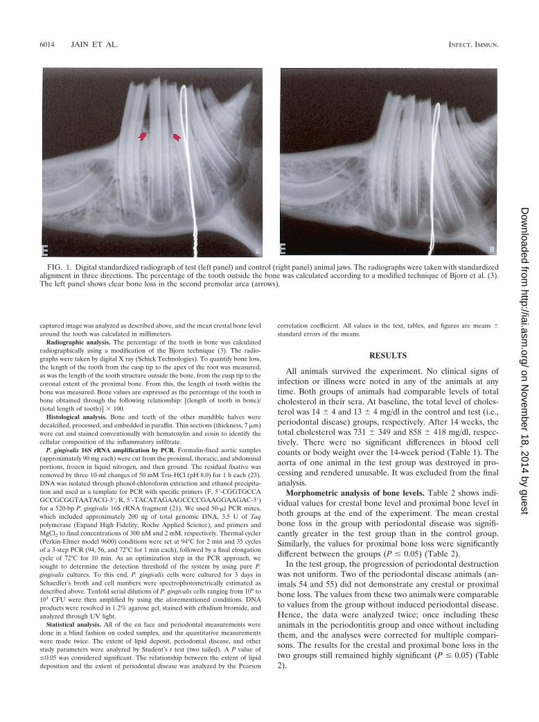
FIG. 1. Digital standardized radiograph of test (left panel) and control (right panel) animal jaws. The radiographs were taken with standardizedalignment in three directions. The percentage of the tooth outside the bone was calculated according to a modified technique of Bjorn et al. (3).The left panel shows clear bone loss in the second premolar area (arrows).
6014 JAIN ET AL. INFECT. IMMUN.
on Novem
ber 18, 2014 by guesthttp://iai.asm
.org/D
ownloaded from
Radiographic analysis. Radiographic bone loss in this modelof periodontal disease is depicted in Fig. 1. There was a sta-tistically significant (P � 0.05) loss of interproximal bone de-tected radiographically in the test group compared to the levelof bone in the control group (Table 2). Again, if the twononreacting animals (54 and 55) in the test group weredropped from the analysis, the significance level was �0.005.
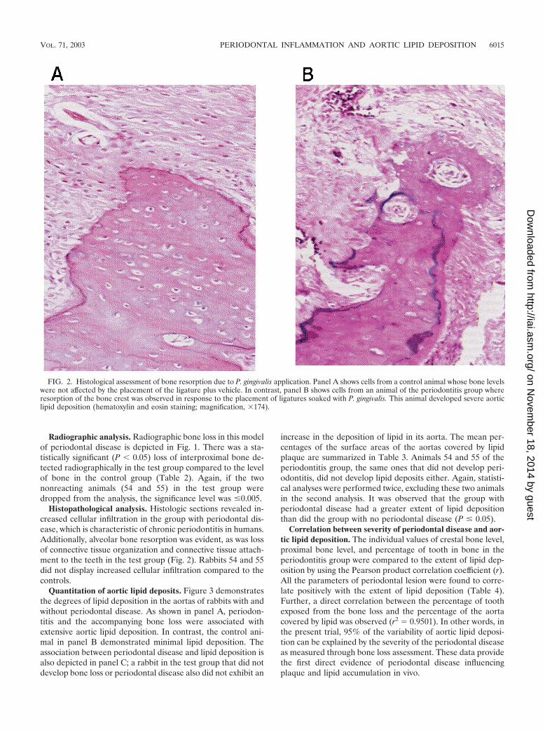
Histopathological analysis. Histologic sections revealed in-creased cellular infiltration in the group with periodontal dis-ease, which is characteristic of chronic periodontitis in humans.Additionally, alveolar bone resorption was evident, as was lossof connective tissue organization and connective tissue attach-ment to the teeth in the test group (Fig. 2). Rabbits 54 and 55did not display increased cellular infiltration compared to thecontrols.
Quantitation of aortic lipid deposits. Figure 3 demonstratesthe degrees of lipid deposition in the aortas of rabbits with andwithout periodontal disease. As shown in panel A, periodon-titis and the accompanying bone loss were associated withextensive aortic lipid deposition. In contrast, the control ani-mal in panel B demonstrated minimal lipid deposition. Theassociation between periodontal disease and lipid deposition isalso depicted in panel C; a rabbit in the test group that did notdevelop bone loss or periodontal disease also did not exhibit an
increase in the deposition of lipid in its aorta. The mean per-centages of the surface areas of the aortas covered by lipidplaque are summarized in Table 3. Animals 54 and 55 of theperiodontitis group, the same ones that did not develop peri-odontitis, did not develop lipid deposits either. Again, statisti-cal analyses were performed twice, excluding these two animalsin the second analysis. It was observed that the group withperiodontal disease had a greater extent of lipid depositionthan did the group with no periodontal disease (P � 0.05).
Correlation between severity of periodontal disease and aor-tic lipid deposition. The individual values of crestal bone level,proximal bone level, and percentage of tooth in bone in theperiodontitis group were compared to the extent of lipid dep-osition by using the Pearson product correlation coefficient (r).All the parameters of periodontal lesion were found to corre-late positively with the extent of lipid deposition (Table 4).Further, a direct correlation between the percentage of toothexposed from the bone loss and the percentage of the aortacovered by lipid was observed (r2 � 0.9501). In other words, inthe present trial, 95% of the variability of aortic lipid deposi-tion can be explained by the severity of the periodontal diseaseas measured through bone loss assessment. These data providethe first direct evidence of periodontal disease influencingplaque and lipid accumulation in vivo.
FIG. 2. Histological assessment of bone resorption due to P. gingivalis application. Panel A shows cells from a control animal whose bone levelswere not affected by the placement of the ligature plus vehicle. In contrast, panel B shows cells from an animal of the periodontitis group whereresorption of the bone crest was observed in response to the placement of ligatures soaked with P. gingivalis. This animal developed severe aorticlipid deposition (hematoxylin and eosin staining; magnification, �174).
VOL. 71, 2003 PERIODONTAL INFLAMMATION AND AORTIC LIPID DEPOSITION 6015
on Novem
ber 18, 2014 by guesthttp://iai.asm
.org/D
ownloaded from

P. gingivalis 16S rRNA amplification by PCR. The minimumamplification detection threshold of the system observed in thePCR optimization procedure was 10 cells of P. gingivalis (Fig.4). P. gingivalis 16S rRNA has not been amplified fromgenomic DNA obtained from any of the aortas analyzed byusing the PCR-optimized conditions.
DISCUSSION
In this study we report that in the rabbit high-fat diet model,animals with experimentally induced periodontitis develop vas-cular lipid deposits to a greater extent than periodontally
healthy controls. Further, there is a positive correlation be-tween the extent of lipid deposition and the severity of peri-odontal disease. There is growing evidence linking periodontaldisease and atherosclerosis on the epidemiologic level (2).Recently, a prospective study using a murine apoE knockoutmodel revealed that repeated systemic injections of P. gingiva-lis contributed to the initiation and progression of lipid accu-mulation in major vessels (11). While the cardiovascular out-comes of that study were similar to the results reported here,there are several important differences between the two mod-els. First, in the murine model, bacteria were recovered fromthe lipid lesions. Bacterial lesion recovery in the mouse modelis most likely a result of the systemic intravenous injection ofbacteria and the well-known fact that P. gingivalis can invadecells, especially endothelial cells (5). Further, the apoE knock-out mouse is a genetically altered animal. The combination ofthe altered host response and the nonphysiologic introductionof an intravenous pathogen make it difficult to directly com-pare these experiments to human disease.
The present study was designed to test the hypothesis thatthe presence of periodontal disease modifies the developmentof atheromatous changes. Our aim was also to examine themechanism by which periodontal disease might contribute tothe development of the vascular lesions. As reported, we havedeveloped an animal model in which both periodontal diseaseand atheromatous changes can be measured simultaneously.Our findings demonstrate that a diet consisting of 0.5% cho-lesterol can induce measurable lipid accumulation in the majorvessels of the rabbit, and periodontal disease accelerates orincreases lipid accumulation. The laboratory parameters mea-sured, including peripheral blood cell counts and body weight,were not different between groups. There were no obvioussystemic indicators of illness in the periodontitis group in spiteof the topical application of the pathogen. This finding is con-sistent with observations from studies of human periodontaldisease, which is also asymptomatic, without fever, weight loss,
FIG. 3. Representative results of periodontal lesion quantification and en face analysis. (A) Test group. The images show an extensive loss ofbone in the second premolar area (arrow) and extensive coverage by atherosclerotic plaque of the same animal’s aorta. (B) Control group. Theimages show the crestal bone with no loss around the second premolar and the aorta of the same animal, where minimal plaque is visible. (C) Testgroup. The images show the crestal bone from an animal that failed to develop any periodontal lesion and the same animal’s aorta, which showsminimal plaque development as well.
TABLE 3. Lipid deposition area analysis
Group and animal % Aortic surface area
Periodontitis54................................................................................. 2.7755................................................................................. 2.2656................................................................................. 56.6157................................................................................. 19.558................................................................................. 26.74
Mean � SEM (n � 3)a ............................................ 34.28 � 19.67b
Mean � SEM (n � 5).............................................. 21.57 � 22.27
Control59................................................................................. 17.360................................................................................. 461................................................................................. 12.4662................................................................................. 10.863................................................................................. 5.7164................................................................................. 17.63
Mean � SEM (n � 3).............................................. 11.31 � 5.6
a Animals 54 and 55 failed to develop periodontal disease and lipid depositionand were therefore excluded from analysis.
b Significantly different (P � 0.05) from value for control group.
6016 JAIN ET AL. INFECT. IMMUN.
on Novem
ber 18, 2014 by guesthttp://iai.asm
.org/D
ownloaded from
or other indicators of infection. It is interesting, however, thatelevated C-reactive protein levels in periodontitis patientswithout cardiovascular disease have been reported (14). Im-portantly, in our study, there was no difference in the serumcholesterol levels between the two groups.
An interesting and unexpected outcome of the study was thevaried levels of susceptibility of the rabbits to periodontal dis-ease. This difference is, however, consistent with observationsof humans, in whom the susceptibility to periodontal break-down is quite varied, seemingly independent of the presence ofpathogens, which are ubiquitous. In humans, this difference inlevels of susceptibility is hypothesized to be linked to hostresponse traits, with individuals with a robust innate immuneresponse being more susceptible to disease (15). Of particularinterest in the context of this work was the observation that twoanimals in the periodontitis group failed to develop periodon-tal disease and aortic lipid deposition. In these animals, peri-odontal infection might not have been successfully induced andthus atherosclerosis did not progress. Another possibility thatshould not be ruled out is that those animals were resistant toperiodontal infection, which may in turn suggest a commonsusceptibility trait for the two diseases. To support the latterpremise, however, more experiments would be necessary.
This is the first report of a positive correlation between theseverity of periodontal disease and the extent of atheromatousvessel changes. There is precedent for a relationship betweeninfection and atherosclerosis (8). It has been previously re-ported that an infectious agent anywhere in the body may leadto coactivation of the innate immune system, which acceleratesatherosclerosis. Richardson et al. (17) have reported that ath-erosclerosis was accelerated in hypercholesterolemic rabbitssimultaneously suffering from respiratory tract (but not vascu-lar) infections with Pasteurella multocida. Based on these ob-
servations, it has been suggested that there may be an infec-tious component in the development of cardiovascular lesions.However, as noted by Libby et al. (12), human atheromas oftenlack any indication of the presence of infectious agents, andeven if an infectious particle is present in the lesion, a patho-genic role is far from established for that organism.
Conversely, there is a body of evidence supported by ourstudy that suggests that an elevated innate host response is arisk factor for cardiovascular disease and periodontitis. Animportant corollary to this observation is the suggestion thatlocal inflammation can alter the systemic inflammatory state.As an example, the work of Pouliot and coworkers (16) re-vealed that initiation of isolated inflammation in the murinedorsal air pouch model resulted in an upregulation of COX-2mRNA in the heart and lung. Taken together, the data supportthe hypothesis that the degree of the inflammatory response toinsult or injury is a major determinant in the pathogenesis ofinflammatory diseases, including periodontitis and cardiovas-cular disease. Moreover, while these traits are known to beunder genetic control, the traits are also modifiable by envi-ronmental factors, such as bacteria, and local inflammation ata single site may modify the response trait at remote locationsin the body.
In conclusion, we have provided prospective data suggestinga link between periodontitis and cardiovascular disease in ananimal model. Further, the evidence suggests that focal inflam-mation in the oral cavity can have direct effects on the innateimmune response in other organ systems by increasing the riskfor inflammatory-cell-mediated tissue injury.
ACKNOWLEDGMENTS
We thank Margaret Morrissey for her excellent technical assistance.This study was supported by U.S. Public Health Service grant
DE13499. Eraldo L. Batista, Jr., is supported by the CAPES Founda-tion, Brasilia, Brazil (grant BEX 1539/99-2).
The T. Van Dyke and G. Stahl laboratories contributed equally tothis study.
REFERENCES
1. Beck, J., R. Garcia, G. Heiss, P. S. Vokonas, and S. Offenbacher. 1996.Periodontal disease and cardiovascular disease. J. Periodontol. 67:1123–1137.
2. Beck, J. D., S. Offenbacher, R. Williams, P. Gibbs, and R. Garcia. 1998.Periodontitis: a risk factor for coronary heart disease? Ann. Periodontol.3:127–141.
3. Bjorn, H., A. Halling, and H. Thyberg. 1969. Radiographic assessment ofmarginal bone loss. Odontol. Revy 20:165–179.
4. Chiu, B., E. Viira, W. Tucker, and I. W. Fong. 1997. Chlamydia pneumoniae,cytomegalovirus, and herpes simplex virus in atherosclerosis of the carotidartery. Circulation 96:2144–2148.
5. Deshpande, R. G., M. B. Khan, and C. A. Genco. 1998. Invasion of aortic and
FIG. 4. For PCR optimization, 10-fold serial dilutions of 3-day P.gingivalis cultures were amplified under the conditions described in thetext. The conditions used in our study were able to detect P. gingivalis16S rRNA genes from as few as 101 CFU obtained from a pure culture.
TABLE 4. Correlation analysis of the periodontitis group
MeasurementCrestal bone loss Proximal bone loss % Tooth in bone SAa
r P r P r P r P
Crestal bone loss 1.000 0.947b 0.014b �0.929 0.022 0.896 0.040Proximal bone loss 0.947b 0.014 1.000 �0.932b 0.021 0.898 0.038% Tooth in bone �0.929b 0.022 �0.932b 0.021 1.000 �0.975c 0.005SA 0.896b 0.040 0.898b 0.038 �0.975c 0.005 1.000
a SA, surface area of the aorta covered by lipid deposits.b Significant correlation at a P of 0.05 (two tailed).c Significant correlation at a P of 0.01 (two tailed).
VOL. 71, 2003 PERIODONTAL INFLAMMATION AND AORTIC LIPID DEPOSITION 6017
on Novem
ber 18, 2014 by guesthttp://iai.asm
.org/D
ownloaded from

heart endothelial cells by Porphyromonas gingivalis. Infect. Immun. 66:5337–5343.
6. Haraszthy, V. I., J. J. Zambon, M. Trevisan, M. Zeid, and R. J. Genco. 2000.Identification of periodontal pathogens in atheromatous plaques. J. Peri-odontol. 71:1554–1560.
7. Kuvin, J. T., and C. D. Kimmelstiel. 1999. Infectious causes of atheroscle-rosis. Am. Heart J. 137:216–226.
8. Lehr, H. A., and K. Kawasaki. 1992. Aggravation of ischaemia-reperfusioninjury in the striated muscle microcirculation by acute endotoxemia, p. 449–452. In E. Faist, J. L. Meakins, and F. W. Schildberg (ed.), Host defensedysfunctions in trauma, shock, and sepsis. Springer-Verlag, Heidelberg, Ger-many.
9. Lehr, H. A., D. A. Mankoff, D. Corwin, G. Santeusanio, and A. M. Gown.1997. Application of Photoshop-based image analysis to quantification ofhormone receptor expression in breast cancer. J. Histochem. Cytochem.45:1559–1565.
10. Lepine, G., and A. Progulske-Fox. 1996. Duplication and differential expres-sion of hemagglutinin genes in Porphyromonas gingivalis. Oral Microbiol.Immunol. 11:65–78.
11. Li, L., E. Messas, E. L. Batista, Jr., R. A. Levine, and S. Amar. 2002.Porphyromonas gingivalis infection accelerates the progression of athero-sclerosis in a heterozygous apolipoprotein E-deficient murine model. Circu-lation 105:861–867.
12. Libby, P., D. Egan, and S. Skarlatos. 1997. Roles of infectious agents inatherosclerosis and restenosis: an assessment of the evidence and need forfuture research. Circulation 96:4095–4103.
13. Marcus, A. J., and D. P. Hajjar. 1993. Vascular transcellular signaling. J.Lipid Res. 34:2017–2031.
14. Noack, B., R. J. Genco, M. Trevisan, S. Grossi, J. J. Zambon, and E. DeNardin. 2001. Periodontal infections contribute to elevated systemic C-re-active protein level. J. Periodontol. 72:1221–1227.
15. Page, R. C., S. Offenbacher, H. E. Schroeder, G. J. Seymour, and K. S.Kornman. 1997. Advances in the pathogenesis of periodontitis: summary ofdevelopments, clinical implications and future directions. Periodontol. 200014:216–248.
16. Pouliot, M., C. B. Clish, N. A. Petasis, T. E. Van Dyke, and C. N. Serhan.2000. Lipoxin A(4) analogues inhibit leukocyte recruitment to Porphyromo-nas gingivalis: a role for cyclooxygenase-2 and lipoxins in periodontal dis-ease. Biochemistry 39:4761–4768.
17. Richardson, M., M. De Reske, K. Delaney, A. Fletch, L. H. Wilcox, and R. L.Kinlough-Rathbone. 1997. Respiratory infection in lipid-fed rabbits en-hances sudanophilia and the expression of VCAM-1. Am. J. Pathol. 151:1009–1017.
18. Ross, R. 1999. Atherosclerosis is an inflammatory disease. Am. Heart J.138:S419–S420.
19. Shah, P. K. 1999. Plaque disruption and thrombosis. Potential role of in-flammation and infection. Cardiol. Clin. 17:271–281.
20. Silver, J. G., A. W. Martin, and B. C. McBride. 1977. Experimental transientbacteraemias in human subjects with varying degrees of plaque accumulationand gingival inflammation. J. Clin. Periodontol. 4:92–99.
21. Slots, J., A. Ashimoto, M. J. Flynn, G. Li, and C. Chen. 1995. Detection ofputative periodontal pathogens in subgingival specimens by 16S ribosomalDNA amplification with the polymerase chain reaction. Clin. Infect. Dis.20(Suppl.):S304–S307.
22. Sztukowska, M., M. Bugno, J. Potempa, J. Travis, and D. M. Kurtz, Jr. 2002.Role of rubrerythrin in the oxidative stress response of Porphyromonasgingivalis. Mol. Microbiol. 44:479–488.
23. Warford, A. 1996. In situ hybridisation, p. 491–512. In J. D. Bancroft (ed.),Theory and practice of histological techniques, 4th ed. Churchill Livingstone,London, United Kingdom.
Editor: J. D. Clements
6018 JAIN ET AL. INFECT. IMMUN.
on Novem
ber 18, 2014 by guesthttp://iai.asm
.org/D
ownloaded from