Periodontitis - Deep Blue Repositories
25
This is the author manuscript accepted for publication and has undergone full peer review but has not been through the copyediting, typesetting, pagination and proofreading process, which may lead to differences between this version and the Version of Record. Please cite this article as doi: 10.1002/JPER.17-0721. This article is protected by copyright. All rights reserved. Periodontitis WORKGROUP 2 Chairs Panos N. Papapanou 1 Mariano Sanz 2 Participants Nurcan Buduneli 3 Thomas Dietrich 4 Magda Feres 5 Dan Fine 6 Thomas Flemmig 7 Raul Garcia 8 William Giannobile 9 David Herrera Gonzalez 2
-
Upload
khangminh22 -
Category
Documents
-
view
0 -
download
0
Transcript of Periodontitis - Deep Blue Repositories

This is the author manuscript accepted for publication and has undergone full peer review but has not
been through the copyediting, typesetting, pagination and proofreading process, which may lead to
differences between this version and the Version of Record. Please cite this article as doi:
10.1002/JPER.17-0721.
This article is protected by copyright. All rights reserved.
Periodontitis
WORKGROUP 2
Chairs
Panos N. Papapanou1
Mariano Sanz2
Participants
Nurcan Buduneli3
Thomas Dietrich4
Magda Feres5
Dan Fine6
Thomas Flemmig7
Raul Garcia8
William Giannobile9
David Herrera Gonzalez2

This article is protected by copyright. All rights reserved.
Filippo Graziani10
Henry Greenwell11
Richard T. Kao12
Moritz Kebschull13
Denis Kinane14
Keith Kirkwood15
Thomas Kocher16
Kenneth Kornman9
Purnima Kumar17
Bruno Loos18
Eli Machtei19
Huanxin Meng20
Andrea Mombelli21
Ian Needleman22
Steven Offenbacher23
Gregory Seymour24
This article is protected by copyright. All rights reserved.
Ricardo Teles14
Maurizio Tonetti7
1. Columbia University, New York, USA
2. Universidad Complutense Madrid, Spain
3. Ege University School of Dentistry, Izmir, Turkey
4. Birmingham Dental School & Hospital, Birmingham, United Kingdom
5. Universidade Guarulhos, Guarulhos, Brazil
6. Rutgers University, Newark, USA
7. Prince Philip Dental Hospital, Hong Kong
8. Boston University, Boston, USA
9. University of Michigan, Ann Arbor, MI
10. University of Pisa, Pisa, Italy
11. University of Louisville, Louisville, USA
12. Private practice, Cupertino, CA
13. Bonn University, Bonn, Germany
14. University of Pennsylvania, Philadelphia, PA
15. University at Buffalo, Buffalo, USA
16. Greifswald University, Greifswald, Germany
17. Ohio State University, Columbus, OH
18. ACTA University, Amsterdam, Netherlands
This article is protected by copyright. All rights reserved.
19. Rambam HCC & Technion I.I.T., Haifa, Israel
20. Beijing Medical University, Beijing, China
21. University of Geneva, Geneva, Switzerland
22. UCL Eastman Dental Institute, London, United Kingdom
23. University of North Carolina, Chapel Hill, NC
24. University of Queensland, Brisbane, Australia
Sponsor Representatives
Marcelo Araujo (ADA)
Paul Eke (CDC)
Fotis Panagakos (Geistlich)
Name (Colgate)
Name (Sunstar)
Running head: Classification and case definitions for Periodontitis
Key words: periodontal disease, periodontitis, acute periodontal conditions,
periodontal abscess, necrotizing periodontitis, necrotizing gingivitits, endo-
periodontal lesion.
This article is protected by copyright. All rights reserved.
ABSTRACT
A new periodontitis classification scheme has been adopted, in which forms of the disease
previously recognized as “Chronic” or “Aggressive” are now grouped under a single category
(“Periodontitis”) and are further characterized based on a multi-dimensional staging and
grading system. Staging is largely dependent upon the severity of disease at presentation as
well as on the complexity of disease management, while grading provides supplemental
information about biological features of the disease including a history-based analysis of the
rate of periodontitis progression; assessment of the risk for further progression; analysis of
possible poor outcomes of treatment; and assessment of the risk that the disease or its
treatment may negatively affect the general health of the patient.
Necrotizing Periodontal Diseases, whose characteristic clinical phenotype includes typical
features (papilla necrosis, bleeding and pain) and are associated with host immune response
impairments, remain a distinct periodontitis category.
Endodontic-Periodontal Lesions, defined by a pathological communication between the
pulpal and periodontal tissues at a given tooth, occur in either an acute or a chronic form,
and are classified according to signs and symptoms that have direct impact on their
prognosis and treatment.
Periodontal Abscesses are defined as acute lesions characterized by localized accumulation
of pus within the gingival wall of the periodontal pocket/sulcus, rapid tissue destruction and
are associated with risk for systemic dissemination.
Periodontitis is a chronic multifactorial inflammatory disease associated with dysbiotic plaque
biofilms and characterized by progressive destruction of the tooth-supporting apparatus. Its
primary features include the loss of periodontal tissue support, manifested through clinical
This article is protected by copyright. All rights reserved.
attachment loss (CAL) and radiographically assessed alveolar bone loss, presence of
periodontal pocketing and gingival bleeding. Periodontitis is a major public health problem
due to its high prevalence, as well as because it may lead to tooth loss and disability,
negatively affect chewing function and aesthetics, be a source of social inequality, and
impair quality of life. Periodontitis accounts for a substantial proportion of edentulism and
masticatory dysfunction, results in significant dental care costs and has a plausible negative
impact on general health.
According to the latest internationally accepted classification scheme (Armitage1 1999),
periodontitis is further subdivided as follows:
Chronic periodontitis, representing the forms of destructive periodontal disease that
are generally characterized by slow progression
Aggressive periodontitis, a diverse group of highly destructive forms of periodontitis
affecting primarily young individuals, including conditions formerly classified as
“Early-Onset Periodontitis” and “Rapidly Progressing Periodontitis”
Periodontitis as a manifestation of systemic disease, a heterogeneous group of
systemic pathological conditions that include periodontitis as a manifestation
Necrotizing Periodontal Diseases, a group of conditions that share a characteristic
phenotype where necrosis of the gingival or periodontal tissues is a prominent
feature
Periodontal Abscesses, a clinical entity with distinct diagnostic features and
treatment requirements
This article is protected by copyright. All rights reserved.
Although the above classification has provided a workable framework that has been used
extensively in both clinical practice and scientific investigation in Periodontology during the
past 17 years, the system suffers from several important shortcomings, including substantial
overlap and lack of clear pathobiology-based distinction between the stipulated categories,
diagnostic imprecision, and implementation difficulties. The objectives of Workgroup 2 were
to revisit the current classification system of periodontitis, incorporate new knowledge
relevant to its epidemiology, etiology and pathogenesis that has accumulated since the
current classification’s inception, and propose a new classification framework along with
case definitions. To this end, five position papers were commissioned, authored, peer-
reviewed and accepted. The first reviewed the classification and diagnosis of aggressive
periodontitis (Fine et al.2 2018); the second focused on the age-dependent distribution of
clinical attachment loss in two population-representative, cross-sectional studies (Billings et
al.3 2018); the third reviewed progression data of clinical attachment loss from existing
prospective, longitudinal studies (Needleman at al.4 2018); the fourth reviewed the
diagnosis, pathobiology and clinical presentation of acute periodontal lesions (periodontal
abscesses, necrotizing periodontal diseases and endo-periodontal lesions; Herrera et al.5
2018); lastly, the fifth focused on periodontitis case definitions (Tonetti et al.6 2018).
The Workgroup reviewed, debated and agreed by consensus on the overall conclusions of
the five position papers, that can be largely summarized as follows:
(i) The conflicting literature findings on aggressive periodontitis are primarily due to the fact
that the currently adopted classification is too broad, the disease has not been studied from
its inception, and there is paucity of longitudinal studies including multiple time points and
different populations. The position paper argued that a more restrictive definition might be
better suited to take advantage of modern methodologies to enhance knowledge on the
diagnosis, pathogenesis, and management of this form of periodontitis.
This article is protected by copyright. All rights reserved.
(ii) Despite substantial differences in the overall severity of attachment loss between the
two population samples analyzed by Billings et al.3 (2018), suggesting presence of cohort
effects, common patterns of clinical attachment loss (CAL) were identified across different
ages, along with consistencies in the relative contribution of recession and pocket depth to
CAL. The findings suggest that it is feasible to introduce empirical evidence-driven
thresholds of attachment loss that signify disproportionate severity of periodontitis with
respect to age.
(iii) Longitudinal mean annual attachment level change was found to vary considerably both
within and between populations. Surprisingly, neither age nor gender had any discernible
effects on CAL change, but geographic location was associated. Overall, the position paper
argued that the existing evidence neither supports nor refutes the differentiation between
forms of periodontal diseases based upon progression of attachment level change.
(iv) Necrotizing periodontal diseases are characterized by three typical clinical features
(papilla necrosis, bleeding and pain) and are associated with host immune response
impairments, which should be considered in the classification of these conditions.
Endodontic-periodontal lesions are defined by a pathological communication between the
pulpal and periodontal tissues at a given tooth, occur in either an acute or a chronic form,
and should be classified according to signs and symptoms that have direct impact on their
prognosis and treatment (i.e., presence or absence of fractures and perforations, and
presence or absence of periodontitis).
Periodontal abscesses most frequently occur in pre-existing periodontal pockets and should
be classified according to their etiology. They are characterized by localized accumulation of
pus within the gingival wall of the periodontal pocket/sulcus, cause rapid tissue destruction
which may compromise tooth prognosis, and are associated with risk for systemic
dissemination.
This article is protected by copyright. All rights reserved.
(v) A periodontitis case definition system should include three components: (a)
identification of a patient as a periodontitis case, (b) identification of the specific type of
periodontitis, and (c) description of the clinical presentation and other elements that affect
clinical management, prognosis, and potentially broader influences on both oral and
systemic health. A framework for developing a multi-dimensional periodontitis staging and
grading system was proposed, in which staging is largely dependent upon the severity of
disease at presentation as well as on the complexity of disease management, while grading
provides supplemental information about biological features of the disease including a
history-based analysis of the rate of periodontitis progression; assessment of the risk for
further progression; analysis of possible poor outcomes of treatment; and assessment of the
risk that the disease or its treatment may negatively affect the general health of the patient.
During the workgroup deliberations, the following questions were formulated and addressed
in order to clarify and substantiate the need for a new classification system for periodontitis:
Which are the main features that identify periodontitis?
Loss of periodontal tissue support due to inflammation is the primary feature of
periodontitis. A threshold of interproximal, clinical attachment loss of ≥2mm or ≥3mm at 2
or more non-adjacent teeth is commonly used. Clinicians typically confirm presence of
interproximal tissue loss through radiographic assessments of bone loss. Clinically
meaningful descriptions of periodontitis should include the proportion of sites that bleed on
probing, and the number and proportion of teeth with probing depth over certain thresholds
(commonly ≥ 4mm and ≥6mm) and of teeth with clinical attachment loss of ≥ 3mm and
≥5mm (Holtfreter et al.7 2015).
This article is protected by copyright. All rights reserved.
Which criteria would need to be fulfilled to support the contention that chronic
and aggressive periodontitis are indeed different diseases? (e.g., etiology,
histology, pathophysiology, clinical presentation, other)
Differences in etiology and pathophysiology are required to indicate presence of distinct
periodontitis entities; variations in clinical presentation per se, i.e. extent and severity, do
not support the concept of different diseases.
Does current evidence suggest that we should continue to differentiate between
“aggressive” and “chronic” periodontitis as two different diseases?
Current evidence does not support the distinction between chronic and aggressive
periodontitis, as defined by the 1999 Classification Workshop, as two separate diseases;
however, a substantial variation in clinical presentation exists with respect to extent and
severity throughout the age spectrum, suggesting that there are population subsets with
distinct disease trajectories due to differences in exposure and/or susceptibility.
Is there evidence suggesting that early-onset forms of periodontitis (currently
classified under “aggressive periodontitis”) have a distinct pathophysiology (e.g.,
genetic background, microbiology, host-response) compared to later-onset
forms?
Although localized early onset periodontitis has a distinct, well-recognized clinical
presentation (early onset, molar/incisor distribution, progression of attachment loss), the
specific etiologic or pathological elements that account for this distinct presentation are
This article is protected by copyright. All rights reserved.
insufficiently defined. Likewise, mechanisms accounting for the development of generalized
periodontitis in young individuals are poorly understood.
What are the determinants for the mean annual attachment loss based on
existing longitudinal studies in adults?
A meta-analysis included in the position paper documented differences in mean annual
attachment loss between studies originating from different geographic regions, but did not
reveal an association with age or gender. It should be emphasized that meta-analysis of
mean data may fail to identify associations due to the loss of information and the lack of
accounting for both disease progression and regression. However, approaches that have
modelled both progression and regression of CAL have also reported no effect of age or
smoking on progression, although age and smoking reduced disease regression (e.g., Faddy
et al.8 2000). Individual studies that could not be included in the meta-analysis have shown
effects of smoking, socioeconomic status, previous attachment loss, ethnicity, age, gender
and calculus on mean annual attachment loss.
This article is protected by copyright. All rights reserved.
How do we define a patient as a periodontitis case?
In the context of clinical care, a patient is a “periodontitis case” if:
1. Interdental CAL is detectable at 2 or more non-adjacent teeth, or
2. Buccal or oral CAL ≥ 3mm with pocketing greater than 3 mm is detectable at 2 or
more teeth
but the observed CAL cannot be ascribed to non-periodontal causes such as: i) gingival
recession of traumatic origin; ii) dental caries extending in the cervical area of the tooth; iii)
the presence of CAL on the distal aspect of a second molar and associated with malposition
or extraction of a third molar, iv) an endodontic lesion draining through the marginal
periodontium; and v) the occurrence of a vertical root fracture.
Which different forms periodontitis are recognized in the present revised
classification system?
Based on pathophysiology, three clearly different forms of periodontitis have been identified:
A. Necrotizing periodontitis
B. Periodontitis as a direct manifestation of systemic diseases
C. Periodontitis
Differential diagnosis is based on history and the specific signs and symptoms of necrotizing
periodontitis, or the presence or absence of an uncommon systemic disease that alters the
host immune response. Periodontitis as a direct manifestation of systemic disease (Albandar
et al.9 2018, Jepsen et al.10 2018) should follow the classification of the primary disease
This article is protected by copyright. All rights reserved.
according to the respective International Statistical Classification of Diseases and Related
Health Problems (ICD) codes.
The remaining clinical cases of periodontitis which do not have the local characteristics of
necrotizing periodontitis or the systemic characteristics of a rare immune disorder with a
secondary manifestation of periodontitis should be diagnosed as “Periodontitis” and be
further characterized using a Staging and Grading system that describes clinical presentation
as well as other elements that affect clinical management, prognosis, and potentially
broader influences on both oral and systemic health.
How is a periodontitis case further characterized by Stage and Grade?
An individual case of periodontitis should be further characterized using a simple matrix that
describes the Stage and Grade of the disease. Stage is largely dependent upon the severity
of disease at presentation as well as on the anticipated complexity of disease management,
and further includes a description of extent and distribution of the disease in the dentition.
Grade provides supplemental information about biological features of the disease including a
history-based analysis of the rate of periodontitis progression; assessment of the risk for
further progression; analysis of possible poor outcomes of treatment; and assessment of the
risk that the disease or its treatment may negatively affect the general health of the patient.
For a complete description of the rationale, determinants and practical implementation of
the Staging and Grading system, the reader is referred to the publication by Tonetti et al.6
2018. Tables 1a and 1b list the framework of the staging and grading system.
This article is protected by copyright. All rights reserved.
Do the acute periodontal lesions have distinct features when compared with
other forms of periodontitis?
Periodontal abscesses, lesions from necrotizing periodontal diseases and acute presentations
of endo-periodontal lesions, share the following features that differentiate them from
periodontitis lesions: (i) rapid-onset, (ii) rapid destruction of periodontal tissues,
underscoring the importance of prompt treatment, and (iii) pain or discomfort, prompting
patients to seek urgent care.
Do periodontal abscesses have a distinct pathophysiology when compared to
other periodontitis lesions?
The first step in the development of a periodontal abscess is bacterial invasion or foreign
body impaction in the soft tissues surrounding the periodontal pocket, which develops into
an inflammatory process that attracts polymorphonuclear neutrophils (PMNs) and low
numbers of other immune cells. If the neutrophil-mediated defense process fails to control
the local bacterial invasion or clear the foreign body, degranulation, necrosis and further
neutrophilic influx may occur, leading to the formation of pus which, if not drained, results in
an abscess. Pathophysiologically, this lesion differs in that the low pH within an abscess
leads to rapid enzymatic disruption of the surrounding connective tissues and, in contrast to
a chronic inflammatory lesion, has a greater potential for resolution if quickly managed.
What is the case definition of a periodontal abscess?
Periodontal abscess is a localized accumulation of pus located within the gingival wall of the
periodontal pocket/sulcus, resulting in a significant tissue breakdown. The primary
detectable signs/symptoms associated with a periodontal abscess may involve ovoid
This article is protected by copyright. All rights reserved.
elevation in the gingiva along the lateral part of the root and bleeding on probing. Other
signs/symptoms that may also be observed include pain, suppuration on probing, deep
periodontal pocket and increased tooth mobility.
A periodontal abscess may develop in a pre-existing periodontal pocket, e.g. in patients with
untreated periodontitis, under supportive therapy or after scaling and root planing or
systemic antimicrobial therapy. A periodontal abscess occurring at a previously periodontally
healthy site is commonly associated with a history of impaction or harmful habits.
Do necrotizing periodontal diseases have a distinct pathophysiology when
compared to other periodontitis lesions?
Yes. Necrotizing gingivitis lesions are characterized by the presence of ulcers within the
stratified squamous epithelium and the superficial layer of the gingival connective tissue,
surrounded by a non-specific acute inflammatory infiltrate. Four zones have been described:
(i) superficial bacterial zone, (ii) neutrophil-rich zone, (iii) necrotic zone and a (iv)
spirochetal/bacterial infiltration zone.
Necrotizing periodontal diseases are strongly associated with impairment of the host
immune system, as follows: (i) in chronically, severely compromised patients (e.g., AIDS
patients, children suffering from severe malnourishment, extreme living conditions or severe
infections) and may constitute a severe or even life-threating condition; (ii) in temporarily
and/or moderately compromised patients (e.g., in smokers or psychosocially stressed adult
patients).
This article is protected by copyright. All rights reserved.
What are the case definitions of Necrotizing Periodontal Diseases?
Necrotizing gingivitis is an acute inflammatory process of the gingival tissues characterized
by presence of necrosis/ulcer of the interdental papillae, gingival bleeding and pain. Other
signs/symptoms associated with this condition may include halitosis, pseudomembranes,
regional lymhadenopathy, fever and sialorrhea (in children).
Necrotizing periodontitis is an inflammatory process of the periodontium characterized by
presence of necrosis/ulcer of the interdental papillae, gingival bleeding, halitosis, pain and
rapid bone loss. Other signs/symptoms associated with this condition may include
pseudomembrane formation, lymphadenopathy and fever.
Necrotizing stomatitis is a severe inflammatory condition of the periodontium and the oral
cavity in which soft tissue necrosis extends beyond the gingiva and bone denudation may
occur through the alveolar mucosa, with larger areas of osteitis and formation of bone
sequestrum. It typically occurs in severely systemically compromised patients. Atypical cases
have also been reported, in which necrotizing stomatitis may develop without prior
appearance of necrotizing gingivitis/periodontitis lesions.
Do endo-periodontal lesions have a distinct pathophysiology when compared to
other periodontitis or endodontic lesions?
The term endo-periodontal lesion (EPL) describes a pathologic communication between the
pulpal and periodontal tissues at a given tooth that may be triggered by a carious or
traumatic lesion that affects the pulp and, secondarily, affects the periodontium; by
periodontal destruction that secondarily affects the root canal; or by concomitant presence
of both pathologies. The review did not identify evidence for a distinct pathophysiology
between an EPL and a periodontal lesion. Nonetheless, the communication between the
This article is protected by copyright. All rights reserved.
pulp/root canal system and the periodontium complicates the management of the involved
tooth.
What is the case definition of an Endo-periodontal lesion?
EPL is a pathologic communication between the pulpal and periodontal tissues at a given
tooth that may occur in an acute or a chronic form. The primary signs associated with this
lesion are deep periodontal pockets extending to the root apex and/or negative/altered
response to pulp vitality tests. Other signs/symptoms may include radiographic evidence of
bone loss in the apical or furcation region, spontaneous pain or pain on
palpation/percussion, purulent exudate/suppuration, tooth mobility, sinus tract/fistula and
crown and/or gingival color alterations. Signs observed in EPLs associated with traumatic
and/or iatrogenic factors may include root perforation, fracture/cracking or external root
resorption. These conditions drastically impair the prognosis of the involved tooth.
Which are the current key gaps in knowledge that would inform a better
classification of periodontitis and should be addressed in future research?
Future research should:
(i) Develop improved methodologies to assess more accurately the longitudinal soft and
hard tissue changes associated with periodontitis progression
This article is protected by copyright. All rights reserved.
(ii) Identify genetic, microbial and host response-associated markers that distinguish
between distinct periodontitis phenotypes, or which can reflect the initiation and progression
of periodontitis.
(iii) Expand existing epidemiological databases to include world regions currently
underrepresented, utilizing consistent, standardized methodologies, and capturing and
reporting detailed data on both patient-related, oral and periodontal variables. Open access
to the detailed data is crucial to facilitate comprehensive analyses.
(iv) Integrate multi-dimensional data platforms (clinical, radiographic, -omics) to facilitate
systems biology approaches to the study of periodontal and peri-implant diseases and
conditions
(v) Use existing databases/ develop new databases that will facilitate the implementation,
validation and continuous refinement of the newly introduced periodontitis classification
system.
REFERENCES
1. Armitage GC. (1999) Development of a classification system for periodontal diseases and
conditions. Ann Periodontol 4, 1-6.
2. Fine, D., Patil, A.G., Loos, B. (2018) Classification and diagnosis of aggressive
periodontitis.
3. Billings, M, Holtfreter, B., Papapanou, P.N., Lopez-Mitnik, G., Kocher, T., Dye, B.A. (2018)
Age-dependent distribution of periodontitis in two countries: Findings from NHANES 2009-
2014 and SHIP-Trend 2008-2012. J Clin Periodontol 45, in press.
This article is protected by copyright. All rights reserved.
4. Needleman, I, Garcia, R., Gkranias, N., Kirkwood, K., Kocher, T., Di Iorio, A., Moreno, F.,
Petrie, A. (2018) Mean annual attachment, bone level and tooth loss - a systematic review. J
Clin Periodontol 45, in press.
5. Herrera, D., Retamal-Valdes, B., Alonso, B., Feres, M. (2018) Acute periodontal lesions
(periodontal abscesses and necrotizing periodontal diseases) and endo-periodontal lesions. J
Clin Periodontol 45, in press.
6. Tonetti, M.S., Greenwell, H., Kornman, K.S. (2018) Periodontitis case definition:
Framework for staging and grading the individual periodontitis case. J Clin Periodontol 45, in
press.
7. Holtfreter, B., Albandar, J. M., Dietrich, T., Dye, B. A., Eaton, K. A., Eke, P. I., Papapanou,
P. N., Kocher, T. (2015) Standards for reporting chronic periodontitis prevalence and
severity in epidemiologic studies: Proposed standards from the Joint EU/USA Periodontal
Epidemiology Working Group. J Clin Periodontol 42, 407-412. doi:10.1111/jcpe.12392.
8. Faddy, M. J., Cullinan, M. P., Palmer, J. E., Westerman, B. & Seymour, G. J. (2000) Ante-
dependence modeling in a longitudinal study of periodontal disease: the effect of age,
gender, and smoking status. J Periodontol 71, 454-459.
9. Albandar, J.M., Susin, C., Hughes, F.J. (2018) Manifestations of systemic diseases and
conditions that affect the periodontal attachment apparatus: Case definitions and diagnostic
considerations. J Clin Periodontol 45, in press.
10. Jepsen, S., Caton, L. Albandar, J.M., Bissada, N., Bouchard, P, Cortellini, P., Demirel, K.,
de Sanctis, M., Ercoli, C., Fan, J., Geurs, N., Hughes, F., Jin, L., Kantarci, A., Lalla, E.,
Madianos, P.N., Matthews, D., McGuire, M.K., Mills, M.P., Preshaw, P.M., Reynolds, M.A.,
Sculean, A., Susin, C., West, N.X., Yamazaki, K. (2018) Consensus Report: Periodontal
manifestations of systemic diseases and developmental and acquired conditions. J Clin
Periodontol 45, in press.
This article is protected by copyright. All rights reserved.
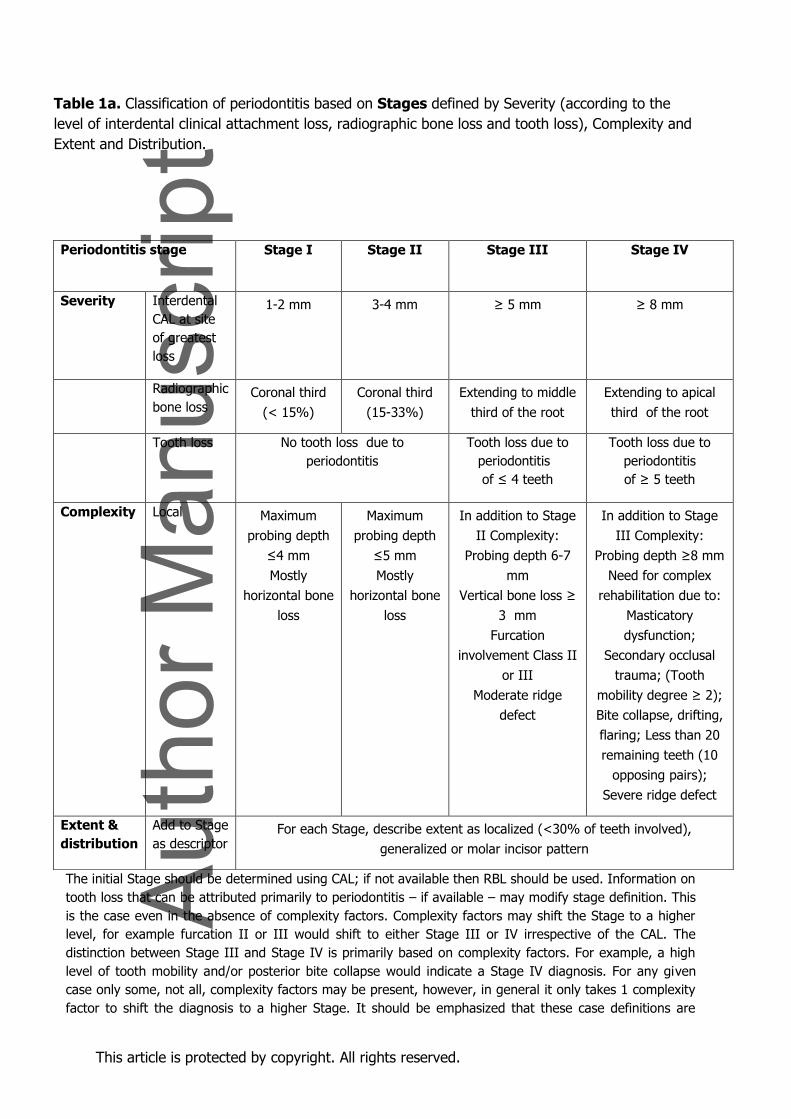
Table 1a. Classification of periodontitis based on Stages defined by Severity (according to the
level of interdental clinical attachment loss, radiographic bone loss and tooth loss), Complexity and
Extent and Distribution.
The initial Stage should be determined using CAL; if not available then RBL should be used. Information on
tooth loss that can be attributed primarily to periodontitis – if available – may modify stage definition. This
is the case even in the absence of complexity factors. Complexity factors may shift the Stage to a higher
level, for example furcation II or III would shift to either Stage III or IV irrespective of the CAL. The
distinction between Stage III and Stage IV is primarily based on complexity factors. For example, a high
level of tooth mobility and/or posterior bite collapse would indicate a Stage IV diagnosis. For any given
case only some, not all, complexity factors may be present, however, in general it only takes 1 complexity
factor to shift the diagnosis to a higher Stage. It should be emphasized that these case definitions are
Periodontitis stage
Stage I Stage II Stage III Stage IV
Severity Interdental
CAL at site
of greatest
loss
1-2 mm 3-4 mm ≥ 5 mm ≥ 8 mm
Radiographic
bone loss Coronal third
(< 15%)
Coronal third
(15-33%)
Extending to middle
third of the root
Extending to apical
third of the root
Tooth loss No tooth loss due to
periodontitis
Tooth loss due to
periodontitis
of ≤ 4 teeth
Tooth loss due to
periodontitis
of ≥ 5 teeth
Complexity Local Maximum
probing depth
≤4 mm
Mostly
horizontal bone
loss
Maximum
probing depth
≤5 mm
Mostly
horizontal bone
loss
In addition to Stage
II Complexity:
Probing depth 6-7
mm
Vertical bone loss ≥
3 mm
Furcation
involvement Class II
or III
Moderate ridge
defect
In addition to Stage
III Complexity:
Probing depth ≥8 mm
Need for complex
rehabilitation due to:
Masticatory
dysfunction;
Secondary occlusal
trauma; (Tooth
mobility degree ≥ 2);
Bite collapse, drifting,
flaring; Less than 20
remaining teeth (10
opposing pairs);
Severe ridge defect
Extent &
distribution
Add to Stage
as descriptor For each Stage, describe extent as localized (<30% of teeth involved),
generalized or molar incisor pattern
This article is protected by copyright. All rights reserved.
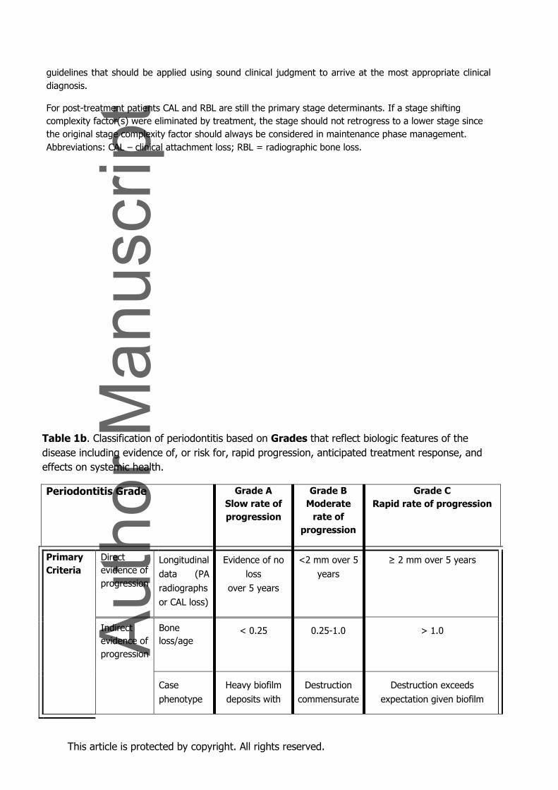
guidelines that should be applied using sound clinical judgment to arrive at the most appropriate clinical
diagnosis.
For post-treatment patients CAL and RBL are still the primary stage determinants. If a stage shifting
complexity factor(s) were eliminated by treatment, the stage should not retrogress to a lower stage since
the original stage complexity factor should always be considered in maintenance phase management.
Abbreviations: CAL – clinical attachment loss; RBL = radiographic bone loss.
Table 1b. Classification of periodontitis based on Grades that reflect biologic features of the
disease including evidence of, or risk for, rapid progression, anticipated treatment response, and
effects on systemic health.
Periodontitis Grade
Grade A
Slow rate of
progression
Grade B
Moderate
rate of
progression
Grade C
Rapid rate of progression
Primary
Criteria
Direct
evidence of
progression
Longitudinal
data (PA
radiographs
or CAL loss)
Evidence of no
loss
over 5 years
<2 mm over 5
years
≥ 2 mm over 5 years
Indirect
evidence of
progression
Bone
loss/age
< 0.25
0.25-1.0
> 1.0
Case
phenotype
Heavy biofilm
deposits with
Destruction
commensurate
Destruction exceeds
expectation given biofilm
This article is protected by copyright. All rights reserved.
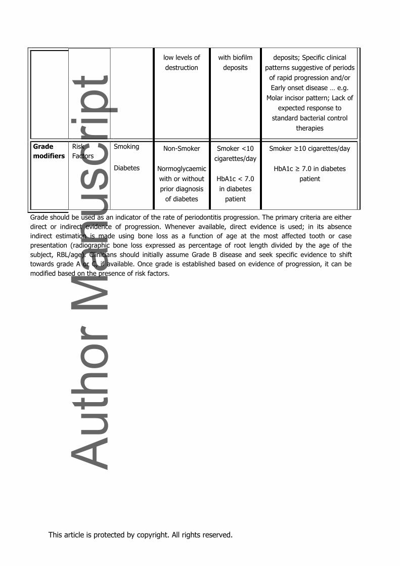
low levels of
destruction
with biofilm
deposits
deposits; Specific clinical
patterns suggestive of periods
of rapid progression and/or
Early onset disease … e.g.
Molar incisor pattern; Lack of
expected response to
standard bacterial control
therapies
Grade
modifiers
Risk
Factors
Smoking
Diabetes
Non-Smoker
Normoglycaemic
with or without
prior diagnosis
of diabetes
Smoker <10
cigarettes/day
HbA1c < 7.0
in diabetes
patient
Smoker ≥10 cigarettes/day
HbA1c ≥ 7.0 in diabetes
patient
Grade should be used as an indicator of the rate of periodontitis progression. The primary criteria are either
direct or indirect evidence of progression. Whenever available, direct evidence is used; in its absence
indirect estimation is made using bone loss as a function of age at the most affected tooth or case
presentation (radiographic bone loss expressed as percentage of root length divided by the age of the
subject, RBL/age). Clinicians should initially assume Grade B disease and seek specific evidence to shift
towards grade A or C, if available. Once grade is established based on evidence of progression, it can be
modified based on the presence of risk factors.
This article is protected by copyright. All rights reserved.
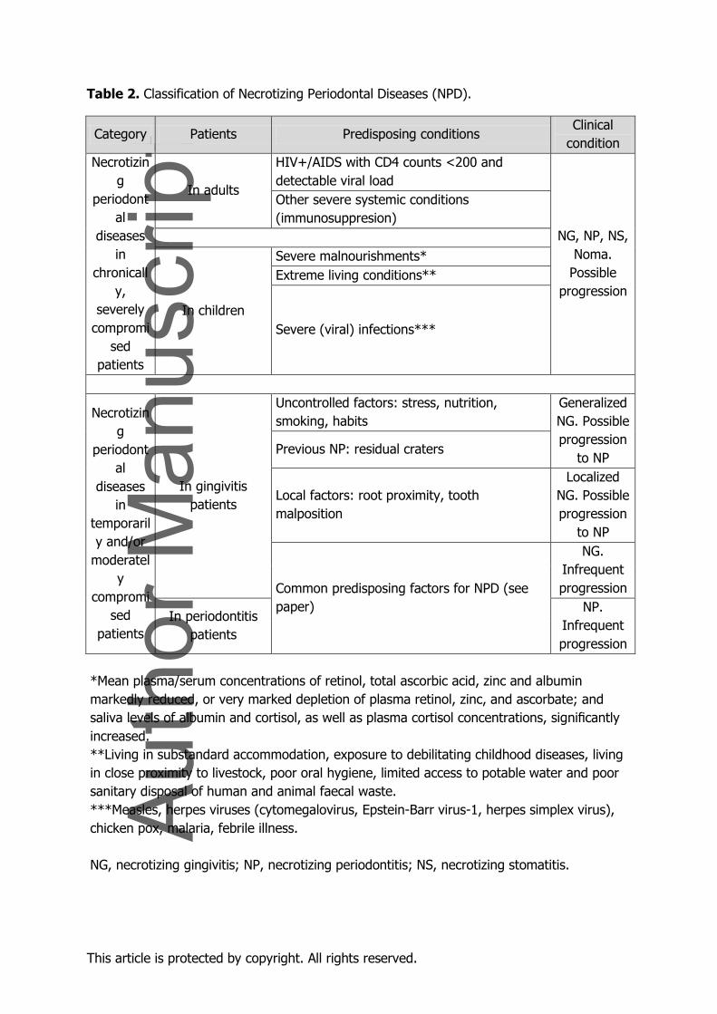
Table 2. Classification of Necrotizing Periodontal Diseases (NPD).
Category Patients Predisposing conditions Clinical
condition
Necrotizin
g
periodont
al
diseases
in
chronicall
y,
severely
compromi
sed
patients
In adults
HIV+/AIDS with CD4 counts <200 and
detectable viral load
NG, NP, NS,
Noma.
Possible
progression
Other severe systemic conditions
(immunosuppresion)
In children
Severe malnourishments*
Extreme living conditions**
Severe (viral) infections***
Necrotizin
g
periodont
al
diseases
in
temporaril
y and/or
moderatel
y
compromi
sed
patients
In gingivitis
patients
Uncontrolled factors: stress, nutrition,
smoking, habits
Generalized
NG. Possible
progression
to NP Previous NP: residual craters
Local factors: root proximity, tooth
malposition
Localized
NG. Possible
progression
to NP
Common predisposing factors for NPD (see
paper)
NG.
Infrequent
progression
In periodontitis
patients
NP.
Infrequent
progression
*Mean plasma/serum concentrations of retinol, total ascorbic acid, zinc and albumin
markedly reduced, or very marked depletion of plasma retinol, zinc, and ascorbate; and
saliva levels of albumin and cortisol, as well as plasma cortisol concentrations, significantly
increased.
**Living in substandard accommodation, exposure to debilitating childhood diseases, living
in close proximity to livestock, poor oral hygiene, limited access to potable water and poor
sanitary disposal of human and animal faecal waste.
***Measles, herpes viruses (cytomegalovirus, Epstein-Barr virus-1, herpes simplex virus),
chicken pox, malaria, febrile illness.
NG, necrotizing gingivitis; NP, necrotizing periodontitis; NS, necrotizing stomatitis.
This article is protected by copyright. All rights reserved.
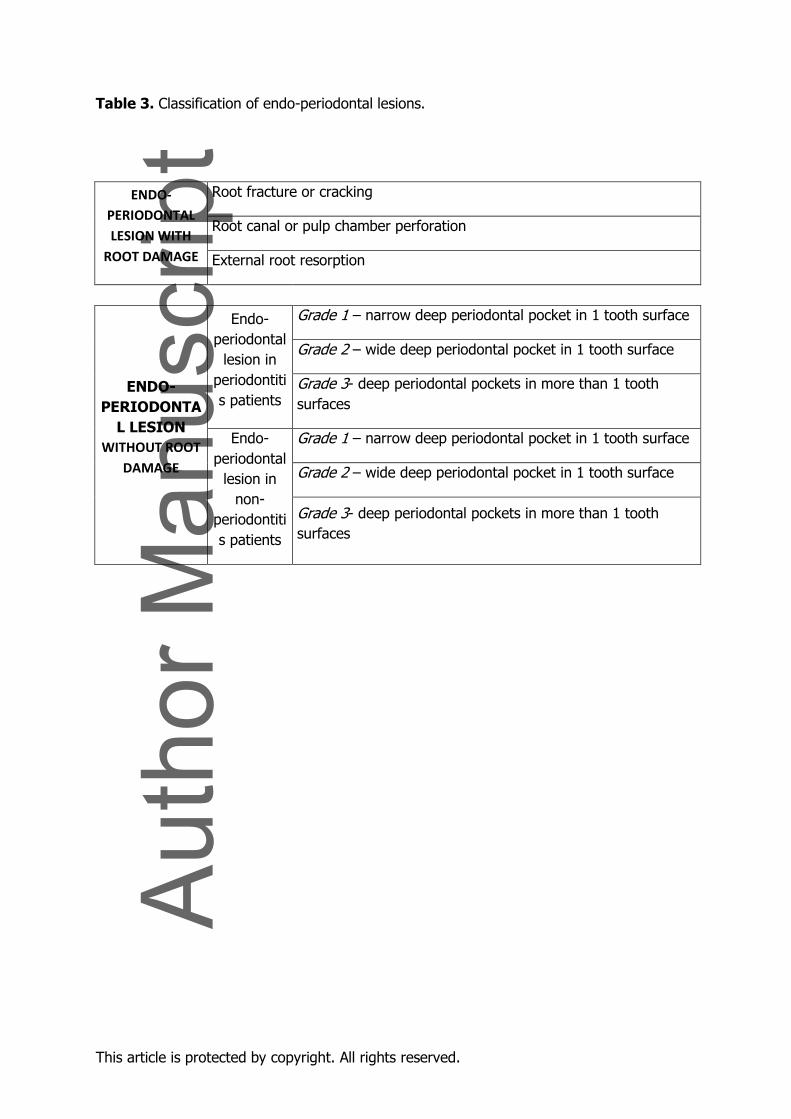
Table 3. Classification of endo-periodontal lesions.
ENDO-
PERIODONTAL
LESION WITH
ROOT DAMAGE
Root fracture or cracking
Root canal or pulp chamber perforation
External root resorption
ENDO-
PERIODONTA
L LESION
WITHOUT ROOT
DAMAGE
Endo-
periodontal
lesion in
periodontiti
s patients
Grade 1 – narrow deep periodontal pocket in 1 tooth surface
Grade 2 – wide deep periodontal pocket in 1 tooth surface
Grade 3- deep periodontal pockets in more than 1 tooth
surfaces
Endo-
periodontal
lesion in
non-
periodontiti
s patients
Grade 1 – narrow deep periodontal pocket in 1 tooth surface
Grade 2 – wide deep periodontal pocket in 1 tooth surface
Grade 3- deep periodontal pockets in more than 1 tooth
surfaces
This article is protected by copyright. All rights reserved.
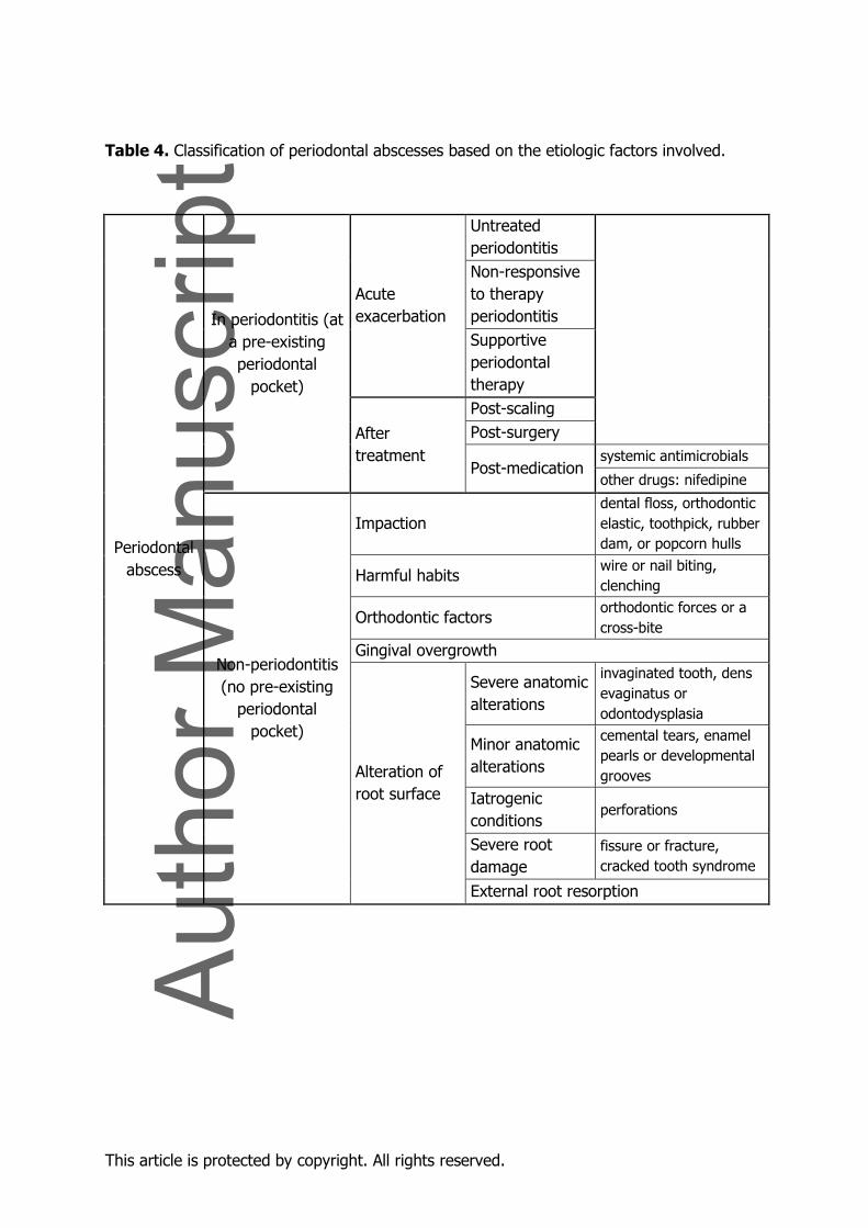
Table 4. Classification of periodontal abscesses based on the etiologic factors involved.
Periodontal
abscess
In periodontitis (at
a pre-existing
periodontal
pocket)
Acute
exacerbation
Untreated
periodontitis
Non-responsive
to therapy
periodontitis
Supportive
periodontal
therapy
After
treatment
Post-scaling
Post-surgery
Post-medication systemic antimicrobials
other drugs: nifedipine
Non-periodontitis
(no pre-existing
periodontal
pocket)
Impaction dental floss, orthodontic
elastic, toothpick, rubber
dam, or popcorn hulls
Harmful habits wire or nail biting,
clenching
Orthodontic factors orthodontic forces or a
cross-bite
Gingival overgrowth
Alteration of
root surface
Severe anatomic
alterations
invaginated tooth, dens
evaginatus or
odontodysplasia
Minor anatomic
alterations
cemental tears, enamel
pearls or developmental
grooves
Iatrogenic
conditions perforations
Severe root
damage
fissure or fracture,
cracked tooth syndrome
External root resorption