Platelet-derived growth factor-induced Akt phosphorylation ...
Upload
independentCategory
view
0download
0
Downloaded from UvA-DARE, the Institutional Repository of the University of Amsterdam (UvA)http://dare.uva.nl/document/119416
Description ThesisFile ID 119416Filename thesis.pdf
SOURCE, OR PART OF THE FOLLOWING SOURCE:Type DissertationTitle Reactivity of neutrophils, monocytes and platelets in periodontitisAuthor E.A. NicuFaculty Faculty of DentistryYear 2008Pages 151
FULL BIBLIOGRAPHIC DETAILS: http://dare.uva.nl/record/288434
Copyrights It is not permitted to download or to forward/distribute the text or part of it without the consent of the copyright holder(usually the author), other then for strictly personal, individual use. UvA-DARE is a service provided by the Library of the University of Amsterdam (http://dare.uva.nl)
Reactivity of neutrophils, monocytes and platelets in periodontitis Elena A. Nicu
Reactivity of neutrophils, monocytes
and platelets in periodontitis
Printing of this thesis was financially supported by:
The Netherlands Institute for Dental Sciences (IOT)
University of Amsterdam
Stichting NVvP
Copyright © 2008, Elena Alina Nicu
Reactivity of neutrophils, monocytes and platelets in periodontitis
ISBN 978-90-9023659-9
Printed by PrintPartners Ipskamp BV, Enschede.
Reactivity of neutrophils, monocytes
and platelets in periodontitis
ACADEMISCH PROEFSCHRIFT
ter verkrijging van de graad van doctor aan de Universiteit van Amsterdam
op gezag van de Rector Magnificus Prof. dr. D.C. van den Boom ten overstaan van een door het college voor promoties ingestelde commissie,
in het openbaar te verdedigen in de Agnietenkapel op donderdag 11 december 2008, te 10:00 uur
door
Elena Alina Nicu
Geboren te Rîmnicu-Vîlcea, Roemenië
Promotiecommissie
Promotores: Prof. dr. B.G. Loos
Prof. dr. U. van der Velden
Co-promotor: Prof. dr. V. Everts
Overige leden: Prof. dr. H.R. Büller
Prof. dr. W. Crielaard
Prof. dr. M.M. Levi
Prof. dr. A. Sturk
Prof. dr. J.G.J. van de Winkel
Faculteit der Tandheelkunde
The research presented in this thesis was performed at the Department of Periodontology of the Academic
Centre for Dentistry Amsterdam (ACTA).
Contents
Chapter 1 General introduction 7
Chapter 2 Hyper-reactive PMNs in FcγRIIa 131H/H genotype periodontitis patients
23
Chapter 3 Expression of FcγRs and mCD14 on polymorphonuclear neutrophils and monocytes may determine periodontal infection
45
Chapter 4 Soluble CD14 as a possible molecular link between atherosclerosis and chronic infectious diseases: periodontitis as model
69
Chapter 5 Periodontitis is associated with platelet activation
87
Chapter 6 Elevated platelet and leukocyte response to oral bacteria in periodontitis
105
Chapter 7 Summary and discussion
123
Samenvatting
135
Acknowledgements
147
List of publications 151


Chapter 1
General Introduction
7
Introduction
Introduction
The periodontium is the tooth-supporting organ. It comprises the periodontal ligament, the
alveolar bone, the radicular cementum and the gingiva. The periodontal ligament is the
soft connective tissue interposed between the root of each tooth and the inner wall of the
alveolar socket. At the soft-hard tissue borders of the periodontal ligament, the principal
periodontal ligament fibers are embedded in alveolar bone on one side and radicular
cementum on the other side. Covering and protecting these structures, the gingiva forms
the fourth component of the periodontium.
The periodontium can become inflamed; the mildest and most frequent
inflammatory condition of the periodontium is gingivitis. Gingivitis is highly prevalent
and readily reversible by effective oral hygiene. Gingivitis affects 50-90% of adults
worldwide, depending on its precise definition (1). Inflammation that extends deep into
the tissues and causes loss of supportive connective tissue and alveolar bone is known as
periodontitis. Periodontitis results in the formation of periodontal pockets; these are
deepened crevices between gingiva and tooth roots eventually leading to tooth loss. After
the age of fifty, virtually all individuals may present with some mild periodontal
destruction, one or two pockets deeper than 4 mm, or small gingival recessions. But the
severe forms of periodontitis will only occur in approximately 10% of the adult population
(2). Treatment of periodontitis includes mechanical removal of subgingival bacterial
plaque with scalers, curettes or ultra-sonic devices and intensive oral hygiene instructions
for the patient. A close to ideal oral hygiene is the only way to prevent formation of new
dental plaque deposits and re-infection of the subgingival tissues.
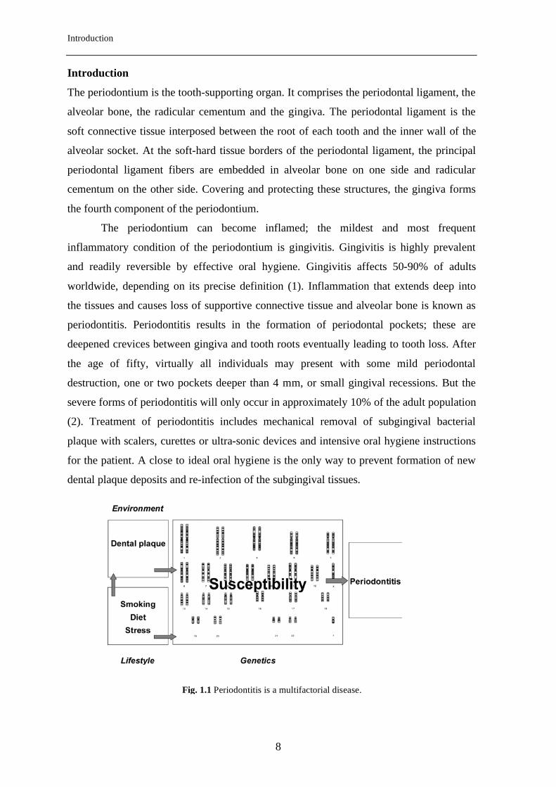
Fig. 1.1 Periodontitis is a multifactorial disease.
8
Chapter 1
Periodontitis is a multifactorial disease. In the light of current knowledge, genetic factors
represent the basis of susceptibility for periodontitis, and environment and lifestyle are
major modifying factors (Fig. 1.1). Dental plaque is the major environmental factor. As
dental plaque matures to a state that is associated with periodontitis, the number of Gram-
negative and anaerobe micro-organisms increases. Certain bacterial species have been
often associated with disease, and intensively studied, including Aggregatibacter
actinomycetemcomitans and Porphyromonas gingivalis (3). Several hundred different
bacterial species colonize the subgingival tissues, covering the root surfaces and the
epithelial lining of the pocket with a complex biofilm. A biofilm is a structured
community of micro-organisms encapsulated within a self-developed polymeric matrix
and adherent to a living or inert surface. Evidence is accumulating that the aggregated
organisms in biofilms are not merely passive neighbors, but rather are involved in a wide
range of physical, metabolic and molecular interactions (4). Dental biofilm pathogenicity
in the oral cavity is magnified by two biofilm characteristics: increased antibiotic
resistance and the inability of the community to be phagocytosed by host inflammatory
cells.
Smoking and emotional stress are lifestyle-associated factors that have been often
incriminated as major disease-modifiers. Smokers are much more likely to develop
periodontitis than non-smokers and smoking has a strong negative effect on the response
to periodontal treatment and other oral surgical interventions (1). Traumatic life events
that lead to depression or individual inability to cope with stress could increase the
person’s risk for periodontitis, most likely due to adverse immune responses (5).
The periodontal immune response has been compared to a double-edged sword, on
the one hand fighting microorganisms with which it comes into contact and, on the other
hand, mediating injury to the host. Maintaining a balance between these two conflicting
properties and the preservation of health is assured by proper immunoregulation.
Therefore, knowledge of the immune response to periodontal pathogens is ultimately
relevant for understanding pathogenesis of periodontitis.
The immune response in periodontitis
The local defense against pathogens from dental plaque is based on the integrity and
activity of the epithelial lining, secretion of gingival crevicular fluid and saliva, and local
inflammatory reactions. Subgingival accumulation of oral bacteria triggers inflammation
in the periodontium. Inflammation is the response of the host to infection or other insults
9
Introduction
and comprises a series of vascular reactions at the site of injury. The results of these
vascular reactions are exudation of fluid and plasma proteins and recruitment of
leukocytes to the site of injury. The goal of inflammation is confining the injury and
initiating the immune response, through which the infection is eliminated and the injury
repaired (6).
Professional phagocytes, comprising polymorphonuclear neutrophilic leukocytes
(PMNs) and monocyte/macrophage cells, play an important role in the host defense
against bacterial infections, and, as such, play an important role in periodontal disease.
The functional responses of the phagocytes to bacterial infections include chemotaxis,
migration, phagocytosis, degranulation and reactive oxygen species generation.
PMNs form the first line of defense, are the most abundant leukocytes in peripheral
blood (50-65%) and are characterized by a segmented nucleus and a rich granular
cytoplasm. The PMN granules contain large amounts of anti-microbial substances and
enzymes. The primary (or azurophilic) granules contain myeloperoxidase (MPO), serine
proteases (elastase, cathepsine G, and others), defensins and lysozyme. The secondary (or
specific) granules contain collagenase, lysozyme, vitamin B-binding protein and lactoferin
(7).
PMNs have the ability of sensing and moving towards chemical signals,
chemotaxins, generated at infectious sites. Among the chemotaxins recognized by PMNs
are N-formyl-methionyl peptides from bacteria, C5a from complement and IL-8 from
epithelial and other cells (8). Activated PMNs adhere to the vessel wall via selectins and
integrins, leave the blood circulation, and migrate to the site of infection with the ultimate
goal to phagocytose and kill pathogens. The bacterial killing by PMN takes place in a
membrane-confined vacuole, the phagolysosome, formed after fusion of PMN granules
with the bacterium-containing phagosome. The bacterial killing and digestion are
accomplished via oxygen-dependent and oxygen-independent pathways. In the oxygen-
dependent the NADPH-oxidase system (residing in the cytosol) is activated and generates
toxic oxygen species; in the oxygen-independent pathway, proteolytic enzymes stored in
the granules are activated and released within the phagolysosome. In periodontitis, a
hyper-production of proteolytic enzymes (9,10) and reactive oxygen species (11) has been
documented and these may play a role in the host-derived destruction of periodontal
tissues.
In inflammation, PMNs arrive at the site of injury first followed by monocytes (6).
Tissue macrophages differentiate from peripheral-blood monocytes. The monocytes, and
10
Chapter 1
their progeny, the macrophages and the dendritic cells are important players in the
pathogenesis of periodontitis. Various bacterial species in dental plaque are gram-negative
and their LPS interacts with CD14 and Toll-like receptors inducing the production of
cytokines and other mediators by macrophages or dendritic cells. A key cytokine in the
monocyte-associated periodontal destruction is interleukin-1 (IL-1), which is capable of
stimulating the collagenolytic and bone-destructive processes (12).
Not only PMNs and monocytes/macrophages can interact with bacteria, platelets
also have the ability to respond to infection. Activated platelets release anti-microbial
peptides and chemokines such as platelet factor 4, RANTES (13), and expose pro-
inflammatory receptors, facilitating their binding to leukocytes and endothelial cells (14).
Due to this interaction, leukocytes and endothelial cells increase the expression of
adhesion molecules and various cytokines (15,16). Bacteria-platelet interactions are made
possible either directly through a bacterial surface protein or indirectly by a bridging
molecule from plasma that links bacteria with platelet surface receptors. Gingipains, a
family of cysteine proteases produced by the periodontal pathogen P. gingivalis, can
directly activate platelets. Streptococcus sanguis was found to require immunoglobulin G
(IgG) interacting with an IgG receptor (FcγRIIa) to mediate platelet activation (17).
Recognition of bacteria by phagocytes: the receptors
In general, phagocytes lack the ability to recognize bacteria, and instead, depend on
opsonization. Opsonization is the process of coating of bacteria with plasma proteins in
order to signal and facilitate phagocytosis. There are three identified mechanisms: I) the
recognition of complement C3b by CR3 and CR4 (complement receptor 3 and 4); II) the
recognition of antibodies by Fc receptors; and III) the recognition of lipopolysaccharide-
binding protein by CD14 (18).
The PMN and monocyte complement receptors - CR3 (Mac-1; CD11b/CD18) and
CR4 (p150,95; CD11c/CD18) recognize and bind particles that have been coated by the
complement-derived opsonin, C3b.
Phagocytes bind and recognize targets also via antibodies (immunoglobulins).
Immunoglobulin G (IgG) is the predominant serum isotype during bacterial infections.
IgG1 and IgG3 are produced in response to proteinaceous antigens (such as P. gingivalis
hemagglutinin) and viruses; IgG2 is formed in response to the bacterial polysaccharides
and outer membrane proteins of gram-negative periodontal pathogens (19). IgG molecules
11
Introduction
contain two regions: one region (the Fab-domain) recognizes the pathogen, whereas the
other region (the Fc-domain) activates the immune system. Depending on their expression
on effector cells, FcγR exert different effects. Based on their affinity for monomeric IgG,
three types of FcγR on phagocytes have been identified: FcγRI, FcγRII, and FcγRIII (20).
FcγRI (CD64) is constitutionally highly expressed by monocytes and
macrophages. Under resting condition, PMNs do not express FcγRI (21). Expression of
FcγRI on PMNs can be induced by stimulation with interferon-γ (IFN-γ) or granulocyte
colony-stimulating factor (G-CSF) (22,23,24). FcγRII (CD32) is the most widely
distributed FcγR, expressed on PMNs, platelets, eosinophils, basophils, lymphocytes and
monocytes. There are several isoforms of FcγRII, with highly similar extracellular and
transmembrane domains, but with different intracellular signaling motifs (25,26). FcγRIIa
and FcγRIIc contain an immunoreceptor tyrosine-based activation motif (ITAM), thus
they function as activating receptors. FcγRIIb, on the other hand, contains an
immunoreceptor tyrosine-based inhibitory motif (ITIM), thus it is an inhibitory receptor
(27,28). PMNs and monocytes express both FcγRIIa and FcγRIIb (29,21). The balance
between activating and inhibitory FcγRII can shift in inflammatory states. IFN-γ induces
upregulation of FcγRIIa and concomitant downregulation of FcγRIIb, whereas interleukin-
4 (IL-4) can have the opposite effect (29). Two genes, FCG3A and FCG3B encode for
FcγRIII. FcγRIIIa is expressed by monocytes and NK cells (30,31,32), whereas FcγRIIIb
is solely expressed by PMNs and eosinophils (33).
Genetic polymorphisms with consequences for the receptor affinities for IgG-
subclasses have been identified in FCG2A, FCG3A and FCG3B (34,35,36,21). At
aminoacid position 131, FcγRIIa expresses either an arginine (R) or a histidine (H). The
genetic variation in the FcγRIIa has functional consequences: the FcγRIIa-H131 variant
binds human IgG2, whereas FcγRIIa-R131 does not (35). It has been speculated that this
polymorphism may have important consequences for the pathogenesis of periodontitis
(37). The polymorphism in FcγRIIIa yields either a valine (V) or a phenylalanine (F) at
aminoacid position 158. This substitution results in an increased affinity for IgG1 and
IgG3 of the FcγRIIIa-V158 variant (36,34). In the case of FcγRIIIb, the polymorphism
involves 4 aminoacids, combined in the NA1 and NA2 variants (38). FcγRIIIb-NA1 binds
IgG1 and IgG3 more efficiently than FcγRIIIb-NA2 (21).
Plasma-derived lipopolysaccharide-binding protein (LBP) and septin can directly
opsonize Gram-negative bacteria by interaction with the lipopolysaccharide (LPS) that
12
Chapter 1
forms part of the Gram-negative bacterial outer membrane (18). CD14 is an LPS co-
receptor and is mainly expressed on mature monocytes, macrophages and activated PMNs
(39). Soluble CD14 (sCD14) can be released from blood monocytes or produced in the
liver. Acting as a soluble LPS-receptor, sCD14 helps inducing responses in cells that
naturally lack CD14, such as endothelial cells (40). Another role is the neutralization and
clearance of LPS in Gram-negative infectious states (endotoxemic states). Acting as a
decoy receptor for serum LPS, sCD14 prevents extreme pro-inflammatory responses from
monocytes/macrophages (41).
Periodontitis in relation to atherosclerosis and cardiovascular diseases
Periodontitis shares its multifactorial character with other frequent diseases of the modern
world, such as diabetes and cardiovascular diseases. Furthermore, it has been postulated
that periodontitis and cardiovascular diseases have more in common than unfavorable
lifestyle (such as smoking and stress), as epidemiologic studies identified periodontitis as
a risk factor for future cardiovascular events such as stroke and myocardial infarction. In a
meta-analysis cumulating the results from several studies investigating the relationship
between periodontitis and coronary artery disease, presence of periodontitis associated
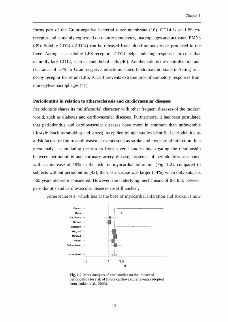
with an increase of 19% in the risk for myocardial infarction (Fig. 1.2), compared to
subjects without periodontitis (42); the risk increase was larger (44%) when only subjects
<65 years old were considered. However, the underlying mechanisms of the link between
periodontitis and cardiovascular diseases are still unclear.
Atherosclerosis, which lies at the base of myocardial infarction and stroke, is now
Fig. 1.2 Meta-analysis of nine studies on the impact of periodontitis for risk of future cardiovascular events (adopted from Janket et al., 2003).
13
Introduction
also considered as an inflammatory disease (43). The atherosclerotic process is initiated
when cholesterol-containing low-density lipoproteins accumulate in the intima and
activate the endothelium. Leukocyte adhesion molecules and chemokines promote
recruitment of monocytes and T cells. Monocytes differentiate into macrophages that
accumulate intracellular lipoprotein, which leads to foam-cell formation. Intensified
inflammatory activation may lead to local proteolysis, plaque rupture, and thrombus
formation, which causes ischemia and infarction. Chronic infections with Chlamydia
pneumoniae, Helicobacter pylori, cytomegalovirus (CMV) have been implicated in
atherosclerosis by stimulating disease progression and possibly plaque activation (43).
This activation could be due either to direct action in plaques or to remote signaling via
inflammatory mediators. Similar mechanisms of action are conceivable in periodontitis,
where chronic subgingival infection with periodontal pathogens is accompanied by
transient, low-grade bacteremias during dental procedures or daily activities like tooth
brushing or chewing (44,45). Moreover, a systemic inflammatory reaction is documented
in periodontitis, with increased production of IL-1β, IL-6, C-reactive protein and tumor-
necrosis factor (TNF)-α (46,47). There is evidence of the presence of periodontal
pathogens in atherosclerotic plaques (48). Therefore, direct or indirect priming of PMNs,
monocytes and platelets by periodontal pathogens, leading to increased inflammation at
sites of atherosclerotic activity, might be an important mechanism underlying the
increased risk for CVD in periodontitis patients.
General aim and outline of this thesis
The general scope of this thesis was to characterize the reactivity of PMNs, monocytes
and platelets against periodontal pathogens. The added knowledge will increase our
understanding of some patho-physiological processes in periodontitis, and may explain
inter-individual variation in clinical responses to pathogens. Moreover, this interaction is
important to dissect possible pathways of the association between periodontitis and
cardiovascular diseases, as endotoxemia, systemic exposure to periodontal pathogens and
the induced systemic inflammation seem to be important (49,50).
There is a need to incorporate host genetic diversity in functional studies in the
pathogenesis of periodontitis. One aspect of the host-derived breakdown of periodontal
tissues seems related to a hyper-reactive trait of PMNs (51,11). The nature of this hyper-
reactivity might be genetically-determined or derived from increased expression levels of
14
Chapter 1
phagocytic receptors (e.g FcγR, complement receptor, CD14) in response to the pathologic
processes accompanying periodontitis. The hypothesis that there are inter individual
differences in phagocyte reactivity due to variations in phagocytic receptors led to three
research questions.
1. Does the PMN activation via FcγRIIa among periodontitis patients with different
FcγRIIa genotypes (R/R and H/H) after stimulation with IgG-opsonized A.
actinomycetemcomitans differ? (Chapter 2)
2. Is the PMN and monocyte reactivity in periodontitis attributable to modified
expression of FcγRI, FcγRIIa, FcγRIII, CR3 or CD14? (Chapter 3)
3. Is periodontitis leading to increased sCD14 levels? (Chapter 4)
Furthermore, we hypothesized that in response to transient bacteremic episodes of
periodontal origin, PMNs, monocytes and platelets are primed in periodontitis patients.
This priming would than be a part of the pathogenic processes linking periodontitis and
cardiovascular disease. We analyzed the cellular response of PMNs, monocytes and
platelets to the periodontal pathogens A. actinomycetemcomitans, P. gingivalis and T.
forsythia. This interest materialized in two research questions.
4. Are circulating platelets from patients with periodontitis more activated than
control platelets? (Chapter 5)
5. Do platelets, PMNs and monocytes from patients with periodontitis respond in a
hyper-reactive fashion to periodontal pathogens compared to cells from
periodontally healthy controls? (Chapter 6)
Finally, Chapter 7 summarizes and interprets the main findings of the studies in the
context of the research questions, and suggests new directions for future research.
The chapters 2-6 of this thesis have been published as individual studies, thus some
repetition, especially in the introductory sections, is inherent.
15
Introduction
REFERENCES
1 Pihlstrom BL, Michalowicz BS, Johnson NW. 2005. Periodontal diseases. Lancet
366:1809-1820.
2 Brown LJ, Oliver RC, Loe H. 1990. Evaluating periodontal status of US employed
adults. J Am Dent Assoc 121:226-232.
3 van Winkelhoff AJ, Loos BG, van der Reijden WA, van der Velden U. 2002.
Porphyromonas gingivalis, Bacteroides forsythus and other putative periodontal
pathogens in subjects with and without periodontal destruction. J Clin Periodontol
29:1023-1028.
4 Thomas JG and Nakaishi LA. 2006. Managing the complexity of a dynamic biofilm.
J Am Dent Assoc 137:10S-115.
5 Genco RJ, Ho AW, Grossi SG, Dunford RG, Tedesco LA. 1999. Relationship of
stress, distress and inadequate coping behaviors to periodontal disease. J Periodontol
70:711-723.
6 Majno G. 1982. Inflammation and infection: historic highlights. Monogr Pathol1-17.
7 Kuijpers TW, Tool AT, van der Schoot CE, Ginsel LA, Onderwater JJ, Roos D,
Verhoeven AJ. 1991. Membrane surface antigen expression on neutrophils: a
reappraisal of the use of surface markers for neutrophil activation. Blood 78:1105-
1111.
8 Zadeh HH, Nichols FC, Miyasaki KT. 1999. The role of the cell-mediated immune
response to Actinobacillus actinomycetemcomitans and Porphyromonas gingivalis in
periodontitis. Periodontology 2000 20:239-288.
9 Figueredo CM, Gustafsson A, Asman B, Bergstrom K. 1999. Increased release of
elastase from in vitro activated peripheral neutrophils in patients with adult
periodontitis. J Clin Periodontol 26:206-211.
16
Chapter 1
10 Gustafsson A and Asman B. 1996. Increased release of free oxygen radicals from
peripheral neutrophils in adult periodontitis after Fc delta-receptor stimulation. J Clin
Periodontol 23:38-44.
11 Matthews JB, Wright HJ, Roberts A, Cooper PR, Chapple IL. 2007. Hyperactivity
and reactivity of peripheral blood neutrophils in chronic periodontitis. Clin Exp
Immunol 147:255-264.
12 Tatakis DN. 1993. Interleukin-1 and bone metabolism: a review. J Periodontol
64:416-431.
13 Fitzgerald JR, Foster TJ, Cox D. 2006. The interaction of bacterial pathogens with
platelets. Nat Rev Microbiol 4:445-457.
14 Klinger MH and Jelkmann W. 2002. Role of blood platelets in infection and
inflammation. J Interferon Cytokine Res 22:913-922.
15 Celi A, Pellegrini G, Lorenzet R, De Blasi A, Ready N, Furie BC, Furie B. 1994.
P-selectin induces the expression of tissue factor on monocytes. PNAS 91:8767-8771.
16 Slupsky JR, Kalbas M, Willuweit A, Henn V, Kroczek RA, Muller-Berghaus G.
1998. Activated platelets induce tissue factor expression on human umbilical vein
endothelial cells by ligation of CD40. Thromb Haemost 80:1008-1014.
17 McNicol A, Zhu R, Pesun R, Pampolina C, Jackson EC, Bowden GH, Zelinski T.
2006. A role for immunoglobulin G in donor-specific Streptococcus sanguis-induced
platelet aggregation. Thromb Haemost 95:288-293.
18 Wright SD, Ramos RA, Patel M, Miller DS. 1992. Septin: a factor in plasma that
opsonizes lipopolysaccharide-bearing particles for recognition by CD14 on
phagocytes. J Exp Med 176:719-727.
19 Ebersole JL and Taubman MA. 1994. The protective nature of host responses in
periodontal diseases. Periodontol 2000 5:112-141.
20 Deo YM, Graziano RF, Repp R, Van de Winkel JG. 1997. Clinical significance of
IgG Fc receptors and Fc gamma R-directed immunotherapies. Immunol Today 18:127-
135.
17

Introduction
21 Huizinga TW, Kerst M, Nuyens JH, Vlug A, dem Borne AE, Roos D, Tetteroo
PA. 1989. Binding characteristics of dimeric IgG subclass complexes to human
neutrophils. J Immunol 142:2359-2364.
22 Repp R, Valerius T, Sendler A, Gramatzki M, Iro H, Kalden JR, Platzer E. 1991.
Neutrophils express the high affinity receptor for IgG (Fc gamma RI, CD64) after in
vivo application of recombinant human granulocyte colony-stimulating factor. Blood
78:885-889.
23 Guyre PM, Morganelli PM, Miller R. 1983. Recombinant immune interferon
increases immunoglobulin G Fc receptors on cultured human mononuclear
phagocytes. J Clin Invest 72:393-397.
24 Perussia B, Kobayashi M, Rossi ME, Anegon I, Trinchieri G. 1987. Immune
interferon enhances functional properties of human granulocytes: role of Fc receptors
and effect of lymphotoxin, tumor necrosis factor, and granulocyte-macrophage colony-
stimulating factor. J Immunol 138:765-774.
25 Brooks DG, Qiu WQ, Luster AD, Ravetch JV. 1989. Structure and expression of
human IgG FcRII(CD32). Functional heterogeneity is encoded by the alternatively
spliced products of multiple genes. J Exp Med 170:1369-1385.
26 Warmerdam PA, Van de Winkel JG, Gosselin EJ, Capel PJ. 1990. Molecular basis
for a polymorphism of human Fc gamma receptor II (CD32). J Exp Med 172:19-25.
27 van den Herik-Oudijk IE, Capel PJ, van der BT, van de Winkel JG. 1995.
Identification of signaling motifs within human Fc gamma RIIa and Fc gamma RIIb
isoforms. Blood 85:2202-2211.
28 van den Herik-Oudijk IE, Westerdaal NA, Henriquez NV, Capel PJ, van de
Winkel JG. 1994. Functional analysis of human Fc gamma RII (CD32) isoforms
expressed in B lymphocytes. J Immunol 152:574-585.
29 Pricop L, Redecha P, Teillaud J-L, Frey J, Fridman WH, Sautes-Fridman C,
Salmon JE. 2001. Differential Modulation of Stimulatory and Inhibitory
Fc{{gamma}} Receptors on Human Monocytes by Th1 and Th2 Cytokines. J
Immunol 166:531-537.
18
Chapter 1
30 Ravetch JV and Kinet JP. 1991. Fc receptors. Annu Rev Immunol 9:457-492.
31 Van de Winkel JG and Anderson CL. 1991. Biology of human immunoglobulin G
Fc receptors. J Leukoc Biol 49:511-524.
32 Hulett MD and Hogarth PM. 1994. Molecular basis of Fc receptor function. Adv
Immunol 57:1-127.
33 Huizinga TW, van der Schoot CE, Jost C, Klaassen R, Kleijer M, dem Borne AE,
Roos D, Tetteroo PA. 1988. The PI-linked receptor FcRIII is released on stimulation
of neutrophils. Nature 333:667-669.
34 De Haas M, Koene HR, Kleijer M, de Vries E, Simsek S, van Tol MJ, Roos D,
dem Borne AE. 1996. A triallelic Fc gamma receptor type IIIA polymorphism
influences the binding of human IgG by NK cell Fc gamma RIIIa. J Immunol
156:2948-2955.
35 Warmerdam PA, Van de Winkel JG, Vlug A, Westerdaal NA, Capel PJ. 1991. A
single amino acid in the second Ig-like domain of the human Fc gamma receptor II is
critical for human IgG2 binding. J Immunol 147:1338-1343.
36 Koene HR, Kleijer M, Algra J, Roos D, dem Borne AE, De Haas M. 1997. Fc
gammaRIIIa-158V/F polymorphism influences the binding of IgG by natural killer cell
Fc gammaRIIIa, independently of the Fc gammaRIIIa-48L/R/H phenotype. Blood
90:1109-1114.
37 Wilson ME and Bronson PM. 1997. Opsonization of Actinobacillus
actinomycetemcomitans by immunoglobulin G antibodies to the O polysaccharide of
lipopolysaccharide. Infect Immun 65:4690-5.
38 Huizinga TW, Kleijer M, Tetteroo PA, Roos D, von dem Borne AE. 1990. Biallelic
neutrophil Na-antigen system is associated with a polymorphism on the phospho-
inositol-linked Fc gamma receptor III (CD16). Blood 75:213-7.
39 Ziegler-Heitbrock HW and Ulevitch RJ. 1993. CD14: cell surface receptor and
differentiation marker. Immunol Today 14:121-125.
19
Introduction
40 Ulevitch RJ and Tobias PS. 1995. Receptor-dependent mechanisms of cell
stimulation by bacterial endotoxin. Annu Rev Immunol 13:437-457.
41 Maliszewski CR. 1991. CD14 and immune response to lipopolysaccharide. Science
252:1321-1322.
42 Janket SJ, Baird AE, Chuang SK, Jones JA. 2003. Meta-analysis of periodontal
disease and risk of coronary heart disease and stroke. Oral Surg Oral Med Oral Pathol
Oral Radiol Endod 95:559-569.
43 Hansson GK, Robertson AK, Soderberg-Naucler C. 2006. Inflammation and
atherosclerosis. Annu Rev Pathol 1:297-329.
44 Forner L, Larsen T, Kilian M, Holmstrup P. 2006. Incidence of bacteremia after
chewing, tooth brushing and scaling in individuals with periodontal inflammation. J
Clin Periodontol 33:401-407.
45 Geerts SO, Nys M, De MP, Charpentier J, Albert A, Legrand V, Rompen EH.
2002. Systemic release of endotoxins induced by gentle mastication: association with
periodontitis severity. J Periodontol 73:73-78.
46 Loos BG. 2005. Systemic markers of inflammation in periodontitis. J Periodontol
76:2106-2115.
47 Paraskevas S, Huizinga JD, Loos BG. 2008. A systematic review and meta-analyses
on C-reactive protein in relation to periodontitis. J Clin Periodontol 35:277-290.
48 Haraszthy VI, Zambon JJ, Trevisan M, Zeid M, Genco RJ. 2000. Identification of
periodontal pathogens in atheromatous plaques. J Periodontol 71:1554-1560.
49 Pussinen PJ, Alfthan G, Tuomilehto J, Asikainen S, Jousilahti P. 2004. High
serum antibody levels to Porphyromonas gingivalis predict myocardial infarction. Eur
J Cardiovasc Prev Rehabil 11:408-411.
50 Pussinen PJ, Tuomisto K, Jousilahti P, Havulinna AS, Sundvall J, Salomaa V.
2007. Endotoxemia, immune response to periodontal pathogens, and systemic
inflammation associate with incident cardiovascular disease events. Arterioscler
Thromb Vasc Biol 27:1433-1439.
20

Chapter 1
51 Fredriksson M, Gustafsson A, Asman B, Bergstrom K. 1998. Hyper-reactive
peripheral neutrophils in adult periodontitis: generation of chemiluminescence and
intracellular hydrogen peroxide after in vitro priming and FcgammaR-stimulation. J
Clin Periodontol 25:394-8.
21

22
Chapter 2
Hyper-reactive PMNs in FcγRIIa 131H/H genotype
periodontitis patients
E.A. Nicu1, U. van der Velden1, V. Everts2, A.J. Van Winkelhoff3, D. Roos4, B.G.
Loos1
Departments of 1 Periodontology , 2 Oral Cell Biology and 3 Oral Microbiology, Academic Centre for
Dentistry Amsterdam (ACTA), University of Amsterdam and VU University Amsterdam, The Netherlands
4 Department of Blood Cell Research, Sanquin Research and Landsteiner Laboratory, Academic Medical
Center (AMC), University of Amsterdam, The Netherlands
Journal of Clinical Periodontology 2007; 34:938-945
23
Hyper-reactive PMNs in FcγRIIa 131H/H genotype periodontitis patients
Abstract Receptors for the Fc part of IgG (FcγRIIa) on polymorphonuclear leukocytes (PMN)
mediate phagocytosis and cell activation. Previous results show that one of the genetic
variants of the FcγRIIa, the 131H/H, is associated with more periodontal breakdown than
the R/R. This may be due to a hyper-reactivity of the H/H-PMNs upon interaction with
bacteria. We aimed to study whether the FcγRIIa genotype modifies the PMN reactivity in
periodontitis patients. A cohort of 98 periodontitis patients was genotyped. From these, 10
H/H and 10 R/R consented to participate. PMNs were incubated with immune serum-
opsonized A. actinomycetemcomitans. Phagocytosis, degranulation (CD63 and CD66b
expression), respiratory burst and elastase release were assessed. Patients of the H/H
genotype showed more bone loss than the H/R or R/R (P=0.038). H/H-PMNs
phagocytosed more opsonized A. actinomycetemcomitans than did R/R-PMNs (P=0.019).
The H/H-PMNs expressed also more CD63 and CD66b than did the R/R-PMNs (P=0.004
and 0.002, respectively) and released more elastase (P=0.001). The genotyping results
confirm previous reports that more periodontal destruction occurs in the H/H genotype
than in H/R or R/R. The functional studies indicate a hyper-reactivity of the H/H- PMN in
response to bacteria that may be one of several pathways leading to more periodontal
breakdown.
24
Chapter 2
Introduction Periodontitis is a chronic infectious disease of the supportive tissues of the teeth
characterized by gradual loss of periodontal attachment and alveolar bone.
Periodontopathic bacteria such as Aggregatibacter actinomycetemcomitans and
Porphyromonas gingivalis have been implicated in the pathogenesis of the disease (1,2).
However, recent literature indicates a genetically determined hyperactivity of the host
response, which constitutes one aspect of the susceptibility to periodontitis (3). Disease
resistance seems to be characterized by a proper defense against periodontal pathogens
without concomitant damage to the host itself.
In the course of periodontal infection, the human polymorphonuclear neutrophilic
leukocyte (PMN) represents the first line of antibacterial defense. The PMNs have also
been implicated in periodontal tissue degradation by releasing proteases and reactive
oxygen species (4,5,6,7). The PMN constitutively expresses two types of Fcγ receptors:
FcγRIIa and FcγRIIIb. The FcγR recognize and bind the constant part of immunoglobulin
G. FcγRIIa mediates phagocytosis (8), killing of opsonized cellular targets via antibody-
dependent cellular cytotoxicity (9), and respiratory burst (10). Important to note is that
FcγRIIa contains a transmembrane domain, that facilitates signal transduction to the cell.
This is not the case for FcγRIIIb, which does not contain a cytoplasmic domain but is
anchored to the cell membrane via a glycosyl-phosphatidylinosytol (GPI)-anchor. The role
of FcγRIIIb in activation of PMNs has been debated. Some investigators have shown that
this receptor is indeed capable of inducing signal transduction, possibly with the help of
FcγRIIa (11), whereas others have suggested that FcγRIIIb does not contribute to effector
functions (12).
FcγRIIa occurs in two allotypic forms, designated FcγRIIa-H131 and FcγRIIa-
R131 due to the genetically determined presence of either a histidine or an arginine
residue at amino acid position 131 (13). The genotype prevalence strongly depends on
ethnicity and is a source of conflicting results. In the general population, the genotype
distribution is similar in African-Americans and Caucasians, and distinct from that of
Japanese or Chinese (14). Among Caucasians, the prevalence of FcγRIIa-131H/H
genotype has been reported to be higher in chronic or aggressive periodontitis than in
healthy subjects (15). On the other hand, Nibali et al. (2006) found no difference in the
genotype frequency between aggressive periodontitis patients and healthy controls when
considering either all subjects or only the Caucasians (16). Moreover, Caucasian
25
Hyper-reactive PMNs in FcγRIIa 131H/H genotype periodontitis patients
periodontitis patients with the H/H genotype have more periodontal breakdown than those
with the H/R or R/R genotype (17).
The genetic variation in the FcγRIIa has functional consequences: the FcγRIIa-
H131 genotype binds IgG2-opsonized particles more efficiently than FcγRIIa-R131
genotype (18). This phenomenon may have important consequences for the pathogenesis
of periodontitis, as has also been speculated by others (19). It is known that IgG2
dominates the humoral immune response against polysaccharide antigens that are
abundant on the cell wall of the gram-negative periodontal pathogens. For example, IgG2
is the main immunoglobulin subclass reactive with A. actinomycetemcomitans (20). It has
been hypothesized that the highly efficient binding of opsonized particles to FcγRIIa in the
H/H genotype may result in a hyper-reactive state of the PMN (15,17). The strongly
activated H/H PMN may release more of its granule contents, thus contributing to
collateral damage, i.e. loss of periodontal connective tissue, periodontal ligament and
alveolar bone in the defense process against periodontal pathogens. However, this
hypothesis has never been tested.
The purpose of the present study was to investigate whether the FcγRIIa H/H and
R/R genotypes have functional consequences for the reactivity of the PMN from
periodontitis patients. This might contribute to the periodontitis phenotype in patients. We
analyzed PMN activation in both H/H and R/R genotypes following incubation with
opsonized A. actinomycetemcomitans. We studied four PMN functional parameters:
phagocytosis, degranulation, respiratory burst, and elastase activity.
Materials and methods
Screening of periodontitis patients
98 periodontitis patients from a cohort within the Department of Periodontology,
Academic Center for Dentistry Amsterdam (21) were genotyped for this study. Genomic
DNA was extracted from blood by means of the Puregene DNA isolation kit (Gentra
Systems, Minneapolis, MN, USA). Five µL of the purified DNA solution (concentration
70 - 100 ng DNA / µL solution) was added to a PCR reaction with allele-specific primers
for FcγRIIa-H131 and FcγRIIa-R131 (22). Based on these PCR results, all FcγRIIa-
H/H131 and FcγRIIa-R/R131 patients were selected as two contrasting groups for the
PMN functional assays.
26
Chapter 2
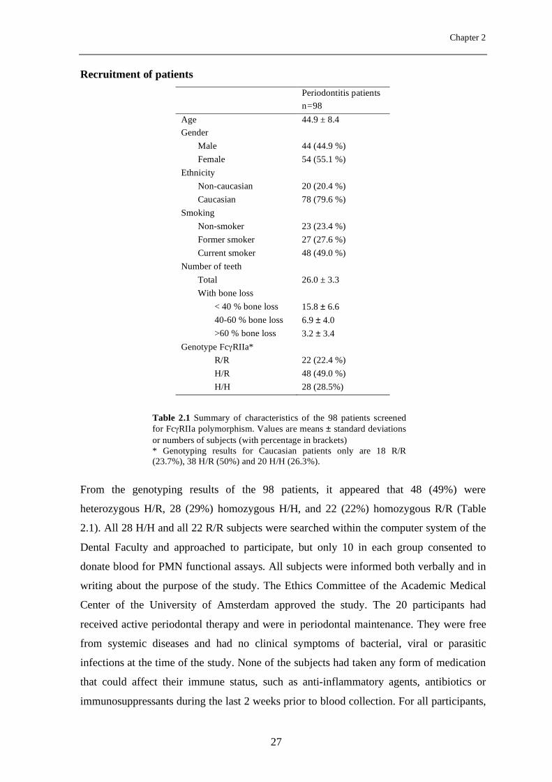
Recruitment of patients Periodontitis patients n=98 Age 44.9 ± 8.4 Gender
Male 44 (44.9 %) Female 54 (55.1 %)
Ethnicity Non-caucasian 20 (20.4 %) Caucasian 78 (79.6 %)
Smoking Non-smoker 23 (23.4 %) Former smoker 27 (27.6 %) Current smoker 48 (49.0 %)
Number of teeth Total 26.0 ± 3.3 With bone loss
< 40 % bone loss 15.8 ± 6.6 40-60 % bone loss 6.9 ± 4.0 >60 % bone loss 3.2 ± 3.4
Genotype FcγRIIa* R/R 22 (22.4 %) H/R 48 (49.0 %) H/H 28 (28.5%)
From the genotyping results of the 98 patients, it appeared that 48 (49%) were
heterozygous H/R, 28 (29%) homozygous H/H, and 22 (22%) homozygous R/R (Table
2.1). All 28 H/H and all 22 R/R subjects were searched within the computer system of the
Dental Faculty and approached to participate, but only 10 in each group consented to
donate blood for PMN functional assays. All subjects were informed both verbally and in
writing about the purpose of the study. The Ethics Committee of the Academic Medical
Center of the University of Amsterdam approved the study. The 20 participants had
received active periodontal therapy and were in periodontal maintenance. They were free
from systemic diseases and had no clinical symptoms of bacterial, viral or parasitic
infections at the time of the study. None of the subjects had taken any form of medication
that could affect their immune status, such as anti-inflammatory agents, antibiotics or
immunosuppressants during the last 2 weeks prior to blood collection. For all participants,
Table 2.1 Summary of characteristics of the 98 patients screened for FcγRIIa polymorphism. Values are means ± standard deviations or numbers of subjects (with percentage in brackets) * Genotyping results for Caucasian patients only are 18 R/R (23.7%), 38 H/R (50%) and 20 H/H (26.3%).
27
Hyper-reactive PMNs in FcγRIIa 131H/H genotype periodontitis patients
smoking status and smoking history were recorded and subjects were classified as non-
smokers (never smokers or those who quit > 10 years ago), former smokers (those who
quit smoking in the last 10 years) and currents smokers. All patients showed on dental
radiographs periodontal bone loss of > 1/3 of the root length on ≥ 2 teeth.
Bacteria and immune serum
To obtain immune serum against A. actinomycetemcomitans, we selected 10 periodontitis
patients who were culture positive for A. actinomycetemcomitans; they were patients from
the departmental clinic who were in active periodontal treatment. The undiluted serum of
these patients was tested against A. actinomycetemcomitans serotype a strain HG 569,
serotype b strain HG 90, serotype c strain HG 683, serotype d strain 3381, and serotype e
strain HG 1650. Bacteria were grown for 18 hours in brain heart infusion broth
supplemented with 5 mg/L hemin and 1 mg/L menadione at 37°C in humidified 5% CO2.
Bacteria were harvested, washed twice in PBS, checked for purity and the concentration
was adjusted to approximately 5 * 108 CFU/mL. All bacterial preparations were sonicated
on ice for 2 min, at 5-sec intervals, amplitude 18, by means of a Soniprep-150 ultrasonic
disintegrator (MSE, London, UK). Immunodiffusion of whole serum was carried out in
1% agarose (Sigma Chemicals Co., St. Louis, MO, USA) in 50 mM Tris-HCl buffer, pH
7.6. Fifteen µL of undiluted serum and 15 µL of the sonic extract were allowed to
precipitate for 48 h at room temperature. A. actinomycetemcomitans serotype c was the
only one of the five serotypes tested that induced immunodiffusion bands with the serum
from the most of the 10 patients: 5 out of the 10 periodontitis patients sera tested were
positive against serotype c. Subsequently we pooled all available sera from these 5
patients (pooled whole serum [Serum]) and stored it at -20°C in 50-µL aliquots to be used
as an opsonization source throughout all experiments. For some experiments, the serum
was incubated at 56°C for 30 minutes to remove complement activity and the resulting
heat-inactivated serum (HIS) was used as a source of immunoglobulins.
Phagocytosis assay
Blood was collected from patients by venous puncture in the antecubital fossa with
minimal stasis, in sodium heparine-containing vacuum tubes (Vacutainer, BD, Alphen a/d
Rijn, the Netherlands). Heparinized blood was diluted 1:1 in PBS containing 10% (w/v)
sodium citrate, layered on Percoll (δ=1.078 g/mL) and centrifuged at 800 x g, at 20°C for
28
Chapter 2
20 min without brake. Supernatant was discarded and the pellet, containing erythrocytes
and PMNs, was washed with ice-cold NH4Cl buffer (155 mM NH4Cl, 10 mM KHCO3, 0.1
mM EDTA at pH 7.4) to lyse the erythrocytes. After a centrifugation step, PMNs were
washed in PBS, counted and the concentration was adjusted to 1 x 10⁷cells/mL in HEPES
buffer (123 mM NaCl, 5 mM KCl, 1mM CaCl2, 1mM MgCl2, 25 mM HEPES, 10 mM
glucose, pH 7.4). Purity and viability was >95%, as determined by flow cytometry and
trypan blue exclusion, respectively.
A. actinomycetemcomitans serotype c (5x 10⁸ CFU/mL) was used in the
phagocytosis assay and was labeled with fluorescein isothiocyanate (FITC 0.015 mg/mL,
Sigma) for 30 minutes at 37°C. After a wash step to remove unbound FITC, the bacteria
were resuspended in PBS and stored in 1 mL aliquots at -20°C until use. Opsonization of
unlabeled or FITC-labeled A. actinomycetemcomitans was performed with 3% (v/v) serum
or HIS for 30 min at 37°C. After opsonization, bacteria were washed and resuspended in
HEPES buffer.
PMNs and opsonized FITC-A. actinomycetemcomitans were mixed at a ratio of 1:
25 in Eppendorf vials containing 175 µL of HEPES buffer and were incubated in a
shaking water bath at 37°C. After 30 minutes, the samples were placed on ice to stop
phagocytosis. Samples were centrifuged at 500 x g for 5 minutes at 4°C and the
supernatants were collected and stored at -20°C for later use. Cells were fixed with 2%
paraformaldehyde in PBS and analyzed within one hour by flow cytometry. Trypan blue
0.064% (w/v) was used to quench fluorescence from adherent, non-ingested FITC-A.
actinomycetemcomitans The percentage (%) of phagocytic PMNs and the mean
fluorescent intensity (MFI) of the phagocytic PMNs were measured in the samples by
means of a FACScan flow cytometer (Becton-Dickinson, San Jose, CA) equipped with a
laser beam emitting at 488 nm. PMNs were gated according to their forward and side-
scatter properties and at least 10 000 cells per sample were counted using the CellQuest
software (Becton-Dickinson). The phagocytic index (PI) was calculated as previously
described (23). Briefly, the PI takes into account the percentage phagocytic PMNs and the
number of fluorescent bacteria per phagocytic PMN (PI = MFI of phagocytic PMNs * %
phagocytic PMNs). Phagocytosis of opsonized A. actinomycetemcomitans is plotted as the
PI of cells when challenged with opsonized A. actinomycetemcomitans minus background
(i.e. negative control, cells incubated with non-opsonized A. actinomycetemcomitans).
29
Hyper-reactive PMNs in FcγRIIa 131H/H genotype periodontitis patients
PMN degranulation assay
PMNs (1 x 10⁷cells/mL) and serum-opsonized A. actinomycetemcomitans, HIS-opsonized
A. actinomycetemcomitans or non-opsonized A. actinomycetemcomitans (5 x 10⁸
CFU/mL) were mixed at a ratio of 1: 25 for PMN and bacteria, respectively, and incubated
in a shaking water bath at 37°C. After 30 minutes, the reaction was stopped by placing the
samples on ice. Samples were washed and the PMNs were resuspended in HEPES buffer.
Fusion of primary (azurophilic) and secondary (specific) granules with the plasma
membrane was quantified by measuring the appearance of the granule markers CD63 and
CD66b, respectively, at the cell surface. The samples were incubated for 30 minutes on ice
with mouse anti-human phycoerythrin (PE)-conjugated CD63, mouse anti-human FITC-
conjugated CD66b, IgG1-PE isotype control and IgG1-FITC isotype control (Sanquin
Reagents, Amsterdam, The Netherlands) at a final concentration of 10 µg/mL. After
incubation, cells were washed twice with HEPES buffer, resuspended in 2% (v/v)
paraformaldehyde in PBS and analyzed by flow cytometry. Data are expressed as the
relative MFI, calculated as the MFI of cells when challenged with A.
actinomycetemcomitans (serum-, HIS-opsonized or non-opsonized) minus background
(i.e. negative control, cells incubated in HEPES buffer).
Respiratory burst assay
Hydrogen peroxide production by PMN was measured by the use of Amplex Red
hydrogen peroxide kit (Molecular Probes, Leiden, the Netherlands) according to the
manufacturer’s protocol. Briefly, the PMN concentration was adjusted to 1 * 106 cells/mL.
Cells were stimulated with serum-opsonized A. actinomycetemcomitans, HIS-opsonized A.
actinomycetemcomitans and non-opsonized A. actinomycetemcomitans in a PMN to A.
actinomycetemcomitans ratio of 1:100. Fifty µL of PMN suspension and 50 µL stimulus
were added to 100 µL of reaction buffer in 96-wells plate. The increase in absorbance at
590 nm was monitored for 90 minutes. The maximal increase in fluorescence was
calculated over a 30-min interval. Data are expressed as relative H2O2 release calculated as
the difference between the H2O2 release from PMN incubated in the presence of A.
actinomycetemcomitans and the H2O2 release from PMN incubated in HEPES buffer as a
negative control.
30
Chapter 2
Elastase activity assay
Elastase activity was measured by the hydrolysis of N-methoxysuccinyl-Ala-Ala-Pro-Val-
pNA (Sigma) as described (24), with minor modifications. Briefly, 75 µL from the
supernatants collected following phagocytosis assay (see section Phagocytosis assay) were
added to 25 µl of 0.1 M Tris-HCl buffer pH 7.2, containing 0.5 M NaCl, and 1 mM N-
methoxysuccinyl-Ala-Ala-Pro-Val-pNA. The absorbance was measured at 405 nm for 30
min after the addition of the substrate. Data are expressed as elastase activity calculated as
the difference between the elastase activity from PMN incubated in the presence of A.
actinomycetemcomitans and the elastase activity from PMN incubated in HEPES buffer as
a negative control.
Statistical analysis
Tabulation of data, box plot generation and data analysis were performed with the SPSS
12.0 package. Means, standard deviations, medians and frequency distributions were
calculated. In the patient screening part of the study, differences between the H/H, H/R,
and R/R genotype groups for the radiographic bone levels were analyzed in a general
linear model (ANCOVA), taking the genotype as fixed factor, and age and Caucasian race
as covariates. In the functional studies, differences between the FcγRIIa-H/H131 and
R/R131 patient groups were statistically analyzed with the Mann-Whitney U test or the
chi-square test, where appropriate. Differences between PMN functional parameters under
different conditions within the FcγRIIa-H/H131 and R/R131 patient groups were
statistically analyzed with the Friedman test for related samples; P-values <0.05 were
considered statistically significant.
The distribution of our data (phagocytosis, CD63, CD66b, respiratory burst,
elastase) was skewed and far from normal. This was the reason for using non-parametric
statistics (Mann-Whitney U tests) when comparing the H/H and R/R. In the cases where
we found statistically significant differences between H/H and R/R, we log-transformed
the experimental data to get a reasonable approximation to a normal distribution. We
performed an exploratory linear regression analysis of the PMN activation parameters
using the log-transformed data. We have used the values of phagocytosis, CD63, CD66b,
or elastase as dependent variable and FcγRIIa genotype, age, gender, race and smoking as
independent variables.
31
Hyper-reactive PMNs in FcγRIIa 131H/H genotype periodontitis patients
Results Screening of periodontitis patients. A description of characteristics of 98 patients
initially genotyped is provided in Table 2.1. The mean age of the screened patients was
44.9 years, and slightly more females participated in the study. The great majority was
Caucasian and almost half of the participants were current smokers. On average, 26.0 teeth
were present and 10.1 (39%) showed bone loss on ≥40% of the root length. The
genotyping showed that 22% of the patients were homozygous R/R, 49% heterozygous
H/R, and 29% homozygous H/H. When we tabulated for Caucasians only, we observed
that 24% were R/R, 50% H/R, and 26% H/H.
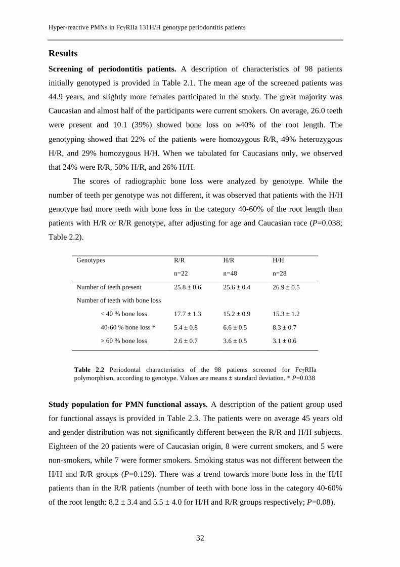
The scores of radiographic bone loss were analyzed by genotype. While the
number of teeth per genotype was not different, it was observed that patients with the H/H
genotype had more teeth with bone loss in the category 40-60% of the root length than
patients with H/R or R/R genotype, after adjusting for age and Caucasian race (P=0.038;
Table 2.2).
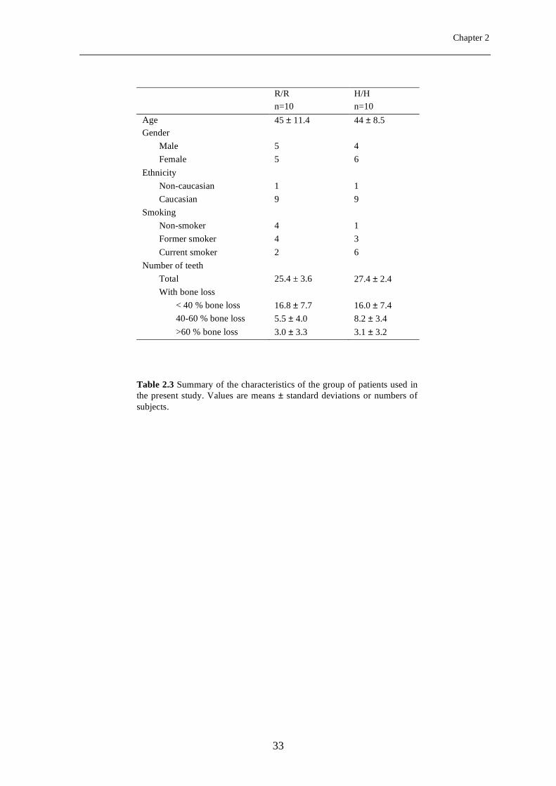
Study population for PMN functional assays. A description of the patient group used
for functional assays is provided in Table 2.3. The patients were on average 45 years old
and gender distribution was not significantly different between the R/R and H/H subjects.
Eighteen of the 20 patients were of Caucasian origin, 8 were current smokers, and 5 were
non-smokers, while 7 were former smokers. Smoking status was not different between the
H/H and R/R groups (P=0.129). There was a trend towards more bone loss in the H/H
patients than in the R/R patients (number of teeth with bone loss in the category 40-60%
of the root length: 8.2 ± 3.4 and 5.5 ± 4.0 for H/H and R/R groups respectively; P=0.08).
Genotypes R/R H/R H/H
n=22 n=48 n=28
Number of teeth present 25.8 ± 0.6 25.6 ± 0.4 26.9 ± 0.5
Number of teeth with bone loss
< 40 % bone loss 17.7 ± 1.3 15.2 ± 0.9 15.3 ± 1.2
40-60 % bone loss * 5.4 ± 0.8 6.6 ± 0.5 8.3 ± 0.7
> 60 % bone loss 2.6 ± 0.7 3.6 ± 0.5 3.1 ± 0.6
Table 2.2 Periodontal characteristics of the 98 patients screened for FcγRIIa polymorphism, according to genotype.
Table 2.2 Periodontal characteristics of the 98 patients screened for FcγRIIa polymorphism, according to genotype. Values are means ± standard deviation. * P=0.038
32
Chapter 2
R/R H/H n=10 n=10 Age 45 ± 11.4 44 ± 8.5 Gender
Male 5 4 Female 5 6
Ethnicity Non-caucasian 1 1 Caucasian 9 9
Smoking Non-smoker 4 1 Former smoker 4 3 Current smoker 2 6
Number of teeth Total 25.4 ± 3.6 27.4 ± 2.4 With bone loss
< 40 % bone loss 16.8 ± 7.7 16.0 ± 7.4 40-60 % bone loss 5.5 ± 4.0 8.2 ± 3.4 >60 % bone loss 3.0 ± 3.3 3.1 ± 3.2
Table 2.3 Summary of the characteristics of the group of patients used in the present study. Values are means ± standard deviations or numbers of subjects.
33
Hyper-reactive PMNs in FcγRIIa 131H/H genotype periodontitis patients
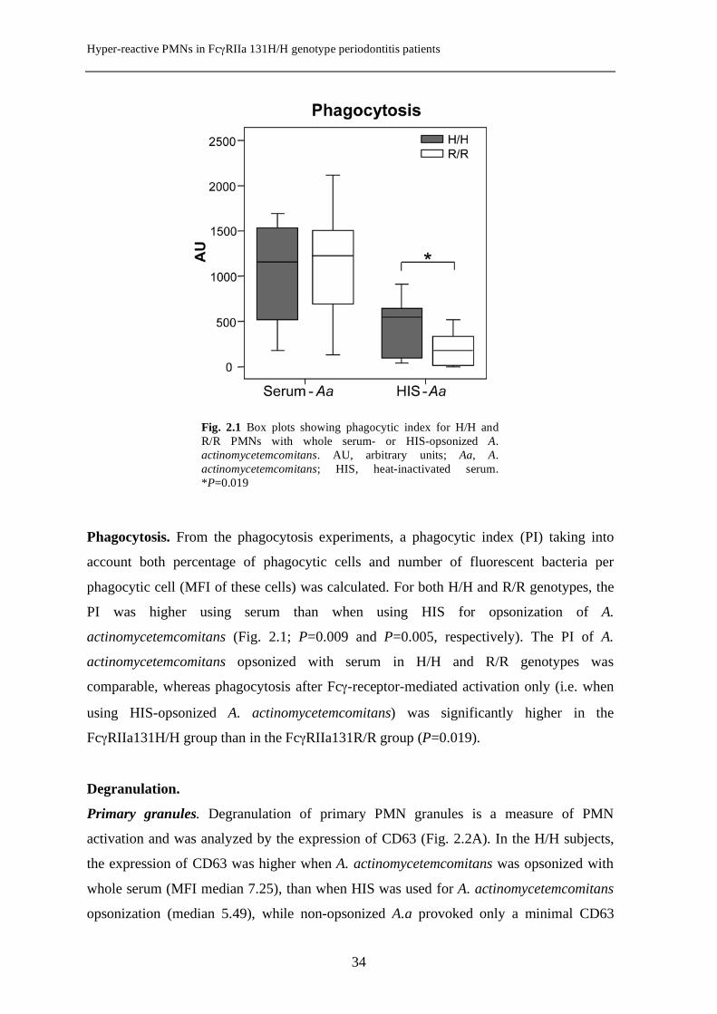
Phagocytosis. From the phagocytosis experiments, a phagocytic index (PI) taking into
account both percentage of phagocytic cells and number of fluorescent bacteria per
phagocytic cell (MFI of these cells) was calculated. For both H/H and R/R genotypes, the
PI was higher using serum than when using HIS for opsonization of A.
actinomycetemcomitans (Fig. 2.1; P=0.009 and P=0.005, respectively). The PI of A.
actinomycetemcomitans opsonized with serum in H/H and R/R genotypes was
comparable, whereas phagocytosis after Fcγ-receptor-mediated activation only (i.e. when
using HIS-opsonized A. actinomycetemcomitans) was significantly higher in the
FcγRIIa131H/H group than in the FcγRIIa131R/R group (P=0.019).
Degranulation.
Primary granules. Degranulation of primary PMN granules is a measure of PMN
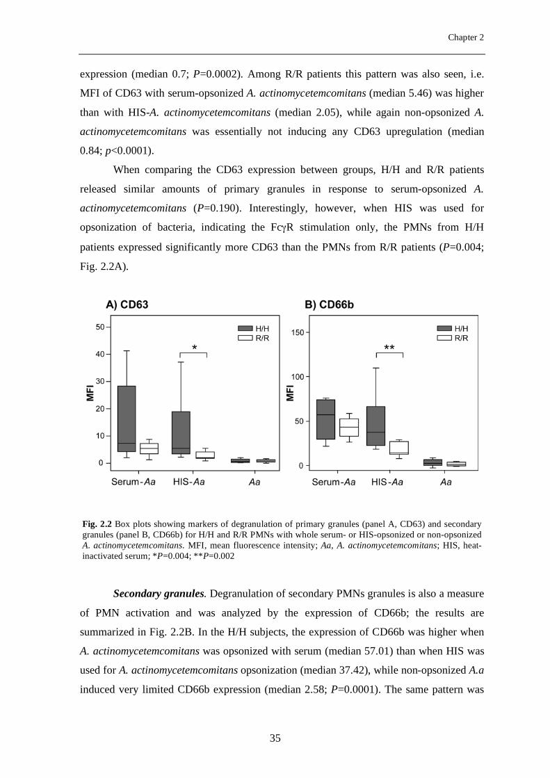
activation and was analyzed by the expression of CD63 (Fig. 2.2A). In the H/H subjects,
the expression of CD63 was higher when A. actinomycetemcomitans was opsonized with
whole serum (MFI median 7.25), than when HIS was used for A. actinomycetemcomitans
opsonization (median 5.49), while non-opsonized A.a provoked only a minimal CD63
Fig. 2.1 Box plots showing phagocytic index for H/H and R/R PMNs with whole serum- or HIS-opsonized A. actinomycetemcomitans. AU, arbitrary units; Aa, A. actinomycetemcomitans; HIS, heat-inactivated serum. *P=0.019
34
Chapter 2
expression (median 0.7; P=0.0002). Among R/R patients this pattern was also seen, i.e.
MFI of CD63 with serum-opsonized A. actinomycetemcomitans (median 5.46) was higher
than with HIS-A. actinomycetemcomitans (median 2.05), while again non-opsonized A.
actinomycetemcomitans was essentially not inducing any CD63 upregulation (median
0.84; p<0.0001).
When comparing the CD63 expression between groups, H/H and R/R patients
released similar amounts of primary granules in response to serum-opsonized A.
actinomycetemcomitans (P=0.190). Interestingly, however, when HIS was used for
opsonization of bacteria, indicating the FcγR stimulation only, the PMNs from H/H
patients expressed significantly more CD63 than the PMNs from R/R patients (P=0.004;
Fig. 2.2A).
Secondary granules. Degranulation of secondary PMNs granules is also a measure
of PMN activation and was analyzed by the expression of CD66b; the results are
summarized in Fig. 2.2B. In the H/H subjects, the expression of CD66b was higher when
A. actinomycetemcomitans was opsonized with serum (median 57.01) than when HIS was
used for A. actinomycetemcomitans opsonization (median 37.42), while non-opsonized A.a
induced very limited CD66b expression (median 2.58; P=0.0001). The same pattern was
Fig. 2.2 Box plots showing markers of degranulation of primary granules (panel A, CD63) and secondary granules (panel B, CD66b) for H/H and R/R PMNs with whole serum- or HIS-opsonized or non-opsonized A. actinomycetemcomitans. MFI, mean fluorescence intensity; Aa, A. actinomycetemcomitans; HIS, heat-inactivated serum; *P=0.004; **P=0.002
35
Hyper-reactive PMNs in FcγRIIa 131H/H genotype periodontitis patients
seen in R/R patients, i.e., the MFI of CD66b in response to serum-opsonized A.
actinomycetemcomitans was higher (median 43.52) than in response to HIS-A.
actinomycetemcomitans (median 14.08), while non-opsonized A. actinomycetemcomitans
was not inducing CD66b expression (median 1.365; P<0.0001).
When comparing the two groups, H/H and R/R PMNs released similar amounts of
secondary granules in response to serum-opsonized A. actinomycetemcomitans (P=0.436).
However, when FcγRIIa were stimulated with HIS-opsonized A. actinomycetemcomitans,
the PMNs from H/H patients expressed significantly more CD66b than did the PMNs from
R/R patients (P=0.002).
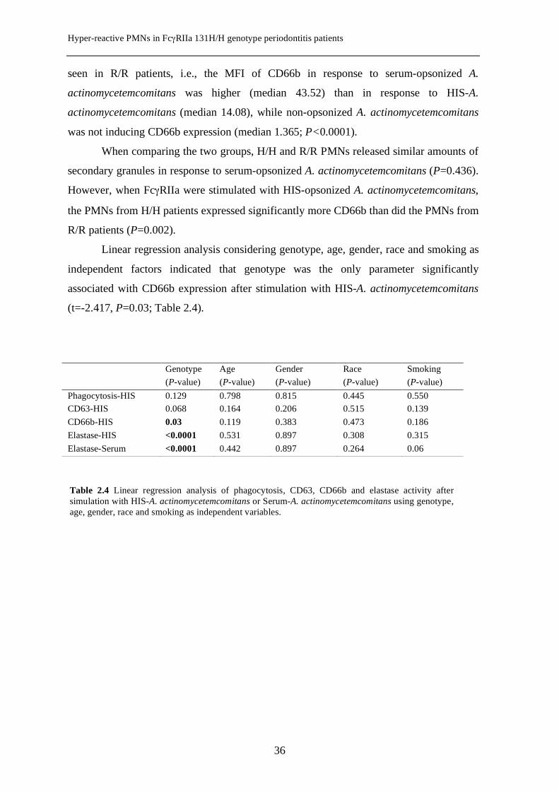
Linear regression analysis considering genotype, age, gender, race and smoking as
independent factors indicated that genotype was the only parameter significantly
associated with CD66b expression after stimulation with HIS-A. actinomycetemcomitans
(t=-2.417, P=0.03; Table 2.4).
Genotype Age Gender Race Smoking (P-value) (P-value) (P-value) (P-value) (P-value) Phagocytosis-HIS 0.129 0.798 0.815 0.445 0.550 CD63-HIS 0.068 0.164 0.206 0.515 0.139 CD66b-HIS 0.03 0.119 0.383 0.473 0.186 Elastase-HIS <0.0001 0.531 0.897 0.308 0.315 Elastase-Serum <0.0001 0.442 0.897 0.264 0.06
Table 2.4 Linear regression analysis of phagocytosis, CD63, CD66b and elastase activity after simulation with HIS-A. actinomycetemcomitans or Serum-A. actinomycetemcomitans using genotype, age, gender, race and smoking as independent variables.
36
Chapter 2
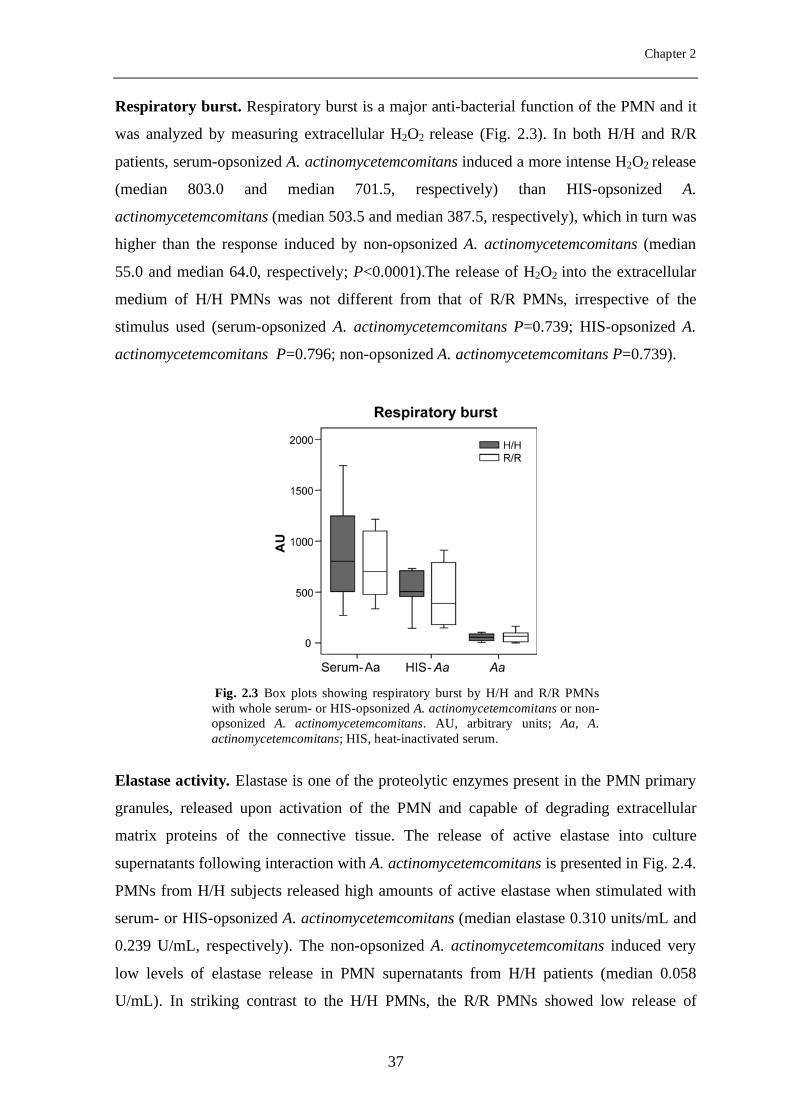
Respiratory burst. Respiratory burst is a major anti-bacterial function of the PMN and it
was analyzed by measuring extracellular H2O2 release (Fig. 2.3). In both H/H and R/R
patients, serum-opsonized A. actinomycetemcomitans induced a more intense H2O2 release
(median 803.0 and median 701.5, respectively) than HIS-opsonized A.
actinomycetemcomitans (median 503.5 and median 387.5, respectively), which in turn was
higher than the response induced by non-opsonized A. actinomycetemcomitans (median
55.0 and median 64.0, respectively; P<0.0001).The release of H2O2 into the extracellular
medium of H/H PMNs was not different from that of R/R PMNs, irrespective of the
stimulus used (serum-opsonized A. actinomycetemcomitans P=0.739; HIS-opsonized A.
actinomycetemcomitans P=0.796; non-opsonized A. actinomycetemcomitans P=0.739).
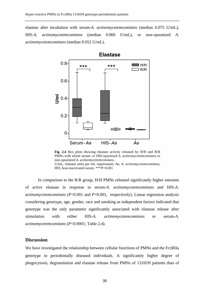
Elastase activity. Elastase is one of the proteolytic enzymes present in the PMN primary
granules, released upon activation of the PMN and capable of degrading extracellular
matrix proteins of the connective tissue. The release of active elastase into culture
supernatants following interaction with A. actinomycetemcomitans is presented in Fig. 2.4.
PMNs from H/H subjects released high amounts of active elastase when stimulated with
serum- or HIS-opsonized A. actinomycetemcomitans (median elastase 0.310 units/mL and
0.239 U/mL, respectively). The non-opsonized A. actinomycetemcomitans induced very
low levels of elastase release in PMN supernatants from H/H patients (median 0.058
U/mL). In striking contrast to the H/H PMNs, the R/R PMNs showed low release of
Fig. 2.3 Box plots showing respiratory burst by H/H and R/R PMNs with whole serum- or HIS-opsonized A. actinomycetemcomitans or non-opsonized A. actinomycetemcomitans. AU, arbitrary units; Aa, A. actinomycetemcomitans; HIS, heat-inactivated serum.
37
Hyper-reactive PMNs in FcγRIIa 131H/H genotype periodontitis patients
elastase after incubation with serum-A. actinomycetemcomitans (median 0.075 U/mL),
HIS-A. actinomycetemcomitans (median 0.060 U/mL), or non-opsonized A.
actinomycetemcomitans (median 0.052 U/mL).
In comparison to the R/R group, H/H PMNs released significantly higher amounts
of active elastase in response to serum-A. actinomycetemcomitans and HIS-A.
actinomycetemcomitans (P<0.001 and P=0.001, respectively). Linear regression analysis
considering genotype, age, gender, race and smoking as independent factors indicated that
genotype was the only parameter significantly associated with elastase release after
stimulation with either HIS-A. actinomycetemcomitans or serum-A.
actinomycetemcomitans (P<0.0001; Table 2.4).
Discussion We have investigated the relationship between cellular functions of PMNs and the FcγRIIa
genotype in periodontally diseased individuals. A significantly higher degree of
phagocytosis, degranulation and elastase release from PMNs of 131H/H patients than of
Fig. 2.4 Box plots showing elastase activity released by H/H and R/R PMNs with whole serum- or HIS-opsonized A. actinomycetemcomitans or non-opsonized A. actinomycetemcomitans. U/mL, elastase units per mL supernatant; Aa, A. actinomycetemcomitans; HIS, heat-inactivated serum. ***P<0.001
38
Chapter 2
131R/R patients was demonstrated when HIS was used for opsonization of A.
actinomycetemcomitans.
The results presented in this study support the hypothesis that the FcγRIIa H/H
genotype may induce a hyper-reactive phenotype of the PMNs, which in response to
periodontal pathogens will release more bioactive molecules that could aggravate the
periodontal destruction. Our results are in line with the idea that the periodontal
breakdown in periodontitis is partly caused by hyper-reactive PMNs (25,5). However,
these latter studies suggested the constitutive nature of the PMN hyper-responsiveness
without shedding light on possible molecular mechanisms involved.
The higher activity of the PMNs of the H/H genotype that we have observed is
most likely the functional consequence of the strong binding of the IgG2 on the bacteria to
the FcγRIIa, because the FcγRIIa H/H is the only PMN receptor that efficiently recognizes
IgG2 (18). The HIS employed in this study contains immunoglobulins specific for the A.
actinomycetemcomitans serotype c strain HG 683 as confirmed in the immunodiffusion
assays. We did not further purify IgG2 from HIS, but it may be the main immunoglobulin
subclass reactive with A. actinomycetemcomitans, as it dominates the antibody response
against polysaccharide antigens that are abundant on the cell wall of the gram-negative
periodontal pathogens (20).
In the case of the more reactive genotype of the FcγRIIa, the H/H, our data
demonstrate a higher release of elastase, contributing to a more severe breakdown of the
extracellular matrix in the periodontal tissues.
Another mechanism proposed for the tissue breakdown induced by hyper-reactive
PMN is the release of oxygen reactive species (26). We observed no significant difference
in H2O2 production between the H/H and R/R PMNs, although degranulation was clearly
increased in the H/H genotype. This unexpected result could be due to a possible
difference in the release of other oxygen species than H2O2. For example, using FcγR-
stimulated PMNs and chemiluminiscence as a measure of all oxygen reactive species
produced, Fredriksson et al. (1998) showed a higher oxidative burst in periodontitis
patients than in controls (27). However, in the same study, there was no difference
between patients and controls when intracellular H2O2 was assessed (27). Therefore, a
future assessment of respiratory burst in H/H and R/R PMNs should take into
consideration the production of all reactive oxygen species rather than relying only on
H2O2 release.
39
Hyper-reactive PMNs in FcγRIIa 131H/H genotype periodontitis patients
A limitation of our study is the low number of participants. From the 98
periodontitis patients genotyped for FcγRIIa 131H/R polymorphism, all H/H (n=28) and
all R/R (n=22) patients were approached to participate in this study. However, only 10
H/H and 10 R/R subjects agreed to donate blood necessary for the PMN functional assays.
We decided to select the homozygous H/H and R/R as two contrasting groups to be
compared with respect to PMN functions. The reason for not including any heterozygous
H/R was first the difficulty in recruiting enough subjects and second, if there is a
difference between genotypes, it should be more clearly visible when using the
homozygous donors. Nevertheless, how PMNs from H/R subjects behave remains to be
elucidated. It was important to note that after performing FcγRIIa genotyping of the 98
periodontitis patients in this cohort, the H/H genotype was associated with more teeth
belonging to the category with bone loss in the 40-60% of the root length. These data
confirmed previous reports and support the view that the H/H genotype may be regarded
as a putative severity factor for periodontitis in Caucasians (15,17). This may be one
important aspect of the genetic make-up of the immune response.
The distribution of smokers and non-smokers was uneven in our R/R and H/H
groups. This drawback was due to the limited number of persons with the required
genotypes who agreed to participate and hence, unavoidable. However, literature reports
on the effect of smoking on the function of PMN are contradictory and do not support a
definitive answer on the matter. Ryder et al. (1998) and Sørensen et al. (2004) describe a
reduced response to stimulation in PMN from smokers when compared to non-smokers
(28,29). Gustafsson et al. (2000) found no difference between the PMN from smokers and
non-smokers after Fcγ-receptors stimulation with opsonized bacteria (30). The issue of the
possible influence of smoking status or other background characteristics on the measured
PMN parameters was a matter of concern for us, as well. We performed an exploratory
linear regression analysis using phagocytosis, CD63, CD66b, or elastase as dependent
variable, and FcγRIIa genotype, age, gender, race and smoking as independent variables.
For none of the parameters of interest, smoking was not significantly contributing to the
measured values nor did age, gender or race. The only parameter significantly associated
with the values of phagocytosis, CD63, CD66b, elastase was the FcγRIIa genotype.
The phagocytosis and degranulation were higher for both H/H and R/R groups
when whole serum-opsonized bacteria were used in comparison to HIS. This indicates that
complement factors in whole serum are important mediators of these processes, thus PMN
40
Chapter 2
reactivity is most likely also affected by complement receptor stimulation. Our data
confirm previous reports that cooperative Fc and C3 receptor interaction is required for
optimal PMN defense against A. actinomycetemcomitans (31).
In conclusion, in the current study we found that FcγRIIa polymorphism influences
the functions of PMNs in periodontitis patients. Individuals with an H/H genotype show a
hyper-reactive phenotype, with increased Fcγ-mediated phagocytosis, degranulation and
granular enzymes release, which may be one of the several factors contributing to the
severity of the periodontitis in these patients.
Acknowledgements The present study was supported by The Netherlands Institute for Dental Sciences. We
thank Nannette Brouwer and Anton Tool for expert assistance in setting up the
phagocytosis and degranulation assays.
REFERENCES
1 Slots J. 1999. Update on Actinobacillus Actinomycetemcomitans and Porphyromonas
gingivalis in human periodontal disease. J Int Acad Periodontol 1:121-126.
2 van Winkelhoff AJ, Loos BG, van der Reijden WA, van der Velden U. 2002.
Porphyromonas gingivalis, Bacteroides forsythus and other putative periodontal
pathogens in subjects with and without periodontal destruction. J Clin Periodontol
29:1023-1028.
3 Van Dyke TE and Sheilesh D. 2005. Risk factors for periodontitis. J Int Acad
Periodontol 7:3-7.
4 Van Dyke TE, Levine MJ, Genco RJ. 1985. Neutrophil function and oral disease. J
Oral Pathol 14:95-120.
5 Fredriksson MI, Gustafsson AK, Bergstrom KG, Asman BE. 2003.
Constitutionally hyperreactive neutrophils in periodontitis. J Periodontol 74:219-24.
6 Janoff A. 1985. Elastase in tissue injury. Annu Rev Med 36:207-216.
41
Hyper-reactive PMNs in FcγRIIa 131H/H genotype periodontitis patients
7 Weiss SJ. 1989. Tissue destruction by neutrophils. N Engl J Med 320:365-376.
8 Anderson CL, Shen L, Eicher DM, Wewers MD, Gill JK. 1990. Phagocytosis
mediated by three distinct Fc gamma receptor classes on human leukocytes. J Exp Med
171:1333-1345.
9 Graziano RF and Fanger MW. 1987. Fc gamma RI and Fc gamma RII on
monocytes and granulocytes are cytotoxic trigger molecules for tumor cells. J
Immunol 139:3536-3541.
10 Anderson CL, Looney RJ, Culp DJ, Ryan DH, Fleit HB, Utell MJ, Frampton
MW, Manganiello PD, Guyre PM. 1990. Alveolar and peritoneal macrophages bear
three distinct classes of Fc receptors for IgG. J Immunol 145:196-201.
11 Chuang FY, Sassaroli M, Unkeless JC. 2000. Convergence of Fc gamma receptor
IIA and Fc gamma receptor IIIB signaling pathways in human neutrophils. J Immunol
164:350-360.
12 Scott-Zaki P, Purkall D, Ruddy S. 2000. Neutrophil chemotaxis and superoxide
production are induced by cross-linking FcgammaRII receptors. Cell Immunol 201:89-
93.
13 Warmerdam PA, Van de Winkel JG, Gosselin EJ, Capel PJ. 1990. Molecular basis
for a polymorphism of human Fc gamma receptor II (CD32). J Exp Med 172:19-25.
14 van der Pol WL and van de Winkel JGJ. 1998. IgG receptor polymorphisms: risk
factors for disease. Immunogenetics 48:222-232.
15 Yamamoto K, Kobayashi T, Grossi S, Ho AW, Genco RJ, Yoshie H, De Nardin E.
2004. Association of Fcgamma receptor IIa genotype with chronic periodontitis in
Caucasians. J Periodontol 75:517-522.
16 Nibali L, Parkar M, Brett P, Knight J, Tonetti MS, Griffiths GS. 2006. NADPH
oxidase (CYBA) and FcgammaR polymorphisms as risk factors for aggressive
periodontitis: A case-control association study. Journal of Clinical Periodontology
33:529-539.
42
Chapter 2
17 Loos BG, Leppers-Van de Straat FG, Van de Winkel JG, van der Velden U. 2003.
Fcgamma receptor polymorphisms in relation to periodontitis. J Clin Periodontol
30:595-602.
18 Warmerdam PA, Van de Winkel JG, Vlug A, Westerdaal NA, Capel PJ. 1991. A
single amino acid in the second Ig-like domain of the human Fc gamma receptor II is
critical for human IgG2 binding. J Immunol 147:1338-1343.
19 Wilson ME and Bronson PM. 1997. Opsonization of Actinobacillus
actinomycetemcomitans by immunoglobulin G antibodies to the O polysaccharide of
lipopolysaccharide. Infect Immun 65:4690-5.
20 Wilson ME and Hamilton RG. 1992. Immunoglobulin G subclass response of
localized juvenile periodontitis patients to Actinobacillus actinomycetemcomitans Y4
lipopolysaccharide. Infect Immun 60:1806-12.
21 Bizzarro S, van der Velden U, ten Heggeler JM, Leivadaros E, Hoek FJ, Gerdes
VE, Bakker SJ, Gans RO, ten Cate H, Loos BG. 2007. Periodontitis is characterized
by elevated PAI-1 activity. J Clin Periodontol 34:574-580
22 Flesch BK, Bauer F, Neppert J. 1998. Rapid typing of the human Fc gamma receptor
IIA polymorphism by polymerase chain reaction amplification with allele-specific
primers. Transfusion 38:174-176.
23 Hildemann S, Hammer C, Krombach F. 1992. Heterogeneity of alveolar
macrophages in experimental silicosis. Environ Health Perspect 97:53-57.
24 Claesson R, Johansson E, Carlsson J. 1994. Oxygen-dependent modulation of
release and activity of polymorphonuclear leukocyte granule products. Oral Microbiol
Immunol 9:81-87.
25 Figueredo CM, Gustafsson A, Asman B, Bergstrom K. 1999. Increased release of
elastase from in vitro activated peripheral neutrophils in patients with adult
periodontitis. J Clin Periodontol 26:206-211.
43
Hyper-reactive PMNs in FcγRIIa 131H/H genotype periodontitis patients
26 Chapple ILC, Brock G, Eftimiadi C, Matthews JB. 2002. Glutathione in gingival
crevicular fluid and its relation to local antioxidant capacity in periodontal health and
disease. Mol Pathol 55:367-373.
27 Fredriksson M, Gustafsson A, Asman B, Bergstrom K. 1998. Hyper-reactive
peripheral neutrophils in adult periodontitis: generation of chemiluminescence and
intracellular hydrogen peroxide after in vitro priming and FcgammaR-stimulation. J
Clin Periodontol 25:394-8.
28 Ryder MI. 2007. The influence of smoking on host responses in periodontal
infections. Periodontology 2000 43:267-277.
29 Sorensen LT, Nielsen HB, Kharazmi A, Gottrup F. 2004. Effect of smoking and
abstention on oxidative burst and reactivity of neutrophils and monocytes. Surgery
136:1047-1053.
30 Gustafsson A, Asman B, Bergstrom K. 2000. Cigarette smoking as an aggravating
factor in inflammatory tissue-destructive diseases. Increase in tumor necrosis Factor-
alpha priming of peripheral neutrophils measured as generation of oxygen radicals. Int
J Clin Lab Res 30:187-190.
31 Wilson ME and Genco RJ. 1989. The role of antibody, complement and neutrophils
in host defense against Actinobacillus actinomycetemcomitans. Immunol Invest
18:187-209.
44
Chapter 3
Expression of FcγRs and mCD14 on polymorphonuclear
neutrophils and monocytes
may determine periodontal infection
E.A. Nicu1, U. van der Velden1, V. Everts2, B.G. Loos1
Departments of 1 Periodontology and 2 Oral Cell Biology, Academic Centre for Dentistry Amsterdam
(ACTA), University of Amsterdam and VU University Amsterdam, the Netherlands
Clinical and Experimental Immunology 2008; 154:177-186
45
Expression of FcγRs and mCD14 on polymorphonuclear neutrophils and monocytes may determine periodontal infection
Abstract Variance in expression of receptors for IgG (FcγRs), complement (CR3) and LPS
(mCD14) on polymorphonuclear neutrophils (PMNs) and monocytes might affect
susceptibility for infection with certain pathogens in periodontitis, a chronic infectious
disease of tooth-supportive tissues. Levels of FcγRI, IIa, III, CR3, and mCD14 on PMNs
and monocytes were measured in 19 periodontitis patients and 18 healthy controls.
Subgingival infection with A. actinomycetemcomitans (Aa) and P. gingivalis (Pg) was
determined. Activation of PMNs and monocytes in response to stimulation with A.
actinomycetemcomitans and P. gingivalis was assessed by means of change in mCD14
expression. Periodontitis is associated with an enrichment of the FcγRIII+ monocytes
(P=0.015) with concomitant low mCD14 (P=0.001). Unadjusted data showed that the
subjects culture-positive for A. actinomycetemcomitans (Aa+) had significantly lower
expression of monocytic FcγRI (P=0.005) and FcγRIIa (P=0.015) than Pg+ subjects. The
FcγRI was still lower on monocytes from Aa+ subjects after adjusting for the background
factors (P=0.037). PMNs from Aa+ subjects responded in a hyper-reactive manner, in
particular when stimulated with A. actinomycetemcomitans (P=0.011). Lower FcγRs
expression by monocytes is related to a higher susceptibility of a subject to become
infected with A. actinomycetemcomitans. The higher proportion of FcγRIII+ monocytes
may be involved in the chronicity of this condition. Hyper-reactive PMNs in Aa+ subjects
may contribute to accelerated breakdown of tooth-supportive tissues.
46
Chapter 3
Introduction
Periodontitis is a chronic infectious disease of the supportive tissues of the teeth
characterized by gradual loss of periodontal attachment and alveolar bone. Approximately
10% of the population suffers from the severe form and, if untreated, it may result in tooth
loss. The major pathogens associated with periodontitis are Aggregatibacter
actinomycetemcomitans (Aa) and Porphyromonas gingivalis (Pg) (1,2). In conjunction
with the bacterial challenge, an important role in the onset and progression of periodontitis
is played by the host immune response (3). A phagocytic response represents the first
barrier to the penetration of bacteria into periodontal tissue (4). However, in addition to
their defensive role, the phagocytes (polymorphonuclear neutrophils [PMNs] and
monocytes) may also be responsible for collateral damage to the periodontal tissues, thus
the host defense may be at the cost of periodontal attachment and alveolar bone (5,6).
One aspect of the host-derived breakdown of periodontal tissues is related to a
hyper-reactive trait of PMNs in response to immunoglobulin G (IgG)-opsonized antigens
(7,8,9). The nature of this hyper-reactivity might be related either to the genetically-
determined increased binding capacity of the receptors for IgG (FcγR) on PMNs or to the
increased expression levels of Fcγ-receptors.
There are several types of FcγRs identified on phagocytes: FcγRI (CD64), FcγRIIa
(CD32) and FcγRIII (CD16) (10). Previous studies have shown that single-nucleotide
polymorphisms, affecting the IgG-binding domain of the genes of FcγRIIa and IIIb, have
functional consequences. PMNs from periodontitis patients bearing the more reactive
genotype (i.e. FcγRIIa-131H/H and FcγRIIIb-NA1/NA1) show hyper-reactivity in
response to stimulation with A. actinomycetemcomitans or P. gingivalis (11,9).
Furthermore, periodontitis patients with FcγRIIa-131H/H genotype have more severe
periodontal breakdown than the patients with FcγRIIa-131H/R or 131R/R genotype
(9,12,13,14). There are several studies that reported the expression of FcγRs on peripheral
PMNs in periodontitis (15,16), but a difference of expression levels between patients and
controls could not be demonstrated.
In contrast to the PMN, little data exist on the expression of FcγR by monocytes in
periodontitis. Nagasawa et al. (17) described an increased percentage of FcγRIII+
monocytes in chronic periodontitis, but did not analyze expression of other FcγRs on
monocytes. Percentages of FcγRIII+ monocytes are also increased in other chronic
inflammatory conditions, such as rheumatoid arthritis (18). Moreover, the monocytes and
47
Expression of FcγRs and mCD14 on polymorphonuclear neutrophils and monocytes may determine the periodontal infection
macrophages of rheumatoid arthritis patients express increased levels of FcγRI and
FcγRIIa. It is conceivable that similar changes in the FcγR expression would occur in
periodontitis, as periodontitis and rheumatoid arthritis appear to share many pathologic
features (19).
In addition to the FcγRs, there are other classes of immune receptors important for
the antibacterial functions of PMNs and monocytes. CD14 is a lipopolysaccharide (LPS)
co-receptor and involved in the recognition of gram-negative bacteria and clearance of
circulating LPS. Membrane-bound CD14 (mCD14) is mainly expressed on mature
monocytes, macrophages and activated neutrophils (20). The β 2-integrins are
heterodimeric receptors consisting of a common β -subunit (CD18) associated with a
unique α -subunit (CD11a, b, c, or d). These receptors are involved in infection and
inflammation by mediating cell-cell, cell-extracellular matrix and cell-pathogen
interactions (21).
The first aim of the present study was to assess the expression of different FcγRs
by PMNs and monocytes from untreated periodontitis patients and healthy controls. In
addition we analyzed the expression levels of two other molecules important for
phagocyte-bacterial interactions: the complement receptor CR3 (CD11b/CD18) and
membrane-bound CD14. The second aim of the study was to explore the activation of
PMNs and monocytes in response to stimulation with two important periodontal
pathogens, A. actinomycetemcomitans and P. gingivalis.
Materials and methods Study population
We recruited 19 periodontitis patients who were referred to the Department of
Periodontology of the Academic Center for Dentistry Amsterdam (ACTA) for diagnosis
and treatment of periodontitis. All patients had ≥8 teeth with radiographic bone loss
beyond 30% of the root length. Age-, gender-, race- and smoking status-matched controls
were selected among subjects registered for restorative dental procedures or who visited
the dental school for regular dental check-ups. Control subjects were selected if they were
not missing more than one tooth per quadrant (3rd molar excluded) and if they showed on
dental bitewing radiographs <1 year old a distance between the cemento-enamel junction
and the alveolar bone crest of <3 mm. Besides their periodontal condition, all participants
48
Chapter 3
were otherwise healthy and had not taken any antibiotics during the last 6 months and no
medication that could influence the immune response during the last 2 weeks.
All patients were initially screened in the departmental clinic and had agreed to accept the
proposed treatment plan. All samples were taken before the start of the periodontal
therapy. All subjects were informed verbally and written about the purposes of the study
and had signed an informed consent. The Medical Ethical Committee of the Academic
Medical Center of the University of Amsterdam approved the study.
Blood sampling
All participants were asked to fast for at least 12 hours before the clinical visit. Fasting
blood samples were obtained between 8.30 and 10.30 AM by venipuncture in the
antecubital fossa. The venous blood was collected into evacuated tubes (BD Vacutainer
System, Plymouth, UK) with anti-coagulant (one 5 mL - EDTA tube for automated
leukocyte counting and leukocyte differential count and one 5 mL Na-citrate tube for flow
cytometric analysis).
Bacterial sampling
Subgingival microbiological samples were taken from all subjects to determine the
presence of the periodontal pathogens A. actinomycetemcomitans and P. gingivalis.
Samples were taken after venous blood collection to avoid possible phagocyte activation.
Sampling, laboratory procedures, and identification of A. actinomycetemcomitans and P.
gingivalis were performed as described before (2).
Flow cytometric analysis
For cell surface staining, citrated whole blood (40 µl) was incubated for 30 min on ice
with saturated concentrations (1-10 µg/mL) of the fluorochrome-conjugated antibodies.
CD14-PE (8G3), CD16-FITC (5D2), and CD11b-FITC (B2) were purchased from
Sanquin (Amsterdam, The Netherlands). CD32-FITC (AT10) and CD64-FITC (10.1) were
obtained from Serotec (Marseille, France). Appropriate isotype controls were used: PE-
IgG2a, FITC-IgG1, FITC-IgG2a from Sanquin and FITC-IgM from Beckman-Coulter
(Fullerton, CA, USA). After incubation, 3 mL of ice-cold NH4Cl buffer (155 mM NH4Cl,
10 mM KHCO3, 0.1 mM EDTA at pH 7.4) was added to lyse the erythrocytes, samples
placed on ice for 15 min and then centrifuged at 500g, 4°C. The cell pellet was washed
49
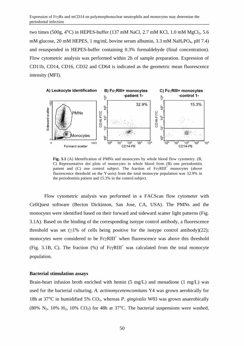
Expression of FcγRs and mCD14 on polymorphonuclear neutrophils and monocytes may determine the periodontal infection
Fig. 3.1 (A) Identification of PMNs and monocytes by whole blood flow cytometry. (B, C) Representative dot plots of monocytes in whole blood from (B) one periodontitis patient and (C) one control subject. The fraction of FcγRIII+ monocytes (above fluorescence threshold on the Y-axis) from the total monocyte population was 32.9% in the periodontitis patient and 15.3% in the control subject.
two times (500g, 4°C) in HEPES-buffer (137 mM NaCl, 2.7 mM KCl, 1.0 mM MgCl2, 5.6
mM glucose, 20 mM HEPES, 1 mg/mL bovine serum albumin, 3.3 mM NaH2PO4, pH 7.4)
and resuspended in HEPES-buffer containing 0.3% formaldehyde (final concentration).
Flow cytometric analysis was performed within 2h of sample preparation. Expression of
CD11b, CD14, CD16, CD32 and CD64 is indicated as the geometric mean fluorescence
intensity (MFI).
Flow cytometric analysis was performed in a FACScan flow cytometer with
CellQuest software (Becton Dickinson, San Jose, CA, USA). The PMNs and the
monocytes were identified based on their forward and sideward scatter light patterns (Fig.
3.1A). Based on the binding of the corresponding isotype control antibody, a fluorescence
threshold was set (≤1% of cells being positive for the isotype control antibody)(22);
monocytes were considered to be FcγRIII+ when fluorescence was above this threshold
(Fig. 3.1B, C). The fraction (%) of FcγRIII+ was calculated from the total monocyte
population.
Bacterial stimulation assays
Brain-heart infusion broth enriched with hemin (5 mg/L) and menadione (1 mg/L) was
used for the bacterial culturing. A. actinomycetemcomitans Y4 was grown aerobically for
18h at 37°C in humidified 5% CO2, whereas P. gingivalis W83 was grown anaerobically
(80% N2, 10% H2, 10% CO2) for 48h at 37°C. The bacterial suspensions were washed,
50
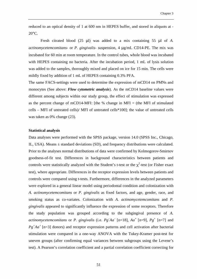
Chapter 3
reduced to an optical density of 1 at 600 nm in HEPES buffer, and stored in aliquots at -
20°C.
Fresh citrated blood (25 µl) was added to a mix containing 55 µl of A.
actinomycetemcomitans- or P. gingivalis- suspension, 4 µg/mL CD14-PE. The mix was
incubated for 60 min at room temperature. In the control tubes, whole blood was incubated
with HEPES containing no bacteria. After the incubation period, 1 mL of lysis solution
was added to the samples, thoroughly mixed and placed on ice for 15 min. The cells were
mildly fixed by addition of 1 mL of HEPES containing 0.3% PFA.
The same FACS-settings were used to determine the expression of mCD14 on PMNs and
monocytes (See above: Flow cytometric analysis). As the mCD14 baseline values were
different among subjects within our study group, the effect of stimulation was expressed
as the percent change of mCD14-MFI: [the % change in MFI = (the MFI of stimulated
cells – MFI of untreated cells)/ MFI of untreated cells*100]; the value of untreated cells
was taken as 0% change (23).
Statistical analysis
Data analyses were performed with the SPSS package, version 14.0 (SPSS Inc., Chicago,
IL, USA). Means ± standard deviations (SD), and frequency distributions were calculated.
Prior to the analyses normal distributions of data were confirmed by Kolmogorov-Smirnov
goodness-of-fit test. Differences in background characteristics between patients and
controls were statistically analyzed with the Student’s t-test or the χ2-test (or Fisher exact
test), where appropriate. Differences in the receptor expression levels between patients and
controls were compared using t-tests. Furthermore, differences in the analyzed parameters
were explored in a general linear model using periodontal condition and colonization with
A. actinomycetemcomitans or P. gingivalis as fixed factors, and age, gender, race, and
smoking status as co-variates. Colonization with A. actinomycetemcomitans and P.
gingivalis appeared to significantly influence the expression of some receptors. Therefore
the study population was grouped according to the subgingival presence of A.
actinomycetemcomitans or P. gingivalis (i.e. Pg–Aa– [n=18], Aa+ [n=9], Pg+ [n=7] and
Pg+Aa+ [n=3] donors) and receptor expression patterns and cell activation after bacterial
stimulation were compared in a one-way ANOVA with the Tukey-Kramer post-test for
uneven groups (after confirming equal variances between subgroups using the Levene’s
test). A Pearson’s correlation coefficient and a partial correlation coefficient correcting for
51
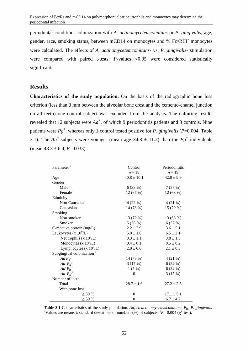
Expression of FcγRs and mCD14 on polymorphonuclear neutrophils and monocytes may determine the periodontal infection
Table 3.1 Characteristics of the study population. Aa, A. actinomycetemcomitans; Pg, P. gingivalis aValues are means ± standard deviations or numbers (%) of subjects; bP =0.004 (χ2-test).
periodontal condition, colonization with A. actinomycetemcomitans or P. gingivalis, age,
gender, race, smoking status, between mCD14 on monocytes and % FcγRIII+ monocytes
were calculated. The effects of A. actinomycetemcomitans- vs. P. gingivalis- stimulation
were compared with paired t-tests; P-values <0.05 were considered statistically
significant.
Results Characteristics of the study population. On the basis of the radiographic bone loss
criterion (less than 3 mm between the alveolar bone crest and the cemento-enamel junction
on all teeth) one control subject was excluded from the analysis. The culturing results
revealed that 12 subjects were Aa+, of which 9 periodontitis patients and 3 controls. Nine
patients were Pg+, whereas only 1 control tested positive for P. gingivalis (P=0.004, Table
3.1). The Aa+ subjects were younger (mean age 34.8 ± 11.2) than the Pg+ individuals
(mean 48.3 ± 6.4, P=0.033).
Parameter a Control Periodontitis n = 18 n = 19 Age 40.8 ± 10.1 42.0 ± 9.9 Gender
Male 6 (33 %) 7 (37 %) Female 12 (67 %) 12 (63 %)
Ethnicity Non-Caucasian 4 (22 %) 4 (21 %) Caucasian 14 (78 %) 15 (79 %)
Smoking Non-smoker 13 (72 %) 13 (68 %) Smoker 5 (28 %) 6 (32 %)
C-reactive protein (mg/L) 2.2 ± 3.9 3.6 ± 5.1 Leukocytes (x 109/L) 5.8 ± 1.6 6.5 ± 2.1
Neutrophils (x 109/L) 3.3 ± 1.1 3.8 ± 1.5 Monocytes (x 109/L) 0.4 ± 0.1 0.5 ± 0.2 Lymphocytes (x 109/L) 2.0 ± 0.6 2.1 ± 0.5
Subgingival colonization b Aa-Pg- 14 (78 %) 4 (21 %) Aa+Pg- 3 (17 %) 6 (32 %) Aa- Pg+ 1 (5 %) 6 (32 %) Aa+Pg+ 0 3 (15 %)
Number of teeth Total 28.7 ± 1.6 27.2 ± 2.5 With bone loss
≥ 30 % 0 17.1 ± 5.1 ≥ 50 % 0 6.7 ± 4.2
52
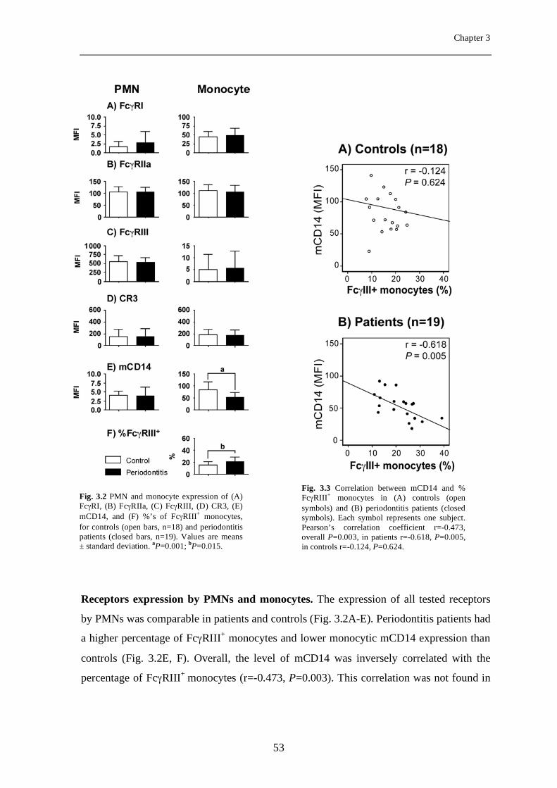
Chapter 3
Receptors expression by PMNs and monocytes. The expression of all tested receptors
by PMNs was comparable in patients and controls (Fig. 3.2A-E). Periodontitis patients had
a higher percentage of FcγRIII+ monocytes and lower monocytic mCD14 expression than
controls (Fig. 3.2E, F). Overall, the level of mCD14 was inversely correlated with the
percentage of FcγRIII+ monocytes (r=-0.473, P=0.003). This correlation was not found in
Fig. 3.2 PMN and monocyte expression of (A) FcγRI, (B) FcγRIIa, (C) FcγRIII, (D) CR3, (E) mCD14, and (F) %’s of FcγRIII+ monocytes, for controls (open bars, n=18) and periodontitis patients (closed bars, n=19). Values are means ± standard deviation. aP=0.001; bP=0.015.
Fig. 3.3 Correlation between mCD14 and % FcγRIII+ monocytes in (A) controls (open symbols) and (B) periodontitis patients (closed symbols). Each symbol represents one subject. Pearson’s correlation coefficient r=-0.473, overall P=0.003, in patients r=-0.618, P=0.005, in controls r=-0.124, P=0.624.
53
Expression of FcγRs and mCD14 on polymorphonuclear neutrophils and monocytes may determine the periodontal infection
control subjects (r=-0.124, P=0.624; Fig. 3.3A), but was strong within periodontitis
patients (r=-0.618, P=0.005; Fig. 3.3B).
In Tables 3.2 and 3.3 we present adjusted data from a general linear model (GLM),
for which periodontal condition and colonization with A. actinomycetemcomitans/P.
gingivalis were entered as fixed factors and age, gender, race and smoking status as co-
variates. Neither colonization with A. actinomycetemcomitans/P. gingivalis nor other
potential confounding factors were associated with expression levels by the PMNs of any
of the receptors tested (Table 3.2). Further, in the GLM we observed that a lower
expression of FcγRI by monocytes was present in the Aa+ subjects (Table 3.3). After
correcting for periodontal condition, colonization with A. actinomycetemcomitans/P.
gingivalis, age, gender, race, smoking status, the correlation between mCD14 and %
FcγRIII+ monocytes was still apparent (radj=-0.382, Padj=0.034).
54
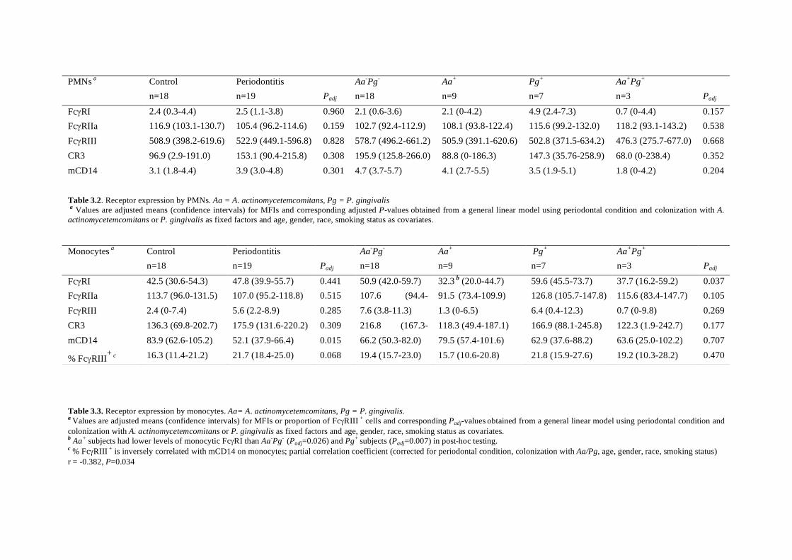
Table 3.2. Receptor expression by PMNs. Aa = A. actinomycetemcomitans, Pg = P. gingivalis a Values are adjusted means (confidence intervals) for MFIs and corresponding adjusted P-values obtained from a general linear model using periodontal condition and colonization with A. actinomycetemcomitans or P. gingivalis as fixed factors and age, gender, race, smoking status as covariates.
Table 3.3. Receptor expression by monocytes. Aa= A. actinomycetemcomitans, Pg = P. gingivalis. a Values are adjusted means (confidence intervals) for MFIs or proportion of FcγRIII + cells and corresponding Padj-values obtained from a general linear model using periodontal condition and colonization with A. actinomycetemcomitans or P. gingivalis as fixed factors and age, gender, race, smoking status as covariates. b Aa+ subjects had lower levels of monocytic FcγRI than Aa-Pg- (Padj=0.026) and Pg+ subjects (Padj=0.007) in post-hoc testing. c % FcγRIII + is inversely correlated with mCD14 on monocytes; partial correlation coefficient (corrected for periodontal condition, colonization with Aa/Pg, age, gender, race, smoking status) r = -0.382, P=0.034
PMNs a Control Periodontitis Aa-Pg- Aa+ Pg+ Aa+Pg+ n=18 n=19 Padj n=18 n=9 n=7 n=3 Padj
FcγRI 2.4 (0.3-4.4) 2.5 (1.1-3.8) 0.960 2.1 (0.6-3.6) 2.1 (0-4.2) 4.9 (2.4-7.3) 0.7 (0-4.4) 0.157 FcγRIIa 116.9 (103.1-130.7) 105.4 (96.2-114.6) 0.159 102.7 (92.4-112.9) 108.1 (93.8-122.4) 115.6 (99.2-132.0) 118.2 (93.1-143.2) 0.538 FcγRIII 508.9 (398.2-619.6) 522.9 (449.1-596.8) 0.828 578.7 (496.2-661.2) 505.9 (391.1-620.6) 502.8 (371.5-634.2) 476.3 (275.7-677.0) 0.668 CR3 96.9 (2.9-191.0) 153.1 (90.4-215.8) 0.308 195.9 (125.8-266.0) 88.8 (0-186.3) 147.3 (35.76-258.9) 68.0 (0-238.4) 0.352 mCD14 3.1 (1.8-4.4) 3.9 (3.0-4.8) 0.301 4.7 (3.7-5.7) 4.1 (2.7-5.5) 3.5 (1.9-5.1) 1.8 (0-4.2) 0.204
Monocytes a Control Periodontitis Aa-Pg- Aa+ Pg+ Aa+Pg+ n=18 n=19 Padj n=18 n=9 n=7 n=3 Padj
FcγRI 42.5 (30.6-54.3) 47.8 (39.9-55.7) 0.441 50.9 (42.0-59.7) 32.3 b (20.0-44.7) 59.6 (45.5-73.7) 37.7 (16.2-59.2) 0.037
FcγRIIa 113.7 (96.0-131.5) 107.0 (95.2-118.8) 0.515 107.6 (94.4-
120.8)
91.5 (73.4-109.9) 126.8 (105.7-147.8) 115.6 (83.4-147.7) 0.105 FcγRIII 2.4 (0-7.4) 5.6 (2.2-8.9) 0.285 7.6 (3.8-11.3) 1.3 (0-6.5) 6.4 (0.4-12.3) 0.7 (0-9.8) 0.269 CR3 136.3 (69.8-202.7) 175.9 (131.6-220.2) 0.309 216.8 (167.3-
266.3)
118.3 (49.4-187.1) 166.9 (88.1-245.8) 122.3 (1.9-242.7) 0.177 mCD14 83.9 (62.6-105.2) 52.1 (37.9-66.4) 0.015 66.2 (50.3-82.0) 79.5 (57.4-101.6) 62.9 (37.6-88.2) 63.6 (25.0-102.2) 0.707
% FcγRIII+ c 16.3 (11.4-21.2) 21.7 (18.4-25.0) 0.068 19.4 (15.7-23.0) 15.7 (10.6-20.8) 21.8 (15.9-27.6) 19.2 (10.3-28.2) 0.470
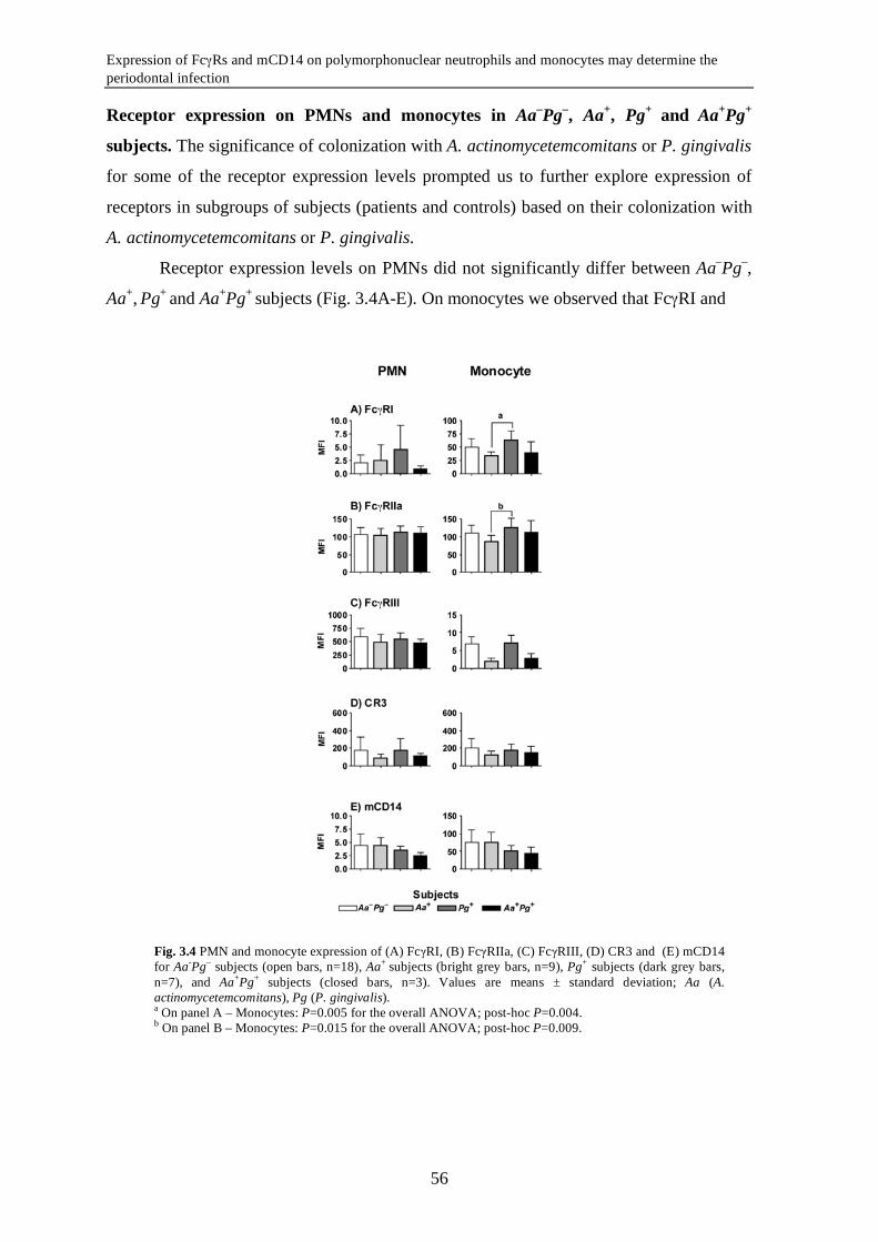
Expression of FcγRs and mCD14 on polymorphonuclear neutrophils and monocytes may determine the periodontal infection
Receptor expression on PMNs and monocytes in Aa–Pg–, Aa+, Pg+ and Aa+Pg+
subjects. The significance of colonization with A. actinomycetemcomitans or P. gingivalis
for some of the receptor expression levels prompted us to further explore expression of
receptors in subgroups of subjects (patients and controls) based on their colonization with
A. actinomycetemcomitans or P. gingivalis.
Receptor expression levels on PMNs did not significantly differ between Aa–Pg–,
Aa+, Pg+ and Aa+Pg+ subjects (Fig. 3.4A-E). On monocytes we observed that FcγRI and
Fig. 3.4 PMN and monocyte expression of (A) FcγRI, (B) FcγRIIa, (C) FcγRIII, (D) CR3 and (E) mCD14 for Aa-Pg– subjects (open bars, n=18), Aa+ subjects (bright grey bars, n=9), Pg+ subjects (dark grey bars, n=7), and Aa+Pg+ subjects (closed bars, n=3). Values are means ± standard deviation; Aa (A. actinomycetemcomitans), Pg (P. gingivalis). a On panel A – Monocytes: P=0.005 for the overall ANOVA; post-hoc P=0.004. b On panel B – Monocytes: P=0.015 for the overall ANOVA; post-hoc P=0.009.
56
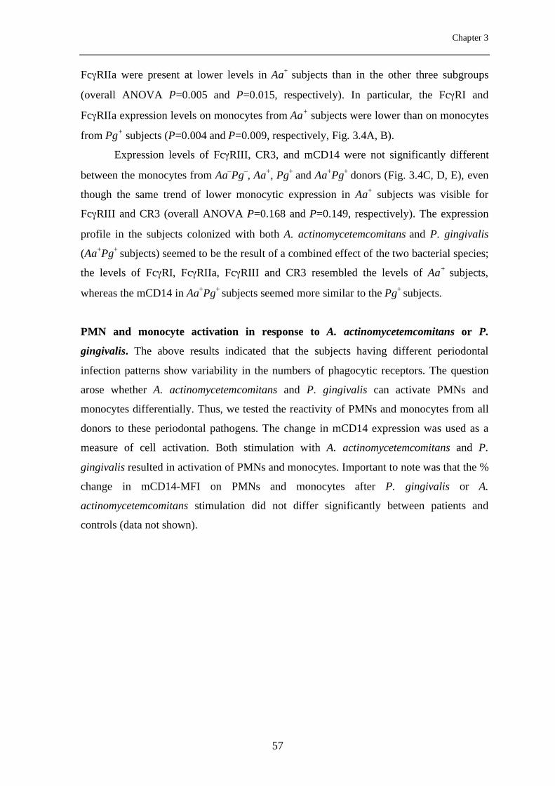
Chapter 3
FcγRIIa were present at lower levels in Aa+ subjects than in the other three subgroups
(overall ANOVA P=0.005 and P=0.015, respectively). In particular, the FcγRI and
FcγRIIa expression levels on monocytes from Aa+ subjects were lower than on monocytes
from Pg+ subjects (P=0.004 and P=0.009, respectively, Fig. 3.4A, B).
Expression levels of FcγRIII, CR3, and mCD14 were not significantly different
between the monocytes from Aa–Pg–, Aa+, Pg+ and Aa+Pg+ donors (Fig. 3.4C, D, E), even
though the same trend of lower monocytic expression in Aa+ subjects was visible for
FcγRIII and CR3 (overall ANOVA P=0.168 and P=0.149, respectively). The expression
profile in the subjects colonized with both A. actinomycetemcomitans and P. gingivalis
(Aa+Pg+ subjects) seemed to be the result of a combined effect of the two bacterial species;
the levels of FcγRI, FcγRIIa, FcγRIII and CR3 resembled the levels of Aa+ subjects,
whereas the mCD14 in Aa+Pg+ subjects seemed more similar to the Pg+ subjects.
PMN and monocyte activation in response to A. actinomycetemcomitans or P.
gingivalis. The above results indicated that the subjects having different periodontal
infection patterns show variability in the numbers of phagocytic receptors. The question
arose whether A. actinomycetemcomitans and P. gingivalis can activate PMNs and
monocytes differentially. Thus, we tested the reactivity of PMNs and monocytes from all
donors to these periodontal pathogens. The change in mCD14 expression was used as a
measure of cell activation. Both stimulation with A. actinomycetemcomitans and P.
gingivalis resulted in activation of PMNs and monocytes. Important to note was that the %
change in mCD14-MFI on PMNs and monocytes after P. gingivalis or A.
actinomycetemcomitans stimulation did not differ significantly between patients and
controls (data not shown).
57
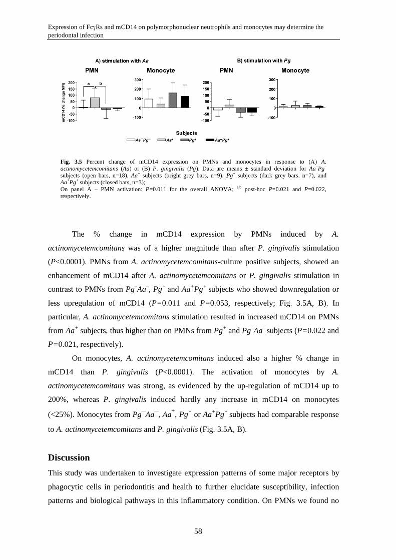
Expression of FcγRs and mCD14 on polymorphonuclear neutrophils and monocytes may determine the periodontal infection
The % change in mCD14 expression by PMNs induced by A.
actinomycetemcomitans was of a higher magnitude than after P. gingivalis stimulation
(P<0.0001). PMNs from A. actinomycetemcomitans-culture positive subjects, showed an
enhancement of mCD14 after A. actinomycetemcomitans or P. gingivalis stimulation in
contrast to PMNs from Pg–Aa–, Pg+ and Aa+Pg+ subjects who showed downregulation or
less upregulation of mCD14 (P=0.011 and P=0.053, respectively; Fig. 3.5A, B). In
particular, A. actinomycetemcomitans stimulation resulted in increased mCD14 on PMNs
from Aa+ subjects, thus higher than on PMNs from Pg+ and Pg–Aa– subjects (P=0.022 and
P=0.021, respectively).
On monocytes, A. actinomycetemcomitans induced also a higher % change in
mCD14 than P. gingivalis (P<0.0001). The activation of monocytes by A.
actinomycetemcomitans was strong, as evidenced by the up-regulation of mCD14 up to
200%, whereas P. gingivalis induced hardly any increase in mCD14 on monocytes
(<25%). Monocytes from Pg¯Aa¯, Aa+, Pg+ or Aa+Pg+ subjects had comparable response
to A. actinomycetemcomitans and P. gingivalis (Fig. 3.5A, B).
Discussion This study was undertaken to investigate expression patterns of some major receptors by
phagocytic cells in periodontitis and health to further elucidate susceptibility, infection
patterns and biological pathways in this inflammatory condition. On PMNs we found no
Fig. 3.5 Percent change of mCD14 expression on PMNs and monocytes in response to (A) A. actinomycetemcomitans (Aa) or (B) P. gingivalis (Pg). Data are means ± standard deviation for Aa–Pg– subjects (open bars, n=18), Aa+ subjects (bright grey bars, n=9), Pg+ subjects (dark grey bars, n=7), and Aa+Pg+ subjects (closed bars, n=3); On panel A – PMN activation: P=0.011 for the overall ANOVA; a,b post-hoc P=0.021 and P=0.022, respectively.
58
Chapter 3
differences between patients and controls in the expression patterns of the tested receptors.
On monocytes we found also a comparable expression of FcγRI, II, and CR3 in patients
and controls. However, an intriguing finding of this study is the heterogeneity of receptor
expression levels between individuals. In the general linear models, bacterial infection
patterns appeared as a major determinant for expression levels by monocytes. When we
subdivided the study population into Pg─Aa─, Aa+, Pg+ and Aa+Pg+ subjects, the
expression of FcγRI and IIa by monocytes was significantly lower in Aa+ subjects than in
Pg+ subjects (Fig. 3.4); after correcting for periodontal condition and potential
confounding factors, the expression of FcγRI monocytes was still decreased in Aa+
subjects (Table 3.3).
One possible explanation for the lower levels of FcγRs on monocytes from Aa+
subjects might be due to genotypic differences, possibly making these individuals more
susceptible to become infected with A. actinomycetemcomitans. Similarly, genetically-
defined deficiencies in various components of the innate immune system (mannose-
binding lectin, vitamin D receptor, mannose-associated serine-protease-2, Toll-like
receptors, etc.) have been associated with a greater risk of infection with Mycobacterium
tuberculosis, meningococci or gram-negative bacteria (24,25,26,27,28). In periodontitis it
has been shown that polymorphisms in the FcγRs and IL-6 genes are associated with
increased odds of detecting A. actinomycetemcomitans, P. gingivalis and Tannerella
forsythia after adjustment for age, ethnicity, smoking and periodontitis extent (29).
However, in our study neither the FcγRIIa 131H+ (131 H/R or H/H) nor the CD14 -260T+
(-260 C/T or T/T) genotypes showed increased odds of detecting A.
actinomycetemcomitans or P. gingivalis in subgingival plaque samples (data not shown).
Alternatively, the lower expression of FcγRs on monocytes in Aa+ subjects might be a
consequence of the infection with A. actinomycetemcomitans. In order to elucidate this,
future studies are needed evaluating the effects of therapeutic elimination of A.
actinomycetemcomitans from the subgingival flora on the expression of FcγRs. In line
with this possibility there are studies demonstrating the effects of medication on FcγRs
expression levels in rheumatoid arthritis. Expression levels of FcγRs with cell activation
potential on peripheral monocytes in patients suffering from rheumatoid arthritis were
increased before therapy in comparison to healthy subjects (30,31,32,33). The levels of
FcγRs are changed to “healthy” levels due to treatment (33,34,35). This change in
expression levels of FcγRs correlates with clinical improvement, thus restoring expression
59
Expression of FcγRs and mCD14 on polymorphonuclear neutrophils and monocytes may determine the periodontal infection
levels of FcγRs can have beneficial effects. Similarly, it is conceivable that periodontal
therapy may have an effect on expression levels of FcγRs.
Normally, the majority of monocytes (90%) do not express FcγRIII and show high
levels of mCD14 (36). However, we found an increased % of FcγRIII+ monocytes and a
lower level of mCD14 on monocytes from periodontitis patients compared to healthy
controls (Fig. 3.2). These results are in line with previous studies showing an increased %
of FcγRIII+ monocytes in chronic periodontitis and lower mCD14 on peripheral monocytes
from early-onset periodontitis patients (37,17). Furthermore, we demonstrated a
correlation between the mCD14 on monocytes and the % of FcγRIII+ monocytes,
suggesting that periodontal infection affects the monocyte phenotype leading to
differentiation of the CD14lowFcγRIII+ cells (36). The CD14lowFcγRIII+ monocytes account
for about 10% of monocytes in healthy adults and are expanding up to 40% in
inflammatory conditions such as rheumatoid arthritis (18,31), sepsis (38) or Kawasaki
disease (39). In our study mCD14 was lowest on monocytes from Pg+ subjects (51.8 ±
13.8 MFI, Fig. 3.4E; adjusted mean 62.9 MFI, Table 3.3) and not significantly different
than the overall value noted for the whole periodontitis patient group (53.1 ± 20.9 MFI,
Fig. 3.2E; adjusted mean 52.1 MFI, Table 3.3). This suggests that individuals infected
with P. gingivalis show the most marked changes in the monocyte phenotype and they
account for the difference in mCD14 expression noted between patients and controls.
The association between P. gingivalis and a lower mCD14 expression on
monocytes is supported by in vitro findings where P. gingivalis LPS was able to induce
maturation of CD14lowFcγRIII+ dendritic cells (40). Compared to the CD14bright-
monocytes, the CD14lowFcγRIII+ monocyte subset can differentiate into dendritic cells that
produce lower amounts of IL-1β, IL-6, IL-12, TNF-α and IL-8, but have higher phagocytic
and oxidative activity (41). These dendritic cells are capable of antigen presentation, but
fail to efficiently stimulate T-cells, possibly due to lack of co-stimulatory molecules (40).
These characteristics of the dendritic cells originating from the CD14lowFcγRIII+
monocytes are possibly contributing to the perpetuation of chronic inflammation in
periodontitis, as they might be unable to efficiently coordinate the antibacterial defense in
the inflamed periodontium. We hypothesize that during periodontal infection frequent
bacteremic episodes, especially of P. gingivalis origin, lead to the early selection of the
CD14lowFcγRIII+ precursors of dendritic cells, which are ultimately unable to clear the
periodontal infection.
60
Chapter 3
PMN receptor expression was essentially not different between patients and
controls and a possible relation to the infection pattern was not found. Thus, in contrast to
monocytes for which we suggest a role in the susceptibility to certain infection patterns,
the PMNs may be solely involved in bacterial clearance. It is commonly accepted that
during their antibacterial functions PMNs induce collateral damage in inflamed
periodontal tissues, which is largely attributable to the production of proteolytic enzymes
(i.e. matrix-metalloproteinases, elastase) and reactive oxygen species (42). In particular
this process is relevant, since hyper-reactive PMNs in periodontitis have been
demonstrated by several research groups and several mechanisms have been proposed
(7,43,8,9). The PMNs from Aa+ subjects showed a stronger response to both P. gingivalis
and A. actinomycetemcomitans (measured by the change in mCD14), further providing
evidence for a hyper-reactive trait of the PMNs from A. actinomycetemcomitans-infected
subjects, possibly aggravating the periodontal inflammatory reactions in this individuals
(7,8,9). This hypothesis may be strengthened by the observations that A.
actinomycetemcomitans induced stronger PMN activation than P. gingivalis (Fig. 3.5A,
B). A. actinomycetemcomitans might be in general a stronger stimulator of phagocytes
than P. gingivalis (44,45,46,47). A. actinomycetemcomitans LPS is inducing higher
amounts of IL-1β, TNF-α and IL-8 in PMNs than P. gingivalis LPS (48), evoking a more
acute, E.coli - like inflammatory immune response. This might help explaining the severe
and relative early onset of periodontal breakdown associated with A.
actinomycetemcomitans infection, especially in patients with aggressive periodontitis (49).
An interesting aspect of the periodontal pathogen P. gingivalis is its capability to produce
bacterial cysteine proteinases (gingipains) which are able to proteolyse human monocyte
mCD14 (50,51). However, protease inhibitors present in serum inhibit the gingipain
activity (51), since in our bacterial stimulation assays A. actinomycetemcomitans and P.
gingivalis were similarly able to induce mCD14 upregulation on monocytes, although of
different amplitude.
In conclusion, we suggest that receptor expression patterns by monocytes may be
related to the susceptibility of a subject to become infected with certain periodontal
pathogens. The enrichment of the FcγRIII+ monocytes in periodontitis, in particular in
patients that are culture-positive for P. gingivalis, may result in formation of a dendritic
cell type that can stimulate T cells less efficiently. As monocytes and their progeny, the
dendritic cells, are expected to orchestrate the immune response in periodontitis, the
61
Expression of FcγRs and mCD14 on polymorphonuclear neutrophils and monocytes may determine the periodontal infection
FcγRIII+ monocytes may be involved in the chronicity of the infection. PMNs from A.
actinomycetemcomitans-culture positive subjects respond in a hyper-reactive fashion to
the infection, and in particular seem to get strongly activated when stimulated with A.
actinomycetemcomitans. In this way PMNs may contribute to advanced breakdown of
tooth-supportive tissues through an enhanced release of a variety of proteolytic enzymes
and reactive oxygen species.
Acknowledgements This study was supported by the Netherlands Institute for Dental Sciences (IOT), the
Department of Periodontology, ACTA and Philips Oral Healthcare EMEA.
We thank Dimitris Papapanagiotou and Denise Duijster for their help in recruiting the
participants of this study.
REFERENCES
1 Slots J. 1999. Update on Actinobacillus Actinomycetemcomitans and Porphyromonas
gingivalis in human periodontal disease. J Int Acad Periodontol 1:121-126.
2 van Winkelhoff AJ, Loos BG, van der Reijden WA, van der Velden U. 2002.
Porphyromonas gingivalis, Bacteroides forsythus and other putative periodontal
pathogens in subjects with and without periodontal destruction. J Clin Periodontol
29:1023-1028.
3 Ebersole JL, Taubman MA, Smith DJ, Frey DE, Haffajee AD, Socransky SS.
1987. Human serum antibody responses to oral microorganisms. IV. Correlation with
homologous infection. Oral Microbiol Immunol 2:53-59.
4 Van Dyke TE, Levine MJ, Genco RJ. 1985. Neutrophil function and oral disease. J
Oral Pathol 14:95-120.
5 Page RC and Schroeder HE. 1976. Pathogenesis of inflammatory periodontal
disease. A summary of current work. Lab Invest 34:235-249.
62
Chapter 3
6 Madianos PN, Bobetsis YA, Kinane DF. 2005. Generation of inflammatory stimuli:
how bacteria set up inflammatory responses in the gingiva. J Clin Periodontol 32
Suppl 6:57-71.
7 Fredriksson M, Gustafsson A, Asman B, Bergstrom K. 1998. Hyper-reactive
peripheral neutrophils in adult periodontitis: generation of chemiluminescence and
intracellular hydrogen peroxide after in vitro priming and FcgammaR-stimulation. J
Clin Periodontol 25:394-8.
8 Matthews JB, Wright HJ, Roberts A, Cooper PR, Chapple IL. 2007. Hyperactivity
and reactivity of peripheral blood neutrophils in chronic periodontitis. Clin Exp
Immunol 147:255-264.
9 Nicu EA, van der Velden U, Everts V, van Winkelhoff AJ, Roos D, Loos BG.
2007. Hyper-reactive PMNs in FcgammaRIIa 131 H/H genotype periodontitis patients.
J Clin Periodontol 34:938-945.
10 Deo YM, Graziano RF, Repp R, Van de Winkel JG. 1997. Clinical significance of
IgG Fc receptors and Fc gamma R-directed immunotherapies. Immunol Today 18:127-
135.
11 Kobayashi T, van der Pol WL, Van de Winkel JG, Hara K, Sugita N, Westerdaal
NA, Yoshie H, Horigome T. 2000. Relevance of IgG receptor IIIb (CD16)
polymorphism to handling of Porphyromonas gingivalis: implications for the
pathogenesis of adult periodontitis. J Periodontal Res 35:65-73.
12 Yamamoto K, Kobayashi T, Grossi S, Ho AW, Genco RJ, Yoshie H, De Nardin E.
2004. Association of Fcgamma receptor IIa genotype with chronic periodontitis in
Caucasians. J Periodontol 75:517-522.
13 Wolf DL, Neiderud AM, Hinckley K, Dahlen G, van de Winkel JGJ, Papapanou
PN. 2006. Fcgamma receptor polymorphisms and periodontal status: a prospective
follow-up study. J Clin Periodontol 33:691-698.
14 Loos BG, Leppers-Van de Straat FG, Van de Winkel JG, van der Velden U. 2003.
Fcgamma receptor polymorphisms in relation to periodontitis. J Clin Periodontol
30:595-602.
63
Expression of FcγRs and mCD14 on polymorphonuclear neutrophils and monocytes may determine the periodontal infection
15 Leino L, Hurttia HM, Sorvajarvi K, Sewon LA. 1994. Increased respiratory burst
activity is associated with normal expression of IgG-Fc-receptors and complement
receptors in peripheral neutrophils from patients with juvenile periodontitis. J
Periodontal Res 29:179-184.
16 Gustafsson A and Asman B. 1996. Increased release of free oxygen radicals from
peripheral neutrophils in adult periodontitis after Fc delta-receptor stimulation. J Clin
Periodontol 23:38-44.
17 Nagasawa T, Kobayashi H, Aramaki M, Kiji M, Oda S, Izumi Y. 2004. Expression
of CD14, CD16 and CD45RA on monocytes from periodontitis patients. J Periodontal
Res 39:72-8.
18 Kawanaka N, Yamamura M, Aita T, Morita Y, Okamoto A, Kawashima M,
Iwahashi M, Ueno A, Ohmoto Y, Makino H. 2002. CD14+,CD16+ blood monocytes
and joint inflammation in rheumatoid arthritis. Arthritis Rheum 46:2578-2586.
19 Mercado FB, Marshall RI, Bartold PM. 2003. Inter-relationships between
rheumatoid arthritis and periodontal disease. A review. J Clin Periodontol 30:761-772.
20 Ziegler-Heitbrock HW and Ulevitch RJ. 1993. CD14: cell surface receptor and
differentiation marker. Immunol Today 14:121-125.
21 Luo BH, Carman CV, Springer TA. 2007. Structural basis of integrin regulation and
signaling. Annu Rev Immunol 25:619-647.
22 Ziegler-Heitbrock HW, Strobel M, Kieper D, Fingerle G, Schlunck T,
Petersmann I, Ellwart J, Blumenstein M, Haas JG. 1992. Differential expression of
cytokines in human blood monocyte subpopulations. Blood 79:503-511.
23 Inoue T, Sakai Y, Morooka S, Hayashi T, Takayanagi K, Takabatake Y. 1996.
Expression of polymorphonuclear leukocyte adhesion molecules and its clinical
significance in patients treated with percutaneous transluminal coronary angioplasty. J
Am Coll Cardiol 28:1127-1133.
24 Garred P, Madsen HO, Svejgaard A, Michaelsen TE. 1999. Mannose-binding
lectin and meningococcal disease. Lancet 354:336-
64
Chapter 3
25 Stengaard-Pedersen K, Thiel S, Gadjeva M, Moller-Kristensen M, Sorensen R,
Jensen LT, Sjoholm AG, Fugger L, Jensenius JC. 2003. Inherited deficiency of
mannan-binding lectin-associated serine protease 2. N Engl J Med 349:554-560.
26 Agnese DM, Calvano JE, Hahm SJ, Coyle SM, Corbett SA, Calvano SE, Lowry
SF. 2002. Human toll-like receptor 4 mutations but not CD14 polymorphisms are
associated with an increased risk of gram-negative infections. J Infect Dis 186:1522-
1525.
27 Bellamy R, Ruwende C, Corrah T, McAdam KP, Thursz M, Whittle HC, Hill
AV. 1999. Tuberculosis and chronic hepatitis B virus infection in Africans and
variation in the vitamin D receptor gene. J Infect Dis 179:721-724.
28 Garred P, Harboe M, Oettinger T, Koch C, Svejgaard A. 1994. Dual role of
mannan-binding protein in infections: another case of heterosis? Eur J Immunogenet
21:125-131.
29 Nibali L, Ready DR, Parkar M, Brett PM, Wilson M, Tonetti MS, Griffiths GS.
2007. Gene polymorphisms and the prevalence of key periodontal pathogens. J Dent
Res 86:416-420.
30 Hepburn AL, Mason JC, Davies KA. 2004. Expression of Fc{gamma} and
complement receptors on peripheral blood monocytes in systemic lupus erythematosus
and rheumatoid arthritis. Rheumatology (Oxford) 43:547-554.
31 Wijngaarden S, van Roon JAG, Bijlsma JWJ, van de Winkel JGJ, Lafeber FPJG.
2003. Fc{gamma} receptor expression levels on monocytes are elevated in rheumatoid
arthritis patients with high erythrocyte sedimentation rate who do not use anti-
rheumatic drugs. Rheumatology (Oxford) 42:681-688.
32 Shinohara S, Hirohata S, Inoue T, Ito K. 1992. Phenotypic analysis of peripheral
blood monocytes isolated from patients with rheumatoid arthritis. J Rheumatol 19:211-
215.
33 Torsteinsdottir I, Arvidson NG, Hallgren R, Hakansson L. 1999. Monocyte
activation in rheumatoid arthritis (RA): increased integrin, Fc gamma and complement
receptor expression and the effect of glucocorticoids. Clin Exp Immunol 115:554-560.
65
Expression of FcγRs and mCD14 on polymorphonuclear neutrophils and monocytes may determine the periodontal infection
34 Magnusson SE, Engstrom M, Jacob U, Ulfgren AK, Kleinau S. 2007. High
synovial expression of the inhibitory FcgammaRIIb in rheumatoid arthritis. Arthritis
Res Ther 9:R51-
35 Wijngaarden S, van Roon JA, Van de Winkel JG, Bijlsma JW, Lafeber FP. 2005.
Down-regulation of activating Fcgamma receptors on monocytes of patients with
rheumatoid arthritis upon methotrexate treatment. Rheumatology (Oxford) 44:729-734.
36 Ziegler-Heitbrock HW. 1996. Heterogeneity of human blood monocytes: the CD14+
CD16+ subpopulation. Immunol Today 17:424-428.
37 Buduneli N, Bicakci N, Keskinoglu A. 2001. Flow-cytometric analysis of
lymphocyte subsets and mCD14 expression in patients with various periodontitis
categories. J Clin Periodontol 28:419-424.
38 Fingerle G, Pforte A, Passlick B, Blumenstein M, Strobel M, Ziegler-Heitbrock
HW. 1993. The novel subset of CD14+/CD16+ blood monocytes is expanded in sepsis
patients. Blood 82:3170-3176.
39 Katayama K, Matsubara T, Fujiwara M, Koga M, Furukawa S. 2000.
CD14+CD16+ monocyte subpopulation in Kawasaki disease. Clin Exp Immunol
121:566-570.
40 Kanaya S, Nemoto E, Ogawa T, Shimauchi H. 2004. Porphyromonas gingivalis
lipopolysaccharides induce maturation of dendritic cells with CD14+CD16+
phenotype. Eur J Immunol 34:1451-1460.
41 Almeida J, Bueno C, Alguero MC, Sanchez ML, de Santiago M, Escribano L,
Diaz-Agustin B, Vaquero JM, Laso FJ, San Miguel JF, Orfao A. 2001.
Comparative Analysis of the Morphological, Cytochemical, Immunophenotypical, and
Functional Characteristics of Normal Human Peripheral Blood Lineage-/CD16+/HLA-
DR+/CD14-/lo Cells, CD14+ Monocytes, and CD16- Dendritic Cells. Clin Immunol
100:325-338.
42 Woessner JF, Jr. 1991. Matrix metalloproteinases and their inhibitors in connective
tissue remodeling. FASEB J 5:2145-2154.
66
Chapter 3
43 Gustafsson A, Ito H, Asman B, Bergstrom K. 2006. Hyper-reactive mononuclear
cells and neutrophils in chronic periodontitis. J Clin Periodontol 33:126-129.
44 Nonnenmacher C, Dalpke A, Zimmermann S, Flores-de-Jacoby L, Mutters R,
Heeg K. 2003. DNA from periodontopathogenic bacteria is immunostimulatory for
mouse and human immune cells. Infect Immun 71:850-856.
45 Harokopakis E, Albzreh MH, Haase EM, Scannapieco FA, Hajishengallis G.
2006. Inhibition of proinflammatory activities of major periodontal pathogens by
aqueous extracts from elder flower (Sambucus nigra). J Periodontol 77:271-279.
46 Kobayashi H, Nagasawa T, Aramaki M, Mahanonda R, Ishikawa I. 2000.
Individual diversities in interferon gamma production by human peripheral blood
mononuclear cells stimulated with periodontopathic bacteria. J Periodontal Res
35:319-328.
47 Reddi K, Wilson M, Nair S, Poole S, Henderson B. 1996. Comparison of the pro-
inflammatory cytokine-stimulating activity of the surface-associated proteins of
periodontopathic bacteria. J Periodontal Res 31:120-130.
48 Yoshimura A, Hara Y, Kaneko T, Kato I. 1997. Secretion of IL-1 beta, TNF-alpha,
IL-8 and IL-1ra by human polymorphonuclear leukocytes in response to
lipopolysaccharides from periodontopathic bacteria. J Periodontal Res 32:279-286.
49 van der Velden U, Abbas F, Van Steenbergen TJ, De Zoete OJ, Hesse M, De
Ruyter C, De Laat VH, de Graaff J. 1989. Prevalence of periodontal breakdown in
adolescents and presence of Actinobacillus actinomycetemcomitans in subjects with
attachment loss. J Periodontol 60:604-610.
50 Duncan L, Yoshioka M, Chandad F, Grenier D. 2004. Loss of lipopolysaccharide
receptor CD14 from the surface of human macrophage-like cells mediated by
Porphyromonas gingivalis outer membrane vesicles. Microb Pathog 36:319-325.
51 Sugawara S, Nemoto E, Tada H, Miyake K, Imamura T, Takada H. 2000.
Proteolysis of human monocyte CD14 by cysteine proteinases (gingipains) from
Porphyromonas gingivalis leading to lipopolysaccharide hyporesponsiveness. J
Immunol 165:411-418.
67

68
Chapter 4
Soluble CD14 as a possible molecular link between
atherosclerosis and chronic infectious diseases: periodontitis
as model
E.A. Nicu1, M.L. Laine2, S.A. Morré3,4, U. van der Velden1 and B.G. Loos1
Departments of 1 Periodontology and 2 Oral Microbiology, Academic Center for Dentistry Amsterdam
(ACTA), Universiteit van Amsterdam and VU University Amsterdam, The Netherlands
3Laboratory of Immunogenetics, Department of Pathology and Section Infectious Diseases, Department of
Internal Medicine, VU University Medical Center, Amsterdam, The Netherlands; and 4Department of
Medical Microbiology, Maastricht University Medical Center, Maastricht, The Netherlands.
Submitted to Innate Immunity
69
Soluble CD14 as a possible molecular link between atherosclerosis and chronic infectious diseases: periodontitis as model
Abstract Lipopolysaccharide (LPS) binds to soluble (s) CD14. The LPS-sCD14 complexes activate
endothelial and smooth muscle cells, exacerbating inflammation in atherosclerotic lesions.
We hypothesized that sCD14 is an important link between atherosclerosis and low-grade
infectious processes, such as chronic periodontitis. sCD14 levels were determined by
ELISA in healthy controls (n=57) and untreated patients (59 moderate and 46 severe) and
their relation with markers of systemic inflammation (CRP levels, leukocyte, neutrophil
and lymphocyte counts) was assessed. Anti-A. actinomycetemcomitans and anti-P.
gingivalis IgG levels were established by ELISA and CD14-260 genotype was determined
in a TaqMan allelic discrimination assay. Increased levels of sCD14 were more frequent
among periodontitis patients (P=0.026) and showed a severity-dependence with increasing
levels of periodontal breakdown (P=0.008). In patients, levels of sCD14 correlated
positively with CRP (P=0.043), leukocyte numbers (P=0.011) and negatively with anti-A.
actinomycetemcomitans IgG (P=0.007). In a multivariate analysis, sCD14 levels were
predicted by ethnicity, age, educational level, and in Caucasian subjects also by the
severity of periodontal destruction, but not by anti-P. gingivalis IgG or the CD14-260
genotype. Periodontitis is associated with elevated levels of sCD14. We suggest that
increased sCD14 levels in periodontitis might lead to increased activation of non-CD14
expressing cells, possibly activating atherogenesis.
70
Chapter 4
Introduction Cardiovascular diseases (CVD) are a leading cause of global mortality, accounting for
almost 17 million deaths annually or 30% of all global mortality (1). The underlying
mechanism associated with CVD is atherosclerosis. In the recent years, a growing interest
is emerging in aspects of infection and inflammation, as they may also be important risk
factors in the initiation and progression of atherosclerosis (2). Longstanding, systemic
chronic inflammation has been attributed to repeated, transient dissemination of certain
micro-organisms in the peripheral circulation. These include Helicobacter pylori,
Chlamydia pneumoniae, and cytomegalovirus (3). The systemic chronic inflammation is
evidenced by elevated C-reactive protein (CRP); this acute-phase protein is regarded as a
risk factor for CVD (4). Periodontitis may be a source of daily, low-grade bacteremias.
The periodontal pockets form ports of entry which lead to transient bacteremias and
leakage of bacterial products into blood (5). Periodontitis is also clearly associated with
elevated levels of CRP and increased risk for CVD (6,7).
Lipopolysaccharide (LPS), the common component of Gram-negative bacteria,
might be the trigger for the events linking some chronic infectious processes and
atherosclerosis (8). LPS is recognized by CD14-expressing immune cells (9). Membrane-
bound CD14 (mCD14) is mainly expressed on mature monocytes, macrophages and
activated neutrophils (10). Soluble (s)CD14 can be the result of shedding / cleavage of
mCD14 or be produced in the liver. Automatic text mining of the literature has identified
the CD14 molecule as an important link between atherosclerosis and periodontitis (11). In
periodontitis, elevated serum levels of LPS have bee reported,(8) as well as elevated levels
of sCD14 (12). Moreover, in the latter, Japanese study, treatment of periodontitis was
shown to decrease the sCD14 levels (12). Gram-negative periodontal pathogens, such as
Aggregatibacter actinomycetemcomitans and Porphyromonas gingivalis, might contribute
to the overall pro-atherogenic burden in periodontitis patients as a result of recurrent
bacteremic episodes (5).
CD14 binds LPS linked with LPS-binding-protein (LBP); the CD14-LPS-LBP
complex activates Toll-like receptors 2 and 4 and will, through activation of downstream
molecules, result in production of cytokines such as tumor necrosis factor (TNF)-alpha,
interleukin (IL)-1, IL-6 and growth factors (13). The resulting pro-inflammatory milieu
can accelerate atherogenesis. Endothelial and smooth muscle cells, lacking their own
mCD14, are directly activated by a LPS-sCD14 complex (14,15), leading to the
71
Soluble CD14 as a possible molecular link between atherosclerosis and chronic infectious diseases: periodontitis as model
expression of cell adhesion molecules, thereby increasing procoagulant activity, and
exacerbating inflammation in atherosclerotic vessel walls (16).
The promotor region of the CD14 gene contains a single nucleotide polymorphism
(C>T) at position -260. The CD14-260 T allele has been associated not only with increased
levels of sCD14 (17), myocardial infarction (18), and increased susceptibility to chronic
C. pneumoniae infection in coronary artery disease patients (19), but also with severe
periodontitis (20).
Given above, we hypothesized that CD14 is an important molecular link between
atherosclerosis and low-grade infectious processes. Using periodontitis as a model, we
investigated sCD14 plasma levels in relation to systemic markers of inflammation,
especially CRP, infection with the periodontal pathogens A. actinomycetemcomitans and
P. gingivalis, and the CD14-260 genotype.
Materials and methods Study population
Our study population was derived from two previous studies.(21,22) We included all
subjects for which the plasma levels of sCD14, serology, and CD14-260 genotype could be
determined. Fifty-seven healthy controls and untreated periodontitis patients (59 moderate
and 46 severe) were included. For details of dental inclusion criteria for patients and
controls, see Bizzarro et al (22). Exclusion criteria for controls and patients were: (I) the
presence of systemic disease (especially cardiovascular disorders, diabetes mellitus,
allergy), (II) a recent history or the presence of any acute or chronic infection, (III)
systemic antibiotic treatment within the last 3 months, (IV) the use of any medication
(including sporadic NSAID’s), and (V) pregnancy. For all participants smoking habits
were recorded; non-smokers were subjects who never smoked or quitted smoking >10
years ago. Educational level was used as surrogate marker for social class and the given
scores were 0 or 1 for the subjects with educational level < highschool or ≥ highschool,
respectively. From height and weight measurements the body mass index (BMI) was
calculated. Subjects from European ancestry were entered as Caucasian and individuals
from other or mixed ancestry were scored as non-Caucasian.
All subjects were both verbally and written informed about the purpose of the
study and gave written informed consent to participate. The Medical Ethical Committee of
the Academic Medical Center of the University of Amsterdam approved the study.
72
Chapter 4
Lipids profile and systemic markers of inflammation.
Plasma levels of total cholesterol, HDL cholesterol and triglycerides were determined by
standard (enzymatic) methods in a hospital based diagnostic clinical laboratory; LDL
cholesterol was calculated (23). Plasma levels of CRP were determined using a high
sensitivity (latex enhanced) nephelometric method on the BN ProSpec analyzer (Dade
Behring, Marburg, Germany). EDTA blood was used for automated leukocyte counting
and leukocyte differentiation.
Infection with A. actinomycetemcomitans and P. gingivalis.
The determination of serum IgG levels against two relevant periodontal pathogens, A.
actinomycetemcomitans and P. gingivalis, was a modification of the enzyme-linked
immunosorbent assay (ELISA) described by Pussinen et al.(24) As antigens we used a
mixture of five strains of A. actinomycetemcomitans and eight strains of P. gingivalis. The
strains were ATCC 29523, Y4, NCTC 9710, 3381 and OM2 534 for A.
actinomycetemcomitans, representing the serotypes a, b, c, d and e, and W83, HG 184,
A7A1-28, ATCC 49417, HG 1690, HG 1691 and 34-4 for P. gingivalis, representing the
capsule serotypes K1-K7, as well as the uncapsulated strain 381. A.
actinomycetemcomitans were grown for 18h in brain heart infusion (BHI) broth (Sigma
Chemical Co., St.Louis, MO) aerobically at 37°C in humidified 5% CO2. P. gingivalis
were grown anaerobically (80% N2, 10% H2, 10% CO2) at 37°C for 18h in BHI broth
supplemented with haemin (5 mg/L) and menadione (1 mg/L) (Sigma). The bacteria were
washed once with phosphate-buffered saline (PBS; 10 mM phosphate, 150 mM NaCl,
pH=7.4) and then fixed overnight at 4°C in 0.5% paraformaldehyde-PBS. The bacterial
suspensions were washed three times in PBS and brought to an optical density
corresponding to an absorbance of 0.15 at 580 nm in ELISA-buffer (PBS, 0.5% bovine
serum albumin, 0.05% Tween 20).
For ELISA, equal volumes of the five A. actinomycetemcomitans or of the eight P.
gingivalis strains were mixed and 150 µl of the mixture was used to coat Microlon ELISA
plates (Greiner Bio-One B.V., Alphen a/d Rijn, The Netherlands). The unspecific binding
was blocked by 5% bovine serum albumin in PBS at room temperature for 30 min. Diluted
(1:1500) serum samples were tested in duplicate. The plates were incubated for 2h at room
temperature, washed 3 times in ELISA-buffer. The horse-radish peroxidase-conjugated
goat anti-human IgG (Vector Laboratories Inc., Burlingame, CA) diluted (1:2000; 150 µl)
73
Soluble CD14 as a possible molecular link between atherosclerosis and chronic infectious diseases: periodontitis as model
was added and the plates were incubated for 2h at room temperature. Substrate was then
added, and absorbance values were measured at 450 nm with a multilabel counter (Wallac
Victor 1420, Perkin-Elmer Life Sciences, Boston, MA).
We selected a cut-off point for defining seropositivity, the 75th percentile of the
control population (OD=0.744 for A. actinomycetemcomitans and OD=0.915 for P.
gingivalis, respectively). The subjects were considered seropositive for A.
actinomycetemcomitans and P. gingivalis when the corresponding IgG value was higher
than the threshold.
CD14 –260 genotype
Genomic DNA was isolated from blood samples by means of the Puregene DNA isolation
kit (Gentra Systems, Minneapolis, MN, USA) according to the manufacture’s instructions.
We assessed the C>T substitution in the proximal CD14 promoter GC box at position –
260 from the translation start site (NCBI SNP CLUSTER ID: rs2569190). A realtime
TaqMan PCR was performed using standard conditions with the following primers and
probes: forward primer GACACTGCCAGGAGACACAGAA and reverse primer
GCCAGCCCCCTTCCTTT, and probes: TGTTACGGCCCCCCT-VIC-MGB (for the
common allele, the C allele), TTACGGTCCCCCTCC-6-FAM-MGB (for the rare allele,
the T allele).
Soluble CD14 (sCD14)
The concentration of sCD14 was measured by a sandwich ELISA using two monoclonal
antibodies (MAbs) against different epitopes of sCD14 (R&D systems, Abingdon, UK) in
accordance to the manufacturer’s instructions. In brief, EDTA plasma specimens were
diluted 1:200 and incubated in duplicate for 3h in a 96-well plate precoated with the anti-
CD14 MAb. The plate was incubated for 1h with the other anti-CD14 MAb conjugated
with peroxidase. Substrate was then added, and absorbance values were measured at 450
nm with a multilabel counter (Wallac Victor 1420). The concentration of each sample was
determined by extrapolation from a standard curve estimated from a panel of sCD14
standards of known concentrations. Serum levels of sCD14 were expressed as mg/L. The
intra- and inter-assay coefficients of variation were 2.4% and 6.3%, respectively.
74
Chapter 4
Statistical analysis
Data analyses were performed with the SPSS 15.0 package (SPSS Inc., Chicago, IL,
USA). Means, standard deviations and frequency distributions were calculated; the
background characteristics were compared with one-way ANOVA or χ2-test (or Fisher
exact test), where appropriate. Normal distribution of data was assessed by Kolmogorov-
Smirnov goodness-of-fit test and if needed, the log-transformed values were employed.
Differences in sCD14 between controls, moderate and severe patients were compared with
the Kruskal-Wallis test. For sCD14 the 75th percentile of values of the control subjects was
used as cut-off point for a frequency distribution analysis followed by χ2-test. Correlations
between sCD14 levels, infection with A. actinomycetemcomitans or P. gingivalis, CRP,
and numbers of leukocytes, neutrophils and lymphocytes, and for patients also between
sCD14 and the number of teeth with ≥50% bone loss, were investigated by Spearman
correlation coefficients, ρ. A multivariate analysis (backward stepwise linear regression
with P=0.10 to enter and P=0.05 to leave) was performed considering the sCD14 levels as
the outcome variable. Predictor variables were age, gender, ethnicity, smoking,
educational level, BMI, CD14-260 genotype, anti-A. actinomycetemcomitans IgG levels,
anti-P. gingivalis IgG levels, total cholesterol, triglycerides, and no. of teeth with ≥50%
bone loss.
Results Study population. Two third of the study population was European Caucasian (Table 4.1)
and the remainder had a mixed racial background. Periodontitis patients had a lower
educational level than the healthy controls (P=0.002).
Lipids profile and systemic markers of inflammation. Levels of total cholesterol,
HDL, LDL, and triglycerides were roughly comparable in controls, moderate or severe
periodontitis patients. The total number of leukocytes was increased in moderate and
severe periodontitis compared to controls (P<0.001; Table 4.1). The increase was largely
explained by the increase in neutrophil counts in periodontitis compared to health
(P<0.001). CRP levels showed a tendency to be increased in moderate and severe
periodontitis groups compared to the control group
(P=0.072).
Infection with A. actinomycetemcomitans and P. gingivalis. The anti-A.
actinomycetemcomitans and the anti-P. gingivalis IgG levels were increased in
75
Soluble CD14 as a possible molecular link between atherosclerosis and chronic infectious diseases: periodontitis as model
periodontitis compared to health (P=0.002 and P=0.006, respectively). As expected, more
A. actinomycetemcomitans-seropositive and P. gingivalis-seropositive subjects were
present among moderate and severe periodontitis patients than among controls (P=0.004
and P=0.049, respectively; Table 4.1).
CD14 –260 genotype. CD14-260 allele frequencies in periodontitis patients and
controls did not show a significant deviation from the Hardy-Weinberg equilibrium
(P>0.05). We did not observe differences in the distribution of the CD14-260 genotypes or
of the allele frequencies between patients and controls, neither when the entire study
population was analyzed, nor when considering only Caucasian subjects (Table 4.1).
76
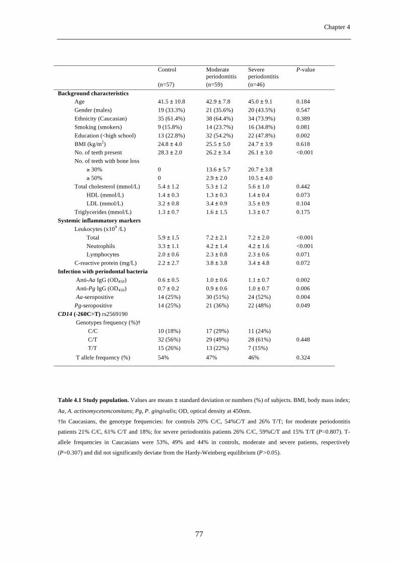
Chapter 4
Table 4.1 Study population. Values are means ± standard deviation or numbers (%) of subjects. BMI, body mass index;
Aa, A. actinomycetemcomitans; Pg, P. gingivalis; OD, optical density at 450nm.
†In Caucasians, the genotype frequencies: for controls 20% C/C, 54%C/T and 26% T/T; for moderate periodontitis
patients 21% C/C, 61% C/T and 18%; for severe periodontitis patients 26% C/C, 59%C/T and 15% T/T (P=0.807). T-
allele frequencies in Caucasians were 53%, 49% and 44% in controls, moderate and severe patients, respectively
(P=0.307) and did not significantly deviate from the Hardy-Weinberg equilibrium (P>0.05).
Control
Moderate periodontitis
Severe periodontitis
P-value
(n=57) (n=59) (n=46) Background characteristics
Age 41.5 ± 10.8 42.9 ± 7.8 45.0 ± 9.1 0.184 Gender (males) 19 (33.3%) 21 (35.6%) 20 (43.5%) 0.547 Ethnicity (Caucasian) 35 (61.4%) 38 (64.4%) 34 (73.9%) 0.389 Smoking (smokers) 9 (15.8%) 14 (23.7%) 16 (34.8%) 0.081 Education (<high school) 13 (22.8%) 32 (54.2%) 22 (47.8%) 0.002 BMI (kg/m2) 24.8 ± 4.0 25.5 ± 5.0 24.7 ± 3.9 0.618 No. of teeth present 28.3 ± 2.0 26.2 ± 3.4 26.1 ± 3.0 <0.001 No. of teeth with bone loss
≥ 30% 0 13.6 ± 5.7 20.7 ± 3.8 ≥ 50% 0 2.9 ± 2.0 10.5 ± 4.0
Total cholesterol (mmol/L) 5.4 ± 1.2 5.3 ± 1.2 5.6 ± 1.0 0.442 HDL (mmol/L) 1.4 ± 0.3 1.3 ± 0.3 1.4 ± 0.4 0.073 LDL (mmol/L) 3.2 ± 0.8 3.4 ± 0.9 3.5 ± 0.9 0.104
Triglycerides (mmol/L) 1.3 ± 0.7 1.6 ± 1.5 1.3 ± 0.7 0.175 Systemic inflammatory markers
Leukocytes (x109 /L) Total 5.9 ± 1.5 7.2 ± 2.1 7.2 ± 2.0 <0.001 Neutrophils 3.3 ± 1.1 4.2 ± 1.4 4.2 ± 1.6 <0.001 Lymphocytes 2.0 ± 0.6 2.3 ± 0.8 2.3 ± 0.6 0.071
C-reactive protein (mg/L) 2.2 ± 2.7 3.8 ± 3.8 3.4 ± 4.8 0.072 Infection with periodontal bacteria
Anti-Aa IgG (OD450) 0.6 ± 0.5 1.0 ± 0.6 1.1 ± 0.7 0.002 Anti-Pg IgG (OD450) 0.7 ± 0.2 0.9 ± 0.6 1.0 ± 0.7 0.006 Aa-seropositive 14 (25%) 30 (51%) 24 (52%) 0.004 Pg-seropositive 14 (25%) 21 (36%) 22 (48%) 0.049
CD14 (-260C>T) rs2569190 Genotypes frequency (%)†
C/C 10 (18%) 17 (29%) 11 (24%) C/T 32 (56%) 29 (49%) 28 (61%) 0.448 T/T 15 (26%) 13 (22%) 7 (15%)
T allele frequency (%) 54% 47% 46% 0.324
77
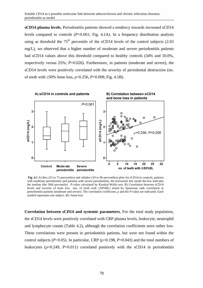
Soluble CD14 as a possible molecular link between atherosclerosis and chronic infectious diseases: periodontitis as model
sCD14 plasma levels. Periodontitis patients showed a tendency towards increased sCD14
levels compared to controls (P=0.061; Fig. 4.1A). In a frequency distribution analysis
using as threshold the 75th percentile of the sCD14 levels of the control subjects (2.03
mg/L), we observed that a higher number of moderate and severe periodontitis patients
had sCD14 values above this threshold compared to healthy controls (34% and 50.0%,
respectively versus 25%; P=0.026). Furthermore, in patients (moderate and severe), the
sCD14 levels were positively correlated with the severity of periodontal destruction (no.
of teeth with ≥50% bone loss, ρ=0.256, P=0.008; Fig. 4.1B).
Correlation between sCD14 and systemic parameters. For the total study population,
the sCD14 levels were positively correlated with CRP plasma levels, leukocyte, neutrophil
and lymphocyte counts (Table 4.2), although the correlation coefficients were rather low.
These correlations were present in periodontitis patients, but were not found within the
control subjects (P>0.05). In particular, CRP (ρ=0.198, P=0.043) and the total numbers of
leukocytes (ρ=0.249, P=0.011) correlated positively with the sCD14 in periodontitis
Fig. 4.1 A) Box (25 to 75 percentiles) and whisker (10 to 90 percentiles) plots for sCD14 in controls, patients with moderate periodontitis and patients with severe periodontitis; the horizontal line inside the box indicates the median (the 50th percentile). P-value calculated by Kruskal-Wallis test. B) Correlation between sCD14 levels and severity of bone loss (no. of teeth with ≥50%BL) tested by Spearman rank correlation in periodontitis patients (moderate and severe). The correlation coefficient, ρ and the P-value are indicated. Each symbol represents one subject. BL=bone loss
78
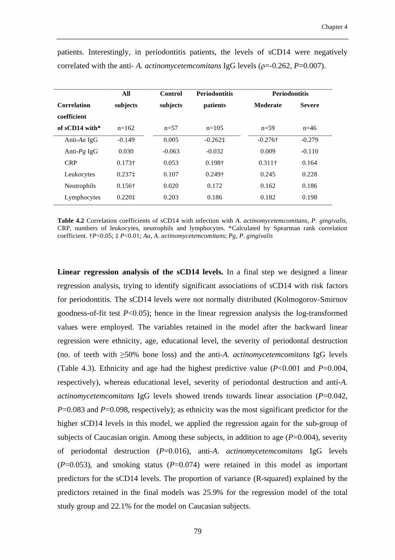
Chapter 4
patients. Interestingly, in periodontitis patients, the levels of sCD14 were negatively
correlated with the anti- A. actinomycetemcomitans IgG levels (ρ=-0.262, P=0.007).
Table 4.2 Correlation coefficients of sCD14 with infection with A. actinomycetemcomitans, P. gingivalis, CRP, numbers of leukocytes, neutrophils and lymphocytes. *Calculated by Spearman rank correlation coefficient. †P<0.05; ‡ P<0.01; Aa, A. actinomycetemcomitans; Pg, P. gingivalis
Linear regression analysis of the sCD14 levels. In a final step we designed a linear
regression analysis, trying to identify significant associations of sCD14 with risk factors
for periodontitis. The sCD14 levels were not normally distributed (Kolmogorov-Smirnov
goodness-of-fit test P<0.05); hence in the linear regression analysis the log-transformed
values were employed. The variables retained in the model after the backward linear
regression were ethnicity, age, educational level, the severity of periodontal destruction
(no. of teeth with ≥50% bone loss) and the anti-A. actinomycetemcomitans IgG levels
(Table 4.3). Ethnicity and age had the highest predictive value (P<0.001 and P=0.004,
respectively), whereas educational level, severity of periodontal destruction and anti-A.
actinomycetemcomitans IgG levels showed trends towards linear association (P=0.042,
P=0.083 and P=0.098, respectively); as ethnicity was the most significant predictor for the
higher sCD14 levels in this model, we applied the regression again for the sub-group of
subjects of Caucasian origin. Among these subjects, in addition to age (P=0.004), severity
of periodontal destruction (P=0.016), anti-A. actinomycetemcomitans IgG levels
(P=0.053), and smoking status (P=0.074) were retained in this model as important
predictors for the sCD14 levels. The proportion of variance (R-squared) explained by the
predictors retained in the final models was 25.9% for the regression model of the total
study group and 22.1% for the model on Caucasian subjects.
All Control Periodontitis Periodontitis
Correlation
coefficient
subjects subjects patients Moderate
Severe
of sCD14 with* n=162 n=57 n=105 n=59 n=46
Anti-Aa IgG -0.149 0.005 -0.262‡ -0.276† -0.279
Anti-Pg IgG 0.030 -0.063 -0.032 0.009 -0.110
CRP 0.173† 0.053 0.198† 0.311† 0.164
Leukocytes 0.237‡ 0.107 0.249† 0.245 0.228
Neutrophils 0.156† 0.020 0.172 0.162 0.186
Lymphocytes 0.220‡ 0.203 0.186 0.182 0.198
79
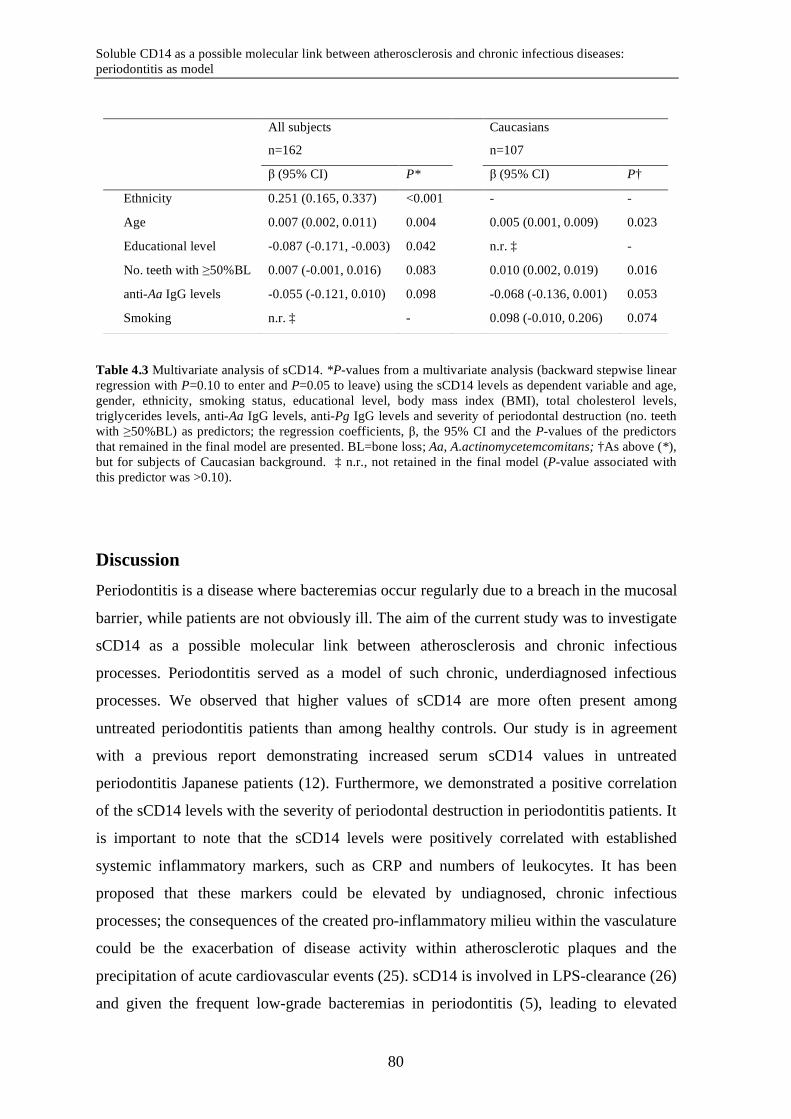
Soluble CD14 as a possible molecular link between atherosclerosis and chronic infectious diseases: periodontitis as model
Table 4.3 Multivariate analysis of sCD14. *P-values from a multivariate analysis (backward stepwise linear regression with P=0.10 to enter and P=0.05 to leave) using the sCD14 levels as dependent variable and age, gender, ethnicity, smoking status, educational level, body mass index (BMI), total cholesterol levels, triglycerides levels, anti-Aa IgG levels, anti-Pg IgG levels and severity of periodontal destruction (no. teeth with ≥50%BL) as predictors; the regression coefficients, β, the 95% CI and the P-values of the predictors that remained in the final model are presented. BL=bone loss; Aa, A.actinomycetemcomitans; †As above (*), but for subjects of Caucasian background. ‡ n.r., not retained in the final model (P-value associated with this predictor was >0.10).
Discussion Periodontitis is a disease where bacteremias occur regularly due to a breach in the mucosal
barrier, while patients are not obviously ill. The aim of the current study was to investigate
sCD14 as a possible molecular link between atherosclerosis and chronic infectious
processes. Periodontitis served as a model of such chronic, underdiagnosed infectious
processes. We observed that higher values of sCD14 are more often present among
untreated periodontitis patients than among healthy controls. Our study is in agreement
with a previous report demonstrating increased serum sCD14 values in untreated
periodontitis Japanese patients (12). Furthermore, we demonstrated a positive correlation
of the sCD14 levels with the severity of periodontal destruction in periodontitis patients. It
is important to note that the sCD14 levels were positively correlated with established
systemic inflammatory markers, such as CRP and numbers of leukocytes. It has been
proposed that these markers could be elevated by undiagnosed, chronic infectious
processes; the consequences of the created pro-inflammatory milieu within the vasculature
could be the exacerbation of disease activity within atherosclerotic plaques and the
precipitation of acute cardiovascular events (25). sCD14 is involved in LPS-clearance (26)
and given the frequent low-grade bacteremias in periodontitis (5), leading to elevated
All subjects
n=162
Caucasians
n=107
β (95% CI) P* β (95% CI) P†
Ethnicity 0.251 (0.165, 0.337) <0.001 - -
Age 0.007 (0.002, 0.011) 0.004 0.005 (0.001, 0.009) 0.023
Educational level -0.087 (-0.171, -0.003) 0.042 n.r. ‡ -
No. teeth with ≥50%BL 0.007 (-0.001, 0.016) 0.083 0.010 (0.002, 0.019) 0.016
anti-Aa IgG levels -0.055 (-0.121, 0.010) 0.098 -0.068 (-0.136, 0.001) 0.053
Smoking n.r. ‡ - 0.098 (-0.010, 0.206) 0.074
80
Chapter 4
blood levels of LPS (8), the positive correlation with disease severity points into the
direction that the sCD14 increases with the need to remove LPS from circulation.
Therefore we propose that periodontitis is one of the undiagnosed infections inducing a
systemic, sub-clinical inflammatory reaction that contributes to an increased
atherothrombotic burden.
In this study we looked for alternative modifying factors that could induce
variation in the sCD14 levels measured. A role for the positive infectious serology with H.
pylori, C. pneumoniae and cytomegalovirus in the chronic inflammatory reaction in
patients with cardiovascular disease has been proposed (3). In our periodontitis model, we
investigated the relationship between history of infection with two Gram-negative
established periodontal pathogens, A. actinomycetemcomitans and P. gingivalis and the
sCD14 levels. The anti-P. gingivalis IgG were not correlated with the sCD14 levels. Our
results showed that the sCD14 levels were negatively associated with the levels of anti-A.
actinomycetemcomitans IgG in periodontitis patients (Table 4.2). We suggest that the
negative correlation demonstrates that with a poor IgG-response to A.
actinomycetemcomitans, sCD14 becomes increasingly important in the clearance of A.
actinomycetemcomitans-derived LPS.
The CD14−260 polymorphism has been described as a modifying factor for
myocardial infarction and atherosclerosis, in Asian and Caucasian populations; the T-
allele carriers have elevated levels of soluble and membrane-bound CD14 (18,27). Albeit
not conclusive, there are indications for the association of the CD14 T/T genotype with
severe periodontal disease (20). In our study, the CD14−260 genotypes were present with
similar frequency in patients and controls. Within the Caucasians, the allele frequencies
are similar to those reported in the literature (28,20). The sCD14 levels were not correlated
with the CD14−260 genotypes, neither in the entire group nor in the Caucasian sub-group.
The lack of association between the CD14−260 genotypes and sCD14 levels is not
completely surprising. Contradictory reports on the correlation between CD14−260
genotypes and sCD14 levels have been documented on even larger groups than the present
relatively-small study population; Koenig et al. (28) found a significant effect of the
CD14−260 genotypes on sCD14 in 312 Germans suffering from coronary artery disease, but
not in their age- and gender-matched 476 healthy controls. It could be speculated that it is
not the CD14-260 polymorphism per se that is responsible for modified CD14 levels, but
the linkage disequilibrium with other genetic variations, leading to conflicting results in
different ethnic groups (27).
81
Soluble CD14 as a possible molecular link between atherosclerosis and chronic infectious diseases: periodontitis as model
In the current study, we found elevated sCD14 levels that were increasing with the
severity of periodontal destruction and were paralleled by markers of a systemic
inflammatory reaction. Our findings support the hypothesis of a pro-atherogenic milieu in
untreated periodontitis patients resulting from recurrent bacteremic episodes. Since
periodontal therapy results in a reduction of sCD14 plasma values (12), future strategies
for reducing the risk for cardiovascular events might consider periodontal therapy among
the prevention methods to reduce the overall pro-atherogenic burden. However, such
studies are still in demand.
Aknowledgements The present study was supported by The Netherlands Institute for Dental Sciences, Philips
Oral Healthcare EMEA and the INFOBIOMED Network of Excellence (IST
2002507585). We thank J. Pleijsters for the CD14 genotyping and J. Brunner, C.J. Bosch-
Tijhof and S. Asikainen for providing the bacterial strains used in the study.
REFERENCES
1 Anderson KM, Castelli WP, Levy D. 1987. Cholesterol and mortality. 30 years of
follow-up from the Framingham study. JAMA 257:2176-2180.
2 Hansson GK, Robertson AK, Soderberg-Naucler C. 2006. Inflammation and
atherosclerosis. Annu Rev Pathol 1:297-329.
3 Anderson JL, Carlquist JF, Muhlestein JB, Horne BD, Elmer SP. 1998.
Evaluation of C-reactive protein, an inflammatory marker, and infectious serology as
risk factors for coronary artery disease and myocardial infarction. J Am Coll Cardiol
32:35-41.
4 Ridker PM, Hennekens CH, Buring JE, Rifai N. 2000. C-reactive protein and other
markers of inflammation in the prediction of cardiovascular disease in women. N Engl
J Med 342:836-843.
5 Forner L, Larsen T, Kilian M, Holmstrup P. 2006. Incidence of bacteremia after
chewing, tooth brushing and scaling in individuals with periodontal inflammation. J
Clin Periodontol 33:401-407.
82
Chapter 4
6 Janket SJ, Baird AE, Chuang SK, Jones JA. 2003. Meta-analysis of periodontal
disease and risk of coronary heart disease and stroke. Oral Surg Oral Med Oral Pathol
Oral Radiol Endod 95:559-569.
7 Paraskevas S, Huizinga JD, Loos BG. 2008. A systematic review and meta-analyses
on C-reactive protein in relation to periodontitis. J Clin Periodontol 35:277-290.
8 Pussinen PJ, Tuomisto K, Jousilahti P, Havulinna AS, Sundvall J, Salomaa V.
2007. Endotoxemia, immune response to periodontal pathogens, and systemic
inflammation associate with incident cardiovascular disease events. Arterioscler
Thromb Vasc Biol 27:1433-1439.
9 Maliszewski CR. 1991. CD14 and immune response to lipopolysaccharide. Science
252:1321-1322.
10 Ziegler-Heitbrock HW and Ulevitch RJ. 1993. CD14: cell surface receptor and
differentiation marker. Immunol Today 14:121-125.
11 Hettne KM, Weeber M, Laine ML, ten CH, Boyer S, Kors JA, Loos BG. 2007.
Automatic mining of the literature to generate new hypotheses for the possible link
between periodontitis and atherosclerosis: lipopolysaccharide as a case study. J Clin
Periodontol 34:1016-1024.
12 Hayashi J, Masaka T, Ishikawa I. 1999. Increased levels of soluble CD14 in sera of
periodontitis patients. Infect Immun 67:417-420.
13 Liuzzo G, Angiolillo DJ, Buffon A, Rizzello V, Colizzi C, Ginnetti F, Biasucci LM,
Maseri A. 2001. Enhanced response of blood monocytes to in vitro
lipopolysaccharide-challenge in patients with recurrent unstable angina. Circulation
103:2236-2241.
14 Pugin J, Ulevitch RJ, Tobias PS. 1995. Activation of endothelial cells by endotoxin:
direct versus indirect pathways and the role of CD14. Prog Clin Biol Res 392:369-373.
15 Loppnow H, Stelter F, Schonbeck U, Schluter C, Ernst M, Schutt C, Flad HD.
1995. Endotoxin activates human vascular smooth muscle cells despite lack of
83
Soluble CD14 as a possible molecular link between atherosclerosis and chronic infectious diseases: periodontitis as model
expression of CD14 mRNA or endogenous membrane CD14. Infect Immun 63:1020-
1026.
16 Lo SK, Cheung A, Zheng Q, Silverstein RL. 1995. Induction of tissue factor on
monocytes by adhesion to endothelial cells. J Immunol 154:4768-4777.
17 Baldini M, Lohman IC, Halonen M, Erickson RP, Holt PG, Martinez FD. 1999. A
Polymorphism* in the 5' flanking region of the CD14 gene is associated with
circulating soluble CD14 levels and with total serum immunoglobulin E. Am J Respir
Cell Mol Biol 20:976-983.
18 Hubacek JA, Pit'ha J, Skodova Z, Poledne R. 1999. C(-260)->T polymorphism in
the promoter of the CD14 monocyte receptor gene as a risk factor for myocardial
infarction. Circulation 99:3218-3220.
19 Rupp J, Goepel W, Kramme E, Jahn J, Solbach W, Maass M. 2004. CD14
promoter polymorphism -159C>T is associated with susceptibility to chronic
Chlamydia pneumoniae infection in peripheral blood monocytes. Genes Immun 5:435-
438.
20 Laine ML, Morre SA, Murillo LS, van Winkelhoff AJ, Pena AS. 2005. CD14 and
TLR4 gene polymorphisms in adult periodontitis. J Dent Res 84:1042-1046.
21 Nicu EA, van der Velden U, Everts V, Loos BG. 2008. Expression of FcRs and
mCD14 on polymorphonuclear neutrophils and monocytes may determine the
periodontal infection. Clinical & Experimental Immunology; in press
22 Bizzarro S, van der Velden U, ten Heggeler JM, Leivadaros E, Hoek FJ, Gerdes
VE, Bakker SJ, Gans RO, ten Cate H, Loos BG. 2007. Periodontitis is characterized
by elevated PAI-1 activity. J Clin Periodontol 34:574-580.
23 Friedewald WT, Levy RI, Fredrickson DS. 1972. Estimation of the concentration of
low-density lipoprotein cholesterol in plasma, without use of the preparative
ultracentrifuge. Clin Chem 18:499-502.
84
Chapter 4
24 Pussinen PJ, Vilkuna-Rautiainen T, Alfthan G, Mattila K, Asikainen S. 2002.
Multiserotype enzyme-linked immunosorbent assay as a diagnostic aid for
periodontitis in large-scale studies. J Clin Microbiol 40:512-518.
25 Danesh J, Collins R, Peto R. 1997. Chronic infections and coronary heart disease: is
there a link? Lancet 350:430-436.
26 Ulevitch RJ and Tobias PS. 1995. Receptor-dependent mechanisms of cell
stimulation by bacterial endotoxin. Annu Rev Immunol 13:437-457.
27 Eng HL, Wang CH, Chen CH, Chou MH, Cheng CT, Lin TM. 2004. A CD14
promoter polymorphism is associated with CD14 expression and Chlamydia-
stimulated TNF alpha production. Genes Immun 5:426-430.
28 Koenig W, Khuseyinova N, Hoffmann MM, Marz W, Frohlich M, Hoffmeister A,
Brenner H, Rothenbacher D. 2002. CD14 C(-260)-->T polymorphism, plasma levels
of the soluble endotoxin receptor CD14, their association with chronic infections and
risk of stable coronary artery disease. J Am Coll Cardiol 40:34-42.
85

86

Chapter 5
Periodontitis is associated with platelet activation
D. Papapanagiotou1, ¶, E.A. Nicu1, ¶, , S. Bizzarro1, V.E.A. Gerdes2,3, J.C. Meijers2, R.
Nieuwland4, U. van der Velden1, B.G. Loos1
1 Department of Periodontology, Academic Centre for Dentistry Amsterdam (ACTA), Universiteit van
Amsterdam and VU University Amsterdam 2 Department of Vascular Medicine, Academic Medical Center (AMC), Universiteit van Amsterdam
3 Department of Internal Medicine, Slotervaart Hospital, Amsterdam 4 Department of Clinical Chemistry, Academic Medical Center (AMC), Universiteit van Amsterdam, The
Netherlands
Atherosclerosis; in press 2008
¶ These authors contributed equally to the present study.
87
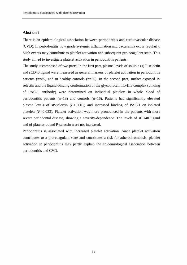
Periodontitis is associated with platelet activation
Abstract There is an epidemiological association between periodontitis and cardiovascular disease
(CVD). In periodontitis, low grade systemic inflammation and bacteremia occur regularly.
Such events may contribute to platelet activation and subsequent pro-coagulant state. This
study aimed to investigate platelet activation in periodontitis patients.
The study is composed of two parts. In the first part, plasma levels of soluble (s) P-selectin
and sCD40 ligand were measured as general markers of platelet activation in periodontitis
patients (n=85) and in healthy controls (n=35). In the second part, surface-exposed P-
selectin and the ligand-binding conformation of the glycoprotein IIb-IIIa complex (binding
of PAC-1 antibody) were determined on individual platelets in whole blood of
periodontitis patients (n=18) and controls (n=16). Patients had significantly elevated
plasma levels of sP-selectin (P<0.001) and increased binding of PAC-1 on isolated
platelets (P=0.033). Platelet activation was more pronounced in the patients with more
severe periodontal disease, showing a severity-dependence. The levels of sCD40 ligand
and of platelet-bound P-selectin were not increased.
Periodontitis is associated with increased platelet activation. Since platelet activation
contributes to a pro-coagulant state and constitutes a risk for atherothrombosis, platelet
activation in periodontitis may partly explain the epidemiological association between
periodontitis and CVD.
88
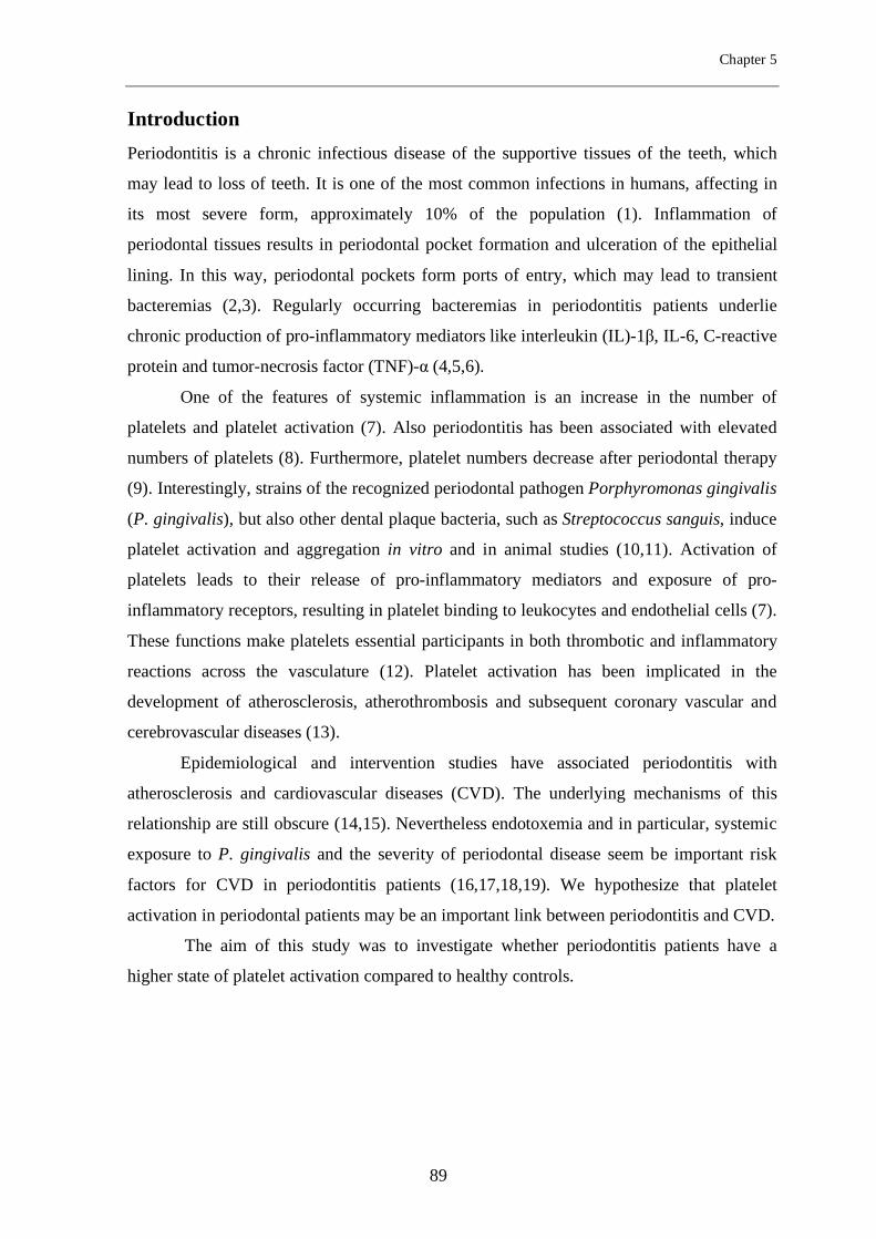
Chapter 5
Introduction Periodontitis is a chronic infectious disease of the supportive tissues of the teeth, which
may lead to loss of teeth. It is one of the most common infections in humans, affecting in
its most severe form, approximately 10% of the population (1). Inflammation of
periodontal tissues results in periodontal pocket formation and ulceration of the epithelial
lining. In this way, periodontal pockets form ports of entry, which may lead to transient
bacteremias (2,3). Regularly occurring bacteremias in periodontitis patients underlie
chronic production of pro-inflammatory mediators like interleukin (IL)-1β, IL-6, C-reactive
protein and tumor-necrosis factor (TNF)-α (4,5,6).
One of the features of systemic inflammation is an increase in the number of
platelets and platelet activation (7). Also periodontitis has been associated with elevated
numbers of platelets (8). Furthermore, platelet numbers decrease after periodontal therapy
(9). Interestingly, strains of the recognized periodontal pathogen Porphyromonas gingivalis
(P. gingivalis), but also other dental plaque bacteria, such as Streptococcus sanguis, induce
platelet activation and aggregation in vitro and in animal studies (10,11). Activation of
platelets leads to their release of pro-inflammatory mediators and exposure of pro-
inflammatory receptors, resulting in platelet binding to leukocytes and endothelial cells (7).
These functions make platelets essential participants in both thrombotic and inflammatory
reactions across the vasculature (12). Platelet activation has been implicated in the
development of atherosclerosis, atherothrombosis and subsequent coronary vascular and
cerebrovascular diseases (13).
Epidemiological and intervention studies have associated periodontitis with
atherosclerosis and cardiovascular diseases (CVD). The underlying mechanisms of this
relationship are still obscure (14,15). Nevertheless endotoxemia and in particular, systemic
exposure to P. gingivalis and the severity of periodontal disease seem be important risk
factors for CVD in periodontitis patients (16,17,18,19). We hypothesize that platelet
activation in periodontal patients may be an important link between periodontitis and CVD.
The aim of this study was to investigate whether periodontitis patients have a
higher state of platelet activation compared to healthy controls.
89
Periodontitis is associated with platelet activation
Materials and methods Study population
The present study consists of two parts. The study population of the first part has been
described previously (20) and included 85 consecutive periodontitis patients and 35 healthy
controls. On the basis of an extensive medical history by a written questionnaire and by
interview, the following subjects were not included in the study: pregnant women and
individuals who suffered from any given disease or chronic medical condition, apart from
periodontitis, or had trauma or tooth extractions in the last two weeks, or received
antibiotics within the last 3 months or any chronic medication. All background
characteristics are derived from the previous study (20).
Concentrations of soluble (s) sP-selectin (sCD62P) and sCD40 ligand were
determined in plasma samples collected from these periodontitis patients and controls. The
results suggested that platelet activation was present in periodontitis. Therefore, the second
study was initiated to further explore the possibility that platelets are activated in
periodontitis. This time the focus was laid on measurements of platelet-bound activation
markers, evaluated by means of flow cytometry of fresh platelets.
For this second study, a new study population was recruited, applying similar
inclusion and exclusion criteria as in the first study. Based on the P-selectin results in the
first study (mean ± SD: 58.1± 26.1 ng/mL for controls, 82.9 ± 34.7 ng/mL for patients) the
sample size for the second study was estimated using α=0.05 and β=0.20 (80% power). The
sample size needed was 18 participants for each of the control and patient groups. Subjects
with platelet counts of >350x109/ L (thrombocytosis) were excluded (13). Eighteen
consecutive periodontitis patients were included and for each patient an age-, gender-, race-
, and smoking status matched healthy control was recruited. After their initial inclusion,
two control subjects were excluded from analysis, because one subject had a platelet count
>350x109/L and the other subject appeared to suffer from mild periodontitis. Both platelet-
bound P-selectin and binding of PAC-1 on individual platelets were measured in this cohort
by whole-blood flow cytometry. All subjects were both verbally and written informed
about the purpose of the study and had signed an informed consent. The Medical Ethical
Committee of the Academic Medical Center of the University of Amsterdam approved the
study.
90
Chapter 5
Blood collection
In the first study, fasting venous blood samples were obtained without stasis by
venipuncture in the antecubital fossa between 8.30 and 11.30 AM. Blood, collected in
EDTA, was used to determine leukocyte counts. In addition, a second EDTA-blood tube
(10 mL) was centrifuged at 3000 rpm for 10 minutes at room temperature. EDTA plasma
was divided in aliquots and stored at -80°C until analysis.
In the second study, fasting venous blood samples were collected by venipuncture
of the antecubital fossa, through a 19G butterfly needle (Vygon Nederland BV,
Valkenswaard, The Netherlands) without venous stasis. For whole-blood flow cytometry,
0.32% citrate-anticoagulated blood was processed within 5 minutes after collection.
Soluble (s) P-selectin and sCD40 ligand
Plasma levels of sP-selectin and sCD40 ligand were determined by ELISA (R&D systems,
Abingdon, UK), according to the manufacturers instructions. EDTA plasma specimens
were diluted 1:20. For the sP-selectin ELISA, the diluted samples (in duplicate) together
with the peroxidase-conjugated polyclonal antibody against P-selectin were incubated for
1h in a 96-well plate precoated with the anti-human P-selectin monoclonal antibody. The
plate was washed three times with 300 µl “Wash buffer”. After washing, the “Substrate”
was added, followed by 30min incubation and the reaction was stopped adding “Stop
Solution” to each well.
For the sCD40 ligand ELISA, the samples were incubated in duplicate for 2h, at
room temperature in a 96-well plate precoated with the anti-human CD40 ligand
monoclonal antibody. The plate was washed four times with 400 µl “Wash buffer” and
incubated for another 2h with the polyclonal antibody against sCD40 ligand conjugated
with peroxidase. Absorbance values were measured at 450 nm with a multilabel counter
(Wallac Victor2 1420, Perkin-Elmer Life Sciences, Boston, MA). The concentration of each
sample was determined by extrapolation from a standard curve estimated from a panel of
standards of known concentrations. Reagents marked in quotes were all from the R&D
ELISA kits. The intra- and inter-assay coefficients of variation were 5.1 and 6.4%, for
sCD40 ligand and 5.1 and 9.9 % for sP-selectin, respectively.
91
Periodontitis is associated with platelet activation
Whole blood flow cytometry
Aliquots of blood (5 µL) were diluted in 30 µL HEPES buffer (137 mM NaCl, 2.7 mM
KCl, 1.0 mM MgCl2, 5.6 mM glucose, 20 mM HEPES, 1 mg/ml bovine serum albumin,
3.3 mM NaH2PO4, pH 7.4). In addition, the labeling tubes contained 4 µg/mL PerCP-
labeled anti-CD61 (5 µL), 4 µg/ml PE-labeled anti-CD62p (5 µL) and 4 µg/mL FITC-
labeled PAC-1 (5 µL). To set fluorescence thresholds, 4 µg/mL PE-IgG1 and 4 µg/mL
FITC-IgM isotype control antibodies were used. After mixing and 30 minutes incubation at
room temperature in the dark, 2.5 mL HEPES buffer containing formaldehyde (0.2% final
concentration) was added. CD61-PerCP and PAC-1 FITC were obtained from Becton
Dickinson Immunocytometry Systems (San Jose, CA, USA), PE-labeled CD62P from
Immunotech (Marseille, France), PE-labelled IgG1 from Sanquin Reagents (Amsterdam,
The Netherlands), and IgM-FITC from Beckman-Coulter (Fullerton, CA, USA). Flow
cytometry was performed as described previously (21,22). After fixation, blood samples
were analyzed in a FACScan flow cytometer with CellQuest software (Becton Dickinson).
Forward and side scatter were set at logarithmic gain. Platelets were identified by
characteristic forward and side scatter, and PerCP fluorescence. Exposure of platelet
activation markers was determined in 5000 platelets. The threshold for platelet activation
was arbitrarily set at 1% fluorescence-positive platelet activation with the appropriate
control antibody.
Statistical analysis
Means, standard deviations, medians and frequency distributions were calculated.
Differences in population background characteristics in both parts of the study were
analyzed by t-tests or χ 2 - test (or Fisher’s exact test, where needed). To correct for
differences in background that were statistically significant between patients and controls
in the first study, a general linear model (GLM) was constructed with periodontal condition
as factor, and age, educational level, systolic blood pressure, total cholesterol, and
triglycerides as co-variates. From the GLM, adjusted means, confidence intervals and
PGLM-values were obtained. In the first study, a χ2-test was performed to test the distribution
of controls and periodontitis patients with sP-selectin and sCD40 ligand levels below and
above the respective population medians. Partial correlation coefficients between sP-
selectin and sCD40 ligand were calculated, both within patients and control groups,
correcting for the same co-variates included in the GLM. To analyze a possible effect of
periodontal disease severity on the soluble platelet parameters, a stepwise linear regression
92
Chapter 5
analysis was conducted, within the patient group in the first study, using sP-selectin or
sCD40 ligand as dependent variables and severity of periodontal disease (severe patients
were those with ≥7 teeth with ≥50% bone loss), age, gender, ethnicity, smoking status,
educational level, body mass index (BMI), systolic blood pressure, total cholesterol, and
triglycerides as predictors.
In the second study, the % of cells exposing P-selectin and the % of cells binding
PAC-1 showed a skewed distribution (Kolmogorov-Smirnov goodness of fit test P<0.05).
These data were log transformed before statistical analysis. Subsequently, t-tests were
employed for the analysis of platelet activation data. To explore the potential confounding
of the non-matched background characteristics on platelet-bound P-selectin and PAC-1
binding, a GLM was constructed using periodontal condition as factor and educational
level, BMI, systolic blood pressure, total cholesterol and triglycerides as co-variates. Also
for the patients in the second part of the study, correlation coefficients between platelet
activation markers and periodontitis severity (number of teeth with ≥50% bone loss) were
computed. P-values <0.05 were considered statistically significant.
Results Study population
Tables 5.1 and 5.2 summarize the background characteristics of the study populations of
the first and second study, respectively. As shown in Table 5.1, several differences were
present in the first study between groups, including age, educational level, systolic blood
pressure, cholesterol, and triglycerides. As defined before (20), in the first study patients
suffering from moderate (n=51) or severe (n=34) periodontitis were included. In the second
part of our study, again moderate (n=10) as well as severe (n=8) periodontitis patients were
included. Patients had significantly higher fibrinogen and platelet counts than controls
(Table 5.2).
93
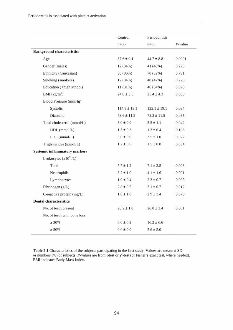
Periodontitis is associated with platelet activation
Control Periodontitis
n=35 n=85 P-value
Background characteristics
Age 37.6 ± 9.1 44.7 ± 8.8 0.0001
Gender (males) 12 (34%) 41 (48%) 0.225
Ethnicity (Caucasian) 30 (86%) 70 (82%) 0.791
Smoking (smokers) 12 (34%) 40 (47%) 0.228
Education (<high school) 11 (31%) 46 (54%) 0.028
BMI (kg/m2) 24.0 ± 3.5 25.4 ± 4.3 0.088
Blood Pressure (mmHg)
Systolic 114.3 ± 13.1 122.1 ± 19.1 0.034
Diastolic 73.6 ± 11.5 75.3 ± 11.5 0.465
Total cholesterol (mmol/L) 5.0 ± 0.9 5.5 ± 1.1 0.042
HDL (mmol/L) 1.5 ± 0.3 1.3 ± 0.4 0.106
LDL (mmol/L) 3.0 ± 0.9 3.5 ± 1.0 0.022
Triglycerides (mmol/L) 1.2 ± 0.6 1.5 ± 0.8 0.034
Systemic inflammatory markers
Leukocytes (x109 /L)
Total 5.7 ± 1.2 7.1 ± 2.5 0.003
Neutrophils 3.2 ± 1.0 4.1 ± 1.6 0.001
Lymphocytes 1.9 ± 0.4 2.3 ± 0.7 0.005
Fibrinogen (g/L) 2.8 ± 0.5 3.1 ± 0.7 0.012
C-reactive protein (mg/L) 1.8 ± 1.8 2.9 ± 3.4 0.078
Dental characteristics
No. of teeth present 28.2 ± 1.8 26.0 ± 3.4 0.001
No. of teeth with bone loss
≥ 30% 0.0 ± 0.2 16.2 ± 6.6
≥ 50% 0.0 ± 0.0 5.6 ± 5.0
Table 5.1 Characteristics of the subjects participating in the first study. Values are means ± SD or numbers (%) of subjects; P-values are from t-test or χ2-test (or Fisher’s exact test, where needed). BMI indicates Body Mass Index.
94
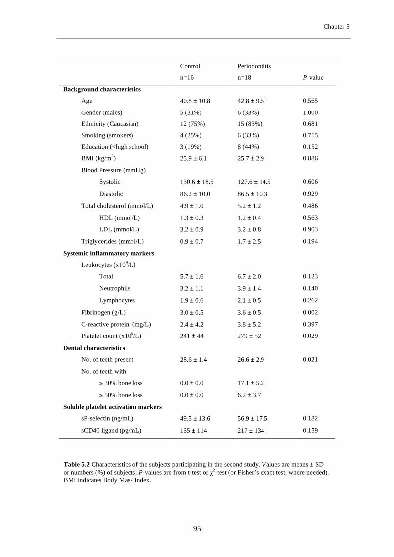
Chapter 5
Control Periodontitis
n=16 n=18 P-value
Background characteristics
Age 40.8 ± 10.8 42.8 ± 9.5 0.565
Gender (males) 5 (31%) 6 (33%) 1.000
Ethnicity (Caucasian) 12 (75%) 15 (83%) 0.681
Smoking (smokers) 4 (25%) 6 (33%) 0.715
Education (<high school) 3 (19%) 8 (44%) 0.152
BMI (kg/m2) 25.9 ± 6.1 25.7 ± 2.9 0.886
Blood Pressure (mmHg)
Systolic 130.6 ± 18.5 127.6 ± 14.5 0.606
Diastolic 86.2 ± 10.0 86.5 ± 10.3 0.929
Total cholesterol (mmol/L) 4.9 ± 1.0 5.2 ± 1.2 0.486
HDL (mmol/L) 1.3 ± 0.3 1.2 ± 0.4 0.563
LDL (mmol/L) 3.2 ± 0.9 3.2 ± 0.8 0.903
Triglycerides (mmol/L) 0.9 ± 0.7 1.7 ± 2.5 0.194
Systemic inflammatory markers
Leukocytes (x109/L)
Total 5.7 ± 1.6 6.7 ± 2.0 0.123
Neutrophils 3.2 ± 1.1 3.9 ± 1.4 0.140
Lymphocytes 1.9 ± 0.6 2.1 ± 0.5 0.262
Fibrinogen (g/L) 3.0 ± 0.5 3.6 ± 0.5 0.002
C-reactive protein (mg/L) 2.4 ± 4.2 3.8 ± 5.2 0.397
Platelet count (x109/L) 241 ± 44 279 ± 52 0.029
Dental characteristics
No. of teeth present 28.6 ± 1.4 26.6 ± 2.9 0.021
No. of teeth with
≥ 30% bone loss 0.0 ± 0.0 17.1 ± 5.2
≥ 50% bone loss 0.0 ± 0.0 6.2 ± 3.7
Soluble platelet activation markers
sP-selectin (ng/mL) 49.5 ± 13.6 56.9 ± 17.5 0.182
sCD40 ligand (pg/mL) 155 ± 114 217 ± 134 0.159
Table 5.2 Characteristics of the subjects participating in the second study. Values are means ± SD or numbers (%) of subjects; P-values are from t-test or χ2-test (or Fisher’s exact test, where needed). BMI indicates Body Mass Index.
95
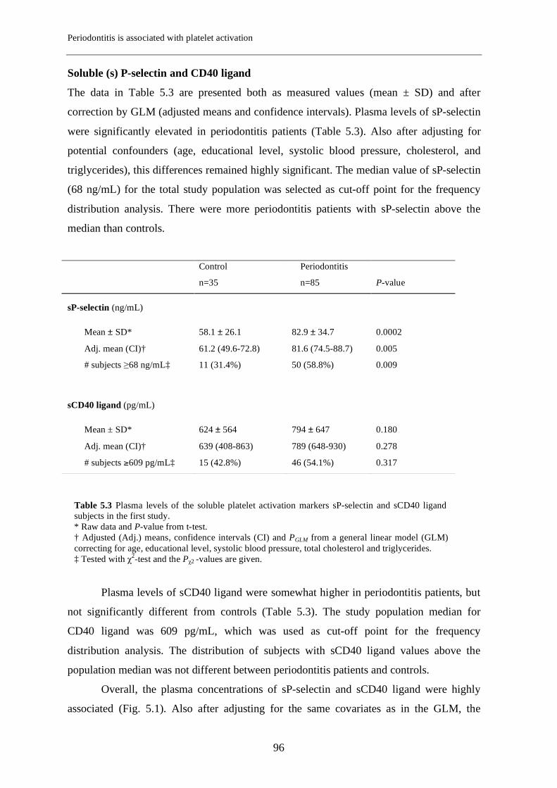
Periodontitis is associated with platelet activation
Soluble (s) P-selectin and CD40 ligand
The data in Table 5.3 are presented both as measured values (mean ± SD) and after
correction by GLM (adjusted means and confidence intervals). Plasma levels of sP-selectin
were significantly elevated in periodontitis patients (Table 5.3). Also after adjusting for
potential confounders (age, educational level, systolic blood pressure, cholesterol, and
triglycerides), this differences remained highly significant. The median value of sP-selectin
(68 ng/mL) for the total study population was selected as cut-off point for the frequency
distribution analysis. There were more periodontitis patients with sP-selectin above the
median than controls.
Plasma levels of sCD40 ligand were somewhat higher in periodontitis patients, but
not significantly different from controls (Table 5.3). The study population median for
CD40 ligand was 609 pg/mL, which was used as cut-off point for the frequency
distribution analysis. The distribution of subjects with sCD40 ligand values above the
population median was not different between periodontitis patients and controls.
Overall, the plasma concentrations of sP-selectin and sCD40 ligand were highly
associated (Fig. 5.1). Also after adjusting for the same covariates as in the GLM, the
Control Periodontitis
n=35 n=85 P-value
sP-selectin (ng/mL)
Mean ± SD* 58.1 ± 26.1 82.9 ± 34.7 0.0002
Adj. mean (CI)† 61.2 (49.6-72.8) 81.6 (74.5-88.7) 0.005
# subjects ≥68 ng/mL‡ 11 (31.4%) 50 (58.8%) 0.009
sCD40 ligand (pg/mL)
Mean ± SD* 624 ± 564 794 ± 647 0.180
Adj. mean (CI)† 639 (408-863) 789 (648-930) 0.278
# subjects ≥609 pg/mL‡ 15 (42.8%) 46 (54.1%) 0.317
Table 5.3 Plasma levels of the soluble platelet activation markers sP-selectin and sCD40 ligand in the subjects in the first study. * Raw data and P-value from t-test. † Adjusted (Adj.) means, confidence intervals (CI) and PGLM from a general linear model (GLM) correcting for age, educational level, systolic blood pressure, total cholesterol and triglycerides. ‡ Tested with χ2-test and the Pχ2 -values are given.
96
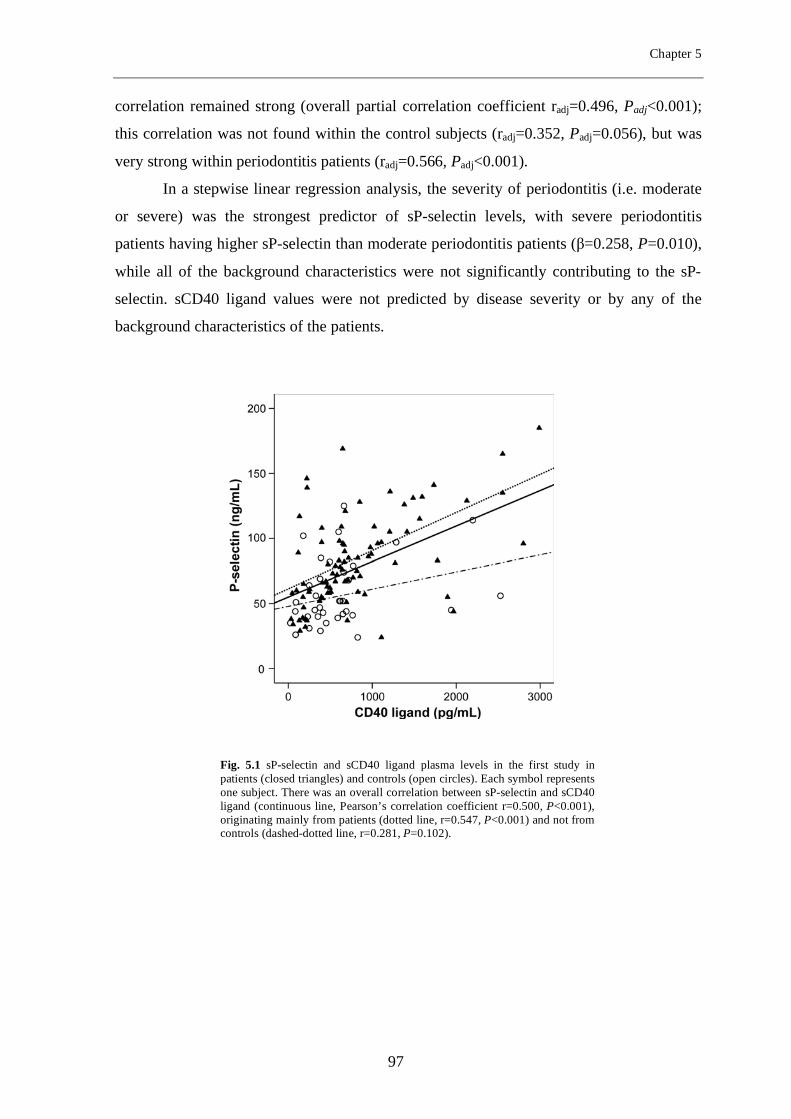
Chapter 5
correlation remained strong (overall partial correlation coefficient radj=0.496, Padj<0.001);
this correlation was not found within the control subjects (radj=0.352, Padj=0.056), but was
very strong within periodontitis patients (radj=0.566, Padj<0.001).
In a stepwise linear regression analysis, the severity of periodontitis (i.e. moderate
or severe) was the strongest predictor of sP-selectin levels, with severe periodontitis
patients having higher sP-selectin than moderate periodontitis patients (β=0.258, P=0.010),
while all of the background characteristics were not significantly contributing to the sP-
selectin. sCD40 ligand values were not predicted by disease severity or by any of the
background characteristics of the patients.
Fig. 5.1 sP-selectin and sCD40 ligand plasma levels in the first study in patients (closed triangles) and controls (open circles). Each symbol represents one subject. There was an overall correlation between sP-selectin and sCD40 ligand (continuous line, Pearson’s correlation coefficient r=0.500, P<0.001), originating mainly from patients (dotted line, r=0.547, P<0.001) and not from controls (dashed-dotted line, r=0.281, P=0.102).
97
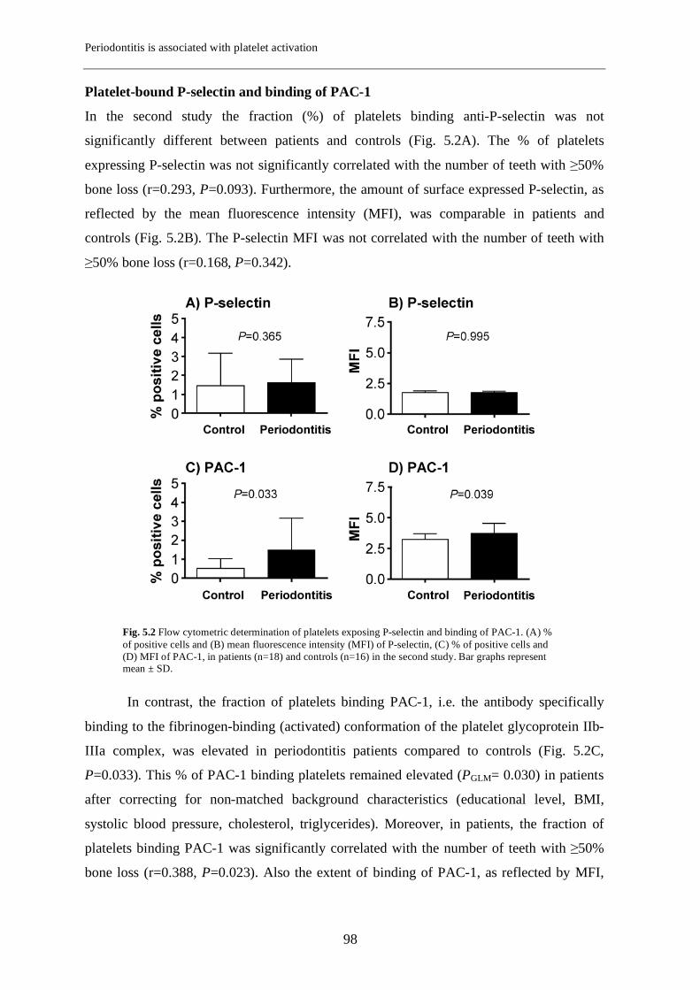
Periodontitis is associated with platelet activation
Platelet-bound P-selectin and binding of PAC-1
In the second study the fraction (%) of platelets binding anti-P-selectin was not
significantly different between patients and controls (Fig. 5.2A). The % of platelets
expressing P-selectin was not significantly correlated with the number of teeth with ≥50%
bone loss (r=0.293, P=0.093). Furthermore, the amount of surface expressed P-selectin, as
reflected by the mean fluorescence intensity (MFI), was comparable in patients and
controls (Fig. 5.2B). The P-selectin MFI was not correlated with the number of teeth with
≥50% bone loss (r=0.168, P=0.342).
In contrast, the fraction of platelets binding PAC-1, i.e. the antibody specifically
binding to the fibrinogen-binding (activated) conformation of the platelet glycoprotein IIb-
IIIa complex, was elevated in periodontitis patients compared to controls (Fig. 5.2C,
P=0.033). This % of PAC-1 binding platelets remained elevated (PGLM= 0.030) in patients
after correcting for non-matched background characteristics (educational level, BMI,
systolic blood pressure, cholesterol, triglycerides). Moreover, in patients, the fraction of
platelets binding PAC-1 was significantly correlated with the number of teeth with ≥50%
bone loss (r=0.388, P=0.023). Also the extent of binding of PAC-1, as reflected by MFI,
Fig. 5.2 Flow cytometric determination of platelets exposing P-selectin and binding of PAC-1. (A) % of positive cells and (B) mean fluorescence intensity (MFI) of P-selectin, (C) % of positive cells and (D) MFI of PAC-1, in patients (n=18) and controls (n=16) in the second study. Bar graphs represent mean ± SD.
98
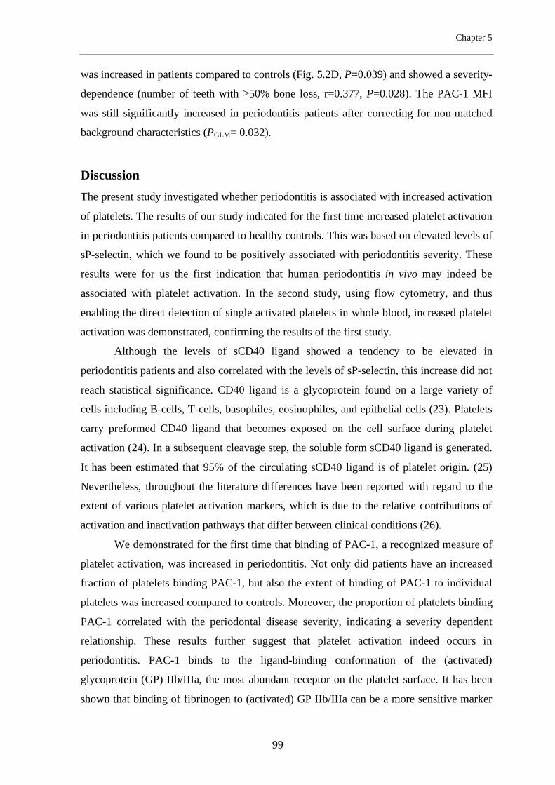
Chapter 5
was increased in patients compared to controls (Fig. 5.2D, P=0.039) and showed a severity-
dependence (number of teeth with ≥50% bone loss, r=0.377, P=0.028). The PAC-1 MFI
was still significantly increased in periodontitis patients after correcting for non-matched
background characteristics (PGLM= 0.032).
Discussion The present study investigated whether periodontitis is associated with increased activation
of platelets. The results of our study indicated for the first time increased platelet activation
in periodontitis patients compared to healthy controls. This was based on elevated levels of
sP-selectin, which we found to be positively associated with periodontitis severity. These
results were for us the first indication that human periodontitis in vivo may indeed be
associated with platelet activation. In the second study, using flow cytometry, and thus
enabling the direct detection of single activated platelets in whole blood, increased platelet
activation was demonstrated, confirming the results of the first study.
Although the levels of sCD40 ligand showed a tendency to be elevated in
periodontitis patients and also correlated with the levels of sP-selectin, this increase did not
reach statistical significance. CD40 ligand is a glycoprotein found on a large variety of
cells including B-cells, T-cells, basophiles, eosinophiles, and epithelial cells (23). Platelets
carry preformed CD40 ligand that becomes exposed on the cell surface during platelet
activation (24). In a subsequent cleavage step, the soluble form sCD40 ligand is generated.
It has been estimated that 95% of the circulating sCD40 ligand is of platelet origin. (25)
Nevertheless, throughout the literature differences have been reported with regard to the
extent of various platelet activation markers, which is due to the relative contributions of
activation and inactivation pathways that differ between clinical conditions (26).
We demonstrated for the first time that binding of PAC-1, a recognized measure of
platelet activation, was increased in periodontitis. Not only did patients have an increased
fraction of platelets binding PAC-1, but also the extent of binding of PAC-1 to individual
platelets was increased compared to controls. Moreover, the proportion of platelets binding
PAC-1 correlated with the periodontal disease severity, indicating a severity dependent
relationship. These results further suggest that platelet activation indeed occurs in
periodontitis. PAC-1 binds to the ligand-binding conformation of the (activated)
glycoprotein (GP) IIb/IIIa, the most abundant receptor on the platelet surface. It has been
shown that binding of fibrinogen to (activated) GP IIb/IIIa can be a more sensitive marker
99
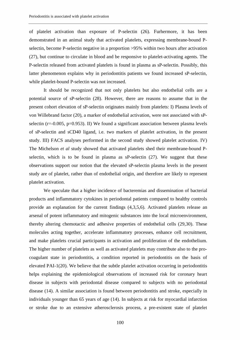
Periodontitis is associated with platelet activation
of platelet activation than exposure of P-selectin (26). Furhermore, it has been
demonstrated in an animal study that activated platelets, expressing membrane-bound P-
selectin, become P-selectin negative in a proportion >95% within two hours after activation
(27), but continue to circulate in blood and be responsive to platelet-activating agents. The
P-selectin released from activated platelets is found in plasma as sP-selectin. Possibly, this
latter phenomenon explains why in periodontitis patients we found increased sP-selectin,
while platelet-bound P-selectin was not increased.
It should be recognized that not only platelets but also endothelial cells are a
potential source of sP-selectin (28). However, there are reasons to assume that in the
present cohort elevation of sP-selectin originates mainly from platelets: I) Plasma levels of
von Willebrand factor (20), a marker of endothelial activation, were not associated with sP-
selectin (r=-0.005, p=0.953). II) We found a significant association between plasma levels
of sP-selectin and sCD40 ligand, i.e. two markers of platelet activation, in the present
study. III) FACS analyses performed in the second study showed platelet activation. IV)
The Michelson et al study showed that activated platelets shed their membrane-bound P-
selectin, which is to be found in plasma as sP-selectin (27). We suggest that these
observations support our notion that the elevated sP-selectin plasma levels in the present
study are of platelet, rather than of endothelial origin, and therefore are likely to represent
platelet activation.
We speculate that a higher incidence of bacteremias and dissemination of bacterial
products and inflammatory cytokines in periodontal patients compared to healthy controls
provide an explanation for the current findings (4,3,5,6). Activated platelets release an
arsenal of potent inflammatory and mitogenic substances into the local microenvironment,
thereby altering chemotactic and adhesive properties of endothelial cells (29,30). These
molecules acting together, accelerate inflammatory processes, enhance cell recruitment,
and make platelets crucial participants in activation and proliferation of the endothelium.
The higher number of platelets as well as activated platelets may contribute also to the pro-
coagulant state in periodontitis, a condition reported in periodontitis on the basis of
elevated PAI-1(20). We believe that the subtle platelet activation occurring in periodontitis
helps explaining the epidemiological observations of increased risk for coronary heart
disease in subjects with periodontal disease compared to subjects with no periodontal
disease (14). A similar association is found between periodontitis and stroke, especially in
individuals younger than 65 years of age (14). In subjects at risk for myocardial infarction
or stroke due to an extensive atherosclerosis process, a pre-existent state of platelet
100
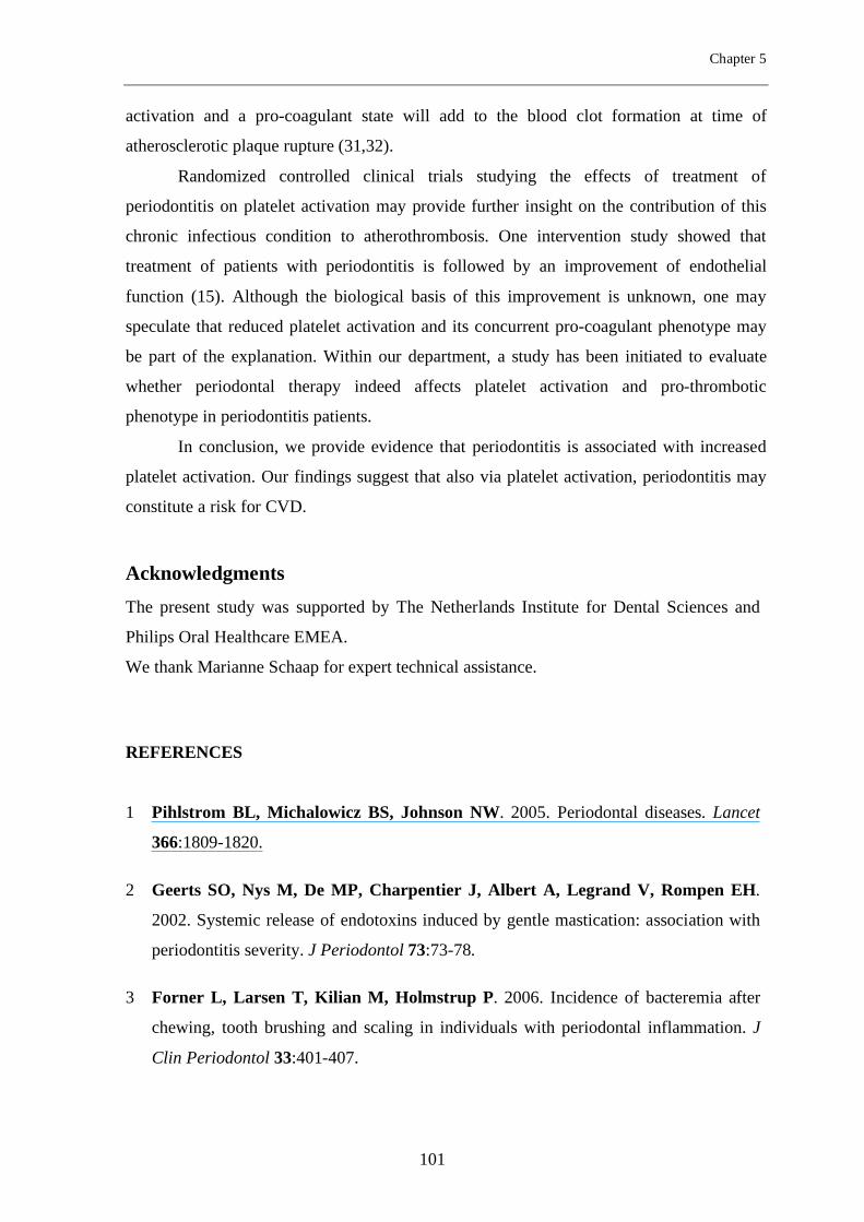
Chapter 5
activation and a pro-coagulant state will add to the blood clot formation at time of
atherosclerotic plaque rupture (31,32).
Randomized controlled clinical trials studying the effects of treatment of
periodontitis on platelet activation may provide further insight on the contribution of this
chronic infectious condition to atherothrombosis. One intervention study showed that
treatment of patients with periodontitis is followed by an improvement of endothelial
function (15). Although the biological basis of this improvement is unknown, one may
speculate that reduced platelet activation and its concurrent pro-coagulant phenotype may
be part of the explanation. Within our department, a study has been initiated to evaluate
whether periodontal therapy indeed affects platelet activation and pro-thrombotic
phenotype in periodontitis patients.
In conclusion, we provide evidence that periodontitis is associated with increased
platelet activation. Our findings suggest that also via platelet activation, periodontitis may
constitute a risk for CVD.
Acknowledgments The present study was supported by The Netherlands Institute for Dental Sciences and
Philips Oral Healthcare EMEA.
We thank Marianne Schaap for expert technical assistance.
REFERENCES
1 Pihlstrom BL, Michalowicz BS, Johnson NW. 2005. Periodontal diseases. Lancet
366:1809-1820.
2 Geerts SO, Nys M, De MP, Charpentier J, Albert A, Legrand V, Rompen EH.
2002. Systemic release of endotoxins induced by gentle mastication: association with
periodontitis severity. J Periodontol 73:73-78.
3 Forner L, Larsen T, Kilian M, Holmstrup P. 2006. Incidence of bacteremia after
chewing, tooth brushing and scaling in individuals with periodontal inflammation. J
Clin Periodontol 33:401-407.
101
Periodontitis is associated with platelet activation
4 D'Aiuto F, Parkar M, Nibali L, Suvan J, Lessem J, Tonetti MS. 2006. Periodontal
infections cause changes in traditional and novel cardiovascular risk factors: results
from a randomized controlled clinical trial. Am Heart J 151:977-984.
5 Loos BG. 2005. Systemic markers of inflammation in periodontitis. J Periodontol
76:2106-2115.
6 Paraskevas S, Huizinga JD, Loos BG. 2008. A systematic review and meta-analyses
on C-reactive protein in relation to periodontitis. J Clin Periodontol
7 Klinger MH and Jelkmann W. 2002. Role of blood platelets in infection and
inflammation. J Interferon Cytokine Res 22:913-922.
8 Wakai K, Kawamura T, Umemura O, Hara Y, Machida J, Anno T, Ichihara Y,
Mizuno Y, Tamakoshi A, Lin Y, Nakayama T, Ohno Y. 1999. Associations of
medical status and physical fitness with periodontal disease. J Clin Periodontol
26:664-672.
9 Christan C, Dietrich T, Hagewald S, Kage A, Bernimoulin JP. 2002. White blood
cell count in generalized aggressive periodontitis after non-surgical therapy. J Clin
Periodontol 29:201-206.
10 Herzberg MC and Weyer MW. 1998. Dental plaque, platelets, and cardiovascular
diseases. Ann Periodontol 3:151-160.
11 Lourbakos A, Yuan Y, Jenkins AL, Travis J, Andrade-Gordon P, Santulli R,
Potempa J, Pike RN. 2001. Activation of protease-activated receptors by gingipains
from Porphyromonas gingivalis leads to platelet aggregation: a new trait in microbial
pathogenicity. Blood 97:3790-3797.
12 Weyrich AS, Lindemann S, Tolley ND, Kraiss LW, Dixon DA, Mahoney TM,
Prescott SP, McIntyre TM, Zimmerman GA. 2004. Change in protein phenotype
without a nucleus: translational control in platelets. Semin Thromb Hemost 30:491-
498.
13 Kamath S, Blann AD, Lip GY. 2001. Platelet activation: assessment and
quantification. Eur Heart J 22:1561-1571.
102
Chapter 5
14 Janket SJ, Baird AE, Chuang SK, Jones JA. 2003. Meta-analysis of periodontal
disease and risk of coronary heart disease and stroke. Oral Surg Oral Med Oral Pathol
Oral Radiol Endod 95:559-569.
15 Tonetti MS, D'Aiuto F, Nibali L, Donald A, Storry C, Parkar M, Suvan J,
Hingorani AD, Vallance P, Deanfield J. 2007. Treatment of periodontitis and
endothelial function. N Engl J Med 356:911-920.
16 Pussinen PJ, Tuomisto K, Jousilahti P, Havulinna AS, Sundvall J, Salomaa V.
2007. Endotoxemia, immune response to periodontal pathogens, and systemic
inflammation associate with incident cardiovascular disease events. Arterioscler
Thromb Vasc Biol 27:1433-1439.
17 Pussinen PJ, Alfthan G, Jousilahti P, Paju S, Tuomilehto J. 2007. Systemic
exposure to Porphyromonas gingivalis predicts incident stroke. Atherosclerosis
193:222-228.
18 Holmlund A, Holm G, Lind L. 2006. Severity of periodontal disease and number of
remaining teeth are related to the prevalence of myocardial infarction and hypertension
in a study based on 4,254 subjects. J Periodontol 77:1173-1178.
19 Mustapha IZ, Debrey S, Oladubu M, Ugarte R. 2007. Markers of systemic bacterial
exposure in periodontal disease and cardiovascular disease risk: a systematic review
and meta-analysis. J Periodontol 78:2289-2302.
20 Bizzarro S, van der Velden U, ten Heggeler JM, Leivadaros E, Hoek FJ, Gerdes
VE, Bakker SJ, Gans RO, ten Cate H, Loos BG. 2007. Periodontitis is characterized
by elevated PAI-1 activity. J Clin Periodontol 34:574-580.
21 Shattil SJ, Motulsky HJ, Insel PA, Flaherty L, Brass LF. 1986. Expression of
fibrinogen receptors during activation and subsequent desensitization of human
platelets by epinephrine. Blood 68:1224-1231.
22 Shattil SJ, Cunningham M, Hoxie JA. 1987. Detection of activated platelets in
whole blood using activation-dependent monoclonal antibodies and flow cytometry.
Blood 70:307-315.
103
Periodontitis is associated with platelet activation
23 Schonbeck U, Mach F, Libby P. 2000. CD154 (CD40 ligand). Int J Biochem Cell
Biol 32:687-693.
24 Gawaz M. 2004. Role of platelets in coronary thrombosis and reperfusion of ischemic
myocardium. Cardiovasc Res 61:498-511.
25 Freedman JE. 2003. CD40-CD40L and platelet function: beyond hemostasis. Circ
Res 92:944-946.
26 Holmes MB, Sobel BE, Howard DB, Schneider DJ. 1999. Differences between
activation thresholds for platelet P-selectin and glycoprotein IIb-IIIa expression and
their clinical implications. Thromb Res 95:75-82.
27 Michelson AD, Barnard MR, Hechtman HB, MacGregor H, Connolly RJ,
Loscalzo J, Valeri CR. 1996. In vivo tracking of platelets: circulating degranulated
platelets rapidly lose surface P-selectin but continue to circulate and function. PNAS
93:11877-11882.
28 Burger PC and Wagner DD. 2003. Platelet P-selectin facilitates atherosclerotic
lesion development. Blood 101:2661-2666.
29 Huo Y and Ley KF. 2004. Role of platelets in the development of atherosclerosis.
Trends Cardiovasc Med 14:18-22.
30 Gawaz M. 2003. Do platelets trigger atherosclerosis? Thromb Haemost 90:971-972.
31 Sakariassen KS and Barstad RM. 1993. Mechanisms of thromboembolism at arterial
plaques. Blood Coagul Fibrinolysis 4:615-625.
32 Holvoet P and Collen D. 1997. Thrombosis and atherosclerosis. Curr Opin Lipidol
8:320-328.
104
Chapter 6
Elevated platelet and leukocyte response to oral bacteria in
periodontitis
E.A. Nicu1, U. van der Velden1, R. Nieuwland2, V. Everts3, B.G. Loos1
Departments of 1 Periodontology and 3 Oral Cell Biology, Academic Centre for Dentistry Amsterdam
(ACTA), University of Amsterdam and VU University Amsterdam, The Netherlands
2 Department of Clini cal Chemistry, Academic Medical Center (AMC), University of Amsterdam, The
Netherlands
Journal of Thrombosis and Haemostasis; in press 2008
105
Chapter 6
Abstract Periodontitis is associated with an increased risk for cardiovascular diseases (CVD), but
the underlying mechanisms are poorly understood. Recently, we showed that platelets
from periodontitis patients are more activated than those from controls. Given the
regularly occurring bacteremic episodes in periodontitis patients, we hypothesized that
platelets and/or leukocytes from periodontitis patients are more sensitive to stimulation by
oral bacteria, in particular the known periodontal pathogens, than platelets from control
subjects. Three-color flow cytometry analysis was performed to quantify activation of
platelets (P-selectin, PAC-1, CD63) and leukocytes (CD11b) in whole blood from patients
with periodontitis (n=19) and controls (n=18), with and without stimulation by oral
bacteria. Phagocytosis was assessed by using green-fluorescent protein (GFP)-expressing
Aggregatibacter actinomycetemcomitans. Neutrophils and monocytes were activated by
all species of oral bacteria tested, but no differences were observed between patients and
controls. In response to several species of oral bacteria, platelets from periodontitis
patients showed –compared to controls– increased exposure of P-selectin (P=0.027) and
increased formation of platelet-monocyte complexes (P=0.040). Platelet-leukocyte
complexes bound and/or phagocytosed more GFP-A. actinomycetemcomitans than
platelet-free leukocytes (for neutrophils and monocytes, in both patients and controls
P<0.001). In periodontitis, increased platelet response to oral bacteria is paralleled by
increased formation of platelet-leukocyte complexes with elevated capacity of bacterial
clearance. We speculate that activated platelets and leukocytes might contribute to
increased atherothrombotic activity.
106
Elevated platelet and leukocyte response to oral bacteria in periodontitis
Introduction Systemic inflammation may have an atherogenic effect at different levels (1). In addition
to induced endothelial dysfunction and induced secondary dyslipidemia, systemic
inflammation can activate the coagulation cascade (2), a process mainly mediated by
tissue factor (3). Platelets also contribute to activation of coagulation when they are
primed during systemic inflammation; this process is closely related to atherothrombosis
(4). Activated platelets release chemokines and cytokines, and expose pro-inflammatory
receptors, facilitating their binding to leukocytes and endothelial cells (5). Platelet-
leukocyte (i.e. platelet-neutrophil and platelet-monocyte) complexes are a sensitive
marker for platelet activation and have a proposed role in plaque instability, thrombosis
and inflammation (6). Increased numbers of circulating complexes have been reported in
patients with unstable angina, myocardial infarction and stroke (7,8,9). The correlation
between systemic inflammation and atherothrombosis is sustained by an increased
incidence of cardiovascular disease (CVD) in patients with systemic lupus erythematosus,
rheumatoid arthritis, inflammatory bowel disease or periodontitis as compared to subjects
without an inflammatory disease (2,10).
Periodontitis is an infectious disease of the supportive tissues of the teeth,
characterized by gradual loss of tooth supporting alveolar bone, and affects up to 10% of
the population in its most severe form (11). The primary etiologic factor of periodontitis is
the subgingival infection with a group of Gram-negative pathogens. The major bacterial
species associated with periodontitis are Aggregatibacter actinomycetemcomitans (A.
actinomycetemcomitans), Porphyromonas gingivalis (P. gingivalis) and Tannerella
forsythia (T. forsythia) (12). Inflammation in the periodontal tissues results in areas of
ulceration of the epithelium in the periodontal pocket, which leads to dissemination of oral
bacteria into the circulation during mastication (13). Transient bacteremias in periodontitis
patients underlie chronic production and systemic increases of various pro-inflammatory
mediators, including interleukin (IL)-1β, IL-6, C-reactive protein and tumor-necrosis
factor (TNF)-α (14,15).
In a previous study, we have shown that platelets from periodontitis patients have
an increased activation status compared to platelets from healthy controls (16). Strains of
the periodontal pathogen P. gingivalis, but also species of the oral commensal microflora,
such as Streptococcus sanguis (S. sanguis), induce platelet activation in vitro and in
animal studies (17,18). Given the regularly occurring bacteremic episodes in periodontitis
107
Chapter 6
patients, we hypothesized that platelets and/or leukocytes from periodontitis patients are
more sensitive to stimulation by oral bacteria, in particular the known periodontal
pathogens, than platelets from control subjects. If true, the association between platelet
activation and oral bacteria improves our understanding of the underlying mechanisms
contributing to the higher risk of CVD in periodontitis patients. Therefore, the aim of the
present study was to investigate whether platelets and/or leukocytes from periodontitis
patients are more sensitive to stimulation by oral bacteria than platelets from matched
controls.
Materials and methods Chemicals and antibodies
All chemicals were from Sigma Chemical Co. (St. Louis, MO, USA). CD61-PerCP and
PAC-1 FITC were obtained from Becton Dickinson Immunocytometry Systems (San
Jose, CA, USA), PE-labeled CD62P from Immunotech (Marseille, France), CD14-PE,
CD11b-FITC and PE-labelled IgG1 from Sanquin (Amsterdam, the Netherlands) and IgM-
FITC from Beckman-Coulter (Fullerton, CA, USA).
Patients
We selected a consecutive series of periodontitis patients who were referred to the
Department of Periodontology of the Academic Centre for Dentistry Amsterdam for
diagnosis and treatment of severe periodontitis, who met the inclusion criteria and who
consented to participate. Inclusion criteria were generalized gingival inflammation,
deepened periodontal pockets and ≥8 teeth with radiographic bone loss ≥ 30% of the root
length. Age-, gender-, race- and smoking status-matched controls were recruited among
subjects registered for restorative dental procedures or who visited the dental school for
regular dental check-ups. Inclusion criteria for control subjects were (i) missing ≤1 tooth
per quadrant (3rd molar excluded), and (ii) showed on <1 year old dental bitewing
radiographs a distance of ≤3 mm between the cemento-enamel junction and the alveolar
bone crest. After initial inclusion, one control subject was excluded from analysis because
subgingival calculus masked initially the presence of minor loss of periodontal attachment
(mild periodontitis). Exclusion criteria for patients and controls were: the presence of
systemic disease (especially cardiovascular disorders, diabetes mellitus, and allergies), a
recent history or the presence of any acute or chronic infection, systemic antibiotic
108
Elevated platelet and leukocyte response to oral bacteria in periodontitis
treatment within the last 3 months or usage of any medication (including sporadic
NSAID’s), pregnancy. Subgingival microbiological samples were taken from all subjects
to determine the presence of the periodontal pathogens A. actinomycetemcomitans, P.
gingivalis and T. forsythia; sampling, laboratory procedures, and identification of A.
actinomycetemcomitans, P. gingivalis and T. forsythia were performed as previously
described (12). All subjects were informed verbally and written about the purposes of the
study and had signed an informed consent. The Medical Ethical Committee of the
Academic Medical Center of the University of Amsterdam approved the study.
Bacterial cultures used in the stimulation assays
A. actinomycetemcomitans (Y4) was grown aerobically in enriched brain-heart infusion
broth (enriched BHI; hemin 5 mg/L, menadione 1 mg/L). P. gingivalis (W83) was grown
anaerobically in enriched BHI. For the anaerobic culturing of T. forsythia (ATCC 43037),
the enriched BHI was supplemented with 5% v/v fetal calf serum, 1 g/L L-cysteine and
15 mg/L N-acetylmuramic acid. S. sanguis (HG1470) was grown aerobically in BHI. The
bacterial suspensions were washed by centrifugation, reduced to an optical density of 1 at
600 nm (corresponding to 5*108 cells/µL) in HEPES buffer (137 mM NaCl, 2.7 mM KCl,
1.0 mM MgCl2, 5.6 mM glucose, 20 mM HEPES, 1 mg/mL bovine serum albumin, 3.3
mM NaH2PO4; pH 7.4), and stored in aliquots at -20°C.
Platelet activation
Fasting venous blood samples were collected as previously described (19). For whole-
blood flow cytometry, 0.32% citrate-anticoagulated blood was processed within 5 minutes
after collection. Aliquots of blood (5 µL) were diluted in 30 µL HEPES buffer. Platelets
were incubated with and without 30 µL of A. actinomycetemcomitans, P. gingivalis, T.
forsythia or S. sanguis for 30 minutes at room temperature. Adenosine diphosphate (ADP,
10 µM) was used as a positive control. The reaction vials contained PerCP-labeled anti-
CD61, FITC-labeled PAC-1 and PE-labeled anti-CD62p (4 µg/mL of each monoclonal
antibody, final concentration) or PerCP-labeled anti-CD61 (4 µg/mL) and PE-labeled anti-
CD63 (10 µg/mL). To set fluorescence thresholds, 4 µg/mL PE-IgG1 and 4 µg/mL FITC-
IgM isotype control antibodies were used. After mixing and 30 minutes incubation at
room temperature in the dark, HEPES buffer containing 0.2% paraformaldehyde (PFA;
2.5 mL) was added. Flow cytometry was performed as described previously and the
109
Chapter 6
geometric mean fluorescence intensity (MFI) was recorded (19). After fixation, blood
samples were analyzed in a FACScan flow cytometer with CellQuest software (Becton
Dickinson). Forward and side scatter were set at logarithmic gain. Platelets were identified
by characteristic forward and side scatter, and binding of anti-CD61. Exposure of platelet
activation markers was determined on 5000 platelets. The threshold for platelet activation
was set at 1% of the appropriate isotype control-antibody. Platelet activation was
expressed as the ratio between the MFI of the indicated activation marker after stimulation
and the MFI of the same marker in the absence of stimulation (cells in HEPES buffer).
Platelet-leukocyte interaction and leukocyte activation
Fresh citrated blood (25 µL) was incubated with and without bacteria or ADP as described
above (see Platelet activation). The samples contained 4 µg/mL (final concentration) of
each CD14-PE, CD11b-FITC and CD61-PerCP. The mix was incubated for 60 min at
room temperature. In the control tubes, whole blood was incubated with HEPES buffer
containing no bacteria. After incubation, 1 mL of ice-cold lysis solution (155 mM NH4Cl,
10 mM KHCO3, 0.1 mM EDTA; pH 7.4) was added to the samples, thoroughly mixed and
placed on ice for 15 min. After lysis, the cells were fixed by addition of 1 mL of HEPES
containing 0.3% PFA. Neutrophils and monocytes were identified on their characteristic
light scatter patterns and extent of CD14-PE expression (see Fig. 6.2A). Dim CD14 and
90° light scatter define neutrophils. Bright CD14 and 90° light scatter define monocytes.
At least 5000 neutrophils and 1000 monocytes were gated within each measurement. In
turn, of the gated neutrophils and monocytes, the extent of binding of anti-CD61 was
determined, reflecting platelet binding. Based on control antibody binding, a fluorescence
threshold was set and neutrophils and monocytes were considered to be CD61-positive
when fluorescence was above this threshold. The CD61-positive neutrophils/monocytes
are the platelet-neutrophil complexes (PNCs) and the platelet-monocyte complexes
(PMCs), respectively (see Fig. 6.2B and 2C). The results are plotted as the % PNCs/ %
PMCs from the total neutrophil/ monocyte population.
Leukocyte activation in response to oral bacteria or ADP was monitored by
measuring the exposure of CD11b on neutrophils, monocytes, PNCs and PMCs. CD11b
or integrin αm is part of MAC-1 (macrophage-1 antigen), a receptor implicated in
leukocyte adhesion to other cells, phagocytosis and cellular activation (20).
110
Elevated platelet and leukocyte response to oral bacteria in periodontitis
Phagocytosis
Green fluorescent protein (GFP)-A. actinomycetemcomitans strain ATCC29522,
containing plasmid pNP3M, a kind gift of Dr. D. Galli, was grown in TSBYE (3%
trypticase soy broth, 0.6% yeast extract) with 100 µg/mL ampicillin at 37°C in humidified
5% CO2; the construct pNP3M is a slight modification of the previously described pNP3
(21). The GFP-containing bacteria allowed us to identify phagocytosing neutrophils and
monocytes by flow cytometry. The GFP-A. actinomycetemcomitans suspension was
washed, reduced to an optical density of 1 at 600 nm in HEPES buffer, and stored in
aliquots at -20°C. Fresh citrate-anticoagulated blood (25 µL) was added to a mixture
containing no bacteria, GFP-A. actinomycetemcomitans (35 µL) or GFP-A.
actinomycetemcomitans (35 µL) plus 10 µL ADP (final concentration 10 µM). The
samples contained 4 µg/mL CD61-PerCP and CD14-PE. Mixtures were incubated for 60
min at room temperature. After incubation, 1 mL of lysis solution was added to the
samples, thoroughly mixed and placed on ice for 15 min. The cells were fixed by addition
of 1 mL of HEPES containing 0.3% PFA. The same flow cytometry settings and the same
gating logic as described above were applied for defining neutrophils, monocytes, PNCs
and PMCs.
Statistical analysis
Differences in background characteristics between patients and controls were compared
by t-tests or χ2-test (or Fisher-exact test, where needed). Our results of the stimulation
assays showed a non-normal distribution and were rank-transformed for subsequent
statistical analysis. To analyze the effects of periodontitis and of the different stimulants
used, results were compared using repeated-measures analysis of variance (ANOVA). In
case of the existence of an overall effect, differences between cells from patients and
controls using the different stimulants were analyzed using an ANOVA followed by
Bonferroni correction. Data are presented as means ± standard errors of the mean (SEM).
Data analyses were performed with the SPSS package, version 14.0 (SPSS Inc.,
Chicago, IL, USA) and GraphPad Prism, version 4.00 for Windows (GraphPad Software,
San Diego, California, USA); P-values<0.05 were considered statistically significant.
111
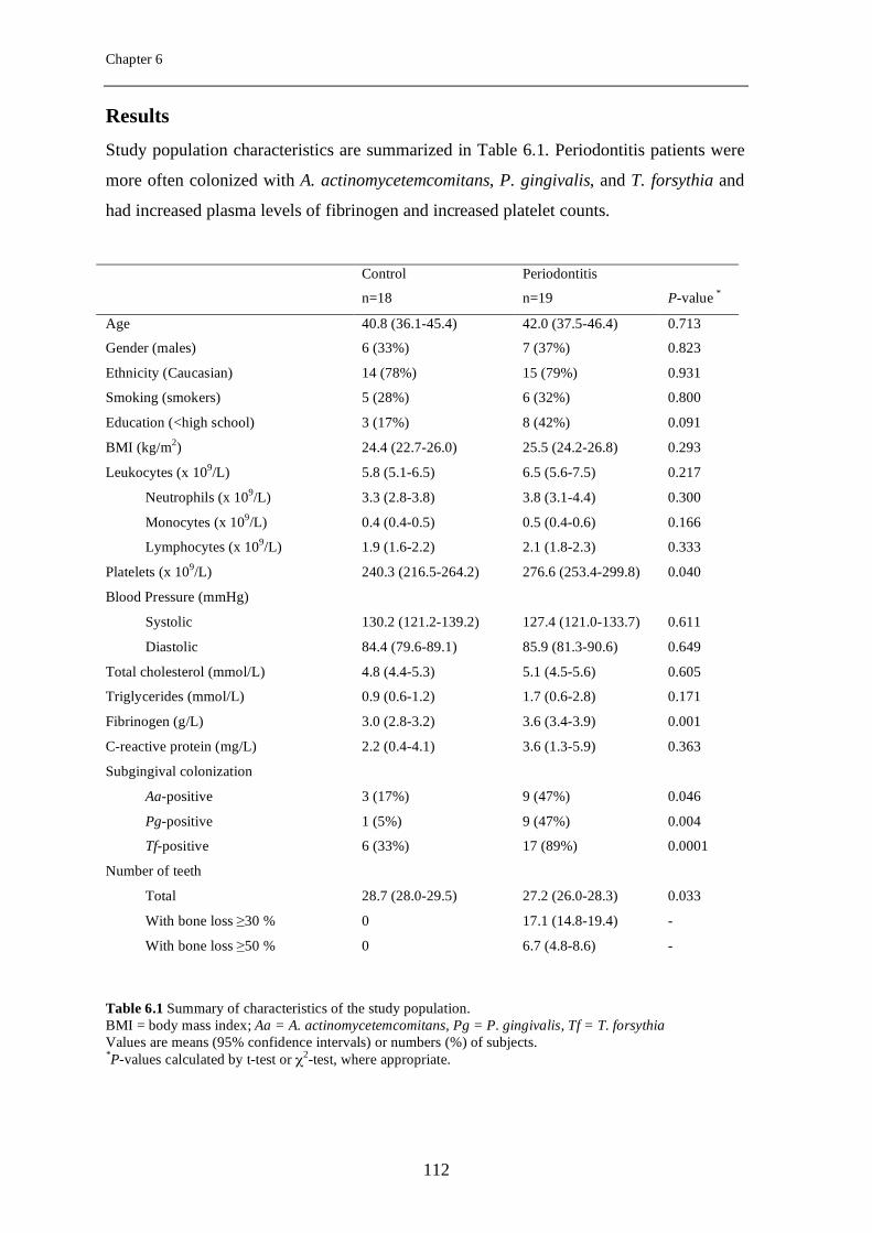
Chapter 6
Results
Study population characteristics are summarized in Table 6.1. Periodontitis patients were
more often colonized with A. actinomycetemcomitans, P. gingivalis, and T. forsythia and
had increased plasma levels of fibrinogen and increased platelet counts.
Table 6.1 Summary of characteristics of the study population. BMI = body mass index; Aa = A. actinomycetemcomitans, Pg = P. gingivalis, Tf = T. forsythia Values are means (95% confidence intervals) or numbers (%) of subjects. *P-values calculated by t-test or χ2-test, where appropriate.
Parameter a
Control
Control
Periodontitis
Periodontitis
n=18 n=19 P-value *
Age 40.8 (36.1-45.4)
4)
42.0 (37.5-46.4) 0.713
Gender (males) 6 (33%) 7 (37%) 0.823
Ethnicity (Caucasian) 14 (78%) 15 (79%) 0.931
Smoking (smokers) 5 (28%) 6 (32%) 0.800
Education (<high school) 3 (17%) 8 (42%) 0.091
BMI (kg/m2) 24.4 (22.7-26.0) 25.5 (24.2-26.8) 0.293
Leukocytes (x 109/L) 5.8 (5.1-6.5) 6.5 (5.6-7.5) 0.217
Neutrophils (x 109/L) 3.3 (2.8-3.8) 3.8 (3.1-4.4) 0.300
Monocytes (x 109/L) 0.4 (0.4-0.5) 0.5 (0.4-0.6) 0.166
Lymphocytes (x 109/L) 1.9 (1.6-2.2) 2.1 (1.8-2.3) 0.333
Platelets (x 109/L) 240.3 (216.5-264.2) 276.6 (253.4-299.8) 0.040
Blood Pressure (mmHg)
Systolic 130.2 (121.2-139.2) 127.4 (121.0-133.7) 0.611
Diastolic 84.4 (79.6-89.1) 85.9 (81.3-90.6) 0.649
Total cholesterol (mmol/L) 4.8 (4.4-5.3) 5.1 (4.5-5.6) 0.605
Triglycerides (mmol/L) 0.9 (0.6-1.2) 1.7 (0.6-2.8) 0.171
Fibrinogen (g/L) 3.0 (2.8-3.2) 3.6 (3.4-3.9) 0.001
C-reactive protein (mg/L) 2.2 (0.4-4.1) 3.6 (1.3-5.9) 0.363
Subgingival colonization
Aa-positive 3 (17%) 9 (47%) 0.046
Pg-positive 1 (5%) 9 (47%) 0.004
Tf-positive 6 (33%) 17 (89%) 0.0001
Number of teeth
Total 28.7 (28.0-29.5) 27.2 (26.0-28.3) 0.033
With bone loss ≥30 %
0
17.1 (14.8-19.4)
-
With bone loss ≥50 % 0 6.7 (4.8-8.6) -
112
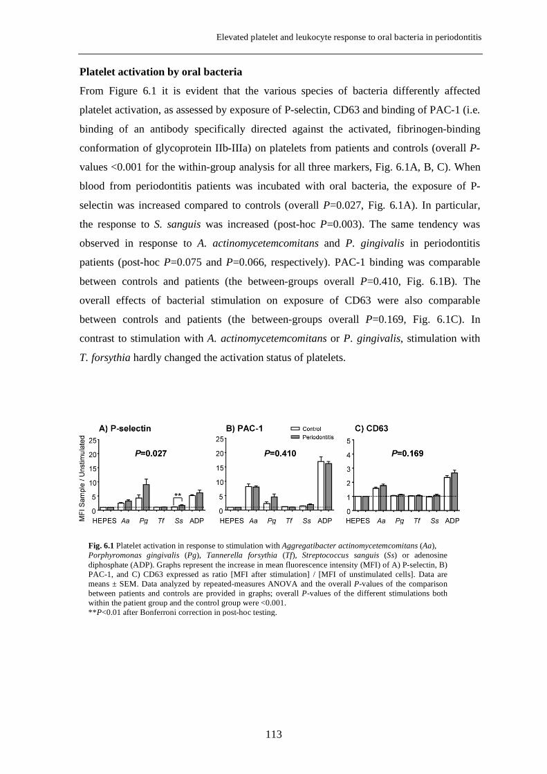
Elevated platelet and leukocyte response to oral bacteria in periodontitis
Platelet activation by oral bacteria
From Figure 6.1 it is evident that the various species of bacteria differently affected
platelet activation, as assessed by exposure of P-selectin, CD63 and binding of PAC-1 (i.e.
binding of an antibody specifically directed against the activated, fibrinogen-binding
conformation of glycoprotein IIb-IIIa) on platelets from patients and controls (overall P-
values <0.001 for the within-group analysis for all three markers, Fig. 6.1A, B, C). When
blood from periodontitis patients was incubated with oral bacteria, the exposure of P-
selectin was increased compared to controls (overall P=0.027, Fig. 6.1A). In particular,
the response to S. sanguis was increased (post-hoc P=0.003). The same tendency was
observed in response to A. actinomycetemcomitans and P. gingivalis in periodontitis
patients (post-hoc P=0.075 and P=0.066, respectively). PAC-1 binding was comparable
between controls and patients (the between-groups overall P=0.410, Fig. 6.1B). The
overall effects of bacterial stimulation on exposure of CD63 were also comparable
between controls and patients (the between-groups overall P=0.169, Fig. 6.1C). In
contrast to stimulation with A. actinomycetemcomitans or P. gingivalis, stimulation with
T. forsythia hardly changed the activation status of platelets.
Fig. 6.1 Platelet activation in response to stimulation with Aggregatibacter actinomycetemcomitans (Aa), Porphyromonas gingivalis (Pg), Tannerella forsythia (Tf), Streptococcus sanguis (Ss) or adenosine diphosphate (ADP). Graphs represent the increase in mean fluorescence intensity (MFI) of A) P-selectin, B) PAC-1, and C) CD63 expressed as ratio [MFI after stimulation] / [MFI of unstimulated cells]. Data are means ± SEM. Data analyzed by repeated-measures ANOVA and the overall P-values of the comparison between patients and controls are provided in graphs; overall P-values of the different stimulations both within the patient group and the control group were <0.001. **P<0.01 after Bonferroni correction in post-hoc testing.
113
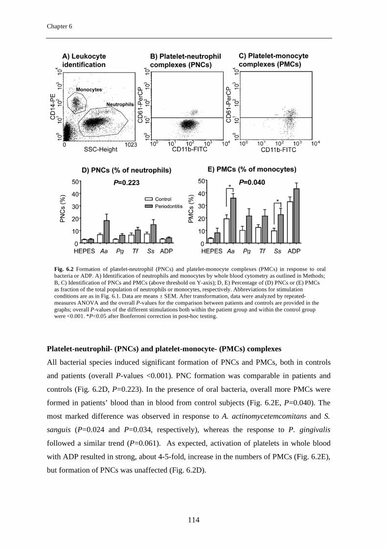
Chapter 6
Platelet-neutrophil- (PNCs) and platelet-monocyte- (PMCs) complexes
All bacterial species induced significant formation of PNCs and PMCs, both in controls
and patients (overall P-values <0.001). PNC formation was comparable in patients and
controls (Fig. 6.2D, P=0.223). In the presence of oral bacteria, overall more PMCs were
formed in patients’ blood than in blood from control subjects (Fig. 6.2E, P=0.040). The
most marked difference was observed in response to A. actinomycetemcomitans and S.
sanguis (P=0.024 and P=0.034, respectively), whereas the response to P. gingivalis
followed a similar trend (P=0.061). As expected, activation of platelets in whole blood
with ADP resulted in strong, about 4-5-fold, increase in the numbers of PMCs (Fig. 6.2E),
but formation of PNCs was unaffected (Fig. 6.2D).
Fig. 6.2 Formation of platelet-neutrophil (PNCs) and platelet-monocyte complexes (PMCs) in response to oral bacteria or ADP. A) Identification of neutrophils and monocytes by whole blood cytometry as outlined in Methods; B, C) Identification of PNCs and PMCs (above threshold on Y-axis); D, E) Percentage of (D) PNCs or (E) PMCs as fraction of the total population of neutrophils or monocytes, respectively. Abbreviations for stimulation conditions are as in Fig. 6.1. Data are means ± SEM. After transformation, data were analyzed by repeated-measures ANOVA and the overall P-values for the comparison between patients and controls are provided in the graphs; overall P-values of the different stimulations both within the patient group and within the control group were <0.001. *P<0.05 after Bonferroni correction in post-hoc testing.
114
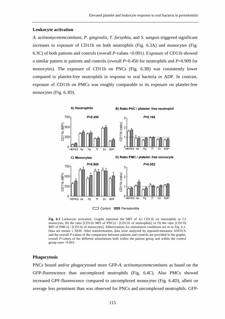
Elevated platelet and leukocyte response to oral bacteria in periodontitis
Leukocyte activation
A. actinomycetemcomitans, P. gingivalis, T. forsythia, and S. sanguis triggered significant
increases in exposure of CD11b on both neutrophils (Fig. 6.3A) and monocytes (Fig.
6.3C) of both patients and controls (overall P-values <0.001). Exposure of CD11b showed
a similar pattern in patients and controls (overall P=0.450 for neutrophils and P=0.909 for
monocytes). The exposure of CD11b on PNCs (Fig. 6.3B) was consistently lower
compared to platelet-free neutrophils in response to oral bacteria or ADP. In contrast,
exposure of CD11b on PMCs was roughly comparable to its exposure on platelet-free
monocytes (Fig. 6.3D).
Phagocytosis
PNCs bound and/or phagocytosed more GFP-A. actinomycetemcomitans as based on the
GFP-fluorescence than uncomplexed neutrophils (Fig. 6.4C). Also PMCs showed
increased GPF-fluorescence compared to uncomplexed monocytes (Fig. 6.4D), albeit on
average less prominent than was observed for PNCs and uncomplexed neutrophils. GFP-
Fig. 6.3 Leukocyte activation. Graphs represent the MFI of A) CD11b on neutrophils or C) monocytes, B) the ratio [CD11b MFI of PNCs] / [CD11b of neutrophils] or D) the ratio [CD11b MFI of PMCs] / [CD11b of monocytes]. Abbreviations for stimulation conditions are as in Fig. 6.1. Data are means ± SEM. After transformation, data were analyzed by repeated-measures ANOVA and the overall P-values of the comparison between patients and controls are provided in the graphs; overall P-values of the different stimulations both within the patient group and within the control group were <0.001.
115
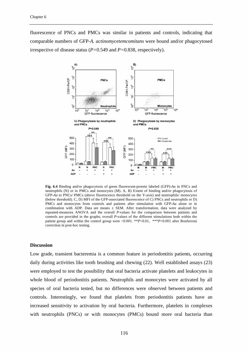
Chapter 6
fluorescence of PNCs and PMCs was similar in patients and controls, indicating that
comparable numbers of GFP-A. actinomycetemcomitans were bound and/or phagocytosed
irrespective of disease status (P=0.549 and P=0.838, respectively).
Discussion
Low grade, transient bacteremia is a common feature in periodontitis patients, occurring
daily during activities like tooth brushing and chewing (22). Well established assays (23)
were employed to test the possibility that oral bacteria activate platelets and leukocytes in
whole blood of periodontitis patients. Neutrophils and monocytes were activated by all
species of oral bacteria tested, but no differences were observed between patients and
controls. Interestingly, we found that platelets from periodontitis patients have an
increased sensitivity to activation by oral bacteria. Furthermore, platelets in complexes
with neutrophils (PNCs) or with monocytes (PMCs) bound more oral bacteria than
Fig. 6.4 Binding and/or phagocytosis of green fluorescent-protein labeled (GFP)-Aa in PNCs and neutrophils (N) or in PMCs and monocytes (M). A, B) Extent of binding and/or phagocytosis of GFP-Aa to PNCs/ PMCs (above fluorescence threshold on the Y-axis) and neutrophils/ monocytes (below threshold). C, D) MFI of the GFP-associated fluorescence of C) PNCs and neutrophils or D) PMCs and monocytes from controls and patients after stimulation with GFP-Aa alone or in combination with ADP. Data are means ± SEM. After transformation, data were analyzed by repeated-measures ANOVA and the overall P-values for the comparison between patients and controls are provided in the graphs; overall P-values of the different stimulations both within the patient group and within the control group were <0.001. **P<0.01, ***P<0.001 after Bonferroni correction in post-hoc testing.
116
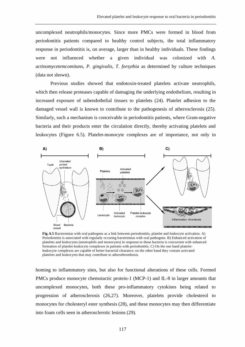
Elevated platelet and leukocyte response to oral bacteria in periodontitis
uncomplexed neutrophils/monocytes. Since more PMCs were formed in blood from
periodontitis patients compared to healthy control subjects, the total inflammatory
response in periodontitis is, on average, larger than in healthy individuals. These findings
were not influenced whether a given individual was colonized with A.
actinomycetemcomitans, P. gingivalis, T. forsythia as determined by culture techniques
(data not shown).
Previous studies showed that endotoxin-treated platelets activate neutrophils,
which then release proteases capable of damaging the underlying endothelium, resulting in
increased exposure of subendothelial tissues to platelets (24). Platelet adhesion to the
damaged vessel wall is known to contribute to the pathogenesis of atherosclerosis (25).
Similarly, such a mechanism is conceivable in periodontitis patients, where Gram-negative
bacteria and their products enter the circulation directly, thereby activating platelets and
leukocytes (Figure 6.5). Platelet-monocyte complexes are of importance, not only in
homing to inflammatory sites, but also for functional alterations of these cells. Formed
PMCs produce monocyte chemotactic protein-1 (MCP-1) and IL-8 in larger amounts that
uncomplexed monocytes, both these pro-inflammatory cytokines being related to
progression of atherosclerosis (26,27). Moreover, platelets provide cholesterol to
monocytes for cholesteryl ester synthesis (28), and these monocytes may then differentiate
into foam cells seen in atherosclerotic lesions (29).
Fig. 6.5 Bacteremias with oral pathogens as a link between periodontitis, platelet and leukocyte activation. A) Periodontitis is associated with regularly occuring bacteremias with oral pathogens. B) Enhanced activation of platelets and leukocytes (neutrophils and monocytes) in response to these bacteria is concurrent with enhanced formation of platelet-leukocyte complexes in patients with periodontitis. C) On the one hand platelet-leukocyte complexes are capable of better bacterial clearance; on the other hand they contain activated platelets and leukocytes that may contribute to atherothrombosis.
117
Chapter 6
It is very interesting to interpret the behavior of activated platelets in the platelet-
leukocyte interaction assays. Activated platelets preferentially adhere to monocytes, rather
than to neutrophils (30). This was also observed in our assays. The platelet agonist ADP
induced increased formation of PMCs and essentially did not induce formation of PNCs.
Moreover, platelet activation by ADP induced monocyte activation rather than neutrophil
activation. Although activated platelets bind to leukocytes, this seems not the only
mechanism for platelet-leukocyte complex formation in our experiments. Stimulation with
oral bacteria induced increased formation of PNCs preferentially via activation of
neutrophils rather than platelets. This phenomenon is similar to PNC formation in
response to stimulation with the bacterial peptide fMLP, which also induces formation of
PNCs more efficiently than ADP (31).
In addition to P. gingivalis and S. sanguis that were shown to promote platelet
aggregation in vitro and in animal studies (32,18), A. actinomycetemcomitans emerged for
the first time as a potent platelet activator in a human ex vivo study. The different species
of oral bacteria, however, induced distinct activation patterns, which are most likely
related to activation of different intracellular signalling pathways (33). At this point it is
unclear which bacterial components are responsible for the platelet activation. This
requires additional experimentation to reach a mechanistical explanation. Especially with
regard to A. actinomycetemcomitans, a marked increase in the exposure of CD63 and
binding of PAC-1 were observed on activated platelets, whereas exposure of P-selectin
was hardly affected. This seemingly discrepant finding is explained by the rapid binding
of P-selectin-exposing platelets to monocytes. Indeed, concurrently an increased number
of PMCs was observed in A. actinomycetemcomitans-treated blood samples.
Randomized controlled clinical trials studying the effects of treatment of
periodontitis on platelet activation may provide further insight on the contribution of this
chronic infectious condition to atherothrombosis. One intervention study showed that
treatment of patients with periodontitis is followed by an improvement of endothelial
function (34). Although the biological basis of this improvement is unknown, one may
speculate that reduced platelet activation and its concurrent pro-coagulant phenotype may
be part of the explanation. Within our department, a study has been initiated to evaluate
whether periodontal therapy indeed affects platelet activation and pro-thrombotic
phenotype in periodontitis patients.
In summary, our data show that not only monocytes but also platelets from
periodontitis patients are more sensitive to stimulation with oral bacteria than cells from
118
Elevated platelet and leukocyte response to oral bacteria in periodontitis
controls. Since oral bacteria regularly get disseminated in blood of periodontitis patients,
this increased cellular sensitivity may contribute to more inflammation and thrombosis at
atherosclerotic sites (Fig. 6.5). Since both processes are involved in the development of
CVD, our findings may in part explain the increased relative risk for cardiovascular events
in periodontitis patients.
Aknowledgements
The present study was supported by The Netherlands Institute for Dental Sciences and
Philips Oral Healthcare EMEA. We thank C. Tijhof, H. de Soet and J. Brunner for
providing the bacterial strains used in the stimulation assays. The GFP-expressing A.
actinomycetemcomitans strain was a gift of Dr. D. Galli (School of Dentistry, Department
of Oral Biology, Indiana University, Indianapolis, USA) and we thank I. Sliepen and Prof.
Dr. W. Teughels for their support in obtaining this strain.
REFERENCES
1 Libby P, Ridker PM, Maseri A. 2002. Inflammation and atherosclerosis. Circulation
105:1135-1143.
2 van Leuven SI, Franssen R, Kastelein JJ, Levi M, Stroes ESG, Tak PP. 2008.
Systemic inflammation as a risk factor for atherothrombosis. Rheumatology (Oxford)
47:3-7.
3 Levi M, Nieuwdorp M, Poll T, Stroes E. 2008. Metabolic modulation of
inflammation-induced activation of coagulation. Semin Thromb Hemost 34:26-32.
4 Wagner DD and Burger PC. 2003. Platelets in inflammation and thrombosis.
Arterioscler Thromb Vasc Biol 23:2131-2137.
5 Klinger MH and Jelkmann W. 2002. Role of blood platelets in infection and
inflammation. J Interferon Cytokine Res 22:913-922.
6 Freedman JE and Loscalzo J. 2002. Platelet-monocyte aggregates: bridging
thrombosis and inflammation. Circulation 105:2130-2132.
119
Chapter 6
7 McCabe DJ, Harrison P, Mackie IJ, Sidhu PS, Purdy G, Lawrie AS, Watt H,
Brown MM, Machin SJ. 2004. Platelet degranulation and monocyte-platelet complex
formation are increased in the acute and convalescent phases after ischaemic stroke or
transient ischaemic attack. Br J Haematol 125:777-787.
8 McEver RP. 2001. Adhesive interactions of leukocytes, platelets, and the vessel wall
during hemostasis and inflammation. Thromb Haemost 86:746-756.
9 Sarma J, Laan CA, Alam S, Jha A, Fox KA, Dransfield I. 2002. Increased platelet
binding to circulating monocytes in acute coronary syndromes. Circulation 105:2166-
2171.
10 Janket SJ, Baird AE, Chuang SK, Jones JA. 2003. Meta-analysis of periodontal
disease and risk of coronary heart disease and stroke. Oral Surg Oral Med Oral Pathol
Oral Radiol Endod 95:559-569.
11 Pihlstrom BL, Michalowicz BS, Johnson NW. 2005. Periodontal diseases. Lancet
366:1809-1820.
12 van Winkelhoff AJ, Loos BG, van der Reijden WA, van der Velden U. 2002.
Porphyromonas gingivalis, Bacteroides forsythus and other putative periodontal
pathogens in subjects with and without periodontal destruction. J Clin Periodontol
29:1023-1028.
13 Geerts SO, Nys M, De MP, Charpentier J, Albert A, Legrand V, Rompen EH.
2002. Systemic release of endotoxins induced by gentle mastication: association with
periodontitis severity. J Periodontol 73:73-78.
14 Loos BG. 2005. Systemic markers of inflammation in periodontitis. J Periodontol
76:2106-2115.
15 Paraskevas S, Huizinga JD, Loos BG. 2008. A systematic review and meta-analyses
on C-reactive protein in relation to periodontitis. J Clin Periodontol 35:277-290.
16 Papapanagiotou D, Nicu EA, Bizzarro S, Gerdes VE, Meijers JC, Nieuwland R,
van der Velden U, Loos BG. 2008. Periodontitis is associated with platelet activation.
Atherosclerosis; in press
120
Elevated platelet and leukocyte response to oral bacteria in periodontitis
17 Herzberg MC and Weyer MW. 1998. Dental plaque, platelets, and cardiovascular
diseases. Ann Periodontol 3:151-160.
18 Lourbakos A, Yuan Y, Jenkins AL, Travis J, Andrade-Gordon P, Santulli R,
Potempa J, Pike RN. 2001. Activation of protease-activated receptors by gingipains
from Porphyromonas gingivalis leads to platelet aggregation: a new trait in microbial
pathogenicity. Blood 97:3790-3797.
19 Konijnenberg A, van der Post JA, Mol BW, Schaap MC, Lazarov R, Bleker OP,
Boer K, Sturk A. 1997. Can flow cytometric detection of platelet activation early in
pregnancy predict the occurrence of preeclampsia? A prospective study. Am J Obstet
Gynecol 177:434-442.
20 Solovjov DA, Pluskota E, Plow EF. 2005. Distinct roles for the alpha and beta
subunits in the functions of integrin alphaMbeta2. J Biol Chem 280:1336-1345.
21 Permpanich P, Kowolik MJ, Galli DM. 2006. Resistance of fluorescent-labelled
Actinobacillus actinomycetemcomitans strains to phagocytosis and killing by human
neutrophils. Cell Microbiol 8:72-84.
22 Forner L, Larsen T, Kilian M, Holmstrup P. 2006. Incidence of bacteremia after
chewing, tooth brushing and scaling in individuals with periodontal inflammation. J
Clin Periodontol 33:401-407.
23 Matzdorff A. 2005. Platelet function tests and flow cytometry to monitor antiplatelet
therapy. Semin Thromb Hemost 31:393-399.
24 Clark SR, Ma AC, Tavener SA, McDonald B, Goodarzi Z, Kelly MM, Patel KD,
Chakrabarti S, McAvoy E, Sinclair GD, Keys EM, len-Vercoe E, Devinney R,
Doig CJ, Green FH, Kubes P. 2007. Platelet TLR4 activates neutrophil extracellular
traps to ensnare bacteria in septic blood. Nat Med 13:463-469.
25 Davi G and Patrono C. 2007. Platelet activation and atherothrombosis. N Engl J Med
357:2482-2494.
26 Weyrich AS, McIntyre TM, McEver RP, Prescott SM, Zimmerman GA. 1995.
Monocyte tethering by P-selectin regulates monocyte chemotactic protein-1 and tumor
121
Chapter 6
necrosis factor-alpha secretion. Signal integration and NF-kappa B translocation. J
Clin Invest 95:2297-2303.
27 Neumann FJ, Marx N, Gawaz M, Brand K, Ott I, Rokitta C, Sticherling C, Meinl
C, May A, Schomig A. 1997. Induction of cytokine expression in leukocytes by
binding of thrombin-stimulated platelets. Circulation 95:2387-2394.
28 Mendelsohn ME and Loscalzo J. 1988. Role of platelets in cholesteryl ester
formation by U-937 cells. J Clin Invest 81:62-68.
29 Curtiss LK, Black AS, Takagi Y, Plow EF. 1987. New mechanism for foam cell
generation in atherosclerotic lesions. J Clin Invest 80:367-373.
30 Ahn KC, Jun AJ, Pawar P, Jadhav S, Napier S, McCarty OJ, Konstantopoulos K.
2005. Preferential binding of platelets to monocytes over neutrophils under flow.
Biochem Biophys Res Commun 329:345-355.
31 Hu H, Varon D, Hjemdahl P, Savion N, Schulman S, Li N. 2003. Platelet-leukocyte
aggregation under shear stress: differential involvement of selectins and integrins.
Thromb Haemost 90:679-687.
32 McNicol A, Zhu R, Pesun R, Pampolina C, Jackson EC, Bowden GH, Zelinski T.
2006. A role for immunoglobulin G in donor-specific Streptococcus sanguis-induced
platelet aggregation. Thromb Haemost 95:288-293.
33 Holmes MB, Sobel BE, Howard DB, Schneider DJ. 1999. Differences between
activation thresholds for platelet P-selectin and glycoprotein IIb-IIIa expression and
their clinical implications. Thromb Res 95:75-82.
34 Tonetti MS, D'Aiuto F, Nibali L, Donald A, Storry C, Parkar M, Suvan J,
Hingorani AD, Vallance P, Deanfield J. 2007. Treatment of periodontitis and
endothelial function. N Engl J Med 356:911-920.
122

Chapter 7
Summary and discussion
123
Summary and discussion
Periodontitis is a chronic infectious condition in the tooth-supporting tissues, which
eventually leads to loss of teeth if left untreated. The host response to bacterial insults
leads to chronic inflammation; the features of periodontal inflammation are gingival
bleeding, formation of deepened periodontal pockets, and progressive destruction of
periodontal ligaments and alveolar bone. While bacteria are essential for the initiation and
progression of periodontitis, genetic and lifestyle factors appear to strongly influence the
severity of the disease and the response to treatment. New models of how to apply this
knowledge for individualized patient care are necessary. An important step forward would
be the identification of individuals with heightened susceptibility for periodontitis. They
would ultimately require more intensive prevention and treatment strategies.
Since bacteria are central in the initiation and progression of periodontitis, one
approach in the direction of identifying modifying factors for susceptibility of
periodontitis, is analyzing the immune response to periodontal pathogens. In this thesis we
analyzed the importance of phagocytes, i.e. PMNs and monocytes in the defense against
periodontal pathogens, and in particular we investigated the function of the receptors in
charge with bacterial recognition. In Chapter 2 we analyzed the FcγRIIa on PMNs, with a
closer look at one single nucleotide polymorphism in the FcγRIIa-gene (FCG2A). FcγRIIa
(one of the types of receptors for the Fc part of immunoglobulins, IgG) mediates
phagocytosis and cell activation. Previous studies showed that one of the genetic variants
of the FcγRIIa, the FcγRIIa131H/H, is capable of efficiently binding human IgG2, whereas
the other two variants, FcγRIIa131H/R or FcγRIIa131R/R do so with reduced affinity. IgG2 is
the predominant IgG subclass against periodontal pathogens and opsonization (with IgG,
complement, or other plasma proteins) is a pre-requisite for efficient recognition of
bacteria by phagocytes. Therefore, we hypothesized that this polymorphism in the FCG2A
is modifying the PMN reactivity in response to periodontal pathogens. Our results
demonstrate that in response to stimulation with the periodontal pathogen A.
actinomycetemcomitans, PMNs from FcγRIIa131H/H carriers phagocytosed more bacteria.
During this phagocytosis step, the FcγRIIa131H/H-PMNs released more granular content and
more active elastase than the FcγRIIa131R/R-PMNs, suggesting a hyper-reactive phenotype
of the FcγRIIa131H/H-PMNs. In addition to the reactive oxygen-species produced during the
respiratory burst, the PMN degranulation products (matrix metallo-proteinases, elastase)
may be responsible for a large part of the PMN-induced collateral damage in periodontitis
(1,2,3). Therefore, the hyper-reactivity of the FcγRIIa131H/H-PMNs resulting in increased
124
Chapter 7
degranulation might be one of the mechanisms by which periodontitis patients with the
FcγRIIa131H/H genotype acquire more severe periodontal destruction compared to patients
with the FcγRIIa131H/R or FcγRIIa131R/R genotypes. Indeed, our results give a mechanistic
explanation for previous observations of an increased severity of periodontal breakdown in
periodontitis patients with the FcγRIIa131H/H genotype (4,5).
The hyper-reactivity of PMNs in periodontitis might result not only from
genetically-encoded affinity of their receptors, but also from differential expression of
these receptors when compared to periodontally-healthy controls. In Chapter 3 we
investigated the cellular expression of IgG-receptors (FcγRI, FcγRIIa, FcγRIIIa and
FcγRIIIb), complement-receptor CR3 and the LPS-co-receptor CD14 on PMNs and
monocytes, as well as activation of these cells in response to two periodontal pathogens, A.
actinomycetemcomitans and P. gingivalis.
Our results demonstrate a similar expression of the analyzed receptors on PMNs in
periodontitis patients and healthy controls, suggesting that the chronic inflammatory
reaction accompanying periodontitis does not affect the PMNs during their formation in
the bone marrow, or in their short-lived blood circulation. The monocytes, on the contrary,
demonstrated a decreased membrane-bound CD14 (mCD14) expression with concomitant
increase in the FcγRIII. These CD14lowFcγRIII+ monocytes have been described in the
literature as precursors of a dendritic cell-type; this cell population is expanding in
inflammatory conditions such as rheumatoid arthritis (6,7), sepsis (8) or Kawasaki disease
(9). The dendritic cells formed from CD14lowFcγRIII+ monocytes have phagocytic and
oxidative capacity, but fail to efficiently stimulate T-cells for a definitive resolution of
inflammation (10). Therefore, we propose a role for the CD14lowFcγRIII+ monocytes in the
chronicization of periodontal inflammation.
Furthermore, our results showed that the subjects culture-positive for A.
actinomycetemcomitans had significantly lower expression of monocytic FcγRI and
FcγRIIa than P. gingivalis-infected subjects. This lower FcγRs expression by monocytes
might be related to a higher susceptibility of a subject to become infected with A.
actinomycetemcomitans or to an adaptation to this particular infection. Indeed, PMNs from
A. actinomycetemcomitans -infected subjects responded in a hyper-reactive manner in
response to bacterial stimulation, in particular when stimulated with A.
actinomycetemcomitans. Taken together, these results suggest a hyper-reactivity of the
PMNs in A. actinomycetemcomitans –infected individuals for which the immune system
125
Summary and discussion
tries to account for by lowering the FcγRs expression on monocytes. Nevertheless, the
hyper-reactive PMNs might contribute to the severe and relatively rapid periodontal
destruction reported in early-onset periodontitis patients sub-gingivally colonized with A.
actinomycetemcomitans (11).
Nevertheless, we believe that the origin of the PMN hyper-reactivity in subjects
colonized with A. actinomycetemcomitans is not clarified yet. We hypothesize that a
certain genetic background could make these individuals more prone for A.
actinomycetemcomitans infection. This scenario has been observed for subjects with
certain genetic variations in the genes coding mannose-binding lectin, vitamin D receptor,
mannose-associated serine-protease-2, and Toll-like receptors; these subjects had a greater
risk of infection with Mycobacterium tuberculosis, meningococci or gram-negative
bacteria (12,13,14,15,16). On the other hand, it could be the infection with A.
actinomycetemcomitans itself responsible for the phenotypic changes of the PMNs. Future
intervention studies that would eliminate A. actinomycetemcomitans from the sub-gingival
microflora might offer the answer to this question.
In addition to the cellular expression of phagocytic receptors, in Chapter 4 we
analyzed the plasmatic levels of the soluble form of CD14 (sCD14) in periodontitis
patients and healthy controls. sCD14 can be the result of shedding / cleavage of mCD14
from monocytes / macrophages or be produced in the liver. Acting as a soluble LPS-
receptor, sCD14 helps inducing responses in cells that naturally lack CD14, such as
endothelial, epithelial and smooth muscle cells (17). Another role is the neutralization of
LPS in Gram-negative infectious states. Acting as a decoy receptor for serum LPS, sCD14
prevents extreme pro-inflammatory responses from monocytes/macrophages (18). Also
periodontitis is a condition associated with Gram-negative pathogens that gain access to
the systemic blood circulation and are responsible for endotoxemia in periodontitis
patients (19). Our results demonstrated an increase in sCD14 plasma levels that positively
correlated with severity of periodontal destruction. This association points into the
direction that the sCD14 increases with the need to remove LPS from circulation. Given
the results from Chapter 3, where we demonstrated a decreased membrane-bound CD14
expression on monocytes from periodontitis patients, the increase in sCD14 might be
partially attributable to the shedding of the membrane-bound form of CD14 from
monocytes. However, we also showed that the increase in sCD14 was positively correlated
with systemic inflammatory markers, such as CRP and numbers of leukocytes. Therefore,
126
Chapter 7
we can assume that also an increased sCD14 production by the liver is accompanying
periodontitis. Interestingly, cleavage of mCD14 by bacterial proteases like the P.
gingivalis gingipains has been described in vitro (20). However, our results do not support
the hypothesis of a direct proteolytic cleavage of mCD14 by gingipains since in our
bacterial stimulation assays A. actinomycetemcomitans and P. gingivalis were similarly
able to induce upregulation of mCD14 on monocytes (Chapter 3). In a future study, an
assay distinguishing between the three forms of sCD14 (resulting from shedding, cleavage
or liver-produced) might bring more insight in the periodontitis-associated increase in
sCD14.
It has been shown that several inflammatory markers predict future vascular events
(21,22,23). C-reactive protein (CRP) and number of leukocytes have been identified as
markers for cardio-vascular disease (CVD) (24,25). Elevated levels of CRP and leukocytes
in patients with CVD may be the result of chronic infectious and inflammatory processes
(26,27,28). Periodontitis is one of the chronic infectious processes that has been associated
with CVD in several epidemiologic studies (29) and elevated blood levels of CRP and
leukocytes have been found in patients with periodontitis (30,31). The underlying
mechanism of CVD is atherosclerosis. CD14 binds LPS in conjunction with LPS-binding-
protein (LBP); the CD14-LPS-LBP complex can directly activate endothelial and smooth
muscle cells (32,33), leading to elevated expression of cell adhesion molecules, thereby
increasing procoagulant activity, and exacerbating inflammation in atherosclerotic lesions
(34). Based on our results in Chapter 4 that showed increased sCD14 levels in
periodontitis, concomitant with elevated CRP and leukocyte levels, we propose a role for
sCD14 in the recurrent episodes of accelerated activity within atherosclerotic plaques in
patients at risk for CVD. We hypothesize that sCD14-mediated signaling is one of the
pathways leading to increased atherogenic activity, and thus, increased risk for CVD in
periodontitis patients.
Not only PMNs and monocytes are capable of interacting with bacteria, also
platelets respond to bacteria by releasing anti-microbial peptides (35), and exposing pro-
inflammatory receptors (36). In Chapters 5 and 6 we analyzed the activation status of
circulating platelets in periodontitis patients, as well as the platelet response to periodontal
pathogens and the interplay between platelets, PMNs and monocytes during stimulation
with bacteria. Periodontitis patients demonstrated increased platelet activation, as revealed
by elevated plasma levels of sP-selectin and increased expression of the glycoprotein IIb-
IIIa, both abundantly expressed receptors, and responsible for adhesive properties of
127

Summary and discussion
platelets. P-selectin is released from platelets within minutes after activation and is found
in plasma as soluble (s)P-selectin (37). Platelet activation was more pronounced in the
patients with more severe periodontal disease, showing a severity-dependence. Moreover,
in Chapter 6 we showed that platelets from periodontitis patients are more sensitive to
oral bacteria than cells from periodontally healthy controls. In response to A.
actinomycetemcomitans, P. gingivalis, and S. sanguis, but not T. forsythia, platelets from
patients expressed more P-selectin than those from controls. Moreover, more platelet-
monocyte complexes were formed in blood from patients in response to oral bacteria than
in blood from controls. In a phagocytosis assay, platelet-PMN and platelet-monocyte
complexes phagocytosed more bacteria than platelet-free cells, suggesting that the
complexes contain primed cells.
Periodontitis is a condition in which due to breaching of the epithelial lining,
small-scale, frequent bacteremias occur during regular activities like chewing or tooth
brushing (38,39). These bacteremic episodes underlie endotoxemia and a chronic systemic
inflammatory reaction in periodontitis patients (40,31,19). In epidemiologic studies,
periodontitis has been associated with increased risk for myocardial infarction and stroke
(29), but the responsible mechanisms are still obscure. Based on our results, we suggest
that the pathologic processes in periodontitis induce platelet and monocyte priming, a
repeatable process with virtually every bacteremic episode of periodontal origin. Activated
platelets produce vast amounts of proinflammatory mediators stored in their α-granules
and dense body systems. Promptly released upon platelet activation, the pro-inflammatory
mediators and receptors induce platelet binding to endothelial cells lining vessel walls
(36). Once adhered, platelets create a platform onto which monocytes can roll and adhere
firmly, leading to increased monocyte recruitment. Monocytes in the subendothelial space
will excessively accumulate intracellular low density lipoprotein leading to accumulation
of intracellular lipid droplets and formation of the foam cells. These functions make
activated platelets and monocytes essential participants in both thrombosis and
atherogenesis and we hypothesize that they explain, at least partially, the
epidemiologically-increased risk for CVD in periodontitis patients.
It is currently unclear whether the increased platelet reactivity in periodontitis
patients is intrinsic, the result of a specific genetic make-up, or extrinsic, the result of
priming by bacterial products streaming from periodontal lesions or by the cytokines
produced as a part of the systemic inflammatory reaction in periodontitis. Future studies,
assessing platelet activation status and reactivity after successful treatment of periodontitis
128
Chapter 7
will shed light on this matter. One published intervention study showed that treatment of
patients with periodontitis is followed by an improvement of endothelial function (41); the
biological basis of this improvement of the vascular condition is as yet unknown.
However, a reduction in the state of platelet activation and in the pro-coagulant state may
be part of the explanation.
Concluding remarks
In this thesis we have investigated the reactivity of PMNs, monocytes and platelets in
periodontitis patients. Individuals at higher risk for periodontal breakdown were
represented by the subjects with the FcγRIIa131H/H genotype and the subjects sub-
gingivally colonized with A. actinomycetemcomitans. These groups of subjects
demonstrated hyper-reactive PMNs in functional assays using periodontal pathogenic
bacteria as stimuli. Whereas for the FcγRIIa genotypic variants, the origin is evidently
genetic and the therapeutic approach goes more in the direction of better prevention of
periodontitis, for the subjects colonized with A. actinomycetemcomitans the answer is not
so straightforward. Future intervention studies that would eliminate A.
actinomycetemcomitans from the sub-gingival microflora might offer the answer to this
question.
Furthermore, we have demonstrated that platelets and monocytes from
periodontitis patients are more sensitive to activation by oral bacterial species, such as A.
actinomycetemcomitans, P. gingivalis, and S. sanguis than cells from healthy controls.
Periodontal therapy results in a reduction of the bacterial burden from the periodontal
pockets, as well as in a reduction of the systemic inflammatory reaction (40). Given the
roles of platelets and monocytes in thrombosis and atherosclerosis, periodontal therapy
might have an added value not only in improving the patients’ oral health, but also as
prevention measure to reduce their overall risk for myocardial infarction and stroke.
129
Summary and discussion
REFERENCES
1 Figueredo CM, Gustafsson A, Asman B, Bergstrom K. 1999. Increased release of
elastase from in vitro activated peripheral neutrophils in patients with adult
periodontitis. J Clin Periodontol 26:206-211.
2 Gustafsson A and Asman B. 1996. Increased release of free oxygen radicals from
peripheral neutrophils in adult periodontitis after Fc delta-receptor stimulation. J Clin
Periodontol 23:38-44.
3 Matthews JB, Wright HJ, Roberts A, Cooper PR, Chapple IL. 2007. Hyperactivity
and reactivity of peripheral blood neutrophils in chronic periodontitis. Clin Exp
Immunol 147:255-264.
4 Loos BG, Leppers-Van de Straat FG, Van de Winkel JG, van der Velden U. 2003.
Fcgamma receptor polymorphisms in relation to periodontitis. J Clin Periodontol
30:595-602.
5 Wolf DL, Neiderud AM, Hinckley K, Dahlen G, van de Winkel JGJ, Papapanou
PN. 2006. Fcgamma receptor polymorphisms and periodontal status: a prospective
follow-up study. J Clin Periodontol 33:691-698.
6 Kawanaka N, Yamamura M, Aita T, Morita Y, Okamoto A, Kawashima M,
Iwahashi M, Ueno A, Ohmoto Y, Makino H. 2002. CD14+,CD16+ blood monocytes
and joint inflammation in rheumatoid arthritis. Arthritis Rheum 46:2578-2586.
7 Wijngaarden S, van Roon JAG, Bijlsma JWJ, van de Winkel JGJ, Lafeber FPJG.
2003. Fc{gamma} receptor expression levels on monocytes are elevated in rheumatoid
arthritis patients with high erythrocyte sedimentation rate who do not use anti-
rheumatic drugs. Rheumatology (Oxford) 42:681-688.
8 Fingerle G, Pforte A, Passlick B, Blumenstein M, Strobel M, Ziegler-Heitbrock
HW. 1993. The novel subset of CD14+/CD16+ blood monocytes is expanded in sepsis
patients. Blood 82:3170-3176.
130
Chapter 7
9 Katayama K, Matsubara T, Fujiwara M, Koga M, Furukawa S. 2000.
CD14+CD16+ monocyte subpopulation in Kawasaki disease. Clin Exp Immunol
121:566-570.
10 Kanaya S, Nemoto E, Ogawa T, Shimauchi H. 2004. Porphyromonas gingivalis
lipopolysaccharides induce maturation of dendritic cells with CD14+CD16+
phenotype. Eur J Immunol 34:1451-1460.
11 van der Velden U, Abbas F, Van Steenbergen TJ, De Zoete OJ, Hesse M, De
Ruyter C, De Laat VH, de Graaff J. 1989. Prevalence of periodontal breakdown in
adolescents and presence of Actinobacillus actinomycetemcomitans in subjects with
attachment loss. J Periodontol 60:604-610.
12 Garred P, Madsen HO, Svejgaard A, Michaelsen TE. 1999. Mannose-binding
lectin and meningococcal disease. Lancet 354:336-
13 Stengaard-Pedersen K, Thiel S, Gadjeva M, Moller-Kristensen M, Sorensen R,
Jensen LT, Sjoholm AG, Fugger L, Jensenius JC. 2003. Inherited deficiency of
mannan-binding lectin-associated serine protease 2. N Engl J Med 349:554-560.
14 Agnese DM, Calvano JE, Hahm SJ, Coyle SM, Corbett SA, Calvano SE, Lowry
SF. 2002. Human toll-like receptor 4 mutations but not CD14 polymorphisms are
associated with an increased risk of gram-negative infections. J Infect Dis 186:1522-
1525.
15 Bellamy R, Ruwende C, Corrah T, McAdam KP, Thursz M, Whittle HC, Hill
AV. 1999. Tuberculosis and chronic hepatitis B virus infection in Africans and
variation in the vitamin D receptor gene. J Infect Dis 179:721-724.
16 Garred P, Harboe M, Oettinger T, Koch C, Svejgaard A. 1994. Dual role of
mannan-binding protein in infections: another case of heterosis? Eur J Immunogenet
21:125-131.
17 Ulevitch RJ and Tobias PS. 1995. Receptor-dependent mechanisms of cell
stimulation by bacterial endotoxin. Annu Rev Immunol 13:437-457.
131
Summary and discussion
18 Maliszewski CR. 1991. CD14 and immune response to lipopolysaccharide. Science
252:1321-1322.
19 Pussinen PJ, Tuomisto K, Jousilahti P, Havulinna AS, Sundvall J, Salomaa V.
2007. Endotoxemia, immune response to periodontal pathogens, and systemic
inflammation associate with incident cardiovascular disease events. Arterioscler
Thromb Vasc Biol 27:1433-1439.
20 Duncan L, Yoshioka M, Chandad F, Grenier D. 2004. Loss of lipopolysaccharide
receptor CD14 from the surface of human macrophage-like cells mediated by
Porphyromonas gingivalis outer membrane vesicles. Microbial Pathogenesis 36:319-
325.
21 Danesh J, Wheeler JG, Hirschfield GM, Eda S, Eiriksdottir G, Rumley A, Lowe
GD, Pepys MB, Gudnason V. 2004. C-reactive protein and other circulating markers
of inflammation in the prediction of coronary heart disease. N Engl J Med 350:1387-
1397.
22 Keaney JF, Jr. and Vita JA. 2002. The value of inflammation for predicting unstable
angina. N Engl J Med 347:55-57.
23 Lindahl B, Toss H, Siegbahn A, Venge P, Wallentin L. 2000. Markers of
myocardial damage and inflammation in relation to long-term mortality in unstable
coronary artery disease. FRISC Study Group. Fragmin during instability in coronary
artery disease. N Engl J Med 343:1139-1147.
24 Ikonomidis I, Stamatelopoulos K, Lekakis J, Vamvakou GD, Kremastinos DT.
2008. Inflammatory and non-invasive vascular markers: the multimarker approach for
risk stratification in coronary artery disease. Atherosclerosis 199:3-11.
25 Rana JS, Boekholdt SM, Ridker PM, Jukema JW, Luben R, Bingham SA, Day
NE, Wareham NJ, Kastelein JJ, Khaw KT. 2007. Differential leucocyte count and
the risk of future coronary artery disease in healthy men and women: the EPIC-
Norfolk Prospective Population Study. J Intern Med 262:678-689.
26 Danesh J, Collins R, Peto R. 1997. Chronic infections and coronary heart disease: is
there a link? Lancet 350:430-436.
132
Chapter 7
27 Lowe GD. 2001. The relationship between infection, inflammation, and cardiovascular
disease: an overview. Ann Periodontol 6:1-8.
28 Maseri A, Biasucci LM, Liuzzo G. 1996. Inflammation in ischaemic heart disease.
BMJ 312:1049-1050.
29 Janket SJ, Baird AE, Chuang SK, Jones JA. 2003. Meta-analysis of periodontal
disease and risk of coronary heart disease and stroke. Oral Surg Oral Med Oral Pathol
Oral Radiol Endod 95:559-569.
30 Paraskevas S, Huizinga JD, Loos BG. 2008. A systematic review and meta-analyses
on C-reactive protein in relation to periodontitis. J Clin Periodontol 35:277-290.
31 Loos BG. 2005. Systemic markers of inflammation in periodontitis. J Periodontol
76:2106-2115.
32 Pugin J, Ulevitch RJ, Tobias PS. 1995. Activation of endothelial cells by endotoxin:
direct versus indirect pathways and the role of CD14. Prog Clin Biol Res 392:369-373.
33 Loppnow H, Stelter F, Schonbeck U, Schluter C, Ernst M, Schutt C, Flad HD.
1995. Endotoxin activates human vascular smooth muscle cells despite lack of
expression of CD14 mRNA or endogenous membrane CD14. Infect Immun 63:1020-
1026.
34 Lo SK, Cheung A, Zheng Q, Silverstein RL. 1995. Induction of tissue factor on
monocytes by adhesion to endothelial cells. J Immunol 154:4768-4777.
35 Fitzgerald JR, Foster TJ, Cox D. 2006. The interaction of bacterial pathogens with
platelets. Nat Rev Microbiol 4:445-457.
36 Klinger MH and Jelkmann W. 2002. Role of blood platelets in infection and
inflammation. J Interferon Cytokine Res 22:913-922.
37 Michelson AD, Barnard MR, Hechtman HB, MacGregor H, Connolly RJ,
Loscalzo J, Valeri CR. 1996. In vivo tracking of platelets: Circulating degranulated
platelets rapidly lose surface P-selectin but continue to circulate and function. PNAS
93:11877-11882.
133
Summary and discussion
38 Forner L, Larsen T, Kilian M, Holmstrup P. 2006. Incidence of bacteremia after
chewing, tooth brushing and scaling in individuals with periodontal inflammation. J
Clin Periodontol 33:401-407.
39 Geerts SO, Nys M, De MP, Charpentier J, Albert A, Legrand V, Rompen EH.
2002. Systemic release of endotoxins induced by gentle mastication: association with
periodontitis severity. J Periodontol 73:73-78.
40 D'Aiuto F, Parkar M, Andreou G, Suvan J, Brett PM, Ready D, Tonetti MS.
2004. Periodontitis and systemic inflammation: control of the local infection is
associated with a reduction in serum inflammatory markers. J Dent Res 83:156-160.
41 Tonetti MS, D'Aiuto F, Nibali L, Donald A, Storry C, Parkar M, Suvan J,
Hingorani AD, Vallance P, Deanfield J. 2007. Treatment of periodontitis and
endothelial function. N Engl J Med 356:911-920.
134

Samenvatting
135
Samenvatting
Parodontitis is een chronische infectieuze aandoening van de tand ondersteunende
weefsels (het parodontium), die uiteindelijk leidt tot het verlies van tanden wanneer er niet
behandeld wordt. De reactie van de gastheer op bacteriële accumulatie op de
gebitselementen leidt tot chronische ontsteking van het parodontium. De kenmerken van
parodontale ontsteking zijn bloeding van de gingiva, vorming van diepe parodontale
pockets en progressieve afbraak van het parodontaal ligament en het alveolaire bot.
Hoewel bacteriën essentieel zijn voor het ontstaan en de progressie van parodontitis,
blijken ook genetische en lifestyle factoren een grote invloed te hebben op de vatbaarheid
voor en de ernst van de ziekte, alsmede de reactie op behandeling. Deze nieuwe inzichten
moeten nog geïmplementeerd worden bij geïndividualiseerde patiëntenzorg. Identificatie
van individuen met verhoogde vatbaarheid voor parodontitis zou een belangrijke stap
voorwaarts zijn. Zij zouden uiteindelijk intensievere preventie en behandelingsstrategieën
nodig hebben.
Het analyseren van de gastheerreactie op de parodontale bacteriële pathogenen is
één van de mogelijke benaderingen in de richting van het identificeren van modificerende
factoren voor de vatbaarheid voor parodontitis. In dit proefschrift hebben we het belang
van de polymorphonucleaire leukocyten (PMNs) en monocyten (deze cel typen zijn beiden
fagocyten), in de verdediging tegen parodontale pathogenen geanalyseerd, en in het
bijzonder hebben we de functie onderzocht van de receptoren die verantwoordelijk zijn
voor de herkenning van de bacteriën.
In Hoofdstuk 2 hebben we FcγRIIa op PMNs geanalyseerd, met een focus op een
single nucleotide polymorfisme in het FcγIIa gen (FCG2A). FcγIIa (één van de typen
receptoren voor het Fc gedeelte van immunoglobulines, IgG) stuurt fagocytose en cel
activering aan. Eerdere studies hebben aangetoond dat één van de genetische varianten van
FcγIIa, FcγIIa131H/H, in staat is om humaan IgG2 efficiënt te binden, terwijl de andere twee
varianten, FcγIIa131H/R of FcγIIa131R/R , dat ook wel kunnen, maar met een verminderde
affiniteit. IgG2 is de voornaamste IgG subklasse tegen parodontale pathogenen en
opsonisatie met IgG, en in parodontitis vooral met IgG2, helpt bij een efficiënte
herkenning van bacteriën door fagocyten. Wij hebben gehypothetiseerd dat dit
polymorfisme in het FCG2A gen, de reactiviteit verandert van PMNs in respons op
parodontale pathogenen. Onze resultaten laten zien dat PMNs van individuen met het
FcγIIa131H/H genotype meer bacteriën fagocyteren als reactie op de stimulatie met de
parodontale paropathogeen A. actinomycetemcomitans. Tijdens dit fagocytose proces
scheiden de granulen van FcγIIa131H/H PMNs meer enzymen uit waaronder meer actief
136
Samenvatting
elastase, dan FcγIIa131R/R PMNs, hetgeen een hyperactief fenotype van de FcγIIa131H/H
PMNs suggereert. Voor het ziekteproces parodontitis betekent dit dat naast de reactieve
zuurstof radicalen die geproduceerd worden tijdens de oxidatieve stress van de PMNs, ook
de degranulatie producten (matrix metallo proteinases, elastase) van de PMNs
verantwoordelijk zijn voor afbraak van bind- en steunweefsel bij parodontitis (1,2,3).
Derhalve zou de hyper-reactiviteit van de FcγIIa131H/H PMNs één van de mechanismen
kunnen zijn waardoor juist deze patiënten een ernstiger parodontale afbraak hebben in
vergelijking met de FcγIIa131H/R en FcγIIa131R/R genotypen. Onze resultaten geven dan ook
een mechanistische verklaring voor eerdere observaties van een toegenomen ernst van
parodontale afbraak bij patiënten met het FcγIIa131H/H genotype (4,5).
De hyperreactiviteit van PMNs bij parodontitis zou, naast de genetisch gecodeerde
sterkere of zwakkere affiniteit van hun receptoren, ook veroorzaakt kunnen worden door
een afwijkende expressie van deze receptoren in vergelijking met parodontaal gezonde
controles. In Hoofdstuk 3 hebben we zowel de cellulaire expressie van IgG receptoren
(FcγRI, FcγRIIa, FcγRIIIa en FcγIIIb), complement receptor CR3 en de LPS co-receptor
CD14 op PMNs en monocyten onderzocht, als ook de activatie van deze cellen in reactie
op de twee parodontale pathogenen A. actinomycetemcomitans en P. gingivalis. Onze
resultaten laten een vergelijkbare expressie zien van de onderzochte receptoren op PMNs
van parodontale patiënten en van gezonde controles. Wij suggereren dat de chronische
ontstekingsreactie die gepaard gaat met parodontitis blijkbaar geen effect heeft op de PMN
receptor expressie tijdens hun vorming in het beenmerg en tijdens hun kortstondige
circulatie in het bloed. De monocyten daarentegen, lieten een verlaagde expressie van
membraan-gebonden CD14 (mCD14) zien, in samenhang met een toename van FcγRIII
expressie. Deze CD14lowFcγRIII+ monocyten zijn in de literatuur beschreven als
voorlopers van een dendritisch celtype; deze celpopulatie neemt toe tijdens ontstekingen
zoals reumatoïde artritis (6,7), sepsis (8) of de Kawasaki ziekte (9). De dendritische cellen
die gevormd worden door de CD14lowFcγRIII+ monocyten hebben een fagocyterende en
oxiderende capaciteit, maar zijn niet in staat om T cellen efficiënt te stimuleren tot een
definitieve actie om de ontsteking te elimineren (10). Met deze bevindingen kennen wij
een rol toe aan de CD14lowFcγRIII+ monocyten, die mogelijk de chroniciteit van
parodontale ontsteking bevorderen dan wel in stand houden.
Daarnaast vonden wij dat personen die geïnfecteerd zijn met A.
actinomycetemcomitans, een significant lagere expressie van monocytaire FcγRI en
FcγRIIa hebben dan personen die met P. gingivalis geïnfecteerd zijn. Deze verlaagde
137
Samenvatting
FcγR expressie door monocyten kan gerelateerd zijn aan een verhoogde gevoeligheid voor
een A. acinomycetemcomitans infectie, of aan een aanpassing aan deze specifieke infectie.
Parallel aan deze bevinding zagen we ook dat PMNs van A. actinomycetemcomitans
geïnfecteerde personen inderdaad op een hyperactieve manier reageerden op een bacteriële
stimulans, in het bijzonder wanneer zij werden gestimuleerd met A.
actinomycetemcomitans. Samenvattend suggereren deze resultaten een hyperreactiviteit
van de PMNs bij A. actinomycetemcomitans geïnfecteerde personen, welke het
immuunsysteem probeert te compenseren door de verlaging van FcγRs expressie op
monocyten. Desondanks zouden de hyperactieve PMNs kunnen bijdragen aan de ernstige
en relatief snelle parodontale afbraak die gerapporteerd is in early-onset parodontitis
patiënten, die zeer frequent geïnfecteerd zijn met A. actinomycetemcomitans (11).
Ondanks deze bevindingen is het toch niet helemaal duidelijk waarom bij personen
die geïnfecteerd zijn met A. actinomycetemcomitans de PMN’s hyperactief zijn. Wij
veronderstellen dat een bepaalde genetische achtergrond deze personen meer vatbaar
maken voor A. actinomycetemcomitans infectie. Dit scenario is al beschreven bij personen
met genetische variaties in de genen coderend voor mannose-bindend lectine, vitamine D
receptor, mannose-geassocieerde serine protease 2 en toll-like receptoren; deze personen
hadden een groter risico op infectie met Mycobacterium tuberculosis, meningococci of
gram negatieve bacteriën (12,13,14,15,16). Aan de andere kant zou het de infectie met A.
actinomycetemcomitans zelf kunnen zijn die verantwoordelijk is voor de fenotypische
veranderingen van de PMNs. Dit dilemma zou verder onderzocht kunnen worden met
interventie studies, welke A. actinomycetemcomitans elimineren uit de subgingivale
microflora.
Naast de cellulaire expressie van receptoren op fagocyten, hebben we in
Hoofdstuk 4 de plasma spiegels van de oplosbare vorm van CD14 (sCD14) geanalyseerd
bij parodontitis patiënten en gezonde controles. sCD14 kan ontstaan door afscheiding /
afknippen van membraan-gebonden CD14 op monocyten / macrofagen of sCD14 kan
geproduceerd worden in de lever. Fungerend als een oplosbare LPS receptor, helpt sCD14
bij de activatie van cellen die van nature geen CD14 hebben, zoals endotheelcellen,
epitheelcellen en gladde spiercellen. Een andere rol is de neutralisatie van LPS tijdens
gram-negatieve infectieuze ziekteprocessen. Als neutraliserende factor voor serum LPS,
verhindert sCD14 extreme proinflammatoire reacties van monocyten / macrofagen (18).
Ook parodontitis is een ziekteproces waarbij gram-negatieve pathogenen de infectie
vormen. Deze pathogenen komen in de bloedcirculatie terecht en zijn verantwoordelijk
138
Samenvatting
voor endotoxemia bij parodontitis patiënten (19). Onze resultaten laten een toename in
sCD14 plasma waardes zien, welke positief correleren met de ernst van de parodontale
afbraak. Deze associatie suggereert dat het sCD14 toeneemt samen met de noodzaak om
LPS uit de circulatie te verwijderen. Gezien de resultaten uit Hoofdstuk 3, waar we een
verminderde membraan-gebonden CD14 expressie op monocyten van parodontitis
patiënten hebben laten zien, zou de toename van het sCD14 gedeeltelijk toegeschreven
kunnen worden aan de afscheiding van het membraan gebonden CD14 van monocyten.
We hebben echter ook aangetoond dat de toename in sCD14 een positieve correlatie laat
zien met de systemische ontstekingsmarker C-reactief proteïn (CRP) en met het aantal
leukocyten in de circulatie. Dus we veronderstellen dat naast CRP, ook een verhoogde
sCD14 productie in de lever bij de parodontitis patiënt plaatsvindt.
Het is aangetoond dat verschillende ontstekingmarkers toekomstige vasculaire
aandoeningen voorspellen (21,22,23). CRP en het aantal leukocyten, zijn o.a. aangewezen
als markers voor hart- en vaatziekten (HVZ) (24,25). Verhoogde waardes van CRP en
leukocyten in patiënten met HVZ worden tegenwoordig ook gezien als een mogelijk
gevolg van chronisch infectieuze processen en ontstekingsreacties (26,27,28). Parodontitis
is één van de chronische infectieuze aandoeningen die geassocieerd is met HVZ in
verschillende epidemiologische studies (29). Daarnaast zijn verhoogde bloedwaarden van
CRP en leukocyten gevonden bij patiënten met parodontitis (30,31). Het onderliggende
mechanisme van HVZ is atherosclerose. In dit verband is het bijzonder interessant dat
CD14 het mogelijk maakt dat LPS gebonden kan worden aan het LPS binding-eiwit
(LBP); het LPS-CD14-LBP complex kan endotheelcellen en gladde spiercellen direct
activeren (32,33), wat leidt tot verhoogde expressie van celadhesie moleculen. Hierdoor
wordt de procoagulante activiteit in bloedvaten verhoogd en de ontstekingreacties
binnenin en rondom de atherosclerotische laesies verergert (34). Gebaseerd op onze
resultaten uit Hoofdstuk 4, waar verhoogde sCD14 waardes bij parodontitis werden
aangetoond, in samenhang met verhoogde CRP waarden en aantallen leukocyten, zouden
verhoogde sCD14 spiegels in het bloed kunnen leiden tot een verhoogde activiteit
binnenin de atherosclerotische plak bij patiënten met een risico voor HVZ. Wij
veronderstellen dat sCD14-gemedieerde signalering één van de routes is die leidt tot
verhoogde atherogene activiteit en daardoor tot een verhoogd risico voor HVZ bij
parodontitis patiënten.
Niet alleen PMNs en monocyten zijn in staat tot interactie met bacteriën, maar ook
bloedplaatjes reageren op bacteriën door middel van uitscheiding van antimicrobiële
139
Samenvatting
eiwitten (35), en de expressie van proinflammatoire receptoren (36). In Hoofdstuk 5 en 6
hebben we de activeringsstatus van circulerende bloedplaatjes bij parodontitis patiënten
geanalyseerd, alsmede de reactie van de plaatjes op parodontale pathogenen en de
interactie tussen bloedplaatjes, PMNs en monocyten tijdens stimulatie met bacteriën.
Parodontitis patiënten lieten een verhoogde activering van de plaatjes zien, dat werd
aangetoond door de verhoogde plasma waardes van P-selectine en de verhoogde expressie
van de glycoproteïne IIb-IIIa. P-selectine en de glycoproteïne IIb-IIIa worden bij
activering van bloedplaatjes in hoge mate tot expressie gebracht en zijn verantwoordelijk
voor de hechtende en aggregerende eigenschappen van bloedplaatjes. P-selectine wordt
enkele minuten na activering uitgescheiden door bloedplaatjes en wordt in het plasma
teruggevonden als oplosbaar (s)P-selectine (37). Wij vonden dat de activering van de
bloedplaatjes intenser was bij parodontitis patiënten naarmate de ziekte ernstiger was, dus
“dosis” afhankelijk was.
Bovendien hebben we in Hoofdstuk 6 laten zien dat bloedplaatjes van parodontitis
patiënten gevoeliger zijn voor orale bacteriën dan bloedplaatjes van parodontaal gezonde
personen. Als reactie op A. actinomycetemcomitans, P. gingivalis en S. sanguis, maar niet
op T. forsythia, brachten de bloedplaatjes van patiënten meer P-selectine tot expressie dan
de bloedplaatjes van controles. Bovendien werden er, ex vivo, als reactie op de genoemde
paropathogene bacterië, meer bloedplaatjes-monocyt complexen gevormd met het bloed
van patiënten, dan met bloed van controles. In een fagocytose experiment, fagocyteerden
bloedplaatjes-PMN- en bloedplaatjes-monocyt complexen meer bacteriën dan
bloedplaatjes-vrije cellen, wat suggereert dat de complexen geactiveerde cellen bevatten.
Parodontitis is een aandoening waarbij, door ulceratie van het pocketepitheel,
beperkte, maar desalniettemin frequente bacteremiën voorkomen tijdens normale
activiteiten zoals kauwen of tanden poetsen (38,39). Deze bacteremiën zijn de oorzaak van
endotoxemia en een chronische systemische ontstekingsreactie bij parodontitis patiënten
(40,31,19). Uit epidemiologisch onderzoek is gebleken dat parodontitis geassocieerd is
met een verhoogd risico op hartinfarcten en beroerte (29), maar de hiervoor
verantwoordelijke mechanismen zijn nog onduidelijk. Gezien onze resultaten, denken wij
dat de pathologische processen bij parodontitis o.a. resulteren in de activering van
bloedplaatjes en monocyten, een steeds terugkerend proces tijdens elke bacteremie bij
parodontitis. Geactiveerde bloedplaatjes produceren grote hoeveelheden proinflammatoire
mediatoren afkomstig vanuit hun alfa-granules en “dense-body” systemen. Bovendien
komen meer receptoren tot expressie. Na activering van bloedplaatjes induceren de
140
Samenvatting
vrijgekomen proinflammatoire mediatoren en de receptoren, de hechting van plaatjes aan
de endotheelcellen die de bloedvatwand bekleden (36). Eenmaal gehecht, creëren de
bloedplaatjes een platform waarop monocyten kunnen rollen en sterk hechten, hetgeen
leidt tot een verhoogde monocyt rekrutering. Monocyten in de subendotheliale ruimte
gaan overmatig intracellulair lipoproteinen opnemen, wat leidt tot accumulatie van
intracellulaire vetdruppels en formatie van schuimcellen. Deze functionaliteit maken
geactiveerde bloedplaatjes en monocyten essentiële deelnemers in zowel trombose als
atherogenese en wij suggereren dan ook dat dit, in ieder geval gedeeltelijk, de
epidemiologische relatie tussen HVZ en parodontitis kan verklaren.
Het is tot op heden onduidelijk of de verhoogde bloedplaatjes reactiviteit bij
parodontitis patiënten intrinsiek is, het resultaat van een specifieke genetische opmaak, of
reactief is, dat wil zeggen het resultaat van priming door bacteriële producten afkomstig
uit de parodontale pockets of door de cytokines die geproduceerd worden als gevolg van
de systemische ontstekingsreactie in parodontitis. Verder onderzoek, waarbij de mate van
plaatjesactivering en de reactiviteit worden vastgesteld na succesvolle behandeling van
parodontitis, zal hier meer duidelijkheid over kunnen verschaffen. Belangrijk is in dit
verband dat een interventie studie liet zien dat de behandeling van patiënten met
parodontitis resulteert in een verbeterde endotheliale functie (41); de biologische basis van
deze verbetering van de vasculaire conditie is tot op heden nog onduidelijk. Een afname in
de mate van bloedplaatjes activering zou dit echter gedeeltelijk kunnen verklaren.
Tot slot
In dit proefschrift hebben we de reactiviteit van PMNs, monocyten en bloedplaatjes
onderzocht. Voorgaande studies hebben aangetoond dat early-onset parodontitis patiënten
o.a. gekarakteriseerd worden door het FcγRIIa131H/H genotype en door de subgingivale
kolonisatie met A. actinomycetemcomitans. In een studie in dit proefschrift vertoonden de
FcγRIIa131H/H genotype patiënten hyperactieve PMNs in functionele assays waarbij A.
actinomycetemcomitans bacteriën werden gebruikt als stimuli. Voor de FcγRIIa varianten
is de origine duidelijk genetisch en zal de therapeutische aanpak meer in de richting gaan
van preventie van parodontitis; pro-actief genetische screening op vatbaarheid voor
parodontitis ligt wellicht in het verschiet.
Daarnaast hebben we aangetoond dat bloedplaatjes en monocyten van parodontitis
patiënten gevoeliger zijn voor activering door orale bacteriële soorten, zoals A.
actinomycetemcomitans, P. gingivalis en S. sanguis, dan cellen van gezonde controles.
141
Samenvatting
Parodontale therapie resulteert zowel in een afname van de hoeveelheid bacteriën in de
parodontale pockets, als in een afname van de systemische ontstekingsreactie. Gegeven de
activiteit van bloedplaatjes en monocyten in trombotische processen en atherosclerose, kan
parodontale therapie een toegevoegde waarde hebben, waarbij niet alleen de mond
gezondheid van de patiënt verbetert, maar ook het algemene risico voor een myocard
infarct en beroerte wellicht afneemt.
REFERENTIES
1 Figueredo CM, Gustafsson A, Asman B, Bergstrom K. 1999. Increased release of
elastase from in vitro activated peripheral neutrophils in patients with adult
periodontitis. J Clin Periodontol 26:206-211.
2 Gustafsson A and Asman B. 1996. Increased release of free oxygen radicals from
peripheral neutrophils in adult periodontitis after Fc delta-receptor stimulation. J Clin
Periodontol 23:38-44.
3 Matthews JB, Wright HJ, Roberts A, Cooper PR, Chapple IL. 2007. Hyperactivity
and reactivity of peripheral blood neutrophils in chronic periodontitis. Clin Exp
Immunol 147:255-264.
4 Loos BG, Leppers-Van de Straat FG, Van de Winkel JG, van der Velden U. 2003.
Fcgamma receptor polymorphisms in relation to periodontitis. J Clin Periodontol
30:595-602.
5 Wolf DL, Neiderud AM, Hinckley K, Dahlen G, van de Winkel JGJ, Papapanou
PN. 2006. Fcgamma receptor polymorphisms and periodontal status: a prospective
follow-up study. J Clin Periodontol 33:691-698.
6 Kawanaka N, Yamamura M, Aita T, Morita Y, Okamoto A, Kawashima M,
Iwahashi M, Ueno A, Ohmoto Y, Makino H. 2002. CD14+,CD16+ blood monocytes
and joint inflammation in rheumatoid arthritis. Arthritis Rheum 46:2578-2586.
7 Wijngaarden S, van Roon JAG, Bijlsma JWJ, van de Winkel JGJ, Lafeber
FPJG. 2003. Fc{gamma} receptor expression levels on monocytes are elevated in
142
Samenvatting
rheumatoid arthritis patients with high erythrocyte sedimentation rate who do not use
anti-rheumatic drugs. Rheumatology (Oxford) 42:681-688.
8 Fingerle G, Pforte A, Passlick B, Blumenstein M, Strobel M, Ziegler-Heitbrock
HW. 1993. The novel subset of CD14+/CD16+ blood monocytes is expanded in sepsis
patients. Blood 82:3170-3176.
9 Katayama K, Matsubara T, Fujiwara M, Koga M, Furukawa S. 2000.
CD14+CD16+ monocyte subpopulation in Kawasaki disease. Clin Exp Immunol
121:566-570.
10 Kanaya S, Nemoto E, Ogawa T, Shimauchi H. 2004. Porphyromonas gingivalis
lipopolysaccharides induce maturation of dendritic cells with CD14+CD16+
phenotype. Eur J Immunol 34:1451-1460.
11 van der Velden U, Abbas F, Van Steenbergen TJ, De Zoete OJ, Hesse M, De
Ruyter C, De Laat VH, de Graaff J. 1989. Prevalence of periodontal breakdown in
adolescents and presence of Actinobacillus actinomycetemcomitans in subjects with
attachment loss. J Periodontol 60:604-610.
12 Garred P, Madsen HO, Svejgaard A, Michaelsen TE. 1999. Mannose-binding
lectin and meningococcal disease. Lancet 354:336-
13 Stengaard-Pedersen K, Thiel S, Gadjeva M, Moller-Kristensen M, Sorensen R,
Jensen LT, Sjoholm AG, Fugger L, Jensenius JC. 2003. Inherited deficiency of
mannan-binding lectin-associated serine protease 2. N Engl J Med 349:554-560.
14 Agnese DM, Calvano JE, Hahm SJ, Coyle SM, Corbett SA, Calvano SE, Lowry
SF. 2002. Human toll-like receptor 4 mutations but not CD14 polymorphisms are
associated with an increased risk of gram-negative infections. J Infect Dis 186:1522-
1525.
15 Bellamy R, Ruwende C, Corrah T, McAdam KP, Thursz M, Whittle HC, Hill
AV. 1999. Tuberculosis and chronic hepatitis B virus infection in Africans and
variation in the vitamin D receptor gene. J Infect Dis 179:721-724.
143
Samenvatting
16 Garred P, Harboe M, Oettinger T, Koch C, Svejgaard A. 1994. Dual role of
mannan-binding protein in infections: another case of heterosis? Eur J Immunogenet
21:125-131.
17 Ulevitch RJ and Tobias PS. 1995. Receptor-dependent mechanisms of cell
stimulation by bacterial endotoxin. Annu Rev Immunol 13:437-457.
18 Maliszewski CR. 1991. CD14 and immune response to lipopolysaccharide. Science
252:1321-1322.
19 Pussinen PJ, Tuomisto K, Jousilahti P, Havulinna AS, Sundvall J, Salomaa V.
2007. Endotoxemia, immune response to periodontal pathogens, and systemic
inflammation associate with incident cardiovascular disease events. Arterioscler
Thromb Vasc Biol 27:1433-1439.
20 Duncan L, Yoshioka M, Chandad F, Grenier D. 2004. Loss of lipopolysaccharide
receptor CD14 from the surface of human macrophage-like cells mediated by
Porphyromonas gingivalis outer membrane vesicles. Microbial Pathogenesis 36:319-
325.
21 Danesh J, Wheeler JG, Hirschfield GM, Eda S, Eiriksdottir G, Rumley A, Lowe
GD, Pepys MB, Gudnason V. 2004. C-reactive protein and other circulating markers
of inflammation in the prediction of coronary heart disease. N Engl J Med 350:1387-
1397.
22 Keaney JF, Jr. and Vita JA. 2002. The value of inflammation for predicting unstable
angina. N Engl J Med 347:55-57.
23 Lindahl B, Toss H, Siegbahn A, Venge P, Wallentin L. 2000. Markers of
myocardial damage and inflammation in relation to long-term mortality in unstable
coronary artery disease. FRISC Study Group. Fragmin during instability in coronary
artery disease. N Engl J Med 343:1139-1147.
24 Ikonomidis I, Stamatelopoulos K, Lekakis J, Vamvakou GD, Kremastinos DT.
2008. Inflammatory and non-invasive vascular markers: the multimarker approach for
risk stratification in coronary artery disease. Atherosclerosis 199:3-11.
144
Samenvatting
25 Rana JS, Boekholdt SM, Ridker PM, Jukema JW, Luben R, Bingham SA, Day
NE, Wareham NJ, Kastelein JJ, Khaw KT. 2007. Differential leucocyte count and
the risk of future coronary artery disease in healthy men and women: the EPIC-
Norfolk Prospective Population Study. J Intern Med 262:678-689.
26 Danesh J, Collins R, Peto R. 1997. Chronic infections and coronary heart disease: is
there a link? Lancet 350:430-436.
27 Lowe GD. 2001. The relationship between infection, inflammation, and cardiovascular
disease: an overview. Ann Periodontol 6:1-8.
28 Maseri A, Biasucci LM, Liuzzo G. 1996. Inflammation in ischaemic heart disease.
BMJ 312:1049-1050.
29 Janket SJ, Baird AE, Chuang SK, Jones JA. 2003. Meta-analysis of periodontal
disease and risk of coronary heart disease and stroke. Oral Surg Oral Med Oral Pathol
Oral Radiol Endod 95:559-569.
30 Paraskevas S, Huizinga JD, Loos BG. 2008. A systematic review and meta-analyses
on C-reactive protein in relation to periodontitis. J Clin Periodontol 35:277-290.
31 Loos BG. 2005. Systemic markers of inflammation in periodontitis. J Periodontol
76:2106-2115.
32 Pugin J, Ulevitch RJ, Tobias PS. 1995. Activation of endothelial cells by endotoxin:
direct versus indirect pathways and the role of CD14. Prog Clin Biol Res 392:369-373.
33 Loppnow H, Stelter F, Schonbeck U, Schluter C, Ernst M, Schutt C, Flad HD.
1995. Endotoxin activates human vascular smooth muscle cells despite lack of
expression of CD14 mRNA or endogenous membrane CD14. Infect Immun 63:1020-
1026.
34 Lo SK, Cheung A, Zheng Q, Silverstein RL. 1995. Induction of tissue factor on
monocytes by adhesion to endothelial cells. J Immunol 154:4768-4777.
35 Fitzgerald JR, Foster TJ, Cox D. 2006. The interaction of bacterial pathogens with
platelets. Nat Rev Microbiol 4:445-457.
145
Samenvatting
36 Klinger MH and Jelkmann W. 2002. Role of blood platelets in infection and
inflammation. J Interferon Cytokine Res 22:913-922.
37 Michelson AD, Barnard MR, Hechtman HB, MacGregor H, Connolly RJ,
Loscalzo J, Valeri CR. 1996. In vivo tracking of platelets: Circulating degranulated
platelets rapidly lose surface P-selectin but continue to circulate and function. PNAS
93:11877-11882.
38 Forner L, Larsen T, Kilian M, Holmstrup P. 2006. Incidence of bacteremia after
chewing, tooth brushing and scaling in individuals with periodontal inflammation. J
Clin Periodontol 33:401-407.
39 Geerts SO, Nys M, De MP, Charpentier J, Albert A, Legrand V, Rompen EH.
2002. Systemic release of endotoxins induced by gentle mastication: association with
periodontitis severity. J Periodontol 73:73-78.
40 D'Aiuto F, Parkar M, Andreou G, Suvan J, Brett PM, Ready D, Tonetti MS.
2004. Periodontitis and systemic inflammation: control of the local infection is
associated with a reduction in serum inflammatory markers. J Dent Res 83:156-160.
41 Tonetti MS, D'Aiuto F, Nibali L, Donald A, Storry C, Parkar M, Suvan J,
Hingorani AD, Vallance P, Deanfield J. 2007. Treatment of periodontitis and
endothelial function. N Engl J Med 356:911-920.
146
Acknowledgements
I have learned a lot in the past four years, not only about research and science, but also
about myself. For all this knowledge I want to thank the people who contributed directly
or indirectly.
Dear Bruno, you gave me the opportunity of doing research on one of the most
intriguing aspects of dentistry, periodontal immunology. I want to thank you for your trust
in my potential and for the freedom you gave me to implement my ideas in the research
design. I have also learned a lot from your way of motivating people to work
independently.
Dear Ubele, thanks for your clinician’s view in my articles and for your critical
eye, which spotted ambiguous sentences. Your input really sped up the last months of the
writing process.
Dear Vincent, you offered me the hospitality of the Oral Cell Biology Department
from my first days in Amsterdam. Your disbelief in biology dogma’s, sincere interest in
scientific findings and your personal way of turning apparently negative results into future
directions of research have inspired me a lot.
I have learned a lot also from people outside ACTA. Dear dr. Nieuwland, beste
Rienk, our collaboration resulted in two publications of which I am very proud. Thanks a
lot for all the time you have invested in our meetings and for your honest opinion about
things that had to be improved. In the beginning of my PhD-training, I spent time at
Sanquin Research Amsterdam, learning about phagocytosis, FcγR-blocking and FcγR-
genotyping. My gratitude goes to Nannette Brouwer, Anton Tool, Edwin van Mirre and
Judy Geissler for their help and to Prof. Dirk Roos for his interest in my research.
The platelet-project wouldn’t have been possible without the help of Dimitris
Papapanagiotou and Denise Duijster. Dear Dimitris, our very early mornings in ACTA
paid-off and I think you are, just like me, happy with the publication we have written
together. Dear Denise, without your motivational skills we could not have seen so many
patients and healthy controls in such a short time. Thanks a lot, also for the determination
of looking for three months for one 48 years old Caucasian control subject who smokes.
We found him in the end…I really hope you will follow your dream of doing research.
Who knows, maybe periodontitis in relation to allergies?
147
Acknowledgements
My studies would not have been possible without all the subjects, who accepted to
participate in the studies and donate blood for the project: periodontitis patients and
periodontally-healthy controls; many thanks to you all. I am grateful to my colleagues who
donated blood for the preliminary experiments, in particular to Cor, Teun and Veerle who
were victims of the Romanian vampire on multiple occasions. Thanks to Ingrid and Gaudy
who helped me with the blood collection for the first study.
The nature of my project obliged me to spend my time in-between ACTA and VU.
Thanks to the colleagues from the Department of Periodontology for their willingness to
help me in recruiting patients and for the good time spent on the days-out. Dinie, bedankt
voor het snel afhandelen van lesmaterial, artikelen en brieven. Thanks to the colleagues
from Medical & Oral Microbiology for their hospitality. Special thanks to Marja Laine,
who agreed to share her office with me for one year. Dear Marja and Servaas, I hope you
like the CD14 paper just as much as I do; I learned a lot about genetic studies from our
discussions around the paper. Dear colleagues from Oral Cell Biology, you have accepted
as one of yours from the very beginning; bedankt voor jullie bereidheid om met mij
Nederlands te praten, mijn fouten te verbeteren en voor de gezelligheid binnen de
afdeling. Teun, jij was mij dagelijkse taaladviseur, naast Ineke en Veerle die hebben voor
mij brieven en stukken tekst verbeterd. Bedankt voor alles! Beste Theo, ik heb jouw kennis
en goed humeur gemist in mijn laatste maanden in Amsterdam.
My friends, Veerle and Ton, thanks a lot for your help and agreeing to play the “
paranimfen”. Especially after my moving to Belgium, you made everything going
smoothly; many thanks for the help translating the Summary. Ton, you are the definition
of a citizen of the world, and your lack of preconceived ideas is genuine. Veerle, you have
helped me not only in Amsterdam, but also in Belgium, you made my transition here
easier. Je bent een echte vriendin, ik kan je het nog niet genoeg bedanken voor de dag-
poetsen-in-Antwerpen en voor de tips voor het overleven in België. Veel succes met het
afronden van jouw PhD!
Last but not least, I want to thank my family for supporting me throughout all these
years, especially to my parents, who found the strength to be happy for my steps-forward
here, although always missed me and wished we lived closer. Dragii mei Mami şi Tati, vă
mulţumesc pentru tot ce aţi făcut pentru mine, pentru că întotdeauna aţi avut încredere în
mine şi m-aţi învăţat şi pe mine să am. Vă iubesc tare!
If I have actually any thesis to defend is mainly Paul’s fault. Meeting him and
falling in love with him motivated me to find a way to be together, and I want to thank
148

Acknowledgements
him for all his love and patience. His unconditioned confidence got us through all the
rough patches inherent to a PhD-training. Dragul meu, sufletul tău cald şi iubitor e
singurul loc din lume unde mă simt acasă. Te iubesc din toată inima mea!
149

150
List of publications
1. Nicu EA, van der Velden U, Everts V, Van Winkelhoff AJ, Roos D, Loos BG.
Hyper-reactive PMNs in FcγRIIa 131H/H genotype periodontitis patients.
Journal of Clinical Periodontology 2007; 34: 938-945.
2. Nicu EA, van der Velden U, Everts V, Loos BG. Expression of FcγRs and
mCD14 on PMNs and monocytes may determine the periodontal infection.
Clinical and Experimental Immunology 2008; 154:177-186.
3. Papapanagiotou D, Nicu EA, Bizzarro S, Gerdes VEA, Meijers JC, Nieuwland R,
van der Velden U, Loos BG. Periodontitis is associated with platelet activation;
Atherosclerosis, in press, 2008.
4. Nicu EA, van der Velden U, Nieuwland R, Everts V, Loos BG. Elevated platelet
and leukocyte response to oral bacteria in periodontitis. Journal of Thrombosis
and Haemostasis, in press, 2008.
5. Nicu EA, Laine ML, Morré SA, van der Velden U, Loos BG. Soluble CD14 as a
possible molecular link between atherosclerosis and chronic infectious diseases:
periodontitis as model. Submitted to Innate Immunity.
6. van Wissen M, Nicu EA, Bizzarro S, Splint LJ, Meijers JCM, Gerdes VEA, Loos
BG. Periodontitis and systemic markers of inflammation and cardiovascular
disease. Submitted to Atherosclerosis.
Professional publications (non-ISI)
Nicu EA, Loos BG. De rol van PMNs bij parodontitis. ACTA-quality practice;
jaargang 3, september 2007.
151

Copyright © 2022 FDOKUMEN