Platelet-derived growth factor-induced Akt phosphorylation ...
12
RESEARCH Open Access Platelet-derived growth factor-induced Akt phosphorylation requires mTOR/Rictor and phospholipase C-γ1, whereas S6 phosphorylation depends on mTOR/Raptor and phospholipase D Masoud Razmara, Carl-Henrik Heldin and Johan Lennartsson * Abstract Mammalian target of rapamycin (mTOR) can be found in two multi-protein complexes, i.e. mTORC1 (containing Raptor) and mTORC2 (containing Rictor). Here, we investigated the mechanisms by which mTORC1 and mTORC2 are activated and their downstream targets in response to platelet-derived growth factor (PDGF)-BB treatment. Inhibition of phosphatidylinositol 3-kinase (PI3K) inhibited PDGF-BB activation of both mTORC1 and mTORC2. We found that in Rictor-null mouse embryonic fibroblasts, or after prolonged rapamycin treatment of NIH3T3 cells, PDGF-BB was not able to promote phosphorylation of Ser473 in the serine/threonine kinase Akt, whereas Thr308 phosphorylation was less affected, suggesting that Ser473 in Akt is phosphorylated in an mTORC2-dependent manner. This reduction in Akt phosphorylation did not influence the phosphorylation of the S6 protein, a well established protein downstream of mTORC1. Consistently, triciribine, an inhibitor of the Akt pathway, suppressed PDGF-BB-induced Akt phosphorylation without having any effect on S6 phosphorylation. Thus, mTORC2 does not appear to be upstream of mTORC1. We could also demonstrate that in Rictor-null cells the phosphorylation of phospholipase Cγ1 (PLCγ1) and protein kinase C (PKC) was impaired, and the PKCα protein levels strongly reduced. Furthermore, interfering with the PLCγ/Ca 2+ /PKC pathway inhibited PDGF-BB-induced Akt phosphorylation. In addition, PDGF-BB-induced activation of mTORC1, as measured by phosphorylation of the downstream S6 protein, was dependent on phospholipase D (PLD). It has been shown that Erk1/2 MAP-kinase directly phosphorylates and activates mTORC1; in partial agreement with this finding, we found that a Mek1/2 inhibitor delayed S6 phosphorylation in response to PDGF-BB, but it did not block it. Thus, whereas both mTORC1 and mTORC2 are activated in a PI3K-dependent manner, different additional signaling pathways are needed. mTORC1 is activated in a PLD-dependent manner and promotes phosphorylation of the S6 protein, whereas mTORC2, in concert with PLCγ signaling, promotes Akt phosphorylation. Keywords: PDGF, PI3K, mTOR, Rictor, Raptor, Akt, PLC, PKC, PLD, S6 Background Platelet-derived growth factor (PDGF) stimulates proli- feration, migration and survival of mesenchymal cells and plays a pivotal role during embryonic development and wound healing [1]. The biologically active form of PDGF consists of disulphide-linked dimers, PDGF-AA, -AB, -BB, -CC and –DD, which bind to two structurally similar tyrosine kinase receptors, i.e. PDGFRα and PDGFRβ [2,3]. PDGFRα binds all PDGF chains except PDGF-D, whereas PDGFRβ interacts only with PDGF B- and D-chains. The binding of the bivalent ligand induces dimerization and activation of PDGFRs, leading to auto- phosphorylation of tyrosine residues in the intracellular re- gion [2]. Thereby, several signal transduction pathways are initiated, including phosphatidylinositol 3-kinase (PI3K), the Src tyrosine kinase, phospholipase Cγ (PLC), and se- veral mitogen-activated protein (MAP) kinase cascades. mTOR is the mammalian ortholog of the yeast serine/ threonine kinase TOR which is involved in the regulation * Correspondence: [email protected] Ludwig Institute for Cancer Research, Science for life laboratory, Box 595, Biomedical Center, SE-751 24, Uppsala, Sweden © 2013 Razmara et al.; licensee BioMed Central Ltd. This is an Open Access article distributed under the terms of the Creative Commons Attribution License (http://creativecommons.org/licenses/by/2.0), which permits unrestricted use, distribution, and reproduction in any medium, provided the original work is properly cited. Razmara et al. Cell Communication and Signaling 2013, 11:3 http://www.biosignaling.com/content/11/1/3
-
Upload
khangminh22 -
Category
Documents
-
view
3 -
download
0
Transcript of Platelet-derived growth factor-induced Akt phosphorylation ...
Razmara et al. Cell Communication and Signaling 2013, 11:3http://www.biosignaling.com/content/11/1/3
RESEARCH Open Access
Platelet-derived growth factor-induced Aktphosphorylation requires mTOR/Rictor andphospholipase C-γ1, whereas S6 phosphorylationdepends on mTOR/Raptor and phospholipase DMasoud Razmara, Carl-Henrik Heldin and Johan Lennartsson*
Abstract
Mammalian target of rapamycin (mTOR) can be found in two multi-protein complexes, i.e. mTORC1 (containingRaptor) and mTORC2 (containing Rictor). Here, we investigated the mechanisms by which mTORC1 and mTORC2are activated and their downstream targets in response to platelet-derived growth factor (PDGF)-BB treatment.Inhibition of phosphatidylinositol 3-kinase (PI3K) inhibited PDGF-BB activation of both mTORC1 and mTORC2. Wefound that in Rictor-null mouse embryonic fibroblasts, or after prolonged rapamycin treatment of NIH3T3 cells,PDGF-BB was not able to promote phosphorylation of Ser473 in the serine/threonine kinase Akt, whereas Thr308phosphorylation was less affected, suggesting that Ser473 in Akt is phosphorylated in an mTORC2-dependentmanner. This reduction in Akt phosphorylation did not influence the phosphorylation of the S6 protein, a wellestablished protein downstream of mTORC1. Consistently, triciribine, an inhibitor of the Akt pathway, suppressedPDGF-BB-induced Akt phosphorylation without having any effect on S6 phosphorylation. Thus, mTORC2 does notappear to be upstream of mTORC1. We could also demonstrate that in Rictor-null cells the phosphorylation ofphospholipase Cγ1 (PLCγ1) and protein kinase C (PKC) was impaired, and the PKCα protein levels strongly reduced.Furthermore, interfering with the PLCγ/Ca2+/PKC pathway inhibited PDGF-BB-induced Akt phosphorylation. Inaddition, PDGF-BB-induced activation of mTORC1, as measured by phosphorylation of the downstream S6 protein,was dependent on phospholipase D (PLD). It has been shown that Erk1/2 MAP-kinase directly phosphorylates andactivates mTORC1; in partial agreement with this finding, we found that a Mek1/2 inhibitor delayed S6phosphorylation in response to PDGF-BB, but it did not block it. Thus, whereas both mTORC1 and mTORC2 areactivated in a PI3K-dependent manner, different additional signaling pathways are needed. mTORC1 is activated ina PLD-dependent manner and promotes phosphorylation of the S6 protein, whereas mTORC2, in concert with PLCγsignaling, promotes Akt phosphorylation.
Keywords: PDGF, PI3K, mTOR, Rictor, Raptor, Akt, PLC, PKC, PLD, S6
BackgroundPlatelet-derived growth factor (PDGF) stimulates proli-feration, migration and survival of mesenchymal cellsand plays a pivotal role during embryonic developmentand wound healing [1]. The biologically active form ofPDGF consists of disulphide-linked dimers, PDGF-AA,-AB, -BB, -CC and –DD, which bind to two structurallysimilar tyrosine kinase receptors, i.e. PDGFRα and
* Correspondence: [email protected] Institute for Cancer Research, Science for life laboratory, Box 595,Biomedical Center, SE-751 24, Uppsala, Sweden
© 2013 Razmara et al.; licensee BioMed CentraCommons Attribution License (http://creativecreproduction in any medium, provided the or
PDGFRβ [2,3]. PDGFRα binds all PDGF chains exceptPDGF-D, whereas PDGFRβ interacts only with PDGFB- and D-chains. The binding of the bivalent ligand inducesdimerization and activation of PDGFRs, leading to auto-phosphorylation of tyrosine residues in the intracellular re-gion [2]. Thereby, several signal transduction pathways areinitiated, including phosphatidylinositol 3-kinase (PI3K),the Src tyrosine kinase, phospholipase Cγ (PLC), and se-veral mitogen-activated protein (MAP) kinase cascades.mTOR is the mammalian ortholog of the yeast serine/
threonine kinase TOR which is involved in the regulation
l Ltd. This is an Open Access article distributed under the terms of the Creativeommons.org/licenses/by/2.0), which permits unrestricted use, distribution, andiginal work is properly cited.
Razmara et al. Cell Communication and Signaling 2013, 11:3 Page 2 of 12http://www.biosignaling.com/content/11/1/3
of various cellular functions, such as initiation of transla-tion, cell growth and proliferation, ribosome biogenesis,transcription and cytoskeletal reorganization [4]. Dysregu-lation of mTOR signaling is frequently seen in cancer andhas attracted attention as a therapeutic target [5,6]. mTORis functional in two distinct complexes, namely mTORC1and mTORC2 [7]. mTORC1 activity is controlled by theG-protein Rheb; Rheb-GTP promotes mTORC1 activityand the tuberous sclerosis complex 1/2 (TSC1/2) acts as aGTPase activating protein for Rheb, consequently inhi-biting mTORC1 activity [8]. Generally, mTORC1 isdescribed as being activated by growth factors throughAkt-mediated phosphorylation which inactivates theTSC1/2 complex [8-10]. In addition, the TSC1/2 complexcan also be phosphorylated and inhibited by AMPK, thusallowing the cellular energy status to impact mTORC1activity [11]. mTORC1 is a rapamycin-sensitive complex,and includes the proteins Raptor (regulatory-associatedprotein of mTOR), mLST8, PRAS40 and Deptor [12].Raptor acts as a scaffold and thereby controls mTORC1activity. Established functions for mTORC1 are to phos-phorylate 4EBP1 and activate S6-kinase, which in turnphosphorylates the S6 protein [13]. Phosphorylated S6and 4EBP1 enhance protein translation. In mTORC2,mTOR occurs in a complex with Rictor (rapamycin-insensitive companion of mTOR), mLST8, mSin1, protor,Deptor and Hsp70 [14-17]. mTORC2 is primarily acti-vated by growth factors, but the mechanism is largelyunknown. It has recently been suggested that mTORC2activation is dependent on PI3-kinase, but independentof Akt [18]. mTORC2 is able to phosphorylate Akt onSer473, at least in some cell types [19]. Other substratesfor mTORC2 include PKCα and paxillin [20]. mTOR canbe activated by growth factor signaling, such as by PDGF,but the roles of mTORC1 and mTORC2 in PDGF-BB-induced signal transduction have not been established.The serine/threonine kinase Akt is activated by PDGF-
BB stimulation in a PI3-kinase-dependent manner. Acti-vation of PI3-kinase generates PIP3 that can interactwith and thereby translocate Akt to the plasmamembrane, where it is activated by phosphorylationon Ser473 in a hydrophobic motif and Thr308 in theactivation loop of the kinase domain [19,21,22]. Thr308is phosphorylated by phosphoinositide-dependent proteinkinase 1 (PDK1), whereas several candidates, includingmTORC2, may perform the Ser473 phosphorylation[19,23-25]. Furthermore, the kinase responsible for theSer473 phosphorylation may be different for different celland receptor types. When activated, Akt transducesimportant survival signals that interfere with the apoptoticprocess, for example by inhibition of Foxo, Bad andcaspase 9 [26-28].Phoshoplipase Cγ catalyzes the hydrolysis of PIP2, thus
releasing the polar head group inositol-1,4,5-trisphosphate
(IP3), while diacylglycerol (DAG) remains embedded in theplasma membrane [29]. IP3 release results in mobilizationof Ca2+ from intracellular stores. Both DAG and Ca2+ par-ticipate in the activation of protein kinase C (PKC) familymembers, some of which require both DAG and Ca2+
(PKCα, β, γ), whereas others require only DAG (PKCδ, ε,η, θ) [30]. In addition, there are atypical PKC isoforms(PKCζ, ι) that are regulated by other means [31]. PLCγ isactivated by direct SH2-domain-dependent interactionwith activated tyrosine kinase receptors and subsequentphosphorylation [32,33]. Another phospholipase that isactivated by receptor tyrosine kinases is phospholipaseD (PLD). PLD acts by hydrolyzing phosphatidylcholinegenerating choline and phosphatidic acid [34] which isrequired for mTORC1 activation by mitogenic factors [35].Regulation of PLD activity is complex and has beenshown to involve small G-proteins, phosphatidylinositol4,5-bisphosphate (PIP2), Ca
2+ and kinases [36]. PDGFhas been demonstrated to promote PLD tyrosine phos-phorylation and activation by a mechanism involvingthe production of reactive oxygen species [37].In this study, we have explored the role of mTOR in the
regulation of PDGF-BB signaling. We found that Rictor,and hence mTORC2, promotes the PDGF-BB-inducedphosphorylation of Akt at Ser473, as well as the phospho-rylation of PLCγ1 and PKCα in addition to promotingPKCα protein stability. Moreover, we show that PLDactivity is important for S6 phosphorylation and that thisoccurs through mTORC1. However, our data suggest thatS6 phosphorylation downstream of PDGFR does not relyon Akt activation. Functionally, mTOR inhibition by rapa-mycin suppressed PDGF-BB-mediated cell proliferation,whereas rapamycin treatment or the loss of Rictor in themTORC2 complex had no significant impact on thechemotactic response toward PDGF-BB.
ResultsInhibition of mTORC2-Akt signaling does not influencethe phosphorylation of the ribosomal S6 proteindownstream of mTORC1Initially, we investigated if mTORC1 and mTORC2function downstream of PI3K using the selective panPI3K inhibitor NVP-BKM120, which in contrast to theclassical PI3K inhibitors wortmannin or LY29004 doesnot inhibit mTOR [38]. NPV-BKM120 inhibited Aktphosphorylation at both Ser473 and Thr308 and alsoreduced mTOR and S6 phosphorylation upon PDGF-BBstimulation (Figure 1A), indicating that PI3K is requiredfor activation of both mTOR complexes.Previous studies have shown that Rictor is an essential
component of the mTORC2 complex, which inducesAkt phosphorylation at Ser473, at least in some celltypes [39]. To elucidate whether mTORC2 is also ne-cessary for PDGF-BB-induced Akt phosphorylation in
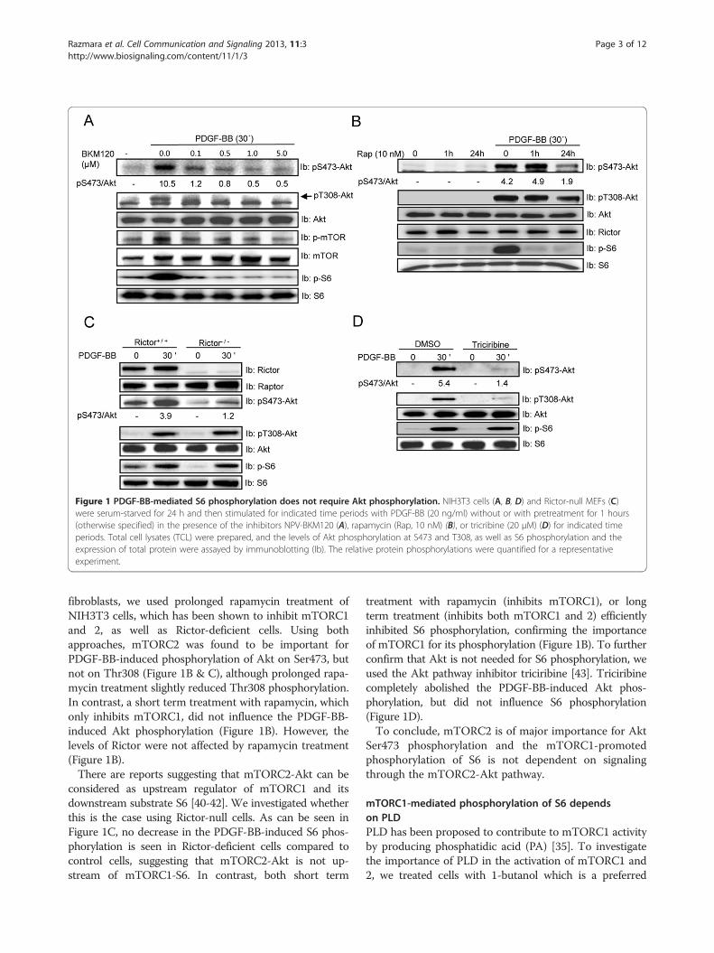
Figure 1 PDGF-BB-mediated S6 phosphorylation does not require Akt phosphorylation. NIH3T3 cells (A, B, D) and Rictor-null MEFs (C)were serum-starved for 24 h and then stimulated for indicated time periods with PDGF-BB (20 ng/ml) without or with pretreatment for 1 hours(otherwise specified) in the presence of the inhibitors NPV-BKM120 (A), rapamycin (Rap, 10 nM) (B), or tricribine (20 μM) (D) for indicated timeperiods. Total cell lysates (TCL) were prepared, and the levels of Akt phosphorylation at S473 and T308, as well as S6 phosphorylation and theexpression of total protein were assayed by immunoblotting (Ib). The relative protein phosphorylations were quantified for a representativeexperiment.
Razmara et al. Cell Communication and Signaling 2013, 11:3 Page 3 of 12http://www.biosignaling.com/content/11/1/3
fibroblasts, we used prolonged rapamycin treatment ofNIH3T3 cells, which has been shown to inhibit mTORC1and 2, as well as Rictor-deficient cells. Using bothapproaches, mTORC2 was found to be important forPDGF-BB-induced phosphorylation of Akt on Ser473, butnot on Thr308 (Figure 1B & C), although prolonged rapa-mycin treatment slightly reduced Thr308 phosphorylation.In contrast, a short term treatment with rapamycin, whichonly inhibits mTORC1, did not influence the PDGF-BB-induced Akt phosphorylation (Figure 1B). However, thelevels of Rictor were not affected by rapamycin treatment(Figure 1B).There are reports suggesting that mTORC2-Akt can be
considered as upstream regulator of mTORC1 and itsdownstream substrate S6 [40-42]. We investigated whetherthis is the case using Rictor-null cells. As can be seen inFigure 1C, no decrease in the PDGF-BB-induced S6 phos-phorylation is seen in Rictor-deficient cells compared tocontrol cells, suggesting that mTORC2-Akt is not up-stream of mTORC1-S6. In contrast, both short term
treatment with rapamycin (inhibits mTORC1), or longterm treatment (inhibits both mTORC1 and 2) efficientlyinhibited S6 phosphorylation, confirming the importanceof mTORC1 for its phosphorylation (Figure 1B). To furtherconfirm that Akt is not needed for S6 phosphorylation, weused the Akt pathway inhibitor triciribine [43]. Triciribinecompletely abolished the PDGF-BB-induced Akt phos-phorylation, but did not influence S6 phosphorylation(Figure 1D).To conclude, mTORC2 is of major importance for Akt
Ser473 phosphorylation and the mTORC1-promotedphosphorylation of S6 is not dependent on signalingthrough the mTORC2-Akt pathway.
mTORC1-mediated phosphorylation of S6 dependson PLDPLD has been proposed to contribute to mTORC1 activityby producing phosphatidic acid (PA) [35]. To investigatethe importance of PLD in the activation of mTORC1 and2, we treated cells with 1-butanol which is a preferred
Razmara et al. Cell Communication and Signaling 2013, 11:3 Page 4 of 12http://www.biosignaling.com/content/11/1/3
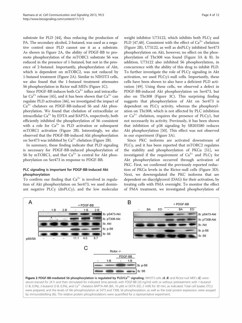
substrate for PLD [44], thus reducing the production ofPA. The secondary alcohol, 2-butanol, was used as a nega-tive control since PLD cannot use it as a substrate.As shown in Figure 2A, the ability of PDGF-BB to pro-mote phosphorylation of the mTORC1 substrate S6 wasreduced in the presence of 1-butanol, but not in the pres-ence of 2-butanol. Importantly, phosphorylation of Akt,which is dependent on mTORC2, was not reduced by1-butanol treatment (Figure 2A). Similar to NIH3T3 cells,we also found that the 1-butanol treatment attenuatesS6 phosphorylation in Rictor null MEFs (Figure 1C).Since PDGF-BB induces both Ca2+ influx and intracellu-
lar Ca2+ release [45], and it has been shown that Ca2+ canregulate PLD activation [46], we investigated the impact ofCa2+ chelators on PDGF-BB-induced S6 and Akt phos-phorylation. We found that chelation of extracellular orintracellular Ca2+ by EDTA and BAPTA, respectively, bothefficiently inhibited the phosphorylation of S6 consistentwith a role for Ca2+ in PLD activation or subsequentmTORC1 activation (Figure 2B). Interestingly, we alsoobserved that the PDGF-BB-induced Akt phosphorylationon Ser473 was inhibited by Ca2+ chelation (Figure 2B).In summary, these finding indicate that PLD signaling
is necessary for PDGF-BB-induced phosphorylation ofS6 by mTORC1, and that Ca2+ is central for Akt phos-phorylation on Ser473 in response to PDGF-BB.
PLC signaling is important for PDGF-BB-induced AktphosphorylationTo confirm our finding that Ca2+ is involved in regula-tion of Akt phosphorylation on Ser473, we used domin-ant negative PLCγ (dnPLCγ), and the low molecular
Figure 2 PDGF-BB-mediated S6 phosphorylation is regulated by PLD/serum-starved for 24 h and then stimulated for indicated time periods with(1-B, 0.3%), 2-butanol (2-B, 0.3%), and Ca2+ chelators BAPTA-AM (BA, 10 μMwere prepared, and the levels of Akt phosphorylation at S473 and T308, S6by immunoblotting (Ib). The relative protein phosphorylations were quanti
weight inhibitor U73122, which inhibits both PLCγ andPLD [47,48]. Consistent with the effect of Ca2+ chelation(Figure 2B), U73122, as well as dnPLCγ inhibited Ser473phosphorylation on Akt, however, no effect on the phos-phorylation of Thr308 was found (Figure 3A & B). Inaddition, U73122 also inhibited S6 phosphorylation, inconcurrence with the ability of this drug to inhibit PLD.To further investigate the role of PLCγ signaling in Aktactivation, we used PLCγ1-null cells. Importantly, thesecells have been shown to also have a deficient PLD acti-vation [49]. Using these cells, we observed a defect inPDGF-BB-induced Akt phosphorylation on Ser473, butalso on Thr308 (Figure 3C). This surprising findingsuggests that phosphorylation of Akt on Ser473 isdependent on PLCγ activity, whereas the phosphoryl-ation on Thr308, which is not affected by PLC inhibitionor Ca2+ chelation, requires the presence of PLCγ1, butnot necessarily its activity. Previously, it has been shownthat inhibition of p38 signaling by SB203580 reducesAkt phosphorylation [50]. This effect was not observedin our experiment (Figure 3A).Since PKC isoforms are activated downstream of
PLCγ, and it has been reported that mTORC2 regulatesthe stability and phosphorylation of PKCα [51], weinvestigated if the requirement of Ca2+ and PLCγ forAkt phosphorylation occurred through activation ofPKC. First, we confirmed the previously reported reduc-tion of PKCα levels in the Rictor-null cells (Figure 3D).Next, we downregulated the PKC isoforms that aredependent on diacylglycerol (DAG) for their activation, bytreating cells with PMA overnight. To monitor the effectof PMA treatment, we investigated phosphorylation of
Ca2+ signaling. NIH3T3 cells (A, B) and Rictor-null MEFs (C) werePDGF-BB (20 ng/ml) with or without pretreatment with 1-butanol
) or EDTA (ED, 2 mM) for 30 min, as indicated. Total cell lysates (TCL)phosphorylation, as well as the total protein expression, were assayedfied for a representative experiment.
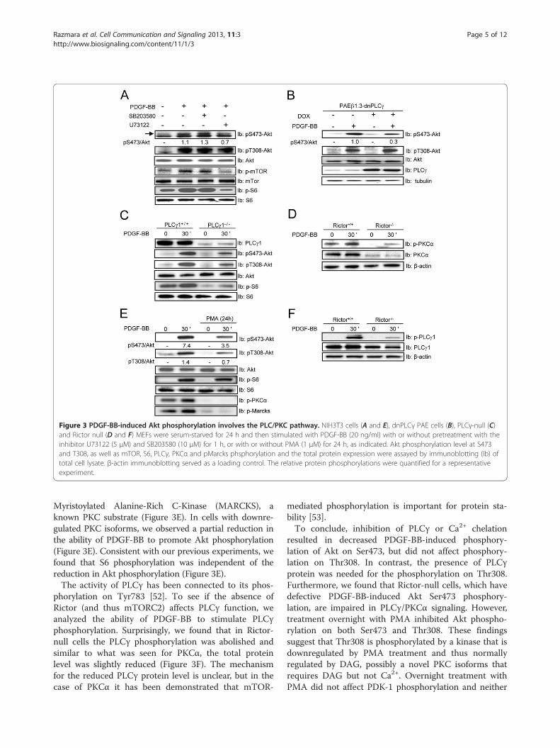
Figure 3 PDGF-BB-induced Akt phosphorylation involves the PLC/PKC pathway. NIH3T3 cells (A and E), dnPLCγ PAE cells (B), PLCγ-null (C)and Rictor null (D and F) MEFs were serum-starved for 24 h and then stimulated with PDGF-BB (20 ng/ml) with or without pretreatment with theinhibitor U73122 (5 μM) and SB203580 (10 μM) for 1 h, or with or without PMA (1 μM) for 24 h, as indicated. Akt phosphorylation level at S473and T308, as well as mTOR, S6, PLCγ, PKCα and pMarcks phsphorylation and the total protein expression were assayed by immunoblotting (Ib) oftotal cell lysate. β-actin immunoblotting served as a loading control. The relative protein phosphorylations were quantified for a representativeexperiment.
Razmara et al. Cell Communication and Signaling 2013, 11:3 Page 5 of 12http://www.biosignaling.com/content/11/1/3
Myristoylated Alanine-Rich C-Kinase (MARCKS), aknown PKC substrate (Figure 3E). In cells with downre-gulated PKC isoforms, we observed a partial reduction inthe ability of PDGF-BB to promote Akt phosphorylation(Figure 3E). Consistent with our previous experiments, wefound that S6 phosphorylation was independent of thereduction in Akt phosphorylation (Figure 3E).The activity of PLCγ has been connected to its phos-
phorylation on Tyr783 [52]. To see if the absence ofRictor (and thus mTORC2) affects PLCγ function, weanalyzed the ability of PDGF-BB to stimulate PLCγphosphorylation. Surprisingly, we found that in Rictor-null cells the PLCγ phosphorylation was abolished andsimilar to what was seen for PKCα, the total proteinlevel was slightly reduced (Figure 3F). The mechanismfor the reduced PLCγ protein level is unclear, but in thecase of PKCα it has been demonstrated that mTOR-
mediated phosphorylation is important for protein sta-bility [53].To conclude, inhibition of PLCγ or Ca2+ chelation
resulted in decreased PDGF-BB-induced phosphory-lation of Akt on Ser473, but did not affect phosphory-lation on Thr308. In contrast, the presence of PLCγprotein was needed for the phosphorylation on Thr308.Furthermore, we found that Rictor-null cells, which havedefective PDGF-BB-induced Akt Ser473 phosphory-lation, are impaired in PLCγ/PKCα signaling. However,treatment overnight with PMA inhibited Akt phospho-rylation on both Ser473 and Thr308. These findingssuggest that Thr308 is phosphorylated by a kinase that isdownregulated by PMA treatment and thus normallyregulated by DAG, possibly a novel PKC isoforms thatrequires DAG but not Ca2+. Overnight treatment withPMA did not affect PDK-1 phosphorylation and neither
Razmara et al. Cell Communication and Signaling 2013, 11:3 Page 6 of 12http://www.biosignaling.com/content/11/1/3
did PDGF-BB treatment (data not shown). In contrast,phosphorylation of Akt on Ser473 is dependent onPLCγ1 activity, Ca2+, DAG and the conventional PKCs.
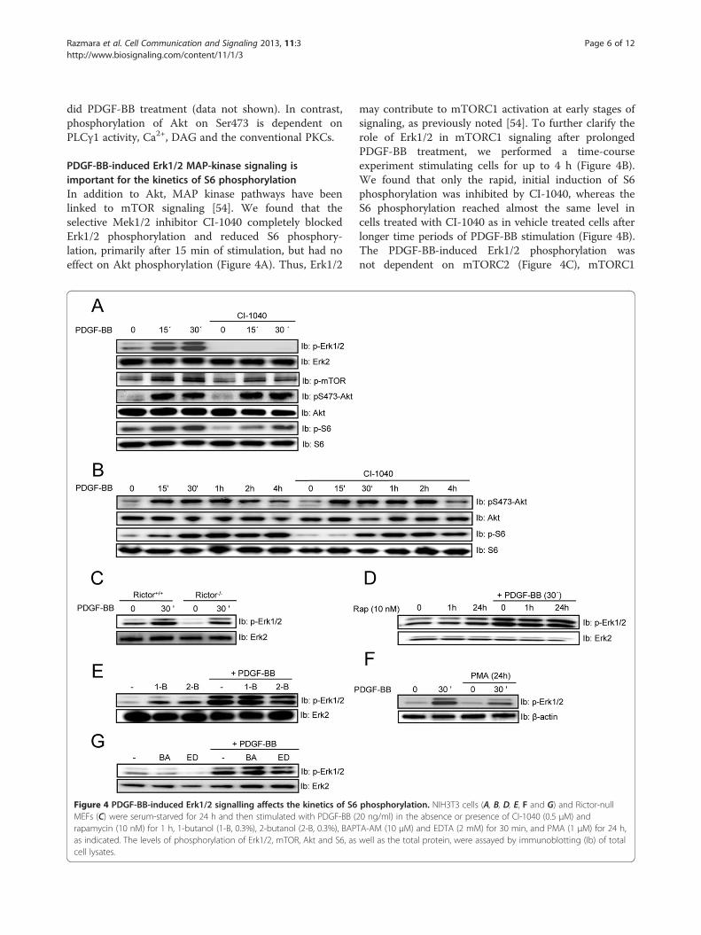
PDGF-BB-induced Erk1/2 MAP-kinase signaling isimportant for the kinetics of S6 phosphorylationIn addition to Akt, MAP kinase pathways have beenlinked to mTOR signaling [54]. We found that theselective Mek1/2 inhibitor CI-1040 completely blockedErk1/2 phosphorylation and reduced S6 phosphory-lation, primarily after 15 min of stimulation, but had noeffect on Akt phosphorylation (Figure 4A). Thus, Erk1/2
Figure 4 PDGF-BB-induced Erk1/2 signalling affects the kinetics of S6MEFs (C) were serum-starved for 24 h and then stimulated with PDGF-BB (2rapamycin (10 nM) for 1 h, 1-butanol (1-B, 0.3%), 2-butanol (2-B, 0.3%), BAPas indicated. The levels of phosphorylation of Erk1/2, mTOR, Akt and S6, ascell lysates.
may contribute to mTORC1 activation at early stages ofsignaling, as previously noted [54]. To further clarify therole of Erk1/2 in mTORC1 signaling after prolongedPDGF-BB treatment, we performed a time-courseexperiment stimulating cells for up to 4 h (Figure 4B).We found that only the rapid, initial induction of S6phosphorylation was inhibited by CI-1040, whereas theS6 phosphorylation reached almost the same level incells treated with CI-1040 as in vehicle treated cells afterlonger time periods of PDGF-BB stimulation (Figure 4B).The PDGF-BB-induced Erk1/2 phosphorylation wasnot dependent on mTORC2 (Figure 4C), mTORC1
phosphorylation. NIH3T3 cells (A, B, D, E, F and G) and Rictor-null0 ng/ml) in the absence or presence of CI-1040 (0.5 μM) andTA-AM (10 μM) and EDTA (2 mM) for 30 min, and PMA (1 μM) for 24 h,well as the total protein, were assayed by immunoblotting (Ib) of total
Razmara et al. Cell Communication and Signaling 2013, 11:3 Page 7 of 12http://www.biosignaling.com/content/11/1/3
(Figure 4D & E), PKCs (Figure 4F), or the presence of Ca2+
(Figure 4G).In summary, PDGF-BB-induced Erk1/2 activity is only
important for the early onset of mTORC1-mediatedphosphorylation of S6. Furthermore, neither mTORC1nor mTORC2 are needed for PDGF-BB-induced Erk1/2activation.
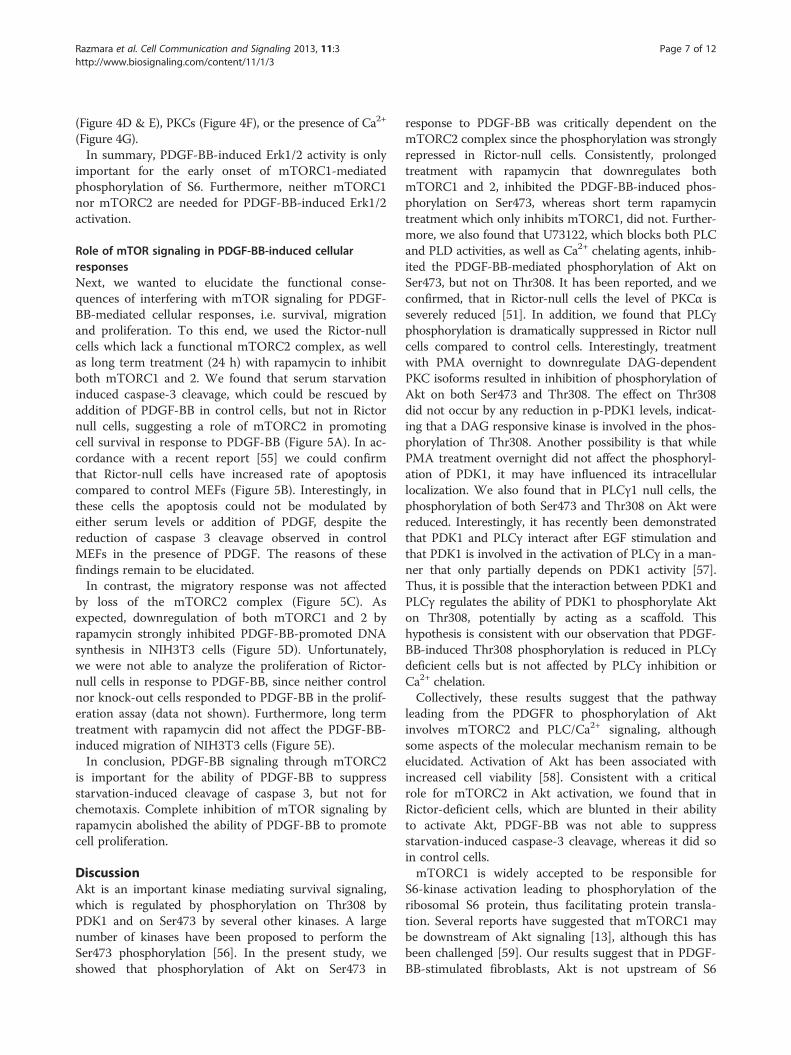
Role of mTOR signaling in PDGF-BB-induced cellularresponsesNext, we wanted to elucidate the functional conse-quences of interfering with mTOR signaling for PDGF-BB-mediated cellular responses, i.e. survival, migrationand proliferation. To this end, we used the Rictor-nullcells which lack a functional mTORC2 complex, as wellas long term treatment (24 h) with rapamycin to inhibitboth mTORC1 and 2. We found that serum starvationinduced caspase-3 cleavage, which could be rescued byaddition of PDGF-BB in control cells, but not in Rictornull cells, suggesting a role of mTORC2 in promotingcell survival in response to PDGF-BB (Figure 5A). In ac-cordance with a recent report [55] we could confirmthat Rictor-null cells have increased rate of apoptosiscompared to control MEFs (Figure 5B). Interestingly, inthese cells the apoptosis could not be modulated byeither serum levels or addition of PDGF, despite thereduction of caspase 3 cleavage observed in controlMEFs in the presence of PDGF. The reasons of thesefindings remain to be elucidated.In contrast, the migratory response was not affected
by loss of the mTORC2 complex (Figure 5C). Asexpected, downregulation of both mTORC1 and 2 byrapamycin strongly inhibited PDGF-BB-promoted DNAsynthesis in NIH3T3 cells (Figure 5D). Unfortunately,we were not able to analyze the proliferation of Rictor-null cells in response to PDGF-BB, since neither controlnor knock-out cells responded to PDGF-BB in the prolif-eration assay (data not shown). Furthermore, long termtreatment with rapamycin did not affect the PDGF-BB-induced migration of NIH3T3 cells (Figure 5E).In conclusion, PDGF-BB signaling through mTORC2
is important for the ability of PDGF-BB to suppressstarvation-induced cleavage of caspase 3, but not forchemotaxis. Complete inhibition of mTOR signaling byrapamycin abolished the ability of PDGF-BB to promotecell proliferation.
DiscussionAkt is an important kinase mediating survival signaling,which is regulated by phosphorylation on Thr308 byPDK1 and on Ser473 by several other kinases. A largenumber of kinases have been proposed to perform theSer473 phosphorylation [56]. In the present study, weshowed that phosphorylation of Akt on Ser473 in
response to PDGF-BB was critically dependent on themTORC2 complex since the phosphorylation was stronglyrepressed in Rictor-null cells. Consistently, prolongedtreatment with rapamycin that downregulates bothmTORC1 and 2, inhibited the PDGF-BB-induced phos-phorylation on Ser473, whereas short term rapamycintreatment which only inhibits mTORC1, did not. Further-more, we also found that U73122, which blocks both PLCand PLD activities, as well as Ca2+ chelating agents, inhib-ited the PDGF-BB-mediated phosphorylation of Akt onSer473, but not on Thr308. It has been reported, and weconfirmed, that in Rictor-null cells the level of PKCα isseverely reduced [51]. In addition, we found that PLCγphosphorylation is dramatically suppressed in Rictor nullcells compared to control cells. Interestingly, treatmentwith PMA overnight to downregulate DAG-dependentPKC isoforms resulted in inhibition of phosphorylation ofAkt on both Ser473 and Thr308. The effect on Thr308did not occur by any reduction in p-PDK1 levels, indicat-ing that a DAG responsive kinase is involved in the phos-phorylation of Thr308. Another possibility is that whilePMA treatment overnight did not affect the phosphoryl-ation of PDK1, it may have influenced its intracellularlocalization. We also found that in PLCγ1 null cells, thephosphorylation of both Ser473 and Thr308 on Akt werereduced. Interestingly, it has recently been demonstratedthat PDK1 and PLCγ interact after EGF stimulation andthat PDK1 is involved in the activation of PLCγ in a man-ner that only partially depends on PDK1 activity [57].Thus, it is possible that the interaction between PDK1 andPLCγ regulates the ability of PDK1 to phosphorylate Akton Thr308, potentially by acting as a scaffold. Thishypothesis is consistent with our observation that PDGF-BB-induced Thr308 phosphorylation is reduced in PLCγdeficient cells but is not affected by PLCγ inhibition orCa2+ chelation.Collectively, these results suggest that the pathway
leading from the PDGFR to phosphorylation of Aktinvolves mTORC2 and PLC/Ca2+ signaling, althoughsome aspects of the molecular mechanism remain to beelucidated. Activation of Akt has been associated withincreased cell viability [58]. Consistent with a criticalrole for mTORC2 in Akt activation, we found that inRictor-deficient cells, which are blunted in their abilityto activate Akt, PDGF-BB was not able to suppressstarvation-induced caspase-3 cleavage, whereas it did soin control cells.mTORC1 is widely accepted to be responsible for
S6-kinase activation leading to phosphorylation of theribosomal S6 protein, thus facilitating protein transla-tion. Several reports have suggested that mTORC1 maybe downstream of Akt signaling [13], although this hasbeen challenged [59]. Our results suggest that in PDGF-BB-stimulated fibroblasts, Akt is not upstream of S6
Figure 5 (See legend on next page.)
Razmara et al. Cell Communication and Signaling 2013, 11:3 Page 8 of 12http://www.biosignaling.com/content/11/1/3
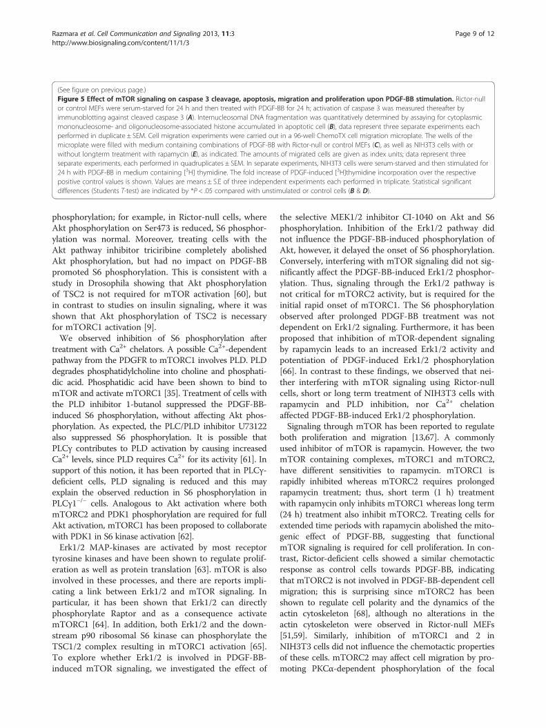
(See figure on previous page.)Figure 5 Effect of mTOR signaling on caspase 3 cleavage, apoptosis, migration and proliferation upon PDGF-BB stimulation. Rictor-nullor control MEFs were serum-starved for 24 h and then treated with PDGF-BB for 24 h; activation of caspase 3 was measured thereafter byimmunoblotting against cleaved caspase 3 (A). Internucleosomal DNA fragmentation was quantitatively determined by assaying for cytoplasmicmononucleosome- and oligonucleosome-associated histone accumulated in apoptotic cell (B), data represent three separate experiments eachperformed in duplicate ± SEM. Cell migration experiments were carried out in a 96-well ChemoTX cell migration microplate. The wells of themicroplate were filled with medium containing combinations of PDGF-BB with Rictor-null or control MEFs (C), as well as NIH3T3 cells with orwithout longterm treatment with rapamycin (E), as indicated. The amounts of migrated cells are given as index units; data represent threeseparate experiments, each performed in quadruplicates ± SEM. In separate experiments, NIH3T3 cells were serum-starved and then stimulated for24 h with PDGF-BB in medium containing [3H] thymidine. The fold increase of PDGF-induced [3H]thymidine incorporation over the respectivepositive control values is shown. Values are means ± S.E of three independent experiments each performed in triplicate. Statistical significantdifferences (Students T-test) are indicated by *P < .05 compared with unstimulated or control cells (B & D).
Razmara et al. Cell Communication and Signaling 2013, 11:3 Page 9 of 12http://www.biosignaling.com/content/11/1/3
phosphorylation; for example, in Rictor-null cells, whereAkt phosphorylation on Ser473 is reduced, S6 phosphor-ylation was normal. Moreover, treating cells with theAkt pathway inhibitor triciribine completely abolishedAkt phosphorylation, but had no impact on PDGF-BBpromoted S6 phosphorylation. This is consistent with astudy in Drosophila showing that Akt phosphorylationof TSC2 is not required for mTOR activation [60], butin contrast to studies on insulin signaling, where it wasshown that Akt phosphorylation of TSC2 is necessaryfor mTORC1 activation [9].We observed inhibition of S6 phosphorylation after
treatment with Ca2+ chelators. A possible Ca2+-dependentpathway from the PDGFR to mTORC1 involves PLD. PLDdegrades phosphatidylcholine into choline and phosphati-dic acid. Phosphatidic acid have been shown to bind tomTOR and activate mTORC1 [35]. Treatment of cells withthe PLD inhibitor 1-butanol suppressed the PDGF-BB-induced S6 phosphorylation, without affecting Akt phos-phorylation. As expected, the PLC/PLD inhibitor U73122also suppressed S6 phosphorylation. It is possible thatPLCγ contributes to PLD activation by causing increasedCa2+ levels, since PLD requires Ca2+ for its activity [61]. Insupport of this notion, it has been reported that in PLCγ-deficient cells, PLD signaling is reduced and this mayexplain the observed reduction in S6 phosphorylation inPLCγ1−/− cells. Analogous to Akt activation where bothmTORC2 and PDK1 phosphorylation are required for fullAkt activation, mTORC1 has been proposed to collaboratewith PDK1 in S6 kinase activation [62].Erk1/2 MAP-kinases are activated by most receptor
tyrosine kinases and have been shown to regulate prolif-eration as well as protein translation [63]. mTOR is alsoinvolved in these processes, and there are reports impli-cating a link between Erk1/2 and mTOR signaling. Inparticular, it has been shown that Erk1/2 can directlyphosphorylate Raptor and as a consequence activatemTORC1 [64]. In addition, both Erk1/2 and the down-stream p90 ribosomal S6 kinase can phosphorylate theTSC1/2 complex resulting in mTORC1 activation [65].To explore whether Erk1/2 is involved in PDGF-BB-induced mTOR signaling, we investigated the effect of
the selective MEK1/2 inhibitor CI-1040 on Akt and S6phosphorylation. Inhibition of the Erk1/2 pathway didnot influence the PDGF-BB-induced phosphorylation ofAkt, however, it delayed the onset of S6 phosphorylation.Conversely, interfering with mTOR signaling did not sig-nificantly affect the PDGF-BB-induced Erk1/2 phosphor-ylation. Thus, signaling through the Erk1/2 pathway isnot critical for mTORC2 activity, but is required for theinitial rapid onset of mTORC1. The S6 phosphorylationobserved after prolonged PDGF-BB treatment was notdependent on Erk1/2 signaling. Furthermore, it has beenproposed that inhibition of mTOR-dependent signalingby rapamycin leads to an increased Erk1/2 activity andpotentiation of PDGF-induced Erk1/2 phosphorylation[66]. In contrast to these findings, we observed that nei-ther interfering with mTOR signaling using Rictor-nullcells, short or long term treatment of NIH3T3 cells withrapamycin and PLD inhibition, nor Ca2+ chelationaffected PDGF-BB-induced Erk1/2 phosphorylation.Signaling through mTOR has been reported to regulate
both proliferation and migration [13,67]. A commonlyused inhibitor of mTOR is rapamycin. However, the twomTOR containing complexes, mTORC1 and mTORC2,have different sensitivities to rapamycin. mTORC1 israpidly inhibited whereas mTORC2 requires prolongedrapamycin treatment; thus, short term (1 h) treatmentwith rapamycin only inhibits mTORC1 whereas long term(24 h) treatment also inhibit mTORC2. Treating cells forextended time periods with rapamycin abolished the mito-genic effect of PDGF-BB, suggesting that functionalmTOR signaling is required for cell proliferation. In con-trast, Rictor-deficient cells showed a similar chemotacticresponse as control cells towards PDGF-BB, indicatingthat mTORC2 is not involved in PDGF-BB-dependent cellmigration; this is surprising since mTORC2 has beenshown to regulate cell polarity and the dynamics of theactin cytoskeleton [68], although no alterations in theactin cytoskeleton were observed in Rictor-null MEFs[51,59]. Similarly, inhibition of mTORC1 and 2 inNIH3T3 cells did not influence the chemotactic propertiesof these cells. mTORC2 may affect cell migration by pro-moting PKCα-dependent phosphorylation of the focal
Razmara et al. Cell Communication and Signaling 2013, 11:3 Page 10 of 12http://www.biosignaling.com/content/11/1/3
adhesion component paxillin [20]. However, it has previ-ously been found that PDGF-BB can promote paxillinphosphorylation through the JNK MAP kinase pathway[69], and this may relieve the absolute requirement ofmTORC2 in PDGF-BB-mediated fibroblast migration.
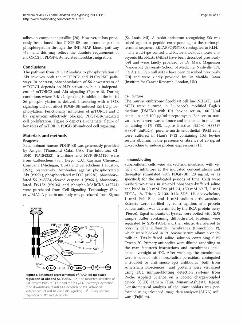
ConclusionsThe pathway from PDGFR leading to phosphorylation ofAkt involves both the mTORC2 and PLCγ/PKC path-ways. In contrast, phosphorylation of S6 downstream ofmTORC1 depends on PLD activation, but is independ-ent of mTORC2 and Akt signaling (Figure 6). Duringconditions where Erk1/2 signaling is inhibited, the initialS6 phosphorylation is delayed. Interfering with mTORsignaling did not affect PDGF-BB-induced Erk1/2 phos-phorylation. Functionally, inhibition of mTORC1 and 2by rapamycin effectively blocked PDGF-BB-mediatedcell proliferation. Figure 6 depicts a schematic figure ofkey roles of mTOR in PDGF-BB-induced cell signaling.
Materials and methodsReagentsRecombinant human PDGF-BB was generously providedby Amgen (Thousand Oaks, CA). The inhibitors CI-1040 (PD184352), triciribine and NVP-BKM120 werefrom Calbiochem (San Diego, CA), Cayman ChemicalCompany (Michigan, USA) and Selleckchem (Houston,USA), respectively. Antibodies against phosphorylatedAkt (#9271), phosphorylated mTOR (#5536), phosphory-lated S6 (#4858), cleaved caspase 3 (#9661), phosphory-lated Erk1/2 (#9106) and phospho-MARCKS (#2741)were purchased from Cell Signaling Technology (Bev-erly, MA). A β-actin antibody was purchased from Sigma
Figure 6 Schematic representation of PDGF-BB-mediatedregulation of Akt and S6. Initially, PDGF-BB-mediated activation ofAkt involves both mTORC2 and the PLCγ/PKC pathways. Activationof S6 downstream of mTORC1 depends on PLD activation,independent of mTORC2 and Akt signaling. Ca2+ is required forregulation of Akt and S6 activity.
(St. Louis, MI). A rabbit antiserum recognizing Erk wasraised against a peptide corresponding to the carboxyl-terminal sequence EETARFQPGYRS conjugated to KLH.The wild-type control and Rictor-knockout mouse em-
bryonic fibroblasts (MEFs) have been described previously[59] and were kindly provided by Dr Mark Magnuson(Vanderbilt University School of Medicine, Nashville, TN,U.S.A.). PLCγ1-null MEFs have been described previously[70] and were kindly provided by Dr Matilda Katan(Institute for Cancer Research, London, UK).
Cell cultureThe murine embryonic fibroblast cell line NIH3T3, andMEFs were cultured in Dulbecco's modified Eagle'smedium (DMEM) with 10% bovine serum, 100 U/mlpenicillin and 100 μg/ml streptomycin. For serum-star-vation, cells were washed once and incubated in mediumcontaining 0.1% FBS. Lipase inactive PLC-γ1 H335F/H380F (dnPLCγ), porcine aortic endothelial (PAE) cellswere cultured in Ham’s F-12 containing 10% bovineserum albumin, in the presence or absence of 20 ng/mldoxycycline to induce protein expression [71].
ImmunoblottingSubconfluent cells were starved and incubated with ve-hicle or inhibitors at the indicated concentrations andthereafter stimulated with PDGF-BB (20 ng/ml, or asspecified) for the indicated periods of time. Cells werewashed two times in ice-cold phosphate-buffered salineand lysed in 20 mM Tris pH 7.4, 150 mM NaCl, 5 mMEDTA, 1% Triton X-100, 0.1% SDS, 1% deoxycholate,1 mM Pefa Bloc and 1 mM sodium orthovanadate.Extracts were clarified by centrifugation, and proteinconcentration was determined by the BCA protein assay(Pierce). Equal amounts of lysates were boiled with SDSsample buffer containing dithiothreitol. Proteins wereseparated by SDS-PAGE and then electro-transferred topolyvinylidene difluoride membranes (Immobilon P),which were blocked in 5% bovine serum albumin or 5%milk in Tris-buffered saline solution containing 0.1%Tween-20. Primary antibodies were diluted according tothe manufacturer’s instructions and membranes incu-bated overnight at 4°C. After washing, the membraneswere incubated with horseradish peroxidase-conjugatedanti-rabbit or anti-mouse IgG antibodies (both fromAmersham Biosciences), and proteins were visualizedusing ECL immunoblotting detection systems fromRoche Applied Science on a cooled charge-coupleddevice (CCD) camera (Fuji, Minami-Ashigata, Japan).Densitometrical analysis of the immunoblots was per-formed using advanced image data analyzer (AIDA) soft-ware (Fujifilm).
Razmara et al. Cell Communication and Signaling 2013, 11:3 Page 11 of 12http://www.biosignaling.com/content/11/1/3
Apoptosis assayControl and Rictor-null MEFs were starved for 24 h,then the extent of apoptosis was determined by quantifi-cation of nucleosomes released into the cytoplasmausing the Cell Death Detection ELISA Plus kit (RocheApplied Science) according to the manufacturer's direc-tions. In the separate experiments the level of caspase-3-cleaved fragments was analyzed by immunoblotting.
[3H]thymidine incorporation assayFor thymidine incorporation assay subconfluent cellcultures were serum-starved in 24-well plates and thenincubated for 24 h in the presence or absence of rapa-mycin with PDGF-BB in DMEM containing [3H]thymi-dine (0.1 μCi/ml). Incorporation of 3H-radioactivity intoacid-insoluble material was measured by a scintillationcounter. The obtained count per minute values in tri-plicate was normalized against the positive control ofcultures incubated in 10% bovine serum for eachexperiment.
Cell migration assaysCell migration was determined as previously described [69].In brief, 96-well ChemoTX (Neuroprobe, Gaithersburg,MD) cell migration microplate filters were coated with50 μg/ml fibronectin (BD Biochemicals, Erembodegem,Belgium) for 1 h at room temperature. Control and Rictornull-MEFs, or NIH3T3 cells treated with or without rapa-mycin, were serum-starved overnight and then trypsinizedinto single cells. The wells of the ChemoTX microplatewere filled with DMEM containing the indicated PDGF-BBconcentrations. The filters were placed in the wells and50,000 cells were added on top of each filter. The chamberwas incubated for 4 h at 37°C, 5% CO2. Cells adhering tothe bottom of the filter were fixed by a 3-min incubationin 96% ethanol. The adherent cells were stained withGiemsa (Sigma)-and the migration indices were assessedby scanning the filter in a CCD camera (Fuji). Quan-tifications were performed using Aida Image Analyzersoftware. All experiments were performed in quadru-plicate, and single representative data of three separateexperiments ± SD are shown.
AbbreviationsErk: Extracellular regulated kinase; MAPK: Mitogen activated protein kinase;MEF: Mouse embryo fibroblast; mTOR: Mammalian target of rapamycin;PDGF: Platelet-derived growth factor; PDGFR: PDGF receptor;PLCγ1: Phospholipase C gamma 1; PI3K: Phosphatidylinositol 3-kinase.
Competing interestsThe authors declare that they have no competing interests.
Authors’ contributionsMR, planned and performed experiments, analyzed the data and contributedto manuscript preparation; CHH, planned experiments, analyzed data andcontributed to manuscript preparation; JL, planned and performedexperiments, analyzed the data and contributed to manuscript preparation.All authors read and approved the final manuscript.
AcknowledgementsThis work was supported by Ludwig Institute for Cancer Research, theSwedish ResearchCouncil (K2011-67X-21859-01-6) and the Swedish Cancer Society(CAN 2009/657).
Received: 25 September 2012 Accepted: 9 January 2013Published: 11 January 2013
References1. Andrae J, Gallini R, Betsholtz C: Role of platelet-derived growth factors in
physiology and medicine. Genes Dev 2008, 22:1276–1312.2. Heldin CH, Westermark B: Mechanism of action and in vivo role of
platelet-derived growth factor. Physiol Rev 1999, 79:1283–1316.3. Fredriksson L, Li H, Eriksson U: The PDGF family: four gene products form
five dimeric isoforms. Cytokine Growth Factor Rev 2004, 15:197–204.4. Sarbassov DD, Ali SM, Sabatini DM: Growing roles for the mTOR pathway.
Curr Opin Cell Biol 2005, 17:596–603.5. Bjornsti MA, Houghton PJ: The TOR pathway: a target for cancer therapy.
Nat Rev Cancer 2004, 4:335–348.6. Sawyers CL: Will mTOR inhibitors make it as cancer drugs?. Cancer Cell
2003, 4:343–348.7. Bhaskar PT, Hay N: The two TORCs and Akt. Dev Cell 2007, 12:487–502.8. Inoki K, Li Y, Zhu T, Wu J, Guan KL: TSC2 is phosphorylated and inhibited
by Akt and suppresses mTOR signalling. Nat Cell Biol 2002, 4:648–657.9. Manning BD, Tee AR, Logsdon MN, Blenis J, Cantley LC: Identification of
the tuberous sclerosis complex-2 tumor suppressor gene producttuberin as a target of the phosphoinositide 3-kinase/akt pathway.Mol Cell 2002, 10:151–162.
10. Mamane Y, Petroulakis E, LeBacquer O, Sonenberg N: mTOR, translationinitiation and cancer. Oncogene 2006, 25:6416–6422.
11. Inoki K, Zhu T, Guan KL: TSC2 mediates cellular energy response tocontrol cell growth and survival. Cell 2003, 115:577–590.
12. Kim DH, Sarbassov DD, Ali SM, King JE, Latek RR, Erdjument-Bromage H,Tempst P, Sabatini DM: mTOR interacts with raptor to form a nutrient-sensitive complex that signals to the cell growth machinery. Cell 2002,110:163–175.
13. Hay N, Sonenberg N: Upstream and downstream of mTOR. Genes Dev2004, 18:1926–1945.
14. Frias MA, Thoreen CC, Jaffe JD, Schroder W, Sculley T, Carr SA, Sabatini DM:mSin1 is necessary for Akt/PKB phosphorylation, and its isoforms definethree distinct mTORC2s. Curr Biol 2006, 16:1865–1870.
15. Pearce LR, Huang X, Boudeau J, Pawlowski R, Wullschleger S, Deak M, IbrahimAF, Gourlay R, Magnuson MA, Alessi DR: Identification of Protor as a novelRictor-binding component of mTOR complex-2. Biochem J 2007, 405:513–522.
16. Martin J, Masri J, Bernath A, Nishimura RN, Gera J: Hsp70 associates withRictor and is required for mTORC2 formation and activity. BiochemBiophys Res Commun 2008, 372:578–583.
17. Populo H, Lopes JM, Soares P: The mTOR Signalling Pathway in HumanCancer. Int J Mol Sci 2012, 13:1886–1918.
18. Dalle Pezze P, Sonntag AG, Thien A, Prentzell MT, Godel M, Fischer S,Neumann-Haefelin E, Huber TB, Baumeister R, Shanley DP, Thedieck K:A dynamic network model of mTOR signaling reveals TSC-independentmTORC2 regulation. Sci Signal 2012, 5:ra25.
19. Sarbassov DD, Guertin DA, Ali SM, Sabatini DM: Phosphorylation and regulationof Akt/PKB by the rictor-mTOR complex. Science 2005, 307:1098–1101.
20. Sarbassov DD, Ali SM, Kim DH, Guertin DA, Latek RR, Erdjument-Bromage H,Tempst P, Sabatini DM: Rictor, a novel binding partner of mTOR, defines arapamycin-insensitive and raptor-independent pathway that regulatesthe cytoskeleton. Curr Biol 2004, 14:1296–1302.
21. Alessi DR, James SR, Downes CP, Holmes AB, Gaffney PR, Reese CB, CohenP: Characterization of a 3-phosphoinositide-dependent protein kinasewhich phosphorylates and activates protein kinase Balpha. Curr Biol 1997,7:261–269.
22. Stokoe D, Stephens LR, Copeland T, Gaffney PR, Reese CB, Painter GF, Holmes AB,McCormick F, Hawkins PT: Dual role of phosphatidylinositol-3,4,5-trisphosphatein the activation of protein kinase B. Science 1997, 277:567–570.
23. Feng J, Park J, Cron P, Hess D, Hemmings BA: Identification of a PKB/Akthydrophobic motif Ser-473 kinase as DNA-dependent protein kinase.J Biol Chem 2004, 279:41189–41196.
Razmara et al. Cell Communication and Signaling 2013, 11:3 Page 12 of 12http://www.biosignaling.com/content/11/1/3
24. Bozulic L, Surucu B, Hynx D, Hemmings BA: PKBalpha/Akt1 actsdownstream of DNA-PK in the DNA double-strand break response andpromotes survival. Mol Cell 2008, 30:203–213.
25. Persad S, Attwell S, Gray V, Mawji N, Deng JT, Leung D, Yan J, Sanghera J,Walsh MP, Dedhar S: Regulation of protein kinase B/Akt-serine 473phosphorylation by integrin-linked kinase: critical roles for kinase activityand amino acids arginine 211 and serine 343. J Biol Chem 2001,276:27462–27469.
26. Greer EL, Brunet A: FOXO transcription factors at the interface betweenlongevity and tumor suppression. Oncogene 2005, 24:7410–7425.
27. Datta SR, Dudek H, Tao X, Masters S, Fu H, Gotoh Y, Greenberg ME:Akt phosphorylation of BAD couples survival signals to the cell-intrinsicdeath machinery. Cell 1997, 91:231–241.
28. Cardone MH, Roy N, Stennicke HR, Salvesen GS, Franke TF, Stanbridge E,Frisch S, Reed JC: Regulation of cell death protease caspase-9 byphosphorylation. Science 1998, 282:1318–1321.
29. Rebecchi MJ, Pentyala SN: Structure, function, and control ofphosphoinositide-specific phospholipase C. Physiol Rev 2000, 80:1291–1335.
30. Mellor H, Parker PJ: The extended protein kinase C superfamily. Biochem J1998, 332(Pt 2):281–292.
31. Nishizuka Y: Protein kinase C and lipid signaling for sustained cellularresponses. FASEB J 1995, 9:484–496.
32. Sierke SL, Koland JG: SH2 domain proteins as high-affinity receptortyrosine kinase substrates. Biochemistry 1993, 32:10102–10108.
33. Heldin CH, Ostman A, Ronnstrand L: Signal transduction via platelet-derivedgrowth factor receptors. Biochim Biophys Acta 1998, 1378:F79–F113.
34. Exton JH: Regulation of phospholipase D. Biochim Biophys Acta 1999,1439:121–133.
35. Fang Y, Vilella-Bach M, Bachmann R, Flanigan A, Chen J: Phosphatidic acid-mediated mitogenic activation of mTOR signaling. Science 2001,294:1942–1945.
36. Vorland M, Thorsen VA, Holmsen H: Phospholipase D in platelets andother cells. Platelets 2008, 19:582–594.
37. Min DS, Kim EG, Exton JH: Involvement of tyrosine phosphorylation andprotein kinase C in the activation of phospholipase D by H2O2 in Swiss3T3 fibroblasts. J Biol Chem 1998, 273:29986–29994.
38. Maira SM, Pecchi S, Huang A, Burger M, Knapp M, Sterker D, Schnell C,Guthy D, Nagel T, Wiesmann M, et al: Identification and characterization ofNVP-BKM120, an orally available pan-class I PI3-kinase inhibitor. MolCancer Ther 2011, 11:317–328.
39. Sarbassov DD, Ali SM, Sengupta S, Sheen JH, Hsu PP, Bagley AF, MarkhardAL, Sabatini DM: Prolonged rapamycin treatment inhibits mTORC2assembly and Akt/PKB. Mol Cell 2006, 22:159–168.
40. Gingras AC, Kennedy SG, O'Leary MA, Sonenberg N, Hay N: 4E-BP1, arepressor of mRNA translation, is phosphorylated and inactivated by theAkt(PKB) signaling pathway. Genes Dev 1998, 12:502–513.
41. Lizcano JM, Alrubaie S, Kieloch A, Deak M, Leevers SJ, Alessi DR: Insulin-inducedDrosophila S6 kinase activation requires phosphoinositide 3-kinase andprotein kinase B. Biochem J 2003, 374:297–306.
42. Miron M, Lasko P, Sonenberg N: Signaling from Akt to FRAP/TOR targets both4E-BP and S6K in Drosophila melanogaster. Mol Cell Biol 2003, 23:9117–9126.
43. Yang L, Dan HC, Sun M, Liu Q, Sun XM, Feldman RI, Hamilton AD, PolokoffM, Nicosia SV, Herlyn M, et al: Akt/protein kinase B signaling inhibitor-2,a selective small molecule inhibitor of Akt signaling with antitumoractivity in cancer cells overexpressing Akt. Cancer Res 2004, 64:4394–4399.
44. Morris AJ, Frohman MA, Engebrecht J: Measurement of phospholipase Dactivity. Anal Biochem 1997, 252:1–9.
45. Clunn GF, Lymn JS, Schachter M, Hughes AD: Differential effects oflovastatin on mitogen induced calcium influx in human culturedvascular smooth muscle cells. Br J Pharmacol 1997, 121:1789–1795.
46. Huang R, Kucera GL, Rittenhouse SE: Elevated cytosolic Ca2+ activatesphospholipase D in human platelets. J Biol Chem 1991, 266:1652–1655.
47. Hu T, Liu Z, Shen X: Roles of phospholipase D in phorbol myristate acetate-stimulated neutrophil respiratory burst. J Cell Mol Med 2011, 15:647–653.
48. Burgdorf C, Schafer U, Richardt G, Kurz T: U73122, an aminosteroidphospholipase C inhibitor, is a potent inhibitor of cardiac phospholipase Dby a PIP2-dependent mechanism. J Cardiovasc Pharmacol 2010, 55:555–559.
49. Hess JA, Ji QS, Carpenter G, Exton JH: Analysis of platelet-derived growthfactor-induced phospholipase D activation in mouse embryo fibroblastslacking phospholipase C-gamma1. J Biol Chem 1998, 273:20517–20524.
50. Rane MJ, Coxon PY, Powell DW, Webster R, Klein JB, Pierce W, Ping P,McLeish KR: p38 Kinase-dependent MAPKAPK-2 activation functions as3-phosphoinositide-dependent kinase-2 for Akt in human neutrophils.J Biol Chem 2001, 276:3517–3523.
51. Guertin DA, Stevens DM, Thoreen CC, Burds AA, Kalaany NY, Moffat J, BrownM, Fitzgerald KJ, Sabatini DM: Ablation in mice of the mTORC componentsraptor, rictor, or mLST8 reveals that mTORC2 is required for signaling toAkt-FOXO and PKCalpha, but not S6K1. Dev Cell 2006, 11:859–871.
52. Kim HK, Kim JW, Zilberstein A, Margolis B, Kim JG, Schlessinger J, Rhee SG:PDGF stimulation of inositol phospholipid hydrolysis requires PLC-gamma1 phosphorylation on tyrosine residues 783 and 1254. Cell 1991, 65:435–441.
53. Ikenoue T, Inoki K, Yang Q, Zhou X, Guan KL: Essential function of TORC2in PKC and Akt turn motif phosphorylation, maturation and signalling.EMBO J 2008, 27:1919–1931.
54. Chiang GG, Abraham RT: Targeting the mTOR signaling network incancer. Trends Mol Med 2007, 13:433–442.
55. Riaz A, Zeller KS, Johansson S: Receptor-specific mechanisms regulatephosphorylation of AKT at Ser473: role of RICTOR in beta1 integrin-mediated cell survival. PLoS One 2012, 7:e32081.
56. Dong LQ, Liu F: PDK2: the missing piece in the receptor tyrosine kinasesignaling pathway puzzle. Am J Physiol Endocrinol Metab 2005, 289:E187–E196.
57. Raimondi C, Chikh A, Wheeler AP, Maffucci T, Falasca M: A novel regulatorymechanism links PLCγ1 to PDK1. J Cell Sci 2012, 125:3153–3163.
58. Brazil DP, Yang ZZ, Hemmings BA: Advances in protein kinase Bsignalling: AKTion on multiple fronts. Trends Biochem Sci 2004, 29:233–242.
59. Shiota C, Woo JT, Lindner J, Shelton KD, Magnuson MA: Multiallelicdisruption of the rictor gene in mice reveals that mTOR complex 2 isessential for fetal growth and viability. Dev Cell 2006, 11:583–589.
60. Dong J, Pan D: Tsc2 is not a critical target of Akt during normalDrosophila development. Genes Dev 2004, 18:2479–2484.
61. El Kirat K, Besson F, Prigent AF, Chauvet JP, Roux B: Role of calcium andmembrane organization on phospholipase D localization and activity.Competition between a soluble and insoluble substrate. J Biol Chem2002, 277:21231–21236.
62. Pullen N, Dennis PB, Andjelkovic M, Dufner A, Kozma SC, Hemmings BA,Thomas G: Phosphorylation and activation of p70s6k by PDK1. Science1998, 279:707–710.
63. Roux PP, Blenis J: ERK and p38 MAPK-activated protein kinases: a familyof protein kinases with diverse biological functions. Microbiol Mol Biol Rev2004, 68:320–344.
64. Carriere A, Romeo Y, Acosta-Jaquez HA, Moreau J, Bonneil E, Thibault P, FingarDC, Roux PP: ERK1/2 phosphorylate Raptor to promote Ras-dependentactivation of mTOR complex 1 (mTORC1). J Biol Chem 2010, 286:567–577.
65. Magnuson B, Ekim B, Fingar DC: Regulation and function of ribosomal proteinS6 kinase (S6K) within mTOR signalling networks. Biochem J 2012, 441:1–21.
66. Albert L, Karsy M, Murali R, Jhanwar-Uniyal M: Inhibition of mTOR Activatesthe MAPK Pathway in Glioblastoma Multiforme. Cancer GenomicsProteomics 2009, 6:255–261.
67. Liu L, Parent CA: Review series: TOR kinase complexes and cell migration.J Cell Biol 2011, 194:815–824.
68. Liu L, Das S, Losert W, Parent CA: mTORC2 regulates neutrophilchemotaxis in a cAMP- and RhoA-dependent fashion. Dev Cell 2010,19:845–857.
69. Amagasaki K, Kaneto H, Heldin CH, Lennartsson J: c-Jun N-terminal kinaseis necessary for platelet-derived growth factor-mediated chemotaxis inprimary fibroblasts. J Biol Chem 2006, 281:22173–22179.
70. Ji QS, Ermini S, Baulida J, Sun FL, Carpenter G: Epidermal growth factorsignaling and mitogenesis in Plcg1 null mouse embryonic fibroblasts.Mol Biol Cell 1998, 9:749–757.
71. Ronnstrand L, Siegbahn A, Rorsman C, Johnell M, Hansen K, Heldin CH:Overactivation of phospholipase C-gamma1 renders platelet-derivedgrowth factor beta-receptor-expressing cells independent of thephosphatidylinositol 3-kinase pathway for chemotaxis. J Biol Chem 1999,274:22089–22094.
doi:10.1186/1478-811X-11-3Cite this article as: Razmara et al.: Platelet-derived growth factor-induced Akt phosphorylation requires mTOR/Rictor and phospholipaseC-γ1, whereas S6 phosphorylation depends on mTOR/Raptor andphospholipase D. Cell Communication and Signaling 2013 11:3.