Anti-Myeloma Activity of Akt Inhibition Is Linked to the Activation Status of PI3K/Akt and MEK/ERK...
11
Anti-Myeloma Activity of Akt Inhibition Is Linked to the Activation Status of PI3K/Akt and MEK/ERK Pathway Vijay Ramakrishnan, Teresa Kimlinger, Jessica Haug, Utkarsh Painuly, Linda Wellik, Timothy Halling, S. Vincent Rajkumar, Shaji Kumar* The Divisions of Hematology, Mayo Clinic, Rochester, Minnesota, United States of America Abstract The PI3K/Akt/mTOR signal transduction pathway plays a central role in multiple myeloma (MM) disease progression and development of therapeutic resistance. mTORC1 inhibitors have shown limited efficacy in the clinic, largely attributed to the reactivation of Akt due to rapamycin induced mTORC2 activity. Here, we present promising anti-myeloma activity of MK- 2206, a novel allosteric pan-Akt inhibitor, in MM cell lines and patient cells. MK-2206 was able to induce cytotoxicity and inhibit proliferation in all MM cell lines tested, albeit with significant heterogeneity that was highly dependent on basal pAkt levels. MK-2206 was able to inhibit proliferation of MM cells even when cultured with marrow stromal cells or tumor promoting cytokines. The induction of cytotoxicity was due to apoptosis, which at least partially was mediated by caspases. MK-2206 inhibited pAkt and its down-stream targets and up-regulated pErk in MM cells. Using MK-2206 in combination with rapamycin (mTORC1 inhibitor), LY294002 (PI3K inhibitor), or U0126 (MEK1/2 inhibitor), we show that Erk- mediated downstream activation of PI3K/Akt pathway results in resistance to Akt inhibition. These provide the basis for clinical evaluation of MK-2206 alone or in combination in MM and potential use of baseline pAkt and pErk as biomarkers for patient selection. Citation: Ramakrishnan V, Kimlinger T, Haug J, Painuly U, Wellik L, et al. (2012) Anti-Myeloma Activity of Akt Inhibition Is Linked to the Activation Status of PI3K/ Akt and MEK/ERK Pathway. PLoS ONE 7(11): e50005. doi:10.1371/journal.pone.0050005 Editor: Rakesh K. Srivastava, The University of Kansas Medical Center, United States of America Received July 10, 2012; Accepted October 15, 2012; Published November 21, 2012 Copyright: ß 2012 Ramakrishnan et al. This is an open-access article distributed under the terms of the Creative Commons Attribution License, which permits unrestricted use, distribution, and reproduction in any medium, provided the original author and source are credited. Funding: This study was supported in part by Hematological Malignancies Program (Mayo Clinic Cancer Center) and CA90628 (SK) from National Cancer Institute. SK received research support from Celgene, Millennium, Novartis, Merck, Cephalon, Genzyme and Bayer. MK-2206 was provided by Merck. & Co., Inc. (Whitehouse Station, New Jersey). The funders had no role in study design, data collection and analysis, decision to publish, or preparation of the manuscript. Competing Interests: SK received research support from Celgene, Millennium, Novartis, Merck, Cephalon, Genzyme and Bayer. SK is on the advisory board of Merck. MK-2206 was provided by Merck. & Co., Inc. (Whitehouse Station, NJ). There are no patents, products in development or marketed products to declare. This does not alter the authors’ adherence to all the PLOS ONE policies on sharing data and materials. * E-mail: [email protected] Introduction Multiple myeloma (MM) is an incurable plasma cell neoplasm that has seen considerable improvement in patient survival in the last decade due to the introduction of several effective therapies [1]. However, the responders eventually relapse and become refractory to current therapies [1]. Novel drugs based on better understanding of the disease biology are urgently required to overcome resistance and improve patient outcomes in MM [2]. Phosphotidylinositol-3-kinase (PI3K) represents a family of serine threonine kinases that initiate a complex signaling cascade in response to the binding of extracellular cytokines to their receptors expressed on cellular membranes [3]. When activated, the PI3Ks phosphorylate phosphotidylinositol-4,5-bisphosphate (PIP 2 ) to PIP 3 . PIP 3 binds to the PH domain of Akt, which causes a conformational change in Akt exposing amino acids (Thr308 and Ser473) that are then phosphorylated and activated [4,5]. Akt then phosphorylates and modulates multiple proteins leading to increased cell growth and survival, decreased apoptosis and drug resistance [6,7,8,9,10,11,12,13]. A critical down-stream member of the PI3K/Akt pathway is the mammalian target of rapamycin complex I (mTORC1) which is activated by Akt either directly by relieving the PRAS40 mediated inhibition of mTORC1 or indirectly through the inhibition of TSC2 [14,15]. Once activated, mTORC1 regulates cell growth, metabolism, translation and autophagy [16,17]. In MM, activating mutations of PI3K/Akt/mTOR pathway members or inactivating mutations of the tumor suppressor PTEN are uncommon events [18,19]. However, the pathway is up regulated in a significant proportion of MM patients due to the interaction of MM cells with non-malignant cells in the microenvironment, increased levels of tumor promoting cytokines and activating mutations or aberrant expression levels of other signaling pathways that feed into the PI3K/Akt pathway [20,21,22,23,24]. Activated PI3K/Akt pathway inactivates cas- pase-9 and offers resistance against Dexamethasone induced apoptosis [20]. IL6 stimulated PI3K/Akt signaling has also been found to phosphorylate and inactivate forkhead transcriptional factor (FKHR) resulting in G1/S phase transition, whereas PI3K inhibitors such as LY294002 block this signaling, resulting in up regulation of p27 (KIP1) and G1 growth arrest [20]. Prevention of IL6 induced mTOR activity by rapamycin and CCI779 results in inhibition of proliferation [23]. IGF1 stimulation also leads to activation of the PI3K/Akt pathway, phosphorylation of the FKHRL1 Forkhead transcription factor and up regulation of the anti apoptotic proteins FLIP, survivin, cIAP2, A1/Bfl1, and Xiap [22]. Thus, it is clear that this pathway is critical to MM pathogenesis and resistance to therapy. However, inhibiting this pathway at the PLOS ONE | www.plosone.org 1 November 2012 | Volume 7 | Issue 11 | e50005
-
Upload
independent -
Category
Documents
-
view
3 -
download
0
Transcript of Anti-Myeloma Activity of Akt Inhibition Is Linked to the Activation Status of PI3K/Akt and MEK/ERK...

Anti-Myeloma Activity of Akt Inhibition Is Linked to theActivation Status of PI3K/Akt and MEK/ERK PathwayVijay Ramakrishnan, Teresa Kimlinger, Jessica Haug, Utkarsh Painuly, Linda Wellik, Timothy Halling,
S. Vincent Rajkumar, Shaji Kumar*
The Divisions of Hematology, Mayo Clinic, Rochester, Minnesota, United States of America
Abstract
The PI3K/Akt/mTOR signal transduction pathway plays a central role in multiple myeloma (MM) disease progression anddevelopment of therapeutic resistance. mTORC1 inhibitors have shown limited efficacy in the clinic, largely attributed to thereactivation of Akt due to rapamycin induced mTORC2 activity. Here, we present promising anti-myeloma activity of MK-2206, a novel allosteric pan-Akt inhibitor, in MM cell lines and patient cells. MK-2206 was able to induce cytotoxicity andinhibit proliferation in all MM cell lines tested, albeit with significant heterogeneity that was highly dependent on basal pAktlevels. MK-2206 was able to inhibit proliferation of MM cells even when cultured with marrow stromal cells or tumorpromoting cytokines. The induction of cytotoxicity was due to apoptosis, which at least partially was mediated by caspases.MK-2206 inhibited pAkt and its down-stream targets and up-regulated pErk in MM cells. Using MK-2206 in combination withrapamycin (mTORC1 inhibitor), LY294002 (PI3K inhibitor), or U0126 (MEK1/2 inhibitor), we show that Erk- mediateddownstream activation of PI3K/Akt pathway results in resistance to Akt inhibition. These provide the basis for clinicalevaluation of MK-2206 alone or in combination in MM and potential use of baseline pAkt and pErk as biomarkers for patientselection.
Citation: Ramakrishnan V, Kimlinger T, Haug J, Painuly U, Wellik L, et al. (2012) Anti-Myeloma Activity of Akt Inhibition Is Linked to the Activation Status of PI3K/Akt and MEK/ERK Pathway. PLoS ONE 7(11): e50005. doi:10.1371/journal.pone.0050005
Editor: Rakesh K. Srivastava, The University of Kansas Medical Center, United States of America
Received July 10, 2012; Accepted October 15, 2012; Published November 21, 2012
Copyright: � 2012 Ramakrishnan et al. This is an open-access article distributed under the terms of the Creative Commons Attribution License, which permitsunrestricted use, distribution, and reproduction in any medium, provided the original author and source are credited.
Funding: This study was supported in part by Hematological Malignancies Program (Mayo Clinic Cancer Center) and CA90628 (SK) from National CancerInstitute. SK received research support from Celgene, Millennium, Novartis, Merck, Cephalon, Genzyme and Bayer. MK-2206 was provided by Merck. & Co., Inc.(Whitehouse Station, New Jersey). The funders had no role in study design, data collection and analysis, decision to publish, or preparation of the manuscript.
Competing Interests: SK received research support from Celgene, Millennium, Novartis, Merck, Cephalon, Genzyme and Bayer. SK is on the advisory board ofMerck. MK-2206 was provided by Merck. & Co., Inc. (Whitehouse Station, NJ). There are no patents, products in development or marketed products to declare. Thisdoes not alter the authors’ adherence to all the PLOS ONE policies on sharing data and materials.
* E-mail: [email protected]
Introduction
Multiple myeloma (MM) is an incurable plasma cell neoplasm
that has seen considerable improvement in patient survival in the
last decade due to the introduction of several effective therapies
[1]. However, the responders eventually relapse and become
refractory to current therapies [1]. Novel drugs based on better
understanding of the disease biology are urgently required to
overcome resistance and improve patient outcomes in MM [2].
Phosphotidylinositol-3-kinase (PI3K) represents a family of serine
threonine kinases that initiate a complex signaling cascade in
response to the binding of extracellular cytokines to their receptors
expressed on cellular membranes [3]. When activated, the PI3Ks
phosphorylate phosphotidylinositol-4,5-bisphosphate (PIP2) to
PIP3. PIP3 binds to the PH domain of Akt, which causes a
conformational change in Akt exposing amino acids (Thr308 and
Ser473) that are then phosphorylated and activated [4,5]. Akt then
phosphorylates and modulates multiple proteins leading to
increased cell growth and survival, decreased apoptosis and drug
resistance [6,7,8,9,10,11,12,13]. A critical down-stream member
of the PI3K/Akt pathway is the mammalian target of rapamycin
complex I (mTORC1) which is activated by Akt either directly by
relieving the PRAS40 mediated inhibition of mTORC1 or
indirectly through the inhibition of TSC2 [14,15]. Once activated,
mTORC1 regulates cell growth, metabolism, translation and
autophagy [16,17].
In MM, activating mutations of PI3K/Akt/mTOR pathway
members or inactivating mutations of the tumor suppressor PTEN
are uncommon events [18,19]. However, the pathway is up
regulated in a significant proportion of MM patients due to the
interaction of MM cells with non-malignant cells in the
microenvironment, increased levels of tumor promoting cytokines
and activating mutations or aberrant expression levels of other
signaling pathways that feed into the PI3K/Akt pathway
[20,21,22,23,24]. Activated PI3K/Akt pathway inactivates cas-
pase-9 and offers resistance against Dexamethasone induced
apoptosis [20]. IL6 stimulated PI3K/Akt signaling has also been
found to phosphorylate and inactivate forkhead transcriptional
factor (FKHR) resulting in G1/S phase transition, whereas PI3K
inhibitors such as LY294002 block this signaling, resulting in up
regulation of p27 (KIP1) and G1 growth arrest [20]. Prevention of
IL6 induced mTOR activity by rapamycin and CCI779 results in
inhibition of proliferation [23]. IGF1 stimulation also leads to
activation of the PI3K/Akt pathway, phosphorylation of the
FKHRL1 Forkhead transcription factor and up regulation of the
anti apoptotic proteins FLIP, survivin, cIAP2, A1/Bfl1, and Xiap
[22].
Thus, it is clear that this pathway is critical to MM pathogenesis
and resistance to therapy. However, inhibiting this pathway at the
PLOS ONE | www.plosone.org 1 November 2012 | Volume 7 | Issue 11 | e50005

mTOR kinase level has seen limited success in the clinic [25].
Increased pAKT (Ser 473) levels post rapamycin or rapalog
treatment has been attributed to be an important factor in the
resistance to these drugs [26]. In addition, down regulation of
activated p70S6K, a substrate of mTOR, by mTOR inhibitors
also leads to the activation of the pathway upstream of PI3K [27].
We therefore examined the effect of MK-2206, a small molecule
Akt kinase specific allosteric inhibitor, in both MM cell lines and
patient cells.
Materials and Methods
EthicsBone marrow aspirates were collected from MM patients after
written informed consent. The Mayo Clinic Institutional Review
Board approved the study in adherence with the Declaration of
Helsinki.
Multiple Myeloma Cell Lines, Bone Marrow Stromal Cellsand Patient Cells
Dexamethasone sensitive (MM1.S) and resistant (MM1.R)
human MM cell lines both from Dr. Steven Rosen’s laboratory
(Northwestern University, Chicago, IL) [28]; doxorubicin resistant
(DOX 40), and melphalan resistant (LR5) RPMI 8226 human
MM cell lines were from Dr. William Dalton’s laboratory (Moffitt
Cancer Center, Tampa, FL) [29,30] and sensitive RPMI 8226 cell
line, NCI-H929 and U266 cell lines were purchased from
American Type Culture Collection (ATCC) (Manassas, VA).
OPM2 was obtained from Dr. Leif Bergsagel’s laboratory (Mayo
Clinic, Scottsdale, AZ) [31]. All cell lines were cultured in RPMI
1640 media (Mediatech Inc., Manassas, VA) containing 10% fetal
bovine serum (Mediatech, Inc.), 2 mM L-glutamine (Invitrogen,
Grand Island, NY), 100 U/mL penicillin, and 100 mg/mL
streptomycin (Invitrogen). Freshly obtained bone marrow aspirates
from patients were collected with informed consent and were
processed to obtain myeloma cells or stromal cells as previously
described [32,33,34]. All patient cells were cultured in RPMI 1640
media that contained 10% fetal bovine serum, 2 mM L-glutamine
(GIBCO), 100 U/mL penicillin, and 100 mg/mL streptomycin.
ReagentsMK-2206 was provided by Merck. & Co., Inc. (Whitehouse
Station, NJ) under a Material Transfer Agreement. Rapamycin
(Sirolimus) and dexamethasone were purchased from EMD
Chemicals Inc. (Gibbstown, NJ). LY294002 and U0126 were
purchased from cell signaling (Danvers, MA). Stock solutions
(10 mM) of each of the above drugs except rapamycin were made
in DMSO, aliquoted and stored at 220uC. Rapamycin stock
solution (5 mM) was made in DMSO. Pan-caspase inhibitor Q-
VD-OPH, caspase 9 inhibitor Ac-LEHD-CMK and caspase 8
inhibitor Z-IETD-FMK were purchased from EMD Chemicals
Inc.
MTT and Proliferation AssaysViability of MM cells post MK-2206 treatment was measured
using 3-(4, 5-dimethylthiazol-2-yl)-2, 5-diphenyl tetrasodium
bromide (MTT) (Chemicon International Inc., Temecula, CA)
colorimetric assay and proliferation of MM cells post MK-2206
treatment was measured by tritiated thymidine uptake as
previously described [32,33,34]. Experiments were performed in
triplicate.
Apoptosis AssayApoptosis of MM cell lines was assayed as described before
[32,33,34]. Briefly, control or treated cells were washed twice with
annexin binding buffer (ABB) (10 mM HEPES pH 7.4, 140 mM
NaCl, 2.5 mM CaCl2). 100 ml cells (107 cells per ml) were stained
with 3 ml of annexin V- FITC (Caltag, Burlingame, CA) for
15 mins at room temperature. Cells were washed again with ABB
and resuspended in 500 ml of ABB containing 5 ml of 1 mg/ml
propidium iodide (PI) (Sigma-Aldrich, St. Louis, MO). The
samples were subsequently run on a Canto flow cytometer (BD
Biosciences, San Jose, CA). Experiments were performed in
triplicate.
Caspase AssayActivated levels of caspases 3, 8 and 9 were measured indirectly
using kits from OncoImmunin (Gaithersburg, MD). PhiPhiLux
G1D2 (catalog# A304R1G-3) was used for the detection of
caspase 3, CaspaLux 8 L1D2 (catalog# CPL8R1L-3) for caspase 8
and CaspaLux 9 M1D2 (catalog# CPL9R1M-3) was used for the
detection of caspase 9. Samples were run on the Canto flow
cytometer (BD Biosciences). Experiments were performed in
triplicate.
Western BlottingMM cell lines or patient cells were incubated with indicated
concentrations of MK-2206 for indicated time periods. Cells were
harvested and lysed with RIPA buffer (50 mM HEPES (pH 7.4),
150 mM NaCl, 1% Triton X-100, 30 mM sodium pyrophos-
phate, 5 mM EDTA, 2 mM Na3VO4, 5 mM NaF, 1 mM
phenylmethyl-sulfonyl-fluoride (PMSF) and protease inhibitor
cocktail). Protein lysate concentrations were measured using
BCA assay (Pierce, Rockford, IL). Equal amounts of protein were
loaded on 12% Tris-Glycine gels and transferred onto nitrocel-
lulose membranes. Antibodies used pAkt (Ser473), pAkt (Thr308),
Akt, pErk, Erk, pmTOR, mTOR, p-p70S6K, p70S6K, p4EBP1,
4EBP1, pS6, S6, pGsk3b, Gsk3b, pFoxO3A, FoxO3A, pStat3,
Stat3, pBad, Bad, Bcl2, Bcl-Xl, Mcl1, Xiap and b actin were all
purchased from Cell Signaling. Antigen-antibody complexes were
detected using enhanced chemiluminescence (Amersham, Arling-
ton Heights, IL). Experiments on MM cell lines were performed
on three separate occasions.
Isobologram AnalysisThe effects of MK-2206 in combination with rapamycin,
LY294002, U0126 or dexamethasone on MM cells were analyzed
using the CalcuSynTM software program (Biosoft, Ferguson, MO).
This program is based upon the Chou-Talalay method, which
calculates a combination index (CI), and analysis is performed
based on the following equation: CI = (D)1/(Dx)1+(D)2/
(Dx)2+(D)1(D)2/(Dx)1(Dx)2, where (D)1 and (D)2 are the doses
of drug 1 and drug 2 that have6effect when used in combination,
and (Dx)1 and (Dx)2 are the doses of drug 1 and drug 2 that have
the same6effect when used alone [35]. Data from the MTT
viability assay was expressed as the fraction of cells killed by the
individual drug or the combination in drug-treated cells compared
with untreated cells. A CI of 1.0 indicates an additive effect,
whereas CI values below 1.0 indicate synergism.
Akt Inhibition in Multiple Myeloma
PLOS ONE | www.plosone.org 2 November 2012 | Volume 7 | Issue 11 | e50005

Results
MK-2206 Induces Cytotoxicity and Inhibits Proliferation inMM Cell Lines
Incubating MM cells with indicated concentrations of MK-2206
for 48 hrs showed heterogeneity among MM cell lines in their
sensitivity to the drug. MM1S, MM1R, OPM2 and H929 cells
were sensitive to drug treatment with IC50 values in the range of
0.5–2.5 mM (Figure 1A and Table S1). However, RPMI,
DOX40 and U266 cells were sensitive only at higher concentra-
tions of MK-2206 with IC50 values in the range of 10–15 mM
(Figure 1A and Table S1). IC50 was not reached in LR5 cells.
Similar differences were observed when the cells were treated with
MK-2206 and proliferation was studied (Figure 1B). We then
examined if there is a correlation between basal levels of activated
Akt pathway members and sensitivity to MK-2206. Like in a
previous study [36], we observed constitutively activated Akt (both
Ser 473 and Thr 308) in MM1S, MM1R, OPM2 and H929 cells
(Figure 1C). We did not observe activated Akt in RPMI8226,
DOX40, LR5 and U266 cells, which were resistant to MK-2206
treatment (Figure 1C). However, we observed constitutively
activated mTOR and downstream substrates of mTOR including
p70S6K and 4EBP1 in these cell lines (Figure 1C). This
suggested that in these cell lines, the downstream pathway is
independent of Akt status. It is known that oncogenic Ras could
activate mTOR in MM [21]. While RPMI8226, D0X40 and LR5
all have K-Ras mutations, U266 has wild type K-Ras and hence
Ras mutation status alone cannot predict for activation of mTOR
independent of Akt. It is also known that the MEK/Erk pathway
can be activated in MM irrespective of Ras mutation [34].
Consistent with this possibility, we observed high levels of
activated Erk in U266, RPMI8226, DOX40, and LR5, suggesting
a non-Akt dependent up-regulation of the downstream mediators
explaining the resistance seen with Akt inhibition in these cell lines
(Figure 1C). Thus, our results indicate that constitutively
activated Akt predicts for sensitivity to MK-2206.
MK-2206 Overcomes the Protective Effect of BoneMarrow Microenvironment
The components of the microenvironment including the bone
marrow stromal cells (BMSCs) and cytokines such as IL6, IGF and
VEGF are well known to be important mediators of tumor growth
and drug resistance in MM. We therefore wanted to examine if
MK-2206 was able to overcome the tumor protective effects
observed when MM cells were co-cultured with BMSCs or these
growth factors. As shown in Figures 1D–E, MK-2206 overcame
the stimulatory effect of the microenvironment and inhibited the
proliferation of MM1S cells when co-cultured with BMSCs or
cytokines (IL6, IGF or VEGF) at concentration similar to that seen
with MM cells alone. Given the importance of IGF-1 in myeloma
biology, we specifically examined the effect of MK-2206 on
inhibiting PI3K/Akt/mTOR pathway in the presence of IGF.
MK-2206 was clearly able to prevent the activation of pAkt and
the canonical pathway members, which were otherwise activated
by IGF (Figure 1F).
MK-2206 Induces Apoptosis in MM Cell Lines and PatientMyeloma Cells
We then wanted to examine if the cytotoxic effects observed
were due to apoptotic cell death. We chose two cell lines that were
sensitive to MK-2206, MM1S and OPM2 and one cell line that
was resistant to MK-2206, U266 to examine if % apoptotic cells
increased post drug treatment. For this, we incubated MM1S,
OPM2 and U266 cells with indicated concentrations of MK-2206
for 48 hrs and examined for apoptotic cells by annexin/PI
staining. In both the sensitive cell lines examined, MK-2206
induced a dose-dependent increase in cells undergoing apoptosis
(Figures 2A). We then incubated MM1S and OPM2 with
2.5 mM of MK-2206 for indicated time points and observed a
time-dependent increase in apoptosis (Figure 2B). However,
MK-2206 was unable to induce apoptosis in the resistant cell line
U266 (Figure S1). The increase in apoptosis was mediated by
both the intrinsic and extrinsic apoptotic pathways as shown by
activation of caspases 9, 8 and 3 in both the sensitive cell lines
MM1S and OPM2 (Figures 2C, D). In U266, as expected, we
did not observe activation of any of the caspases (Figure S2). In
order to further understand if caspase activation is essential for
MK-2206 to induce apoptosis, we treated MM1S and OPM2 cells
with 10 mM of the pan-caspase inhibitor Q-VD-OPH in
combination with MK-2206. Q-VD-OPH was able to partially
protect both MM1S and OPM2 cells from cell death induced by
MK-2206 (Figures 2E and F). This effect was more pronounced
in OPM2 cells where IC50 increased from 1 mM to 5 mM when
cells were cultured with MK-2206 in the presence of Q-VD-OPH.
This indicated that though caspase activation was important for
MK-2206 to induce cell death, caspase independent mechanisms
could also be involved. To further examine whether the extrinsic
or intrinsic pathways were more involved in the partial protection,
we treated MM1S and OPM2 cells with 10 mM of Z-IETD-FMK
(a caspase 8 specific inhibitor) or 10 mM of Ac-LEHD-CMK (a
caspase 9 specific inhibitor) and MK-2206. Treatment with either
of these inhibitors was not able to protect MM cells from MK-
2206 induced cell death indicating that both the extrinsic and
intrinsic pathways were activated by the drug (Figures 2E andF).
Next, we examined if MK-2206 was able to induce apoptosis in
primary cells from MM patients. As shown in figures 2G, MK-
2206 was able to induce apoptosis in primary cells from MM
patients. However, like in MM cell lines, few MM patient cells
were sensitive and few were resistant to MK-2206 treatment
in vitro. MK-2206 did not induce apoptosis in lymphocyte
populations indicating the specificity of MK-2206 to MM cells
(data not shown). We have however not performed any studies
examining the effect of MK-2206 on other hematopoietic subsets
which is a limitation of this study. We also examined the
cytogenetic abnormalities in these patients based on FISH studies
done as part of their clinical care, in order to identify any specific
abnormalities that might predict for sensitivity to MK-2206. We
did not observe any particular cytogenetic abnormality that
predicted for sensitivity to the drug (data not shown).
MK-2206 Inhibits Activated Akt and DownstreamMembers of the Akt Pathway
From figure 1F it was clear that MK-2206 was able to
substantially inhibit pAkt after 8 hrs of incubation with the drug
and this decrease was observed even in the presence of IGF.
However, pmTOR levels were not inhibited to similar extent in
the presence or absence of IGF. We therefore examined the effects
of MK-2206 in more detail on both PI3K/Akt/mTOR pathway
and other signaling pathway proteins that cross talk with this
pathway. For this, we used MM1S and OPM2 cells and incubated
them with 2.5 mM of MK-2206 for indicated time points. MK-
2206 was able to inhibit pAkt (both Ser 473 and Thr 308) levels in
both cell lines after as early as 1 hr of treatment. Activated levels of
proteins down-stream of Akt including mTOR, p70S6K and
4EBP1were also down regulated though not to a similar degree
(Figures 3A, B). Other substrates of Akt including pS6 and
Akt Inhibition in Multiple Myeloma
PLOS ONE | www.plosone.org 3 November 2012 | Volume 7 | Issue 11 | e50005

pFoxO3A were significantly down regulated in MM1S and OPM2
cells (Figures 3A and B). However, in OPM2 cells pGsk3bshowed only a transient down regulation after MK-2206 treatment
with higher levels of expression observed after 8 hrs of drug
treatment (Figure 3B). Thus, MK-2206 greatly inhibited Akt
activity as observed by down regulation of its substrates. We then
examined if MK-2206 was able to inhibit pAkt in patient cells
extracted from myeloma patients. For this, we purified CD138+plasma cells from two myeloma patients and incubated them with
indicated concentration of MK-2206 for indicated time points.
Like in MM cell lines, MK-2206 was able to inhibit pAkt in both
patient cells and the inhibition of Akt activity translated to lower
levels of pmTOR, p-p70S6K, p4EBP1 and pGsk3b (Figures 3C,D). It must be noted that due to limited sample availability from
the two patients, we were unable to monitor the expression levels
of other pAkt substrates in the patient cells.
Additional Mechanisms are Involved in the Anti-myelomaActivity of MK-2206
In MM, the PI3K/Akt/mTOR pathway crosstalks with other
important signaling pathways implicated in myeloma including the
MEK/Erk and Jak/Stat pathways [33,34]. Hence, we wanted to
examine if MK-2206 treatment modulates the expression levels of
the above mentioned pathway proteins. As shown in figures 3Eand F, MK-2206 was not observed to modulate pStat3 levels
whereas pErk levels were up regulated at the concentration used in
both MM1S and OPM2 cells respectively. However, in the
primary cells from two MM patient cells (the same patients as
above), MK-2206 treatment did not lead to upregulation of pErk
(Figures 3G, H).
Next, we wanted to examine the effect of MK-2206 on the pro
and anti-apoptotic proteins. Bad, a pro apoptotic BH3 only
protein of the Bcl2 family is a direct substrate of Akt and is
phosphorylated by activated Akt preventing its binding to Bcl-Xl.
MK-2206 down regulated pBad levels in both MM1S and OPM2
cells. Upon examining levels of anti-apoptotic proteins post MK-
2206 treatment, we observed no difference in the expression levels
of Bcl2, Mcl1 and Xiap and down regulation of Bcl-Xl in both cell
lines tested (Figures 3E, F). In the two patient cells, however,
MK-2206 treatment caused a significant down regulation of Mcl1
and no significant impact on Bcl2 and Bcl-Xl levels (Figures 3G,H). Thus, MK-2206 treatment could induce apoptosis through
different mechanisms down-stream of Akt in different cells mainly
due to the wide array of proteins modulated by activated Akt.
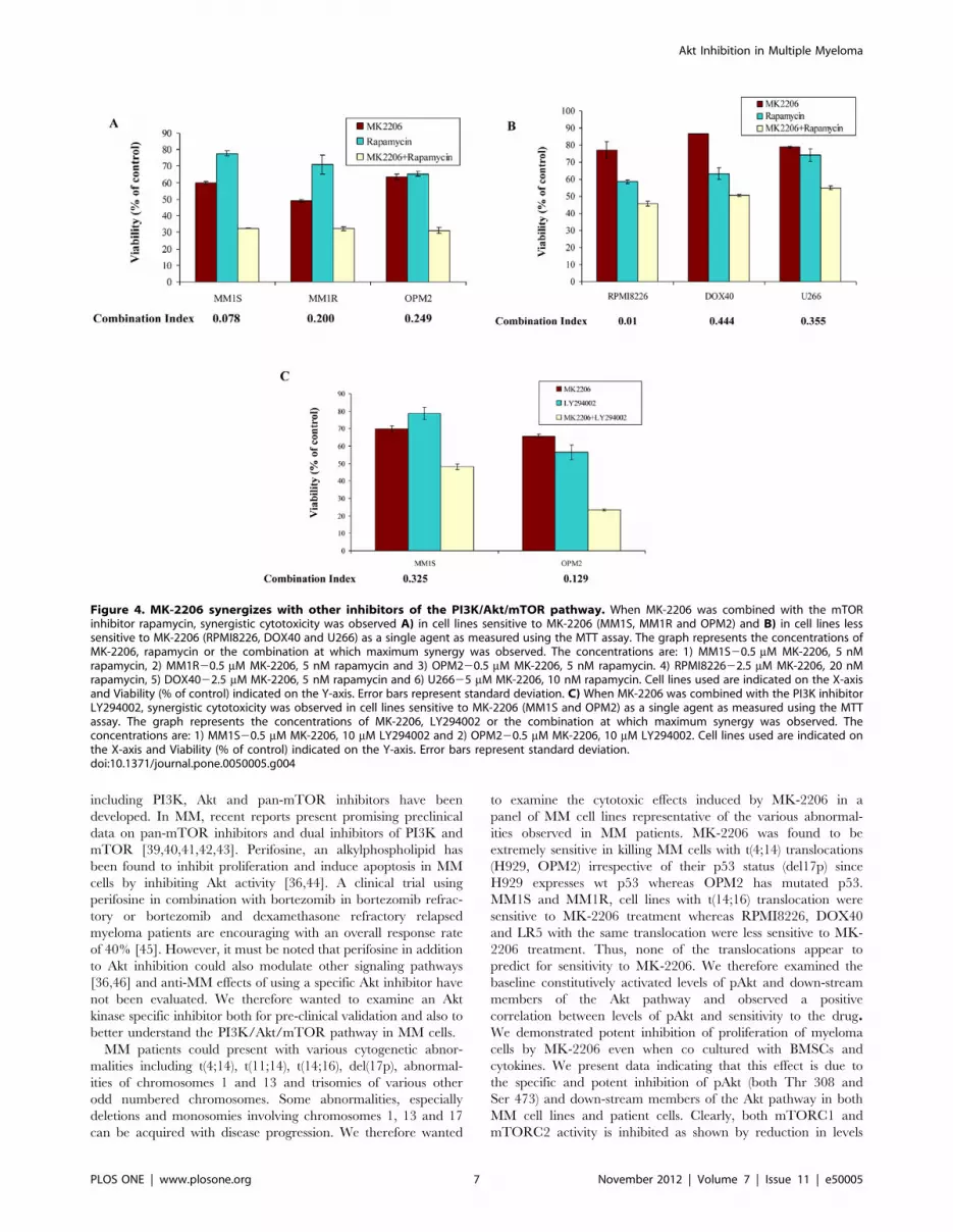
Figure 1. MK-2206 induced cytotoxicity, inhibited proliferation and was able to overcome the protective effects of the tumormicroenvironment. When we cultured MM cell lines with indicated concentrations of MK-2206 for 48 hrs we observed dose dependent decrease inA) viability as observed by MTT assays and B) tritiated thymidine uptake indicative of inhibition of proliferation albeit with differences in thesensitivity of cell lines to MK-2206. MK-2206 concentration is indicated along the6axis and in A) viability is expressed as % of control is indicatedalong the Y axis and in B) proliferation is expressed as % cpm of control. Error bars represent standard deviation. C) We evaluated baseline levels ofactivated Akt and down-stream members of the PI3K/Akt/mTOR pathway including pmTOR, p-p70S6K, p4EBP1 and in addition pErk levels by westernblotting. We observed that cell lines sensitive to MK-2206 expressed high levels of pAkt whereas the cell lines less sensitive to MK-2206 expressed lowlevels of pAkt but high levels of pErk. When MM1S cells were co-cultured with D) bone marrow stromal cells (BMSCs) or E) cytokines IL6 (25 ng/ml),IGF (50 ng/ml) or VEGF (50 ng/ml), we observed increase in proliferation measured by increase in cpm. MK-2206 was able to inhibit this increase inproliferation. MK-2206 concentrations are indicated on X-axis and % cpm is indicated on the Y-axis. Error bars represent standard deviation. F) Wecultured MM1S cells with or without 2.5 mM of MK-2206 for 8 hrs. In the last thirty minutes of incubation, cells were treated with IGF (50 ng/ml) forindicated time points. We then made lysates and checked for the ability of MK-2206 to inhibit pAkt and down-stream members of the PI3K/Akt/mTORpathway. We observed potent down regulation of pAkt and downstream members with no differences observed in total proteins and actin, whichserve as loading controls.doi:10.1371/journal.pone.0050005.g001
Akt Inhibition in Multiple Myeloma
PLOS ONE | www.plosone.org 4 November 2012 | Volume 7 | Issue 11 | e50005

Inhibition of PI3K/Akt Pathway at Multiple Levels Resultin Synergistic Anti-myeloma Activity
From the results so far, it is clear that lack of dependence on the
Akt in some cell lines precludes a relevant level of cytotoxicity with
Akt inhibitors. Hence, we wanted to examine if simultaneously
inhibiting the pathway at multiple levels or inhibiting multiple
pathways can result in synergistic effects. We first incubated three
sensitive cell lines MM1S, MM1R and OPM2 with various
concentrations of MK-2206 and rapamycin for 48 hrs. In all the
three cell lines, we observed marked synergy. The results presented
represents the concentration at which maximum synergy was
observed (Figure 4A). It is well known that mTOR inhibition
leads to activation of Akt. Therefore, we wanted to test if using
rapamycin with MK-2206 could sensitize the MM cell lines that
do not express basal levels of activated Akt and were less sensitive
to MK-2206 treatment. MK-2206 was observed to synergize with
these cell lines as well at various concentrations. The results
presented in figure 4B represents the concentration at which
maximum synergy was observed in RPMI8226, DOX40 and
U266 cells. It is also known that p-p70S6K when inhibited leads to
up regulation of the PI3K/Akt pathway by activating insulin
receptor substrate (IRS), which might also contribute to resistance
in cells exposed to MK-2206 treatment. We therefore wanted to
examine MK-2206 in combination with the PI3K inhibitor
LY294002. Like in combination with rapamycin, MK-2206
synergized with LY294002 in killing cell lines sensitive to MK-
2206 treatment (Figure 4C). However, we did not observe
synergy when MK-2206 was used in combination with LY294002
in MM cell lines less sensitive to MK-2206, as expected since Akt
status probably does not play a role in the survival of these cells.
(data not shown).
Activation of MEK/Erk Pathway Plays an Important Rolein Resistance to Akt Inhibition in Myeloma Cells
Constitutive levels of activated Erk are observed in almost all
MM cell lines examined with increased levels observed in cell lines
lacking pAkt expression and less sensitive to MK-2206
(Figure 1C). Also, MK-2206 treatment led to an upregulation
of pErk in the two MM cell lines used (Figures 3E and F). In
addition, we observed that MK-2206 potently inhibited pAkt
levels without comparable level of inhibition of the proteins down-
stream of pAkt (Figures 3A and B). It is known that activated
Erk feeds into the PI3K/Akt/mTOR pathway at the mTOR level
and hence we hypothesized that this could be a factor contributing
to the lack of significant pmTOR inhibition [23]. We therefore
examined if MK-2206 can synergize with inhibitors of the Mek/
Erk pathway. More importantly, we examined whether pretreat-
ing the cells less sensitive to MK-2206 with a MEK inhibitor
would sensitize these cells to MK-2206 through mTORC2
induced pAkt activation. To test this, we pretreated MM cells
with indicated concentrations of U0126 for 24 hrs. Following this,
we treated these cells with indicated concentrations of MK-2206
for 48 hrs and examined the viability of cells. We observed marked
synergy when MK-2206 was used in combination with U0126
both in cell lines sensitive to MK-2206 (Figure 5A) and more
importantly in lines less sensitive to MK-2206 including
Figure 2. MK-2206 induced apoptosis in MM cell lines and patient cells. We incubated MM1S and OPM2 cells with A) indicatedconcentrations of MK-2206 for 48 hrs and B) with 2.5 mM of MK-2206 for 12, 24 or 48 hrs. We observed dose and time dependent increase inapoptosis as measured by annexin/PI staining. % cells in each quadrant is indicated. C) MM1S or D) OPM2 with indicated concentrations of MK-2206for 48 hrs and measured activation of caspases 9, 8 and 3. We observed dose dependent increase in activation of each of the three caspases. In all theabove experiments, control refers to cells untreated with MK-2206. We incubated E) MM1S or F) OPM2 cells with indicated concentrations of MK-2206, MK-2206 plus Q-VD-OPH (10 mM), MK-2206 plus Z-IETD-FMK (10 mM) or MK-2206 plus Ac-LEHD-CMK(10 mM) for 48 hrs. We observed dosedependent decrease in cell viability, which was partially blocked by Q-VD-OPH. MK-2206 concentration is indicated along6axis and viability (% ofcontrol) is indicated along Y-axis. Error bars represent standard deviation. G) We incubated cells from MM patients with indicated concentrations ofMK-2206 for 48 hrs. We then measured for plasma cells undergoing apoptosis by annexin/PI staining. MK-2206 concentration is indicated along6axisand viability (% of control) is indicated along Y axis.doi:10.1371/journal.pone.0050005.g002
Akt Inhibition in Multiple Myeloma
PLOS ONE | www.plosone.org 5 November 2012 | Volume 7 | Issue 11 | e50005

RPMI8226, DOX and U266 (Figure 5B). We then examined the
mechanism of action of the combination to better understand the
synergistic response observed. For this, we pretreated MM1S cells
with 10 mM of U0126 for 24 hrs. Following this, we treated these
cells with 2.5 mM of MK-2206 for 1, 2, 4 or 8 hrs. As controls, we
treated MM1S cells with just U0126 or MK-2206 or left them
untreated. We then examined the levels of pAkt, pErk, pmTOR,
p-p70S6K, p4EBP1, pS6 and pGsk3b. We observed that MK-
2206 clearly inhibited pAkt alone or in combination with U0126
(Figure 5C). U0126, as expected, leads to up regulation of pAkt
(both Ser 473 and Thr308) which was efficiently down-regulated
by MK-2206 (Figure 5C). U0126 clearly inhibited pErk to
similar extents as a single agent or in combination with MK-2206
(Figure 5C). The combination of MK-2206 and U0126 inhibited
mTOR activity more effectively than either of these agents as
single agents (Figure 5C). Similarly, pGsk3b that is down-stream
of both pAkt and pErk, was down-regulated more significantly
when MM1S cells were treated with the drug combination
(Figure 5C). This once again indicated that activated Erk feeds
into the PI3K/Akt/mTOR pathway and inhibiting pErk could
sensitize cells to PI3K pathway inhibitors.
Finally, given the significant role of dexamethasone as a
therapeutic agent for myeloma we examined if pAkt inhibition
can synergize with corticosteroid therapy. The PI3K/Akt/mTOR
pathway is also known to be a critical pathway contributing to
dexamethasone resistance in MM cells [20,37]. For example,
activated PI3K/Akt pathway inactivates caspase-9 and offers
resistance against Dexamethasone induced apoptosis [20]. We
therefore examined the cytotoxicity of the combination of MK-
2206 and dexamethasone in MM cell lines. We observed marked
synergy when the drugs were used in combination in both cell lines
sensitive to MK-2206 (Figure 6A) and those resistant to MK-
2206 (Figure 6B).
Discussion
The PI3K/Akt/mTOR pathway has long been shown to be
important in MM disease progression and resistance to therapy.
However, the limited availability of drugs specifically targeting this
pathway has been a limiting factor to understand the clinical
benefits of inhibiting this pathway. In addition, mTORC1
inhibitor rapamycin treatment resulted in a cytostatic effect with
minimal cytotoxicity in various tumor systems [38]. The reasons
for this are now better understood and have been attributed
mainly to increased Akt activation by mTORC2 [26]. Over the
last few years numerous drugs specifically targeting this pathway
Figure 3. MK-2206 inhibits pAkt and downstream members of the PI3K/Akt/mTOR pathway and modulates expression levels ofother signaling pathway proteins. We incubated A and E) MM1S or B and F) OPM2 cells with 2.5 mM of MK-2206 for 1, 2, 4 or 8 hrs. Weobserved down regulation of pAkt and downstream members of the pathway in both MM1S and OPM2 cells. We observed up regulation of pErk inboth cell lines. We also observed down regulation of pBad and Bcl-Xl. No differences were observed in total proteins and actin, which serve as loadingcontrols. We incubated CD138+ cells from C and G) Patient 1 or D and H) Patient 2 with 2.5 mM of MK-2206 for indicated time points and observeddown regulation of pAkt, pmTOR, p-p70S6K, p4EBP1 and pGsk3b. We observed down regulation of Mcl1 and Xiap levels in patient 1 and downregulation of Mcl1 in patient 2. In all the above experiments control refers to cells untreated with MK-2206.doi:10.1371/journal.pone.0050005.g003
Akt Inhibition in Multiple Myeloma
PLOS ONE | www.plosone.org 6 November 2012 | Volume 7 | Issue 11 | e50005
including PI3K, Akt and pan-mTOR inhibitors have been
developed. In MM, recent reports present promising preclinical
data on pan-mTOR inhibitors and dual inhibitors of PI3K and
mTOR [39,40,41,42,43]. Perifosine, an alkylphospholipid has
been found to inhibit proliferation and induce apoptosis in MM
cells by inhibiting Akt activity [36,44]. A clinical trial using
perifosine in combination with bortezomib in bortezomib refrac-
tory or bortezomib and dexamethasone refractory relapsed
myeloma patients are encouraging with an overall response rate
of 40% [45]. However, it must be noted that perifosine in addition
to Akt inhibition could also modulate other signaling pathways
[36,46] and anti-MM effects of using a specific Akt inhibitor have
not been evaluated. We therefore wanted to examine an Akt
kinase specific inhibitor both for pre-clinical validation and also to
better understand the PI3K/Akt/mTOR pathway in MM cells.
MM patients could present with various cytogenetic abnor-
malities including t(4;14), t(11;14), t(14;16), del(17p), abnormal-
ities of chromosomes 1 and 13 and trisomies of various other
odd numbered chromosomes. Some abnormalities, especially
deletions and monosomies involving chromosomes 1, 13 and 17
can be acquired with disease progression. We therefore wanted
to examine the cytotoxic effects induced by MK-2206 in a
panel of MM cell lines representative of the various abnormal-
ities observed in MM patients. MK-2206 was found to be
extremely sensitive in killing MM cells with t(4;14) translocations
(H929, OPM2) irrespective of their p53 status (del17p) since
H929 expresses wt p53 whereas OPM2 has mutated p53.
MM1S and MM1R, cell lines with t(14;16) translocation were
sensitive to MK-2206 treatment whereas RPMI8226, DOX40
and LR5 with the same translocation were less sensitive to MK-
2206 treatment. Thus, none of the translocations appear to
predict for sensitivity to MK-2206. We therefore examined the
baseline constitutively activated levels of pAkt and down-stream
members of the Akt pathway and observed a positive
correlation between levels of pAkt and sensitivity to the drug.We demonstrated potent inhibition of proliferation of myeloma
cells by MK-2206 even when co cultured with BMSCs and
cytokines. We present data indicating that this effect is due to
the specific and potent inhibition of pAkt (both Thr 308 and
Ser 473) and down-stream members of the Akt pathway in both
MM cell lines and patient cells. Clearly, both mTORC1 and
mTORC2 activity is inhibited as shown by reduction in levels
Figure 4. MK-2206 synergizes with other inhibitors of the PI3K/Akt/mTOR pathway. When MK-2206 was combined with the mTORinhibitor rapamycin, synergistic cytotoxicity was observed A) in cell lines sensitive to MK-2206 (MM1S, MM1R and OPM2) and B) in cell lines lesssensitive to MK-2206 (RPMI8226, DOX40 and U266) as a single agent as measured using the MTT assay. The graph represents the concentrations ofMK-2206, rapamycin or the combination at which maximum synergy was observed. The concentrations are: 1) MM1S20.5 mM MK-2206, 5 nMrapamycin, 2) MM1R20.5 mM MK-2206, 5 nM rapamycin and 3) OPM220.5 mM MK-2206, 5 nM rapamycin. 4) RPMI822622.5 mM MK-2206, 20 nMrapamycin, 5) DOX4022.5 mM MK-2206, 5 nM rapamycin and 6) U26625 mM MK-2206, 10 nM rapamycin. Cell lines used are indicated on the X-axisand Viability (% of control) indicated on the Y-axis. Error bars represent standard deviation. C) When MK-2206 was combined with the PI3K inhibitorLY294002, synergistic cytotoxicity was observed in cell lines sensitive to MK-2206 (MM1S and OPM2) as a single agent as measured using the MTTassay. The graph represents the concentrations of MK-2206, LY294002 or the combination at which maximum synergy was observed. Theconcentrations are: 1) MM1S20.5 mM MK-2206, 10 mM LY294002 and 2) OPM220.5 mM MK-2206, 10 mM LY294002. Cell lines used are indicated onthe X-axis and Viability (% of control) indicated on the Y-axis. Error bars represent standard deviation.doi:10.1371/journal.pone.0050005.g004
Akt Inhibition in Multiple Myeloma
PLOS ONE | www.plosone.org 7 November 2012 | Volume 7 | Issue 11 | e50005

of p-p70S6K, p4EBP1 (mTORC1 substrates) and pAkt (Ser
473) (mTORC2 substrate). Though we observed inhibition of
pmTOR and its substrates, the extent of the down regulation
was not as pronounced as pAkt down regulation. It has been
reported that p-p70S6K inhibition leads to PI3K mediated up
regulation of the Mek/Erk pathway [47]. Additionally, activated
Erk has been shown to phosphorylate TSC2 and raptor, both
events leading to activation of mTOR [38,48]. It has long been
suggested that the Mek/Erk pathway and the PI3K/Akt
pathway cross inhibit each other and this cross inhibition is
why inhibitors of either pathway lead to upregulation of the
other pathway [49,50]. In our studies, we observed that MK-
2206 either had no effect on pErk levels or in some cases
increased pErk levels. In OPM2 cells where we observed
significant up regulation of pErk post MK-2206, we also
observed the lack of down regulation of pGsk3b. Gsk3b has
been shown to be phosphorylated by activated Erk in addition
to pAkt and this once again indicated the complex interaction
between these two pathways [51]. Pre treating MM cells with
U0126 (Mek1/2 inhibitor) followed by MK-2206 treatment
demonstrated synergy in killing MM cells and our results clearly
indicated that the drug combination inhibited mTOR activity
significantly. In addition, it has recently been shown that Akt
inhibition leads to reactivation of multiple receptor tyrosine
kinases in several different tumor systems through the inhibition
of Akt induced negative feedback loop [52]. Thus it is very
likely that additional mechanisms could contribute to resistance
to MK-2206 in MM cells. We are currently examining the
activated levels of Akt and Erk in MM patient cells to evaluate
if these could serve as biomarkers to predict for sensitivity to
MK-2206 and other PI3K/Akt/mTOR inhibitors.
Our results clearly indicate the complex nature of the PI3K/
Akt/mTOR pathway, the benefits of inhibiting this pathway at
multiple levels and the synergistic effect of simultaneously
inhibiting both this and the Mek/Erk pathways in MM cells.
Specifically, we present evidence in support of the Mek/Erk
pathway driving the down-stream elements of the Akt pathway,
providing a resistance mechanism in myeloma cells. Taken
together, we present clear evidence for MK-2206 alone or in
combination with rapamycin, a PI3K inhibitor, an inhibitor of the
Mek/Erk pathway or dexamethasone to be taken up for clinical
investigation in MM patients, and most importantly, to use
baseline expression of pAkt and pERK as companion biomarkers
in patient selection for clinical trials with this agent.
Figure 5. MK-2206 synergizes with the Mek inhibitor U0126 and the drug combination leads to increased inhibition of mTORactivity. When MK-2206 was combined with the Mek1/2 inhibitor U0126, synergistic cytotoxicity was observed A) in cell lines sensitive to MK-2206(MM1S, MM1R and OPM2) and B) in cell lines less sensitive to MK-2206 (RPMI8226, DOX40 and U266) as a single agent as measured using the MTTassay. The graph represents the concentrations of MK-2206, U0126 or the combination at which maximum synergy was observed. The concentrationsare: 1) MM1S20.5 mM MK-2206, 10 mM U0126, 2) MM1R20.5 mM MK-2206, 10 mM U0126 and 3) OPM221 mM MK-2206, 20 mM U0126. 4)RPMI822622.5 mM MK-2206, 10 mM U0126, 5) DOX4025 mM MK-2206, 20 mM U0126 and 6) U26625 mM MK-2206, 30 mM U0126. Cell lines used areindicated on the X-axis and Viability (% of control) indicated on the Y-axis. Error bars represent standard deviation. C) MM1S cells were left untreatedor treated with U0126 (10 mM) for 24 hrs. Following this, cells were treated with 2.5 mM of MK-2206 for 1, 2, 4 or 8 hrs. As a control, MM1S cells weretreated with U0126 alone (10 mM) for 32 hrs or MK-2206 alone (2.5 mM) for 8 hrs. We observed significant down regulation of pAkt and otherdownstream members of the pathway and pErk when MK-2206 was used in combination with U0126 with no difference in total proteins and actin,which serve as loading controls.doi:10.1371/journal.pone.0050005.g005
Akt Inhibition in Multiple Myeloma
PLOS ONE | www.plosone.org 8 November 2012 | Volume 7 | Issue 11 | e50005

Supporting Information
Figure S1 Apoptosis is not induced by MK2206 in U266 cell
line. We incubated U266 cells with A) indicated concentrations of
MK-2206 for 48 hrs and B) with 2.5 mM of MK-2206 for 12, 24
or 48 hrs. We observed absence of dose and time dependent
increase in apoptosis as measured by annexin/PI staining. % cells
in each quadrant is indicated. In all the above experiments, control
refers to cells untreated with MK-2206.
(TIF)
Figure S2 Caspases are not activated by MK2206 in U266 cell
line. U266 cells were incubated with indicated concentrations of
MK-2206 for 48 hrs and we measured activation of caspases 9, 8
and 3. None of the caspases were activated by MK2206. In all the
above experiments, control refers to cells untreated with MK-
2206.
(TIF)
Table S1 IC50 values of MK2206 on MM cell lines. This table
indicates the IC50 values of MK2206 on all MM cell lines
examined. From this table, it is clear that MK2206 exhibits
preferential cytotoxicity on a few MM cell lines.
(PDF)
Acknowledgments
We would like to acknowledge Kimberly Henderson, RobertaDeGoey, Steven Zincke and Beatrice Hartke for their assistance
with processing of tumor cells and all of the patients who provided us with
Figure 6. MK-2206 synergizes with dexamethasone. When MK-2206 was combined with dexamethasone, synergistic cytotoxicity wasobserved A) in cell lines sensitive to MK-2206 (MM1S and OPM2) and B) in cell lines less sensitive to MK-2206 (RPMI8226 and DOX40) as a singleagent as measured using the MTT assay. The graph represents the concentrations of MK-2206, dexamethasone or the combination at whichmaximum synergy was observed. The concentrations are: 1) MM1S20.5 mM MK-2206, 10 nM dexamethasone and 2) OPM221 mM MK-2206, 40 nMdexamethasone. 3) RPMI822625 mM MK-2206, 20 nM dexamethasone and 4) DOX4022.5 mM MK-2206, 50 nM dexamethasone. Cell lines used areindicated on the X-axis and Viability (% of control) indicated on the Y-axis. Error bars represent standard deviation.doi:10.1371/journal.pone.0050005.g006
Akt Inhibition in Multiple Myeloma
PLOS ONE | www.plosone.org 9 November 2012 | Volume 7 | Issue 11 | e50005
the tumor samples. MK-2206 was provided by Merck. & Co., Inc.
(Whitehouse Station, NJ).Author Contributions
Conceived and designed the experiments: VR SVR SK. Performed the
experiments: VR TK JH UP LW TH. Analyzed the data: VR SVR SK.
Contributed reagents/materials/analysis tools: SVR SK. Wrote the paper:
VR SVR SK.
References
1. Rajkumar SV (2011) Treatment of multiple myeloma. Nat Rev Clin Oncol 8:
479–491.
2. Kumar SK, Lee JH, Lahuerta JJ, Morgan G, Richardson PG, et al. (2012) Riskof progression and survival in multiple myeloma relapsing after therapy with
IMiDs and bortezomib: a multicenter international myeloma working groupstudy. Leukemia 26: 149–157.
3. Fruman DA, Meyers RE, Cantley LC (1998) Phosphoinositide kinases. Annu
Rev Biochem 67: 481–507.
4. Alessi DR, Andjelkovic M, Caudwell B, Cron P, Morrice N, et al. (1996)
Mechanism of activation of protein kinase B by insulin and IGF-1. Embo J 15:
6541–6551.
5. Kohn AD, Takeuchi F, Roth RA (1996) Akt, a pleckstrin homology domain
containing kinase, is activated primarily by phosphorylation. J Biol Chem 271:21920–21926.
6. Brunet A, Bonni A, Zigmond MJ, Lin MZ, Juo P, et al. (1999) Akt promotes cell
survival by phosphorylating and inhibiting a Forkhead transcription factor. Cell96: 857–868.
7. del Peso L, Gonzalez-Garcia M, Page C, Herrera R, Nunez G (1997)
Interleukin-3-induced phosphorylation of BAD through the protein kinase Akt.Science 278: 687–689.
8. Harris TE, Lawrence JC Jr (2003) TOR signaling. Sci STKE 2003: re15.
9. Junttila TT, Akita RW, Parsons K, Fields C, Lewis Phillips GD, et al. (2009)Ligand-independent HER2/HER3/PI3K complex is disrupted by trastuzumab
and is effectively inhibited by the PI3K inhibitor GDC-0941. Cancer Cell 15:429–440.
10. Kennedy SG, Kandel ES, Cross TK, Hay N (1999) Akt/Protein kinase B
inhibits cell death by preventing the release of cytochrome c from mitochondria.Mol Cell Biol 19: 5800–5810.
11. Kulik G, Klippel A, Weber MJ (1997) Antiapoptotic signalling by the insulin-like
growth factor I receptor, phosphatidylinositol 3-kinase, and Akt. Mol Cell Biol17: 1595–1606.
12. Shaw RJ, Bardeesy N, Manning BD, Lopez L, Kosmatka M, et al. (2004) The
LKB1 tumor suppressor negatively regulates mTOR signaling. Cancer Cell 6:91–99.
13. Wendel HG, De Stanchina E, Fridman JS, Malina A, Ray S, et al. (2004)Survival signalling by Akt and eIF4E in oncogenesis and cancer therapy. Nature
428: 332–337.
14. Long X, Ortiz-Vega S, Lin Y, Avruch J (2005) Rheb binding to mammaliantarget of rapamycin (mTOR) is regulated by amino acid sufficiency. J Biol Chem
280: 23433–23436.
15. Sancak Y, Thoreen CC, Peterson TR, Lindquist RA, Kang SA, et al. (2007)PRAS40 is an insulin-regulated inhibitor of the mTORC1 protein kinase. Mol
Cell 25: 903–915.
16. Sengupta S, Peterson TR, Sabatini DM (2010) Regulation of the mTORcomplex 1 pathway by nutrients, growth factors, and stress. Mol Cell 40: 310–
322.
17. Tee AR, Fingar DC, Manning BD, Kwiatkowski DJ, Cantley LC, et al. (2002)
Tuberous sclerosis complex-1 and -2 gene products function together to inhibit
mammalian target of rapamycin (mTOR)-mediated downstream signaling. ProcNatl Acad Sci U S A 99: 13571–13576.
18. Chang H, Qi XY, Claudio J, Zhuang L, Patterson B, et al. (2006) Analysis of
PTEN deletions and mutations in multiple myeloma. Leuk Res 30: 262–265.
19. Ismail SI, Mahmoud IS, Msallam MM, Sughayer MA (2010) Hotspot mutations
of PIK3CA and AKT1 genes are absent in multiple myeloma. Leuk Res 34:824–826.
20. Hideshima T, Nakamura N, Chauhan D, Anderson KC (2001) Biologic
sequelae of interleukin-6 induced PI3-K/Akt signaling in multiple myeloma.Oncogene 20: 5991–6000.
21. Hu L, Shi Y, Hsu JH, Gera J, Van Ness B, et al. (2003) Downstream effectors of
oncogenic ras in multiple myeloma cells. Blood 101: 3126–3135.
22. Mitsiades CS, Mitsiades N, Poulaki V, Schlossman R, Akiyama M, et al. (2002)
Activation of NF-kappaB and upregulation of intracellular anti-apoptotic
proteins via the IGF-1/Akt signaling in human multiple myeloma cells:therapeutic implications. Oncogene 21: 5673–5683.
23. Shi Y, Hsu JH, Hu L, Gera J, Lichtenstein A (2002) Signal pathways involved inactivation of p70S6K and phosphorylation of 4E-BP1 following exposure of
multiple myeloma tumor cells to interleukin-6. J Biol Chem 277: 15712–15720.
24. Steinbrunn T, Stuhmer T, Gattenlohner S, Rosenwald A, Mottok A, et al.(2011) Mutated RAS and constitutively activated Akt delineate distinct
oncogenic pathways, which independently contribute to multiple myeloma cellsurvival. Blood 117: 1998–2004.
25. Farag SS, Zhang S, Jansak BS, Wang X, Kraut E, et al. (2009) Phase II trial of
temsirolimus in patients with relapsed or refractory multiple myeloma. Leuk Res33: 1475–1480.
26. Hresko RC, Mueckler M (2005) mTOR.RICTOR is the Ser473 kinase for Akt/
protein kinase B in 3T3-L1 adipocytes. J Biol Chem 280: 40406–40416.
27. Harrington LS, Findlay GM, Gray A, Tolkacheva T, Wigfield S, et al. (2004)
The TSC1–2 tumor suppressor controls insulin-PI3K signaling via regulation of
IRS proteins. J Cell Biol 166: 213–223.
28. Krett NL, Zell JL, Halgren RG, Pillay S, Traynor AE, et al. (1997) Cyclic
adenosine-39,59-monophosphate-mediated cytotoxicity in steroid sensitive and
resistant myeloma. Clin Cancer Res 3: 1781–1787.
29. Dalton WS, Durie BG, Alberts DS, Gerlach JH, Cress AE (1986) Character-
ization of a new drug-resistant human myeloma cell line that expresses P-
glycoprotein. Cancer Res 46: 5125–5130.
30. Bellamy WT, Dalton WS, Gleason MC, Grogan TM, Trent JM (1991)
Development and characterization of a melphalan-resistant human multiple
myeloma cell line. Cancer Res 51: 995–1002.
31. Katagiri S, Yonezawa T, Kuyama J, Kanayama Y, Nishida K, et al. (1985) Two
distinct human myeloma cell lines originating from one patient with myeloma.
Int J Cancer 36: 241–246.
32. Ramakrishnan V, Ansell S, Haug J, Grote D, Kimlinger T, et al. (2012)
MRK003, a gamma-secretase inhibitor exhibits promising in vitro pre-clinical
activity in multiple myeloma and non-Hodgkin’s lymphoma. Leukemia 26: 340–
348.
33. Ramakrishnan V, Kimlinger T, Haug J, Timm M, Wellik L, et al. (2010)
TG101209, a novel JAK2 inhibitor, has significant in vitro activity in multiple
myeloma and displays preferential cytotoxicity for CD45+ myeloma cells.
Am J Hematol 85: 675–686.
34. Ramakrishnan V, Timm M, Haug JL, Kimlinger TK, Wellik LE, et al. (2010)
Sorafenib, a dual Raf kinase/vascular endothelial growth factor receptor
inhibitor has significant anti-myeloma activity and synergizes with common anti-
myeloma drugs. Oncogene 29: 1190–1202.
35. Chou TC, Talalay P (1984) Quantitative analysis of dose-effect relationships: the
combined effects of multiple drugs or enzyme inhibitors. Adv Enzyme Regul 22:
27–55.
36. Hideshima T, Catley L, Yasui H, Ishitsuka K, Raje N, et al. (2006) Perifosine, an
oral bioactive novel alkylphospholipid, inhibits Akt and induces in vitro and
in vivo cytotoxicity in human multiple myeloma cells. Blood 107: 4053–4062.
37. Moreaux J, Legouffe E, Jourdan E, Quittet P, Reme T, et al. (2004) BAFF and
APRIL protect myeloma cells from apoptosis induced by interleukin 6
deprivation and dexamethasone. Blood 103: 3148–3157.
38. Zoncu R, Efeyan A, Sabatini DM (2011) mTOR: from growth signal integration
to cancer, diabetes and ageing. Nat Rev Mol Cell Biol 12: 21–35.
39. Baumann P, Schneider L, Mandl-Weber S, Oduncu F, Schmidmaier R (2012)
Simultaneous targeting of PI3K and mTOR with NVP-BGT226 is highly
effective in multiple myeloma. Anticancer Drugs 23: 131–138.
40. Cirstea D, Hideshima T, Rodig S, Santo L, Pozzi S, et al. (2010) Dual inhibition
of akt/mammalian target of rapamycin pathway by nanoparticle albumin-
bound-rapamycin and perifosine induces antitumor activity in multiple
myeloma. Mol Cancer Ther 9: 963–975.
41. Hoang B, Frost P, Shi Y, Belanger E, Benavides A, et al. (2010) Targeting
TORC2 in multiple myeloma with a new mTOR kinase inhibitor. Blood 116:
4560–4568.
42. Maiso P, Liu Y, Morgan B, Azab AK, Ren P, et al. (2011) Defining the role of
TORC1/2 in multiple myeloma. Blood 118: 6860–6870.
43. McMillin DW, Ooi M, Delmore J, Negri J, Hayden P, et al. (2009) Antimyeloma
activity of the orally bioavailable dual phosphatidylinositol 3-kinase/mammalian
target of rapamycin inhibitor NVP-BEZ235. Cancer Res 69: 5835–5842.
44. Hideshima T, Catley L, Raje N, Chauhan D, Podar K, et al. (2007) Inhibition of
Akt induces significant downregulation of survivin and cytotoxicity in human
multiple myeloma cells. Br J Haematol 138: 783–791.
45. Richardson PG, Wolf J, Jakubowiak A, Zonder J, Lonial S, et al. (2011)
Perifosine Plus Bortezomib and Dexamethasone in Patients With Relapsed/
Refractory Multiple Myeloma Previously Treated With Bortezomib: Results of a
Multicenter Phase I/II Trial. J Clin Oncol 29: 4243–4249.
46. Gills JJ, Dennis PA (2009) Perifosine: update on a novel Akt inhibitor. Curr
Oncol Rep 11: 102–110.
47. Carracedo A, Ma L, Teruya-Feldstein J, Rojo F, Salmena L, et al. (2008)
Inhibition of mTORC1 leads to MAPK pathway activation through a PI3K-
dependent feedback loop in human cancer. J Clin Invest 118: 3065–3074.
48. Carriere A, Romeo Y, Acosta-Jaquez HA, Moreau J, Bonneil E, et al. (2011)
ERK1/2 phosphorylate Raptor to promote Ras-dependent activation of mTOR
complex 1 (mTORC1). J Biol Chem 286: 567–577.
49. Hoeflich KP, O’Brien C, Boyd Z, Cavet G, Guerrero S, et al. (2009) In vivo
antitumor activity of MEK and phosphatidylinositol 3-kinase inhibitors in basal-
like breast cancer models. Clin Cancer Res 15: 4649–4664.
Akt Inhibition in Multiple Myeloma
PLOS ONE | www.plosone.org 10 November 2012 | Volume 7 | Issue 11 | e50005

50. Zimmermann S, Moelling K (1999) Phosphorylation and regulation of Raf by
Akt (protein kinase B). Science 286: 1741–1744.51. Ding Q, Xia W, Liu JC, Yang JY, Lee DF, et al. (2005) Erk associates with and
primes GSK-3beta for its inactivation resulting in upregulation of beta-catenin.
Mol Cell 19: 159–170.
52. Chandarlapaty S, Sawai A, Scaltriti M, Rodrik-Outmezguine V, Grbovic-Huezo
O, et al. (2011) AKT inhibition relieves feedback suppression of receptor
tyrosine kinase expression and activity. Cancer Cell 19: 58–71.
Akt Inhibition in Multiple Myeloma
PLOS ONE | www.plosone.org 11 November 2012 | Volume 7 | Issue 11 | e50005