HijAkt: The PI3K/Akt pathway in virus replication and pathogenesis
28
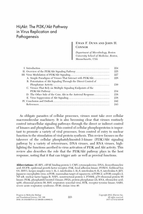
HijAkt: The PI3K/Akt Pathway in Virus Replication and Pathogenesis Ewan F. Dunnand John H. Connor Department of Microbiology, Boston University School of Medicine, Boston, Massachusetts, USA I. Introduction ................................................................................. 224 II. Overview of the PI3K/Akt Signaling Pathway ....................................... 225 III. Virus Modulation of PI3K/Akt Signaling.............................................. 227 A. Simple Paradigms of Viruses That Interact with PI3K/Akt................... 228 B. Potentiation of Akt Signaling Through the Direct Control of Phosphatase Activity .................................................................. 230 C. Viruses That Rely on Multiple Signaling Endpoints of the PI3K/Akt Pathway ..................................................................... 234 D. The Other Side of the Coin: Akt in the Antiviral Response.................. 238 E. Virus Suppression of Akt Signaling ................................................ 238 IV. Conclusion and Outlook .................................................................. 242 References ................................................................................... 243 As obligate parasites of cellular processes, viruses must take over cellular macromolecular machinery. It is also becoming clear that viruses routinely control intracellular signaling pathways through the direct or indirect control of kinases and phosphatases. This control of cellular phosphoproteins is impor- tant to promote a variety of viral processes, from control of entry to nuclear function to the stimulation of viral protein synthesis. This review focuses on the takeover of the cellular phosphatidylinositol-3-kinase (PI3K)/Akt signaling pathway by a variety of retroviruses, DNA viruses, and RNA viruses, high- lighting the functions ascribed to virus activation of PI3K and Akt activity. This review also describes the role that the PI3K/Akt pathway plays in the host response, noting that it that can trigger anti- as well as proviral functions. Abbreviations: 4E-BP1, eIF4E binding protein 1; CMV, cytomegalovirus; DNA, deoxyribonucleic acid; EGFR, epidermal growth factor receptor; FAK, focal adhesion kinase; FOXO1, forkhead box O1; HSV1, herpes simplex virus 1; IL-1, interleukin-1; IL-6, interleukin-6; IL-8, interleukin-8; JEV, Japanese encephalitis virus; mTOR, mammalian target of rapamycin; mTORC2, mTOR complex 2; NF-kB, nuclear factor kappa B; NSP1, nonstructural protein 1; P70S6k, p70 ribosomal protein S6 kinase; PI3K, phosphatidyl inositol 3 kinase; PP2A, protein phosphatase 2A; RNA, ribonucleic acid; rpS6, ribosomal protein S6; RSV, respiratory syncytial virus; RTK, receptor tyrosine kinase; SARS, severe acute respiratory syndrome; SV40, simian virus 40. Progress in Molecular Biology Copyright 2012, Elsevier Inc. and Translational Science, Vol. 106 223 All rights reserved. DOI: 10.1016/B978-0-12-396456-4.00002-X 1877-1173/12 $35.00
-
Upload
independent -
Category
Documents
-
view
4 -
download
0
Transcript of HijAkt: The PI3K/Akt pathway in virus replication and pathogenesis
HijAkt: The PI3K/Akt Pathwayin Virus Replication andPathogenesis
Progress in Molecular Biologyand Translational Science, Vol. 106 223DOI: 10.1016/B978-0-12-396456-4.00002-X
Ewan F. Dunn and John H.Connor
Department of Microbiology, BostonUniversity School of Medicine, Boston,Massachusetts, USA
I. I
ntroduction ..... ... .. ... .. ... .. .. ... .. ... .. ... .. .. ... .. ... .. ... .. ... .. .. ... .. ... .. ... .. ... ..Copyright 2A
187
2
012, Ell righ7-117
24
lsevts re3/12
II. O
verview of the PI3K/Akt Signaling Pathway ..... .. ... .. ... .. .. ... .. ... .. ... .. ... .. 2 25 I II. V irus Modulation of PI3K/Akt Signaling.... ... .. ... .. ... .. ... .. .. ... .. ... .. ... .. ... .. 2 27A
. S imple Paradigms of Viruses That Interact with PI3K/Akt.... ... .. ... .. ... .. 2 28 B . P otentiation of Akt Signaling Through the Direct Control ofPhosphatase Activity ..... .. ... .. ... .. ... .. .. ... .. ... .. ... .. ... .. .. ... .. ... .. ... .. ... ..
230 C . V iruses That Rely on Multiple Signaling Endpoints of thePI3K/Akt Pathway...... .. .. ... .. ... .. ... .. .. ... .. ... .. ... .. ... .. .. ... .. ... .. ... .. ... ..
234 D . T he Other Side of the Coin: Akt in the Antiviral Response...... .. ... .. ... .. 2 38 E . V irus Suppression of Akt Signaling .... .. ... .. ... .. ... .. ... .. .. ... .. ... .. ... .. ... .. 2 38I
V. C onclusion and Outlook..... .. ... .. ... .. ... .. .. ... .. ... .. ... .. ... .. .. ... .. ... .. ... .. ... .. 2 42 R eferences..... .. ... .. ... .. ... .. .. ... .. ... .. ... .. .. ... .. ... .. ... .. ... .. .. ... .. ... .. ... .. ... .. 2 43As obligate parasites of cellular processes, viruses must take over cellularmacromolecular machinery. It is also becoming clear that viruses routinelycontrol intracellular signaling pathways through the direct or indirect controlof kinases and phosphatases. This control of cellular phosphoproteins is impor-tant to promote a variety of viral processes, from control of entry to nuclearfunction to the stimulation of viral protein synthesis. This review focuses on thetakeover of the cellular phosphatidylinositol-3-kinase (PI3K)/Akt signalingpathway by a variety of retroviruses, DNA viruses, and RNA viruses, high-lighting the functions ascribed to virus activation of PI3K and Akt activity. Thisreview also describes the role that the PI3K/Akt pathway plays in the hostresponse, noting that it that can trigger anti- as well as proviral functions.
Abbreviations: 4E-BP1, eIF4E binding protein 1; CMV, cytomegalovirus; DNA, deoxyribonucleicacid; EGFR, epidermal growth factor receptor; FAK, focal adhesion kinase; FOXO1, forkhead boxO1; HSV1, herpes simplex virus 1; IL-1, interleukin-1; IL-6, interleukin-6; IL-8, interleukin-8; JEV,Japanese encephalitis virus; mTOR, mammalian target of rapamycin; mTORC2, mTOR complex 2;NF-kB, nuclear factor kappa B; NSP1, nonstructural protein 1; P70S6k, p70 ribosomal protein S6kinase; PI3K, phosphatidyl inositol 3 kinase; PP2A, protein phosphatase 2A; RNA, ribonucleic acid;rpS6, ribosomal protein S6; RSV, respiratory syncytial virus; RTK, receptor tyrosine kinase; SARS,severe acute respiratory syndrome; SV40, simian virus 40.
ier Inc.served.$35.00

224 DUNN AND CONNOR
I. Introduction
Viral disease is one of the most significant human health concerns inmodern medicine, manifesting as a broad range of acute infections, chronicdiseases, and virus-associated malignancies. Viruses such as human immuno-deficiency virus (HIV), hepatitis C, influenza, rotavirus, chikungunya, anddengue affect millions of people each year worldwide. The emergence ofnovel strains of H1N1 flu1 and previously unrecognized pathogens such assevere acute respiratory syndrome (SARS) coronavirus2 and SFTS bunya-virus3,4 has highlighted the threats of viral emergence and pandemic withwhich humanity regularly contends. This, along with the emergence of antiviralresistance among viruses such as influenza, highlights the need to betterunderstand virus replication and the virus–host interaction to identify criticalrestriction points that can be targets for antiviral drug development and vaccinegeneration.
One controlling aspect of viral pathogenesis and replication is their depen-dence on the cells that they infect. Unlike other microbial pathogens such asbacteria and fungi which are capable of autonomous growth, viruses arecompletely dependent on cellular processes for replication. It has long beenrecognized that viruses are dependent upon the use of cellular machinery forprotein synthesis: nucleotide and protein building blocks, ribonucleic acid(RNA) and deoxyribonucleic acid (DNA) polymerases, and membranes.More recently, it has become clear that viruses also rely upon intracellularcommunication.5,6 Particularly, viruses appear to be adept at capturing proteinphosphorylation cascades for their own use.
Most, if not all, viruses require protein phosphorylation in order to repli-cate. This would initially suggest that viruses would gain a replication edge bygenerally promoting protein phosphorylation. However, simply promoting allcellular protein phosphorylation is not likely to be advantageous to viruses, asthe activity of some host kinases (and the resultant phosphorylation events) areknown to antagonize viral replication. Thus, a nonspecific increase in phos-phorylation would trigger an antiviral response. Viruses have a need for speci-ficity in activating host kinases and phosphatases. Simply stated, viruses need toactivate signaling pathways that promote their replication7–9 while blocking oravoiding the activation of antiviral signaling pathways.10
Controlling kinase activity can promote protein translation,11 increasemetabolic activity, drive cell division, and inhibit cellular apoptosis. All ofthese are advantageous for virus replication. In addition, many viruses producevirus proteins that require direct phosphorylation in order to properly functionbut make no kinase that will carry out this phosphorylation.9,10 This dictatesthat they must recruit kinase activity from their host.

HIJAKT: THE PI3K/AKT PATHWAY IN VIRUS REPLICATION AND PATHOGENESIS 225
It is therefore not surprising that viruses have developed mechanisms tocontrol cellular signaling through regulating kinase and phosphatase signalingcascades. What is surprising is the commonality of host pathways that aretargeted. In theory, any number of kinases might be important for the replicationof any particular virus, but this possibility of each virus using different kinases isnot actually observed. Rather, as is emphasized below, one signaling pathway hasemerged as a common player in the replication of many different viruses—thephosphatidylinositol-3-kinase (PI3K)/Akt phosphorylation cascade.
The repeated identification of the PI3K/Akt pathway as a central player invirus replication has underscored the importance of this pathway as a centralregulator of cell health and metabolism. As a central regulator of multiple cellarprocesses that control translation, metabolism, and cell death, active PI3K/Aktsignaling can fulfill many viral ‘‘needs.’’ This is not to suggest that this signalingpathway only provides positive feedback for the virus. Studies of virus interac-tion with this pathway have also shown that the pathway is not simply a‘‘proviral’’ kinase when activated, but that it also acts as part of the hostresponse to viral infection. This suggests that PI3K/Akt signaling represents atwo-faced player in interactions between the virus and cell, able to bothpromote viral replication and be an active part of the immune response thatwill eventually quash virus replication. This review will describe the differentapproaches that viruses have taken to dominate the PI3K/Akt signaling path-way. The viruses chosen in this review highlight the varied manner in which thispathway is manipulated and turned to the advantage of the invader, while alsopointing out how the pathway also plays a role in host defense.
II. Overview of the PI3K/Akt Signaling Pathway
The PI3K/Akt signal transduction cascade is a classical phosphorylationcascade that utilizes tyrosine, lipid, and serine–threonine phosphorylation totransduce external signals to internal responses (see Fig. 1 for a schematicdiagram). Normally, signaling through this pathway is initiated by the stimulationof a receptor tyrosine kinase (RTK) by a cytokine or a growth factor at the cellsurface. Activation of the RTK recruits and activates PI3K. PI3K is a heterodimerconsisting of a p110 catalytic and a p85 regulatory subunit.12 PI3K is responsiblefor converting phosphatidylinositol 4,5-bisphosphate (PIP2) to 3,4,5-tripho-sphorylated phosphoinositide (PIP3). PIP3-rich membrane domains serve asdocking sites for proteins that contain a pleckstrin homology domain such as Akt.
The recruitment of inactive Akt protein to PIP3-rich areas of the plasmamembrane results in a conformational change that exposes the activation loopof Akt.13 Akt’s activating kinase, phosphoinositide-dependent protein kinase

PDK1p8
5 p110
PI3K
mTORC2
RTKAkt
P308
473 P
AntiapoptoticGrowth/survival
Translational control
AMPK
mTORC1
GSK3
BADp21
FOXO1
p70S6k 4E-BP1
P
P
PPP P
PP
P
Key:
PIP2
PIP3
Phosphorylation P
P
P
FIG. 1. Overview of the PI3K/Akt pathway. The diagram illustrates the two subunits of PI3K(p85 and p110) bound to an activated receptor tyrosine kinase (classical examples are the epidermalgrowth factor and the insulin receptor). Active PI3K catalyzes the addition of a single phosphate tothe 3 position of PIP4,5-bisphosphate to make PIP3. This triphosphorylated inositol serves as anaffinity ligand for the pleckstrin homology domains of the Akt kinase PDK1 and Akt, with thebinding of PIP3 by the Akt PH domain serving to uncover the activation domain of Akt. Phosphor-ylation by PDK1 occurs at position T308, and a second activating phosphorylation occurs at S473.Active Akt will phosphorylate various downstream protein targets that control cell growth (AMPK,p21, and GSK3 are illustrated) and translational control (mTOR and its downstream effectorsp70S6K and 4E-BP1 are illustrated) and act to suppress apoptosis (inhibitory phosphorylation ofBAD and FOXO1 are illustrated).
226 DUNN AND CONNOR
(PDK1)14, is also recruited to PIP3 microdomains. PDK1 phosphorylates Akton threonine 308 (Thr308) of the exposed activation loop, activating Akt andleading to a second phosphorylation of Akt at serine 473 (Ser473) by a kinasepresumed to be mTORC2 that further potentiates kinase activity.13,15–17
Activated Akt can control the signaling of several key pathways supportingdifferent types of cellular functions (Fig. 1). The PI3/Akt signaling pathwaypromotes cell growth, cell survival, and tumorigenesis through the phosphory-lation and inactivation of cell cycle kinase inhibitors such as p21 and the

HIJAKT: THE PI3K/AKT PATHWAY IN VIRUS REPLICATION AND PATHOGENESIS 227
inactivation of transcription factors that can inhibit cell cycle progression andpromote apoptosis such as the forkhead family of proteins (e.g., FOXO118). Aktalso can block the actions of metabolically repressive kinases such as AMPK19
and so can drive the up regulation of metabolic activity in the cell.20
Akt also promotes cellular translation through GSK3 phosphorylation andthe activation of mammalian target of rapamycin complex 1 (mTORC1), akinase complex that activates ribosomal S6 kinase and inhibits the translationalrepressor eIF4E-BP1 (4E-BP1).21 Akt activation of mTORC1 is indirect. Aktphosphorylates and represses the hamartin and tuberin complex (TSC1 andTSC222). Inhibition of TSC1/TSC2 allows Rheb-mediated activation ofmTORC1 complex kinase activity. MTORC1 phosphorylates and activates theribosomal protein S6 kinase (p70S6K) and inactivates the translation suppres-sor 4EBP1, leading to increased translation.
Akt activity antagonizes apoptotic signaling. This is accomplished throughdirect actions such as inhibiting pro-apoptotic factors (e.g., phosphorylationand inhibition of BAD,23 a pro-apoptotic member of the BCL-2 family) andthrough more indirect actions, such as the activation of the transcription factornuclear factor kappa B (NF-kB)24 and, as mentioned above, the inactivation ofFOXO1. With actions that influence multiple phosphoprotein cascadesinvolved in different core aspects of cell function, PI3K/Akt signaling pathwayis a prime candidate for a ‘‘hub’’ kinase whose activation can have a diverse setof consequences, all of which are centered on promoting cell survival andincreasing the metabolic capacity of the cell.
III. Virus Modulation of PI3K/Akt Signaling
With the central nature of the PI3K/Akt pathway in controlling cell func-tions, it stands to reason that the control of such a signaling ‘‘hub’’ would greatlybenefit an invading virus that is dependent upon cellular functions. Extensiveresearch supports this hypothesis, finding viral domination of this pathway invarious contexts.5,25,26 To illustrate the different ways in which viruses attackand control the pathway, this review will explain different examples of howviruses co-opt the host signaling apparatus through the PI3K/Akt pathway forselfish gain. The review will also highlight how viral activation of this pathwaycan contribute to the host’s response to viral infection. This review will classifyvirus interactions with the PI3K/Akt pathway based on the aspect(s) of PI3K/Akt signaling from which each virus benefits. This approach is complementaryto other reviews which have taken the approach of classifying the interactionbased on virus families.5,26

228 DUNN AND CONNOR
The first examples presented here highlight viruses that are suggested tohave ‘‘simple’’ interaction paradigms. For the purpose of this review, a ‘‘simple’’interaction is one where the virus or virus family appears to activate the PI3K/Akt pathway to benefit from just one aspect of its cellular control, such as thepromotion of cell cycle progression, or the inhibition of apoptosis. Followingthis, examples of viruses that benefit from several of the outcomes of PI3K/Aktsignaling will be discussed.
A. Simple Paradigms of Viruses That Interact withPI3K/Akt
1. VIRAL PROTEINS THAT USE MOLECULAR MIMICRY TO ACTIVATE THE
PI3K/AKT SIGNALING AXIS
Several viral proteins are known to directly activate the PI3K/Akt signalingaxis through molecular mimicry or replacement (Fig. 2). These viral proteinshelped in elucidating the initial understanding of the PI3K/Akt signalingcascade. The paradigm example v-akt was originally described as a retroviraloncogene encoded by the AKT8 mouse transforming retrovirus.27 The kinasewas described as a fusion protein with the viral gag (matrix protein). It wasquickly recognized that the protein had a cellular homolog,28 which was namedc-akt. This protein was later determined to be the same as the cellular kinasesthat had been characterized under the names of PKB29 and RAC-PK.30
Though the role of v-akt in the life cycle of the AKT8 virus was never estab-lished, v-akt is capable of transforming cells, suggesting that the virus utilizedthe v-akt protein to drive cell cycle progression. The finding that a cellularkinase had been essentially directly incorporated into the viral genome as afusion protein with the viral gag protein was the first demonstration of theimportance of this kinase for viral replication.
2. ONCORETROVIRUS CONTROL OF PI3K/AKT SIGNALING
The control of the PI3K/Akt signaling pathway by molecular replacementextends to several other viruses, notably avian sarcoma viruses. The AV16sarcoma virus encodes for a PI3K homolog, named vP3K,31 which encodes ahomolog of the cellular catalytic subunit of PI3K (p110). Like v-Akt, vP3K actsto provide constitutively active PI4,5-bisphosphate kinase activity without theneed for the p85 regulatory subunit, generating high levels of PIP3. This servesto activate Akt,32 which phosphorylates and inactivates the FOXO1 transcrip-tion repressor33 and promotes cellular translation through the phosphorylationof 4E-BP1.34 These actions remove a cell-cycle brake, allowing proliferativesignals to dominate.

PDK1
p85
v-aktP
P
vP3k
Avian retroviruses
Polyomavirus
PP
2A APP2A C
ST
p85 p110
PI3KvCrk
FAK
Promotion of cell growth and cell cycle
Mammalian retrovirus
FIG. 2. Viral mimicry of PI3K pathway members. The diagram illustrates steps at which viralproteins either replace or actively modulate Akt signaling. Shown are the vPI3K (p110 subunit)produced by the avian retrovirus A16, and the vAkt protein produced by the mammalian AKT8retrovirus which directly activates Akt signaling. Also shown is vCrk, which stimulates Akt signalingin a focal adhesion kinase (FAK)-dependent manner. Illustrated on the right-hand side of the figureis the SV40 small t (smt) protein. The smt protein binds the PP2A/C heterodimer and inactivatesAkt dephosphorylation, providing an indirect but potent activation of Akt kinase activity. Theactions of all these proteins can drive cell proliferation in a manner that is dependent on Akt-phosphorylated proteins.
HIJAKT: THE PI3K/AKT PATHWAY IN VIRUS REPLICATION AND PATHOGENESIS 229
A slightly different approach to activation of the PI3K/Akt pathwayactivation is taken by the CT10 and AV1 viruses. These viruses each encodea homolog to the cellular Crk protein.35–37 vCrk activates the PI3K/Akt signal-ing pathway through the recruitment of PI3K to the focal adhesion kinase.38
This activity leads to the transformation of vCrk-infected cells39 presumably bymechanisms similar to those described for vP3K (activation of cellular transla-tion and inactivation of transcription factors such as FOXO1).
230 DUNN AND CONNOR
In all of these examples, a consistent theme is evident—retroviruses haverepeatedly captured host proteins that can activate the PI3K/Akt pathway. It isthought that the cell proliferation induced by these proteins is critical for thereplication of these viruses, as the breakdown of the nuclear envelope duringthe cell cycle gives these retroviruses access to cellular DNA and thereby allowsthe virus to integrate within this DNA. Further, proliferating cells are morelikely to activate retroviral gene expression to stimulate virus production.However, this may not be the only role for the PI3K/Akt pathway in theretrovirus life cycle, and more recent research into viruses, such as HIV,suggests that the virus may benefit from inducing Akt activity to preventpremature apoptosis.40
B. Potentiation of Akt Signaling Through the DirectControl of Phosphatase Activity
1. POLYOMAVIRUS CONTROL OF AKT SIGNALING
Viruses have also adopted approaches of inducing PI3K/Akt pathway sig-naling by attacking phosphatase activity. As is true in most kinase cascades, theactivity of Akt signaling can be decreased through phosphatase-mediateddephosphorylation of Akt activation domains. Conversely, kinase signalingcan be potentiated by decreasing phosphatase activity. For PI3K/Akt signaling,the cellular phosphatase PP2A is at least partially responsible for the dephos-phorylation and inactivation of Akt.41,42 By decreasing the activity of PP2A,more Akt will remain in the active state. This approach to activating Aktsignaling has been adopted by the genus Polyomaviridae. Simian virus 40(SV40) is the prototype example of a polyomavirus that stimulates Akt activa-tion by inhibiting the dephosphorylation of Akt.43 To do this, the SV40 smallt antigen of SV40 (SV40ST) binds the cellular phosphatase PP2A that isresponsible for a large fraction of Akt phosphatase activity.44 PP2A is a hetero-trimeric protein comprising a scaffolding subunit (A), a catalytic subunit (C),and a regulatory subunit (B). There are many variants of the B subunit, andeach is believed to be responsible for directing the phosphatase catalyticsubunit directly to a single substrate (such as Akt) or set of substrates.SV40ST displaces the B subunit from the A and C PP2A subunits, inhibitingmuch of the phosphatase activity which would normally dampen or reverse Aktand mTOR activation (see Fig. 2). The resulting potentiation of Akt signalinghelps drive cell proliferation, moving the cell into S phase, where virus DNAsynthesis is initiated.
This modulation of PP2A to alter PI3K/Akt signaling is utilized by otherviruses in the polyomavirus genus as well, including BK virus,45 mouse poly-oma virus,46 and JC virus.47 In addition, polyomaviruses can indirectly
HIJAKT: THE PI3K/AKT PATHWAY IN VIRUS REPLICATION AND PATHOGENESIS 231
stimulate the PI3K/Akt pathway through additional mechanisms,48–50 indicat-ing that these viruses attack the PI3K/Akt pathway at multiple points to ensurepathway activation.
2. PAPILLOMAVIRUS CONTROL OF AKT SIGNALING
A similar mechanism of activating Akt signaling (inhibition of PP2A activitythat increases Akt activation) has been proposed for the E7 oncoprotein ofpapillomavirus HPV.51 The HPV E7 protein is critical for the maintenance ofcellular DNA synthesis in HPV-infected cells,52 which is a necessity, as HPVrequires the host cell DNA synthesis machinery for its replication.53 Thus,activation of Akt to remove brakes on cell DNA synthesis (i.e., FOXO) is ofclear value to virus replication. This action is also important in the cases wherethe E7 gene is incorporated into host DNA (not part of the normal virus lifecycle). Under these circumstances, Akt signaling may promote malignanttransformation,51 thereby underscoring a potential link between viral controlof cellular phosphorylation for its own means and the induction of cancer.
3. VIRUS ACTIVATION OF AKT TO PROMOTE AN ANTIAPOPTOTIC SIGNAL
In the examples presented above, virus takeover of the Akt signalingpathway was associated with the induction of cellular proliferation. In manyexamples, though, viruses that activate the PI3K/Akt pathway do not benefitfrom the promotion of cellular proliferation or cellular transformation. Formany viruses, the PI3K/Akt pathway can provide the short-term benefit ofkeeping the infected cell alive through the suppression of apoptotic signals.The critical nature of PI3K/Akt-mediated suppression of apoptosis is bestillustrated by viruses that cause acute infections, such as the RNA viruses.
For most RNA viruses, their life cycle is carried out in the cytoplasm oftheir host cell. These viruses therefore have minimal requirements for nuclearcomponents or cell cycle progression, and in many cases block nuclear func-tions and cell-cycle progression.54–56 Thus, PI3K/Akt-mediated promotion ofcell division or alteration of transcription does not play an essential role in virusreplication. Instead, phosphorylation of cytoplasmic proteins in order to delayapoptosis keeps the host cell alive until viral progeny can be produced.
4. ACTIVATION OF AKT SIGNALING BY PARAMYXOVIRUSES
The trigger for PI3K/Akt activation in infection with paramyxoviruses oftenoccurs early in infection. As an example, respiratory syncytial virus (RSV), asignificant pediatric pathogen, activates PI3K/Akt signaling at the level of PI3Kactivation.57 Pathway stimulation appears to involve the activation of sphingo-sine kinase, which, in turn, leads to the phosphorylation and activation of Akt

232 DUNN AND CONNOR
through a mechanism that may involve the extracellular signaling of sphingo-sine 1 phosphate.58 Akt activity is critical for delaying cellular apoptosis duringRSV infection.57
Similarly, the genetically related Sendai virus, human parainfluenza virus 5(HPIV5), and HPIV3 have also been shown59 or suggested59,60 to activate PI3Kand Akt early in infection, an action associated with delaying apoptosis andpotentially activating the viral polymerase. For all of these viruses, signalingthrough the Akt pathway allows the completion of the viral replication cycle.Inhibition of PI3K or Akt signaling leads to faster apoptosis59 and, in somereports, reduced virus growth.60 In the case of Sendai virus, Akt activation mayalso play a role in the ability of this virus to establish a persistent infection incells in tissue culture.59
5. ACTIVATION OF AKT SIGNALING BY PICORNAVIRUSES
Viruses in the picornavirus family are also reported to activate PI3K/Aktsignaling. For both poliovirus and the common-cold-causing rhinovirus, viralattachment and entry serve as the trigger for PI3K/Akt activity.61 For poliovi-rus, the exact trigger is unknown; for rhinovirus, binding of the virus to itsreceptor recruits the tyrosine kinase Syk, which interacts directly with the p85subunit of PI3K and activates PI3K/Akt signaling.62,63 It is likely that theactivation of Akt is important for avoiding cell death in the case of both viruses.An important role for Akt in forestalling apoptosis has been shown in the case ofpoliovirus, where the Akt phosphorylation of Ask1 limits the activity of itsdownstream effector Jnk1 and thereby decreases apoptotic signals.
Cardioviruses also appear to manipulate Akt activity to limit apoptosis ininfected cells. For both encephalomyocarditis virus (EMCV) and coxsackie-virus, viral infection is associated with the stimulation of PI3K/Akt activity.64
Unlike poliovirus and rhinovirus infection, the activation of PI3K does notappear to be entry dependent.64 Activation is important for suppression ofapoptosis and promotion of virus replication. For coxsackievirus, Akt activationresults in the stimulation of NF-kB activity, which promotes cell survival.65
6. ACTIVATION OF AKT SIGNALING BY REOVIRUSES
PI3K/Akt activation to promote the survival of infected cells is also seenduring infection of cells with double-stranded RNA viruses of the reovirusfamily. As an example, rotavirus stimulates PI3K/Akt activity within the first 2h of infection.66 Through the use of both mutant virus and viral protein over-expression approaches, it has been shown that Akt activation is carried out bythe rotavirus nonstructural protein 1 (NSP1).67 The activation of Akt by NSP1can occur even in the absence of other viral components and involves a directinteraction between NSP1 and PI3K (see Fig. 3), suggesting that the proteinalters kinase activity. The mechanism of this targeting is currently unknown.

Influenzavirus
NS1
NSP1
UL3
Akt substrates
Promotion of cell growthand translation
M-T5
Akt308 P
P
P
P
PP
473
PDK1
UL38TSC1/2
FKBP38NS5A
HCV
Rheb
mTORC1
p85 p110
PI3K
Myxomavirus
CMV
Rotavirus
Herpesvirus
FIG. 3. Virus proteins that directly interact with the Akt signaling pathway. Illustrated on theleft-hand side of the diagram are activation of PI3K by the influenza NS1 protein through directinteraction with the p85 subunit and the activation of Akt by the rotavirus NSP1 protein. Illustratedin the center of the image are the myxomavirus Akt activator M-T5 and the herpes virus kinaseUL3, a viral ortholog of the Akt kinase. Illustrated on the right-hand side of the diagram is action ofthe hepatitis C protein NS5A to bind FKBP38, relieving an inhibitory regulation to activate mTOR.Also illustrated is action of UL38, which inhibits TSC2, also relieving an inhibitory regulation toactivate mTOR.
HIJAKT: THE PI3K/AKT PATHWAY IN VIRUS REPLICATION AND PATHOGENESIS 233
Activation of Akt has also been suggested for the avian reovirus A1133.68 Theactivation of Akt in rotavirus-infected cells suppresses apoptosis to allow thecompletion of the virus life cycle.69 Akt signaling in infected cells is alsorequired for the expression of integrin proteins66 which can strongly promoteboth survival signaling and virus attachment, suggesting that Akt activation maysend survival signals and promote viral attachment in infected cells.
7. FLAVIVIRUS ACTIVATION OF PI3K/AKT SIGNALING
Like many viruses, the flaviviruses dengue virus and Japanese encephalitisvirus70 stimulate PI3K/Akt activation within the first few hours of infection.For these viruses, the mechanism of activation is unknown, but activationappears to be transient70 and curiously does not seem to impact virus replicationin any fashion. Hepatitis C, another flavivirus, has not been reported to activate

234 DUNN AND CONNOR
either PI3K or Akt but has been shown to promote cell survival through theactivation of the downstream effector, mTOR kinase.71 This activation is facili-tated by the viral nonstructural protein NS5A, which has the capacity to binddirectly to FKBP38, a negative regulator ofmTOR72 (see Fig. 3). The associationof FKBP38 with mTORC1 is blocked by NS5A binding, thereby stimulatingmTORC1 activity, promoting translation, and limiting apoptosis. This mecha-nism of targeting mTORC1 by relieving an inhibitor action is seen in the actionsof a cytomegalovirus (CMV) virus protein, UL38 (see below).
C. Viruses That Rely on Multiple Signaling Endpoints ofthe PI3K/Akt Pathway
As described in the introduction, the PI3K/Akt pathway acts as a signaling‘‘hub’’ triggering a branching phosphorylation cascade that alters multiplecellular processes. The viruses described above appear to benefit primarilyfrom one aspect of ‘‘hub’’ activation or one subset of the phosphoproteins. Thisis not true of all viruses. Some viruses benefit frommore than one signaling armof the PI3K/Akt pathway. Three examples of this more complex virus/hostinteraction illustrate this well: the domination of signaling by herpes viruses,poxviruses, and the influenza virus.
1. HERPES VIRUSES BENEFIT FROM THE STIMULATION OF TRANSLATION
THROUGH PI3K/AKT SIGNALING
The herpes virus family consists of several viruses that have each takendiffering approaches to achieve the same result: preservation of a favorableenvironment for both viral transcription and viral translation. The life cycle ofthese viruses is particularly pertinent to their interaction with the PI3K/Aktpathway, as herpes viruses have both a lytic phase, where they actively replicatein cells, and a latent phase, where they are maintained as episomes in the hostgenome. This dual life cycle means the virus requires different cell environ-ments at different times. During latent infection, the virus requires little fromthe host, while during the lytic phase, many host factors and a high metabolicthroughput are desirable.
2. EPSTEIN-BARR VIRUS ACTIVATION OF PI3K/AKT SIGNALING
One example of how herpes viruses interact with PI3K/Akt signaling is theEpstein–Barr virus (EBV). EBV is associated with Burkitt lymphoma/leukemiaand other lymphoproliferative disorders. EBVestablishes a latent stage in whichmost viral proteins are not produced and replication is dormant. In this phase,virus-infected cells produce a latency protein LMP2A. LMP2A promotes Aktphosphorylation, provides a general antiapoptotic signal, and also promotes c-MYC translation through translational upregulation caused by mTOR

HIJAKT: THE PI3K/AKT PATHWAY IN VIRUS REPLICATION AND PATHOGENESIS 235
activation.73 The development of EBV-driven Burkitt lymphoma/leukemia isdependent upon the constitutive expression of c-MYC, which modulates theexpression of target genes that encode many cellular processes including cellgrowth, division, and apoptosis. LMP2A protects cells that express c-MYC fromapoptosis through the upregulation of antiapoptotic genes.74
LMP1, another latency-associated protein produced by the EBV genome,also activates the Akt pathway.75 For LMP1, the activation of Akt activity isassociated with a nuclear function—the inhibition of DNA damage repair—and not protein synthesis promotion. This suggests the LMP1 and LMP2proteins of EBV target different pools of PI3K and Akt to control the functionof this signaling pathway in different cellular compartments.
The activity of Akt is also targeted in the lytic phase of EBV replication.Expression of BRLF1, a protein that induces lytic replication of EBV, results inthe activation of Akt in overexpression studies, suggesting that pathway PI3Kactivity is an important signal for the emergence of the virus from latency.76
Consistent with this, inhibition of PI3K signaling blocks EBV reactivation froma latent state.
This dual use of the PI3K/Akt signaling for both latent and lytic replicationreveals an interesting paradigm for cellular signaling in virus infection. Aktactivity appears to be a basic requirement for the survival of EBV-infectedcells, but cannot in itself be a trigger for reactivation. Akt activity is muchmore likely to be a basal maintenance factor. Further research will undoubtedlyuncover true triggers that cooperate with the PI3K/Akt pathway to drive reacti-vation. In the interim, the apparent requirement for PI3K/Akt signaling tomaintain a latent state and help drive reactivation has identified an Achillesheel of EBV-infected cells. Targeting EBV-infected cells for death by addinginhibitors of this pathway are showing initial promise as a way to inhibit EBVþtumor growth.77
3. HERPES VIRUS ACTIVATION OF PI3K/AKT SIGNALING
Unlike EBV, the activation of the PI3K/Akt phosphorylation cascade inherpes simplex virus 1 infection is transient.78 The activation has been pro-posed to be due to the actions of the viral VP11/12 protein,79 though virusbinding to the cell surface has also been proposed as the activating trigger.80
Despite the transient nature of Akt activation,78 downstream effectors of Aktsuch as 4E-BP1 remain phosphorylated throughout virus infection.81 Thepreservation of the phosphorylated state appears to be the responsibility ofthe viral kinase UL3. UL3 is capable of phosphorylating multiple Akt substratesincluding TSC2, GSK3, and the transcription factor FOXO. UL3 appears tostimulate the assembly of translation initiation factor complexes that drive thetranslation of viral mRNAs, though additional actions seem likely81 as thephosphorylation of multiple Akt substrates suggest UL3’s kinase activity

236 DUNN AND CONNOR
impacts multiple signaling pathways (Fig. 3). This host kinase mimicry isfunctionally analogous to that seen for the retroviruses described above,where a virus acquires or evolves a kinase that phosphorylates the same sub-strates as Akt. The HSV example is distinct in that UL3 is not homologous toAkt at a protein sequence level.
4. CYTOMEGALOVIRUS ACTIVATION OF PI3K/AKT SIGNALING
Unlike EBV and HSV, CMV is a herpes virus family member that has notbeen shown to directly or indirectly activate PI3K or Akt. Instead, CMV targetsthe downstream effector mTOR through a viral protein/host protein interac-tion strategy. CMV encodes a protein, named UL38,82 that interacts directlywith the tuberous sclerosis protein 2 (TSC2), a protein that is involved insuppressing mTOR activity. Binding of UL38 acts to ‘‘silence’’ the TSC2inhibition of mTOR activity (Fig. 3), resulting in an increase in mTOR sub-strate phosphorylation.82,83 This action phenocopies Akt activation, as Akt willdirectly phosphorylate and inhibit TSC2. By producing UL38, CMV replacesthis function of Akt activation. Further, the UL38 inactivation of TSC2 alsoblocks the ability of the metabolic sensor AMPK to modulate mTOR activity byactivating TSC2. In CMV-infected cells, expression of UL38 acts to preservethe mTOR arm of the PI3K/Akt pathway, an action that promotes viral transla-tion and replication.
Similar to the multifactor targeting of the PI3K/Akt pathway seen in EBV-infected cells, it has also been reported that CMV-infected cells show a changein the substrate specificities of the mTOR complexes mTORC1 andmTORC2.84 These changes are not associated with the UL38 protein butmay be important for a change in substrate specificities that accompany alocalization of mTORC1 to the replication centers of CMV in infected cells.85
These findings suggest that there are mechanisms of mTOR control in additionto UL38-mediated activation of mTOR.
5. POXVIRUS UTILIZES PI3K/AKT ACTIVITY TO DRIVE TRANSLATION, AND
VIRAL ASSEMBLY
Poxviruses are some of the most genetically complex viruses known, expres-sing more than 200 genes from a DNA genome, including viral homologs ofmany cellular proteins. While a homolog of PI3K or Akt is not present, signalingthrough the PI3K/Akt pathway is critical for virus replication. For vaccinia,cowpox,86 and myxoma viruses,87 PI3K/Akt activity is rapidly induced followingproductive infection. This activation appears to play at least three distinct rolesduring poxvirus infection. One is the suppression of apoptosis, which has beensuggested to be important to allow proper viral replication.86 The second is thepromotion of assembly of the eIF4F translation initiation factor complex throughmTOR-mediated inhibition of 4E-BP1 function. This promotes virus mRNA

HIJAKT: THE PI3K/AKT PATHWAY IN VIRUS REPLICATION AND PATHOGENESIS 237
translation.88,89 PI3K/Akt activity appears to be especially critical for allowing thetranscription of mRNAs that encode for proteins involved in the assembly andbudding of poxviruses.86 The third role for PI3K/Akt is at a late stage of viralinfection, where it is important for the proper progression of morphogenesis.89,90
Addition of PI3K inhibitors during infection or deletion of the p85 alpha andbeta subunits of PI3K altered multiple aspects of vaccinia replication.90 Of notewere a decrease in late gene expression and the progression of the virus particlefrom an immature form to the intracellular mature virus.
At least one poxvirus, the rabbitpox virus (myxoma), produces a proteindedicated to harnessing and activating Akt. This protein, named M-T5, is anankyrin-repeat protein. It was recently shown that this protein binds directly toAkt (see Fig. 3) and promotes its activation and signaling in a manner that isanalogous to the cellular protein PikeA.87,91M-T5 not only activates Akt but alsoforces relocalization of nuclear Akt to the cytoplasm.92 Removal of M-T5 fromthe myxomavirus genome results in a markedly attenuated virus that is incapa-ble of causing lethal disease in rabbits.93 This highlights the importance of Aktactivity for poxvirus pathogenesis. How Akt activation supports the pathogene-sis of virus replication in an animal model is not fully resolved. Also unknown atthis point is whether other poxviruses such as smallpox or monkeypox haveproteins analogous to M-T5 that function to activate PI3K/Akt signaling.
6. INFLUENZA BENEFITS FROM ENTRY ASSISTANCE AND SUPPRESSION OF
APOPTOSIS THROUGH PI3K/AKT PATHWAY ACTIVATION
As one of the most significant endemic pathogens confronting humanity,influenza virus represents a virus that has repeatedly shown its ability to infectand adapt. As a virus that produces fewer than a dozen proteins, it is notsurprising that host factors aid in influenza replication. What is striking aboutinfluenza is the extent to which the virus appears to utilize Akt-related signalingthroughout the viral life cycle. Binding of influenza virus to the cell surfaceleads to the activation of Akt, potentially through clustering of RTKs such as theepidermal growth factor receptor (EGFR).94 While a role for PI3K in flu virusentry seems clear, other signaling pathways (e.g., the ras signaling pathway) alsoappear to cooperate with the PI3K/Akt pathway to promote virus entry.95
The importance of PI3K/Akt signaling for influenza continues after infec-tion. Following virus entry, influenza A virus directly activates Akt signaling96,97
through the binding of the viral NSP1 to the P85 subunit of PI3K96,98 (Fig. 3).NS1 binds to p85 at a coiled-coil region of p85,99,100 relieving contacts betweenp85 and p110 subunit of PI3K that normally inhibit activity. The resultingactivation of PI3K dramatically stimulates Akt activity in influenza-infectedcells. For some strains of flu, it has been shown that NS1 will also bind thecellular adaptor Crk, an action which further potentiates PI3K/Akt signaling.101
Functionally, this activation of Akt signaling has been proposed to lead to the

238 DUNN AND CONNOR
inhibition of apoptosis and to allow the preservation of cell integrity during theviral replication cycle.102,103 However, this may not be universally true. Viruseslacking NS1 function are stronger inducers of apoptosis during infection, butstimulation of Akt activity in viruses that lack the NS1 protein does not inhibitapoptosis. This suggests the true role for Akt activation during the replication ofinfluenza is yet to be defined.
D. The Other Side of the Coin: Akt in the AntiviralResponse
The PI3K/Akt pathway is also a signaling pathway that is utilized in thecellular defense against invading pathogens. As described above, phosphoryla-tion signals are the backbone of the antiviral response. Akt signaling is increas-ingly recognized to play a role in this response.104 This is particularly true forthe host response to interferon, where PI3K and Akt signaling potentiates thetranslation of interferon-stimulated genes through the actions of mTOR andp70S6K which stimulate interferon-responsive gene translation.105,106 Akt alsoplays a role in stimulating the production of inflammatory cytokines by con-tributing to the activation of the NF-kB transcription factor.57
There are many examples of virus-mediated activation of Akt signalingleading to the activation of host innate immune genes such as inflammatorycytokines. Akt activation results in the increased production of proinflamma-tory cytokines IL-6 and IL-1b in avian reovirus infected cells.68 In cells infectedwith RSV, the stimulation of Akt activity leads to the expression of inflammatorycytokines such as interleukin-8 (IL-8) during infection.57,107,108 EMCV alsostimulates inflammatory cytokine production through the activation of thePI3K/Akt pathway.109
Influenza-infected cells produce IL-8 andRANTES in aPI3K/Akt-dependentmanner,110 and virus activation of Akt potentiates the expression of interferon-b.97
Influenza virus-infectedmacrophages can be kept alive by chemokine receptor-5-mediated activationof theAktpathway,whichallows the infectedcell toclearotherinfected cells.111 Thus activation of Akt signaling can act as a double-edged swordduring infection, promoting viral infection and replication and driving the expres-sion of host danger signals that can recruit immune cells to the site of infection.Thus, thoughmany viruses appear to activate thePI3K/Aktpathway, this activationalso seems to carry with it somenegative consequences. One approach to avoidingthese negative consequences is to inactivate this signaling pathway upon infection.
E. Virus Suppression of Akt Signaling
With the importance of Akt signaling in the host antiviral response, it comesas little surprise that some viruses appear to avoid the use of the PI3K/Aktpathway altogether and instead appear to inhibit its function. This is a stark

HIJAKT: THE PI3K/AKT PATHWAY IN VIRUS REPLICATION AND PATHOGENESIS 239
departure from the many examples of viruses that activate the signaling path-way, but underscores that while activation is common, it is not a requirementfor virus replication.
1. MEASLES VIRUS INHIBITION OF PI3K/AKT SIGNALING
The first example of virus inhibition of PI3K/Akt signaling was describedfor measles virus, a pediatric pathogen whose hallmark of infection is thesuppression of immune function. This immune suppression occurs throughthe decreased proliferation and responsiveness of lymphocytes.112,113 While itis not fully understood how measles induces immune cell quiescence, recentstudies point to a strong role of the PI3K/Akt pathway in this effect. Both in vivoand in vitro, the attachment of measles virus to the surface of T cells, can forceAkt dephosphorylation in uninfected cells (Fig. 4). This inactivation of PI3K/Aktsignaling is not a consequence of infection but merely requires virus binding to acell surface receptor.114 The receptor that is responsible for signaling thisdownregulation is currently unidentified, but following the binding of measlesvirus particles to the cell surface, there is both a decrease in PI3K and Akt
Measlesvirus
MV
RTK
p85 p110
PI3KPDK1
M
Akt308
473
Vesicularstomatitis
virus
Rift Valleyfever virus
Sindbisvirus
?
?
FIG. 4. Virus inactivation of Akt signaling. Four different viral approaches are shown. Measlesvirus suppresses Akt signaling from outside the cell through binding to a currently undefinedreceptor to block the accumulation of PIP3. The matrix protein of vesicular stomatitis virus blocksAkt activation through an alteration of Akt movement. PIP3 accumulation occurs in the presence ofVSV infection, but phosphorylation of Akt is suppressed. Both the Rift Valley fever virus andSindbis virus (an alphavirus) inhibit Akt activity during infection, but through a currently undefinedmechanism (illustrated with a question mark).

240 DUNN AND CONNOR
signaling and an upregulation of the inositol phosphatase SIP110115 that lowersthe overall level of PIP3 in the cell membrane. This promotes long-term sup-pression of PI3K/Akt signaling. The artificial stimulation of Akt activity over-comes measles-induced immunosuppression, suggesting that the inhibition ofAkt is a central cause of measles virus-induced immune suppression.
In addition to the effect of virus particle binding on Akt signaling, activeinfection of cells by measles virus continues to block Akt activity even afterentry. This suppression of a central cell-viability pathway has no demonstrablenegative effect on measles replication.116 Thus, unlike most viruses (evenrelated viruses such as RSV and HPIV5), measles replication appears to beinsensitive to the activity of the PI3K/Akt pathway and does not require theantiapoptotic protection that could be afforded by activating this pathway.
2. RHABDOVIRUS INHIBITION OF PI3K/AKT SIGNALING
A different mechanism of Akt suppression is seen upon infection by anotherRNA virus, vesicular stomatitis virus (VSV). VSV is a veterinary pathogen that isbeing developed both as an oncolytic agent117 and as a platform for effectivevaccines.118 VSV replication is unaffected by Akt signaling.119 As is seen inmeasles virus, infection of cells with VSV results in a rapid inhibition of Aktsignaling in infected insect120 and mammalian cells.121 This inhibition, whichextends to inhibit the downstream components mTOR,122 4E-BP1,123 andrpS6,124 makes cells insensitive to normal Akt-stimulating factors such as insulinand epidermal growth factor and is due in large part to the actions of the viralmatrix protein (Fig. 4).121 Unlike measles, VSV does not block Akt activitythrough inhibition of PI3K signaling. Instead, the virus blocks the activatingphosphorylation of Akt by inhibiting the phosphorylation of Akt at the plasmamembrane through the actions of the viral matrix protein.
3. BUNYAVIRUS INHIBITION OF PI3K/AKT SIGNALING
The inhibition of PI3K/Akt signaling is also noted in cells infected with RiftValley fever virus (RVFV). Recent phosphoproteome analysis of RVFV-infectedcells showed that infection led to the dephosphorylation of the Akt-phosphory-lated sites of FOXO1 and GSK3b.125 The authors also observed a dephosphor-ylation of IRS1, an insulin-receptor adaptor that can facilitate PI3K/Aktactivation. While the proteomics analysis did not identify Akt phosphorylationchanges, the results are very much in line with RVFV inhibition of the Aktpathway. It is not clear how this virus evokes that inhibition of signaling at thispoint, but the analysis is consistent with the interpretation that it is down-regulating RTK activity.

HIJAKT: THE PI3K/AKT PATHWAY IN VIRUS REPLICATION AND PATHOGENESIS 241
4. ALPHAVIRUS INHIBITION OF PI3K/AKT SIGNALING
Recent studies of alphaviruses show that these viruses also appear todownregulate the PI3K/Akt signaling pathway. Analysis of cells infected withthe prototype alphavirus, Sindbis virus, showed that the virus downregulatesPI3K/Akt activity. This downregulation of Akt phosphorylation had little effecton virus replication, and mTOR inhibitors did not significantly alter the repli-cation of Sindbis virus.126 This shows that alphaviruses are also capable ofreplicating without the contribution of these signaling pathways, which isdirectly analogous to what is seen in measles and VSV replication. The inhibi-tion of PI3K/Akt signaling by Sindbis is likely relevant to the pathogenesis ofalphaviruses, as these viruses are known to be neurotropic, and Akt activity isan important survival factor for these cells.127
5. AKT ANTAGONISM AS ANTIVIRAL THERAPY?
The startling regularity with which viruses appear to utilize the Akt pathwayhas not gone unnoticed in the antiviral drug discovery field. There have beenmany suggestions that Akt inhibitors or inhibitors of downstream effectors suchas mTOR might be used as either specific or general antiviral compounds,targeting steps of the virus lifestyle ranging from fusion and entry128 to tran-scription inhibition60 to triggering cell death of infected cells.40 However, giventhe important role of Akt in promoting immune cell responsiveness114,129 andthe negative effect of PI3K/Akt inhibition on host immune cell function seenduring measles virus infection, such an approach seems likely to dampen thehost immune response. This latter conclusion is supported by reports showingthat the PI3K/Akt pathway controls interferon production in dendriticcells130,131 and that it is important for controlling an autophagy-based antiviralresponse.120 This suggests that such an approach is unlikely to succeed as abroad-spectrum approach. However, for viruses that activate PI3K/Akt signal-ing to maintain a latent infection such as the EBV, inhibiting signaling throughthis pathway appear to be successful in limiting downstream effects of latency,such as cellular transformation.132 This suggests that there may be limitedsituations in which this approach will be successful.
The PI3K/Akt signaling pathway itself may represent an evolutionary ‘‘tug-of-war’’ for viruses. If the PI3K/Akt signaling pathway is just as essential forhost response and survival as it is for viral function, inactivation is not aneffective response for the infected host. Similarly, for viruses that have adaptedto activate the pathway and that rely on the downstream effect of this activation,they must also contend with ‘‘unwelcome’’ results of such pathway activation,including the stimulation of cytokine production. This suggests that PI3K/Akt

242 DUNN AND CONNOR
signaling is part of the evolutionary ‘‘red queen race’’ between virus and host,where virus attempts at controlling the host cell are often thwarted by hostevolution.
IV. Conclusion and Outlook
While it has long been recognized that viruses are wholly dependent uponthe use of cellular machinery for macromolecular events such as proteinsynthesis, it is now clear that viruses additionally rely upon cellular phospho-proteins and signaling cascades to mold the cellular environment to theiradvantage. The PI3K/Akt phosphorylation cascade is an emerging paradigmfor how these interactions occur. Many viruses have incorporated methods toactivate PI3K/Akt signaling in order to forestall apoptosis, drive the cell cycle,and thereby support their replication processes. Somewhat paradoxically, otherviruses have evolved an opposite approach, short-circuiting the PI3K/Aktsignaling axis. In this latter case, viruses appear to benefit from the repressionof the host antiviral response that comes from blocking Akt signaling. Why dodifferent viruses, some from the same families, take different approaches? Thisis a question that will need to be answered in future research.
It is likely that a better understanding of the importance of viral control ofthe PI3K/Akt signaling will be gained when the Akt substrates that are vital forviral replication are defined. Some significant progress has been made already.The phosphorylation of host factors such as the mTOR substrate 4E-BP isimportant for the robust translation of many viral messages, and it is likely thatother Akt substrates that stimulate translation will also be sown to be important.It is likely that still more Akt-regulated phosphoproteins play a role in viralprocesses. Perhaps the most obvious but most poorly understood is the possi-bility that viral proteins themselves serve as substrates for Akt or effectors suchas mTOR and GSK3. Recent work has suggested that Akt may phosphorylatethe phosphoprotein of several negative-stranded RNA viruses60 and that it mayalso control calicivirus polymerase function.133 Future discoveries will likelygreatly expand the number of viral substrates and more clearly point out theviral dependencies on this signaling axis.
Acknowledgments
The authors would like to thank Rachel Fearns and Erin Hodges for helpful conversations,Rahm Gummuluru for insight regarding retroviruses and lentiviruses, and Rebecca Connor forsignificant help with editing and revision. We apologize that a number of references could not beincorporated because of space consideration.

HIJAKT: THE PI3K/AKT PATHWAY IN VIRUS REPLICATION AND PATHOGENESIS 243
References
1. Neumann G, Noda T, Kawaoka Y. Emergence and pandemic potential of swine-origin H1N1influenza virus. Nature 2009;459:931–9.
2. Frieman M, Baric R. Mechanisms of severe acute respiratory syndrome pathogenesis andinnate immunomodulation. Microbiol Mol Biol Rev 2008;72:672–85. Table of contents.
3. Feldmann H. Truly emerging - a new disease caused by a novel virus. N Engl J Med 2011;364(16):1561–3.
4. Yu XJ, Liang MF, Zhang SY, Liu Y, Li JD, Sun YL, et al. Fever with thrombocytopeniaassociated with a novel bunyavirus in China. N Engl J Med 2011;364:1523–32.
5. Buchkovich NJ, Yu Y, Zampieri CA, Alwine JC. The TORrid affairs of viruses: effects ofmammalian DNA viruses on the PI3K-Akt-mTOR signalling pathway. Nat Rev Microbiol2008;6:266–75.
6. Munter S, WayM, Frischknecht F. Signaling during pathogen infection. Sci STKE 2006;2006:re5.
7. Hale BG, Randall RE, Ortin J, Jackson D. The multifunctional NS1 protein of influenza Aviruses. J Gen Virol 2008;89:2359–76.
8. Langland JO, Jacobs BL. The role of the PKR-inhibitory genes, E3L and K3L, in determiningvaccinia virus host range. Virology 2002;299:133–41.
9. Xiang Y, Condit RC, Vijaysri S, Jacobs B, Williams BR, Silverman RH. Blockade of interferoninduction and action by the E3L double-stranded RNA binding proteins of vaccinia virus.J Virol 2002;76:5251–9.
10. Berro R, Pedati C, Kehn-Hall K, Wu WL, Klase Z, Even Y, et al. CDK13, a new potentialhuman immunodeficiency virus type 1 inhibitory factor regulating viral mRNA splicing.J Virol 2008;82:7155–66.
11. Arias C, Walsh D, Harbell J, Wilson AC, Mohr I. Activation of host translational controlpathways by a viral developmental switch. PLoS Pathog 2009;5:e1000334.
12. Vogt PK, Hart JR, Gymnopoulos M, Jiang H, Kang S, Bader AG, et al. Phosphatidylinositol3-kinase: the oncoprotein. Curr Top Microbiol Immunol 2010;347:79–104.
13. Calleja V, Alcor D, Laguerre M, Park J, Vojnovic B, Hemmings BA, et al. Intramolecular andintermolecular interactions of protein kinase B define its activation in vivo. PLoS Biol 2007;5:e95.
14. Downward J. Mechanisms and consequences of activation of protein kinase B/Akt. Curr OpinCell Biol 1998;10:262–7.
15. Facchinetti V, Ouyang W, Wei H, Soto N, Lazorchak A, Gould C, et al. The mammalian targetof rapamycin complex 2 controls folding and stability of Akt and protein kinase C. EMBO J2008;27:1932–43.
16. Andjelkovic M, Alessi DR, Meier R, Fernandez A, Lamb NJ, Frech M, et al. Role of transloca-tion in the activation and function of protein kinase B. J Biol Chem 1997;272:31515–24.
17. Galetic I, AndjelkovicM,Meier R, Brodbeck D, Park J, Hemmings BA.Mechanism of proteinkinase B activation by insulin/insulin-like growth factor-1 revealed by specific inhibitors ofphosphoinositide 3-kinase—significance for diabetes and cancer. Pharmacol Ther1999;82:409–25.
18. Biggs WH, 3rd, Meisenhelder J, Hunter T, Cavenee WK, Arden KC. Protein kinase B/Akt-mediated phosphorylation promotes nuclear exclusion of the winged helix transcription factorFKHR1. Proc Natl Acad Sci USA 1999;96:7421–6.
19. Hahn-Windgassen A, Nogueira V, Chen CC, Skeen JE, Sonenberg N, Hay N. Akt activates themammalian target of rapamycin by regulating cellular ATP level and AMPK activity. J BiolChem 2005;280:32081–9.

244 DUNN AND CONNOR
20. Robey RB, Hay N. Is Akt the ‘‘Warburg kinase’’?—Akt-energy metabolism interactions andoncogenesis. Semin Cancer Biol 2009;19:25–31.
21. Wullschleger S, Loewith R, Hall MN. TOR signaling in growth and metabolism. Cell2006;124:471–84.
22. Gao X, Zhang Y, Arrazola P, Hino O, Kobayashi T, Yeung RS, et al. Tsc tumour suppressorproteins antagonize amino-acid-TOR signalling. Nat Cell Biol 2002;4:699–704.
23. Ramaswamy S, Nakamura N, Vazquez F, Batt DB, Perera S, Roberts TM, et al. Regulation ofG1 progression by the PTEN tumor suppressor protein is linked to inhibition of the phos-phatidylinositol 3-kinase/Akt pathway. Proc Natl Acad Sci USA 1999;96:2110–5.
24. Kane LP, Shapiro VS, Stokoe D, Weiss A. Induction of NF-[kappa]B by the Akt/PKB kinase.Curr Biol 1999;9:601–4.
25. Cooray S. The pivotal role of phosphatidylinositol 3-kinase-Akt signal transduction in virussurvival. J Gen Virol 2004;85:1065–76.
26. Norman KL, Sarnow P. Herpes Simplex Virus is Akt-ing in translational control. Genes Dev2010;24:2583–6.
27. Bellacosa A, Testa JR, Staal SP, Tsichlis PN. A retroviral oncogene, akt, encoding a serine-threonine kinase containing an SH2-like region. Science 1991;254:274–7.
28. Bellacosa A, Franke TF, Gonzalez-Portal ME, Datta K, Taguchi T, Gardner J, et al. Structure,expression and chromosomal mapping of c-akt: relationship to v-akt and its implications.Oncogene 1993;8:745–54.
29. Coffer PJ, Woodgett JR. Molecular cloning and characterisation of a novel putative protein-serine kinase related to the cAMP-dependent and protein kinase C families. Eur J Biochem1991;201:475–81.
30. Jones PF, Jakubowicz T, Pitossi FJ, Maurer F, Hemmings BA. Molecular cloning and identifi-cation of a serine/threonine protein kinase of the second-messenger subfamily. Proc Natl AcadSci USA 1991;88:4171–5.
31. Chang HW, Aoki M, Fruman D, Auger KR, Bellacosa A, Tsichlis PN, et al. Transformation ofchicken cells by the gene encoding the catalytic subunit of PI 3-kinase. Science1997;276:1848–50.
32. Aoki M, Batista O, Bellacosa A, Tsichlis P, Vogt PK. The akt kinase: molecular determinants ofoncogenicity. Proc Natl Acad Sci USA 1998;95:14950–5.
33. Aoki M, Jiang H, Vogt PK. Proteasomal degradation of the FoxO1 transcriptional regulator incells transformed by the P3k andAkt oncoproteins.ProcNatl Acad Sci USA 2004;101:13613–7.
34. Aoki M, Blazek E, Vogt PK. A role of the kinase mTOR in cellular transformation induced bythe oncoproteins P3k and Akt. Proc Natl Acad Sci USA 2001;98:136–41.
35. Reichman CT, Mayer BJ, Keshav S, Hanafusa H. The product of the cellular crk gene consistsprimarily of SH2 and SH3 regions. Cell Growth Differ 1992;3:451–60.
36. Tsuchie H, Chang CH, Yoshida M, Vogt PK. A newly isolated avian sarcoma virus, ASV-1,carries the crk oncogene. Oncogene 1989;4:1281–4.
37. Matsuda M, Mayer BJ, Hanafusa H. Identification of domains of the v-crk oncogene productsufficient for association with phosphotyrosine-containing proteins. Mol Cell Biol1991;11:1607–13.
38. Akagi T, Murata K, Shishido T, Hanafusa H. v-Crk activates the phosphoinositide 3-kinase/AKT pathway by utilizing focal adhesion kinase and H-Ras. Mol Cell Biol 2002;22:7015–23.
39. Akagi T, Shishido T, Murata K, Hanafusa H. v-Crk activates the phosphoinositide 3-kinase/AKT pathway in transformation. Proc Natl Acad Sci USA 2000;97:7290–5.
40. Chugh P, Bradel-Tretheway B, Monteiro-Filho CM, Planelles V, Maggirwar SB, Dewhurst S,et al. Akt inhibitors as an HIV-1 infected macrophage-specific anti-viral therapy. Retrovirology2008;5:11.

HIJAKT: THE PI3K/AKT PATHWAY IN VIRUS REPLICATION AND PATHOGENESIS 245
41. Gao T, Furnari F, Newton AC. PHLPP: a phosphatase that directly dephosphorylates Akt,promotes apoptosis, and suppresses tumor growth. Mol Cell 2005;18:13–24.
42. Arroyo JD, Hahn WC. Involvement of PP2A in viral and cellular transformation. Oncogene2005;24:7746–55.
43. Yuan H, Veldman T, Rundell K, Schlegel R. Simian virus 40 small tumor antigen activatesAKT and telomerase and induces anchorage-independent growth of human epithelial cells.J Virol 2002;76:10685–91.
44. Yang SI, Lickteig RL, Estes R, Rundell K, Walter G, Mumby MC. Control of proteinphosphatase 2A by simian virus 40 small-t antigen. Mol Cell Biol 1991;11:1988–95.
45. Liacini A, SeamoneME,Muruve DA, Tibbles LA. Anti-BK virus mechanisms of sirolimus andleflunomide alone and in combination: toward a new therapy for BK virus infection. Trans-plantation 2010;90:1450–7.
46. Andrabi S, Gjoerup OV, Kean JA, Roberts TM, Schaffhausen B. Protein phosphatase 2Aregulates life and death decisions via Akt in a context-dependent manner. Proc Natl Acad SciUSA 2007;104:19011–6.
47. Bollag B, Hofstetter CA, Reviriego-Mendoza MM, Frisque RJ. JC virus small T antigen bindsphosphatase PP2A and Rb family proteins and is required for efficient viral DNA replicationactivity. PLoS One 2010;5:e10606.
48. Utermark T, Schaffhausen BS, Roberts TM, Zhao JJ. The p110alpha isoform of phosphatidy-linositol 3-kinase is essential for polyomavirus middle T antigen-mediated transformation.J Virol 2007;81:7069–76.
49. Yu Y, Alwine JC. Interaction between simian virus 40 large T antigen and insulin receptorsubstrate 1 is disrupted by the K1 mutation, resulting in the loss of large T antigen-mediatedphosphorylation of Akt. J Virol 2008;82:4521–6.
50. Yu Y, Alwine JC. Human cytomegalovirus major immediate-early proteins and simian virus 40large T antigen can inhibit apoptosis through activation of the phosphatidylinositide 30-OHkinase pathway and the cellular kinase Akt. J Virol 2002;76:3731–8.
51. Pim D, Massimi P, Dilworth SM, Banks L. Activation of the protein kinase B pathway by theHPV-16 E7 oncoprotein occurs through a mechanism involving interaction with PP2A.Oncogene 2005;24:7830–8.
52. Cheng S, Schmidt-Grimminger DC, Murant T, Broker TR, Chow LT. Differentiation-dependent up-regulation of the human papillomavirus E7 gene reactivates cellular DNAreplication in suprabasal differentiated keratinocytes. Genes Dev 1995;9:2335–49.
53. McLaughlin-Drubin ME, Munger K. The human papillomavirus E7 oncoprotein. Virology2009;384:335–44.
54. Lin GY, Lamb RA. The paramyxovirus simian virus 5 V protein slows progression of the cellcycle. J Virol 2000;74:9152–66.
55. Gustin KE. Inhibition of nucleo-cytoplasmic trafficking by RNA viruses: targeting the nuclearpore complex. Virus Res 2003;95:35–44.
56. Lyles DS. Cytopathogenesis and inhibition of host gene expression by RNA viruses.MicrobiolMol Biol Rev 2000;64:709–24.
57. Thomas KW, Monick MM, Staber JM, Yarovinsky T, Carter AB, Hunninghake GW. Respira-tory syncytial virus inhibits apoptosis and induces NF-kappa B activity through a phosphati-dylinositol 3-kinase-dependent pathway. J Biol Chem 2002;277:492–501.
58. Monick MM, Cameron K, Powers LS, Butler NS, McCoy D, Mallampalli RK, et al. Sphingo-sine kinase mediates activation of extracellular signal-related kinase and Akt by respiratorysyncytial virus. Am J Respir Cell Mol Biol 2004;30:844–52.
59. Peters K, Chattopadhyay S, Sen GC. IRF-3 activation by Sendai virus infection is required forcellular apoptosis and avoidance of persistence. J Virol 2008;82:3500–8.

246 DUNN AND CONNOR
60. Sun M, Fuentes SM, Timani K, Sun D, Murphy C, Lin Y, et al. Akt plays a critical role inreplication of nonsegmented negative-stranded RNA viruses. J Virol 2008;82:105–14.
61. Autret A, Martin-Latil S, Brisac C, Mousson L, Colbere-Garapin F, Blondel B. Early phos-phatidylinositol 3-kinase/Akt pathway activation limits poliovirus-induced JNK-mediated celldeath. J Virol 2008;82:3796–802.
62. Bentley JK, Newcomb DC, Goldsmith AM, Jia Y, Sajjan US, Hershenson MB. Rhinovirusactivates interleukin-8 expression via a Src/p110beta phosphatidylinositol 3-kinase/Akt path-way in human airway epithelial cells. J Virol 2007;81:1186–94.
63. Lau C, Wang X, Song L, North M, Wiehler S, Proud D, et al. Syk associates with clathrin andmediates phosphatidylinositol 3-kinase activation during human rhinovirus internalization.J Immunol 2008;180:870–80.
64. Esfandiarei M, Luo H, Yanagawa B, Suarez A, Dabiri D, Zhang J, et al. Protein kinase B/Aktregulates coxsackievirus B3 replication through a mechanism which is not caspase dependent.J Virol 2004;78:4289–98.
65. Esfandiarei M, Boroomand S, Suarez A, Si X, Rahmani M, McManus B. Coxsackievirus B3activates nuclear factor kappa B transcription factor via a phosphatidylinositol-3 kinase/proteinkinase B-dependent pathway to improve host cell viability. Cell Microbiol 2007;9:2358–71.
66. Halasz P, Holloway G, Turner SJ, Coulson BS. Rotavirus replication in intestinal cellsdifferentially regulates integrin expression by a phosphatidylinositol 3-kinase-dependentpathway, resulting in increased cell adhesion and virus yield. J Virol 2008;82:148–60.
67. Bagchi P, Dutta D, Chattopadhyay S, Mukherjee A, Halder UC, Sarkar S, et al. Rotavirusnonstructural protein 1 suppresses virus-induced cellular apoptosis to facilitate viral growthby activating the cell survival pathways during early stages of infection. J Virol2010;84:6834–45.
68. Lin PY, Liu HJ, Liao MH, Chang CD, Chang CI, Cheng HL, et al. Activation of PI 3-kinase/Akt/NF-kappaB and Stat3 signaling by avian reovirus S1133 in the early stages of infectionresults in an inflammatory response and delayed apoptosis. Virology 2010;400:104–14.
69. Halasz P, Holloway G, Coulson BS. Death mechanisms in epithelial cells following rotavirusinfection, exposure to inactivated rotavirus or genome transfection. J Gen Virol2010;91:2007–18.
70. Lee C-J, Liao C-L, Lin Y-L. Flavivirus activates phosphatidylinositol 3-kinase signaling toblock caspase-dependent apoptotic cell death at the early stage of virus infection. J Virol2005;79:8388–99.
71. Peng L, LiangD, TongW, Li J, Yuan Z. Hepatitis C virus NS5A activates themammalian targetof rapamycin (mTOR) pathway, contributing to cell survival by disrupting the interactionbetween FK506-binding protein 38 (FKBP38) and mTOR. J Biol Chem 2010;285:20870–81.
72. Bai X, Ma D, Liu A, Shen X, Wang QJ, Liu Y, et al. Rheb activates mTOR by antagonizing itsendogenous inhibitor, FKBP38. Science 2007;318:977–80.
73. Moody CA, Scott RS, Amirghahari N, Nathan CO, Young LS, Dawson CW, et al. Modulationof the cell growth regulator mTOR by Epstein-Barr virus-encoded LMP2A. J Virol2005;79:5499–506.
74. Bultema R, Longnecker R, Swanson-Mungerson M. Epstein-Barr virus LMP2A acceleratesMYC-induced lymphomagenesis. Oncogene 2009;28:1471–6.
75. Chen YR, Liu MT, Chang YT, Wu CC, Hu CY, Chen JY. Epstein-Barr virus latent membraneprotein 1 represses DNA repair through the PI3K/Akt/FOXO3a pathway in human epithelialcells. J Virol 2008;82:8124–37.
76. Darr CD, Mauser A, Kenney S. Epstein-Barr virus immediate-early protein BRLF1 inducesthe lytic form of viral replication through a mechanism involving phosphatidylinositol-3 kinaseactivation. J Virol 2001;75:6135–42.

HIJAKT: THE PI3K/AKT PATHWAY IN VIRUS REPLICATION AND PATHOGENESIS 247
77. Cen O, Longnecker R. Rapamycin reverses splenomegaly and inhibits tumor development ina transgenic model of Epstein-Barr virus-related Burkitt’s lymphoma. Mol Cancer Ther2011;10:679–86.
78. Benetti L, Roizman B. Protein kinase B/Akt is present in activated form throughout the entirereplicative cycle of deltaU(S)3 mutant virus but only at early times after infection withwild-type herpes simplex virus 1. J Virol 2006;80:3341–8.
79. Wagner MJ, Smiley JR. Herpes simplex virus requires VP11/12 to activate Src family kinase-phosphoinositide 3-kinase-Akt signaling. J Virol 2011;85:2803–12.
80. MacLeod IJ, Minson T. Binding of herpes simplex virus type-1 virions leads to the induction ofintracellular signalling in the absence of virus entry. PLoS One 2010;5:e9560.
81. Chuluunbaatar U, Roller R, Feldman ME, Brown S, Shokat KM, Mohr I. ConstitutivemTORC1 activation by a herpesvirus Akt surrogate stimulates mRNA translation and viralreplication. Genes Dev 2010;24:2627–39.
82. Moorman NJ, Cristea IM, Terhune SS, Rout MP, Chait BT, Shenk T. Human cytomegalovirusprotein UL38 inhibits host cell stress responses by antagonizing the tuberous sclerosis proteincomplex. Cell Host Microbe 2008;3:253–62.
83. Kudchodkar SB, Yu Y, Maguire TG, Alwine JC. Human cytomegalovirus infection inducesrapamycin-insensitive phosphorylation of downstream effectors of mTOR kinase. J Virol2004;78:11030–9.
84. Kudchodkar SB, Yu Y, Maguire TG, Alwine JC. Human cytomegalovirus infection alters thesubstrate specificities and rapamycin sensitivities of raptor- and rictor-containing complexes.Proc Natl Acad Sci USA 2006;103:14182–7.
85. Clippinger AJ, Maguire TG, Alwine JC. Human cytomegalovirus infection maintains mTORactivity and its perinuclear localization during amino acid deprivation. J Virol2011;85:9369–76.
86. Soares JA, Leite FG, Andrade LG, Torres AA, De Sousa LP, Barcelos LS, et al. Activation ofthe PI3K/Akt pathway early during vaccinia and cowpox virus infections is required for bothhost survival and viral replication. J Virol 2009;83:6883–99.
87. Wang G, Barrett JW, Stanford M, Werden SJ, Johnston JB, Gao X, et al. Infection of humancancer cells with myxoma virus requires Akt activation via interaction with a viral ankyrin-repeat host range factor. Proc Natl Acad Sci USA 2006;103:4640–5.
88. Zaborowska I, Walsh D. PI3K signaling regulates rapamycin-insensitive translation initiationcomplex formation in vaccinia virus-infected cells. J Virol 2009;83:3988–92.
89. Hu N, Yu R, Shikuma C, Shiramizu B, Ostrwoski MA, Yu Q. Role of cell signaling in poxvirus-mediated foreign gene expression in mammalian cells. Vaccine 2009;27:2994–3006.
90. McNulty S, Bornmann W, Schriewer J, Werner C, Smith SK, Olson VA, et al. Multiplephosphatidylinositol 3-kinases regulate vaccinia virus morphogenesis. PLoS One 2010;5:e10884.
91. Werden SJ, Barrett JW, Wang G, Stanford MM, McFadden G. M-T5, the ankyrin repeat, hostrange protein of myxoma virus, activates Akt and can be functionally replaced by cellularPIKE-A. J Virol 2007;81:2340–8.
92. Werden SJ, Lanchbury J, Shattuck D, Neff C, Dufford M, McFadden G. The myxoma virusm-t5 ankyrin repeat host range protein is a novel adaptor that coordinately links the cellularsignaling pathways mediated by Akt and Skp1 in virus-infected cells. J Virol 2009;83:12068–83.
93. Mossman K, Lee S, Barry M, Boshkov L, McFadden G. Disruption of M-T5, a novel myxomavirus gene member of poxvirus host range superfamily, results in dramatic attenuation ofmyxomatosis in infected European rabbits. J Virol 1996;70:4394–410.
94. Eierhoff T, Hrincius ER, Rescher U, Ludwig S, Ehrhardt C. The epidermal growth factorreceptor (EGFR) promotes uptake of influenza A viruses (IAV) into host cells. PLoS Pathog2010;6:12585–93.

248 DUNN AND CONNOR
95. Fujioka Y, Tsuda M, Hattori T, Sasaki J, Sasaki T, Miyazaki T, et al. The Ras-PI3K signalingpathway is involved in clathrin-independent endocytosis and the internalization of influenzaviruses. PLoS One 2011;6:e16324.
96. Hale BG, Jackson D, Chen YH, Lamb RA, Randall RE. Influenza A virus NS1 protein bindsp85beta and activates phosphatidylinositol-3-kinase signaling. Proc Natl Acad Sci USA2006;103:14194–9.
97. Ehrhardt C, Marjuki H, Wolff T, Nurnberg B, Planz O, Pleschka S, et al. Bivalent role of thephosphatidylinositol-3-kinase (PI3K) during influenza virus infection and host cell defence.Cell Microbiol 2006;8:1336–48.
98. Shin YK, Liu Q, Tikoo SK, Babiuk LA, Zhou Y. Influenza A virus NS1 protein activates thephosphatidylinositol 3-kinase (PI3K)/Akt pathway by direct interaction with the p85 subunitof PI3K. J Gen Virol 2007;88:13–8.
99. Hale BG, Batty IH, Downes CP, Randall RE. Binding of influenza A virus NS1 protein tothe inter-SH2 domain of p85 suggests a novel mechanism for phosphoinositide 3-kinaseactivation. J Biol Chem 2008;283:1372–80.
100. Hale BG, Kerry PS, Jackson D, Precious BL, Gray A, Killip MJ, et al. Structural insights intophosphoinositide 3-kinase activation by the influenza A virus NS1 protein. Proc Natl Acad SciUSA 2010;107:1954–9.
101. Heikkinen LS, Kazlauskas A, Melen K, Wagner R, Ziegler T, Julkunen I, et al. Avian and 1918Spanish influenza a virus NS1 proteins bind to Crk/CrkL Src homology 3 domains to activatehost cell signaling. J Biol Chem 2008;283:5719–27.
102. Ehrhardt C, Wolff T, Pleschka S, Planz O, BeermannW, Bode JG, et al. Influenza A virus NS1protein activates the PI3K/Akt pathway to mediate antiapoptotic signaling responses. J Virol2007;81:3058–67.
103. Zhirnov OP, Klenk HD. Control of apoptosis in influenza virus-infected cells by up-regulationof Akt and p53 signaling. Apoptosis 2007;12:1419–32.
104. Kaur S, Katsoulidis E, Platanias LC. Akt and mRNA translation by interferons. Cell Cycle2008;7:2112–6.
105. Kaur S, Sassano A, Joseph AM,Majchrzak-Kita B, Eklund EA, Verma A, et al. Dual regulatoryroles of phosphatidylinositol 3-kinase in IFN signaling. J Immunol 2008;181:7316–23.
106. Kaur S, Sassano A, Dolniak B, Joshi S, Majchrzak-Kita B, Baker DP, et al. Role of the Aktpathway in mRNA translation of interferon-stimulated genes. Proc Natl Acad Sci USA2008;105:4808–13.
107. Mastronarde JG, He B, Monick MM, Mukaida N, Matsushima K, Hunninghake GW. Induc-tion of interleukin (IL)-8 gene expression by respiratory syncytial virus involves activation ofnuclear factor (NF)-kappa B and NF-IL-6. J Infect Dis 1996;174:262–7.
108. Mastronarde JG, Monick MM, Hunninghake GW. Oxidant tone regulates IL-8 production inepithelium infected with respiratory syncytial virus. Am J Respir Cell Mol Biol 1995;13:237–44.
109. Freudenburg W, Moran JM, Lents NH, Baldassare JJ, Buller RM, Corbett JA. Phosphatidy-linositol 3-kinase regulates macrophage responses to double-stranded RNA and encephalo-myocarditis virus. J Innate Immun 2009;2:77–86.
110. Guillot L, Le Goffic R, Bloch S, Escriou N, Akira S, ChignardM, et al. Involvement of toll-likereceptor 3 in the immune response of lung epithelial cells to double-stranded RNA andinfluenza A virus. J Biol Chem 2005;280:5571–80.
111. Tyner JW, Uchida O, Kajiwara N, Kim EY, Patel AC, O’Sullivan MP, et al. CCL5-CCR5interaction provides antiapoptotic signals for macrophage survival during viral infection. NatMed 2005;11:1180–7.
112. Griffin DE. Measles virus-induced suppression of immune responses. Immunol Rev2010;236:176–89.

HIJAKT: THE PI3K/AKT PATHWAY IN VIRUS REPLICATION AND PATHOGENESIS 249
113. Avota E, Gassert E, Schneider-Schaulies S. Measles virus-induced immunosuppression: fromeffectors to mechanisms. Med Microbiol Immunol 2010;199:227–37.
114. Avota E, Avots A, Niewiesk S, Kane LP, Bommhardt U, ter Meulen V, et al. Disruption of Aktkinase activation is important for immunosuppression induced by measles virus. Nat Med2001;7:725–31.
115. Avota E, Harms H, Schneider-Schaulies S. Measles virus induces expression of SIP110, aconstitutively membrane clustered lipid phosphatase, which inhibits T cell proliferation. CellMicrobiol 2006;8:1826–39.
116. Carsillo M, Kim D, Niewiesk S. Role of AKT kinase in measles virus replication. J Virol2010;84:2180–3.
117. Stojdl DF, Lichty B, Knowles S, Marius R, Atkins H, Sonenberg N, et al. Exploiting tumor-specific defects in the interferon pathway with a previously unknown oncolytic virus. Nat Med2000;6:821–5.
118. Geisbert TW, Bausch DG, Feldmann H. Prospects for immunisation against Marburg andEbola viruses. Rev Med Virol 2010;20:344–57.
119. Dunn EF, Fearns R, Connor JH. Akt inhibitor Akt-IV blocks virus replication through an Akt-independent mechanism. J Virol 2009;83:11665–72.
120. Shelly S, Lukinova N, Bambina S, Berman A, Cherry S. Autophagy is an essential componentof Drosophila immunity against vesicular stomatitis virus. Immunity 2009;30:588–98.
121. Dunn EF, Connor JH. Dominant inhibition of Akt/protein kinase B signaling by the matrixprotein of a negative-strand RNA virus. J Virol 2011;85:422–31.
122. Alain T, Lun X, Martineau Y, Sean P, Pulendran B, Petroulakis E, et al. Vesicular stomatitisvirus oncolysis is potentiated by impairing mTORC1-dependent type I IFN production. ProcNatl Acad Sci USA 2010;107:1576–81.
123. Connor JH, Lyles DS. Vesicular stomatitis virus infection alters the eIF4F translation initia-tion complex and causes dephosphorylation of the eIF4E binding protein 4E-BP1. J Virol2002;76:10177–87.
124. Minami K, Tambe Y, Watanabe R, Isono T, Haneda M, Isobe K, et al. Suppression of viralreplication by stress-inducible GADD34 protein via the mammalian serine/threonine proteinkinase mTOR pathway. J Virol 2007;81:11106–15.
125. Popova TG, Turell MJ, Espina V, Kehn-Hall K, Kidd J, Narayanan A, et al. Reverse-phasephosphoproteome analysis of signaling pathways induced by Rift valley fever virus in humansmall airway epithelial cells. PLoS One 2010;5:e13805.
126. Mohankumar V, Dhanushkodi NR, Raju R. Sindbis virus replication, is insensitive to rapa-mycin and torin1, and suppresses Akt/mTOR pathway late during infection in HEK cells.Biochem Biophys Res Commun 2011;406:262–7.
127. Yuan J, Lipinski M, Degterev A. Diversity in the mechanisms of neuronal cell death. Neuron2003;40:401–13.
128. Sharma NR, Mani P, Nandwani N, Mishra R, Rana A, Sarkar DP. Reciprocal regulation ofAKT and MAP kinase dictates virus-host cell fusion. J Virol 2010;84:4366–82.
129. Hand TW, Cui W, Jung YW, Sefik E, Joshi NS, Chandele A, et al. Differential effects of STAT5and PI3K/AKTsignaling on effector and memory CD8 T-cell survival. Proc Natl Acad Sci USA2010;107:16601–6.
130. Guiducci C, Ghirelli C, Marloie-Provost MA, Matray T, Coffman RL, Liu YJ, et al. PI3K iscritical for the nuclear translocation of IRF-7 and type I IFN production by human plasma-cytoid predendritic cells in response to TLR activation. J Exp Med 2008;205:315–22.
131. Cao W, Manicassamy S, Tang H, Kasturi SP, Pirani A, Murthy N, et al. Toll-like receptor-mediated induction of type I interferon in plasmacytoid dendritic cells requires the rapamy-cin-sensitive PI(3)K-mTOR-p70S6K pathway. Nat Immunol 2008;9:1157–64.

250 DUNN AND CONNOR
132. Nepomuceno RR, Balatoni CE, Natkunam Y, Snow AL, Krams SM, Martinez OM. Rapamy-cin inhibits the interleukin 10 signal transduction pathway and the growth of Epstein Barrvirus B-cell lymphomas. Cancer Res 2003;63:4472–80.
133. Eden JS, Sharpe LJ, White PA, Brown AJ. Norovirus RNA-dependent RNA-polymerase isphosphorylated by an important survival kinase, Akt. J Virol 2011;85:10894–8.