Highly Active Microbial Phosphoantigen Induces Rapid yet Sustained MEK/Erk and PI3K/Akt-Mediated...
12
Highly Active Microbial Phosphoantigen Induces Rapid yet Sustained MEK/Erk- and PI-3K/Akt-Mediated Signal Transduction in Anti-Tumor Human cd T-Cells Daniel V. Correia 1,4. , Francisco d’Orey 1,4. , Bruno A. Cardoso 2 , Telma Lanc ¸a 1,4 , Ana R. Grosso 3 , Ana deBarros 1,4 , Leila R. Martins 2 , Joa ˜o T. Barata 2 , Bruno Silva-Santos 1,4 * 1 Molecular Immunology Unit, Instituto de Medicina Molecular, Faculdade de Medicina da Universidade de Lisboa, Lisbon, Portugal, 2 Cancer Biology Unit, Instituto de Medicina Molecular, Faculdade de Medicina da Universidade de Lisboa, Lisbon, Portugal, 3 Cellular Biology Unit, Instituto de Medicina Molecular, Faculdade de Medicina da Universidade de Lisboa, Lisbon, Portugal, 4 Instituto Gulbenkian de Cie ˆ ncia, Oeiras, Portugal Abstract Background: The unique responsiveness of Vc9Vd2 T-cells, the major cd subset of human peripheral blood, to non-peptidic prenyl pyrophosphate antigens constitutes the basis of current cd T-cell-based cancer immunotherapy strategies. However, the molecular mechanisms responsible for phosphoantigen-mediated activation of human cd T-cells remain unclear. In particular, previous reports have described a very slow kinetics of activation of T-cell receptor (TCR)-associated signal transduction pathways by isopentenyl pyrophosphate and bromohydrin pyrophosphate, seemingly incompatible with direct binding of these antigens to the Vc9Vd2 TCR. Here we have studied the most potent natural phosphoantigen yet identified, (E)-4-hydroxy-3-methyl-but-2-enyl pyrophosphate (HMB-PP), produced by Eubacteria and Protozoa, and examined its cd T-cell activation and anti-tumor properties. Methodology/Principal Findings: We have performed a comparative study between HMB-PP and the anti-CD3e monoclonal antibody OKT3, used as a reference inducer of bona fide TCR signaling, and followed multiple cellular and molecular cd T-cell activation events. We show that HMB-PP activates MEK/Erk and PI-3K/Akt pathways as rapidly as OKT3, and induces an almost identical transcriptional profile in Vc9 + T-cells. Moreover, MEK/Erk and PI-3K/Akt activities are indispensable for the cellular effects of HMB-PP, including cd T-cell activation, proliferation and anti-tumor cytotoxicity, which are also abolished upon antibody blockade of the Vc9 + TCR Surprisingly, HMB-PP treatment does not induce down- modulation of surface TCR levels, and thereby sustains cd T-cell activation upon re-stimulation. This ultimately translates in potent human cd T-cell anti-tumor function both in vitro and in vivo upon transplantation of human leukemia cells into lymphopenic mice, Conclusions/Significance: The development of efficient cancer immunotherapy strategies critically depends on our capacity to maximize anti-tumor effector T-cell responses. By characterizing the intracellular mechanisms of HMB-PP- mediated activation of the highly cytotoxic Vc9 + T-cell subset, our data strongly support the usage of this microbial antigen in novel cancer clinical trials. Citation: Correia DV, d’Orey F, Cardoso BA, Lanc ¸a T, Grosso AR, et al. (2009) Highly Active Microbial Phosphoantigen Induces Rapid yet Sustained MEK/Erk- and PI-3K/Akt-Mediated Signal Transduction in Anti-Tumor Human cd T-Cells. PLoS ONE 4(5): e5657. doi:10.1371/journal.pone.0005657 Editor: Derya Unutmaz, New York University School of Medicine, United States of America Received February 10, 2009; Accepted April 20, 2009; Published May 21, 2009 Copyright: ß 2009 Correia et al. This is an open-access article distributed under the terms of the Creative Commons Attribution License, which permits unrestricted use, distribution, and reproduction in any medium, provided the original author and source are credited. Funding: This work was funded by Fundac ¸a ˜o para a Cie ˆ ncia e Tecnologia (PTDC/SAU-MII/71662/2006 and PPCDT/SAU-OBS/58913/2004), who also supported DVC, BAC, ARG, AdB and LM through individual grants. BSS is an EMBO Installation Grantee and acknowledges the EMBO Young Investigator Program. The funders had no role in study design, data collection and analysis, decision to publish, or preparation of the manuscript. Competing Interests: The authors have declared that no competing interests exist. * E-mail: [email protected] . These authors contributed equally to this work. Introduction The capacity to recognize and eliminate transformed cells is common to several lymphocyte subsets of both the adaptive and the innate immune systems that are being targeted in cancer immunotherapy [1,2]. One population that appears to bridge these two systems in humans is characterized by the expression of aVc9Vd2 T-cell receptor and represents 1–10% of peripheral blood lymphocytes (PBL) of healthy individuals, but expands up to 30–50% upon bacterial or protozoan infection [3]. In line with the cancer susceptibility phenotype of mice devoid of cd T-cells [4], human Vc9Vd2 T-cells are endowed with notable anti-tumor activity toward a large spectrum of malignant cell lines of diverse tissue origin, particularly among lymphomas and leukemias [5], but also including melanomas and carcinomas [6], and are being explored in various clinical trials [7,8]. Unexpectedly, Vc9Vd2 cells were shown to respond to self- and foreign non-peptidic low molecular weight antigens with phosphate moieties (‘‘phosphoantigens’’), in what turns out to be an exclusive property of this lymphocyte subset [9,10,11]. Indeed, no other PLoS ONE | www.plosone.org 1 May 2009 | Volume 4 | Issue 5 | e5657
-
Upload
independent -
Category
Documents
-
view
0 -
download
0
Transcript of Highly Active Microbial Phosphoantigen Induces Rapid yet Sustained MEK/Erk and PI3K/Akt-Mediated...

Highly Active Microbial Phosphoantigen Induces Rapidyet Sustained MEK/Erk- and PI-3K/Akt-Mediated SignalTransduction in Anti-Tumor Human cd T-CellsDaniel V. Correia1,4., Francisco d’Orey1,4., Bruno A. Cardoso2, Telma Lanca1,4, Ana R. Grosso3, Ana
deBarros1,4, Leila R. Martins2, Joao T. Barata2, Bruno Silva-Santos1,4*
1 Molecular Immunology Unit, Instituto de Medicina Molecular, Faculdade de Medicina da Universidade de Lisboa, Lisbon, Portugal, 2 Cancer Biology Unit, Instituto de
Medicina Molecular, Faculdade de Medicina da Universidade de Lisboa, Lisbon, Portugal, 3 Cellular Biology Unit, Instituto de Medicina Molecular, Faculdade de Medicina
da Universidade de Lisboa, Lisbon, Portugal, 4 Instituto Gulbenkian de Ciencia, Oeiras, Portugal
Abstract
Background: The unique responsiveness of Vc9Vd2 T-cells, the major cd subset of human peripheral blood, to non-peptidicprenyl pyrophosphate antigens constitutes the basis of current cd T-cell-based cancer immunotherapy strategies. However,the molecular mechanisms responsible for phosphoantigen-mediated activation of human cd T-cells remain unclear. Inparticular, previous reports have described a very slow kinetics of activation of T-cell receptor (TCR)-associated signaltransduction pathways by isopentenyl pyrophosphate and bromohydrin pyrophosphate, seemingly incompatible withdirect binding of these antigens to the Vc9Vd2 TCR. Here we have studied the most potent natural phosphoantigen yetidentified, (E)-4-hydroxy-3-methyl-but-2-enyl pyrophosphate (HMB-PP), produced by Eubacteria and Protozoa, andexamined its cd T-cell activation and anti-tumor properties.
Methodology/Principal Findings: We have performed a comparative study between HMB-PP and the anti-CD3emonoclonal antibody OKT3, used as a reference inducer of bona fide TCR signaling, and followed multiple cellular andmolecular cd T-cell activation events. We show that HMB-PP activates MEK/Erk and PI-3K/Akt pathways as rapidly as OKT3,and induces an almost identical transcriptional profile in Vc9+ T-cells. Moreover, MEK/Erk and PI-3K/Akt activities areindispensable for the cellular effects of HMB-PP, including cd T-cell activation, proliferation and anti-tumor cytotoxicity,which are also abolished upon antibody blockade of the Vc9+ TCR Surprisingly, HMB-PP treatment does not induce down-modulation of surface TCR levels, and thereby sustains cd T-cell activation upon re-stimulation. This ultimately translates inpotent human cd T-cell anti-tumor function both in vitro and in vivo upon transplantation of human leukemia cells intolymphopenic mice,
Conclusions/Significance: The development of efficient cancer immunotherapy strategies critically depends on ourcapacity to maximize anti-tumor effector T-cell responses. By characterizing the intracellular mechanisms of HMB-PP-mediated activation of the highly cytotoxic Vc9+ T-cell subset, our data strongly support the usage of this microbial antigenin novel cancer clinical trials.
Citation: Correia DV, d’Orey F, Cardoso BA, Lanca T, Grosso AR, et al. (2009) Highly Active Microbial Phosphoantigen Induces Rapid yet Sustained MEK/Erk- andPI-3K/Akt-Mediated Signal Transduction in Anti-Tumor Human cd T-Cells. PLoS ONE 4(5): e5657. doi:10.1371/journal.pone.0005657
Editor: Derya Unutmaz, New York University School of Medicine, United States of America
Received February 10, 2009; Accepted April 20, 2009; Published May 21, 2009
Copyright: � 2009 Correia et al. This is an open-access article distributed under the terms of the Creative Commons Attribution License, which permitsunrestricted use, distribution, and reproduction in any medium, provided the original author and source are credited.
Funding: This work was funded by Fundacao para a Ciencia e Tecnologia (PTDC/SAU-MII/71662/2006 and PPCDT/SAU-OBS/58913/2004), who also supportedDVC, BAC, ARG, AdB and LM through individual grants. BSS is an EMBO Installation Grantee and acknowledges the EMBO Young Investigator Program. Thefunders had no role in study design, data collection and analysis, decision to publish, or preparation of the manuscript.
Competing Interests: The authors have declared that no competing interests exist.
* E-mail: [email protected]
. These authors contributed equally to this work.
Introduction
The capacity to recognize and eliminate transformed cells is
common to several lymphocyte subsets of both the adaptive and
the innate immune systems that are being targeted in cancer
immunotherapy [1,2]. One population that appears to bridge
these two systems in humans is characterized by the expression of
a Vc9Vd2 T-cell receptor and represents 1–10% of peripheral
blood lymphocytes (PBL) of healthy individuals, but expands up to
30–50% upon bacterial or protozoan infection [3].
In line with the cancer susceptibility phenotype of mice devoid
of cd T-cells [4], human Vc9Vd2 T-cells are endowed with
notable anti-tumor activity toward a large spectrum of malignant
cell lines of diverse tissue origin, particularly among lymphomas
and leukemias [5], but also including melanomas and carcinomas
[6], and are being explored in various clinical trials [7,8].
Unexpectedly, Vc9Vd2 cells were shown to respond to self- and
foreign non-peptidic low molecular weight antigens with phosphate
moieties (‘‘phosphoantigens’’), in what turns out to be an exclusive
property of this lymphocyte subset [9,10,11]. Indeed, no other
PLoS ONE | www.plosone.org 1 May 2009 | Volume 4 | Issue 5 | e5657
human T-cell subset (namely Vd1 cells), or any of the murine cdpopulations, respond to phosphoantigens such as prenyl pyro-
phosphates [3].
From its early isolation from mycobacteria, isopentenyl
pyrophosphate (IPP) [10] became the model phosphoantigen for
studies on Vc9Vd2 activation. However, it is now clear that this
class of compounds contains multiple members, either naturally
occurring or synthetic, which span an extremely diverse range of
bioactivities, up to 1010 fold differences. To date, the natural
phosphoantigen with highest bioactivity known (32 picomolar) is
(E)-4-hydroxy-3-methyl-but-2-enyl pyrophosphate (HMB-PP), an
intermediate of the 2-C-methyl-D-erythritol 4-phosphate (MEP)
pathway employed by Eubacteria and apicomplexan Protozoa but
not by eukaryotes [12]. Although HMB-PP is respectively 30,000
and 100 times more potent than IPP and bromohydrin
pyrophosphate (BrH-PP, also known as ‘‘Phosphostim’’), most of
the studies on phosphoantigens have been performed with these
compounds (already applied in the clinic) due to their historical
precedence [3]. Such studies revealed a very slow kinetics of
activation of TCR-associated signal transduction pathways, and
conflicting results regarding their potential interactions with the
Vc9Vd2 TCR [12,13,14]. This, added to the consistent failure to
demonstrate cognate interactions between Vc9Vd2 TCRs and
phosphoantigens in acellular systems [15], has shed some
skepticism regarding the action of phosphoantigens as direct
TCRcd agonists. As HMB-PP is considered for cd T-cell-based
cancer clinical trials, hoping to improve the performance of
previous phosphoantigens [7,8], it is crucial to clarify its own
molecular/cellular mechanisms of action, including its potential
capacity to trigger bona fide Vc9Vd2 TCR signaling. Consistent
with such potential, it has been recently shown that HMB-PP has
the capacity to induce the formation of high-density TCR
nanoclusters on the surface of human cd T-cells [16], and a
newly-developed tetramer reagent for the Vc9Vd2 TCR of rhesus
macaques was reported to bind to HMB-PP loaded on the surface
of human antigen presenting cells (APC) [17].
In this study we have analyzed the intracellular effects of HMB-
PP stimulation of human cd T-cells. Our data show that HMB-PP
induces the activation of MEK/Erk and PI-3K/Akt signaling
pathways with similar kinetics to direct cross-linking of the TCR
complex in human cd T-cells, and requires those activities to
mediate effective cd T-cell activation, including a full repertoire of
TCR-associated transcriptional signatures and the secretion of
pro-inflammatory cytokines IFN-c and TNF-a. Although TCR
accessibility is required for HMB-PP activity, this phosphoantigen
does not lead to ligand-induced TCR internalization, which
appears to be advantageous for sustaining the cells’ activation
status upon re-stimulation. Finally, very low amounts of HMB-PP
in conjugation with interleukin-2 (IL-2) confers human cd T-cells
with very potent anti-lymphoma/leukemia activity both in vitro and
in a human/SCID mouse model for the transplantation of human
tumors, thus attesting the therapeutic potential of HMB-PP for
cancer immunotherapy.
Results
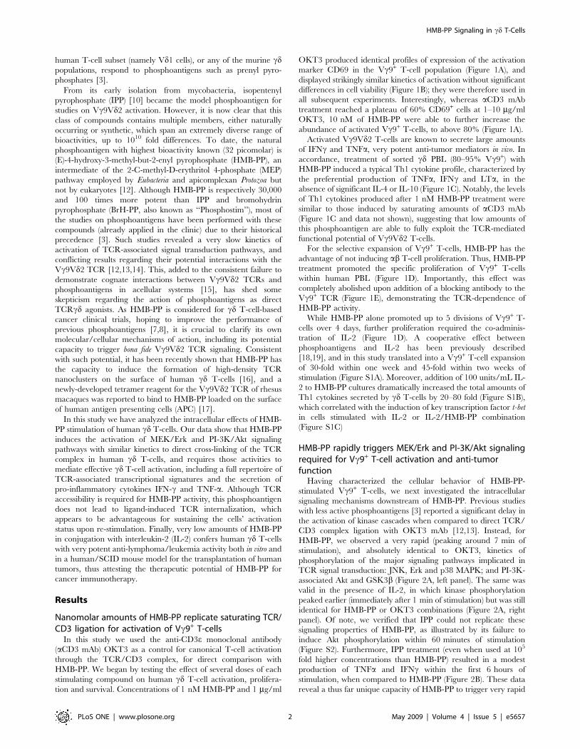
Nanomolar amounts of HMB-PP replicate saturating TCR/CD3 ligation for activation of Vc9+ T-cells
In this study we used the anti-CD3e monoclonal antibody
(aCD3 mAb) OKT3 as a control for canonical T-cell activation
through the TCR/CD3 complex, for direct comparison with
HMB-PP. We began by testing the effect of several doses of each
stimulating compound on human cd T-cell activation, prolifera-
tion and survival. Concentrations of 1 nM HMB-PP and 1 mg/ml
OKT3 produced identical profiles of expression of the activation
marker CD69 in the Vc9+ T-cell population (Figure 1A), and
displayed strikingly similar kinetics of activation without significant
differences in cell viability (Figure 1B); they were therefore used in
all subsequent experiments. Interestingly, whereas aCD3 mAb
treatment reached a plateau of 60% CD69+ cells at 1–10 mg/ml
OKT3, 10 nM of HMB-PP were able to further increase the
abundance of activated Vc9+ T-cells, to above 80% (Figure 1A).
Activated Vc9Vd2 T-cells are known to secrete large amounts
of IFNc and TNFa, very potent anti-tumor mediators in vivo. In
accordance, treatment of sorted cd PBL (80–95% Vc9+) with
HMB-PP induced a typical Th1 cytokine profile, characterized by
the preferential production of TNFa, IFNc and LTa, in the
absence of significant IL-4 or IL-10 (Figure 1C). Notably, the levels
of Th1 cytokines produced after 1 nM HMB-PP treatment were
similar to those induced by saturating amounts of aCD3 mAb
(Figure 1C and data not shown), suggesting that low amounts of
this phosphoantigen are able to fully exploit the TCR-mediated
functional potential of Vc9Vd2 T-cells.
For the selective expansion of Vc9+ T-cells, HMB-PP has the
advantage of not inducing ab T-cell proliferation. Thus, HMB-PP
treatment promoted the specific proliferation of Vc9+ T-cells
within human PBL (Figure 1D). Importantly, this effect was
completely abolished upon addition of a blocking antibody to the
Vc9+ TCR (Figure 1E), demonstrating the TCR-dependence of
HMB-PP activity.
While HMB-PP alone promoted up to 5 divisions of Vc9+ T-
cells over 4 days, further proliferation required the co-adminis-
tration of IL-2 (Figure 1D). A cooperative effect between
phosphoantigens and IL-2 has been previously described
[18,19], and in this study translated into a Vc9+ T-cell expansion
of 30-fold within one week and 45-fold within two weeks of
stimulation (Figure S1A). Moreover, addition of 100 units/mL IL-
2 to HMB-PP cultures dramatically increased the total amounts of
Th1 cytokines secreted by cd T-cells by 20–80 fold (Figure S1B),
which correlated with the induction of key transcription factor t-bet
in cells stimulated with IL-2 or IL-2/HMB-PP combination
(Figure S1C)
HMB-PP rapidly triggers MEK/Erk and PI-3K/Akt signalingrequired for Vc9+ T-cell activation and anti-tumorfunction
Having characterized the cellular behavior of HMB-PP-
stimulated Vc9+ T-cells, we next investigated the intracellular
signaling mechanisms downstream of HMB-PP. Previous studies
with less active phosphoantigens [3] reported a significant delay in
the activation of kinase cascades when compared to direct TCR/
CD3 complex ligation with OKT3 mAb [12,13]. Instead, for
HMB-PP, we observed a very rapid (peaking around 7 min of
stimulation), and absolutely identical to OKT3, kinetics of
phosphorylation of the major signaling pathways implicated in
TCR signal transduction: JNK, Erk and p38 MAPK; and PI-3K-
associated Akt and GSK3b (Figure 2A, left panel). The same was
valid in the presence of IL-2, in which kinase phosphorylation
peaked earlier (immediately after 1 min of stimulation) but was still
identical for HMB-PP or OKT3 combinations (Figure 2A, right
panel). Of note, we verified that IPP could not replicate these
signaling properties of HMB-PP, as illustrated by its failure to
induce Akt phosphorylation within 60 minutes of stimulation
(Figure S2). Furthermore, IPP treatment (even when used at 105
fold higher concentrations than HMB-PP) resulted in a modest
production of TNFa and IFNc within the first 6 hours of
stimulation, when compared to HMB-PP (Figure 2B). These data
reveal a thus far unique capacity of HMB-PP to trigger very rapid
HMB-PP Signaling in cd T-Cells
PLoS ONE | www.plosone.org 2 May 2009 | Volume 4 | Issue 5 | e5657
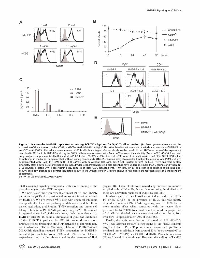
TCR-associated signaling, compatible with direct binding of the
phosphoantigen to the TCR complex.
We next tested the requirement on intact PI-3K and MAPK
pathways for cd T-cell activation and anti-tumor function induced
by HMB-PP. We pre-treated cd T-cells with chemical inhibitors
that specifically block those pathways and then analyzed the effects
on cell activation, proliferation, TNFa secretion and tumor cell
killing. Inhibition of PI-3K/Akt pathway using LY294002 resulted
in approximately half of the cells losing their responsiveness to
HMB-PP after 24–46 hours of stimulation (Figure 3A). Inhibition
of the MEK/Erk pathway by UO126 produced even more
dramatic effects, precluding HMB-PP-activation of approximately
two thirds of Vc9+ T-cells. Moreover, inhibition of PI-3K/Akt and
MEK/Erk signaling reduced TNFa production by HMB-PP-
activated cd T-cells to around 20% and 10% of control levels,
respectively, both in the absence and in the presence of IL-2
(Figure 3B). These effects were remarkably mirrored in cultures
supplied with aCD3 mAb, further demonstrating the similarity of
these two activation regimens (Figures 3A and 3B).
In what regards cd T-cell proliferation induced either by HMB-
PP or by OKT3 (in the presence of IL-2), this was mostly
dependent on intact PI-3K/Akt signaling, since UO126 had a
more modest effect when compared with the severe block
produced by LY294002 treatment, which reduced the proportion
of cd cells that divided twice or more over 4 days in culture, from
over 80% to approximately 20% (Figure 3C).
Finally, the anti-tumor function of sorted cd PBL (80–95%
Vc9+) was assessed through in vitro killing of the Jurkat leukemic
target cell line. HMB-PP pre-treatment augmented cd T-cell-
mediated tumor cell death from around 20% (non-activated cd) to
40% (1 nM HMB-PP) or 70% (10 nM HMB-PP) in a 6 hour assay
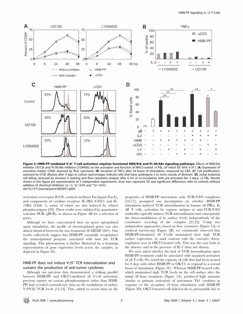
(Figure 3D and data not shown). However, the addition of UO126
Figure 1. Nanomolar HMB-PP replicates saturating TCR/CD3 ligation for Vc9+ T-cell activation. (A) Flow cytometry analysis for theexpression of the activation marker CD69 in MACS-sorted (97–98% purity) cd PBL, stimulated for 48 hours with the indicated amounts of HMB-PP oranti-CD3 mAb (OKT3). Shaded are non-stimulated Vc9+ T-cells. Percentages refer to cells above the threshold bar. (B) Time-course of the experimentdescribed in (A) for 1 nM HMB-PP and 1 mg/ml OKT3; cells were also stained with Annexin V to assess their viability (Annexin V2). (C) Cytokine beadarray analysis of supernatants of MACS-sorted cd PBL (of which 80–90% Vc9+) cultures after 24 hours of stimulation with HMB-PP or OKT3. RPMI refersto cells kept in media not supplemented with activating compounds. (D) CFSE dilution assays to monitor T-cell proliferation in total PBMC culturessupplemented with HMB-PP (1 nM) or OKT3 (1 mg/ml), with or without 100 U/mL rhIL-2. Cells (gated on Vc9+ or CD4+) were analyzed by flowcytometry after 4 days in culture; shaded are non-divided cells. Percentages indicate cells that have undergone more than 5 rounds of division. (E)CFSE dilution in gated Vc9+ T-cells within 6-day cultures of total PBMC activated with 1 nM HMB-PP in the presence or absence of blocking anti-TcRVc9 antibody. Dashed is a control incubated in 10% RPMI without HMB-PP. Results shown in this figure are representative of 3 independentexperiments.doi:10.1371/journal.pone.0005657.g001
HMB-PP Signaling in cd T-Cells
PLoS ONE | www.plosone.org 3 May 2009 | Volume 4 | Issue 5 | e5657
or/and LY294002 to the treatment reduced posterior leukemia
targeting to basal (20–30%) levels; this was also the case for the
more efficient (over 80% killing) combination of HMB-PP with IL-
2 (Figure 3D). Collectively, these data demonstrate an absolute
requirement of PI-3K/Akt- and MEK/Erk-mediated signal
transduction for HMB-PP-induced activation of anti-tumor
Vc9Vd2 T-cells.
HMB-PP signaling mimics the transcriptional eventsdownstream of TCR ligation
Signaling cascades ultimately produce alterations in gene
transcription, which can be effectively tracked by microarray
analysis. We employed this technology to compare the transcrip-
tomes of Vc9Vd2 T-cells activated with either HMB-PP or
OKT3. Both stimuli produced dramatic transcriptional changes:
when compared to non-stimulated cells, HMB-PP and OKT3
treatment resulted in 1359 and 1080 differences in gene expression
of 4-fold or above, respectively (Figure 4A; full microarray data
available on ArrayExpress via http://www.ebi.ac.uk/; accession
E-MEXP-1601). These were consistent across 3 individual
microarray experiments (Figure S2). Strikingly, a direct compar-
ison of the two stimuli revealed that they affected essentially the
same genes, as only 6 were differentially expressed (.4-fold)
between them (Table 1). Therefore, the transcriptional program
downstream HMB-PP appears to be extremely similar to that
induced by bona fide TCR signaling, as clearly illustrated by the
Volcano plots of Figure 4A.
The gene expression program shared by HMB-PP treatment
and direct TCR cross-linking involves, among many others targets
(E-MEXP-1601), the very high (above 16-fold) up-regulation of
pro-inflammatory genes IFNc and LTa, chemokines CCL8,
CCL2, CXCL9 and CXCL10, cell cycle mediator cyclin D2,
Figure 2. HMB-PP stimulation kinetically mimics Vc9+ TCR/CD3 signal transduction. (A) Phosphoimmunoblotting for kinases implicated inTCR signaling. MACS-sorted cd PBL (of which 80–95% Vc9+) were incubated with OKT3 (1 mg/ml) or HMB-PP (1 nM), in the absence (left panel) orpresence (right panel) of 100 U/mL rhIL-2, for the times indicated, or kept in control media (NS, non-stimulated). Results shown in this figure arerepresentative of 4 independent experiments. (B) TNFa and IFNc levels were measured by CBA in the culture supernatants of MACS-sorted cd PBL.Results were compared with the total amounts present in parallel cultures stimulated with 1 nM HMB-PP, and were expressed as percentages (IPP/HMB-PP).doi:10.1371/journal.pone.0005657.g002
HMB-PP Signaling in cd T-Cells
PLoS ONE | www.plosone.org 4 May 2009 | Volume 4 | Issue 5 | e5657
activation co-receptor ICOS, cytolysis mediator Fas ligand (Fas-L),
and components of cytokine receptors IL-2Ra (CD25) and IL-
15Ra (Table 1), many of which are also induced by related
phosphoantigens [20]. These results were validated by quantitative
real-time PCR (qPCR), as shown on Figure 4B for a selection of
genes.
Although we have concentrated here on genes upregulated
upon stimulation, the profile of downregulated genes was also
almost identical between the two treatments (E-MEXP-1601). Our
results collectively suggest that HMB-PP essentially recapitulates
the transcriptional program associated with bona fide TCR
signaling. This phenomenon is further illustrated by a heatmap
representation of gene expression levels across the samples, as
depicted in Figure S3.
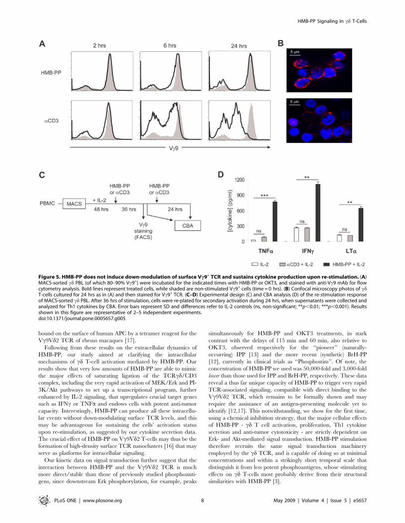
HMB-PP does not induce Vc9+ TCR internalization andsustains the production of anti-tumor cytokines
Although our previous data demonstrated a striking parallel
between HMB-PP- and OKT3-mediated cd T-cell activation,
previous reports on various phosphoantigens (other than HMB-
PP) had revealed contradictory data on the modulation of surface
Vc9Vd2 TCR levels [13,14]. This, added to recent data on the
properties of HMB-PP interactions with TCR/CD3 complexes
[16,17], prompted our investigation on whether HMB-PP
stimulation induced TCR internalization in human cd PBLs. In
ab T cells, activation by cognate antigen or anti-TCR/CD3
antibodies typically induces TCR internalization and consequently
the down-modulation of its surface levels independently of the
constitutive recycling of the complex [21,22]. Using two
independent approaches, based on flow cytometry (Figure 5A) or
confocal microscopy (Figure 5B), we consistently observed that
HMB-PP-stimulated cd T-cells maintained their high TCR
surface expression, in stark contrast with the extensive down-
regulation seen in OKT3-treated cells. This was the case both in
the absence and in the presence of IL-2 (data not shown).
We next asked whether the lack of TCR internalization upon
HMB-PP treatment could be associated with sustained activation
of cd T-cells. We tested the capacity of cells that had been treated
for 2 days with either HMB-PP or OKT3, to respond to a second
boost of stimulation (Figure 5C). Whereas HMB-PP-treated cells,
which maintained high TCR levels on the cell surface after the
initial 48 hour treatment (Figure 5A), produced high amounts
(similar to primary activation) of anti-tumor Th1 cytokines in
response to the secondary 24 hour stimulation with HMB-PP
(Figure 5D), OKT3-treated cells failed to do so, presumably due to
Figure 3. HMB-PP-mediated Vc9+ T-cell activation requires functional MEK/Erk and PI-3K/Akt signaling pathways. Effects of MEK/Erkinhibitor UO126 and PI-3K/Akt inhibitor LY294002 on the activation and function of MACS-sorted cd PBL (of which 85–95% Vc9+). (A) Expression ofactivation marker CD69, assessed by flow cytometry. (B) secretion of TNFa after 24 hours of stimulation, measured by CBA. (C) Cell proliferation,assessed by CFSE dilution after 4 days in culture (percentages indicate cells that have undergone 2 or more rounds of division). (D) Jurkat leukemiacell killing, assessed by Annexin V staining and flow cytometry analysis after 6 hrs of co-incubation with pre-activated (for 3 days) cd PBL. Resultsshown in this figure are representative of 3 independent experiments. Error bars represent SD and significant differences refer to controls withoutaddition of chemical inhibitors (n = 3, *p,0.05 and **p,0.01).doi:10.1371/journal.pone.0005657.g003
HMB-PP Signaling in cd T-Cells
PLoS ONE | www.plosone.org 5 May 2009 | Volume 4 | Issue 5 | e5657
their inability to respond to the mAb once their TCR complexes
have been internalized (Figure 5A–B). Although upon restimula-
tion with HMB-PP, IFNc became more abundant than TNFa(Figure 5D), contrary to the primary activation data (Figure 1C
and Figure S1B), the cytokine profile of the two HMB-PP-based
protocols were qualitatively very similar and consistently Th1-
biased (Figure 5D and data not shown). These data show that
HMB-PP is remarkably capable of sustaining Vc9Vd2 T-cell
activation and the production of anti-tumor cytokines, which are
critical parameters in immunotherapy protocols.
HMB-PP plus IL-2 treatment promotes leukemia cellkilling in vitro and in vivo
Having characterized the intracellular mechanisms of HMB-
PP-mediated cd T-cell activation, we next evaluated the anti-
tumor potential of HMB-PP-based regimens. We selected
Figure 4. HMB-PP treatment reproduces the transcriptional alterations induced by TCR/CD3 ligation on cd T-cells. (A) Volcano plots ofDNA microarray comparisons between aCD3 (OKT3) mAb-treated, HMB-PP-treated and non-stimulated (‘‘resting’’) MACS-sorted cd PBL (of which 85–95% Vc9+). After 18 hours of incubation with the stimuli, RNA was extracted and submitted to Affymetrix GeneChip analysis. Represented are Fold-changes (‘‘biological significance’’) versus statistical significance (B-values). Black dots represent genes over 4-fold differentially expressed (DE)between samples; all other probed genes are depicted in grey. Genes selected as differentially expressed had adjusted p-values lower than 0.005.Results are representative of 3 independent microarray experiments (see Figure S2). (B) Real-time PCR validation of microarray results for a selectionof genes similarly induced by OKT3 and HMB-PP (from Table 1). Gene expression was quantified in independent samples of control and treated cells,also including an IL-2-treated sample. Error bars represent SD and significant differences refer to ‘‘resting’’ cells (n = 3, *p,0.05 and **p,0.01).doi:10.1371/journal.pone.0005657.g004
HMB-PP Signaling in cd T-Cells
PLoS ONE | www.plosone.org 6 May 2009 | Volume 4 | Issue 5 | e5657

leukemias as model tumors to employ in both in vitro and in vivo
assays. The in vitro system previously used with Jurkat cells
(Figure 3D) was applied to a larger panel of leukemia cell lines:
Molt-4 (T-cell), RCH-ACV (pre-B cell) and HL-60 (myeloid)
(Figures 6A–B). cd PBL (80–95% Vc9+) were treated with the
different stimulating agents for 72 hours, and then transferred to
plain media in co-culture with the leukemia cells. In just 3 hours,
more than 80% of leukemia cells were killed by the cd T-cells that
had been stimulated with a combination of HMB-PP with IL-2
(compared to less than 20% by non-activated cd T-cells), and such
a regimen was at least as effective as saturating aCD3 plus IL-2
(Figures 6A–B). Of note, aCD3 mAb or HMB-PP used in isolation
produced more modest increases in target-cell lysis (Figures 6A–B),
highlighting the importance of exogenous IL-2 for the full
activation of Vc9Vd2 T-cells [18,23] (Figure S1).
Taking into account the added relevance of pre-clinical in vivo
systems for the evaluation of the anti-tumor potential of
immunotherapy strategies, we adapted a model of transplantation
of human tumors into lymphopenic SCID mice, previously used
with human cd T-cells by Kabelitz and colleagues [24], and added
bioluminescent analysis of tumor development, which allows early
detection of tumors and temporal evaluation throughout the
course of treatment, in live animals and in real-time [25]. Four
weeks after tumor injection, mice that had received HMB-PP plus
IL-2-treated (activated and expanded over 12 days) cd PBL
showed significantly reduced tumor load (derived from Molt-4
leukemia cells) compared to control mice that did not receive cd
T-cells (Figures 6C–E). Furthermore, while most control had to be
sacrificed at week 4 due to excessive body weight loss, cd-treated
animals resisted wasting for longer, up to week 6 (Figures 6C–D
and data not shown). These results attest the capacity of HMB-PP-
expanded and activated cd T-cells to induce anti-tumor responses
in vivo, and support the application of this phosphoantigen in
conjugation with low amounts of IL-2 in clinical cancer settings.
Discussion
The stimulatory effect prenyl pyrophosphates have on Vc9Vd2
T-cells has been well documented and seems to require TCR
expression, as indicated by antibody blocking and gene transfer
experiments [26,27]. However, some of these experiments have
been difficult to reproduce, and all attempts at showing cognate
interactions between Vc9Vd2 TCRs and phosphoantigens in
acellular systems (including surface plasmon resonance and X-ray
crystallography of isolated complexes) have failed [15], probably
due to the requirement of an unknown phosphoantigen-presenting
molecule [12]. This has raised some skepticism on phosphoanti-
gens as TCRcd agonists, also stemming from the lack of precedent
for such type of compounds interacting with any other variable
region molecule, including all other cd TCRs in humans or mice.
However, recent data have highlighted particular properties of
HMB-PP within the large family of phophoantigens. Namely,
HMB-PP induces the formation of high-density Vc9Vd2 TCR
nanoclusters on the membrane of human cd T-cells [16], and is
Table 1. Transcriptional changes induced by HMB-PP or OKT3 (anti-CD3 mAb) in Vc9Vd2 T cells.
Similarly induced by HMB-PP and OKT3(a)
Linkb Gene Description Function HMBc OKT3c Differd
3458 IFNc Interferon-c Cytokine 9.53 8.70 0.83
6355 CCL8 Chemokine CC motif 8 Chemokine 8.09 8.08 0.01
114614 MIRN155 MicroRNA 155 MicroRNA 7.83 7.95 20.12
4049 LTa Lymphotoxin-a Cytokine 7.35 6.64 0.71
6347 CCL2 Chemokine CC motif 2 Chemokine 6.49 6.19 0.30
3559 IL2Ra IL-2R a chain Cytokine-R 6.34 7.10 20.76
4283 CXCL9 Chemokine CXC motif 9 Chemokine 6.14 6.14 0.00
3627 CXCL10 Chemokine CXC motif 10 Chemokine 5.75 4.87 0.88
894 CCND2 Cyclin D2 Cell cycle 5.62 5.25 0.37
3902 LAG3 Lymphocyte activation gene Activation-R 4.46 4.08 0.38
29851 ICOS Inducible T cell costimulator Activation-R 4.46 5.40 20.94
6504 SLAMF1 Signal transducer SLAM-1 Signaling 4.23 4.34 20.11
Differentially induced by HMB-PP or OKT3
Linkb Gene Description Function HMBc OKT3c Differd
7412 VCAM1 Vascular cell adhesion-R Adhesion 4.50 1.45 3.05
6373 CXCL11 Chemokine CXC motif 11 Chemokine 5.45 2.59 2.86
1493 CTLA4 Co-receptor CTLA-4 Activation-R 4.53 7.23 22.70
112744 IL17F IL-17 isoform F Cytokine 1.29 4.81 23.52
4094 MAF Transcription factor Maf Signaling 0.04 3.83 23.79
6374 CXCL5 Chemokine CXC motif 5 Chemokine 0.04 7.14 27.10
Values are log2[fold change] compared to non-stimulated cells, based on triplicate microarray experiments. (-R, receptor).aListed is a selection of genes implicated in T cell activation. Full cDNA microarray data available on ArrayExpress (E-MEXP-1601).bLocus link gene ID (for unequivocal gene identification).cLog2[fold change] relative to non-stimulated cells.dDifference in fold induction between HMB-PP-treated and OKT3-treated cells.doi:10.1371/journal.pone.0005657.t001
HMB-PP Signaling in cd T-Cells
PLoS ONE | www.plosone.org 7 May 2009 | Volume 4 | Issue 5 | e5657
bound on the surface of human APC by a tetramer reagent for the
Vc9Vd2 TCR of rhesus macaques [17].
Following from these results on the extracellular dynamics of
HMB-PP, our study aimed at clarifying the intracellular
mechanisms of cd T-cell activation mediated by HMB-PP. Our
results show that very low amounts of HMB-PP are able to mimic
the major effects of saturating ligation of the TCRcd/CD3
complex, including the very rapid activation of MEK/Erk and PI-
3K/Akt pathways to set up a transcriptional program, further
enhanced by IL-2 signaling, that upregulates crucial target genes
such as IFNc or TNFa and endows cells with potent anti-tumor
capacity. Interestingly, HMB-PP can produce all these intracellu-
lar events without down-modulating surface TCR levels, and this
may be advantageous for sustaining the cells’ activation status
upon re-stimulation, as suggested by our cytokine secretion data.
The crucial effect of HMB-PP on Vc9Vd2 T-cells may thus be the
formation of high-density surface TCR nanoclusters [16] that may
serve as platforms for intracellular signaling.
Our kinetic data on signal transduction further suggest that the
interaction between HMB-PP and the Vc9Vd2 TCR is much
more direct/stable than those of previously studied phosphoanti-
gens, since downstream Erk phosphorylation, for example, peaks
simultaneously for HMB-PP and OKT3 treatments, in stark
contrast with the delays of 115 min and 60 min, also relative to
OKT3, observed respectively for the ‘‘pioneer’’ (naturally-
occurring) IPP [13] and the more recent (synthetic) BrH-PP
[12], currently in clinical trials as ‘‘Phosphostim’’. Of note, the
concentration of HMB-PP we used was 50,000-fold and 3,000-fold
lower than those used for IPP and BrH-PP, respectively. These data
reveal a thus far unique capacity of HMB-PP to trigger very rapid
TCR-associated signaling, compatible with direct binding to the
Vc9Vd2 TCR, which remains to be formally shown and may
require the assistance of an antigen-presenting molecule yet to
identify [12,17]. This notwithstanding, we show for the first time,
using a chemical inhibition strategy, that the major cellular effects
of HMB-PP - cd T cell activation, proliferation, Th1 cytokine
secretion and anti-tumor cytotoxicity - are strictly dependent on
Erk- and Akt-mediated signal transduction. HMB-PP stimulation
therefore recruits the same signal transduction machinery
employed by the cd TCR, and is capable of doing so at minimal
concentrations and within a strikingly short temporal scale that
distinguish it from less potent phosphoantigens, whose stimulating
effects on cd T-cells most probably derive from their structural
similarities with HMB-PP [3].
Figure 5. HMB-PP does not induce down-modulation of surface Vc9+ TCR and sustains cytokine production upon re-stimulation. (A)MACS-sorted cd PBL (of which 80–90% Vc9+) were incubated for the indicated times with HMB-PP or OKT3, and stained with anti-Vc9 mAb for flowcytometry analysis. Bold lines represent treated cells, while shaded are non-stimulated Vc9+ cells (time = 0 hrs). (B) Confocal microscopy photos of cdT-cells cultured for 24 hrs as in (A) and then stained for Vc9+ TCR. (C–D) Experimental design (C) and CBA analysis (D) of the re-stimulation responseof MACS-sorted cd PBL. After 36 hrs of stimulation, cells were re-plated for secondary activation during 24 hrs, when supernatants were collected andanalyzed for Th1 cytokines by CBA. Error bars represent SD and differences refer to IL-2 controls (ns, non-significant; **p,0.01; ***p,0.001). Resultsshown in this figure are representative of 2–5 independent experiments.doi:10.1371/journal.pone.0005657.g005
HMB-PP Signaling in cd T-Cells
PLoS ONE | www.plosone.org 8 May 2009 | Volume 4 | Issue 5 | e5657

The use of a microbial compound for the activation of human
anti-tumor lymphocytes fits the overall strategy of providing
immune adjuvants (like viral nucleic acids for CD8+ T-cells) for
cancer therapy. Compared to other T-cell agonists, HMB-PP
offers the advantage of specifically activating a T-cell population
with overt effector function, devoid of known immune suppressive
(‘‘regulatory’’) subsets. Moreover, Vc9Vd2 T-cells are broadly
reactive to tumors, potentially allowing them to be used to treat a
variety of cancers.
The data presented in this report provide a framework for
designing novel immunotherapy protocols using cd T-cells, and
encourage the use of HMB-PP in clinical settings. cd T-cell-
mediated tumor surveillance should evidently be seen as
complementary to the adaptive component provided by MHC-
restricted ab T-cells upon priming by dendritic cells. Importantly,
Vc9Vd2 T-cells can also induce monocyte and DC maturation
[28,29,30], on one hand; and even act as CD80/86-expressing
antigen-presenting cells that prime ab T-cells, on the other [31].
Furthermore, cd T-cells are prototypic representatives of uncon-
ventional lymphocytes with innate anti-tumor capacity, alike NK
and NKT-cells, all of which recognize tumors independently of
classical MHC presentation [2]. We believe the success of cancer
immunotherapy will critically depend on the integration of
conventional and unconventional lymphocyte responses [32] to
tackle the multiple immune evasion strategies developed by
tumors.
Materials and Methods
Ethics statementAll experiments involving animals (rodents) were performed in
compliance with the relevant laws and institutional guidelines and
have been approved by the Instituto de Medicina Molecular
animal ethics committee.
In vitro cultures of human peripheral blood lymphocytesPeripheral blood was collected from anonymous healthy
volunteers, diluted 1:1 (v/v) with PBS(16) (Invitrogen Gibco)
Figure 6. Leukemia cell killing by HMB-PP-activated cd T-cells. (A) In vitro lysis of Molt-4 leukemia cells. MACS-sorted cd PBL (of which 85–95% Vc9+) were pre-activated for 72 hours with 1 or 10 mg/ml aCD3 mAb (OKT3), or 1 or 10 nM HMB-PP in the absence of IL-2, and also combined atthe lower concentrations with IL-2 (100 U/ml). For the killing assay, DDAO-SE-labelled Molt-4 cells and pre-activated cd PBL were co-incubated for3 hours in media devoid of activating compounds. Samples were then stained with Annexin V to identify dying (Annevin V+) tumor (DDAO-SE+) cellsby flow cytometry. (B) Data summary for killing assays (as in A) performed with three distinct leukemia cell lines. (C–D) Bioluminescent imaging ofNOD/SCID mice inoculated with luciferase+ Molt-4 leukemic cells, with (D) or without (C) co-injection of pre-activated cd PBL, analyzed weekly asdescribed in Materials and Methods. (E) LivingImage quantification of photon signals (tumor load) collected at day 28 of the experiment illustrated in(C–D). Comparison of cd-treated and control animals (n = 5, p,0.05). Data in this figure are representative of 3 (A–B) or 2 (C–E) independentexperiments.doi:10.1371/journal.pone.0005657.g006
HMB-PP Signaling in cd T-Cells
PLoS ONE | www.plosone.org 9 May 2009 | Volume 4 | Issue 5 | e5657
and centrifuged in LSM Lymphocyte Separation Medium (MP
Biomedicals) in a volume ratio of 3:4 (3 parts of LSM for 4 of
diluted blood) for 15 minutes at 1500 rpm and 25uC. The
interfase containing PBMC was collected, washed in PBS (16)
and cultured at 16106 cells/mL at 37uC, 5% CO2 in round-
bottom 96 well plates with RPMI 1640 with 2 mM L-Glutamine
(Invitrogen Gibco) supplemented with 10% foetal bovine serum
(Invitrogen Gibco), 1 mM Sodium Pyruvate (Invitrogen Gibco),
50 mg/mL of penicillin/streptomycin (Invitrogen Gibco), in the
presence or absence of 100 U/mL of rhIL-2 (Roche Applied
Science), 1–10 nM of HMB-PP (4-hydroxy-3-methyl-but-2-enyl
pyrophosphate) (a kind gift from H. Jomaa and M. Eberl), and 1-
10 ug/ml of soluble anti-CD3 antibody (eBioscience, clone
OKT3).
For TCR blockade, freshly-isolated PBMC were CFSE-labeled
and then incubated for 6 days with anti- TcRVc9 (Beckman
Coulter, clone IMMU360) diluted 1:20 in complete medium
supplemented with 1 nM HMB-PP.
For the phophoimmunoblotting experiments, MACS-isolated
cd T cells were expanded with 100 U/mL rhIL-2 for 15 days.
To study the effects of chemical inhibitors of signal transduc-
tion, the MEK inhibitor UO126 and the PI-3K inhibitor
LY294002 (both from Calbiochem) were added at 10 mM for a
2-hour incubation period, and then transferred to fresh medium
(without inhibitors).
Magnetic cell sorting and flow cytometry analysiscd T-cells were isolated (to above 95% purity) from PBMC by
magnetic cell sorting via positive selection with a FITC-labeled
anti-TCRcd antibody (Miltenyi Biotec). For flow cytometry
analysis (on a FACSCalibur, BD Biosciences), cells were labelled
with fluorescent monoclonal antibodies: anti-CD69-PE (BD
Pharmingen), anti-TcRVc9-PC5 (Beckman Coulter) and anti-
CD4-PerCP (BD Pharmingen). In all cultures the percentage of
Vc9+ T-cells was evaluated by flow cytometry. Cell proliferation
was measured by following a standard CFSE staining protocol
(CellTrace CFSE Cell Proliferation Kit, Invitrogen; final concen-
tration 0.5 mM), while apoptosis was assessed by AnnexinV-FITC
(BD Pharmingen) staining. Cells were counted in Mossbauer
chambers using 0.4% Trypan Blue solution (Sigma-Aldrich) for
viability control.
Cytometric Bead Array (CBA)Cytokine secretion was measured using Cytometric Bead Array
(CBA) technology (BD Biosciences). Cells were seeded with the
respective activators at 26105 cells/well, culture supernatants
were collected at different time points and analyzed on a
FACSCanto (BD Biosciences) using a custom-made Flex Set with
five different cytokine capture beads: LT-a, IL-10, IL-4, TNF-aand IFN-c. Data were analyzed using the FCAP Array Software
v1.0.1 running on BD FACSDiva (BD Biosciences).
Protein isolation and phosphoimmunoblottingCells were incubated at 37uC with pre-warmed PBS alone or
with HMB-PP (1 nM) or OKT3 (1 mg/mL). Reactions were
stopped by placing samples on ice and adding ice-cold PBS. Cell
lysates were prepared and equal amounts of protein were analyzed
by 10% SDS-PAGE electrophoresis, transferred onto nitrocellu-
lose membranes, and immunoblotted with the following mAbs or
antisera: Actin, phospho-Erk (Y204) (Santa Cruz Biotechnology),
ZAP-70 and phospho-STAT5A/B (Y694/Y699) (Upstate Bio-
technology), phospho-Akt (S473), phospho-GSK-3b (S9), phos-
pho-JNK/SAPK (Y183/185), phospho-p38 MAPK (Y180/182)
(Cell Signalling Technology), and phospho-LCK (Y505) (Trans-
duction Laboratories). Immunodetection was performed with
horseradish peroxidase-conjugated secondary antibody and devel-
oped by chemiluminescence as described [33]. Whenever
necessary membranes were striped using 15 mM TRIS pH 6.8
plus 2% SDS and b-Mercaptoethanol (100 mM) for 40 minutes at
57uC.
RNA isolation and Affymetrix GeneChip analysisRNA labeling, hybridization to the Affymetrix GeneChip
Human Genome U133 plus 2.0 Arrays and scanning was
performed by the Affymetrix Core Facility, Instituto Gulbenkian
de Ciencia, Portugal as described below.
Total RNA was extracted using the RNeasy Mini Kit according
to manufacture’s protocol (Qiagen, Hilden, Germany). Concen-
tration and purity was determined by spectrophotometry and
integrity was confirmed using an Agilent 2100 Bioanalyzer with a
RNA 6000 Nano Assay (Agilent Technologies, Palo Alto, CA).
RNA was processed for use on Affymetrix (Santa Clara, CA, USA)
GeneChip Human Genome U133 Plus 2.0 Arrays, according to
the manufacturer’s One-Cycle Target Labeling Assay. Arrays
were scanned on an Affymetrix GeneChip scanner 3000 7G.
All the microarray data analysis was done using R and several
packages available from CRAN (R Development Core Team,
2008) and Bioconductor. The raw data (CEL files) was normalized
and summarized with the Robust MultiArray Average method
from the affy package.
The differentially expressed genes were selected using linear
models and empirical Bayes methods as implemented in limma
package, verifying the p-values corresponding to moderated F-
statistics, and selecting as differentially expressed genes those that
had adjusted p-values lower than 0.005.
Real-time quantitative PCRTotal RNA was reverse-transcribed into cDNA using random
hexamers and Superscript II first strand synthesis reagents
(Invitrogen). qPCR was performed on ABI Prism 7700 Sequence
Detection System using SYBR Green detection system (both from
PE Applied Biosystems). Primers were designed using Primer3
v.0.4.0 online program (http://primer3.sourceforge.net). Primer
sequences are available upon request. For each transcript,
quantification was done using the calibration curve method. b2-
microglobulin was used as the internal control for normalization.
All samples were run in triplicate and repeated three times.
Analysis of the qPCR results was performed using the ABI SDS
v1.1 sequence analysis software (Applied Biosystems).
Tumor cell cultures and in vitro killing assaysAll tumor cell lines were cultured in complete 10% RPMI 1640
(as above), maintained at 16105 up to 26106 cells/mL by dilution
and splitting 1:3 every 3–4 days.
For cytotoxicity assays, magnetically purified cd PBL were pre-
activated for 72 hours with 1–10 mg/mL aCD3 mAb (OKT3) or
1–10 nM HMB-PP either in the absence or presence of IL-2
(100 U/mL). Tumor cell lines were stained with CellTracer Far
Red DDAO-SE (1 mM) (Molecular Probes, Invitrogen) and each
36104 tumour cells were incubated with 36105 cd T-cells in
RPMI devoid of activating compounds, for 3 hours at 37uC and
5% CO2 on a round-bottom 96 well plate. Cells were then stained
with Annexin V-FITC and analyzed by flow cytometry.
Confocal microscopyCells were stained at 4uC with mouse anti-human TCR
Vgamma9-PC5 (Beckman Coulter) primary antibody, and with
HMB-PP Signaling in cd T-Cells
PLoS ONE | www.plosone.org 10 May 2009 | Volume 4 | Issue 5 | e5657

anti-mouse Alexa Fluor 633 (Invitrogen, Molecular Probes)
secondary antibody. Cells were then fixed with 4% Paraformalde-
heyde for 15 minutes at 4uC. Nuclear DNA content was stained
for with DAPI Fluoromount G (Southern Biotech). Immunoflu-
orescence microscopy was performed with a LSM 510 META
confocal microscope (Zeiss). Separate images were collected with a
636objective for each fluorochrome and then overlaid to obtain a
multicolor image.
Bioluminescent imaging of transplanted leukemiadevelopment in SCID mice
107 Molt-4 T-cell leukemia cells stably expressing firefly
luciferase and GFP were injected i.v. in groups of 6 NOD/SCID
mice per experiment, either in isolation or together with 56107 cdPBL (.80% Vc9+), previously expanded and activated in vitro with
1 nM HMB-PP for 12 days. Treated mice received boosts of
56107 cd PBL i.v. on day 14 and 10,000 U IL-2 i.p. twice every
week, whereas control mice received only IL-2. All mice were
analyzed on a weekly basis by in vivo imaging (IVIS, Caliper
Lifesciences) upon intra-peritoneal injection of luciferin. Photon
signals were quantified with LivingImage software (Caliper
Lifesciences). Mouse body weight was measured weekly, and
animals suffering from wasting (loss of over 20% of initial body
weight) were sacrificed.
Statistical analysisStatistical significance of differences between subpopulations
was assessed using Student’s t-test and is indicated when significant
as *, p,0.05; **, p,0.01; ***, p,0.001.
Supporting Information
Figure S1 Exogenous IL-2 expands HMB-PP-activated Vg9+T-cells and up-regulates their Th1 cytokine profile. (A) Absolute
numbers of Vg9+ cells in PBMC cultures stimulated with HMB-
PP (1 nM) or OKT3 (1 ug/ml), supplemented or not with IL-2
(100 U/ml). Cells were analyzed by flow cytometry and light
microscopy (Mossbauer chamber cell counts). (B) Cytokine bead
array (CBA) analysis of supernatants of MACS-sorted gd PBL (of
which 80–90% Vg9+) after 24 hours of stimulation with HMB-PP
or anti-CD3 mAb (OKT3). Represented is the ratio between the
cytokine amounts produced in the presence (100 U/ml) and in the
absence of IL-2. (C) Real-time PCR quantification of t-bet mRNA
expression in activated Vg9Vd2 T-cells, normalized with Beta2-
microglobulin. Cells were pre-incubated for 6 hours with the
activating compounds (or kept in RPMI as control). Significant
differences refer to cells cultured in RPMI in the absence of IL-2
(n = 3, *p,0.05 and **p,0.01).
Found at: doi:10.1371/journal.pone.0005657.s001 (0.07 MB TIF)
Figure S2 Akt phosphorylation in response to IPP versus HMB-
PP stimulation of gd PBL. MACS-sorted gd PBL were activated
with 10 uM IPP or 1 nM HMB-PP for the indicated times. Cell
lysates were analyzed by SDS-PAGE and immunoblotted for
Phospho-Akt (P-Akt) or Beta-Actin on nitrocellulose membranes.
Densitometry for P-Akt bands was normalized with Beta-Actin
loading controls. Data correspond to the induction of Akt
phosphorylation above basal levels, i.e., after subtraction of the
unstimulated control levels.
Found at: doi:10.1371/journal.pone.0005657.s002 (0.05 MB TIF)
Figure S3 Heatmap of non-stimulated, HMB-PP-treated and
anti-CD3 mAb (OKT3)-treated gd T-cells. The DNA microarray
expression value for each gene is normalized across the samples;
levels greater than the mean in a given sample are colored in red,
and those below the mean are depicted in blue. Exp1-3 are
triplicate independent microarray experiments. Note the striking
similarity between HMB-PP-treated and anti-CD3-treated sam-
ples.
Found at: doi:10.1371/journal.pone.0005657.s003 (0.34 MB TIF)
Acknowledgments
The authors greatly thank A. Hayday, H. Sicard, Innate Pharma, H.
Jomaa, M. Eberl, E. Scotet, D. Pennington, D. Vermijlen, A. Roberts, S.
Dias, C. Casalou, J. Becker, A. Gomes, L. Graca and GenoMed.
Author Contributions
Conceived and designed the experiments: DVC FSd JTB BSS. Performed
the experiments: DVC FSd BAC TL Ad. Analyzed the data: DVC FSd
ARG JTB BSS. Contributed reagents/materials/analysis tools: LRM.
Wrote the paper: BSS.
References
1. Stagg J, Johnstone RW, Smyth MJ (2007) From cancer immunosurveillance to
cancer immunotherapy. Immunol Rev 220: 82–101.
2. Gomes AQ, Correia DV, Silva-Santos B (2007) Non-classical major histocom-
patibility complex proteins as determinants of tumour immunosurveillance.EMBO Rep 8: 1024–1030.
3. Morita CT, Jin C, Sarikonda G, Wang H (2007) Nonpeptide antigens,
presentation mechanisms, and immunological memory of human Vgamma2V-
delta2 T cells: discriminating friend from foe through the recognition of prenylpyrophosphate antigens. Immunol Rev 215: 59–76.
4. Girardi M, Oppenheim DE, Steele CR, Lewis JM, Glusac E, et al. (2001)
Regulation of cutaneous malignancy by gammadelta T cells. Science 294:605–609.
5. Kunzmann V, Wilhelm M (2005) Anti-lymphoma effect of gammadelta T cells.Leuk Lymphoma 46: 671–680.
6. Corvaisier M, Moreau-Aubry A, Diez E, Bennouna J, Mosnier JF, et al. (2005) Vgamma 9V delta 2 T cell response to colon carcinoma cells. J Immunol 175:
5481–5488.
7. Wilhelm M, Kunzmann V, Eckstein S, Reimer P, Weissinger F, et al. (2003)
Gammadelta T cells for immune therapy of patients with lymphoidmalignancies. Blood 102: 200–206.
8. Dieli F, Vermijlen D, Fulfaro F, Caccamo N, Meraviglia S, et al. (2007)
Targeting human {gamma}delta} T cells with zoledronate and interleukin-2 for
immunotherapy of hormone-refractory prostate cancer. Cancer Res 67:7450–7457.
9. Constant P, Davodeau F, Peyrat MA, Poquet Y, Puzo G, et al. (1994)
Stimulation of human gamma delta T cells by nonpeptidic mycobacterial
ligands. Science 264: 267–270.
10. Tanaka Y, Morita CT, Tanaka Y, Nieves E, Brenner MB, et al. (1995) Natural
and synthetic non-peptide antigens recognized by human gamma delta T cells.Nature 375: 155–158.
11. Gober HJ, Kistowska M, Angman L, Jeno P, Mori L, et al. (2003) Human T cellreceptor gammadelta cells recognize endogenous mevalonate metabolites in
tumor cells. J Exp Med 197: 163–168.
12. Thedrez A, Sabourin C, Gertner J, Devilder MC, Allain-Maillet S, et al. (2007)
Self/non-self discrimination by human gammadelta T cells: simple solutions for
a complex issue? Immunol Rev 215: 123–135.
13. Lafont V, Liautard J, Sable-Teychene M, Sainte-Marie Y, Favero J (2001)
Isopentenyl pyrophosphate, a mycobacterial non-peptidic antigen, triggersdelayed and highly sustained signaling in human gamma delta T lymphocytes
without inducing eown-modulation of T cell antigen receptor. J Biol Chem 276:15961–15967.
14. Sireci G, Espinosa E, Di Sano C, Dieli F, Fournie JJ, et al. (2001) Differentialactivation of human gammadelta cells by nonpeptide phosphoantigens.
Eur J Immunol 31: 1628–1635.
15. Bonneville M, Scotet E (2006) Human Vgamma9Vdelta2 T cells: promising newleads for immunotherapy of infections and tumors. Curr Opin Immunol 18:
539–546.
16. Chen Y, Shao L, Ali Z, Cai J, Chen ZW (2008) NSOM/QD-based nanoscale
immunofluorescence imaging of antigen-specific T-cell receptor responsesduring an in vivo clonal V{gamma}2V{delta}2 T-cell expansion. Blood 111:
4220–4232.
17. Wei H, Huang D, Lai X, Chen M, Zhong W, et al. (2008) Definition of APC
presentation of phosphoantigen (E)-4-hydroxy-3-methyl-but-2-enyl pyrophos-
phate to Vgamma2Vdelta2 TCR. J Immunol 181: 4798–4806.
HMB-PP Signaling in cd T-Cells
PLoS ONE | www.plosone.org 11 May 2009 | Volume 4 | Issue 5 | e5657
18. Casetti R, Perretta G, Taglioni A, Mattei M, Colizzi V, et al. (2005) Drug-
induced expansion and differentiation of V gamma 9V delta 2 T cells in vivo: therole of exogenous IL-2. J Immunol 175: 1593–1598.
19. Ali Z, Shao L, Halliday L, Reichenberg A, Hintz M, et al. (2007) Prolonged (E)-
4-hydroxy-3-methyl-but-2-enyl pyrophosphate-driven antimicrobial and cyto-toxic responses of pulmonary and systemic Vgamma2Vdelta2 T cells in
macaques. J Immunol 179: 8287–8296.20. Yamashita S, Tanaka Y, Tsutsumi S, Aburatani H, Minato N, et al. (2005)
Analysis of mechanism for human gammadelta T cell recognition of nonpeptide
antigens. Biochem Biophys Res Commun 334: 349–360.21. Dietrich J, Menne C, Lauritsen JP, von Essen M, Rasmussen AB, et al. (2002)
Ligand-induced TCR down-regulation is not dependent on constitutive TCRcycling. J Immunol 168: 5434–5440.
22. Naramura M, Jang IK, Kole H, Huang F, Haines D, et al. (2002) c-Cbl and Cbl-b regulate T cell responsiveness by promoting ligand-induced TCR down-
modulation. Nat Immunol 3: 1192–1199.
23. Vermijlen D, Ellis P, Langford C, Klein A, Engel R, et al. (2007) Distinctcytokine-driven responses of activated blood gammadelta T cells: insights into
unconventional T cell pleiotropy. J Immunol 178: 4304–4314.24. Kabelitz D, Wesch D, Pitters E, Zoller M (2004) Characterization of tumor
reactivity of human V gamma 9V delta 2 gamma delta T cells in vitro and in
SCID mice in vivo. J Immunol 173: 6767–6776.25. Shu ST, Nadella MV, Dirksen WP, Fernandez SA, Thudi NK, et al. (2007) A
novel bioluminescent mouse model and effective therapy for adult T-cellleukemia/lymphoma. Cancer Res 67: 11859–11866.
26. Bukowski JF, Morita CT, Band H, Brenner MB (1998) Crucial role of TCR
gamma chain junctional region in prenyl pyrophosphate antigen recognition bygamma delta T cells. J Immunol 161: 286–293.
27. Bukowski JF, Morita CT, Tanaka Y, Bloom BR, Brenner MB, et al. (1995) V
gamma 2V delta 2 TCR-dependent recognition of non-peptide antigens andDaudi cells analyzed by TCR gene transfer. J Immunol 154: 998–1006.
28. Ismaili J, Olislagers V, Poupot R, Fournie JJ, Goldman M (2002) Humangamma delta T cells induce dendritic cell maturation. Clin Immunol 103:
296–302.
29. Devilder MC, Maillet S, Bouyge-Moreau I, Donnadieu E, Bonneville M, et al.(2006) Potentiation of antigen-stimulated V gamma 9V delta 2 T cell cytokine
production by immature dendritic cells (DC) and reciprocal effect on DCmaturation. J Immunol 176: 1386–1393.
30. Eberl M, Roberts GW, Meuter S, Williams JD, Topley N, et al. (2009) A rapidcrosstalk of human gammadelta T cells and monocytes drives the acute
inflammation in bacterial infections. PLoS Pathog 5: e1000308.
31. Brandes M, Willimann K, Moser B (2005) Professional antigen-presentationfunction by human gammadelta T Cells. Science 309: 264–268.
32. Pennington DJ, Vermijlen D, Wise EL, Clarke SL, Tigelaar RE, et al. (2005)The integration of conventional and unconventional T cells that characterizes
cell-mediated responses. Adv Immunol 87: 27–59.
33. Barata JT, Silva A, Brandao JG, Nadler LM, Cardoso AA, et al. (2004)Activation of PI3K is indispensable for interleukin 7-mediated viability,
proliferation, glucose use, and growth of T cell acute lymphoblastic leukemiacells. J Exp Med 200: 659–669.
HMB-PP Signaling in cd T-Cells
PLoS ONE | www.plosone.org 12 May 2009 | Volume 4 | Issue 5 | e5657