E3 ubiquitin protein ligase, E6-associated protein (E6AP) regulates PI3K-Akt signaling and prostate...
9
E3 ubiquitin protein ligase, E6-associated protein (E6-AP) regulates PI3K-Akt signaling and prostate cell growth ☆ Sathish Srinivasan, Zafar Nawaz ⁎ Department of Biochemistry & Molecular Biology, University of Miami Miller School of Medicine, 1580 NW 10 Avenue, Miami, FL 33136, USA abstract article info Article history: Received 18 March 2010 Received in revised form 20 August 2010 Accepted 27 August 2010 Available online 6 September 2010 Keywords: E6-AP PI3K-AKT Androgen receptor Prostate gland RhoA This study elucidates the role of E6-associated protein, E6-AP (a dual function steroid hormone receptor coactivator and ubiquitin–protein ligase) in the regulation of PI3K-Akt signaling pathway, prostate gland growth and proliferation. Here, we report the generation of transgenic mice and prostate cancer cell line, LNCaP cells that overexpress E6-AP protein. Using these models we show that the levels of total Akt and phosphorylated Akt (active Akt) are increased in E6-AP overexpressing prostate gland and LNCaP cells suggesting that E6-AP regulates the PI3K-Akt signaling pathway. The prostate glands in our transgenic mice are ~20% larger and produce preneoplastic lesions at the age of 18 months. Our data also suggest that E6-AP modulates PI3K-Akt signaling pathway by both androgen-independent and -dependent mechanisms. In the androgen-independent mechanism, E6-AP modulates PI3K-Akt signaling by regulating the protein levels of RhoA, a small GTPase, which is a negative regulator of the Akt signaling pathway. Further, we show that E6-AP, a known coactivator of AR, amplifies the androgen-dependent activation of PI3K-Akt signaling pathway. In addition, we show that stable overexpression of E6-AP in prostate cancer cells results in increased cell size and proliferation. Overall our data suggests that E6-AP regulates both the positive and negative modulators of the PI3K-Akt pathway in prostate cells which results in increased prostate cell growth, proliferation and decreased apoptosis.This article is part of a Special Issue entitled The 26S Proteasome: When degradation is just not enough! © 2010 Elsevier B.V. All rights reserved. 1. Introduction E3 ubiquitin–protein ligase enzyme, E6-associated protein (E6- AP), is a novel dual function steroid hormone receptor coactivator [1,2]. E6-AP not only interacts with and enhances the hormone-dependent transcriptional activities of various steroid hormone receptors, includ- ing androgen receptor (AR), but also is a member of the E3 class of functionally related ubiquitin–protein ligases [3–5]. E3 ubiquitin– protein ligases have been proposed to play a major role in defining the substrate specificity of the ubiquitin–proteasome system. Protein ubiquitination involves two other classes of enzymes, the E1 ubiquitin- activating enzyme (UBA) and E2 ubiquitin-conjugating enzymes (UBCs). UBA first activates ubiquitin in an ATP-dependent manner, and the activated ubiquitin then forms a thioester bond between the carboxyl-terminal glycine residue of ubiquitin and a cysteine residue of the UBA. Next, ubiquitin is transferred from the E1 to one of several E2s (UBCs), preserving the high energy thioester bond. In some cases, ubiquitin is transferred directly from E2 to the target protein through an isopeptide bond between the ε-amino group of lysine residues of the target protein and the carboxy terminus of ubiquitin. In other instances, the transfer of ubiquitin from UBCs to target proteins proceeds through an E3 ubiquitin–protein ligase intermediate, such as E6-AP. Finally, the ubiquitin-tagged target proteins undergo degradation via the 26 S proteasome pathway. Since, E6-AP acts both as a coactivator and an E3 ubiquitin–protein ligase, it is postulated that E6-AP serves to link two important and opposing activities in a cell, the transcription and the protein degradation [6,7]. Previously, we have shown that the prostate gland is smaller in E6-AP knockout animals implying that E6-AP is required for the proper development and growth of prostate gland [1]. Furthermore, we also showed that protein levels of the components of phospha- tidylinositol 3-kinase/protein kinase B (PI3K-Akt) signaling pathway are decreased in E6-AP knockout animals. In addition, we showed that E6-AP regulates the protein levels and transcriptional activity of AR in prostate cells, suggesting that E6-AP plays important roles in the cytoplasm in addition to acting as a coactivator in the nucleus. Since, the PI3K-Akt pathway has been described as a dominant growth survival pathway in prostate cells and elevated PI3K-Akt signaling is correlated with prostate cancer progression [8–12], the main focus of this study is to understand the effect of modulation of this pathway by E6-AP in prostate gland growth and proliferation in different model systems like E6-AP overexpressing transgenics and LNCaP prostate cancer cells. Akt, also known as protein kinase B (PKB), is a proto-oncogene with a pleckstrin homology and serine/threonine kinase domains. The Biochimica et Biophysica Acta 1809 (2011) 119–127 ☆ This article is part of a Special Issue entitled The 26S Proteasome: When degradation is just not enough! ⁎ Corresponding author. Department of Biochemistry & Molecular Biology, Braman Family Breast Cancer Institute (M-877), University of Miami Miller School of Medicine, Batchelor Bldg., Room 416, 1580 NW 10 Avenue, Miami, FL 33136, USA. Tel.: +1 305 243 1456; fax: +1 305 243 4787. E-mail address: [email protected] (Z. Nawaz). 1874-9399/$ – see front matter © 2010 Elsevier B.V. All rights reserved. doi:10.1016/j.bbagrm.2010.08.011 Contents lists available at ScienceDirect Biochimica et Biophysica Acta journal homepage: www.elsevier.com/locate/bbagrm
-
Upload
independent -
Category
Documents
-
view
6 -
download
0
Transcript of E3 ubiquitin protein ligase, E6-associated protein (E6AP) regulates PI3K-Akt signaling and prostate...

Biochimica et Biophysica Acta 1809 (2011) 119–127
Contents lists available at ScienceDirect
Biochimica et Biophysica Acta
j ourna l homepage: www.e lsev ie r.com/ locate /bbagrm
E3 ubiquitin protein ligase, E6-associated protein (E6-AP) regulates PI3K-Aktsignaling and prostate cell growth☆
Sathish Srinivasan, Zafar Nawaz ⁎Department of Biochemistry & Molecular Biology, University of Miami Miller School of Medicine, 1580 NW 10 Avenue, Miami, FL 33136, USA
☆ This article is part of a Special Issue entitled The 26S Pis just not enough!⁎ Corresponding author. Department of Biochemistry
Family Breast Cancer Institute (M-877), University of MBatchelor Bldg., Room 416, 1580 NW 10 Avenue, Miam243 1456; fax: +1 305 243 4787.
E-mail address: [email protected] (Z. Nawaz)
1874-9399/$ – see front matter © 2010 Elsevier B.V. Aldoi:10.1016/j.bbagrm.2010.08.011
a b s t r a c t
a r t i c l e i n f oArticle history:Received 18 March 2010Received in revised form 20 August 2010Accepted 27 August 2010Available online 6 September 2010
Keywords:E6-APPI3K-AKTAndrogen receptorProstate glandRhoA
This study elucidates the role of E6-associated protein, E6-AP (a dual function steroid hormone receptorcoactivator andubiquitin–protein ligase) in the regulation of PI3K-Akt signalingpathway, prostate glandgrowthand proliferation. Here, we report the generation of transgenic mice and prostate cancer cell line, LNCaP cellsthat overexpress E6-APprotein. Using thesemodelswe show that the levels of total Akt and phosphorylated Akt(active Akt) are increased in E6-AP overexpressing prostate gland and LNCaP cells suggesting that E6-APregulates the PI3K-Akt signaling pathway. The prostate glands in our transgenic mice are ~20% larger andproduce preneoplastic lesions at the age of 18 months. Our data also suggest that E6-AP modulates PI3K-Aktsignaling pathway by both androgen-independent and -dependent mechanisms. In the androgen-independentmechanism, E6-APmodulates PI3K-Akt signaling by regulating the protein levels of RhoA, a small GTPase,whichis a negative regulator of the Akt signaling pathway. Further, we show that E6-AP, a known coactivator of AR,amplifies the androgen-dependent activation of PI3K-Akt signaling pathway. In addition, we show that stableoverexpression of E6-AP in prostate cancer cells results in increased cell size and proliferation. Overall our datasuggests that E6-AP regulates both the positive and negative modulators of the PI3K-Akt pathway in prostatecellswhich results in increasedprostate cell growth, proliferation anddecreasedapoptosis.This article is part of aSpecial Issue entitled The 26S Proteasome: When degradation is just not enough!
roteasome:When degradation
& Molecular Biology, Bramaniami Miller School of Medicine,i, FL 33136, USA. Tel.: +1 305
.
l rights reserved.
© 2010 Elsevier B.V. All rights reserved.
1. Introduction
E3 ubiquitin–protein ligase enzyme, E6-associated protein (E6-AP), is a novel dual function steroid hormone receptor coactivator [1,2].E6-AP not only interacts with and enhances the hormone-dependenttranscriptional activities of various steroid hormone receptors, includ-ing androgen receptor (AR), but also is a member of the E3 class offunctionally related ubiquitin–protein ligases [3–5]. E3 ubiquitin–protein ligases have been proposed to play a major role in definingthe substrate specificity of the ubiquitin–proteasome system. Proteinubiquitination involves two other classes of enzymes, the E1 ubiquitin-activating enzyme (UBA) and E2 ubiquitin-conjugating enzymes(UBCs). UBA first activates ubiquitin in an ATP-dependent manner,and the activated ubiquitin then forms a thioester bond between thecarboxyl-terminal glycine residue of ubiquitin and a cysteine residue ofthe UBA. Next, ubiquitin is transferred from the E1 to one of several E2s(UBCs), preserving the high energy thioester bond. In some cases,ubiquitin is transferred directly fromE2 to the target protein through an
isopeptide bond between the ε-amino group of lysine residues of thetarget protein and the carboxy terminus of ubiquitin. In other instances,the transfer of ubiquitin from UBCs to target proteins proceeds throughan E3 ubiquitin–protein ligase intermediate, such as E6-AP. Finally, theubiquitin-tagged target proteins undergo degradation via the 26 Sproteasome pathway. Since, E6-AP acts both as a coactivator and an E3ubiquitin–protein ligase, it is postulated that E6-AP serves to link twoimportant and opposing activities in a cell, the transcription and theprotein degradation [6,7].
Previously, we have shown that the prostate gland is smaller inE6-AP knockout animals implying that E6-AP is required for theproper development and growth of prostate gland [1]. Furthermore,we also showed that protein levels of the components of phospha-tidylinositol 3-kinase/protein kinase B (PI3K-Akt) signaling pathwayare decreased in E6-AP knockout animals. In addition, we showed thatE6-AP regulates the protein levels and transcriptional activity of AR inprostate cells, suggesting that E6-AP plays important roles in thecytoplasm in addition to acting as a coactivator in the nucleus. Since, thePI3K-Akt pathway has been described as a dominant growth survivalpathway in prostate cells and elevated PI3K-Akt signaling is correlatedwith prostate cancer progression [8–12], the main focus of this study isto understand the effect of modulation of this pathway by E6-AP inprostate gland growth and proliferation in different model systems likeE6-AP overexpressing transgenics and LNCaP prostate cancer cells.
Akt, also known as protein kinase B (PKB), is a proto-oncogenewith a pleckstrin homology and serine/threonine kinase domains. The

120 S. Srinivasan, Z. Nawaz / Biochimica et Biophysica Acta 1809 (2011) 119–127
kinase activity of Akt is stimulated by a variety of extracellular stimuli,such as growth factors and cytokines leading to the phosphorylationand regulation of a wide spectrum of its substrates involved inmultiple cellular processes, including cell survival, cell growth, celldifferentiation, cell cycle progression, cell proliferation and cellularmetabolism [13]. Akt is activated by PI3K, a heterodimer composedof a p85 regulatory and a p110 catalytic subunit. In response to avariety of stimuli, Akt is phosphorylated at Threonone-308 (Thr-308) by 3-phosphoinositide-dependent protein kinase-1 and atSerine-473 (Ser-473) by integrin-linked kinase. Increasing evidenceindicates that Akt plays an important role in tumorigenesis [14–16]. ThePI3K-Akt pathway which is normally activated by growth factors is alsoactivated by steroid hormones like estrogens and androgens [17–20].The activation of PI3K-Akt pathway by androgens results in increasedcell growth and anti-apoptotic activity [17,20]. Previously publishedstudies indicate that androgen regulates PI3K-Akt pathway via the ARbut independent of its transcriptional activity, suggesting an importantrole of androgen receptors within the cytoplasm. However, the role forcoactivator proteins in the regulation of PI3K-Akt pathway wasunknown until the recent finding that steroid receptor coactivator-3(SRC-3) is able to inducePI3K-Akt pathway in an androgen-independentmanner [21]. The induction of cell proliferation and growth by SRC-3 hasbeen attributed to its ability to induce Akt activity and expression.Therefore steroid hormone receptor coactivator proteins in addition totheir transcriptional activation can also contribute to growth andproliferation by modulating important growth signaling pathways inthe cytoplasm, independent of transcription.
In order to elucidate the specific roles of E6-AP (a dual functionsteroid receptor coactivator) in the regulation of PI3K-Akt pathway,prostate cell growth and survival, we have generated transgenic micewhich specifically overexpresses E6-AP in the prostate gland and alsoE6-AP stable LNCaP cells (here on referred as E6-AP-LNCaP) thatoverexpress exogenous E6-AP [1]. Our data from thesemodels suggestthat E6-AP is involved in modulation of PI3K-Akt signaling pathway,prostate cell growth and proliferation, prostate gland growth andtumorigenesis.
2. Materials and methods
2.1. Cell culture
LNCaP (parental cell line) and E6-AP-stable cell line (E6-AP-LNCaP) were maintained in RPMI 1640 medium (Invitrogen, GrandIsland, NY) supplemented with 10% fetal bovine serum at 37 °C in 5%CO2. E6-AP stable cells were maintained in RPMI 1640 mediumsupplemented with 10% fetal bovine serum at 37 °C in 5% CO2 in thepresence of Doxycycline (2 μg/ml).
2.2. Generation of E6-AP transgenic mice
Transgenic vector containing wild-type E6-AP with Flag tag(MKbpAII/Pb-Flag-E6-AP) was constructed and digested with NotIand KpnI to generate vector free fragments, purified and wasmicroinjected into fertilized C57BL6 one-cell embryos. The injectedembryos were then implanted into the oviducts of pseudo-pregnantrecipientmothers. The pups received from the transgenic facility werescreened for the presence of E6-AP transgene by PCR. Three wild-typeE6-AP transgenic lines were established.
2.3. siRNA transfection
E6-AP siRNAswere procured from Dharmacon (Chicago, IL) as ON-TARGET plus Smart Pools. Non-targeting siRNAs (siScramble) wereused as negative controls. siRNAs were reverse transfected into LNCaPcells using Lipofectamine RNAimax (Invitrogen) as per the manufac-turer's protocol. Briefly, 25 nM siRNA was diluted in 0.5 ml of
OPTIMEM reduced serum medium (Invitrogen) along with 5 μl ofLipofectamine RNAimax on each well of a 6 well plate. 1×10⁵ cells in1.5 ml of antibiotic free RPMI 1640 was added to the transfectionmixture. 24 h later the media was replaced with phenol red free RPMI1640 supplemented with 5% charcoal stripped FBS with antibiotics.
2.4. Western blot analysis
Cells were grown as indicated; afterward cells were harvested andwashed in TEN buffer (40 mM Tris-HCl (pH 7.5), 1 mM EDTA, and150 mM NaCl). Then cells were lysed in ice-cold RIPA buffer (20 mMTris pH 7.5, 150 mM Nacl, 1% NP40, 0.5% Sodium deoxycholate, 1 mMEDTA and 0.1% SDS) by pipetting up and down. Thereafter, cell lysateswere placed on ice for 30 min and cleared by centrifugation at 12,000gfor 20 min at 4 °C. The supernatants were collected and frozen at−80 °C until used for analysis. The protein concentrations of lysateswere measured using the Bio-Rad protein assay kit.
Twenty-five to 50 μg of total protein from each sample wasresolved on 10% sodium dodecyl sulfate-polyacrylamide gels (SDS-PAGE) and transferred onto nitrocellulose membranes (Protran,Schleicher & Schuell, Inc., Keene, NH). Membranes were blockedwith 5% nonfat dry milk in Tris-buffered saline (20 mM Tris base (pH7.5) and 150 mM NaCl) containing 0.05% Tween 20 (TBS-T), thenprobed with the primary antibody. The primary antibodies werediluted in 1% nonfat dry milk in TBS-T as indicated and used forimmunoblotting: Anti-E6-AP (1:1000 dilution; Bethyl laboratories, Inc.,Montgomery, TX); anti-total Akt (1:1200 dilution), anti-phosphoAkt(1:1000 dilution), anti-PI3-Kinase and p85 (1:1000 dilution), anti-caspase-3 (8G10; 1:1000 dilution), and anti-Bax (1:750 dilution; CellSignaling Technology, Beverly, MA); anti-RhoA (26C4; 1:800 dilution;Santa Cruz Biotechnology, Inc., Santa Cruz, CA); and anti-ß-actin(1:10,000 dilution; Sigma-Aldrich Corp.). After washing in TBS-T,membranes were incubated with their appropriate horseradishperoxidase-conjugated secondary antibodies (Bio-Rad Laboratories,Inc.) and developed using an enhanced chemiluminescence detec-tion system (Amersham Biosciences, Arlington Heights, IL) accordingto the instructions of the manufacturer and were exposed to X-Rayfilm (Phenix research products, Hayward, CA).
2.5. In vitro protein–protein interaction assay
Radiolabeled RhoA protein was synthesized using a rabbitreticulocyte-coupled in vitro transcription and translation (TNT) kitin the presence of 35 S-methionine according to the manufacturer'srecommendations (Promega Corp.). GST-E6-AP and control GSTproteins were expressed in Escherichia coli DH-5 α cells andimmobilized on glutathione-Sepharose beads. The glutathione-bound GST and GST-E6-AP were incubated with in vitro-synthesizedRhoA G14V protein in NETN buffer (100 mM NaCl, 1 mM EDTA,20 mM Tris (pH 8.0), and 0.5% Nonidet P-40) for 2–3 h at roomtemperature or overnight at 4 °C. After washing four times with NETNbuffer, E6-AP-bound RhoA protein was isolated by boiling theSepharose beads in 1× sodium dodecyl sulfate gel loading bufferand were separated on a 10% SDS-PAGE, followed by autoradiograpy.
2.6. Proliferation assays
Cell proliferation was measured using TACS MTT assay (Trevigen,Inc., Gaithersburg, MD). Briefly, LNCaP and E6-AP-LNCaP cells weregrown in 96-well cell culture plates at a density of 2×10³ cells/well in100 μl stripped and normal serum containing medium for 24 h in 5%CO2 at 37 °C. Every day, 10 μl of MTT reagent was added to each well,and the cells were further incubated for 4 h at 37 °C to allow theformation of a purple precipitate. The precipitates were dissolved byadding 100 μl of detergent reagent to each well and the plates wereincubated in dark over night. The absorbance at 570 nm was
121S. Srinivasan, Z. Nawaz / Biochimica et Biophysica Acta 1809 (2011) 119–127
measured using a microplate reader (Biorad laboratories, Hercules,CA). For siRNA experiments, cell proliferation was measured bycounting total cell number by hemocytometer.
2.7. Statistical analysis
Data were presented as mean ± SE from 3 independentexperiments. Statistical analysis was carried out with Student's ttest. Pb0.05 was considered as statistically significant difference.
3. Results
3.1. Characterization of E6-AP overexpressing transgenic mice
Previous studies using E6-AP null mice have shown that E6-AP isrequired for normal prostate gland development. To test the effect ofover-expression of wild-type E6-AP in the development of the normalprostate gland, we generated a new transgenic mouse model, whichutilizes the prostate-specific rat probasin (rPB) promoter to target theexpressionofwild-typehumanE6-AP to themouseprostate epithelium.The efficiency of expression of transgenes in prostate gland will becontingent upon both the sites of integration of the transgene and thenumber of integrated copies per genome. This was assayed usingWestern blot analysis of tissue-specific expression of the transgenes in avariety of organs such as brain, prostate gland, testes and seminal
Fig. 1. Characterization of E6-AP transgenic mice. (A & B) RepresentativeWestern blots fromAP in different tissues. E6-AP is specifically over expressed in the prostate glands of transgenitype and E6-AP over expressing transgenic mice. The wet weights were normalized with bothan wild-type prostate glands (*Pb0.05, n=15). (D) Western blots showing levels of totaWestern blots showing lobe specific expression of E6-AP, p-Akt and phospho GSK β (p-GSK
vesicle. As shown in Fig. 1A and B, wild-type E6-AP is specifically overexpressed in transgenicmiceprostate glandswhen comparedwith non-transgenic littermate controls. Since E6-AP over expression was notobserved in other tissues of transgenic lines, this indicates that thetransgene expression was specific to the prostate gland.
To study the consequences of overexpression of wild-type E6-AP inprostate gland development, the total prostate glands from non-transgenic and transgenic males (12–18 weeks old) were comparedmorphologically. As shown in Fig. 1C, the wet weight of total prostateglands of wild-type E6-AP transgenic glands were approximately 20%more than their littermate controls. We have previously characterizedE6-AP knockout prostate glands by analyzing the expression of variousproteins like PI3K, Akt and RhoA [1]. These studies revealed the possiblemechanismbywhichE6-APmight influence thedevelopment of normalprostate gland. Now, it is of interest to know how over expression ofwild-type E6-AP affects the expression of these proteins during prostateglanddevelopment. The levels of PI3K, total Akt andphosphorylatedAkt(Ser 473) from prostate glands of wild-type E6-AP transgenic mice wascompared with non transgenic mice (24 weeks) by Western blotanalyses. Fig. 1D shows that the levels of total and phosphorylated Aktare increased inWild-type E6-AP transgenics comparedwith littermatecontrols.
Mice prostate is structurally divided into four pairs of lobes: anterior,dorsal, ventral and lateral. It has been reported that there are lobespecific differences in transgene expression driven by the probasin
two lines of E6-AP over expressing transgenic mice showing the expression levels of E6-c mice. (WT—Wild-type, Tr—Transgenic). (C)Wet weights of prostate glands fromwild-dy weights. Prostate glands from E6-AP overexpressing transgenics weigh ~20% morel and phospho Akt (p-Akt) are elevated in E6-AP over-expressing transgenic mice. (E)β) in E6-AP over-expressing transgenic mice.
122 S. Srinivasan, Z. Nawaz / Biochimica et Biophysica Acta 1809 (2011) 119–127
promoter. In addition to that there are lobe specific differences in certainbiochemical and pathological conditions. Hence we wanted to know ifthere is a lobe specific expression of E6-AP in the transgenic mice.Prostate gland from transgenic and Wild-type mice was individuallydissected into anterior, ventral and dorsolateral lobes, proteins wereisolated and Western blots were performed. Fig. 1E shows that E6-APwas over expressed in all the lobes, when compared with Wild-typelittermates, indicating that transgene is expressed in all lobes. Eventhough, there was a difference in the basal level of expression of E6-APamong the different lobes, there was no difference in the extent of overexpression of transgene among the different lobes. We also wanted toexamine if there are lobe specific differences inAktprotein levels. Fig. 1Eshows the expression levels of Phosho-Akt are different in differentprostate gland lobes. The p-Akt is over expressed in E6-AP transgeniclobes compared to theWild-type gland.We also examined the levels ofphosphorylated formof glycogensynthase kinase-3β (p-Gskβ),which isa target of activated Akt. We observed that p-Gskβwas also elevated inE6-AP transgenic prostate glands (Fig. 1E).
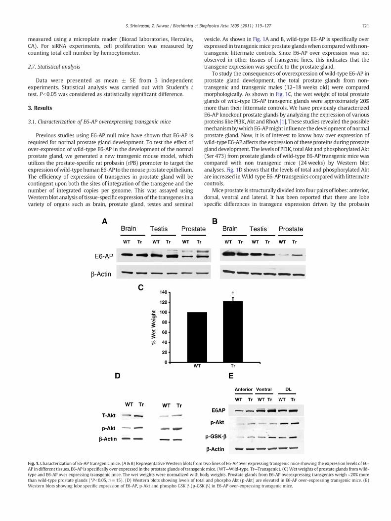
3.2. Over expression of E6-AP in prostate glands leads to preneoplasticlesions
Initiation and progression of prostate cancer is a multistage processinvolving a characteristic lesion termed as prostate intraepithelial
Fig. 2. Histology of prostate glands from E6-AP transgenic mice. Representative sections fromPIN can be identified in transgenic sections (arrows).
neoplasia (PIN), believed to be the precursor for the formation ofprostate cancers. Since E6-AP transgenic mice did not give raise topalpable prostate tumors, we decided to perform a histologicalobservation of the prostate glands. Prostate glands from N18monthold transgenic and Wild-type litter mate controls were dissected intoindividual lobes (n=10). These tissues were processed, embedded inparaffin and 5 m sections were sliced. These sectioned tissues werestained for haematoxylin and eosin to determine the morphologicalfeatures. Fig. 2 shows that E6-AP transgenic mice develop hyperplasiccharacteristics resembling PIN when compared with Wild-type littermate controls (arrows). This finding suggests that over expression ofE6-AP could result in excessive proliferation and formation ofpreneoplastic lesions, which resembles PIN. These lesions were foundin 7 out of 10 transgenicmouse prostate glands, but nonewere found inWild-type prostate glands.
3.3. E6-AP modulates PI3K-Akt signaling
It is known that PI3K-Akt signaling pathway plays a central role inthe development and progression of prostate cancer [14–16]. Ourprevious studies showed that PI3K-Akt signaling is down regulated inthe prostate glands of E6-AP knockout mice [1]. In order to study theeffect of over expression of E6-AP on PI3K-Akt signaling pathway, weutilized E6-AP-LNCaP cells and examined the expression levels of
wild-type and transgenic mice stained for H&E. Precancerous lesions characteristic of

123S. Srinivasan, Z. Nawaz / Biochimica et Biophysica Acta 1809 (2011) 119–127
PI3K, total Akt and p-Akt (active Akt) and compared it with that ofcontrol parental LNCaP cells. Fig. 3A shows that the levels ofexogenously expressed Flag-E6-AP are less in uninduced condition(Dox treated cells) when compared to induced conditions (Doxuntreated cells), indicating that E6-AP expression is regulated in Dox-dependent manner. Furthermore, E6-AP-LNCaP stable clones expresshigh levels of E6-AP compared to control LNCaP cells. As shown inFig. 3B, the levels of total and p-Akt (Ser-473) is higher in the E6-AP-LNCaP cells compared to the control parental LNCaP cells. Similarly,the levels of PI3K are also high in E6-AP-LNCaP cells compared to thatof untransfected control LNCaP cells (Fig. 3C). However, underconditions where the expression of exogenous E6-AP (+Dox) isblocked, the levels of PI3K, total Akt and phospho-Akt are notsignificantly different from that of control LNCaP cells. The treatmentof untransfected LNCaP cells with Dox does not significantly affect thelevels of these proteins, which serves as a control. These experimentssuggest that E6-AP is a key regulator of PI3K-Akt signaling pathway.These results are consistent with our previously published data fromE6-AP knockout animals, suggesting that E6-AP is an importantmodulator of PI3K-Akt pathway in prostate glands. To test if E6-APamplifies the hormone-dependent up-regulation of PI3K-Akt signal-ing, we cultured untransfected control LNCaP cells and E6-AP-LNCaPcells in 5% charcoal stripped medium supplemented with or withoutsynthetic androgen R1881 for 24 h. As shown in Fig. 3D, treatment ofcontrol LNCaP cells with R1881 for 1 h and 36 h increases the levels ofp-Akt and E6-AP further amplifies the hormone-dependent activationof Akt in E6-AP-LNCaP cells (Fig. 3D).
3.4. E6-AP regulates Akt activity via RhoA
Our previous studies indicate that small GTPase, RhoA, a negativeregulator of Akt activity is increased in E6-AP knockout prostate glands
Fig. 3. Over expression of E6-AP induces PI3K-Akt signaling pathway. (A) Western blots shLNCaP cells and E6-AP-LNCaP cells were treated with or without Dox for 48 h. Increased proteLNCaP cells under E6-AP over expressing conditions (−Dox). β-actin was used as a loading cofor 48 h. Increased protein levels of PI3K was observed in E6-AP-LNCaP cells under E6-AP oanalysis of p-Akt (Ser-473) levels from LNCaP cells and E6-AP-LNCaP stable cells grown in 5%1 h and 36 h.
[1]. We have also shown that inhibition of RhoA activity in prostatecancer cells increases Akt activity. Here we tested the levels of RhoA inour E6-AP-LNCaP cells, Fig. 4A shows that the levels of RhoA aredecreased under E6-AP induced conditions (−Dox) in our E6-AP-LNCaPcells. Since RhoA is a known target of the ubiquitin–proteasomepathway and its levels are decreased in E6-AP-LNCaP cells underE6-AP over expressing conditions, we hypothesized that, E6-APinteracts with RhoA, ubiquitinates it and induces its degradation via theubiquitin–proteasome pathway. To investigatewhether RhoA levels arecontrolled by the ubiquitin–proteasome pathway in prostate cancercells, we treated LNCaP cells with either vehicle DMSO or proteasomeinhibitor, MG132. Fig. 4B shows that RhoA protein levels are stabilizedby MG132, indicating that RhoA protein levels in prostate cells areregulated by the ubiquitin–proteasome pathway. Since, E6-AP is an E3ubiquitin–protein ligase enzyme, in order forE6-AP toact asa specific E3ubiquitin–protein ligase for RhoA it should interact with RhoA. To testthis possibility, we utilized glutathione-S-transferase (GST) pull downassay. 35S-Methionine-labeled RhoA protein was incubated with eithercontrol protein (GST only) or GST-E6-AP protein. Fig. 4C depicts asignificant interaction of RhoAwith E6-AP suggesting that E6-APmaybea putative E3 ubiquitin–protein ligase for RhoA in prostate cells.
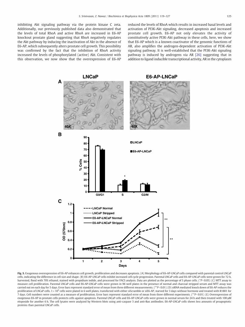
3.5. Over expression of E6-AP increases the cell growth and proliferation
The members of the PI3K-Akt signaling pathway have beenimplicated in cell growth and proliferation. Since, over expression ofE6-AP induces the PI3K-Akt pathway, we predicted that E6-AP-LNCaPcells will exhibit increased cell growth and proliferation compared tothat of untransfected control LNCaP cells. As shown in Fig. 5A, E6-AP-LNCaP cells that over express E6-AP exhibit changes in cell shapecompared with control LNCaP cells. E6-AP-LNCaP cells lacks cellprocesses and also clump together. Furthermore, E6-AP stable LNCaP
owing inducible expression of Flag-E6-AP in E6-AP-LNCaP stable clones. (B) Parentalin levels of total Akt and phosphorylated-Akt (p-Akt, Ser-473) were observed in E6-AP-ntrol. (C) Parental LNCaP cells and E6-AP-LNCaP cells were treated with or without Doxver expressing conditions (−). β-actin was used as a loading control. (D) Western blotcharcoal stripped serum supplemented with or without synthetic androgen R1881 for

Fig. 4. Involvement of RhoA in E6-AP-mediated regulation of Akt activity. (A) Westernblots showing that the levels of RhoA are decreased under E6-AP overexpressingconditions (−Dox) in E6-AP-LNCaP stable cells compared to that of parental LNCaPcells. (B) To determine if RhoA is target of the ubiquitin–proteasome pathway inprostate cells, LNCaP cells were treated with DMSO or proteasome inhibitor, MG132and cell lysates were examined for RhoA levels. Western blots showing RhoAstabilization with MG132 treatment. (C) In vitro interaction of E6-AP with RhoA. Thehuman RhoA protein was synthesized in vitro using the TNT-coupled reticulocyte lysatesystem. RhoA protein was then incubated with GST-E6-AP fusion protein that wasbounded to glutathione-Sepharose beads. The glutathione-bounded proteins wereseparated on SDS-PAGE, followed by autoradiography. Twenty percent of the TNTreaction was used as input, and GST alone was used as a negative control.
124 S. Srinivasan, Z. Nawaz / Biochimica et Biophysica Acta 1809 (2011) 119–127
cells show increase in cell size compared to that of control LNCaP cells.In addition to cell shape, we also examined the effects of E6-APoverexpression on prostate cell cycle progression using propidiumiodide staining of the DNA content of E6-AP-LNCaP cells and controlLNCaP cells. As shown in Fig. 5B, E6-AP-LNCaP cells have increasednumber of cells in S phase compared with that of control LNCaP cells.These data suggest that over expression of E6-AP leads to prolifer-ation, whichmay be due to the upregulation of PI3K-Akt pathway.Wealso confirmed our results using MTT (3-(4,5-Dimethylthiazol-2-yl)-2,5-diphenyltetrazolium bromide) growth assays. E6-AP-LNCaP cellsand untransfected control LNCaP cells were grown for a period of5 days in media containing either normal or charcoal stripped serum.Fig. 5C shows a significant difference in proliferation of E6-AP-LNCaPcells compared with untransfected control LNCaP cells both in normaland stripped serum suggesting that over expression of E6-AP resultsin increased cell proliferation both in an androgen dependent andindependent manner. These data confirm that over expression ofE6-AP leads to prostate cell proliferation and growth, which correlateswith our previous finding that loss of E6-AP leads to reduced prostategland size and increased apoptosis.We also tested if loss of E6-AP couldaffect proliferation of LNCaP cells. siScramble or siE6-AP transfectedLNCaP cells were starved without hormone for 72 h and then treatedwith R1881 for 7 days and proliferation was assessed by enumeratingthe cell number at the end of each day. As shown in Fig. 5D siScrambletransfected cells proliferate normally in the presence of R1881, whereassiE6-AP transfected cells do no proliferate as siScramble transfected
cells. This indicates that E6-AP is required for the normal proliferationof LNCaP cells.
Our previous data from E6-AP knock-out mice shows that loss ofE6-AP leads to impaired prostate gland development. We also showedthat this might be due to elevated apoptosis in E6-AP knockoutprostate glands. It is of interest to see if an E6-AP over expressingcondition provides a protective effect against apoptosis. To test thiswe cultured parental LNCaP cells and E6-AP-LNCaP cells and treatedthem with 100 μM etoposide to induce apoptotic stress. Then celllysates were probed for pro-apoptotic markers like caspase 3 and Baxusing Western immunobloting. As shown in Fig. 5E, the levels ofcleaved caspase 3 and Bax are decreased in E6-AP-LNCaP cellscompared to untransfected LNCaP cells indicating that E6-AP overexpression provides the cells with survival advantage by protectingthem from apoptosis.
4. Discussion
Coactivators enhance the transactivation functions of nuclearhormone receptors by means of their diverse array of enzymaticactivities [22]. Recent studies have suggested that coactivators canalso act as key regulators of signaling cascades, indicating thatcoactivators also possess functions that are distinct from theirtranscriptional activation functions [23]. E6-AP is an importantmember of the ubiquitin–proteasome pathway and has also beencharacterized as a coactivator for several steroid hormone receptorsincluding AR [2]. In our previous report, we have shown that loss ofE6-AP affects the growth of the prostate gland [1]. We also identifiedthe mechanisms by which E6-AP affects the prostate gland growthand we showed that E6-AP affects both the protein levels andfunctions of AR. In addition, we also demonstrated that the proteinlevels of PI3K, total Akt and p-Akt are decreased in E6-AP knockoutprostate gland, suggesting that apart from its coactivation function,E6-AP may also modulate the functions of PI3K-Akt signalingpathway. In this study, we have utilized E6-AP over expressingtransgenic mouse models to verify the observations from the E6-APnull mouse model. Here we show that over expression of E6-AP in themouse prostate has a modest increase in wet weight of prostate gland.Additionally, prostate glands in these mice show elevated Akt levelsand activity. Although not dramatic, our transgenic mouse model hasthe opposite phenotypic characteristics as that of E6-AP null mice.Also in this study, we provide evidence which suggests that E6-APmodulates the activity and functions of PI3K-Akt signaling andregulates prostate cell proliferation, prostate gland growth andtumorigenesis. Furthermore, our data also indicate that the compo-nents of the PI3K-Akt signaling pathway are elevated when E6-AP isover expressed in prostate cells.
Based on our data, we postulate that E6-AP regulates PI3K-Aktsignaling by both androgen-independent and dependent mechan-isms. In the androgen-independent mechanism, E6-AP enhances thebasal protein levels and activity of the PI3K-Akt signaling pathway.The hormone-independent activation of PI3K-Akt signaling by E6-APis consistent with previously published study which suggests that thesteroid receptor coactivator, SRC-3 can stimulate cell growth bymodulating the Akt signaling pathway in an androgen-independentmanner, but it is still unknown how SRC-3 regulates Akt signaling[21,24]. However, in this manuscript, we show the androgen-independent activation of PI3K-Akt signaling by E6-AP is throughthe down regulation of small GTPase, RhoA in prostate cancer cells.The idea that E6-AP regulates PI3K-Akt signaling and prostate cellgrowth by modulating the protein levels of RhoA is supported by ourdata which show that E6-AP interacts with RhoA and may potentiallyinduce its degradation via the ubiquitin–proteasome pathway inprostate cells. Previously, we and others have shown that RhoAnegatively regulates PI3K-Akt signaling pathway [1,25]. It has alsobeen shown that RhoA can promote prostate cancer cell apoptosis by
125S. Srinivasan, Z. Nawaz / Biochimica et Biophysica Acta 1809 (2011) 119–127
inhibiting Akt signaling pathway via the protein kinase C zeta.Additionally, our previously published data also demonstrated thatthe levels of total RhoA and active RhoA are increased in E6-APknockout prostate gland suggesting that RhoA negatively regulatesthe Akt pathway by inducing the inactivation of Akt in the absence ofE6-AP, which subsequently alters prostate cell growth. This possibilitywas confirmed by the fact that the inhibition of RhoA activityincreased the levels of phosphorylated (active) Akt. Consistent withthis observation, we now show that the overexpression of E6-AP
Fig. 5. Exogenous overexpression of E6-AP enhances cell growth, proliferation and decreasescells, indicating the difference in cell size and shape. (B) E6-AP-LNCaP cells exhibit increasedharvested, fixed with 70% ethanol, stained with propidium iodide, and processed for FACS ameasure cell proliferation. Parental LNCaP cells and E6-AP-LNCaP cells were grown in 96 wcarried out on each day for 5 days. Error bars represent standard error of mean from three difproliferation of LNCaP cells. 1×10⁵ cells were plated in 6 well plates, transfected with either7 days. Cell numbers were counted as a measure of proliferation. Error bars represent standexogenous E6-AP in prostate cells protects cells against apoptosis. Parental LNCaP cells andetoposide for another 6 h. The cell lysates were analyzed by Western blots using anti-caspproteins than parental LNCaP cells.
reduced the levels of RhoA which results in increased basal levels andactivation of PI3K-Akt signaling, decreased apoptosis and increasedprostate cell growth. E6-AP not only elevates the activity ofconstitutively active PI3K-Akt pathway in these cells, here, we showthat E6-AP which is a known coactivator of the genomic functions ofAR, also amplifies the androgen-dependent activation of PI3K-Aktsignaling pathway. It is well-established that the PI3K-Akt signalingpathway is induced by androgens via AR [26] suggesting that inaddition to ligand inducible transcriptional activity, AR in the cytoplasm
apoptosis. (A) Morphology of E6-AP-LNCaP cells compared with parental control LNCaPcell cycle progression. Parental LNCaP cells and E6-AP-LNCaP cells were grown for 72 h,nalysis. Data are plotted as the percentage of S phase cells. (*Pb0.05) (C) MTT assay toell plates in the presence of normal and charcoal stripped serum and MTT assay was
ferentmeasurements. (**Pb0.01) (D) siRNAmediated knock down of E6-AP reduces thesiScramble or siE6-AP, starved for 3 days without hormone and treated with R1881 forard error of mean from three different experiments. (**Pb0.01) (E) Overexpression ofE6-AP-LNCaP cells were grown in normal serum for 24 h and then treated with 100 μMase 3 and anti-Bax antibodies. E6-AP-LNCaP cells shows less amounts of proapoptotic
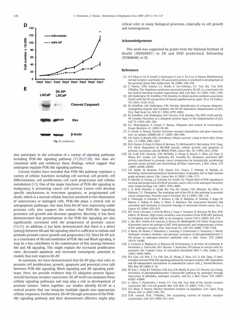
Fig. 5 (continued).
126 S. Srinivasan, Z. Nawaz / Biochimica et Biophysica Acta 1809 (2011) 119–127
also participate in the activation of a variety of signaling pathwaysincluding PI3K-Akt signaling pathway [17,20,27,28]. Our data areconsistent with and reinforces these findings, which suggest thatandrogens regulate PI3K-Akt signaling pathway.
Current studies have revealed that PI3K-Akt pathway regulates avariety of cellular functions including cell survival, cell growth, celldifferentiation, cell proliferation, cell cycle progression and cellularmetabolism [13]. One of the major functions of PI3K-Akt signaling inmalignancy is promoting cancer cell survival. Cancer cells developspecific mechanisms to overcome apoptosis, or programmed celldeath, which is a normal cellular function involved in the eliminationof unnecessary or damaged cells. PI3K-Akt plays a central role inantiapoptotic pathways. Our data from E6-AP over expressing stableprostate cells also support the notion that PI3K-Akt signalingpromotes cell growth and decrease apoptosis. Recently, it has beendemonstrated that perturbations in the PI3K-Akt signaling are alsosignificantly correlated with the progression of prostate cancer[14,15]. In addition, it has been demonstrated that there is a directsynergy between AR and Akt signalingwhich is sufficient to initiate andpromote prostate cancer growth and progression [16]. Since E6-AP actsas a coactivator of AR and modulator of PI3K-Akt and RhoA signaling, itmay be a key contributor in the maintenance of this synergy betweenAkt and AR signaling. This might explain the increased proliferativerates, decreased apoptosis and increased tumorigenic potential inmodels that over express E6-AP.
In summary, we have demonstrated that E6-AP play vital roles inprostate cell proliferation, growth, apoptosis and potential cross talkbetween PI3K-Akt signaling, RhoA signaling and AR signaling path-ways. Here, we provide evidence that E3 ubiquitin–protein ligase/steroid hormone receptor coactivator, E6-AP itself canmodulate thesecellular signaling pathways and may play a role in development ofprostate tumors. Taken together, our studies identify E6-AP as acentral protein that can integrate multiple signals into appropriatecellular responses. Furthermore, E6-AP through activation of the PI3K-Akt signaling pathway and their downstream effectors might play
critical roles in many biological processes, especially in cell growthand tumorigenesis.
Acknowledgements
This work was supported by grants from the National Institute ofHealth (DK060907) to ZN and DOD predoctoral fellowship(PC060648) to SS.
References
[1] O.Y. Khan, G. Fu, A. Ismail, S. Srinivasan, X. Cao, Y. Tu, S. Lu, Z. Nawaz, Multifunctionsteroid receptor coactivator, E6-associated protein, is involved in development ofthe prostate gland, Mol. Endocrinol. 20 (2006) 544–559.
[2] Z. Nawaz, D.M. Lonard, C.L. Smith, E. Lev-Lehman, S.Y. Tsai, M.J. Tsai, B.W.O'Malley, The Angelman syndrome-associated protein, E6-AP, is a coactivator forthe nuclear hormone receptor superfamily, Mol. Cell. Biol. 19 (1999) 1182–1189.
[3] J.M. Huibregtse, M. Scheffner, P.M. Howley, A cellular proteinmediates associationof p53 with the E6 oncoprotein of human papillomavirus types 16 or 18, Embo J.10 (1991) 4129–4135.
[4] M. Scheffner, J.M. Huibregtse, P.M. Howley, Identification of a human ubiquitin-conjugating enzyme that mediates the E6-AP-dependent ubiquitination of p53,Proc. Natl Acad. Sci. USA 91 (1994) 8797–8801.
[5] M. Scheffner, J.M. Huibregtse, R.D. Vierstra, P.M. Howley, The HPV-16 E6 and E6-AP complex functions as a ubiquitin–protein ligase in the ubiquitination of p53,Cell 75 (1993) 495–505.
[6] S.C. Dhananjayan, A. Ismail, Z. Nawaz, Ubiquitin and control of transcription,Essays Biochem. 41 (2005) 69–80.
[7] A. Ismail, Z. Nawaz, Nuclear hormone receptor degradation and gene transcrip-tion: an update, IUBMB Life 57 (2005) 483–490.
[8] S.R. Datta, A. Brunet, M.E. Greenberg, Cellular survival: a play in three Akts, GenesDev. 13 (1999) 2905–2927.
[9] M.A. Davies, D. Koul, H. Dhesi, R. Berman, T.J. McDonnell, D. McConkey, W.K. Yung,P.A. Steck, Regulation of Akt/PKB activity, cellular growth, and apoptosis inprostate carcinoma cells by MMAC/PTEN, Cancer Res. 59 (1999) 2551–2556.
[10] J.R. Graff, B.W. Konicek, A.M. McNulty, Z. Wang, K. Houck, S. Allen, J.D. Paul, A.Hbaiu, R.G. Goode, G.E. Sandusky, R.L. Vessella, B.L. Neubauer, Increased AKTactivity contributes to prostate cancer progression by dramatically acceleratingprostate tumor growth and diminishing p27Kip1 expression, J. Biol. Chem. 275(2000) 24500–24505.
[11] S.N. Malik, M. Brattain, P.M. Ghosh, D.A. Troyer, T. Prihoda, R. Bedolla, J.I.Kreisberg, Immunohistochemical demonstration of phospho-Akt in high Gleasongrade prostate cancer, Clin. Cancer Res. 8 (2002) 1168–1171.
[12] H. Murillo, H. Huang, L.J. Schmidt, D.I. Smith, D.J. Tindall, Role of PI3K signaling insurvival and progression of LNCaP prostate cancer cells to the androgen refractorystate, Endocrinology 142 (2001) 4795–4805.
[13] L. Li, M.M. Ittmann, G. Ayala, M.J. Tsai, R.J. Amato, T.M. Wheeler, B.J. Miles, D.Kadmon, T.C. Thompson, The emerging role of the PI3-K-Akt pathway in prostatecancer progression, Prostate Cancer Prostatic Dis. 8 (2005) 108–118.
[14] E. Tokunaga, A. Kataoka, Y. Kimura, E. Oki, K. Mashino, K. Nishida, T. Koga, M.Morita, Y. Kakeji, H. Baba, S. Ohno, Y. Maehara, The association between Aktactivation and resistance to hormone therapy in metastatic breast cancer, Eur. J.Cancer 42 (2006) 629–635.
[15] M.I. Torres-Arzayus, J. Font de Mora, J. Yuan, F. Vazquez, R. Bronson, M. Rue, W.R.Sellers, M. Brown, High tumor incidence and activation of the PI3K/AKT pathwayin transgenic mice define AIB1 as an oncogene, Cancer Cell 6 (2004) 263–274.
[16] L. Xin, M.A. Teitell, D.A. Lawson, A. Kwon, I.K. Mellinghoff, O.N. Witte, Progressionof prostate cancer by synergy of AKT with genotropic and nongenotropic actionsof the androgen receptor, Proc. Natl Acad. Sci. USA 103 (2006) 7789–7794.
[17] S. Baron, M. Manin, C. Beaudoin, L. Leotoing, Y. Communal, G. Veyssiere, L. Morel,Androgen receptor mediates non-genomic activation of phosphatidylinositol 3-OH kinase in androgen-sensitive epithelial cells, J. Biol. Chem. 279 (2004)14579–14586.
[18] G. Castoria, A. Migliaccio, A. Bilancio, M. Di Domenico, A. de Falco, M. Lombardi, R.Fiorentino, L. Varricchio, M.V. Barone, F. Auricchio, PI3-kinase in concert with Srcpromotes the S-phase entry of oestradiol-stimulated MCF-7 cells, Embo J. 20(2001) 6050–6059.
[19] R.X. Guo, L.H. Wei, Z. Tu, P.M. Sun, J.L. Wang, D. Zhao, X.P. Li, J.M. Tang, 17 beta-estradiol activates PI3K/Akt signaling pathwayby estrogen receptor (ER)-dependentand ER-independent mechanisms in endometrial cancer cells, J. Steroid Biochem.Mol. Biol. 99 (2006) 9–18.
[20] M. Sun, L. Yang, R.I. Feldman, X.M. Sun, K.N. Bhalla, R. Jove, S.V. Nicosia, J.Q. Cheng,Activation of phosphatidylinositol 3-kinase/Akt pathway by androgen throughinteraction of p85alpha, androgen receptor, and Src, J. Biol. Chem. 278 (2003)42992–43000.
[21] G. Zhou, Y. Hashimoto, I. Kwak, S.Y. Tsai, M.J. Tsai, Role of the steroid receptorcoactivator SRC-3 in cell growth, Mol. Cell. Biol. 23 (2003) 7742–7755.
[22] O.Y. Khan, Z. Nawaz, Nuclear hormone receptor co-regulators, Curr. Opin. DrugDiscov. Dev. 6 (2003) 692–701.
[23] D.M. Lonard, B.W. O'Malley, The expanding cosmos of nuclear receptorcoactivators, Cell 125 (2006) 411–414.

127S. Srinivasan, Z. Nawaz / Biochimica et Biophysica Acta 1809 (2011) 119–127
[24] H.J. Zhou, J. Yan, W. Luo, G. Ayala, S.H. Lin, H. Erdem, M. Ittmann, S.Y. Tsai, M.J. Tsai,SRC-3 is required for prostate cancer cell proliferation and survival, Cancer Res. 65(2005) 7976–7983.
[25] P.M. Ghosh, R. Bedolla, M. Mikhailova, J.I. Kreisberg, RhoA-dependent murineprostate cancer cell proliferation and apoptosis: role of protein kinase Czeta,Cancer Res. 62 (2002) 2630–2636.
[26] A. Mani, A.S. Oh, E.T. Bowden, T. Lahusen, K.L. Lorick, A.M. Weissman, R. Schlegel,A. Wellstein, A.T. Riegel, E6AP mediates regulated proteasomal degradation of the
nuclear receptor coactivator amplified in breast cancer 1 in immortalized cells,Cancer Res. 66 (2006) 8680–8686.
[27] M.R. Freeman, B. Cinar, M.L. Lu, Membrane rafts as potential sites of nongenomichormonal signaling in prostate cancer, Trends Endocrinol. Metab. 16 (2005)273–279.
[28] C.A. Heinlein, C. Chang, The roles of androgen receptors and androgen-bindingproteins in nongenomic androgen actions, Mol. Endocrinol. 16 (2002)2181–2187.