Over-expression of severe acute respiratory syndrome coronavirus 3b protein induces both apoptosis...
8
Virus Research 122 (2006) 20–27 Over-expression of severe acute respiratory syndrome coronavirus 3b protein induces both apoptosis and necrosis in Vero E6 cells Sehaam Khan,Burtram C. Fielding, Timothy H.P. Tan, Chih-Fong Chou, Shuo Shen, Seng Gee Lim, Wanjin Hong, Yee-Joo Tan ∗ Collaborative Anti-Viral Research Group, Institute of Molecular and Cell Biology, 61 Biopolis Drive, Proteos, Singapore 138673, Singapore Received 21 March 2006; received in revised form 24 May 2006; accepted 7 June 2006 Available online 11 September 2006 Abstract The genome of the severe acute respiratory syndrome coronavirus encodes for eight accessory viral proteins with no known homologues in other coronaviruses. One of these is the 3b protein, which is encoded by the second open reading frame in subgenomic RNA 3 and contains 154 amino acids. Here, a detailed time-course study was performed to compare the apoptosis and necrosis profiles induced by full-length 3b, a 3b mutant that was deleted by 30 amino acids from the C terminus (3b124-154) and the classical apoptosis inducer, Bax. Our results showed that Vero E6 cells transfected with a construct for expressing 3b underwent necrosis as early as 6 h after transfection and underwent simultaneous necrosis and apoptosis at later time-points. At all the time-points analysed, the apoptosis induced by the expression of 3b was less than the level induced by Bax but the level of necrosis was comparable. The 3b124-154 mutant behaves in a similar manner indicating that the localization of the 3b protein does not seems to be important for the cell-death pathways since full-length 3b is localized predominantly to the nucleolus, while the mutant is found to be concentrated in the peri-nuclear regions. To our knowledge, this is the first report of the induction of necrosis by a SARS-CoV protein. © 2006 Elsevier B.V. All rights reserved. Keywords: Coronavirus; SARS coronavirus; Necrosis; Apoptosis; 3b Protein 1. Introduction Severe acute respiratory syndrome (SARS) originated in early November 2002 in Guangdong province, People’s Repub- lic of China. The disease soon spread worldwide infecting more than 8000 people, with more than 700 fatalities (World Health Organization, http://www.who.int/csr/sars/country/en/). The causative agent of SARS was identified as a novel coron- avirus, now known as SARS-CoV (for reviews, see Berger et al., 2004; Christian et al., 2004; Peiris et al., 2004). SARS-CoV contains a RNA genome of ∼30 kilobases, which encodes for up to 14 potential open reading frames (ORFs) (Marra et al., 2003; Rota et al., 2003). In addition to the replicase polyproteins (pp1a and pp1ab) and structural proteins, spike, membrane, nucleo- capsid and envelope, which are common to all members of the genus coronavirus, the SARS-CoV genome also encodes eight putative proteins with no significant sequence homology to viral ∗ Corresponding author. Tel.: +65 65869625; fax: +65 67791117. E-mail address: [email protected] (Y.-J. Tan). proteins of other known coronaviruses (i.e. ORF 3a, 3b, 6, 7a, 7b, 8a, 8b and 9b) (Marra et al., 2003; Snijder et al., 2003; Tan et al., 2005a). It has not yet been established which of the SARS-CoV accessory proteins are essential for viral replication and/or for viral–host interactions. However, the over-expression of some of these SARS-CoV accessory proteins (3a, 7a, 3b) have been shown to induce apoptosis in cell culture (Law et al., 2005; Tan et al., 2004a; Yuan et al., 2005a), suggesting that they may play a role in viral pathogenesis. The second largest subgenomic RNA of SARS-CoV (sgRNA3) contains two ORFs, 3a and 3b. The 3a protein has been characterized to a great extent and has been shown to be expressed in infected cells both in vitro and in vivo (Chan et al., 2005; Ito et al., 2005; Law et al., 2005; Tan et al., 2004c; Yu et al., 2004; Zeng et al., 2004). It is a novel coronavirus structural protein (Ito et al., 2005; Shen et al., 2005) and can up-regulate the expression of fibrinogen in lung cells (Tan et al., 2005b). The 3b protein is likely to be expressed via an internal ribo- somal entry mechanism since its translation initiation codon is not the first AUG in sgRNA3 (Snijder et al., 2003). It has been detected in SARS-CoV infected cells (Chan et al., 2005), and 0168-1702/$ – see front matter © 2006 Elsevier B.V. All rights reserved. doi:10.1016/j.virusres.2006.06.005
-
Upload
independent -
Category
Documents
-
view
2 -
download
0
Transcript of Over-expression of severe acute respiratory syndrome coronavirus 3b protein induces both apoptosis...
A
catcabdf©
K
1
elmHTaactRacgp
0d
Virus Research 122 (2006) 20–27
Over-expression of severe acute respiratory syndrome coronavirus 3bprotein induces both apoptosis and necrosis in Vero E6 cells
Sehaam Khan, Burtram C. Fielding, Timothy H.P. Tan, Chih-Fong Chou,Shuo Shen, Seng Gee Lim, Wanjin Hong, Yee-Joo Tan ∗
Collaborative Anti-Viral Research Group, Institute of Molecular and Cell Biology, 61 Biopolis Drive, Proteos, Singapore 138673, Singapore
Received 21 March 2006; received in revised form 24 May 2006; accepted 7 June 2006Available online 11 September 2006
bstract
The genome of the severe acute respiratory syndrome coronavirus encodes for eight accessory viral proteins with no known homologues in otheroronaviruses. One of these is the 3b protein, which is encoded by the second open reading frame in subgenomic RNA 3 and contains 154 aminocids. Here, a detailed time-course study was performed to compare the apoptosis and necrosis profiles induced by full-length 3b, a 3b mutanthat was deleted by 30 amino acids from the C terminus (3b�124-154) and the classical apoptosis inducer, Bax. Our results showed that Vero E6ells transfected with a construct for expressing 3b underwent necrosis as early as 6 h after transfection and underwent simultaneous necrosis andpoptosis at later time-points. At all the time-points analysed, the apoptosis induced by the expression of 3b was less than the level induced by Bax
ut the level of necrosis was comparable. The 3b�124-154 mutant behaves in a similar manner indicating that the localization of the 3b proteinoes not seems to be important for the cell-death pathways since full-length 3b is localized predominantly to the nucleolus, while the mutant isound to be concentrated in the peri-nuclear regions. To our knowledge, this is the first report of the induction of necrosis by a SARS-CoV protein.2006 Elsevier B.V. All rights reserved.
p82avosea
(be2
eywords: Coronavirus; SARS coronavirus; Necrosis; Apoptosis; 3b Protein
. Introduction
Severe acute respiratory syndrome (SARS) originated inarly November 2002 in Guangdong province, People’s Repub-ic of China. The disease soon spread worldwide infecting
ore than 8000 people, with more than 700 fatalities (Worldealth Organization, http://www.who.int/csr/sars/country/en/).he causative agent of SARS was identified as a novel coron-virus, now known as SARS-CoV (for reviews, see Berger etl., 2004; Christian et al., 2004; Peiris et al., 2004). SARS-CoVontains a RNA genome of ∼30 kilobases, which encodes for upo 14 potential open reading frames (ORFs) (Marra et al., 2003;ota et al., 2003). In addition to the replicase polyproteins (pp1and pp1ab) and structural proteins, spike, membrane, nucleo-
apsid and envelope, which are common to all members of theenus coronavirus, the SARS-CoV genome also encodes eightutative proteins with no significant sequence homology to viral∗ Corresponding author. Tel.: +65 65869625; fax: +65 67791117.E-mail address: [email protected] (Y.-J. Tan).
aptTsnd
168-1702/$ – see front matter © 2006 Elsevier B.V. All rights reserved.oi:10.1016/j.virusres.2006.06.005
roteins of other known coronaviruses (i.e. ORF 3a, 3b, 6, 7a, 7b,a, 8b and 9b) (Marra et al., 2003; Snijder et al., 2003; Tan et al.,005a). It has not yet been established which of the SARS-CoVccessory proteins are essential for viral replication and/or foriral–host interactions. However, the over-expression of somef these SARS-CoV accessory proteins (3a, 7a, 3b) have beenhown to induce apoptosis in cell culture (Law et al., 2005; Tant al., 2004a; Yuan et al., 2005a), suggesting that they may playrole in viral pathogenesis.
The second largest subgenomic RNA of SARS-CoVsgRNA3) contains two ORFs, 3a and 3b. The 3a protein haseen characterized to a great extent and has been shown to bexpressed in infected cells both in vitro and in vivo (Chan et al.,005; Ito et al., 2005; Law et al., 2005; Tan et al., 2004c; Yu etl., 2004; Zeng et al., 2004). It is a novel coronavirus structuralrotein (Ito et al., 2005; Shen et al., 2005) and can up-regulatehe expression of fibrinogen in lung cells (Tan et al., 2005b).
he 3b protein is likely to be expressed via an internal ribo-omal entry mechanism since its translation initiation codon isot the first AUG in sgRNA3 (Snijder et al., 2003). It has beenetected in SARS-CoV infected cells (Chan et al., 2005), and
esear
a(3acaa
itft1d(t
2
2
(gbmC5
2
vC(pw2ed3CuAA
2
at1aaoFi
6woNPoTbTttwpsw1m
2
Sswp(a(ao0gcpweP
2
ttaahtd
2i
S. Khan et al. / Virus R
nti-3b antibody has been detected in a SARS patient’s serumGuo et al., 2004), suggesting that it is expressed in vivo. Theb protein contains two predicted nuclear localization signalsnd has been shown to localize to the nucleolus of transfectedells in the absence of any other SARS-CoV proteins (Yuan etl., 2005b). In addition, the over-expression of 3b causes G0/G1rrest and induces apoptosis (Yuan et al., 2005a).
In this study, we determined the mechanism of cell-deathnduced by the over-expression of full-length 3b or a 3b mutanthat lacks the C-terminal 30 amino acids (3b�124-154). Byollowing the time-course of expression, we compared the apop-osis and necrosis profiles induced by full-length 3b, 3b�124-54 mutant and the classical apoptosis inducer, Bax. Terminaleoxynucleotidyl transferase mediated dUTP nick-end-labelingTUNEL) assays and subcellular fractionation were performedo further delineate the cell-death pathways.
. Methods
.1. Materials
All reagents used in this study were purchased from SigmaSt. Louis, MO, USA) unless otherwise stated. Vero E6 (Africanreen monkey kidney epithelial) cells were grown in Dul-ecco’s modified Eagle medium containing 0.1 mg/ml strepto-ycin and 100 U penicillin and 5% FBS (HyClone, UT, USA).ells were cultured at 37 ◦C in an incubator supplied with% CO2.
.2. Construction of plasmids
To clone the 3b gene into pXJ40HA mammalian expressionector, the 3b ORF was amplified using 3b-forward (5′-CGCTCGAGGCCACCATGATGCCAA-3′) and 3b-reverse
5′-ATAAGAATGCGGCCGCTTAACGTACCTGTTTC-3′)rimers. cDNA prepared from SARS-CoV infected cellsas used as template as previously described (Tan et al.,004b). The PCR product was digested with restrictionnzymes (XhoI and NotI) and ligated to linearized pXJ40HAigested with the same restriction enzymes. The pXJ40HA-b�124-154 plasmid for expressing a 3b mutant without the-terminal 30 amino acids was made in a similar mannersing the 3b-forward primer and 3b�124-154-reverse (5′-TAAGAATGCGGCCGCTTACATCATAAATTG-3′) primer.ll plasmid sequences were confirmed by sequence analysis.
.3. Transfection and CaspACE fluorometric assay
Vero E6 cells were plated on 6 cm tissue culture dishesnd allowed to grow to 70% confluence before the cells wereransfected with 3 �g of pXJ40HA-3b or pXJ40HA-3b�124-54 using Lipofectamine reagent (Invitrogen, Carlsbad, CA),ccording to manufacturer’s protocol. To reduce the rate of
poptosis induced by the potent apoptosis inducer, Bax, 1 �gf pXJ40HA-Bax was used with 2 �g of empty vector instead.or mock transfection, no DNA was used but the cells werencubated with the same amount of Lipofectamine reagent.
dba
ch 122 (2006) 20–27 21
Each plate of cells was harvested at various time-points, i.e., 12, 18 and 48 h, after transfection. The cells were then washedith phosphate-buffered saline (PBS) and resuspended in 110 �lf lysis buffer (50 mM Tris–HCl (pH 8.0), 150 mM NaCl, 0.5%P40, 0.5% deoxycholic acid, 0.005% SDS), containing 1 mMMSF. Half of the suspension was then subjected to three roundsf freeze-thaw cycles and then centrifuged to remove cell debris.he total protein concentration of the lysate was determinedy using Coomassie Plus reagent from Pierce (Rockford, IL).he amount of caspase-3 activity in 10 �g of total protein was
hen determined by using the CaspACE fluorometric assay sys-em from Promega Corporation (Madison, WI). All experimentsere performed in duplicates. Laemmli’s sodium dodecyl sul-hate buffer (SDS buffer) was added to the other half of the celluspension. As the 3b protein tends to form large aggregateshen boiled, the SDS–cell mixture was incubated at 50 ◦C for5 min before Western blot analysis. For HA-Bax, the SDS–cellixture was heated at 100 ◦C for 10 min.
.4. Western blot analysis
For Western blot analysis, cell lysates were separated onDS-PAGE and transferred onto Hybond-C membranes (Amer-ham Pharmacia Biotech, Uppsala, Sweden). The membranesere then blocked with 5% non-fat milk for 30 min androbed with primary antibodies (anti-HA monoclonal antibody1:1000; Roche Molecular Biochemicals, Indianapolis, IN) ornti-poly(ADP-ribose) polymerase (PARP) polyclonal antibody1:1000; Cell Signaling Technology, Inc., Beverly, MA) or anti-ctin monoclonal antibody (1:3000; Sigma)) with rolling at 4 ◦Cvernight. After three washes (15 min each) with PBS containing.05% Tween 20 (PBST), the membranes were incubated withoat anti-rabbit or anti-mouse horse-radish peroxidase (HRP)-onjugated secondary antibodies (1:2000, Pierce) at room tem-erature, with rolling for 1 h. The membranes were then washedith PBST three times for 15 min each, and visualized using an
nhanced chemiluminescence method (Supersignal West Pico,ierce).
.5. CytoTox-ONE homogenous membrane integrity assay
Vero E6 cells were plated in 6 cm tissue culture dish andransfected as described above. At each time-point, 110 �l ofhe culture medium were transferred to a 96-well plate and themount of lactate dehydrogenase (LDH) released from dam-ged necrotic cells was determined using the CytoTox-ONEomogenous membrane integrity assay (Promega) accordingo manufacturer’s protocol. All experiments were performed inuplicates.
.6. DeadENd fluorometric TUNEL andmmunofluorescence experiments
Vero E6 cells were seeded on cover slips and transfected asescribed above. Cells were analysed for DNA fragmentationy using the DeadENd fluorometric TUNEL system (Promega)ccording to manufacturer’s protocol. Cells treated with DNase

2 esear
It
fiswww(S
2
t3pcSb
t(latatfmralw3Abtafb
3
ecswbscaaiat
cgi(LTpsaanasntc
fasmti6ia3Pftls6tdtai
1ggrttad1pfir3
2 S. Khan et al. / Virus R
(Roche) served as a positive control. The cell nuclei were coun-erstained with 1 �g/ml propidium iodide.
For immunofluorescence experiments, the cover slips werexed in methanol for 5 min at −20 ◦C, after which the coverlips were completely air-dried. Cells were blocked with PBSith 1% bovine serum albumin for 30 min, and then incubatedith anti-HA antibody at a dilution of 1:200 for 1 h. Followingashing, cells were incubated with the fluorescein isothicyanate
FITC)-conjugated goat anti-mouse secondary antibody (1:100;anta Cruz Biotechnology, Santa Cruz, CA) for 1 h.
.7. Subcellular fractionation
Vero E6 cells were plated in 10 cm tissue culture dish andransfected as described above. In this case, 6 �g of pXJ40HA-b or pXJ40HA-3b�124-154 was used. Fractionation was thenerformed to separate the cytoplasmic, nuclear and membraneomponents of the cell. The method used was adapted frompector et al. (1988) with some modifications and is describedelow.
At different time-points, the cells were harvested and washedwice with cold PBS and resuspended in 200 �l hypotonic buffer10 mM Tris–HCl (pH 7.5), 2 mM MgCl2, 0.5 mM PMSF) andeft in ice-water for 5 min. 0.25% Triton-X 100 was then addednd the cells were sheared by passing through a 21G needlehrice. The cells was then left in ice-water for 5 min and spunt 3000 rpm at 4 ◦C for 5 min (step A). The supernatant wasransferred to a clean tube and spun again at 14,000 rpm at 4 ◦Cor 20 min (step B) and the supernatant was collected (cytoplas-ic fraction). The pellet after step B (membrane fraction) was
esuspended in 200 �l of lysis buffer with DNase I (1 �l/ml),nd left at room temperature for 15 min to degrade the cellu-ar DNA. To wash the pellet from step A (nuclear fraction), itas resuspended in 0.5 ml ice-cold hypotonic buffer and spun at000 rpm at 4 ◦C for 5 min and the supernatant was discarded.fter two washes, the pellet was resuspended in 200 �l of lysisuffer with DNase I. Finally, 50 �l of 5XSDS buffer was addedo each of the fractions and the mixtures were vortexed and leftt room temperature for 15 min, followed by incubation at 50 ◦Cor 15 min. Then, 20 �l of each fraction was subjected to Westernlot analysis as described above.
. Results and discussion
Recently, Yuan and co-workers showed that the over-xpression of SARS-CoV 3b in COS-7, Vero and 293 inducesell-cycle arrest but they could only observed significant apopto-is in COS-7 cells (Yuan et al., 2005a). Here, a time-course studyas performed to characterize the profile of cell-death inducedy the over-expression of 3b in Vero E6 cells in order to under-tand the role of 3b during the viral replication cycle in Vero E6ells. In addition, the cell-death profiles of full-length 3b anddeletion mutant, 3b�124-154, which lacks the C-terminal 30
mino acids, were compared to that of Bax, a classical apoptosisnducer. The general opinion is that cell death can either be asresult of necrosis, defined as a passive and non-physiological
ype of death caused by accidental and acute damage to the
otie
ch 122 (2006) 20–27
ell, or the consequence of apoptosis, defined as an active andenetically regulated process of cell suicide by which an organ-sm eliminates senescent, abnormal and potentially harmful cellsfor reviews, see Ameison, 2002; Guimaraes and Linden, 2004;os et al., 2002; Nelson and White, 2004; Zhivotosky, 2004).hese two forms of cell death are distinguishable by their mor-hological and biochemical effects on the cell. However, recenttudies have shown that necrosis is also highly regulated and
fine regulatory line exists between necrosis and apoptosiss many anti-apoptotic mechanisms are also effective againstecrosis (for reviews, see Proskuryakov et al., 2003; Syntichakind Tavernarakis, 2002, 2003). For the sake of simplicity, wehall use the term necrosis to refer to the form of cell deathot involving the apoptotic early steps, and secondary necrosiso describe the subsequent degradative changes that apoptoticells undergo at the late stages of apoptosis.
Vero E6 cells were analysed at 6, 12, 18 and 48 h after trans-ection with plasmids for expressing HA-3b, HA-3b�124-154nd HA-Bax to determine the degree of apoptosis and necro-is. The expression of the proteins were detected using anti-HAonoclonal antibody (Fig. 1A and B). As shown in Fig. 1C,
he over-expression of 3b and 3b�124-154 resulted in a slightncrease in caspase 3 activity, which is a hallmark of apoptosis, ath post-transfection. The level of caspase 3 activity continued to
ncrease until 12 h after transfection but showed gradual decreaset 18 and 48 h after transfection. Consistently, the expression ofb and 3b�124-154 also resulted in cleavage of endogenousARP, which is a substrate of activated caspase-3 (Fig. 1A)rom 12 h onwards. This profile mirrored that of Bax, excepthat the level of caspase 3 activities for 3b and 3b�124-154 wereower than for Bax at all the time-points and that Bax alreadyhowed high level of caspase 3 activities and PARP cleavage ath (Fig. 1B and C). These results are somewhat different from
he report by Yuan et al. (2005a) where the over-expression of 3bid not induce apoptosis in Vero cells, and furthermore, showedhat the C-terminal 30 amino-acids of 3b are not important forpoptosis induction. The discrepancy may be due to differencesn transfection efficiency and/or sensitivity of the assays used.
In all cases, we noted that a decease in caspase 3 activity after8 h of transfection, suggesting that the apoptotic cells wereoing into secondary necrosis. A key marker of a cell under-oing necrosis is the loss of their membrane integrity and theelease of their cytoplasmic contents into the surrounding cul-ure. Thus, the CytoTox-ONE assay (Promega), which measureshe release of lactate dehydrogenase (LDH) from cells with dam-ged membranes, was used to quantify the degree of necrosis atifferent time-points. For the over-expression of 3b and 3b�124-54, significant amounts of LDH were released as early as 6 host-transfection as compared to the low level of LDH releasedrom mock transfected cells (Fig. 1D). The levels of LDH thenncreased gradually up to 12 h. Interestingly, the level of LDHeleased from cells expressing Bax was about the same as forb and 3b�124-154 at 6 h, 12 h and 18 h, even though the level
f apoptosis caused by the over-expression of Bax was consis-ently higher that caused by 3b or 3b�124-154. The gradualncrease in the level of necrosis in the Bax expressing cells wasxpected as apoptotic cells would undergo secondary necrosis
S. Khan et al. / Virus Research 122 (2006) 20–27 23
Fig. 1. Time-course study to determine the effects of the expression of SARS-CoV 3b proteins on apoptosis and necrosis. (A and B) Western blot analysis wasperformed to determine the expression of full-length SARS-CoV 3b ((A), lanes 1–4)), a 3b mutant which lacks the C-terminal 30 amino acids (3b�124-154) ((A),lanes 5–8) and Bax ((B), lanes 1–4), a potent apoptosis inducer in transiently transfected Vero E6 cells. All proteins were tagged at the N-terminal with a HA motiffor easy detection. Cells were harvested at 6, 12, 18 and 48 h post-transfection and the expression levels of HA-tagged proteins were determined with anti-HAantibody (middle panel). Mock transfected cells were used as negative control. The same samples were assayed for the cleavage of endogenous full-length PARP,which is a hallmark of apoptosis, from 116 to 83 kDa (upper panel). Equal loading of total cell lysates were verified by using an antibody to detect endogenous actin(lower panel). (C) The CaspACE fluorometric assay system from Promega Corporation was used to measure the activation of caspase-3 protease activity, which isanother hallmark of apoptosis. (D) The CytoTox-ONE homogenous membrane integrity assay from Promega Corporation was used to measure the amount of lactatedehydrogenase (LDH) released from necrotic cells. For (C and D), these assays were performed at each time-point for HA-3b (solid black bars), HA-3b�124-154( d barss
aLTsw3iasoLeLcTp
aptEscfiif
BIct
e1(tTtcfea
1owoita
unshaded bars), HA-Bax (solid gray bars) and mock transfected cells (striatetandard deviations are plotted.
t the late stages (Ameison, 2002; Guimaraes and Linden, 2004;os et al., 2002; Nelson and White, 2004; Zhivotosky, 2004).hese results showed that the expression of 3b induces necro-is as early as 6 h post-transfection when the level of apoptosisas very much lower than that for Bax. Again, the C-terminal0 amino acids of 3b do not have any effects on its ability tonduce necrosis. At time-points greater than 6 h post-infection, itppears that both necrosis and apoptosis (and secondary necro-is) were occurring simultaneously in cell over-expressing 3br 3b�124-154. At 48 h post-transfection, there was a surge ofDH release from cells expressing 3b or 3b�124-154 or Bax orven the mock transfected cells, indicating that by this time, theipofectamine reagent used for transfection had some unspe-ific cytotoxic effects or the cells were over-grown (Fig. 1D).hus, subsequent experiments were only performed up to 18 host-transfection.
TUNEL assays were performed to examine whether the 3bnd 3b�124-154 could induce DNA fragmentation, a commonhenomenon of apoptosis. At all the time-points, DNA fragmen-ations were observed in 3b- and 3b�124-154-transfected Vero6 cells. A representative set of data for 18 h post-transfection ishown in Fig. 2 (first and second rows). DNA fragmentations inells expressing 3b or 3b�124-154 (note that the typical trans-
ection efficiency achieved in this study was about 30%) werendicated by FITC positive signals while nuclei of all the cellsn the field were counterstained using propidium iodide. DNAragmentation was also be detected in the cells transfected withtwfp
). All experiments were performed in duplicates and the average values with
ax (Fig. 2, third row) and in cells that were treated with DNase(positive control) (Fig. 2, fifth row). Under the same exposureonditions, there was no sign of DNA fragmentation in the mockransfected cells (Fig. 2, fourth row).
Next, the subcellular localization of 3b and 3b�124-154 wasxamined by subcellular fractionation studies performed at 6,2 and 18 h post-transfection. Consistent with previous reportYuan et al., 2005b), the full-length 3b protein was found to parti-ion solely in the nuclear fraction at all the time-points (Fig. 3A).he 3b deletion mutant, 3b�124-154, was initially found in
he cytoplasmic fraction (6 h post-transfection), but with time,ould be more efficiently detected in the membrane and nuclearractions (Fig. 3B). As a control for the fractionation method,ndogenous actin was detected in the cytoplasmic fractions forll transfections.
Next, indirect immunofluorescence was also performed at8 h post-transfection to compare the subcellular localizationf 3b and 3b�124-154 in Vero E6 cells (Fig. 3C). Consistentith the fractionation experiments above, indirect immunoflu-rescence revealed that the 3b protein was localized primarilyn the nucleolus of transfected cells and somewhat less aroundhe nucleus. The latter is likely to be the nuclear membranes 3b was isolated in the nuclear fraction (Fig. 3A). In con-
rast, 3b�124-154 was localized to the perinuclear regions,hile Bax was diffusely localized in the cytoplasm. Since theractionation experiments showed that 3b�124-154 was foundredominantly in the membrane and nuclear fractions at late
24 S. Khan et al. / Virus Research 122 (2006) 20–27
Fig. 2. DNA fragmentation in Vero E6 cells expressing HA-3b, HA-3b�124-154 and HA-Bax, detected by TUNEL assay. Fragmented DNA showed FITC (green,l fieldp agmenc
ttlnnve
3t
eft panels) staining in the nucleus while the nuclei of all the cells in the sameanels showed the merged image of FITC and PI staining and cells containing frells and negative control was mock transfected cells.
ime-points (Fig. 3B), it is likely that the intense staining aroundhe nucleus represents both perinuclear membranous organelles,ike the endoplasmic reticulum or Golgi apparatus, as well as
uclear membrane, with the latter being partitioned into theuclear fraction. Amino acids 134–154 of 3b have been pre-iously shown to contain a nucleolar localization signal (Yuant al., 2005b). Consistently, we observed that unlike full-lengthw6at
were counterstained with propidium iodide (PI, red, middle panels). The rightted DNA would have a yellow colour. The positive control was DNase I treated
b, the 3b�124-154 mutant was not efficiently transported tohe nucleolus.
In summary, we demonstrated that Vero E6 cells transfected
ith a construct for expressing 3b underwent necrosis as early ash after transfection and underwent simultaneous necrosis andpoptosis at later time-points. At all the time-points analysed,he apoptosis induced by the expression of 3b was less than the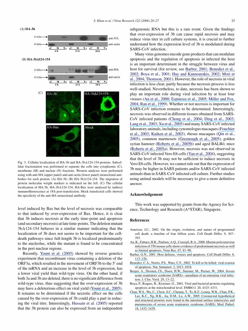
S. Khan et al. / Virus Resear
Fig. 3. Cellular localization of HA-3b and HA-3b�124-154 proteins. Subcel-lular fractionation was performed to separate the cells into cytoplasmic (C),membrane (M) and nuclear (N) fractions. Western analyses were performedusing with anti-HA (upper panel) and anti-actin (lower panel) monoclonal anti-bodies for each protein: (A) HA-3b; (B) HA-3b�124-154. The migration ofprotein molecular weight markers is indicated on the left. (C) The cellularlocalization of HA-3b, HA-3b�124-154, HA-Bax were analysed by indirectit
ltt(3ldti
eOoabwmIcit
struS
aih2aiwpv2SnCLle2s(StV3aua
A
e
R
A
A
B
B
B
B
Chan, W.S., Wu, C., Chow, S.C., Cheung, T., To, K.F., Leung, W.K., Chan, P.K.,
mmunofluorescence at 18 h post-transfection. Mock transfected cells showedhe specificity of the anti-HA monoclonal antibody.
evel induced by Bax but the level of necrosis was comparableo that induced by over-expression of Bax. Hence, it is clearhat 3b induces necrosis at the early time-point and apoptosisand secondary necrosis) at late time-points. The deletion mutantb�124-154 behaves in a similar manner indicating that theocalization of 3b does not seems to be important for the cell-eath pathways since full-length 3b is localized predominantlyo the nucleolus, while the mutant is found to be concentratedn the peri-nuclear regions.
Recently, Yount et al. (2005) showed by reverse geneticsxperiment that recombinant virus containing a deletion of theRF3a, which resulted in the movement of ORF3b to the 5′ endf the mRNA and an increase in the level of 3b expression, haslower viral yield than wild-type virus. On the other hand, if
oth 3a and 3b are deleted, there is no significant difference fromild-type virus, thus suggesting that the over-expression of 3bay have a deleterious effect on viral yield (Yount et al., 2005).
t remains to be determined if the necrotic effect on the cellsaused by the over-expression of 3b could play a part in reduc-ng the viral titre. Interestingly, Hussain et al. (2005) reportedhat the 3b protein can also be expressed from an independent
ch 122 (2006) 20–27 25
ubgenomic RNA but this is a rare event. Given the findingshat over-expression of 3b can cause rapid necrosis and mayeduce virus titer in cell culture systems, it is crucial to furthernderstand how the expression level of 3b is modulated duringARS-CoV infection.
Many virus genomes encode gene products that can modulatepoptosis and the regulation of apoptosis in infected the hosts an important determinant in the struggle between virus andost for survival (for review, see Barber, 2001; Benedict et al.,002; Boya et al., 2001; Hay and Kannourakis, 2002; Mori etl., 2004; Thomson, 2001). However, the role of necrosis in viralnfection is less clear, partly because the necrosis process is lessell-studied. Nevertheless, to date, necrosis has been shown tolay an important role during viral infection by at least fouriruses (An et al., 2000; Espinoza et al., 2005; Miller and Fox,004; Ran et al., 1999). Whether or not necrosis is important forARS-CoV infection remains to be determined. Interestingly,ecrosis was observed in different tissues obtained from SARS-oV infected patients (Chong et al., 2004; Ding et al., 2003;ang et al., 2003; Xu et al., 2005) and many SARS-CoV infected
aboratory animals, including cynomologus macaques (Fouchiert al., 2003; Kuiken et al., 2003), rhesus macaques (Qin et al.,005), common marmosets (Greenough et al., 2005), goldenyrian hamster (Roberts et al., 2005b) and aged BALB/c miceRoberts et al., 2005a). However, necrosis was not observed inARS-CoV infected Vero E6 cells (Yan et al., 2004), suggesting
hat the level of 3b may not be sufficient to induce necrosis inero E6 cells. However, we cannot rule out that the expression ofb may be higher in SARS patients and/or SARS-CoV infectednimals than in SARS-CoV infected cell culture. Further studiessing animal models will be necessary to give a more definitivenswer.
cknowledgement
This work was supported by grants from the Agency for Sci-nce, Technology and Research (A*STAR), Singapore.
eferences
meison, J.C., 2002. On the origin, evolution, and nature of programmedcell death: a timeline of four billion years. Cell Death Differ. 9, 367–393.
n, K., Fattaey, H.K., Paulsen, A.Q., Consigli, R.A., 2000. Murine polyomavirusinfection of 3T6 mouse cells shows evidence of predominant necrosis as wellas limited apoptosis. Virus Res. 67, 81–90.
arber, G.N., 2001. Host defense, viruses and apoptosis. Cell Death Differ. 8,113–126.
enedict, C.A., Norris, P.S., Ware, C.F., 2002. To kill or be killed: viral evasionof apoptosis. Nat. Immunol. 3, 1013–1018.
erger, A., Drosten, Ch., Doerr, H.W., Sturmer, M., Preiser, W., 2004. Severeacute respiratory syndrome (SARS)—paradigm of an emerging viral infec-tion. J. Clin. Virol. 29, 13–22.
oya, P., Roques, B., Kroemer, G., 2001. Viral and bacterial proteins regulatingapoptosis at the mitochondrial level. EMBO J. 20, 4325–4331.
Lee, K.C., Ng, H.K., Au, D.M., Lo, A.W., 2005. Coronaviral hypotheticaland structural proteins were found in the intestinal surface enterocytes andpneumocytes of severe acute respiratory syndrome (SARS). Mod. Pathol.18, 1432–1439.
2 esear
C
C
D
E
F
G
G
G
H
H
I
K
L
L
L
M
M
M
N
P
P
Q
R
R
R
R
S
S
S
S
S
T
T
T
T
6 S. Khan et al. / Virus R
hong, P.Y., Chui, P., Ling, A.E., Franks, T.J., Tai, D.Y., Leo, Y.S., Kaw,G.J., Wansaicheong, G., Chan, K.P., Ean Oon, L.L., Teo, E.S., Tan, K.B.,Nakajima, N., Sata, T., Travis, W.D., 2004. Analysis of deaths during thesevere acute respiratory syndrome (SARS) epidemic in Singapore: chal-lenges in determining a SARS diagnosis. Arch. Pathol. Lab. Med. 128,195–204.
hristian, M.D., Poutanen, S.M., Loutfy, M.R., Muller, M.P., Low, D.E., 2004.Severe acute respiratory syndrome. Clin. Infect. Dis. 38, 1420–1427.
ing, Y., Wang, H., Shen, H., Li, Z., Geng, J., Han, H., Cai, J., Li, X., Kang, W.,Weng, D., Lu, Y., Wu, D., He, L., Yao, K., 2003. The clinical pathology ofsevere acute respiratory syndrome (SARS): a report from China. J. Pathol.200, 282–289.
spinoza, J.C., Cortes-Gutierrez, M., Kuznar, J., 2005. Necrosis of infectiouspancreatic necrosis virus (IPNV) infected cells rarely is preceded by apop-tosis. Virus Res. 109, 133–138.
ouchier, R.A., Kuiken, T., Schutten, M., van Amerongen, G., van Doornum,G.J., van den Hoogen, B.G., Peiris, M., Lim, W., Stohr, K., Osterhaus, A.D.,2003. Aetiology: Koch’s postulates fulfilled for SARS virus. Nature 423,240.
reenough, T.C., Carville, A., Coderre, J., Somasundaran, M., Sullivan, J.L.,Luzuriaga, K., Mansfield, K., 2005. Pneumonitis and multi-organ systemdisease in common marmosets (Callithrix jacchus) infected with the severeacute respiratory syndrome-associated coronavirus. Am. J. Pathol. 167,455–463.
uimaraes, C.A., Linden, R., 2004. Programmed cell death: apoptosis and alter-native deathstyles. Eur. J. Biochem. 271, 1638–1650.
uo, J.P., Petric, M., Campbell, W., McGeer, P.L., 2004. SARS corona viruspeptides recognized by antibodies in the sera of convalescent cases. Virology324, 251–256.
ay, S., Kannourakis, G., 2002. A time to kill: viral manipulation of the celldeath program. J. Gen. Virol. 83, 1547–1564.
ussain, S., Pan, J., Chen, Y., Yang, Y., Xu, J., Peng, Y., Wu, Y., Li, Z., Zhu,Y., Tien, P., Guo, D., 2005. Identification of novel subgenomic RNA’s andnoncanonical transcription initiation siganals of severe acute respiratory syn-drome coronavirus. J. Virol. 79, 5288–5295.
to, N., Mossel, E.C., Narayanan, K., Popov, V.L., Huang, C., Inoue, T., Peters,C.J., Makino, S., 2005. Severe acute respiratory syndrome coronavirus 3aprotein is a viral structural protein. J. Virol. 79, 3182–3186.
uiken, T., Fouchier, R.A., Schutten, M., Rimmelzwaan, G.F., van Amerongen,G., van Riel, D., Laman, J.D., de Jong, T., van Doornum, G., Lim, W., Ling,A.E., Chan, P.K., Tam, J.S., Zambon, M.C., Gopal, R., Drosten, C., van derWerf, S., Escriou, N., Manuguerra, J.C., Stohr, K., Peiris, J.S., Osterhaus,A.D., 2003. Newly discovered coronavirus as the primary cause of severeacute respiratory syndrome. Lancet 362, 263–270.
ang, Z.W., Zhang, L.J., Zhang, S.J., Meng, X., Li, J.Q., Song, C.Z., Sun, L.,Zhou, Y.S., Dwyer, D.E., 2003. A clinicopathological study of three casesof severe acute respiratory syndrome (SARS). Pathology 35, 526–531.
aw, P.T., Wong, C.H., Au, T.C., Chuck, C.P., Kong, S.K., Chan, P.K., To, K.F.,Lo, A.W., Chan, J.Y., Suen, Y.K., Chan, H.Y., Fung, K.P., Waye, M.M., Sung,J.J., Lo, Y.M., Tsui, S.K., 2005. The 3a protein of severe acute respiratorysyndrome-associated coronavirus induces apoptosis in Vero E6 cells. J. Gen.Virol. 86, 1921–1930.
os, M., Mozoluk, M., Ferrari, D., Stepczynska, A., Stroh, C., Renz, A.,Herceg, Z., Wang, Z.-Q., Schulze-Osthoff, K., 2002. Activation and caspase-mediated inhibition of PARP: A molecular switch between fibroblast necro-sis and apoptosis in death receptor signalling. Mol. Biol. Cell. 13, 978–988.
arra, M.A., Jones, S.J., Astell, C.R., Holt, R.A., Brooks-Wilson, A., Butter-field, Y.S., Khattra, J., Asano, J.K., Barber, S.A., Chan, S.Y., Cloutier, A.,Coughlin, S.M., Freeman, D., Girn, N., Griffith, O.L., Leach, S.R., Mayo,M., McDonald, H., Montgomery, S.B., Pandoh, P.K., Petrescu, A.S., Robert-son, A.G., Schein, J.E., Siddiqui, A., Smailus, D.E., Stott, J.M., Yang, G.S.,Plummer, F., Andonov, A., Artsob, H., Bastien, N., Bernard, K., Booth, T.F.,Bowness, D., Czub, M., Drebot, M., Fernando, L., Flick, R., Garbutt, M.,
Gray, M., Grolla, A., Jones, S., Feldmann, H., Meyers, A., Kabani, A., Li,Y., Normand, S., Stroher, U., Tipples, G.A., Tyler, S., Vogrig, R., Ward,D., Watson, B., Brunham, R.C., Krajden, M., Petric, M., Skowronski, D.M.,Upton, C., Roper, R.L., 2003. The Genome sequence of the SARS-associatedcoronavirus. Science 300, 1399–1404.T
T
ch 122 (2006) 20–27
iller, L.C., Fox, J.M., 2004. Apoptosis and porcine reproductive and respiratorysyndrome virus. Vet Immunol. Immunopathol. 102, 131–142.
ori, I., Nishiyama, Y., Yokochi, T., Kimura, Y., 2004. Virus-induced neuronalapoptosis as pathological and protective responses of the host. Rev. Med.Virol. 14, 209–216.
elson, D.A., White, E., 2004. Exploiting different ways to die. Genes Dev. 18,1223–1226.
eiris, J.S., Guan, Y., Yuen, K.Y., 2004. Severe acute respiratory syndrome. Nat.Med. 10 (Suppl.), S88–S97.
roskuryakov, S.Y., Konoplyannikov, A.G., Gabai, V.L., 2003. Necrosis: a spe-cific form of programmed cell death? Exp. Cell Res. 283, 1–16.
in, C., Wang, J., Wei, Q., She, M., Marasco, W.A., Jiang, H., Tu, X., Zhu, H.,Ren, L., Gao, H., Guo, L., Huang, L., Yang, R., Cong, Z., Guo, L., Wang,Y., Liu, Y., Sun, Y., Duan, S., Qu, J., Chen, L., Tong, W., Ruan, L., Liu, P.,Zhang, H., Zhang, J., Zhang, H., Liu, D., Liu, Q., Hong, T., He, W., 2005.An animal model of SARS produced by infection of Macaca mulatta withSARS coronavirus. J. Pathol. 206, 251–259.
an, Z., Rayet, B., Rommelaere, J., Faisst, S., 1999. Parvovirus H-1-inducedcell death: influence of intracellular NAD consumption on the regulation ofnecrosis and apoptosis. Virus Res. 65, 161–174.
oberts, A., Paddock, C., Vogel, L., Butler, E., Zaki, S., Subbarao, K., 2005a.Aged BALB/c mice as a model for increased severity of severe acute respi-ratory syndrome in elderly humans. J. Virol. 79, 5833–5838.
oberts, A., Vogel, L., Guarner, J., Hayes, N., Murphy, B., Zaki, S., Subbarao, K.,2005b. Severe acute respiratory syndrome coronavirus infection of goldenSyrian hamsters. J. Virol. 79, 503–511.
ota, P.A., Oberste, M.S., Monroe, S.S., Nix, W.A., Campagnoli, R., Icenogle,J.P., Penaranda, S., Bankamp, B., Maher, K., Chen, M.H., Tong, S., Tamin,A., Lowe, L., Frace, M., DeRisi, J.L., Chen, Q., Wang, D., Erdman, D.D.,Peret, T.C., Burns, C., Ksiazek, T.G., Rollin, P.E., Sanchez, A., Liffick, S.,Holloway, B., Limor, J., McCaustland, K., Olsen-Rasmussen, M., Fouchier,R., Gunther, S., Osterhaus, A.D., Drosten, C., Pallansch, M.A., Anderson,L.J., Bellini, W.J., 2003. Characterization of a novel coronavirus associatedwith severe acute respiratory syndrome. Science 300, 1394–1399.
hen, S., Lin, P.S., Chao, Y.-C., Zhang, A., Yang, X., Lim, S.G., Hong, W., Tan,Y.-J., 2005. The severe acute respiratory syndrome coronavirus 3a is a novelstructural protein. Biochem. Biophys. Res. Commun. 330, 286–292.
nijder, E.J., Bredenbeek, P.J., Dobbe, J.C., Thiel, V., Ziebuhr, J., Poon, L.L.,Guan, Y., Rozanov, M., Spaan, W.J., Gorbalenya, A.E., 2003. Unique andconserved features of genome and proteome of SARS-coronavirus, an earlysplit-off from the coronavirus group 2 lineage. J. Mol. Biol. 331, 991–1004.
pector, D.L., Goldman, R.D., Leinwand, L.A., 1988. Cells, a laboratory man-ual. Culture and Biochemical Analysis of Cells, vol. 1. Cold Spring HarborLaboratory Press, USA.
yntichaki, P., Tavernarakis, N., 2002. Death by necrosis. Uncontrollable catas-trophe, or is there order behind the chaos? EMBO Rep. 3, 604–609.
yntichaki, P., Tavernarakis, N., 2003. The biochemistry of neuronal necrosis:rogue biology? Nat. Rev. Neurosci. 4, 672–684.
an, Y.J., Fielding, B.C., Goh, P.Y., Shen, S., Tan, T.H.P., Lim, S.G., Hong, W.,2004a. Overexpression of 7a, a protein specifically encoded by the severeacute respiratory syndrome coronavirus, induces apoptosis via a caspase-dependent pathway. J. Virol. 78, 14043–14047.
an, Y.-J., Goh, P.-Y., Fielding, B.C., Shen, S., Chou, C.-F., Fu, J.-L., Leong,H.N., Leo, Y.S., Ooi, E.E., Ling, A.E., Lim, S.G., Hong, W., 2004b. Profileof antibody responses against SARS-Coronavirus recombinant proteins andtheir potential use as diagnostic markers. Clin. Diag. Lab. Immunol. 11,362–371.
an, Y.-J., Teng, E., Shen, S., Tan, T.H.P., Goh, P.-Y., Fielding, B.C., Ooi, E.-E.,Tan, H.-C., Lim, S.G., Hong, W., 2004c. A novel SARS coronavirus protein,U274, is transported to the cell surface and undergoes endocytosis. J. Virol.78, 6723–6734.
an, Y.-J., Lim, S.G., Hong, W., 2005a. Characterization of viral proteinsencoded by the SARS-coronavirus genome. Antiviral Res. 65, 69–78.
an, Y.-J., Tham, P.Y., Chan, D.Z., Chou, C.F., Shen, S., Fielding, B.C., Tan,T.H.P., Lim, S.G., Hong, W., 2005b. The severe acute respiratory syndromecoronavirus 3a protein up-regulates expression of fibrinogen in lung epithe-lial cells. J. Virol. 79, 10083–10087.
homson, B.J., 2001. Viruses and apoptosis. Int. J. Exp. Pathol. 82, 65–76.

esear
X
Y
Y
Y
Y
Y
Z
S. Khan et al. / Virus R
u, J., Zhong, S., Liu, J., Li, L., Li, Y., Wu, X., Li, Z., Deng, P., Zhang, J.,Zhong, N., Ding, Y., Jiang, Y., 2005. Detection of severe acute respiratorysyndrome coronavirus in the brain: potential role of the chemokine mig inpathogenesis. Clin. Infect. Dis. 41, 1089–1096.
an, H., Xiao, G., Zhang, J., Hu, Y., Yuan, F., Cole, D.K., Zheng, C., Gao, G.F.,2004. SARS coronavirus induces apoptosis in Vero E6 cells. J. Med. Virol.73, 323–331.
ount, B., Roberts, R.S., Sims, A.C., Deming, D., Frieman, M.B., Sparks, J.,Denison, M.R., Davis, N., Baric, R.S., 2005. Severe acute respiratory syn-drome coronavirus group-specific open reading frames encode nonessential
functions for replication in cell cultures and mice. J. Virol. 79, 14909–14922.u, C.-J., Chen, Y.-C., Hsiao, C.-H., Kuo, T.-C., Chang, S.C., Lu, C.-Y., Wei, W.-C., Lee, C.-H., Huang, L.-M., Chang, M.-F., Ho, H.-N., Lee, F.-J.S., 2004.Identification of a novel protein 3a from severe acute respiratory syndromecoronavirus. FEBS Lett. 565, 111–116. Z
ch 122 (2006) 20–27 27
uan, X., Shan, Y., Zhao, Z., Chen, J., Cong, Y., 2005a. G0/G1 arrest and apop-tosis induced by SARS-CoV 3b protein in transfected cells. Virol. J. 2,66.
uan, X., Yao, Z., Shan, Y., Chen, B., Yang, Z., Wu, J., Zhao, Z., Chen, J., Cong,Y., 2005b. Nucleolar localization of non-structural protein 3b, a proteinspecifically encoded by the severe acute respiratory syndrome coronavirus.Virus Res. 114, 70–79.
eng, R., Yang, R.F., Shi, M.D., Jiang, M.R., Xie, Y.H., Ruan, H.Q., Jiang, X.S.,Shi, L., Zhou, H., Zhang, L., Wu, X.D., Lin, Y., Ji, Y.Y., Xiong, L., Jin, Y.,Dai, E.H., Wang, X.Y., Si, B.Y., Wang, J., Wang, H.X., Wang, C.E., Gan,
Y.H., Li, Y.C., Cao, J.T., Zuo, J.P., Shan, S.F., Xie, E., Chen, S.H., Jiang,Z.Q., Zhang, X., Wang, Y., Pei, G., Sun, B., Wu, J.R., 2004. Characterizationof the 3a protein of SARS-associated coronavirus in infected vero E6 cellsand SARS patients. J. Mol. Biol. 341, 271–279.hivotosky, B., 2004. Apoptosis, necrosis and between. Cell Cycle 3, 64–66.