Biochemical filtering of a protein-protein docking simulation ...
16
Biochemical filtering of a protein-protein docking simulation identifies the structure of a complex between a recombinant antibody fragment and α-bungarotoxin Luisa Bracci 1 *, Alessandro Pini*, Andrea Bernini* † , Barbara Lelli*, Claudia Ricci*, Maria Scarselli* † , Neri Niccolai* † and Paolo Neri* *Department of Molecular Biology and † Biomolecular Structure Research Center, University of Siena, I-53100 Siena, Italy 1 To whom correspondence should be addressed (e-mail [email protected] ) KEYWORDS: protein-protein interaction; protein complex modelling; acetylcholine receptor; scFv fragment; site directed mutagenesis. ABBREVIATIONS: PDB, protein data bank; scFv, single chain Fv; nAchR, nicotinic acetylcholine receptor; CDR, complementarity determining region. RUNNING TITLE: Biochemically driven docking simulations. Biochemical Journal Immediate Publication. Published on 9 Jan 2003 as manuscript BJ20021369 Copyright 2003 Biochemical Society
-
Upload
khangminh22 -
Category
Documents
-
view
1 -
download
0
Transcript of Biochemical filtering of a protein-protein docking simulation ...
Biochemical filtering of a protein-protein docking simulation identifies the structure of a
complex between a recombinant antibody fragment and α-bungarotoxin
Luisa Bracci1*, Alessandro Pini*, Andrea Bernini*†, Barbara Lelli*, Claudia Ricci*, Maria
Scarselli*†, Neri Niccolai*† and Paolo Neri*
*Department of Molecular Biology and †Biomolecular Structure Research Center, University of
Siena, I-53100 Siena, Italy
1 To whom correspondence should be addressed (e-mail [email protected])
KEYWORDS: protein-protein interaction; protein complex modelling; acetylcholine receptor; scFv
fragment; site directed mutagenesis.
ABBREVIATIONS: PDB, protein data bank; scFv, single chain Fv; nAchR, nicotinic acetylcholine
receptor; CDR, complementarity determining region.
RUNNING TITLE: Biochemically driven docking simulations.
Biochemical Journal Immediate Publication. Published on 9 Jan 2003 as manuscript BJ20021369
Copyright 2003 Biochemical Society

SYNOPSIS
The structural characterisation of a complex, which a recombinant antibody fragment mimicking
the acetylcholine receptor forms with α-bungarotoxin, was achieved by using docking simulation
procedures. To drive the computer simulation towards a limited set of solutions with biological
significance, a filter incorporating general considerations of antigen-antibody interactions,
specificity of the selected antibody fragment and results from α-bungarotoxin epitope mapping was
adopted. Two similar structures were obtained for the complex, both of them stabilised by cation-π
and hydrophobic interactions, due to tyrosilyl residues of the antibody fragment. Site-directed
mutagenesis studies removing each of the latter aromatic residues, causing full inactivation of the
interaction process between the antibody fragment and the neurotoxin, support the validity of the
calculated structure of the complex.
INTRODUCTION
Nowadays it is more and more evident that proteins work in a concerted way [1] and, therefore, the
analysis of protein-protein interactions represent a fundamental step for the understanding
biological processes at molecular level. Thus, several interactome projects have been launched to
define clusters of interacting proteins in high throughput investigations, related to genomic and/or
proteomic studies [2]. Among all the different kinds of protein-protein interactions, the ones
occurring between protein ligands and their receptors assume a prominent relevance, as their
delineation can be the rational basis for therapeutic interferences.
Unfortunately, structural characterisations of protein-protein complexes available in the Protein
Data Bank (PDB) [3] are not enough for a general predictive set of references in interactome
projects.
Computer simulations of intermolecular protein interactions could be extensively used through
suitable docking algorithms [4-5]. Nonetheless, unrestrained docking simulations do not converge
in general towards a limited number of structures and filters are required to reduce the possible
solutions given by calculations.
Here we present the structural characterisation of the molecular complex which a recombinant
single chain antibody fragment (scFv) mimicking the nicotinic receptor binding site forms with the
snake neurotoxin α-bungarotoxin, α-bgt.
The anti-α-bgt scFv C12 we used in this work, was selected from a large phage library by
competitive panning with the muscle acetylcholine nicotinic receptor (nAchR) [6]: this method,
quicker than conventional immunisation with receptor ligands, allows direct selection of receptor-
Biochemical Journal Immediate Publication. Published on 9 Jan 2003 as manuscript BJ20021369
Copyright 2003 Biochemical Society
competing anti-ligand scFv. Moreover it allows the controlled modification of antibody sequence
by gene mutation in order to increase affinity or to test the relevance of single aminoacids in ligand
binding.
The scFv C12 bind α-bgt with an affinity constant of 5.56×10⁷ M-1 and seems to mimic the receptor
toxin-binding site since: i) it fully competes with the receptor for toxin binding; ii) the epitope
recognised by the scFv, mapped on α-bgt sequence by overlapping synthetic peptides, is extremely
similar to the region recognised by nAchR, and peptides covering the α-bgt second loop sequence
and a C-terminal region were recognised by both scFv and nAchR; iii) several positively charged
residues included in these regions were found to be critical for peptide recognition by both scFv and
nAchR.
The fact that functional aspects of this system have been already investigated in detail [6] offers the
possibility to test the reliability of “biological filters” as a procedure to obtain structural results
which can be verified through biological tests.
EXPERIMENTAL
Computer modelling of the antibody fragment
The three-dimensional model of the scFv C12, henceforth called C12 (for aminoacid sequence see
Table 1), was built by using the on-line modelling service WAM (http://antibody.bath.ac.uk) [7],
which uses an updated version of the algorithm first implemented in AbM (Oxford Molecular Ltd.).
The framework was modelled using sequence-homologous antibodies of known structure as
template; CDRs from VL and CDR1 and CDR2 from VH were built using sequence-homologous
known loops of the same canonical class; the non-canonical CDR3-VH was modelled using a
modified CAMAL method [8], which is based on a combined database/conformational search
approach.
The resulting structure was minimised with a 900 cycles run with the AMBER software suite [9]
and the linker chain between the two domains was manually inserted since there is no available
structure in PDB databases for this moiety. The obtained substructure was optimised by a simulated
annealing dynamic with AMBER. Although its conformation is not accurate, this linker was
considered in the final C12 model, since its high hydrophobicity could be a relevant factor in the
formation of large overlapping complexes.
Biochemical Journal Immediate Publication. Published on 9 Jan 2003 as manuscript BJ20021369
Copyright 2003 Biochemical Society
Docking simulation
The docking procedure was carried out with ESCHER software [10], whose algorithm is based on a
rigid body approach using polygons. The structures of a target and a probe protein are first
simplified in stacks of polygons separated by 1.5 Å and aligned along parallel axes, then the probe
is translated against the target along such axis in 1.5 Å steps. For each translation the probe
undergoes a full rotation around the axis perpendicular to polygons with a configurable step. For
each step the shape and charge complementarity of adjacent polygon edges from target and probe is
evaluated and a score given according to a particular scoring function (see [10] for details). All the
translation/rotation pairs are then sorted according to the score obtained. The entire process is
repeated after using a second set of polygons orthogonal to the first. In our case, the scFv was
chosen as the target protein, and a first coarse run with rotation step of 10 degrees was carried out
and approximately 5000 results were collected. The models that passed the biochemical filtering
(described in Results and Discussion section) were subjected to a second run with a rotation step of
2 degrees between ±20° of the starting position in order to refine the most probable structures. The
major side-chain clashes were removed by a 900 cycles minimisation in the AMBER force field [9].
Site-directed mutagenesis
For each mutation, C12 gene was used as template for the amplification of segments with primers
holding each codon substitution (oligo 1 for Y32VH in Gly, oligo 2 for Y100VH in Gly, oligo 3 for
Y33VL in Gly, oligo 4 for Y50VL, oligo 9 for Y32VH in Ala, oligo 10 for Y100VH in Ala, oligo
11 for Y33VL in Ala and oligo 12 for Y50VL in Ala; see Table 2 for the sequences) and a reverse
primer (HisFlagFor; see Table 2) lying out the C-terminal scFv region. A parallel amplification was
carried out with primers overlapping the 5’ end of the first forward primers (oligo 5 for Y32VH,
oligo 6 for Y100VH, oligo 7 for Y33VL and oligo 8 for Y50VL; these oligos are used either for
Gly or Ala substitution), and an oligonucleotide (PelBback) out of the N-terminal scFv region (see
Table 2 for primer sequences). For each mutated clone we had two amplified fragments which
were gel purified in order to eliminate traces of initial C12 template, and then assembled by
polymerase chain reaction with primers PelBback and HisFlagFor.
The band of the right molecular weight (whole mutated scFv gene; mutations were confirmed by
DNA sequencing) was gel purified and cloned between NcoI and NotI restriction sites of pDN268
[11] expression vector and electroporated in TG1 E.Coli cells. The pDN268 plasmid allows the
expression of recombinant proteins in bacterial periplasmic space and in supernatants of cell
culture. A rapid purification by Ion Metal Affinity Cromatography onto Nickel resin columns
Biochemical Journal Immediate Publication. Published on 9 Jan 2003 as manuscript BJ20021369
Copyright 2003 Biochemical Society
(Qiagen, Chatsworth, CA) was performed taking advantage of this expression system. Expression
and purification of scFvs were performed following protocols described in [12].
ELISA
ELISA of bacterial supernatants and purified antibodies was performed on streptavidin precoated
microplates (SA plates, Boehringer Mannheim, Mannheim, Germany) coated with 10 nM
biotinylated α-bgt and blocked with 3% bovine serum albumin (BSA). The anti-FLAG M2
monoclonal antibody (Kodak, Milan, Italy) followed by a peroxidase-conjugated anti-mouse IgG
monoclonal antibody (Sigma Aldrich, Milan, Italy) was used to detect the binding.
RESULTS AND DISCUSSION
C12 three-dimensional model.
In the absence of experimental structural data, a model for C12, a single chain antibody fragment
which mimics the acetylcholine receptor site for neurotoxin binding [6], was built as described in
the experimental section. As expected for large antigen binding [13], the combining site of the
antibody fragment appears as a planar surface. From the analysis of the side chain composition of
the combining site exposed surface, a marked dual polar/hydrophobic nature is apparent. Indeed, of
the 57 residues constituting the six CDRs, 38 show a surface exposed side chain, being 19 of polar
type (14 Serines, 3 Threonines, 2 Glutamines), six aromatic (5 Tyrosines, 1 Phenilalanin), 10 apolar
(6 Glycines, 3 Alanines, 1 Proline), and only 4 charged (2 Arginines, 1 Lysine, 1 Aspartic acid).
Charged aminoacids are a few and located in positions near the edge of the combining site surface.
Filtering criteria for the docking simulation of C12 with α-bgt.
Once the three-dimensional model of the scFvC12 was obtained, the α-bgt solution structure
determined by NMR [14] was used for the docking simulation. As described in the experimental
section, such process has been carried out in two steps, as a high number of different C12/α-bgt
complexes were obtained from a first docking simulation. Afterwards, these structural solutions
were filtered through a set of rules, derived from experimental observations, to decrease the
quantity of the candidate structures.
Biochemical criteria can be introduced for this structural selection, such as the followings: i) since
the complex of an antibody with a large antigen usually involve a large number of CDRs [13] only
those complexes which showed contacts between the toxin and 3 or more CDRs were considered;
Biochemical Journal Immediate Publication. Published on 9 Jan 2003 as manuscript BJ20021369
Copyright 2003 Biochemical Society
ii) since CDR3s are the only variable CDRs in the phage library used to select C12 [15-16], only
complexes involving, at least, one CDR3 were selected; iii) since results from epitope mapping
obtained by overlapping synthetic peptides [6] indicated the critical role of toxin residues Arg36,
Lys70, Arg25, Lys26 for the interaction with C12, only complexes where Arg36, Lys70 and one
from the couple Arg25, Lys26 were in contact with the antibody were taken into account. Models,
which passed through the biochemical filters, were clustered in groups whenever they differed less
than 5 Å in translation and less than 20 degrees in rotation. Only two main families populated by 20
and 16 complexes each were obtained (for a total of 36 models accepted by the filter), and on the
most representative structure of each group a second docking fine run was carried out. At this step,
the best solutions of each run were chosen and then carefully visually analysed.
Analysis of the resulting models
The resulting two final models, henceforth called M34 and M38, are represented in Figure 1. The
difference between the two that is clearly revealed by a first visual analysis is the orientation of α-
bgt, that appears to be rotated by about 180 degrees along the axis perpendicular to the centre of the
combining site. Beside this, a similar role in the interaction is suggested for toxin fingers I (i.e.
residues T5-I11) and II (i.e. residues W28-G37), in both cases inserted at the two sides of the
variable heavy domain (VH) CDR3 loop, but in swapped positions, and lying across the combining
site. In more details, the M34 model presents the α-bgt loop II and loop I inserted respectively
between CDR3, and CDR2 of the variable light domain (VL), and between CDR3-VH and CDR3-
VL, with the α-bgt C-terminal K70-G74 sequence inserted between CDR1-VL and CDR2-VL. It
should be noted that the C-terminal portion of α-bgt shows disordered conformations in both X-ray
and NMR structures [14, 17-18], so it could be arranged in a hardly predictable manner. As already
mentioned, the other model, M38, presents the α-bgt rotated by about 180 degrees and loop I and II
swapped in position respect to M34, while the toxin C-terminal segment lays on top of CDR1-VH
and CDR2-VH. It is worth noting that all the six CDRs are in contact with the toxin in both models
A number of possible interactions between scFv and toxin residues can be predicted in both models.
In particular, in the M34 model cation-π interactions can be predicted between scFv Y33-VL
(CDR1-VL) and toxin K70, scFv Y50-VL (framework, close to CDR2-VL which starts at residue
51) and toxin R36, scFv Y32-VH (CDR1-VH) and toxin K26 (Figura 3). Furthermore, in the M34
model, toxin W28 appears to interact with two opposite tyrosines of scFv, namely Y100-VH of
CDR3-VH and Y32-VH of CDR1-VH. Consequently, a double cation-π/hydrophobic interaction
appears to involve scFv Y32-VH which interacts with both K26 and W28 (Figure 2). In the M38
model, hydrophobic interactions can be predicted between scFv W47-VH (framework, two residues
Biochemical Journal Immediate Publication. Published on 9 Jan 2003 as manuscript BJ20021369
Copyright 2003 Biochemical Society
before CDR2-VH) and scFv Y33-VL (CDR1-VL) with respectively toxin F32 and W28. Moreover,
scFv Y33-VL seems to interact also with toxin K26, while another cation-π interaction can be
predicted between toxin K70 and scFv Y32-VH (CDR1-VH).
By a comparison of the main features of the two complexes, summarised in Table 3, it is apparent
that M34 has more favourable energy contributions and, at the same time, a larger interaction
surface.
Site-directed mutagenesis studies in relation to the proposed complex structure
Selection and characterisation of C12 has been previously described [6], whilst production and
purification of C12 were performed according to procedures reported elsewhere [12].
For the reasons above outlined, M34 was preferred to M38 as a reference model for rational design
of mutants. From M34 model, Tyr32VH, Tyr100VH, Tyr33VL, Tyr 50VL appear to be critical for
binding α-bgt (Figure 2). In order to confirm the reliability of the proposed filtered docking
simulation, all these Tyr were systematically mutated in Gly and in Ala. These substitutions were
performed by site-directed mutagenesis using polymerase chain reaction and designed primers
(Table 2). Mutated scFv genes were cloned in the expression vector pDN268, described elsewhere
[11], and new clones were analysed by DNA sequencing and then tested by ELISA and BIACORE.
ELISA tests of culture supernatants on α-bgt-coated wells, showed that mutations of each of these
tyrosines (either substituted in Gly or in Ala) caused the full drop of antibody activity (not shown),
confirming their fundamental role in the complex stability. The same culture supernatants were also
assayed in BIACORE on SA sensor chip coated with biotin-α-bgt (not shown). The presence of
scFvs in culture supernatants was checked by a further ELISA test where wells were coated with
anti-flag antibody and scFvs revealed by an anti-His-tag antibody. Results from this test allowed to
verify that the absence of α-bgt-binding activity was not due to lacking or lower scFv expression in
mutants culture supernatants.
CONCLUSIONS
With the aim to construct a model system for the rational design of interaction surfaces we propose
the computational complex of a α-bgt interacting scFv. For the structure reconstruction of the
complex we propose molecular docking in conjunction with binding data from toxin epitope
mapping, in order to achieve a simulation driven by case-specific experimental criteria. Indeed, the
compact structure of the two proposed α-bgt/scFv complexes (Figure 2) by construction fully
Biochemical Journal Immediate Publication. Published on 9 Jan 2003 as manuscript BJ20021369
Copyright 2003 Biochemical Society
accounts for the previously reported binding data [11]. It is worth noting that both complexes
obtained by docking simulation of C12 with α-bgt, share the same pattern of interaction, with α-bgt
fingers I and II lying on the combining site across the CDR3-VH loop. A large number of possible
interactions between residues of C12 and α-bgt can be deducted on the basis of both models. In
particular, in M34 model cation-π interactions can be predicted between i) C12 Tyr33, located
within the CDR1-VL and toxin K70; ii) C12 Y50-VL and toxin R36 and iii) C12 Y32, located
within the CDR1-VH and α-bgt K26 (see Figure 2). Furthermore, in the same model, W28 of α-bgt
appears to interact at the same time with two opposite tyrosines of C12, namely Y100 of CDR3-VH
and Y32 of CDR1-VH. Consequently, C12 Y32, simultaneously involved in cation-π and
hydrophobic interactions with both K26 and W28, seem to play a central role in stabilising the
complex formation.
In M38, hydrophobic interactions can be predicted between C12 W47, located in proximity of
CDR2-VH, and C12 Y33, CDR1-VL, respectively with α-bgt F32 and W28. Moreover, C12 Y33-
VL seems to interact also with K26 of the toxin, while another cation-π interaction is predictable
between α-bgt K70 and C12 Y32 of CDR1-VH. The resulting groove type of interaction is
consistent with other experimentally derived structures of antibody-antigen complexes [13] which
supports the reliability of the calculated models. Thus, both models can provide useful information
about the molecular basis of toxin-receptor interaction, as they point out the presence of
hydrophobic and cation-π interactions involving tyrosine residues of the antibody with aromatic and
cationic residues of α-bgt. This kind of interaction could reproduce, indeed, the molecular basis of
neurotoxin binding by nicotinic receptors, as aromatic and hydrophobic residues located in receptor
binding site have been described as critical for neurotoxin binding [19-20]. Moreover, cation-π
interactions are most probably involved in agonist [21] and neurotoxin binding to nicotinic
receptors [20]. Interestingly, the snake neurotoxin invariant residue K26 has been proposed to
interact with receptor Y190 through a cation-π interaction [20, 22].
These results support the hypothesis that from docking calculations it is possible to obtain reliable
structures of interacting protein systems, provided that the computer simulation can be driven by
experimental biological data. The biochemically restrained computational procedure may yield
information particularly useful for rational design of ligands with enhanced affinity and to define, in
silico, sterical aspects of protein-protein interactions.
ACKNOWLEDGEMENTS
The authors thank Silvia Scali, Serena Lorenzini, and Camilla Bacci for their technical support.
Biochemical Journal Immediate Publication. Published on 9 Jan 2003 as manuscript BJ20021369
Copyright 2003 Biochemical Society
This work was supported by grants from the italian “Ministero dell’Università e della Ricerca
Scientifica e Tecnologica” (MURST-PRIN 2000) and from the University of Siena.
REFERENCES
[1] Pawson, T. (1995) Protein modules and signalling networks. Nature 373, 573-580
[2] Pawson, T., Raina, M., Nash, P. (2002) Interaction domains: from simple binding events to
complex cellular behavior. FEBS Lett. 513, 2-10
[3] Berman, H.M., Westbrook, J., Feng, Z., Gilliland, G., Bhat, T. N., Weissig, H., Shindyalov, I.N.,
Bourne, P.E. (2000) The Protein Data Bank. Nucleic Acids Res. 28, 235-242
[4] Smith. G,R,, Sternberg, M.J. (2002) Prediction of protein-protein interactions by docking
methods. Curr. Opin. Struct. Biol. 12, 28-35
[5] Halperin, I., Ma, B., Wolfson, H., Nussinov, R. (2002) Principles of docking: An overview of
search algorithms and a guide to scoring functions. Proteins. 47, 409-443
[6] Bracci, L., Pini, A., Lozzi, L., Lelli, B., Battestin, P., Spreafico, A., Bernini, A., Niccolai, N.,
Neri, P. (2001) Mimicking the nicotinic receptor binding site by a single chain Fv selected by
competitive panning from a synthetic phage library. J. Neurochem. 78, 24-31
[7] Whitelegg, N. R. J., Rees, A. R. (2000) WAM: an improved algorithm for modelling antibodies
on the WEB. Protein Eng. 13, 819-824
[8] Martin, A.C.R., Cheetham, J.C., Rees, A.R. (1989) Modelling antibody hypervariable loops: a
combined algorithm. Proc. Natl. Acad. Sci. USA, 86, 9268-9272
[9] Pearlaman, D.A., Case, D.A., Caldwell, J.W., Ross, W.S., Cheatman, T.E. III, Ferguson, D.M.,
Seibel, G.L., Chandra Singh, U., Weiner, P.K., Kollman, P.A. (1995) AMBER 4.1, University of
California, San Francisco
[10] Ausiello, G., Cesareni, G., Helmer Citterich, M. (1997) ESCHER: a new docking procedure
applied to the reconstruction of protein tertiary structure. Proteins 28, 556-567
[11] Neri, D., Petrul, H., Light, Y., Marais, R., Britton, K.E., Winter, G., Creighton, A.M. (1996)
Radioactive labeling of recombinant antibody fragments by phosphorylation using human casein
kinase II and [gamma-32P]-ATP. Nature Biotechnol. 14, 385-39
[12] Pini, A., Spreafico, A., Botti, R., Neri, D., Neri, P. (1997) Hierarchical affinity maturation of a
phage library derived antibody for the selective removal of cytomegalovirus from plasma. J.
Immunol. Methods, 206, 171-182
[13] Webster, D.M., Henry, A.H., Rees, A.R. (1994) Antibody–antigen interactions Curr. Opin.
Struc. Biol. 4, 123-129
Biochemical Journal Immediate Publication. Published on 9 Jan 2003 as manuscript BJ20021369
Copyright 2003 Biochemical Society
[14] Scarselli, M., Spiga, O., Ciutti, A., Bracci, L., Lelli, B., Lozzi, L., Calamandrei, D., Bernini,
A., Di Maro, D., Klein, S., Niccolai N. (2002) NMR structure of α-bungarotoxin free and bound to
a mimotope of the nicotinic acetylcholine receptor. Biochemistry 41, 1457-1463
[15] Pini, A., Viti, F., Santucci, A., Carnemolla, B., Zardi, L., Neri, P., Neri, D. (1998) Design and
use of a phage display library. Human antibodies with subnanomolar affinity against a marker of
angiogenesis eluted from a two-dimensional gel. J. Biol. Chem. 273, 21769-21776
[16] Viti, F., Nilsson, F., Demartis, S., Huber, A., Neri, D. (2000) Design and use of phage display
libraries for the selection of antibodies and enzymes. Methods Enzymol. 326, 480-505
[17] Zeng, H., Moise, L., Grant, M.A., Hawrot, E. (2001) The solution structure of the complex
formed between alpha-bungarotoxin and an 18-mer cognate peptide derived from the alpha 1
subunit of the nicotinic acetylcholine receptor from Torpedo californica. J. Biol. Chem. 276, 22930-
22940
[18] Love, R.A., Stroud, R.M. (1986) The crystal structure of alpha-bungarotoxin at 2.5 A
resolution: relation to solution structure and binding to acetylcholine receptor. Protein Eng. 1, 37-46
[19] Spura, A., Russin, T.S., Freedman, N.D., Grant, M., McLaughlin, J.T., Hawrot, E. (1999)
Probing the agonist domain of the nicotinic acetylcholine receptor by cysteine scanning
mutagenesis reveals residues in proximity to the alpha-bungarotoxin binding site. Biochemistry 38,
4912-4921
[20] Ackermann, E.J., Ang, E.T., Kanter, J.R., Tsigelny, I., Taylor, P. (1998) Identification of
pairwise interactions in the alpha-neurotoxin-nicotinic acetylcholine receptor complex through
double mutant cycles. J. Biol. Chem. 273, 10958-10964
[21] Zhong, W., Gallivan, J.P., Zhang, Y., Li, L., Lester, H.A. and Dougherty, D.A. (1998) From ab
initio quantum mechanics to molecular neurobiology: a cation-pi binding site in the nicotinic
receptor. Proc. Natl. Acad. Sci. USA 95, 12088-12093
[22] Ackermann, E.J., Taylor, P. (1997) Nonidentity of the alpha-neurotoxin binding sites on the
nicotinic acetylcholine receptor revealed by modification in alpha-neurotoxin and receptor
structures. Biochemistry 36, 12836-12844
Biochemical Journal Immediate Publication. Published on 9 Jan 2003 as manuscript BJ20021369
Copyright 2003 Biochemical Society
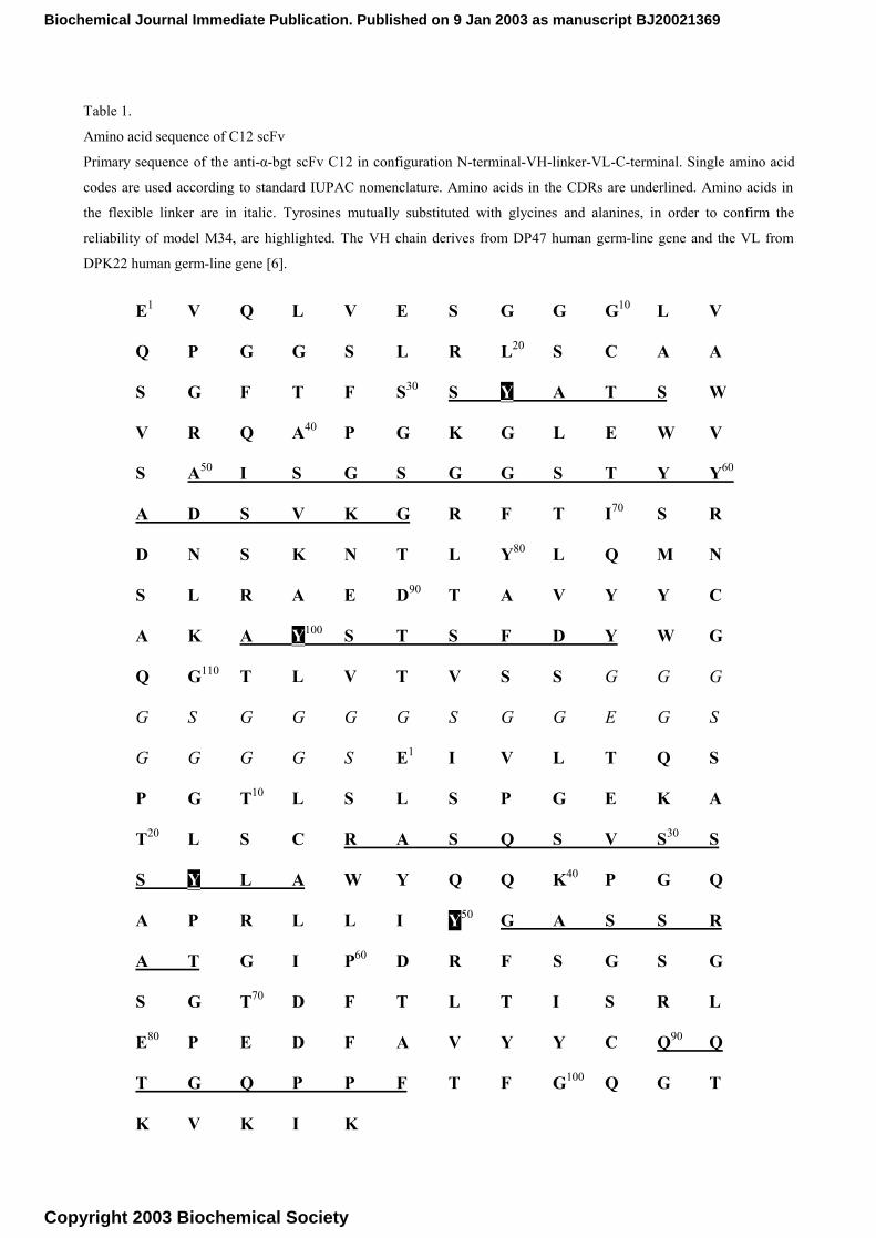
Table 1.
Amino acid sequence of C12 scFv
Primary sequence of the anti-α-bgt scFv C12 in configuration N-terminal-VH-linker-VL-C-terminal. Single amino acid
codes are used according to standard IUPAC nomenclature. Amino acids in the CDRs are underlined. Amino acids in
the flexible linker are in italic. Tyrosines mutually substituted with glycines and alanines, in order to confirm the
reliability of model M34, are highlighted. The VH chain derives from DP47 human germ-line gene and the VL from
DPK22 human germ-line gene [6].
E1 V Q L V E S G G G10 L V
Q P G G S L R L20 S C A A
S G F T F S30 S Y A T S W
V R Q A40 P G K G L E W V
S A50 I S G S G G S T Y Y60
A D S V K G R F T I70 S R
D N S K N T L Y80 L Q M N
S L R A E D90 T A V Y Y C
A K A Y100 S T S F D Y W G
Q G110 T L V T V S S G G G
G S G G G G S G G E G S
G G G G S E1 I V L T Q S
P G T10 L S L S P G E K A
T20 L S C R A S Q S V S30 S
S Y L A W Y Q Q K40 P G Q
A P R L L I Y50 G A S S R
A T G I P60 D R F S G S G
S G T70 D F T L T I S R L
E80 P E D F A V Y Y C Q90 Q
T G Q P P F T F G100 Q G T
K V K I K
Biochemical Journal Immediate Publication. Published on 9 Jan 2003 as manuscript BJ20021369
Copyright 2003 Biochemical Society
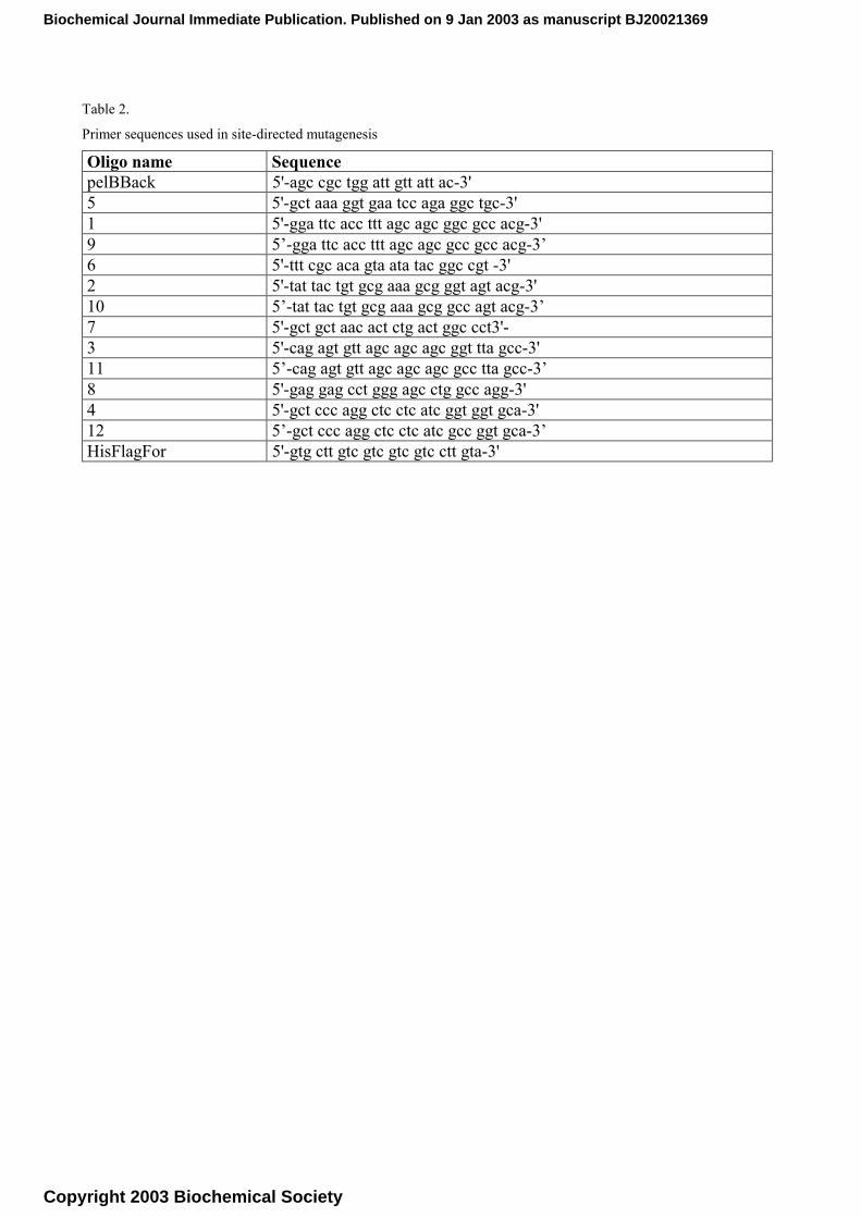
Table 2.
Primer sequences used in site-directed mutagenesis
Oligo name Sequence pelBBack 5'-agc cgc tgg att gtt att ac-3' 5 5'-gct aaa ggt gaa tcc aga ggc tgc-3' 1 5'-gga ttc acc ttt agc agc ggc gcc acg-3' 9 5’-gga ttc acc ttt agc agc gcc gcc acg-3’ 6 5'-ttt cgc aca gta ata tac ggc cgt -3' 2 5'-tat tac tgt gcg aaa gcg ggt agt acg-3' 10 5’-tat tac tgt gcg aaa gcg gcc agt acg-3’ 7 5'-gct gct aac act ctg act ggc cct3'- 3 5'-cag agt gtt agc agc agc ggt tta gcc-3' 11 5’-cag agt gtt agc agc agc gcc tta gcc-3’ 8 5'-gag gag cct ggg agc ctg gcc agg-3' 4 5'-gct ccc agg ctc ctc atc ggt ggt gca-3' 12 5’-gct ccc agg ctc ctc atc gcc ggt gca-3’ HisFlagFor 5'-gtg ctt gtc gtc gtc gtc ctt gta-3'
Biochemical Journal Immediate Publication. Published on 9 Jan 2003 as manuscript BJ20021369
Copyright 2003 Biochemical Society
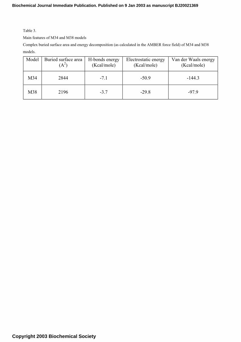
Table 3.
Main features of M34 and M38 models
Complex buried surface area and energy decomposition (as calculated in the AMBER force field) of M34 and M38
models. Model Buried surface area
(A2) H-bonds energy
(Kcal/mole) Electrostatic energy
(Kcal/mole) Van der Waals energy
(Kcal/mole)
M34 2844 -7.1 -50.9 -144.3
M38 2196 -3.7 -29.8 -97.9
Biochemical Journal Immediate Publication. Published on 9 Jan 2003 as manuscript BJ20021369
Copyright 2003 Biochemical Society

Caption to figure 1:
Models of the resulting scFv C12/α-bgt complexes
The scFv C12/α-bgt complexes M34 (top) and M38 (bottom) as resulted from docking study represented as spacefill:
the C12 L chain is shown in blue with CDR1, CDR2 and CDR3 coloured in purple, cyan and blue, respectively, while
the H chain is shown in brown, with CDR1, CDR2 and CDR3 coloured in orange, yellow and red, respectively. The α-
bungarotoxin is represented in ivory as backbone only. α-bgt loop numbering is reported as roman numbers.
Caption to figure 2:
Close view of the scFv C12/α-bgt interface in the M34 and M38 models
The scFv C12/α-bgt complex M34 (top) and M38 (bottom) as resulted from docking study represented as ribbons: the
C12 L chain is shown in blue, the H chain in red and the toxin in green. The aminoacids involved in key interactions are
also shown.
Biochemical Journal Immediate Publication. Published on 9 Jan 2003 as manuscript BJ20021369
Copyright 2003 Biochemical Society
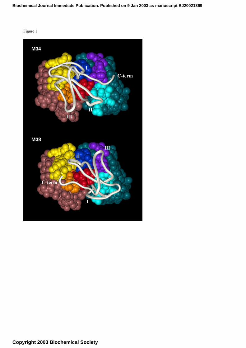
Figure 1
Biochemical Journal Immediate Publication. Published on 9 Jan 2003 as manuscript BJ20021369
Copyright 2003 Biochemical Society
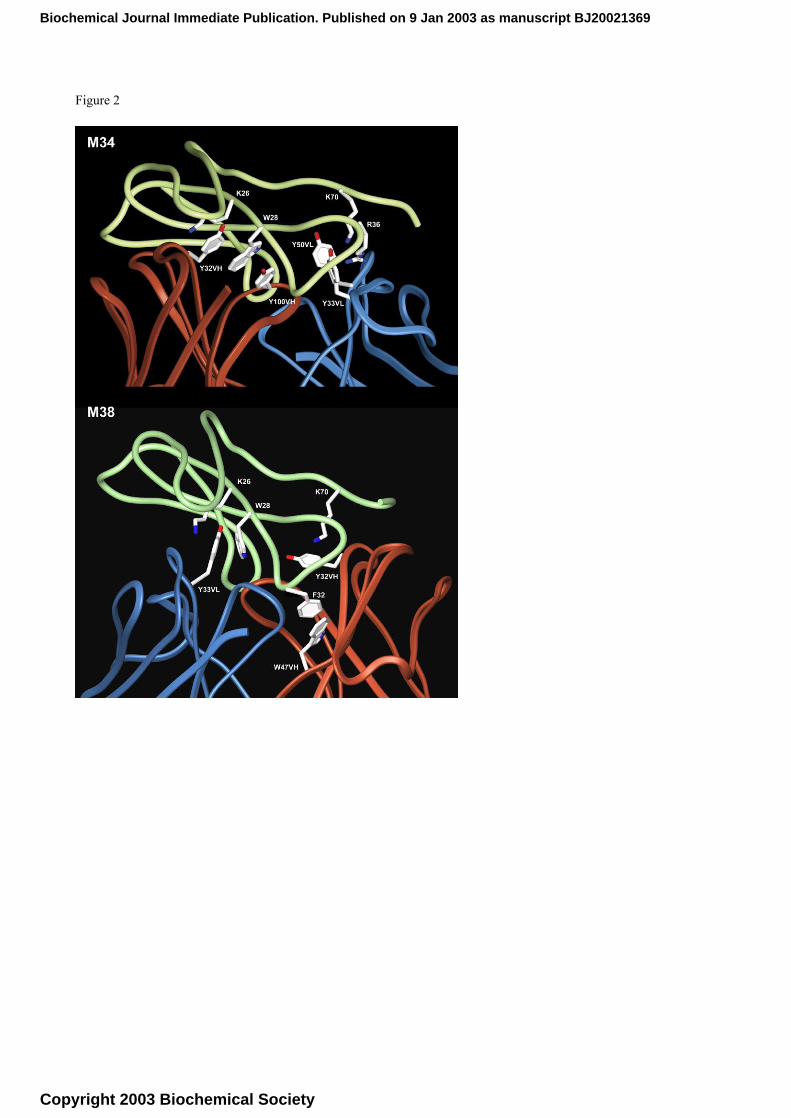
Figure 2
Biochemical Journal Immediate Publication. Published on 9 Jan 2003 as manuscript BJ20021369
Copyright 2003 Biochemical Society