DOCKING AND ADME ANALYSIS
13
Ramya et al RJLBPCS 2018 www.rjlbpcs.com Life Science Informatics Publications © 2018 Life Science Informatics Publication All rights reserved Peer review under responsibility of Life Science Informatics Publications 2018 Sept – Oct RJLBPCS 4(5) Page No.218 Original Research Article DOI: 10.26479/2018.0405.17 ANTICANCER POTENTIALS OF QUASSINOIDS FROM SIMAROUBA GLAUCA – DOCKING AND ADME ANALYSIS K. S. Ramya 1 , Saleem Iqbal 2 , K. Gunasekaran 2 , A. Radha 1 * 1. Department of Botany, Bharathi Women’s College, Chennai, India. 2.CAS in Crystallography and Biophysics, University of Madras, Guindy Campus, Chennai, India. ABSTRACT: The objective of this study was to analyze the inhibitory activity of selected quassinoids from the plant Simarouba glauca using docking studies against Phosphoinositide 3- kinases (PI3Ks). In silico technique was applied to screen abilities of the quassinoids as a potent inhibitor of PI3Ks. The 3D structure of the protein was obtained from PDB database and of the ligands from PUBCHEM database. Docking analysis of the compounds was performed using Ligpep 2.3, Schrodinger Suite 2009. The comparison of the docking score indicated that the compounds exhibited better binding affinity similar to that of the known drug Idelalisib. Further analysis of the drug likeness by means of ADME properties were predicted using Swissadme online server. None of the compounds violated Lipinski’s parameters, making them potentially promising agents for biological activities. Finally, the results indicated that these compounds are potential inhibitor of PI3K and expected to be effective in cancer treatment. KEYWORDS: Simarouba glauca, Quassinoids, ADME Properties, Phosphoinositide 3-kinases (PI3Ks), Anti – Cancer. Corresponding Author: Dr. A. Radha* Ph.D. Department of Botany, Bharathi Women’s College, Chennai, India. Email Address: [email protected] 1. INTRODUCTION Simarouba glauca, commonly known as Laxmitaru or paradise tree belongs to the family Simaroubaceae. Common names for S.glauca are bitter ash, bitter damson princess tree and Simarouba. This tree is also known as “Tree of solace of cancer” as it is widely used in cancer treatment [1]. It produces bright green leaves of 20-50 cm length, yellow flowers and oval elongated
-
Upload
khangminh22 -
Category
Documents
-
view
0 -
download
0
Transcript of DOCKING AND ADME ANALYSIS
Ramya et al RJLBPCS 2018 www.rjlbpcs.com Life Science Informatics Publications
© 2018 Life Science Informatics Publication All rights reserved
Peer review under responsibility of Life Science Informatics Publications
2018 Sept – Oct RJLBPCS 4(5) Page No.218
Original Research Article DOI: 10.26479/2018.0405.17
ANTICANCER POTENTIALS OF QUASSINOIDS FROM SIMAROUBA
GLAUCA – DOCKING AND ADME ANALYSIS
K. S. Ramya1, Saleem Iqbal2, K. Gunasekaran2, A. Radha1*
1. Department of Botany, Bharathi Women’s College, Chennai, India.
2.CAS in Crystallography and Biophysics, University of Madras, Guindy Campus, Chennai, India.
ABSTRACT: The objective of this study was to analyze the inhibitory activity of selected
quassinoids from the plant Simarouba glauca using docking studies against Phosphoinositide 3-
kinases (PI3Ks). In silico technique was applied to screen abilities of the quassinoids as a potent
inhibitor of PI3Ks. The 3D structure of the protein was obtained from PDB database and of the
ligands from PUBCHEM database. Docking analysis of the compounds was performed using
Ligpep 2.3, Schrodinger Suite 2009. The comparison of the docking score indicated that the
compounds exhibited better binding affinity similar to that of the known drug Idelalisib. Further
analysis of the drug likeness by means of ADME properties were predicted using Swissadme online
server. None of the compounds violated Lipinski’s parameters, making them potentially promising
agents for biological activities. Finally, the results indicated that these compounds are potential
inhibitor of PI3K and expected to be effective in cancer treatment.
KEYWORDS: Simarouba glauca, Quassinoids, ADME Properties, Phosphoinositide 3-kinases
(PI3Ks), Anti – Cancer.
Corresponding Author: Dr. A. Radha* Ph.D.
Department of Botany, Bharathi Women’s College, Chennai, India.
Email Address: [email protected]
1. INTRODUCTION
Simarouba glauca, commonly known as Laxmitaru or paradise tree belongs to the family
Simaroubaceae. Common names for S.glauca are bitter ash, bitter damson princess tree and
Simarouba. This tree is also known as “Tree of solace of cancer” as it is widely used in cancer
treatment [1]. It produces bright green leaves of 20-50 cm length, yellow flowers and oval elongated
Ramya et al RJLBPCS 2018 www.rjlbpcs.com Life Science Informatics Publications
© 2018 Life Science Informatics Publication All rights reserved
Peer review under responsibility of Life Science Informatics Publications
2018 Sept – Oct RJLBPCS 4(5) Page No.219
purple colored fleshy fruits. It is suited for temperature range of 10-40ºC, with pH of the soil to be
5.5-8.0 [2]. The bark and leaf extracts of Simarouba is well known for its different types of
pharmacological properties such as haemostatic, anthelminthic, antiparasitic, antidysentric,
antipyretic, anticancerous[3], antimicrobial, antiherpetic, antiprotozoal [4], antiamoebic,
antimalarial, antifungal, antioxidant and antiulcer[5] activities along with hepatoprotective property.
The main group of chemicals in Simarouba glauca is quassinoids, which belong to the triterpene
family. This includes: ailanthinone, canthin, dehydroglaucarubinone, glaucarubine, glaucarubolone,
glaucarubinone, holacanthone, melianone, simaroubidin, simarolide, simarubin, simarubolide,
sitosterol, tirucalla etc. [3]. After the initial discovery of the antileukemic activity of bruceantin, a
quassinoids, these molecules gained much attention [6]. Based on the chemical structures and
biological properties nearly 150 quassinoids have been isolated and classified. A wide range of
inhibitory effects has been shown by quassinoids which includes anti-inflammatory,
antiproliferative effects on tumor cells types [7]. Considering the future in generating anticancer
agents with more active and less toxic compounds, natural quassinoids represent a promising source
of small molecules. From the quassinoids listed, four were selected (Glaucarubine, Glaucarubolone,
Glaucarubinone, Melianone) based on the PASS (Prediction of Active Spectra for Substances)
online server prediction and target hunter databases which are used to find the biological activities
of the particular compounds. In order to understand the biological activity of the compounds,
structural scaffold of the ligands was taken into consideration. The selected ligands were allowed
for their prediction against novel targets for which PASS prediction server is used [8]
(www.way2drug.com). This server predicts more than 300 pharmacological factors and biochemical
mechanisms on the basis of the structural formula of the compounds. The given ligands were
predicted to interact with 15 targets, among which the anticancer target PI3K was the most profound
target for all the ligands. Smiles format of the ligands were used as input for the PASS online server.
From 1980’s the family of lipid kinases termed Phosphoinositide 3-kinases (PI3Ks) has been found
to play key regulatory roles in many cellular processes including cell survival, proliferation and
differentiation [9]. The discovery of PIK3CA gene which encodes p110α confirmed the importance
of PI3Ks in cancer, as these genes are frequently mutated in most of the common human tumors [10,
11, 12]. PI3Ks are divided into three classes, based on their structural characteristics and substrate
specificity [13, 14]. Class IA PI3Ks are heterodimers consisting of p110 catalytic subunit and p85
regulatory subunit. There are three isoforms of p110 which include p110α, p110β which are
expressed ubiquitously and p110δ which is restricted to immune system [15]. PI3Kδ is selectively
expressed in leukocytes which made them a therapeutic target for diseases in which there is
pathological activation of the Akt pathway in hematopoietic cells [16]. PI3K/Akt signaling pathways
are activated in hematological malignancies of B cells which include indolent non-Hodgkin
lymphoma, chronic lymphocytic leukemia and mantle cell lymphoma which responds to pathway
Ramya et al RJLBPCS 2018 www.rjlbpcs.com Life Science Informatics Publications
© 2018 Life Science Informatics Publication All rights reserved
Peer review under responsibility of Life Science Informatics Publications
2018 Sept – Oct RJLBPCS 4(5) Page No.220
inhibition [17]. Idelalisib is a potent selective inhibitor of p110δ kinases activity. This drug along
with rituximab was clinically approved by the United States and European Union and is now
recommended for treatment of patients with relapsed chronic lymphocytic leukemia and as a
monotherapy for patients with relapsed follicular B cell non-Hodgkin lymphoma and small
lymphocytic leukemia [18]. In the present study, comparative analysis of the selected quassinoids
from the plant Simarouba glauca with the known drug Idelalisib was carried out using molecular
docking studies and ADME properties which analyzed the drug likeness of the ligands.
2. MATERIALS AND METHODS
2.1. Target and Binding site
The three dimensional structure of Phosphoinositide 3-kinases (PI3Ks) complexed with known
drug Idelalisib (PDB ID: 4XE0) was retrieved from the Research Collaboratory for Structural
Bioinformatics (RCSB) protein Data Bank [29]. Binding sites of the protein were identified by
FT site server [19]. Binding site identification has a wide range of applications which includes
structure-based prediction of function, the elucidation of functional relationships among proteins,
protein engineering and drug design. FT Site Server describes an accurate method of binding site
identification [20].
2.2. Ligands preparation
The ligands are small molecules which bind to the Protein’s binding sites. The SDF (Structure-Data
Format) files of all compounds i.e. Glaucarubine, Glaucarubolone, Glaucarubinone, Melianone
were obtained from Pubchem database [21] and analyzed by Marvin view [30]. The compounds
were converted to 3D structure (PDB) using PyMol version 1.3 [31] (The PyMol Molecular
Graphics System). The physicochemical properties of the ligands are estimated by Pubchem open
chemistry database (www.pubchem.com).
2.3. Analysis of drug likeness of the ligands
The drug likeness prediction of the ligands were carried out by Lipinski filter, according to which
an orally active drug should fulfill a minimum of four out of five criteria for drug likeness namely
molecular mass, cLogP, hydrogen donor, hydrogen acceptor and molar refractive index [22]. These
properties were analyzed by SWISS ADME (http://www.swissadme.ch), which is reported as a
convenient tool in drug discovery [23]. Also, the properties of ligands with respect to adsorption,
distribution, metabolism and excretion (ADME) were analyzed by SWISS ADME. Gastrointestinal
absorption and brain access are two pharmacokinetic behaviors crucial to estimate various stages of
the drug discovery processes. The Brain Or IntestinaL EstimateD permeation method (BOILED-
Egg) is proposed as an accurate predictive model that works by computing the lipophilicity and
polarity of small molecules [24].
Ramya et al RJLBPCS 2018 www.rjlbpcs.com Life Science Informatics Publications
© 2018 Life Science Informatics Publication All rights reserved
Peer review under responsibility of Life Science Informatics Publications
2018 Sept – Oct RJLBPCS 4(5) Page No.221
2.4. Molecular Docking Procedures
The aim of molecular docking is to predict the binding modes of ligand and thus define the
orientation of molecule with respect to the active or binding site. In this method according to the
affinity score in terms of (kcal/mol), ranking all the binding poses of the molecule inside the
catalytic site of an enzyme is being done .The selected compounds were taken for minimization
using Ligprep module of Schrodinger 09 where probable tautomeric and ionization states at pH = 7
± 1 followed by energy minimization with OPLS 2005 force field [25, 32] was carried out. The
protein preparation of the target (PDB ID 4XE0) was performed using Protein preparation wizard
of Schrodinger 09 where missing hydrogen bond order were assigned followed by energy
minimization.
2.4.1 Molecular Docking
The receptor grid was prepared keeping cocrystal (Idelalisib) ligand on PI3Ks (PDB ID 4XE0) at
the center of grid with 20 Ǻ edges bearing catalytic site. Initially docking study on the cocrystal was
performed on prepared receptor grid for cross-validating the binding mode with respect to X-ray
crystal structure binding mode. Further, molecular docking was performed for given ligands
against PDB ID using Glide XP 5.8 Program [26, 27, 28]. The top analogs based on docking score
as well as binding interaction with catalytic residues were allowed for induced fit docking and
results were compared with the cocrystal after Glide XP. The docked conformation corresponding
to the lowest free energy (or highest score) provided by Glide program was selected as the most
probable binding pose of top compound.
2.4.2. Structural features of the complex
Once the docking was performed, best poses for hydrogen bonding, Hydrophobic and π interactions
were analyzed using PyMol version 1.3 (The PyMol Molecular Graphics System).
3. RESULTS AND DISCUSSION
Owing to the important functions of PI3Ks and challenges in designing specific inhibitors for kinase
proteins because of highly conserved active site architecture, there is big demand for structure
analysis to identify new inhibitors targeting various diseases. Together with the above mentioned
fact, the usefulness of the Simarouba glauca plant against many illness and important compounds
were selected to screen their affinity towards PI3Ks. This selection was guided by PASS prediction
server.
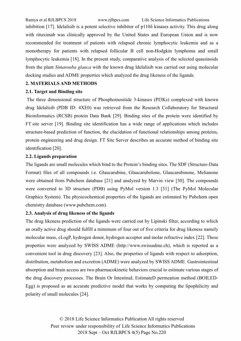
3.1. Macromolecule
The target Phosphoinositide 3-kinases (PI3Ks) complexed with known drug Idelalisib
(PDBID:4XE0) was used for docking studies (Figure 1).
Ramya et al RJLBPCS 2018 www.rjlbpcs.com Life Science Informatics Publications
© 2018 Life Science Informatics Publication All rights reserved
Peer review under responsibility of Life Science Informatics Publications
2018 Sept – Oct RJLBPCS 4(5) Page No.222
Fig 1. Cartoon representation of PI3K (PDB Code: 4XE0). Idelalisib is
shown as solid ball and stick.
This protein comprised of 939 residues, out of which 815 were observed under electron density. The
kinase domain and ATP binding sites are well defined by the electron density whereas certain
fractions of the protein have weak electron density and correspondingly high temperature factors.
The drug Idelalisib binds in the ATP binding pocket. The inhibitor forms hydrogen bonds to the
hinge region that is similar to those made by ATP. The 2.4A˚ crystal structure of the Idelalisib-PI3Kδ
complex showed the inhibitor binding in the ATP site and revealed the specific and entirely
noncovalent interactions between protein and inhibitor [18].
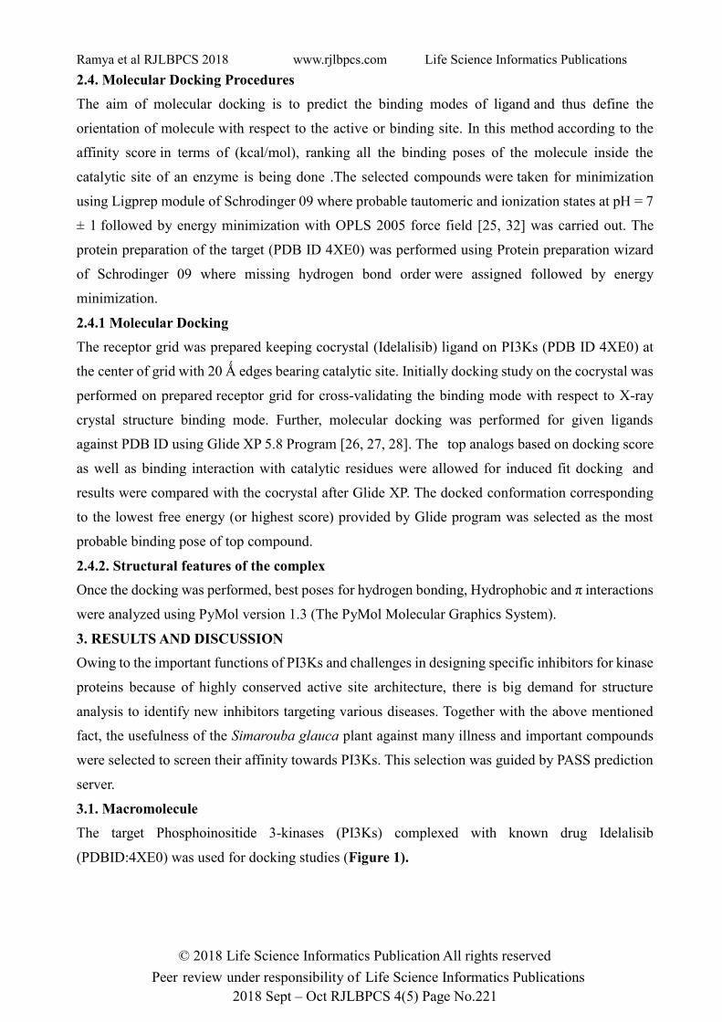
3.2. Active sites of the Protein molecule
In this study, FT Site Server predicted the active sites of the targeted protein PI3K. Three active sites
are projected in this protein (Figure 2). The active sites comprises of amino acid residues are Active
site 1 : PHE 609, PHE 646, HIS 650, PRO 734, LEU 735, ASP 787, MET 788, LEU 791, GLN 792,
GLN 795, PRO 812, TYR 813, GLY 814, CYS 815, LEU 816, Active site 2: PHE 609, GLN 610,
TYR 611, LEU 612, LEU 613, PHE 646, HIS 650, MET 788, GLN 792, Active site 3: ARG 246,
ASP 736, THR 739, GLU 826.
Pink: Active site 1, Green: Active site 2, Blue: Active site 3 (as predicted by FT Site Server)
Fig 2. Active Sites of PI3K
Ramya et al RJLBPCS 2018 www.rjlbpcs.com Life Science Informatics Publications
© 2018 Life Science Informatics Publication All rights reserved
Peer review under responsibility of Life Science Informatics Publications
2018 Sept – Oct RJLBPCS 4(5) Page No.223
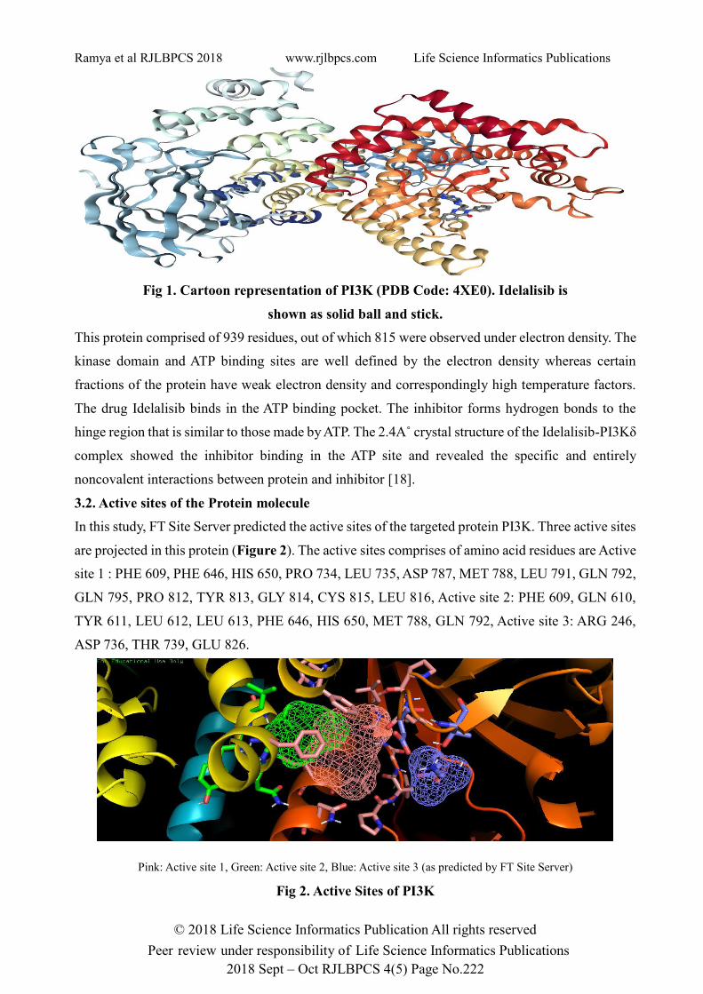
3.3. Ligands
The physicochemical properties of the ligands those 3D structures shown in Figure 3 are mentioned
in table 1.
3a: Glaucarubine 3b: Glaucarubinone
3c: Glaucarubolone 3d: Melianone 3e: Idelalisib (Cocrystal)
Figure 3. 3D structures of the Ligands
Table 1: Physicochemical Properties Of The Ligands
Idelalisib’s exposed polar surface area (PSA) is 99.2 A˚2 which is less than the selected compounds
except Melianone whose PSA is 59.1 A˚2 though the mass is 470 g/mol.
3.4. Drug likeness analysis
Results of Lipinski filter analysis is tabulated (Table 2a) which explains the rigidity of all
compounds to be considered for structure based drug design. Also table 2b list out the properties of
compounds with relevance to their usage as explained by ADME properties.
Sr.
No.
Ligand Molecular
formula
Molecular
weight
Monoisotropic
mass
Heavy
atom
count
Topological
Polar Surface
Area
1 Glaucarubine C25H36O10 496.553 g/mol 496.231 g/mol 35 163 A˚2
2 Glaucarubinone C25H34O10 494.537 g/mol 494.215 g/mol 35 160 A˚2
3 Glaucarubolone C20H26O8 394.42 g/mol 394.163 g/mol 28 134 A˚2
4 Melianone C30H46O4 470.694 g/mol 470.34 g/mol 34 59.1 A˚2
5 Idelalisib
(Cocrystal)
C22H18FN7O 415.432 g/mol 415.156 g/mol 31 99.2 A˚2
Ramya et al RJLBPCS 2018 www.rjlbpcs.com Life Science Informatics Publications
© 2018 Life Science Informatics Publication All rights reserved
Peer review under responsibility of Life Science Informatics Publications
2018 Sept – Oct RJLBPCS 4(5) Page No.224
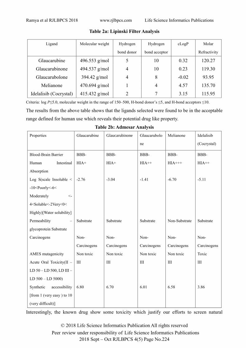
Table 2a: Lipinski Filter Analysis
Ligand Molecular weight Hydrogen
bond donor
Hydrogen
bond acceptor
cLogP Molar
Refractivity
Glaucarubine 496.553 g/mol 5 10 0.32 120.27
Glaucarubinone 494.537 g/mol 4 10 0.23 119.30
Glaucarubolone 394.42 g/mol 4 8 -0.02 93.95
Melianone 470.694 g/mol 1 4 4.57 135.70
Idelalisib (Cocrystal) 415.432 g/mol 2 7 3.15 115.95
Criteria: log P≤5.0, molecular weight in the range of 150–500, H-bond donor’s ≤5, and H-bond acceptors ≤10.
The results from the above table shows that the ligands selected were found to be in the acceptable
range defined for human use which reveals their potential drug like property.
Table 2b: Admesar Analysis
Properties Glaucarubine Glaucarubinone
Glaucarubolo
ne
Melianone Idelalisib
(Cocrystal)
Blood-Brain Barrier BBB- BBB- BBB- BBB- BBB-
Human Intestinal
Absorption
HIA+ HIA+ HIA++ HIA+++ HIA++
Log S(scale Insoluble <
-10<Poorly<-6<
Moderately <-
4<Soluble<-2Very<0<
Highly)[Water solubility]
-2.76 -3.04 -1.41 -6.70 -5.11
Permeability –
glycoprotein Substrate
Substrate Substrate Substrate Non-Substrate Substrate
Carcinogens Non-
Carcinogens
Non-
Carcinogens
Non-
Carcinogens
Non-
Carcinogens
Non-
Carcinogens
AMES mutagenicity Non toxic Non toxic Non toxic Non toxic Toxic
Acute Oral Toxicity(II –
LD 50 – LD 500, LD III –
LD 500 – LD 5000)
III III III III III
Synthetic accessibility
[from 1 (very easy ) to 10
(very difficult)]
6.80 6.70 6.01 6.58 3.86
Interestingly, the known drug show some toxicity which justify our efforts to screen natural

Ramya et al RJLBPCS 2018 www.rjlbpcs.com Life Science Informatics Publications
© 2018 Life Science Informatics Publication All rights reserved
Peer review under responsibility of Life Science Informatics Publications
2018 Sept – Oct RJLBPCS 4(5) Page No.225
compounds.
3.4.1. Brain Or IntestinaL EstimateD permeation method (BOILED-Egg)
The prediction also reveals that Melianone has high GI absorption followed by the known drug
Idelalisib which is then followed by Glaucarubolone. Glaucarubine and Glaucarubinone have low
GI absorption (Figure 4).
Figure 4. BOILED EGG Model: The white region is the physicochemical space of molecules with
highest probability of being absorbed by the gastrointestinal tract, and the yellow region (yolk) is the
physicochemical space of molecules with highest probability to permeate to the brain.
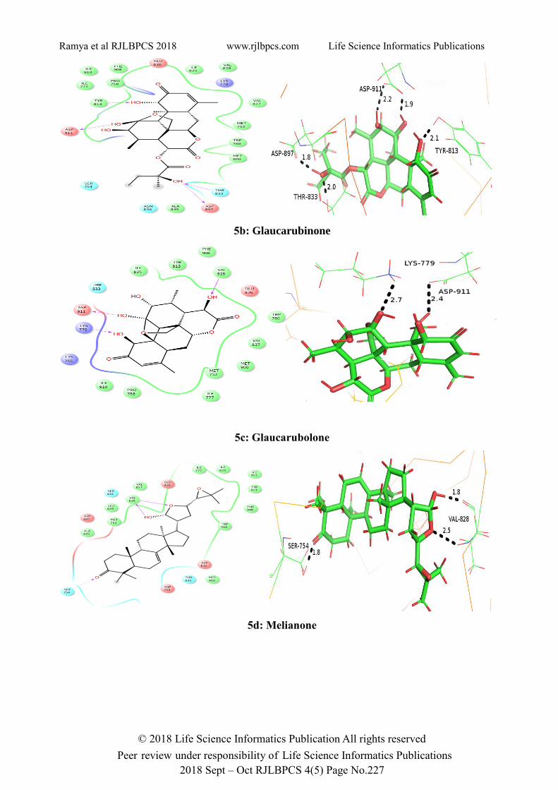
3.5. Docking Results:
Results obtained from induced fit docking of compounds of the active site of PI3K (4XE0) are
reported in Table 3. The molecular docking study of the compounds with PI3K receptor shows that,
all the compounds are showing better docking score than that of the known drug Idelalisib which
predicts that the compounds chosen have the better binding affinity to the receptor than the cocrystal.
The hydrogen binding interactions of the compounds with PI3K is shown in figure 5 (both 2D and
3D interactions). This prediction leads us to believe that, the compounds will possibly be suitable
for cancer treatment.
Table 3: Docking Results
Ligand Docking
Score
Glide Energy
(Kcal/mol)
H – Bond Bond
length (A˚)
Hydrophobic
Interactions
Glaucarubine -12.307
-78.917
(O-H---O) Asp 911
Tyr 813 (O-H---O)
Thr 833 (O-H---O)
(O-H---O) Asp 897
1.6
2.0
2.6
2.5
Pro 758, Trp 760,
Val 828, Ile 825, Ile
777, Met 752
Glaucarubinone
-9.313
-71.535
Thr 813 (H---O-H)
(O-H---O) Asp 911
(O-H---O) Asp 911
(O-H---O) Asp 897
2.1
1.9
2.2
1.8
Met 900, Trp 760,
Met 752, Val 827,
Ile 825, Phe 908,
Pro 758, Ile 777,

Ramya et al RJLBPCS 2018 www.rjlbpcs.com Life Science Informatics Publications
© 2018 Life Science Informatics Publication All rights reserved
Peer review under responsibility of Life Science Informatics Publications
2018 Sept – Oct RJLBPCS 4(5) Page No.226
Thr 833 (H ---O-H) 2.0
Glaucarubolone -11.31
-67.601
Lys 779 (N-H---O)
(O-H---H) Asp 911
2.7
2.4
Val 828, Tyr 813,
Phe 908, Ile 825
Melianone -11.193
-80.162
Val 828 (N-H---O)
(O-H---O) Val 828
(O-H---O) Ser 754
2.5
1.8
1.8
Ala 835, Met 752,
Leu 829, Val 827,Ile
777, Ile 825, Tyr
813, Trp 760
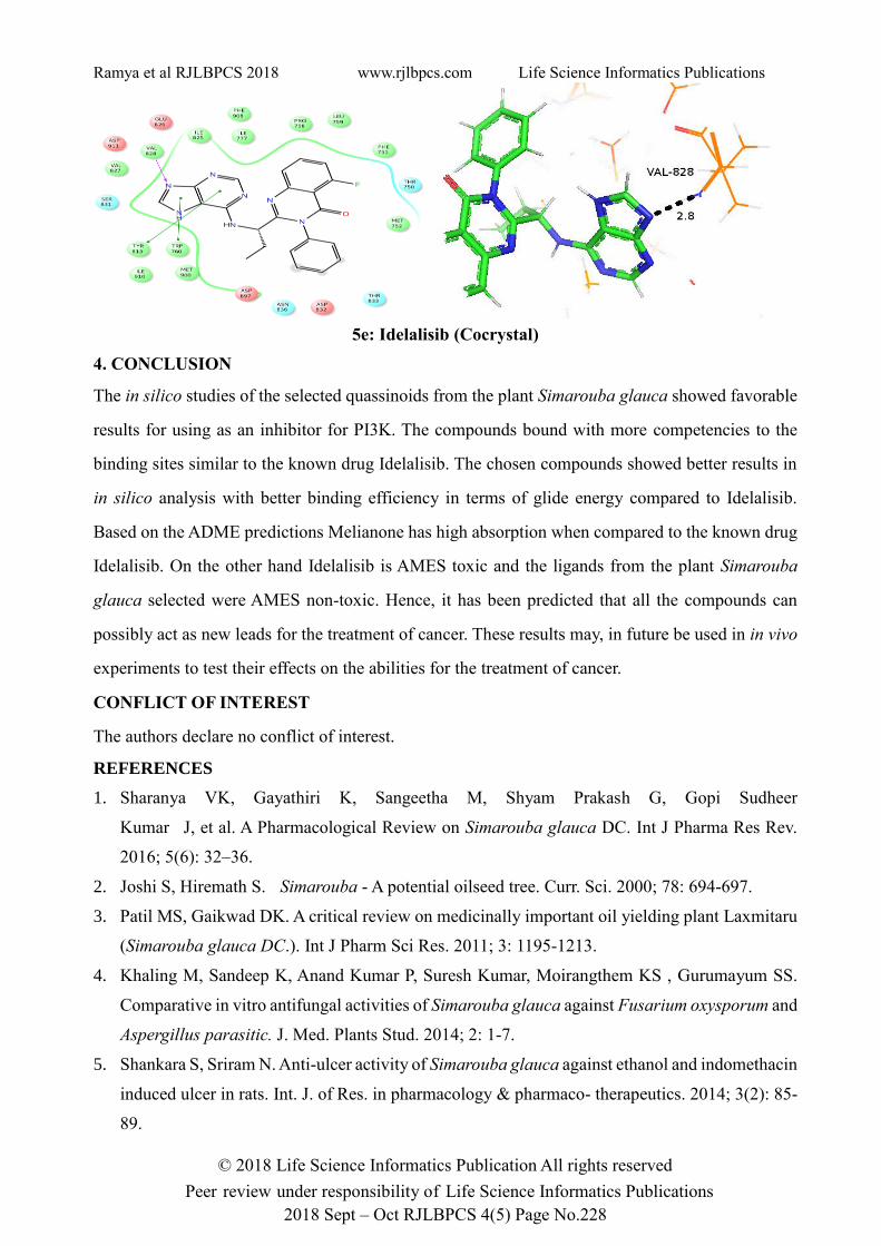
Idelalisib
(Cocrystal)
-10.242 -76.018 Val 828 (N-H---N) 2.8 Tyr 813, Trp 760,
Val 827, Ile 825, Ile
777, Pro 758, Phe
751.
The docking results mentioned in the above table clearly shows that all the compounds selected for
docking study displayed excellent binding affinity when compared to the known drug Idelalisib.
Melianone showed best glide energy (-80.162) followed by Glaucarubine (-78.916) which is much
better than the cocrystal used Idelalisib (-76.018). The hydrogen bond interactions along with the
bond distance (A˚) are shown in the PyMol interactions (Figure 5). The hydrophobic interactions
are mentioned in table 3.
Figure 5. 2D and 3D interactions of the compounds
5a: Glaucarubine
Ramya et al RJLBPCS 2018 www.rjlbpcs.com Life Science Informatics Publications
© 2018 Life Science Informatics Publication All rights reserved
Peer review under responsibility of Life Science Informatics Publications
2018 Sept – Oct RJLBPCS 4(5) Page No.227
5b: Glaucarubinone
5c: Glaucarubolone
5d: Melianone
Ramya et al RJLBPCS 2018 www.rjlbpcs.com Life Science Informatics Publications
© 2018 Life Science Informatics Publication All rights reserved
Peer review under responsibility of Life Science Informatics Publications
2018 Sept – Oct RJLBPCS 4(5) Page No.228
5e: Idelalisib (Cocrystal)
4. CONCLUSION
The in silico studies of the selected quassinoids from the plant Simarouba glauca showed favorable
results for using as an inhibitor for PI3K. The compounds bound with more competencies to the
binding sites similar to the known drug Idelalisib. The chosen compounds showed better results in
in silico analysis with better binding efficiency in terms of glide energy compared to Idelalisib.
Based on the ADME predictions Melianone has high absorption when compared to the known drug
Idelalisib. On the other hand Idelalisib is AMES toxic and the ligands from the plant Simarouba
glauca selected were AMES non-toxic. Hence, it has been predicted that all the compounds can
possibly act as new leads for the treatment of cancer. These results may, in future be used in in vivo
experiments to test their effects on the abilities for the treatment of cancer.
CONFLICT OF INTEREST
The authors declare no conflict of interest.
REFERENCES
1. Sharanya VK, Gayathiri K, Sangeetha M, Shyam Prakash G, Gopi Sudheer
Kumar J, et al. A Pharmacological Review on Simarouba glauca DC. Int J Pharma Res Rev.
2016; 5(6): 32–36.
2. Joshi S, Hiremath S. Simarouba - A potential oilseed tree. Curr. Sci. 2000; 78: 694-697.
3. Patil MS, Gaikwad DK. A critical review on medicinally important oil yielding plant Laxmitaru
(Simarouba glauca DC.). Int J Pharm Sci Res. 2011; 3: 1195-1213.
4. Khaling M, Sandeep K, Anand Kumar P, Suresh Kumar, Moirangthem KS , Gurumayum SS.
Comparative in vitro antifungal activities of Simarouba glauca against Fusarium oxysporum and
Aspergillus parasitic. J. Med. Plants Stud. 2014; 2: 1-7.
5. Shankara S, Sriram N. Anti-ulcer activity of Simarouba glauca against ethanol and indomethacin
induced ulcer in rats. Int. J. of Res. in pharmacology & pharmaco- therapeutics. 2014; 3(2): 85-
89.
Ramya et al RJLBPCS 2018 www.rjlbpcs.com Life Science Informatics Publications
© 2018 Life Science Informatics Publication All rights reserved
Peer review under responsibility of Life Science Informatics Publications
2018 Sept – Oct RJLBPCS 4(5) Page No.229
6. Kupchan SM, Britton RW, Lacadie JA, Ziegler MF, Sigel CW. The isolation and structural
elucidation of bruceantin and bruceantinol, new potent antileukemic quassinoids from Brucea
antidysenterica. J Org Chem. 1975; 40(5): 648-654.
7. Fiaschetti G, Grotzer MA, Shalaby T, Castelletti D, Arcaro A. Quassinoids: From Traditional
Drugs to New Cancer Therapeutics. Curr Med Chem. 2011; 18: 316-328.
8. Filimonov DA, Lagunin AA, Gloriozova TA, Rudik AV, Druzhilovskii DS, Pogodin PV, et al.
Prediction of the biological activity spectra of organic compounds using the PASS online web
resource. Chem. Heterocycl. Compd. 2014; 50 (3): 444-457.
9. Engelman JA, Luo J, Cantley LC. The evolution of phosphatidylinositol 3-kinases as regulators
of growth and metabolism. Nat Rev Genet. 2006; 7: 606–619.
10. Thomas RK, Baker AC, Debiasi RM, Winckler W, Laframboise T, Lin WM, et al. High-
throughput oncogene mutation profiling in human cancer. Nat Genet. 2007; 39: 347–351.
11. Parsons DW, Jones S, Zhang X, Lin JC, Leary RJ, Angenendt P, et al. An integrated genomic
analysis of human glioblastoma multiforme. Science. 2008; 321: 1807–1812.
12. Samuels Y, Wang Z, Bardelli A, Silliman N, Ptak J, Szabo S, et al. High frequency of mutations
of the PIK3CA gene in human cancers. Science. 2004; 304: 554.
13. Fruman DA, Meyers RE, Cantley LC. Phosphoinositide kinases. Annu Rev Biochem. 1998; 67:
481– 507.
14. Katso R, Okkenhaug K, Ahmadi K, White S, Timms J, Waterfield MD. Cellular function of
Phosphoinositide 3-kinases: implications for development, homeostasis, and cancer. Annu Rev
Cell Dev Biol. 2001; 17: 615–675.
15. Pixu Liu, Hailing Cheng, Thomas M. Roberts, Jean J. Zhao. Targeting the Phosphoinositide 3-
kinase pathway in cancer. Nat Rev Drug Discov. 2009; 8: 627–644.
16. Bader AG, Kang S, Zhao L, Vogt PK. Oncogenic PI3K deregulates transcription and translation.
Nat Rev Cancer. 2005; 5: 921–929.
17. Hennessy BT, Smith DL, Ram PT, Lu Y, Mills GB. Exploiting the PI3K/AKT pathway for cancer
drug discovery. Nat Rev Drug Discov. 2005; 4: 988–1004.
18. Somoza JR, Koditek D, Villasenor AG, Novikov N, Wong MH, Liclican A, et al. Structural,
Biochemical, and Biophysical Characterization of Idelalisib Binding to Phosphoinositide 3-
Kinase delta. J. Biol. Chem. 2015; 290: 8439-8446.
19. Kozakov D, Grove LE, Hall DR, Bohnuud T, Mottarella SE, Luo L, et al. The FT Map family
of web servers for determining and characterizing ligand-binding hot spots of proteins. Nat
Protoc. 2015; 10(5): 733-755.
20. Ngan CH, Hall DR, Zerbe BS, Grove LE, Kozakov D, Vajda S. FTSite: high accuracy detection
of ligand binding sites on unbound protein structures. Bioinformatics. 2012; 28: 286-287.

Ramya et al RJLBPCS 2018 www.rjlbpcs.com Life Science Informatics Publications
© 2018 Life Science Informatics Publication All rights reserved
Peer review under responsibility of Life Science Informatics Publications
2018 Sept – Oct RJLBPCS 4(5) Page No.230
21. Wang Y, Xiao J, Suzek TO, Zhang J, Wang J, Bryant SH. PubChem: a public information system
for analyzing bioactivities of small molecules. Asian J of Biotech. 2009; 4: 120-128.
22. Lipinski CA. Lead- and drug-like compounds: the rule-of-five revolution. Drug Discov Today
Technol. 2005; 1: 337-341.
23. Daina A, Michielin O, Zoete V. SwissADME: a free web tool to evaluate pharmacokinetics,
drug-likeness and medicinal chemistry friendliness of small molecules. Sci Rep. 2017; 7: 42717.
24. Daina A, Zoete V. A BOILED-Egg to predict gastrointestinal absorption and brain penetration
of small molecules. ChemMedChem. 2016; 11(11): 1117-1121.
25. Sastry GM, Adzhigirey M, Day T, Annabhimoju R, Sherman W. Protein and ligand preparation:
parameters, protocols, and influence on virtual screening enrichments. J Comput Aided Mol Des.
2013; 27(3): 221-234.
26. Halgren TA, Murphy RB, Friesner RA, Beard HS, Frye LL, Pollard WT, et al. Glide: a new
approach for rapid, accurate docking and scoring 2. Enrichment factors in database screening. J
Med Chem. 2004; 47(7): 1750-1759.
27. Friesner RA, Banks JL, Murphy RB, Halgren TA, Klicic JJ, Mainz DT, et al. Glide: a new
approach for rapid, accurate docking and scoring. J Med Chem. 2004; 47(7): 1739-1749.
28. Friesner RA, Murphy RB, Repasky MP, Frye LL, Greenwood JR, Halgren TA, et al. Extra
Precision Glide: Docking and Scoring Incorporating a Model of Hydrophobic Enclosure for
Protein−Ligand Complexes. J Med Chem. 2006; 49(21): 6177–6196.
29. Available from: http://www.rcsb.org/pdb.
30. Marvin View (version 18.10.0) calculation module developed by ChemAxon,
http://www.chemaxon.com/products/marvin, 2014.
31. PyMol - The PyMol Molecular Graphics System, Version 1.3 Schrodinger, LLC.
http://www.pymol.org.
32. Schrodinger Suite 2009 Protein Preparation Wizard; Epik version 2.0, Schrodinger, LLC, New
York, NY, 2009; Impact version 5.5, Schrodinger, LLC, New York, NY, 2009; Prime version 2.1,
Schrodinger, LLC, New York, NY, 2009, Glide, version 5.5, Schrodinger, LLC, New York, NY,
2009.