Protective Effect of Lycopene against Reperfusion Injury in ...
Upload
independentCategory
view
1download
0
Neuroprotective effect of humanin on cerebral ischemia/reperfusion injury is mediated by a PI3K/Akt pathway
Xingshun Xu, Chu Chang Chua, Jinping Gao, Kao-Wei Chua, Hong Wang, Ronald C.Hamdy, and Balvin H.L. ChuaDepartment of Pharmacology and Cecile Cox Quillen Laboratory of Geriatric Research James H.Quillen College of Medicine East Tennessee State University James H. Quillen Veterans AffairsMedical Center Johnson City, TN 37614
AbstractHumanin (HN) is an anti-apoptotic peptide that suppresses neuronal cell death induced byAlzheimer's disease, prion protein fragments, and serum deprivation. Recently, we demonstrated thatGly14-HN (HNG), a variant of HN in which the 14th amino acid serine is replaced with glycine, candecrease apoptotic neuronal death and reduce infarct volume in a focal cerebral ischemia/reperfusionmouse model. In this study, we postulate that the mechanism of HNG's neuroprotective effect ismediated by the PI3K/Akt pathway. Oxygen-glucose deprivation (OGD) was performed in culturedmouse primary cortical neurons for 60 min. The effect of HNG and PI3K/Akt inhibitors on OGD-induced cell death was examined at 24 h after reperfusion. HNG increased cell viability after OGDin primary cortical neurons, whereas the PI3K/Akt inhibitors wortmannin and Akti-1/2 attenuatedthe protective effect of HNG. HNG rapidly increased Akt phosphorylation, an effect that wasinhibited by wortmannin and Akti-1/2. Mouse brains were injected intraventricularly with HNGbefore being subjected to middle cerebral artery occlusion (MCAO) for 75 min followed by 24 hreperfusion. HNG treatment significantly elevated p-Akt levels after cerebral I/R injury anddecreased infarct volume. The protective effect of HNG on infarct size was attenuated by wortmanninand Akti-1/2. Taken as a whole, these results suggest that PI3K/Akt activation mediates HNG'sprotective effect against hypoxia/ischemia reperfusion injury.
KeywordsHumanin; PI3K/Akt; OGD; cortical neuron; MCAO
1. IntroductionHumanin (HN) is a recently identified 24-amino acid anti-apoptotic peptide that is best knownfor its ability to suppress neuronal cell death induced by Alzheimer's disease-related insults,such as amyloid beta (Aβ) toxicity (Tajima et al., 2002). Recent studies have shown that HNnot only has neuroprotective effects on Aβ toxicity but also inhibits apoptosis in many differentdiseases. HN and a highly potent HN variant, Gly14-HN (HNG), prevent apoptotic cell death
Correspondence: Dr. Balvin H.L. Chua PO Box 70,432 James H. Quillen College of Medicine East Tennessee State University JohnsonCity, TN 37614 Phone: (423) 926-1171 Ext. 7674 Fax: (423) 979-3408 E-Mail: [email protected]'s Disclaimer: This is a PDF file of an unedited manuscript that has been accepted for publication. As a service to our customerswe are providing this early version of the manuscript. The manuscript will undergo copyediting, typesetting, and review of the resultingproof before it is published in its final citable form. Please note that during the production process errors may be discovered which couldaffect the content, and all legal disclaimers that apply to the journal pertain.Section: Cellular and Molecular Biology of Nervous Systems
NIH Public AccessAuthor ManuscriptBrain Res. Author manuscript; available in PMC 2009 August 28.
Published in final edited form as:Brain Res. 2008 August 28; 1227: 12–18. doi:10.1016/j.brainres.2008.06.018.
NIH
-PA Author Manuscript
NIH
-PA Author Manuscript
NIH
-PA Author Manuscript

induced by soluble prion protein fragments in rat cortical neurons (Sponne et al., 2004). HNcan reverse memory impairments induced by different toxic agents in vivo (Mamiya et al.,2001; Krejcova et al., 2004). We recently showed that HNG protects against cerebral ischemia/reperfusion (I/R) injury by inhibiting apoptotic cell death, reducing infarct volume, anddecreasing neurological deficits in mice (Xu et al., 2006).
Despite the above studies, the protective mechanisms and signaling pathways of HN are notclear. Most importantly, the putative HN receptor on the cell membrane has not been clonedor characterized (Hashimoto et al., 2001). Ying et al. (2004) reported that HN binds to a humanG protein-coupled formylpeptide receptor-like-1 (FPRL1), which induces chemotaxis ofmononuclear phagocytes and prevents Aβ-induced apoptotic cell death by competitivelyinhibiting Aβ binding to FPRL1. Harada et al. (2004) also published findings that supportedthis theory. However, Hashimoto et al. (2005) proposed that the HN receptor might belong toa tyrosine kinase receptor family since a tyrosine kinase inhibitor (genistein) attenuates theneuroprotective activity of HNG against Aβ toxicity in F11 neuronal hybrid cells. Theseinvestigators also found that HN-mediated protection is completely blocked by the expressionof a dominant-negative STAT3, suggesting that STAT3 is involved in HN's protectivemechanism. Our previous studies demonstrated that HNG exerts its neuroprotective effects oncerebral ischemia injury by inhibiting the activation of the extracellular signal regulated kinase(ERK), indicating that HNG's protective effect is at least partially mediated by the tyrosinekinase Ras/MEK/ERK pathway (Xu et al., 2006). Finally, several studies reported that HNinhibits apoptosis by binding intracellularly to Bax and Bid, thus blocking apoptosisindependently of receptors (Guo et al., 2003; Zhai et al., 2005).
The PI3K/Akt pathway is a central mediator in signal transduction pathways involved in cellgrowth, cell survival, and metabolism (Brazil, Yang and Hemmings, 2004). Theneuroprotective role of the PI3K/Akt pathway in cerebral ischemia has been widely studied(Janelidze et. al., 2001; Noshita et al., 2001; Shibata et al., 2002). In models of cerebralischemia, Akt phosphorylation increases at 1 h and 4 h after I/R, but decreases significantly24 h after reperfusion (Janelidze et al., 2001, Shibata et al., 2002). Akt blocks apoptotic stimuliby inactivating pro-apoptotic proteins such as Bad, caspase-9, and glycogen synthasekinase-3β. Akt also exerts anti-apoptotic effects by activating endothelial nitric oxide synthase(eNOS) (Brazil, Yang and Hemmings, 2004)
Given the neuroprotective role of the PI3K/Akt pathway in cerebral ischemia, we hypothesizedthat HN's neuroprotective effect involves the activation of the PI3K/Akt pathway after cerebralI/R injury. In this study, we provide evidence that this is indeed the case by using an in vitrooxygen-glucose deprivation (OGD) cell model and a focal cerebral I/R mouse model.
2. Results2.1. HNG protects against OGD-induced neuronal death
In our study, HNG (0.2 μM) treatment alone had no effect on the viability of the mouse corticalneurons. After 60 min of OGD and 24 h reperfusion, cell viability decreased to 52.9 ± 0.93%of the control. HNG treatment significantly reduced cell death induced by OGD (69.1 ± 0.77%of the control) (Figure 1, P<0.05).
2.2. PI3K/Akt inhibitors attenuate HNG's neuroprotective effectsTo examine whether the protective effects of HNG are mediated by the PI3K/Akt pathway,OGD-treated cortical neurons were incubated with HNG and a PI3K inhibitor wortmannin (1μM) or an Akt inhibitor Akti-1/2 (1 μM). In our study, administration of wortmannin orAkti-1/2 alone had no effect on cell survival after OGD. Cells exposed to either HNG and
Xu et al. Page 2
Brain Res. Author manuscript; available in PMC 2009 August 28.
NIH
-PA Author Manuscript
NIH
-PA Author Manuscript
NIH
-PA Author Manuscript

wortmannin or HNG and Akti-1/2 had significantly decreased cell viability compared with theHNG-treated group (P<0.05) (Figure 1).
2.3. HNG activates Akt in primary cortical neuronsWe next investigated whether the addition of HNG activates Akt. Treatment of mouse corticalneurons with 0.2 μM HNG led to a time-dependent phosphorylation of Akt, with the maximallevel occurring at 10 min (Figure 2A). Wortmannin and Akti-1/2 were used to block PI3K andAkt pathways, respectively. Mouse cortical neurons were pretreated with 100 nM wortmanninfor 15 min or 1 μM Akti-1/2 for 30 min before incubating with 0.2 μM HNG for 10 min.Quantification was performed with densitometric analysis of p-Akt and total Akt. As shownin Figure 2 B,C, HNG upregulated p-Akt levels by 1.8-fold (P<0.05); this effect was attenuatedwith wortmannin or Akti-1/2. A lower concentration of wortmannin was chosen becausewortmannin at 1 μM completely blunted p-Akt levels and made densitometric analysisimpossible.
2.4. HNG activates Akt in vivoGiven the protective effect of HNG in vitro, we tested the effect of HNG in an in vivo mousemodel of middle cerebral artery occlusion. As shown in our previous study (Xu et al, 2006),mouse brains subjected to 75 min of ischemia experienced greater than 50% infarction 24 hafter ischemia/reperfusion. The heterogeneity of populations of live and dead cells hampersthe interpretation of p-Akt result. Therefore, Western blot analysis was performed after a periodof 30 min of ischemia and 6 h of reperfusion in the absence of Cell death. HNG treatmentincreased the p-Akt level by two-fold compared with the saline-treated group (P<0.05, Figure3).
2.5. Wortmannin reduces the protection of HNG on cerebral infarct volumeWe investigated whether the protective effect of HNG on cerebral I/R injury could be blockedby wortmannin, a PI3K inhibitor. The infarct size of wortmannin-treated mice (56.6 ± 2.0%)was comparable to that of vehicle-treated control mice (59.3 ± 2.6%, P>0.05, Figure 4C). Theinfarct size in the HNG-treated group was significantly decreased compared to other groups(29.2 ± 1.1%, P<0.01 for all other groups). The combination of HNG and wortmannin partiallyblocked the protective effect of HNG on infarct size (44.2 ± 1.3%).
2.6. Akti-1/2 reduces the protection of HNG on cerebral infarct volumeWe next examined whether the protective effect of HNG on cerebral I/R injury could be blockedby an Akt inhibitor, Akti-1/2 (Cheng et al., 2005). Akti-1/2 alone had no effect after cerebralI/R compared with the vehicle-treated I/R control group (56.9 ± 2.1% vs. 59.3 ± 2.6%,respectively; P>0.05, Figure 5). Infarct volume was significantly lower in the HNG andAkti-1/2-treated I/R groups, compared with the HNG-treated I/R group (29.2 ± 1.1%, P<0.01).The combination of HNG and Akti-1/2 partially blocked the protective effect of HNG on infarctsize (47.0 ± 2.0%).
3. DiscussionThis study demonstrated for the first time that HNG protects against hypoxia/ischemia bothin vitro and in vivo. The study also provides evidence that the mechanism of this protectiveeffect is mediated by the PI3K/Akt pathway. First, the protective effect of HNG in OGD-treatedneurons was attenuated in the presence of wortmannin and Akti-1/2 (Figure 1). Second, HNGsignificantly increased p-Akt levels in cortical neurons, an increase that was abolished by bothwortmannin and Akti-1/2 (Figure 2). Finally, wortmannin and Akti-1/2 attenuated HNG'sprotective effect on infarct volume (Figures 4, 5).
Xu et al. Page 3
Brain Res. Author manuscript; available in PMC 2009 August 28.
NIH
-PA Author Manuscript
NIH
-PA Author Manuscript
NIH
-PA Author Manuscript

Activation of Akt has been shown to play an important role in neuronal survival after cerebralI/R injury. One study showed that transgenic mice overexpressing Akt1 had 35% reducedinfarct volume after I/R (Ohba et al, 2004). Recently, ischemic post-conditioning by an episodeof 10 min of ischemia followed by 10 min of reperfusion has been demonstrated to reduceinfarct volume by 50%; the neuroprotective effect is thought to be mediated by the activationof Akt (Pignataro et al, 2008). A number of neuroprotectants, including VEGF (Kaya et al.,2005), GDNF (Jin et al., 2003), and erythropoietin (Kilic et al., 2005), exert their protectiveeffect through the PI3K/Akt pathway. Our study provides evidence that HNG's protective effectalso relies on the activation of the PI3K/Akt pathway, which agrees with the results of previousstudies.
In the initial step of Akt activation, membrane-bound receptor tyrosine kinase (RTK) isphosphorylated. RTK phosphorylation activates PI3K, which generatesphosphatidylinositol-3,4,5-trisphosphate (PIP3) from phosphatidylinositol-4,5-bisphosphate(PIP2). 3-Phosphoinositide-dependent protein kinase-1 (PDK1) in turn phosphorylates Akt atSer 473 or Thr 308. Activated Akt blocks apoptosis by inactivating several targets, includingBad, glycogen synthase kinase-3β, forkhead transcription factors, or caspase-9 (Zhao et al.,2006 for review). In our study, wortmannin and Akti-1/2 lead to an attenuation of HNG-mediated protection, indicating that PI3K/Akt pathway plays a role in HNG's protection. Oneissue that was not addressed in this study is whether p-Akt is inhibited completely bywortmannin and Akti-1/2 in vivo, although we have shown complete blockade in vitro (Figure2). It possible that wortmannin and Akti-1/2 only partially reduce Akt phosphorylation in vivo,although it is also possible that HNG's protection is mediated by multiple signaling pathways.This question will need to be addressed in future studies.
Hashimoto et al. (2005) demonstrated that HN can inhibit cell death induced by ASK1/JNK(apoptosis signal-regulating kinase1/c-Jun N-terminal kinase). Wang et al. (2005) found thatHN delays serum deprivation-induced apoptosis in K562 cells by downregulating theexpression of p38 MAP kinase. Our previous study demonstrated that HNG had no effect onischemia-induced p38 MAPK activation in vivo (Xu et al., 2006). A recent study indicated thatthe Jak2/STAT3 pathway is involved in the protective mechanism of HN against Aβ toxicity(Hashimoto et al., 2005). Finally, our previous study showed that HNG can decrease phospho-ERK levels in a focal cerebral I/R model (Xu et al., 2006). In the current study, we found thatwortmannin did not change the effect of HNG on ERK activation (data not shown), indirectlysupporting the idea of multiple signaling pathways. The full characterization of these othersignaling pathways awaits further investigation.
One issue not addressed by the current study is whether HN acts via a receptor or whether itacts independently of a receptor on an intracellular level. The Jak2/STAT3 pathway plays animportant role in cerebral ischemia injury (Satriotomo et al., 2006). The possible involvementof this pathway in HNG's neuroprotective effect warrants further investigation. Investigatorshave argued that both the G protein-coupled receptor FPRL1/FPR2 and tyrosine kinase receptorare related to HN's function (Hashimoto et al., 2001; Ying et al., 2004; Harada et al., 2004;Hashimoto et al., 2005). It is possible that there is a tyrosine kinase in the putative HN receptorthat activates the PI3K/Akt pathway. The resolution of this question will require the cloningof the HN receptor.
We have shown previously that intraperitoneal administration of HNG after ischemia/reperfusion reduces infarct volume (Xu et al, 2006), which suggests that HNG could be usedas a neuroprotective agent in the treatment of stroke. A complete understanding of themechanisms of HNG's protective effects will greatly facilitate the realization of its clinicalpotential.
Xu et al. Page 4
Brain Res. Author manuscript; available in PMC 2009 August 28.
NIH
-PA Author Manuscript
NIH
-PA Author Manuscript
NIH
-PA Author Manuscript

4. Experimental Procedure4.1. Materials and Animals
Humanin (HNG) was purchased from Peptide International, Inc. (Lexington, KY).Wortmannin was obtained from Alomone (Israel). Akti-1/2 (Akt inhibitor VIII) was purchasedfrom Calbiochem (San Diego, CA).
Male CD-1 mice, 25-30g, were purchased from Harlan (Indianapolis, IN). All animalprocedures were approved by the University Committee on Animal Care and Use of EastTennessee State University.
4.2. Middle cerebral artery occlusion modelThe MCAO model was performed as described previously (Xu et al., 2006). Briefly, after amouse was anesthetized with 7.2% chloral hydrate (400 mg/kg, i.p.), the right common carotidartery (CCA), the right external carotid artery (ECA), and the internal carotid artery (ICA)were exposed through a ventral midline neck incision. A 6-0 nylon monofilament (Ethilon,Ethicon Inc., Somervill, NJ) coated with silicon resin (Heraeus, Kulzer, Germany) wasintroduced into the right CCA and advanced until faint resistance was felt. Reperfusion wasachieved by withdrawing the suture after 75 min of occlusion to restore blood supply to themiddle cerebral artery (MCA) territory. Body temperature was maintained at 36.5-37.5°Cthroughout the procedure from the start of the surgery until the animals recovered fromanesthesia. Occlusion and reperfusion of the MCA was monitored by a laser Doppler bloodflowmeter (Periflux 5010, PERIMED, Sweden) positioned 1 mm posterior and 3 mm lateralto the bregma bilaterally.
4.3. Animal experimental groupsIn each experiment, CD-1 mice were randomly divided into four groups (n=6-7): (1) vehicle-treated I/R control group; (2) HNG-treated I/R group; (3) wortmannin or Akti-1/2-treated I/Rgroup; (4) HNG and wortmannin- or HNG and Akti-1/2-treated I/R groups. HNG-treatedgroups were administered 0.1 μg HNG intraventricularly (i.c.v.) in 5 μl saline and 1 μl DMSO.This dose was chosen based on results from our previous study (Xu et al., 2006). WortmanninorAkti-1/2-treated I/R groups were administered 43 ng wortmannin or 55 ng Akti-1/2 in 1 μlDMSO with or without HNG. Saline-treated groups were administered 5 μl saline and 1 μlDMSO. For the injection of HNG or saline into the contralateral ventricle to the ischemic side,a small burr hole was made in the parietal region (0.5 mm posterior and 1.0 mm lateral to thebregma on the left side). A 28G needle on a Hamilton syringe was inserted into the lateralventricle 2.5 mm in depth.
4.4. Evaluation of infarct volumeMice were anesthetized and the brains were removed. Brains with subarachnoid hemorrhageand/or clot formation in the MCA were eliminated from the analysis in this study. All brainswere sliced into 1 mm sections. Slices were incubated for 30 min in a 0.1% solution of 2,3,5-triphenyltetrazolium chloride (TTC; Sigma, St. Louis, MO) at 37°C and then fixed in 10%buffered formaldehyde solution. For analysis, the sections were photographed by a high-resolution digital camera (Nikon Coolpix 5700). The cross-sectional area of infarction in theright MCA territory of each brain slice was determined with a computerized image analysissystem (AlphaEase Image Analysis Software V 3.1.2). The total mean infarct area of eachsection was calculated as the average of the area on its rostral and caudal surface. Thehemispheric lesion volume was calculated by multiplying the area by the thickness of slices.The areas of the infarcted tissue and the areas of both hemispheres were calculated for each
Xu et al. Page 5
Brain Res. Author manuscript; available in PMC 2009 August 28.
NIH
-PA Author Manuscript
NIH
-PA Author Manuscript
NIH
-PA Author Manuscript

brain slice. The percent hemispheric infarct volume was calculated as described by Giffard andSwanson (2005).
4.5. Primary cortical neuron cultureEmbryonic day 16-18 mice were obtained from pregnant CD-1 mice anesthetized withtribromoethanol (350 mg/kg, i.p.). Meninges were carefully removed and cerebral corticeswere isolated from the mouse brains. Cerebral cortices were dissociated with 8.2 U/ml papain(Worthington Biochemical, Lakewood, NJ) for 30 min at 37°C. Fetal bovine serum and trypsininhibitor were used to stop digestion. Tissues were then triturated with a Pasteur pipette. Freshlydissociated cells were seeded at 2×10⁵ cells/cm2 into 96-well plastic plates coated with L-polyornithine (10 μg/ml) and then incubated in Neurobasal medium (Invitrogen, Carlsbad, CA)with 2% B-27 supplement, Glutamax (1:100) (Invitrogen, Calsbad, CA), penicillin, andstreptomycin at 37°C with 5% CO2 and 95% air. The medium was changed 24 h after plating,and half of the medium was changed every 3 d. Experiments were conducted at the DIV 10after culture. Immunocytochemical analysis of neuronal marker protein gene product 9.5 (PGP9.5) (Chemicon International, Inc., Temecula, CA) was used to confirm the purity of neuronalcells.
4.6. Oxygen-glucose deprivation (OGD)Culture medium in a 96-well plate was removed and rinsed with 1× Hank's balanced saltsolution (HBSS, 140 mM NaCl, 5 mM KCl, 2 mM CaCl2, 10 mM HEPES, 30 μM glycine, pH7.4). Cultured cortical neurons were incubated in the pre-gassed HBSS buffer containing 0.2μM HNG, 0.1 μM wortmannin, or 1 μM Akti-1/2 and then the plate was placed in a Billups-Rothenberg modular incubator chamber (Del Mar, CA), flushed with mixed gas of 5% CO2and 95% N2 for 10 min. The chamber was then sealed and placed into a humidified CO2incubator at 37°C. After 60 min in the hypoxic chamber, the OGD treatment was stopped byreplacing HBSS with Neurobasal medium supplemented with B27 containing HNG and/orwortmannin or Akti-1/2. The plate was placed back to normoxic conditions and incubated for24 h for the cell viability determination. Control culture plates in the presence of HNG orinhibitors were exposed to oxygenated HBSS containing 5.5 mM glucose in normoxicconditions during the same period as the OGD cultures.
4.7. Cell viability assayCell viability was assessed by the ability of the viable cells to metabolize 3-(4,5-dimethylthiazol-2-yl)-5-(3-carboxymethoxyphenyl)-2(4-sulfophenyl)2-H-tetrazolium, innersalt (MTS), as described previously (Cory et al., 1991). At 24 h of OGD treatment in culturedcortical neurons, 10 μl of MTS solution (5 mg/ml; Promega, Madison, WI) was added to eachwell and the cells were maintained in growth medium for 3 h at 37°C. Absorbance wassubsequently measured at 490 nm. Untreated cells were considered as control and the culturemedium without cells in the presence of MTS solution was used as solution background. Cellviability was expressed as the percentage of the untreated control. For the MTS assay, therewere eight samples in each group, and the experiment was repeated at least 3 times.
4.8. Western blot analysisMouse primary cortical neurons were incubated for 4 h in Neurobasal medium without B27.HNG was added directly to the conditioned medium for 10 min or 30 min at a finalconcentration of 0.2 μM. This concentration was chosen based on our previous studies onmouse heart-derived endothelial cells and rat myoblast H9c2 cells (unpublished data). In someexperiments, cells were pretreated with 0.1 μM wortmannin for 15 min or 1 μM Akti-1/2 for30 min before HNG treatment. Cells were harvested in a buffer containing 20 mM Tris-HCl(pH 7.8), 137 mM NaCl, 15% glycerol, 1% Triton X-100, 2 μg/ml each of leupeptin, aprotinin,
Xu et al. Page 6
Brain Res. Author manuscript; available in PMC 2009 August 28.
NIH
-PA Author Manuscript
NIH
-PA Author Manuscript
NIH
-PA Author Manuscript

and pepstatin, 2 mM benzamidine, 20 mM NaF, 10 mM sodium pyrophosphate, 1 mM sodiumvanadate, 25 mM beta-glycerophosphate, and 1 mM phenylmethylsulfonylfluoride andPhosSTOP (Roche, Indianapolis, IN).
To study the activation of Akt by HNG in vivo, HNG-treated mice were sacrificed after 30 minof ischemia followed by 6 h of reperfusion. The right hemisphere was quickly removed andpulverized into powder in liquid nitrogen. Brains were homogenized in the lysis buffer asdescribed above. Protein concentration was determined by the Bio-Rad protein assay (BioRad,Hercules, CA). Aliquots of 30 μg of lysates were electrophoresed on 12% SDS-PAGE andtransferred to nitrocellulose membranes. Western blot analysis was carried out with antibodiesagainst p-Akt (Ser473) and total Akt (Cell Signaling Technology). Blots were developed withthe ECL chemiluminescence system (GE Healthcare, Piscataway, NJ) and were captured onautoradiographic films (Kodak). Films were scanned, and densitometric analysis of the bandswas performed with AlphaEase Image Analysis Software.
4.9. Statistical analysisAll data are expressed as mean ± SEM. Differences between groups were determined with theStudent's t test for p-Akt level; differences among groups were compared by one-way analysisof variance (ANOVA) followed by Tukey's multiple-comparison test if there was a significantdifference between groups. Differences were deemed statistically significant if P <0.05.
AcknowledgmentsWe appreciate the help of Dr. Deling Yin, Dr. Meng-Yang Zhu and Jennifer Hoard on culturing the primary mouseneurons. This study was supported by a grant from the Department of Veterans Affairs Merit Review, an NIH grantHL087271, and a grant-in-aid from the American Heart Association Southeast Affiliate to B.H.L.C.
AbbreviationsHN, humanin; MCAO, middle cerebral artery occlusion; OGD, Oxygen-glucose deprivation.
Literature references1. Brazil DP, Yang ZZ, Hemmings BA. Advances in protein kinase b signalling: Aktion on multiple
fronts. Trends Biochem. Sci 2004;29:233–242. [PubMed: 15130559]2. Cheng JQ, Lindsley CW, Cheng GZ, Yang H, Nicosia SV. The akt/pkb pathway: Molecular target for
cancer drug discovery. Oncogene 2005;24:7482–7492. [PubMed: 16288295]3. Cory AH, Owen TC, Barltrop JA, Cory JG. Use of an aqueous soluble tetrazolium/formazan assay for
cell growth assays in culture. Cancer Commun 1991;3:207–212. [PubMed: 1867954]4. Giffard RG, Swanson RA. Ischemia-induced programmed cell death in astrocytes. Glia 2005;50:299–
306. [PubMed: 15846803]5. Guo B, Zhai D, Cabezas E, Welsh K, Nouraini S, Satterthwait AC, Reed JC. Humanin peptide
suppresses apoptosis by interfering with bax activation. Nature 2003;423:456–461. [PubMed:12732850]
6. Harada M, Habata Y, Hosoya M, Nishi K, Fujii R, Kobayashi M, Hinuma S. N-formylated humaninactivates both formyl peptide receptor-like 1 and 2. Biochem. Biophys. Res. Commun 2004;324:255–261. [PubMed: 15465011]
7. Hashimoto Y, Niikura T, Tajima H, Yasukawa T, Sudo H, Ito Y, Kita Y, Kawasumi M, Kouyama K,Doyu M, Sobue G, Koide T, Tsuji S, Lang J, Kurokawa K, Nishimoto I. A rescue factor abolishingneuronal cell death by a wide spectrum of familial alzheimer's disease genes and abeta. Proc. Natl.Acad. Sci. U. S. A 2001;98:6336–6341. [PubMed: 11371646]
8. Hashimoto Y, Suzuki H, Aiso S, Niikura T, Nishimoto I, Matsuoka M. Involvement of tyrosine kinasesand stat3 in humanin-mediated neuroprotection. Life Sci 2005;17:3092–104. [PubMed: 16005025]
Xu et al. Page 7
Brain Res. Author manuscript; available in PMC 2009 August 28.
NIH
-PA Author Manuscript
NIH
-PA Author Manuscript
NIH
-PA Author Manuscript

9. Janelidze S, Hu BR, Siesjo P, Siesjo BK. Alterations of akt1 (pkbalpha) and p70(s6k) in transient focalischemia. Neurobiol. Dis 2001;8:147–154. [PubMed: 11162248]
10. Jin G, Omori N, Li F, Nagano I, Manabe Y, Shoji M, Abe K. Protection against ischemic brain damageby GDNF affecting cell survival and death signals. Neurol. Res 2003;25:249–253. [PubMed:12739232]
11. Kaya D, Gursoy-Ozdemir Y, Yemisci M, Tuncer N, Aktan S, Dalkara T. VEGF protects brain againstfocal ischemia without increasing blood-brain permeability when administeredintracerebroventricularly. J. Cereb. Blood Flow Metab 2005;25:1111–1118. [PubMed: 15829918]
12. Kilic E, Kilic U, Soliz J, Bassetti CL, Gassmann M, Hermann DM. Brain-derived erythropoietinprotects from focal cerebral ischemia by dual activation of ERK-1/-2 and Akt pathways. FASEB J2005;19:2026–2028. [PubMed: 16207820]
13. Krejcova G, Patocka J, Slaninova J. Effect of humanin analogues on experimentally inducedimpairment of spatial memory in rats. J. Pept. Sci 2004;10:636–639. [PubMed: 15526713]
14. Mamiya T, Ukai M. [gly(14)]-humanin improved the learning and memory impairment induced byscopolamine in vivo. Br J Pharmacol 2001;134:1597–159. [PubMed: 11739234]
15. Noshita N, Lewen A, Sugawara T, Chan PH. Evidence of phosphorylation of akt and neuronal survivalafter transient focal cerebral ischemia in mice. J. Cereb. Blood Flow Metab 2001;21:1442–1450.[PubMed: 11740206]
16. Ohba N, Kiryu-Seo S, Maeda M, Muraoka M, Ishii M, Kiyama H. Transgenic mouse overexpressingthe Akt reduced the volume of infarct area after middle cerebral artery occlusion. Neurosci. Lett2004;359:159–62. [PubMed: 15050688]
17. Pignataro G, Meller R, Inoue K, Ordonez AN, Ashley MD, Xiong Z, Simon RP. In vivo and in vitrocharacterization of a novel neuroprotective strategy for stroke: ischemic postconditioning. J. Cereb.Blood Flow Metab 2008;28:232–41. [PubMed: 17882162]
18. Satriotomo I, Bowen KK, Vemuganti R. Jak2 and stat3 activation contributes to neuronal damagefollowing transient focal cerebral ischemia. J. Neurochem 2006;98:1353–1368. [PubMed: 16923154]
19. Shibata M, Yamawaki T, Sasaki T, Hattori H, Hamada J, Fukuuchi Y, Okano H, Miura M.Upregulation of akt phosphorylation at the early stage of middle cerebral artery occlusion in mice.Brain Res 2002;942:1–10. [PubMed: 12031847]
20. Sponne I, Fifre A, Koziel V, Kriem B, Oster T, Pillot T. Humanin rescues cortical neurons from prion-peptide-induced apoptosis. Mol. Cell Neurosci 2004;25:95–102. [PubMed: 14962743]
21. Tajima H, Niikura T, Hashimoto Y, Ito Y, Kita Y, Terashita K, Yamazaki K, Koto A, Aiso S,Nishimoto I. Evidence for in vivo production of humanin peptide, a neuroprotective factor againstalzheimer's disease-related insults. Neurosci. Lett 2002;324:227–231. [PubMed: 12009529]
22. Wang D, Li H, Yuan H, Zheng M, Bai C, Chen L, Pei X. Humanin delays apoptosis in K562 cellsby downregulation of P38 MAP kinase. Apoptosis 2005;10:963–71. [PubMed: 16151632]
23. Xu X, Chua CC, Gao J, Hamdy RC, Chua BH. Humanin is a novel neuroprotective agent againststroke. Stroke 2006;37:2613–2619. [PubMed: 16960089]
24. Ying G, Iribarren P, Zhou Y, Gong W, Zhang N, Yu ZX, Le Y, Cui Y, Wang JM. Humanin, a newlyidentified neuroprotective factor, uses the g protein-coupled formylpeptide receptor-like-1 as afunctional receptor. J. Immunol 2004;172:7078–7085. [PubMed: 15153530]
25. Zhai D, Luciano F, Zhu X, Guo B, Satterthwait AC, Reed JC. Humanin binds and nullifies bid activityby blocking its activation of bax and bak. J. Biol. Chem 2005;280:15815–15824. [PubMed:15661737]
26. Zhao H, Sapolsky RM, Steinberg GK. Phosphoinositide-3-kinase/Akt survival signaling pathwaysare implicated in neuronal survival after stroke. Molecular Neurobiology 2006;34:249–269.[PubMed: 17308356]
Xu et al. Page 8
Brain Res. Author manuscript; available in PMC 2009 August 28.
NIH
-PA Author Manuscript
NIH
-PA Author Manuscript
NIH
-PA Author Manuscript

Figure 1.The effects of HNG and the PI3K/Akt inhibitors wortmannin and Akti-1/2 on OGD-inducedcell death in primary cortical neurons. OGD experiments were conducted in cultured mousecortical neurons at DIV 10. Primary cortical neurons were incubated with 0.2 μM HNG, 0.1μM wortmannin (W), or 1 μM Akti-1/2 (Akti) in glucose-free HBSS in a hypoxia chamber for60 min. The plate was then restored to normoxic conditions. Cell viability was assessed by theMTS assay at 24 h of reperfusion. Control culture plates in the presence of HNG, wortmannin,or Akti-1/2 were exposed to oxygenated HBSS containing 5.5 mM glucose in normoxiccondition. Bars represent mean ± SEM of 8 samples. * P<0.01 versus the OGD group; #, versusthe OGD+HNG group.
Xu et al. Page 9
Brain Res. Author manuscript; available in PMC 2009 August 28.
NIH
-PA Author Manuscript
NIH
-PA Author Manuscript
NIH
-PA Author Manuscript
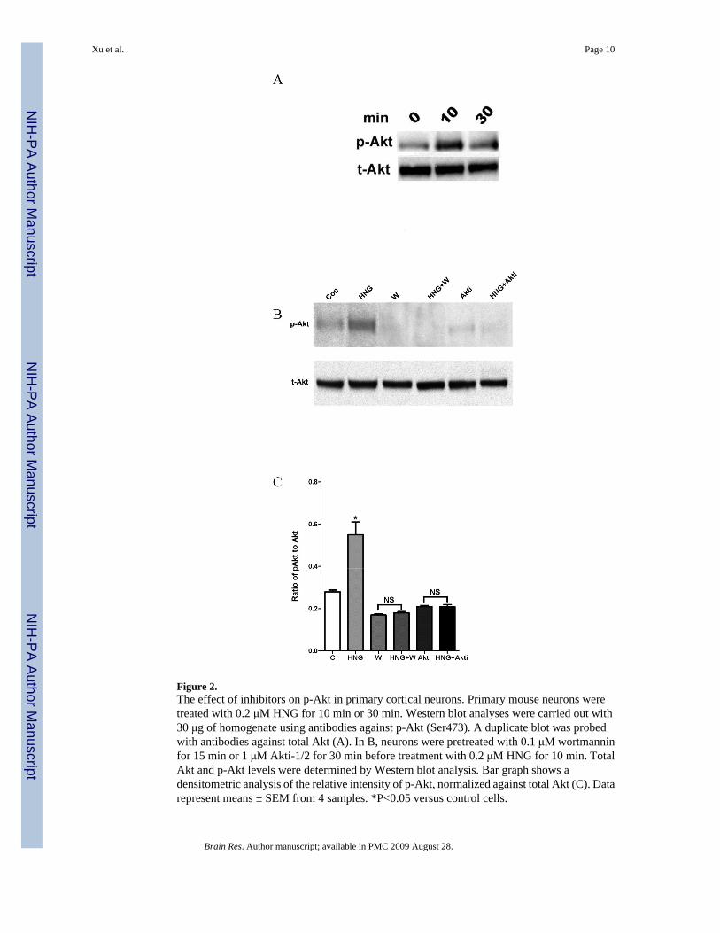
Figure 2.The effect of inhibitors on p-Akt in primary cortical neurons. Primary mouse neurons weretreated with 0.2 μM HNG for 10 min or 30 min. Western blot analyses were carried out with30 μg of homogenate using antibodies against p-Akt (Ser473). A duplicate blot was probedwith antibodies against total Akt (A). In B, neurons were pretreated with 0.1 μM wortmanninfor 15 min or 1 μM Akti-1/2 for 30 min before treatment with 0.2 μM HNG for 10 min. TotalAkt and p-Akt levels were determined by Western blot analysis. Bar graph shows adensitometric analysis of the relative intensity of p-Akt, normalized against total Akt (C). Datarepresent means ± SEM from 4 samples. *P<0.05 versus control cells.
Xu et al. Page 10
Brain Res. Author manuscript; available in PMC 2009 August 28.
NIH
-PA Author Manuscript
NIH
-PA Author Manuscript
NIH
-PA Author Manuscript

Figure 3.The effect of HNG on p-Akt level after cerebral I/R injury. Mice were treated with HNG (0.1μg, i.c.v.) or a same volume of saline 30 min before ischemia. After 30 min of ischemia and 6h of reperfusion, brains were removed and the ischemic hemispheres were used for Westernblot analysis. (A) Representative photographs of p-Akt and total Akt. (B) Quantitative analysisof the ratio of p-Akt to total Akt after cerebral ischemia in saline (NS) group and HNG group.Bars represent mean ± SEM of 3 brains. *P<0.01 versus NS group.
Xu et al. Page 11
Brain Res. Author manuscript; available in PMC 2009 August 28.
NIH
-PA Author Manuscript
NIH
-PA Author Manuscript
NIH
-PA Author Manuscript
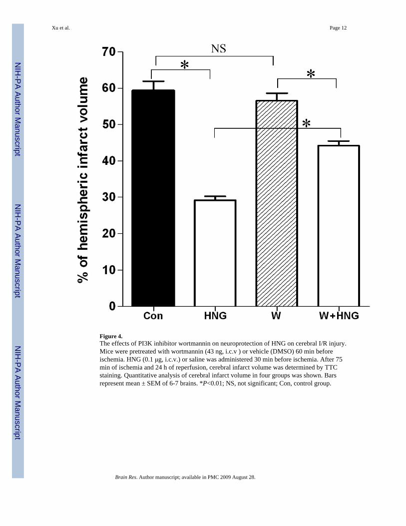
Figure 4.The effects of PI3K inhibitor wortmannin on neuroprotection of HNG on cerebral I/R injury.Mice were pretreated with wortmannin (43 ng, i.c.v ) or vehicle (DMSO) 60 min beforeischemia. HNG (0.1 μg, i.c.v.) or saline was administered 30 min before ischemia. After 75min of ischemia and 24 h of reperfusion, cerebral infarct volume was determined by TTCstaining. Quantitative analysis of cerebral infarct volume in four groups was shown. Barsrepresent mean ± SEM of 6-7 brains. *P<0.01; NS, not significant; Con, control group.
Xu et al. Page 12
Brain Res. Author manuscript; available in PMC 2009 August 28.
NIH
-PA Author Manuscript
NIH
-PA Author Manuscript
NIH
-PA Author Manuscript

Figure 5.The effect of Akti-1/2 on neuroprotection of HNG on cerebral I/R injury. Mice were pretreatedwith Akti-1/2 (55 ng, i.c.v.) or vehicle (DMSO) 60 min before ischemia. HNG (0.1 μg, i.c.v.)or saline was administered 30 min before ischemia. After 75 min of ischemia and 24 h ofreperfusion, cerebral infarct volume was determined by TTC staining. Quantitative analysis ofcerebral infarct volume in four groups was shown. Bars represent mean ± SEM of 6-7 brains.*P<0.01; NS, not significant; Con, control group.
Xu et al. Page 13
Brain Res. Author manuscript; available in PMC 2009 August 28.
NIH
-PA Author Manuscript
NIH
-PA Author Manuscript
NIH
-PA Author Manuscript
Copyright © 2022 FDOKUMEN