Endovascular treatment of cerebrovascular diseases and intracranial neoplasms
Upload
independentCategory
view
1download
0
ACCEPTED MANUSCRIPT
Hydrogen is neuroprotective and preserves cerebrovascular reactivity in asphyxiated newborn pigs
Ferenc Domoki, Orsolya Oláh,, Aliz Zimmermann, István Németh, Valéria Tóth-Szűki, Marietta Hugyecz, Péter Temesvári, and Ferenc Bari
Received: March 10, 2010 Accepted: July 14, 2010
Copyright © 2010 International Pediatric Research Foundation, Inc. All rights reserved.
Pediatric Research Articles Ahead of Print contains articles in unedited manuscript form that have been peer-reviewed and accepted for publication. As a service to our readers, we are providing this early version of the manuscript. The manuscript will undergo copyediting, typesetting and review of the resulting proof before it is published in its final definitive form. Please note that during the production process errors may be discovered, which could affect the content, and all legal disclaimers that apply to the journal pertain.
ACCEPTED

Pediatric Research Articles Ahead of Print DOI: 10.1203/PDR.0b013e3181f2e81c
Copyright © 2010, International Pediatric Foundation, Inc. All rights reserved.
Title: Hydrogen is neuroprotective and preserves cerebrovascular reactivity in asphyxiated
newborn pigs
Running title: Neuroprotection by hydrogen
Ferenc Domoki, Orsolya Oláh,, Aliz Zimmermann, István Németh, Valéria Tóth-Szűki, Marietta
Hugyecz, Péter Temesvári, and Ferenc Bari
Department of Physiology [F.D., O.O., A.Z., V.T., M.H.], Department of Dermatology and
Allergology [I.N.], Department of Medical Informatics [F.B.] University of Szeged School of
Medicine, Szeged, H-6720, Hungary; Department of Pediatrics [P.T.], University Teaching
Hospital Orosháza, Orosháza, H-5900, Hungary
Address for correspondence:
Ferenc Domoki M.D., Ph.D.
Department of Physiology, Faculty of Medicine
University of Szeged
Dóm tér 10
H-6720 Szeged, Hungary
Tel.: +36-62-545923
Fax: +36-62-544978
E-mail: [email protected]
Financial support: This study was supported by grants from the National Scientific Research
Fund of Hungary (OTKA, K68976, K63401). F.D. was supported by the János Bolyai Research
Scholarship of the Hungarian Academy of Sciences.
F.D. and O.O. contributed equally to this manuscript.
ACCEPTED
Copyright © 2010, International Pediatric Foundation, Inc. All rights reserved.
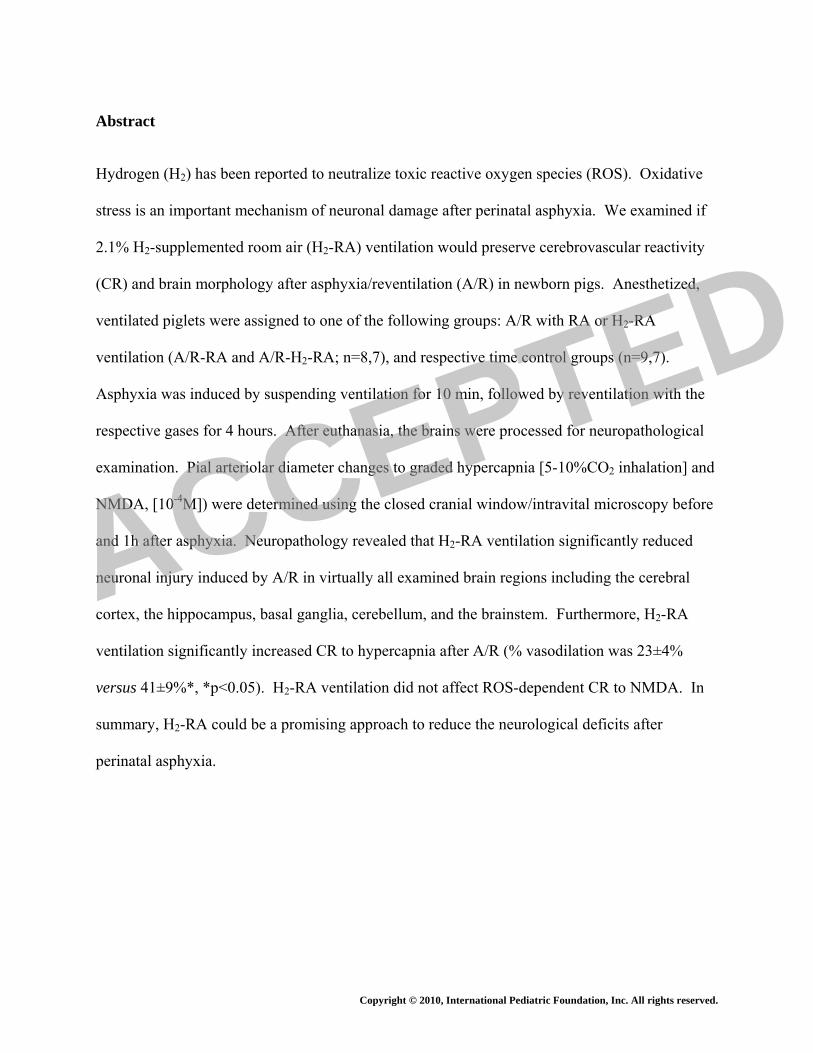
Abstract
Hydrogen (H2) has been reported to neutralize toxic reactive oxygen species (ROS). Oxidative
stress is an important mechanism of neuronal damage after perinatal asphyxia. We examined if
2.1% H2-supplemented room air (H2-RA) ventilation would preserve cerebrovascular reactivity
(CR) and brain morphology after asphyxia/reventilation (A/R) in newborn pigs. Anesthetized,
ventilated piglets were assigned to one of the following groups: A/R with RA or H2-RA
ventilation (A/R-RA and A/R-H2-RA; n=8,7), and respective time control groups (n=9,7).
Asphyxia was induced by suspending ventilation for 10 min, followed by reventilation with the
respective gases for 4 hours. After euthanasia, the brains were processed for neuropathological
examination. Pial arteriolar diameter changes to graded hypercapnia [5-10%CO2 inhalation] and
NMDA, [10-4M]) were determined using the closed cranial window/intravital microscopy before
and 1h after asphyxia. Neuropathology revealed that H2-RA ventilation significantly reduced
neuronal injury induced by A/R in virtually all examined brain regions including the cerebral
cortex, the hippocampus, basal ganglia, cerebellum, and the brainstem. Furthermore, H2-RA
ventilation significantly increased CR to hypercapnia after A/R (% vasodilation was 23±4%
versus 41±9%*, *p<0.05). H2-RA ventilation did not affect ROS-dependent CR to NMDA. In
summary, H2-RA could be a promising approach to reduce the neurological deficits after
perinatal asphyxia.
ACCEPTED

Copyright © 2010, International Pediatric Foundation, Inc. All rights reserved.
List of abbreviations:
A/R – asphyxia/reventilation
aCSF – artificial cerebrospinal fluid
CoBF – cortical blood flow
CR – cerebrovascular reactivity
H2-RA – hydrogen-supplemented room air
MABP – mean arterial blood pressure
PAD – pial arteriolar diameter
RA – room air
ROS – reactive oxygen species ACCEPTED

Copyright © 2010, International Pediatric Foundation, Inc. All rights reserved.
Introduction
Perinatal asphyxia induces brain injury that can result in death of the affected infants or
severe neurodevelopmental deficits of the survivors. According to WHO estimates, perinatal
asphyxia is responsible for 8% of the yearly 10.6 million deaths in children under the age of five
years world wide (1). Thus it is important to seek suitable therapies that can increase survival
and attenuate the neurological deficits. Despite the large number of affected infants, the
incentives and involvement of the pharmaceutical industry has been limited so far perhaps due to
financial concerns and ethical issues of initiating clinical trials in neonates (2). In fact, there is
currently no effective neuroprotective pharmacotherapy to treat perinatal asphyxial
encephalopathy. However, recently the Total Body Hypothermia for Neonatal Encephalopathy
Trial (TOBY) study group has shown that moderate whole-body hypothermia improves outcome
in full-term infants by decreasing the incidence of severe neurodevelopmental deficits, and thus
body cooling is expected to become standard care very soon in this group of patients (3, 4).
Therapeutic hypothermia can be initiated up to six hours after birth, by the time the
patient reaches a neonatal intensive care unit equipped to provide this care. Thus, there would be
a window of opportunity for the employment of an immediate neuroprotective therapy until the
neuroprotective cooling starts. The management of asphyxiated infants usually starts with
resuscitation and involves mechanical ventilation. Reoxygenation of the tissues is a primary goal
of resuscitation efforts, however, reoxygenation and reperfusion provides oxygen for the
synthesis of reactive oxygen species (ROS) that can result in oxidative stress and subsequent
reoxygenation/reperfusion injury (5-9). Resuscitation/ventilation with pure oxygen has been
shown to be detrimental in neonatal large animal models (10-12), and in clinical trials as well
ACCEPTED

Copyright © 2010, International Pediatric Foundation, Inc. All rights reserved.
(13, 14). Resuscitation is now recommended with air to be supplemented with oxygen in infants
with persistent central cyanosis (15). However, the gas mixture used for resuscitation could
perhaps be used to deliver neuroprotective agents to the brain. Molecular hydrogen (H2) has
recently been shown to exert antioxidant properties (16). H2 was indeed neuroprotective against
hypoxic/ischemic injury in adult and neonatal rats (16-18), and also in a rat model of Parkinson s
disease (19). However, the possible protective effect of H2 against perinatal asphyxia has not
been tested in a large animal model yet.
Therefore, the purpose of the present study was to investigate whether ventilation with
H2-supplemented room air (H2-RA) preserves hypoxia-sensitive cerebrovascular reactivity (CR)
and attenuates neuronal injury in asphyxiated newborn piglets. We also studied whether H2-RA
ventilation in the absence of asphyxia/reventilation (A/R) stress affects CR or brain morphology.
Materials and Methods
Animals
Newborn (less than 1-day old) piglets of either sex (n=34) were delivered from a local
farm to the laboratory on the morning of the experiments. All procedures were approved by the
Animal Care and Use Committee of the University of Szeged. The piglets were anaesthetized
with sodium thiopental (45 mg/kg ip, (Biochemie, Vienna, Austria) followed by intravenous
application of α-chloralose (30-40 mg/kg iv, Sigma, St. Louis, MO, USA). Supplemental doses
of α-chloralose (3-7 mg/kg/h) were administered to maintain a stable level of anesthesia
determined by continuous monitoring the blood pressure and responsiveness to tactile stimuli.
ACCEPTED

Copyright © 2010, International Pediatric Foundation, Inc. All rights reserved.
The femoral artery and vein were catheterized to monitor arterial pH, blood gases and blood
pressure, and to inject drugs and fluids, respectively. The animals were intubated through
tracheotomy and artificially ventilated with RA (21% O2, 79% N2). The ventilation rate (~
28/min) and tidal volume (~20ml) were adjusted to maintain physiological blood gas values.
Core body temperature was kept in the physiological range (~38-39 ºC) with electrical heating
pads.
To determine CR, the piglets were equipped with a closed cranial window filled with
artificial cerebrospinal fluid (aCSF) to measure pial arteriolar diameter (PAD) via intravital
microscopy as described previously (20). Cortical blood flow (CoBF) was monitored using a
two-channel laser-Doppler flowmeter (LDF, Periflux 4001, Perimed, Sweden) as described
previously (12). In each experiment, a pial arteriole with a baseline diameter of 80-120 μm was
selected to study CR. Ten-minute periods of arterial blood pressure and CoBF LDF data were
recorded before, during, and after asphyxia, and at each hour of the reventilation period. Data
were stored on a personal computer and were evaluated off-line using the Perisoft 1.30 software
(Perimed).
Experimental protocol
The instrumented piglets were divided into four experimental groups 1) A/R with RA
(21% O2, 79% N2) ventilation (A/R-RA; n=8), 2) A/R with H2-RA (2.1% H2; 21% O2; 76.9%
N2) ventilation (A/R-H2-RA; n=7), 3) time control group ventilated with RA (SHAM-RA; n=9);
and 4) time control group ventilated with hydrogen-supplemented room air (SHAM-H2-RA;
n=7).
First, CR was determined to graded hypercapnia and NMDA. Graded hypercapnia was
elicited by ventilating the animals with a gas mixture containing 5% CO2, (21% O2, balance N2)
ACCEPTED

Copyright © 2010, International Pediatric Foundation, Inc. All rights reserved.
for 5-8 minutes, then with 10% CO2 for additional 5-8 minutes. Ventilation was then switched
back to RA, and PAD was allowed to return to baseline diameters. During a ten minute
stabilization period the cranial window was gently flushed with aCSF, then NMDA dissolved in
aCSF (10-4M, 5ml) was applied onto the cerebral cortex for 5-6 min, then the cranial window
was repeatedly washed with aCSF. PAD was measured each minute during hypercapnia and
NMDA stimulation, and the maximal vasodilation maintained for at least 2 min was used to
determine CR to hypercapnia or NMDA, respectively.
After obtaining baseline CR values, asphyxia was induced (groups A/R-RA, A/R-H2-RA)
by suspending the artificial ventilation and clamping the endotracheal tube for 10 minutes.
Reventilation started with the gas mixtures according to the protocol of the respective
experimental groups. There was no need to apply chest compressions or drugs for resuscitation.
In the SHAM groups, ventilation was switched to the respective gas mixtures after a RA
ventilation time control period (10 min). After 1h ventilation, CR to hypercapnia and NMDA
were again determined in all experimental groups, then ventilation was continued for an
additional three hours. At the end of the 4h survival period, the anaesthetized piglets were
euthanized with KCl (2 M, 10ml iv).
Neuropathology
An additional group of piglets (NAÏVE, n=3) were euthanized with sodium thiopental
(150 mg/kg, ip) to provide an absolute control for the neuropathology studies. After euthanasia
in all experimental groups, the brains were rapidly and carefully removed and immersion-fixed
in 4% paraformaldehyde. Tissue blocks from the frontal, parietal, temporal and occipital
cortices, the hippocampus, the cerebellum, the basal ganglia, the pons, and the medulla oblongata
were embedded in paraffin, and sections were double stained with haematoxylin and eosin. Two
ACCEPTED

Copyright © 2010, International Pediatric Foundation, Inc. All rights reserved.
independent observers (O. Oláh and I. Németh), who were blinded to the treatments,
quantitatively determined, then averaged the percentage of injured neurons in each region based
on the presence of shrunken hyperchromatic (red) neurons with pyknotic nuclei.
Statistical analysis
Blood chemistry, mean arterial blood pressure (MABP), CoBF, and PAD data are
expressed as mean±SEM, and have been analyzed using 2-way repeated measures ANOVA
followed by the Student-Newman-Keuls post hoc test (SigmaStat, SPSS, Chicago, IL).
Histopathology data were not normally distributed and were analyzed with Kruskal-Wallis
ANOVA on Ranks followed by the Dunn’s post hoc test for multiple comparisons. p values of
<0.05 were considered statistically significant.
Results
Hemodynamics and blood chemistry
The MABP, pH and blood gas values were in the physiological range and did not differ
significantly between the experimental groups before asphyxia (Table 1-2). Ventilation with gas
mixtures containing 5% or 10% CO2 to determine CR to hypercapnia resulted in graded increase
in arterial pCO2 with simultaneous drops in pH that were similar between all experimental
groups before and after A/R injury (Table 1.). Asphyxia caused significant hypoxia,
hypercapnia, acidosis, hypotension, and decrease in CoBF that were similar in both groups
subjected to A/R (Table 1-2.). At the onset of reventilation, MABP rapidly increased then
stabilized at near baseline values by the end of first hour (Table 2). MABP values were thus
similar when determining CR to hypercapnia and NMDA before and after A/R. However, at 30
ACCEPTED

Copyright © 2010, International Pediatric Foundation, Inc. All rights reserved.
min and 1h after asphyxia, pCO2 was significantly higher in the A/R-RA group compared to the
A/R-H2-RA group, albeit pH and pO2 values were not significantly different (Table 1). In
contrast, CoBF was ~20-25% lower in the A/R-H2-RA group compared to the A/R-RA (Table 2).
In the SHAM groups, the MABP remained in the normal ranges during the whole course of the
experiment (Table 2). CoBF also appeared to be decreased by 15-20% in the SHAM-H2-RA
group compared to the SHAM-RA group, however, this difference did not reach statistical
significance (Table 2).
H2-RA preserves CR to hypercapnia after A/R injury
The first stimulation with graded hypercapnia elicited significant, dose-dependent, pial
arteriolar vasodilation in all experimental groups (Figure 1A-B). Baseline PAD-s were not
significantly different between repeated hypercapnia stimulations, their values were (first versus
second stimulation): 88±4 μm versus 94±8 μm for group A/R-RA, 87±4 μm versus 92±7 μm for
group A/R-H2-RA, 85±4 μm versus 91±3 μm for group SHAM-RA, and 86±6 μm versus 82±7
μm for group SHAM-H2-RA. A/R significantly attenuated CR to hypercapnia in the A/R-RA
(Figure 1A) but not in the other experimental groups (Figure 1A-B). Thus, ventilation with H2-
RA under normoxic conditions in the SHAM-H2-RA group did not affect CR to hypercapnia.
Furthermore, ventilation with H2-RA after asphyxia prevented the attenuation of CR to
hypercapnia in the A/R-H2-RA group (Figure 1A).
H2-RA ventilation does not affect CR to NMDA
NMDA induced significant, repeatable pial arteriolar vasodilation in all experimental
groups (Figure 1A-B). Thus, ventilation with H2-RA did not affect CR to NMDA. Baseline
ACCEPTED

Copyright © 2010, International Pediatric Foundation, Inc. All rights reserved.
PAD-s were not significantly different between repeated NMDA applications, their values were
(first versus second application): 87±4 μm versus 85±9 μm for group A/R-RA, 85±4 μm versus
91±6 μm for group A/R-H2-RA, 86±4 μm versus 82±4 μm for group SHAM-RA, and 84±5 μm
versus 80±6 μm for group SHAM-H2-RA.
H2-RA protects neurons after A/R injury
The percentage of damaged, red neurons was negligible in any brain region of the
untreated control animals (NAÏVE animals): the median value was 0% in 7 regions, 2% in 3
regions and 3% in 2 regions (data not shown). In the time control animals (SHAM-RA, SHAM-
H2-RA groups), there was also only a minimal (1-4%) neuronal damage depending on the brain
region studied, indicating that H2-RA ventilation alone did not affect neuronal viability (Figure
2A-C, Figure 3). A/R significantly increased neuronal injury in all brain regions studied in the
A/R-RA group. However, the increase in injured, red neurons was smaller in the A/R-H2-RA
group, showing a clear neuroprotective effect in virtually all brain regions examined (Figure 2A-
C, Figure 3).
Discussion
The major findings of the present study are the following: ventilation with H2-RA after
asphyxia preserves CR to hypercapnia, and protects the brain from A/R-induced neuronal injury
in piglets. Furthermore, H2-RA ventilation per se does not affect CR or neuronal viability.
Oxidative stress induced by ROS produced during/after a hypoxic/ischemic episode is a
widely accepted pathomechanism of perinatal brain injury (2, 21). Thus, administration of
antioxidant drugs either to decrease ROS production or to neutralize ROS appears to be a simple,
ACCEPTED

Copyright © 2010, International Pediatric Foundation, Inc. All rights reserved.
straightforward approach to alleviate brain damage. However, the antioxidant drug needs to be
present at the ROS level surge in the early reventilation/reperfusion exert its effect. For
example, the xanthine oxidase inhibitor allopurinol was found ineffective when administered to
severely asphyxiated term infants 4 hours after birth (22). The authors speculated that drug
administration was too late to successfully target the immediate ROS surge following
resuscitation. In fact, in a more recent study, the same research group has found that maternal
treatment of in utero asphyxiated infants with allopurinol reduced the concentration of the brain
injury marker S-100B in infants with therapeutic allopurinol levels (23). The disadvantage of
maternal treatment is that placental transport of the drugs can vary greatly and the drug may not
reach therapeutic levels in a significant portion of patients (23). H2-RA ventilation could be an
appealing technique for antioxidant neuroprotective therapy since administration could start
simultaneously with the resuscitation effort to provide reoxygenation.
ROS also play physiological roles that have been demonstrated in numerous processes
including neuronal and vascular signaling (24). Potent antioxidants may interfere with these
physiological ROS-mediated mechanisms that can attenuate or even nullify their beneficial
effects. Hydrogen gas has been suggested to selectively reduce the most toxic ROS: hydroxyl
radicals and peroxinitrate anions (16), thus leaving the less deleterious/physiological ROS intact.
Our present study provides evidence that H2-RA ventilation does not affect a ROS-dependent
neuronal-vascular response: NMDA-induced vasodilation. NMDA-induced vasodilation has
been first described in the piglet (25), and CR to NMDA can be used to assess the integrity of the
neurovascular unit (26). The mechanism of dose-dependent NMDA-induced pial arteriolar
dilation and subsequent cortical hyperemia involves the neuronal synthesis and vascular actions
of NO (20, 27). In the present study, the intact CR to NMDA during H2-RA ventilation indicates
ACCEPTED

Copyright © 2010, International Pediatric Foundation, Inc. All rights reserved.
that H2 does not interfere with the increases in NO levels necessary for the induction of pial
arteriolar vasodilation. NMDA-induced vasodilation has been repeatedly shown to be sensitive
to global cerebral ischemia/reperfusion, CR to NMDA (10-4 M) is attenuated by ~50% 1h after
10 min ischemia (26, 28). Interestingly, we have not observed a decrease in CR to NMDA in the
present study after 10 min of asphyxia. The only study in the literature investigating the effect of
A/R on NMDA-induced vasodilation reported “altered” CR to NMDA based on progressively
increasing CR to NMDA in the reventilation period (29). However, in that study CR to NMDA
was not determined prior to asphyxia. Furthermore, the vasodilation to 10-5M NMDA used in
that study corresponds with our later findings with this dose of NMDA under normoxic
conditions (20). Thus it is conceivable that A/R represents a less severe stress than global
ischemia/reperfusion, and CR to 10-4M NMDA does not change appreciably using this stress.
However, A/R significantly attenuated CR to hypercapnia. In the piglet, hypercapnia-
induced pial arteriolar vasodilation is dependent on the function of microvascular endothelial
cells (20, 30). In the present study, H2-RA ventilation preserved CR to hypercapnia after A/R
indicating intact endothelial function. The preservation of CR likely results in more adequate
microvascular blood flow matching neuronal needs and thus likely contributes to the
neuroprotective effect of H2-RA ventilation.
Our study is the first demonstrating the early neuroprotective effect of H2-RA ventilation
in a large animal model of perinatal asphyxia. We have previously shown the neurotoxic effect
of 100% oxygen ventilation compared to RA ventilation after the same A/R stress used in the
present study (12). The degree of neuronal injury was similar in the RA ventilated control
groups in both studies, albeit in the present study we used a quantitative cell counting approach
in contrast to the semiquantitative scoring system utilized in the previous study. Thus, our
ACCEPTED

Copyright © 2010, International Pediatric Foundation, Inc. All rights reserved.
simple A/R model of perinatal asphyxia is capable to detect both neuroprotective and neurotoxic
effects related to the changes in the composition of gas mixture used for ventilation. We would
like to emphasize that we observed a beneficial effect of H2-RA ventilation in virtually all brain
regions studied. In the previous study, the detrimental effects of 100% oxygen were significant
only in the hippocampus and the cerebellum where reactive hyperemia during reventilation was
twice higher than in the parietal cortex suggesting that enhanced oxygen delivery may exacerbate
oxidative stress (12). We observed a mild decrease in CoBF during H2-RA ventilation that may
also contribute to its neuroprotective effect after A/R via limiting luxury oxygen delivery to the
metabolically compromised brain parenchyma. The observed neuroprotective effect of H2-RA
ventilation of the present study is in accordance with previous data reporting neuroprotection by
2% H2 gas in a neonatal rat hypoxia/ischemia model (18), and also in an adult rat stroke model
(16).
The selection of gas used for the resuscitation of infants affected by perinatal asphyxia
may represent an ideal way to implement an instant neuroprotective therapy. The importance of
careful selection of inhaled oxygen concentration level is already well-established, and the
present study confirms that addition of 2% H2 gas may likely confer neuroprotection by
combating ROS-inflicted neuronal damage during the early reperfusion/reoxygenation period.
This concentration of H2 (2-2.1%) is safe since H2 has no risk of flammability below 4.7% in
RA, and this H2-RA ventilation protocol yielded H2 concentration of ~20ng/ml (~10-5 M) in the
arterial blood of adult rats suggesting an effective antioxidant capacity at this dose (16).
Furthermore, other gases may potentially be added to the neuroprotective gas mixture to enhance
neuronal survival and function. The anesthethic gas, xenon (50% inhaled concentration) has
been found to augment neuroprotection afforded by hypothermia in the neonatal rat
ACCEPTED

Copyright © 2010, International Pediatric Foundation, Inc. All rights reserved.
hypoxia/ischemia model (31, 32), and helium (70% He/ 30% O2) provided excellent
neuroprotection in adult rat stroke models (33, 34).
The current study has several important limitations. The neuroprotective effect of H2-RA
ventilation was demonstrated in α-chloralose anesthetized piglets using only one A/R insult
severity and one H2-RA ventilation protocol. The anesthesia might enhance the neuroprotective
potential of H2-RA, and the neuroprotection afforded by H2 can be diminished by increasing
insult severity. H2-RA ventilation likely exerts its greatest beneficial effect when administered
immediately after reoxygenation, but it is unknown whether delayed administration of H2-RA
ventilation would have any protective effect. These clinically relevant topics warrant further
studies to elucidate the full neuroprotective potential of H2 perhaps in concert with other inert
gases like helium, and therapeutic hypothermia.
In conclusion, H2-RA ventilation after A/R provides early neuroprotection after asphyxia
in newborn piglets, at least in part via preservation of neurovascular unit function. Ironically,
after fruitless research for a “magic bullet” neuroprotective drug, the optimal combination of the
simplest elements: hydrogen, helium, oxygen with appropriate body temperature control emerges
as the most promising neuroprotective treatment in the management of perinatal asphyxia.
ACCEPTED

Copyright © 2010, International Pediatric Foundation, Inc. All rights reserved.
References
1. Bryce J, Boschi-Pinto C, Shibuya K, Black RE 2005 WHO estimates of the causes of death in children. Lancet 365:1147-1152
2. Degos V, Loron G, Mantz J, Gressens P 2008 Neuroprotective strategies for the neonatal brain. Anesth Analg 106:1670-1680
3. Azzopardi DV, Strohm B, Edwards AD, Dyet L, Halliday HL, Juszczak E, Kapellou O, Levene M, Marlow N, Porter E, Thoresen M, Whitelaw A, Brocklehurst P 2009 Moderate hypothermia to treat perinatal asphyxial encephalopathy. N Engl J Med 361:1349-1358
4. Rutherford M, Ramenghi LA, Edwards AD, Brocklehurst P, Halliday H, Levene M, Strohm B, Thoresen M, Whitelaw A, Azzopardi D 2010 Assessment of brain tissue injury after moderate hypothermia in neonates with hypoxic-ischaemic encephalopathy: a nested substudy of a randomised controlled trial. Lancet Neurol 9:39-45
5. Vento M, Asensi M, Sastre J, Garcia-Sala F, Pallardo FV, Vina J 2001 Resuscitation with room air instead of 100% oxygen prevents oxidative stress in moderately asphyxiated term neonates. Pediatrics 107:642-647
6. Buonocore G, Perrone S, Longini M, Vezzosi P, Marzocchi B, Paffetti P, Bracci R 2002 Oxidative stress in preterm neonates at birth and on the seventh day of life. Pediatr Res 52:46-49
7. Saugstad OD 2005 Room air resuscitation-two decades of neonatal research. Early Hum Dev 81:111-116
8. Solberg R, Andresen JH, Escrig R, Vento M, Saugstad OD 2007 Resuscitation of hypoxic newborn piglets with oxygen induces a dose-dependent increase in markers of oxidation. Pediatr Res 62:559-563
9. Kumar VH, Patel A, Swartz DD, Wang H, Wynn KA, Nielsen LC, Ryan RM 2010 Exposure to supplemental oxygen and its effects on oxidative stress and antioxidant enzyme activity in term newborn lambs. Pediatr Res 67:66-71
10. Munkeby BH, Borke WB, Bjornland K, Sikkeland LI, Borge GI, Halvorsen B, Saugstad OD 2004 Resuscitation with 100% O2 increases cerebral injury in hypoxemic piglets. Pediatr Res 56:783-790
11. Temesvári P, Karg E, Bodi I, Nemeth I, Pinter S, Lazics K, Domoki F, Bari F 2001 Impaired early neurologic outcome in newborn piglets reoxygenated with 100% oxygen compared with room air after pneumothorax-induced asphyxia. Pediatr Res 49:812-819
12. Domoki F, Zimmermann A, Cserni G, Bori R, Temesvari P, Bari F 2006 Reventilation with room air or 100% oxygen after asphyxia differentially affects cerebral neuropathology in newborn pigs. Acta Paediatr 95:1109-1115
13. Davis PG, Tan A, O'Donnell CP, Schulze A 2004 Resuscitation of newborn infants with 100% oxygen or air: a systematic review and meta-analysis. Lancet 364:1329-1333
14. Saugstad OD, Ramji S, Vento M 2005 Resuscitation of depressed newborn infants with ambient air or pure oxygen: a meta-analysis. Biol Neonate 87:27-34
ACCEPTED

Copyright © 2010, International Pediatric Foundation, Inc. All rights reserved.
15. ILCOR 2006 The International Liaison Committee on Resuscitation (ILCOR) consensus on science with treatment recommendations for pediatric and neonatal patients: neonatal resuscitation. Pediatrics 117:e978-e988
16. Ohsawa I, Ishikawa M, Takahashi K, Watanabe M, Nishimaki K, Yamagata K, Katsura K, Katayama Y, Asoh S, Ohta S 2007 Hydrogen acts as a therapeutic antioxidant by selectively reducing cytotoxic oxygen radicals. Nat Med 13:688-694
17. Cai J, Kang Z, Liu K, Liu W, Li R, Zhang JH, Luo X, Sun X 2009 Neuroprotective effects of hydrogen saline in neonatal hypoxia-ischemia rat model. Brain Res 1256:129-137
18. Cai J, Kang Z, Liu WW, Luo X, Qiang S, Zhang JH, Ohta S, Sun X, Xu W, Tao H, Li R 2008 Hydrogen therapy reduces apoptosis in neonatal hypoxia-ischemia rat model. Neurosci Lett 441:167-172
19. Fu Y, Ito M, Fujita Y, Ito M, Ichihara M, Masuda A, Suzuki Y, Maesawa S, Kajita Y, Hirayama M, Ohsawa I, Ohta S, Ohno K 2009 Molecular hydrogen is protective against 6-hydroxydopamine-induced nigrostriatal degeneration in a rat model of Parkinson's disease. Neurosci Lett 453:81-85
20. Domoki F, Perciaccante JV, Shimizu K, Puskar M, Busija DW, Bari F 2002 N-methyl-D-aspartate-induced vasodilation is mediated by endothelium-independent nitric oxide release in piglets. Am J Physiol Heart Circ Physiol 282:H1404-H1409
21. Ferriero DM 2004 Neonatal brain injury. N Engl J Med 351:1985-1995
22. Benders MJ, Bos AF, Rademaker CM, Rijken M, Torrance HL, Groenendaal F, van Bel F 2006 Early postnatal allopurinol does not improve short term outcome after severe birth asphyxia. Arch Dis Child Fetal Neonatal Ed 91:F163-F165
23. Torrance HL, Benders MJ, Derks JB, Rademaker CM, Bos AF, Van Den Berg P, Longini M, Buonocore G, Venegas M, Baquero H, Visser GH, Van Bel F 2009 Maternal allopurinol during fetal hypoxia lowers cord blood levels of the brain injury marker S-100B. Pediatrics 124:350-357
24. Sauer H, Wartenberg M, Hescheler J 2001 Reactive oxygen species as intracellular messengers during cell growth and differentiation. Cell Physiol Biochem 11:173-186
25. Busija DW, Leffler CW 1989 Dilator effects of amino acid neurotransmitters on piglet pial arterioles. Am J Physiol 257:H1200-H1203
26. Busija DW, Bari F, Domoki F, Louis T 2007 Mechanisms involved in the cerebrovascular dilator effects of N-methyl-d-aspartate in cerebral cortex. Brain Res Rev 56:89-100
27. Meng W, Tobin JR, Busija DW 1995 Glutamate-induced cerebral vasodilation is mediated by nitric oxide through N-methyl-D-aspartate receptors. Stroke 26:857-862, discussion 863
28. Busija DW, Meng W, Bari F, McGough PS, Errico RA, Tobin JR, Louis TM 1996 Effects of ischemia on cerebrovascular responses to N-methyl-D-aspartate in piglets. Am J Physiol 270:H1225-H1230
29. Busija DW, Wei M 1993 Altered cerebrovascular responsiveness to N-methyl-D-aspartate after asphyxia in piglets. Am J Physiol 265:H389-H394
ACCEPTED

Copyright © 2010, International Pediatric Foundation, Inc. All rights reserved.
30. Leffler CW, Mirro R, Shanklin DR, Armstead WM, Shibata M 1994 Light/dye microvascular injury selectively eliminates hypercapnia-induced pial arteriolar dilation in newborn pigs. Am J Physiol 266:H623-H630
31. Hobbs C, Thoresen M, Tucker A, Aquilina K, Chakkarapani E, Dingley J 2008 Xenon and hypothermia combine additively, offering long-term functional and histopathologic neuroprotection after neonatal hypoxia/ischemia. Stroke 39:1307-1313
32. Thoresen M, Hobbs CE, Wood T, Chakkarapani E, Dingley J 2009 Cooling combined with immediate or delayed xenon inhalation provides equivalent long-term neuroprotection after neonatal hypoxia-ischemia. J Cereb Blood Flow Metab 29:707-714
33. Pan Y, Zhang H, VanDeripe DR, Cruz-Flores S, Panneton WM 2007 Heliox and oxygen reduce infarct volume in a rat model of focal ischemia. Exp Neurol 205:587-590
34. David HN, Haelewyn B, Chazalviel L, Lecocq M, Degoulet M, Risso JJ, Abraini JH 2009 Post-ischemic helium provides neuroprotection in rats subjected to middle cerebral artery occlusion-induced ischemia by producing hypothermia. J Cereb Blood Flow Metab 29:1159-1165
ACCEPTED

Copyright © 2010, International Pediatric Foundation, Inc. All rights reserved.
Figure legends
Figure 1. H2-RA ventilation preserves cerebrovascular reactivity to hypercapnia one hour after
A/R. Graded hypercapnia induced by ventilation with 5-10% CO2, and topical application of 10-
4 M NMDA elicited similar, significant increases in pial arteriolar diameters in all experimental
groups (Panel A-B). In time controls (Panel B), pial arteriolar responses were repeatable, and
similar between the SHAM-RA (n=9, white bars) and the SHAM-H2-RA (n=7, light gray bars)
groups. Importantly, H2-RA ventilation did not affect NMDA-induced, NO-dependent
vasodilation (Panel B). After A/R, however (Panel A), pial arteriolar dilation to 10% CO2 was
significantly reduced in the A/R-RA group (n=8, black bars). However, in the A/R-H2-RA (n=7,
dark gray bars) group, pial arteriolar dilation to 10% CO2 was significantly higher than in group
A/R-RA, and not significantly different from the respective baseline response before A/R.
p<0.05, * versus corresponding response before A/R, † corresponding responses after A/R in the
A/R-RA versus the A/R-H2-RA groups.
Figure 2. H2-RA ventilation attenuates A/R-induced early neuronal injury. The percentage of
damaged neurons was quantitatively determined at four hours after ten minutes of asphyxia, and
in corresponding time control animals. The examined brain regions were: Panel A: CA1, CA2,
CA3 and stratum granulosum (Str. Gr.) of the hippocampus; Panel B: frontal (Fr. Cx.), parietal
(Par Cx.), temporal (Temp Cx.) and occipital (Occ. Cx.) cortices, Panel C: basal ganglia (Basal
ggl.), pons, medulla, and cerebellum. Summated data from the hippocampal (ΣHippoc.) and
cortical subregions (ΣCortex) are also shown on Panels A and B, respectively. The box plots
show the median (horizontal line), the 25th-75th percentile (box), the 10th-90th percentiles (error
ACCEPTED

Copyright © 2010, International Pediatric Foundation, Inc. All rights reserved.
bars) and any outlying data points (bullets). The experimental groups are represented by gray,
gray-hatched, white, and white hatched boxes for A/R-RA, A/R-H2-RA SHAM-RA and
SHAM-H2-RA (n=8,7,9 and 7), respectively. A/R resulted in significant increases in neuronal
injury in virtually all examined brain regions in the A/R-RA but not in the A/R-H2-RA group
compared to the respective time controls. Thus, H2-RA ventilation attenuated the detrimental
effect of A/R on neuronal viability. p<0.05, * versus corresponding time control (A/R-RA versus
SHAM-H2-RA and A/R-H2-RA versus SHAM-H2-RA) A/R, † A/R-RA versus the A/R-H2-RA.)
Figure 3
Representative photomicrographs from the parietal cortex, the stratum granulare of the
hippocampus, and the cerebellar cortex showing neuroprotection by H2-RA ventilation.
Shrunken, hyperchromatic (red) neurons with pyknotic nuclei are more common in samples
obtained from animals of the A/R-RA group as compared with A/R-H2-RA group. Time control
groups (SHAM-RA, SHAM-H2-RA) show minimal neuronal damage, the samples are similar to
those obtained from untreated (NAÏVE) animals. Scale bar: 100μM.
ACCEPTED

Copyright © 2010, International Pediatric Foundation, Inc. All rights reserved.
Figure 1
ACCEPTED

Copyright © 2010, International Pediatric Foundation, Inc. All rights reserved.
Figure 2
ACCEPTED

Copyright © 2010, International Pediatric Foundation, Inc. All rights reserved.
Figure 3
ACCEPTED

Copyright © 2010, International Pediatric Foundation, Inc. All rights reserved.
Table 1. *p<0.05 significantly smaller than corresponding values in group A/R-RA.
Hypercapnia before asphyxia Reventilation Hypercapnia after
asphyxia
Baseline 5%CO2 10%CO2
Asphyxia10’ 30’ 1h 5%CO2 10%CO2
pH 7,3±0,10 7,06±0,13 7,09±0,04 6,76±0,03 7,06±0,07 7,14±0,09 7,20±0,09 7,05±0,09 7,04±0,04pCO2 (mmHg) 29±1 51±6 80±5 133±10 59±8 43±5 56±9 71±12 90±11
A/R- RA
(n=8) pO2 (mmHg) 82±4 86±4 85±5 11±1 66±8 61±5 60±8 70±9 70±13
pH 7,28±0,09 7,20±0,07 7,05±0,05 6,85±0,07 7,12±0,07 7,21±0,08 7,24±0,09 7,10±0,08 7,01±0,06pCO2 (mmHg) 31±4 48±5 77±5 123±12 44±5 32±4* 31±3* 54±5 82±3
A/R-H2-RA (n=7)
pO2 (mmHg) 71±4 81±4 86±3 5±0,4 69±6 66±4 68±4 78±5 77±3 pH 7,53±0,02 7,32±0,03 7,15±0,02 N/A 7,45±0,06 7,46±0,05 7,44±0,06 7,30±0,03 7,19±0,04pCO2 (mmHg) 26±2 44±3 65±10 N/A 29±6 32±7 35±6 51±5 71±8
SHAM-RA
(n=9) pO2 (mmHg) 89±8 89±7 93±5 N/A 92±9 92±9 84±9 85±7 93±5
pH 7,53±0,06 7,30±0,02 7,15±0,02 N/A 7,46±0,04 7,44±0,05 7,42±0,05 7,26±0,04 7,09±0,03pCO2 (mmHg) 33±2 51±5 85±6 N/A 34±3 35±4 37±6 57±3 85±5
SHAM-H2-RA (n=7) pO2 (mmHg) 78±3 75±4 66±7 N/A 74±7 75±9 70±7 77±6 70±12
ACCEPTED

Copyright © 2010, International Pediatric Foundation, Inc. All rights reserved.
Table 2.
*p<0.05 significantly smaller than corresponding values in group A/R-RA.
Reventilation
Baseline Asphyxia10min 1h 2h 3h 4h
MABP (mmHg) 77±4 47±8 70±3 66±6 60±4 58±4 65±4 A/R- RA
(n=7) CoBF (%) 100±0 31±7 83±4 107±15 101±9 103±5 105±7
MABP (mmHg) 74±5 41±12 70±3 51±3 63±5 55±4 57±4 A/R-H2-RA
(n=7) CoBF (%) 100±0 29±5 96±5 69±3* 81±6 76±8* 79±10*
MABP (mmHg) 78±3 N/A N/A 69±4 65±3 62±4 63±4 SHAM-RA
(n=9) CoBF (%) 100±0 N/A N/A 119±19 121±22 108±15 117±21
MABP (mmHg) 70±7 N/A N/A 63±4 57±7 59±7 63±8 SHAM-H2-RA (n=7) CoBF (%) 100±0 N/A N/A 93±5 80±8 80±8 85±11
ACCEPTED
Copyright © 2022 FDOKUMEN