Kinin Receptor Antagonists as Potential Neuroprotective Agents in Central Nervous System Injury
Upload
independentCategory
view
1download
0
Ann. N.Y. Acad. Sci.
993: 334–344 (2003). ©2003 New York Academy of Sciences.
Mitochondria as a Target for Neurotoxins and Neuroprotective Agents
SERGEY O. BACHURIN,
a
ELENA P. SHEVTSOVA,
a
ELENA G. KIREEVA,
a
GREGORY F. OXENKRUG,
b,c
AND SERGEY O. SABLIN
d
a
Institute of Physiologically Active Compounds, Chernogolovka, Russia
b
Department of Psychiatry and Pineal Research Laboratory, St. Elizabeth’s Medical Center, Boston, Massachusetts, USA
c
Department of Veterans Affairs Medical Center, Providence, Rhode Island, USA
d
Selena Pharmaceuticals, Inc., San Francisco, California, USA
A
BSTRACT
: Mitochondrial permeability transition pores represent a multipro-tein complex that includes components of both inner and outer membrane. Thepores regulate transport of ions and peptides in and out of mitochondria, andtheir regulation is associated with a general mechanism for maintaining Ca
2+
homeostasis in the cell and apoptosis. Various pathologic factors may induce apathologic activation of the permeability transition and an irreversible open-ing of mitochondria pores. This event is a major step in the development ofneurotoxicity and neurodegeneration. This paper explores the effect of MPP
+
and
�
-amyloid fragment 25–35, neurotoxins that are known to generate Par-kinson’s-like syndrome and Alzheimer’s disease, on the regulation of the mito-chondrial pores. Both neurotoxins induce opening of mitochondrial pores,which is prevented by cyclosporin A, a specific inhibitor of the permeabilitytransition. The effect of MPP
+
and
�
-amyloid may be also prevented by anendogenous precursor of melatonin,
N
-acetylserotonin, by an anti-Alzheimer’smedication tacrine, and by dimebon, which is in development as an agent forthe therapy of Alzheimer’s disease and other types of dementia. The paperillustrates that the effect on mitochondrial pores is an important aspect of themechanism of neurotoxicity. Substances that may prevent opening of mito-chondrial pores induced by neurotoxins may preserve the mitochondrial func-tion and, thus, may have potential as neuroprotective agents.
K
EYWORDS
: mitochondria; permeability transition pores;
N
-acetylserotonin;tacrine; dimebon; neurodegeneration; apoptosis
INTRODUCTION
A general deficit of the biological energy and a malfunction of brain mito-chondria is the earliest and the most specific feature of aging and a wide range ofneurodegenerative disorders.
1,2
It is also known that many so-called mitochondrialdiseases are always accompanied with brain malfunctions.
3
Address for correspondence: Sergey O. Sablin, Ph.D., Selena Pharmaceuticals, Inc., 167Skyview Way, San Francisco, CA 94131, USA. Voice: 415-990-4761.
335BACHURIN
et al.
: MITOCHONDRIAL PERMEABILITY PORE REGULATION
One general characteristic of aging and neurodegeneration is an increase in thenumber of neural cells showing signs of an apoptotic degeneration. Recently it wasfound
4
that a key role in apoptotic processes is attributable to so-called mitochondri-al permeability transition pores (PTP), which provide transport in and out of mito-chondria for both calcium ions and for compounds with molecular weight of up to1.5kDa. It was proposed that PTP represent a multiprotein complex, which is locatedat the contact sites of the mitochondrial membrane and which includes componentsof both the inner and the outer membrane.
5
An outer membrane fragment of PTPincludes the so-called
porin
, a voltage-dependent anion channel, antiapoptotic pro-teins of the Bcl-2 family, and, probably, a peripheral benzodiazepine receptor. Theinner membrane fragment of PTP contains an adenine nucleotide translocator (ANT)and cyclophilin, which may interact with proapoptotic proteins of the Bax family. Itis likely that the enzymes creatine kinase and hexokinase also participate in the reg-ulation of PTP.
In the
normal
(low-conductive) state of PTP at physiologic concentration of cyto-sol calcium ions, opening and closing of PTP is reversible and is regulated by themembrane potential. This function of PTP provides calcium homeostasis in the cell.The influx of calcium ions into mitochondria is accompanied by the simultaneousefflux of protons, resulting in an increase of pH in the mitochondrial matrix, fol-lowed by opening of PTP. This ion flux also results in a collapse of both proton gra-dient and a membrane potential, a collapse of Ca
2
+
reuptake, and acidification of themitochondrial matrix. The latter event induces closing of PTP. Functioning of themitochondrial respiratory chain restores the proton gradient that allows calcium ionsto reenter mitochondria, and so forth. Thus, under
normal
physiologic conditions,mitochondria serve as a buffer to compensate for the
excessive
calcium. In patholo-gy, an excessive Ca
2
+
influx into the cell (e.g., via the NMDA receptor system) leadsto the accumulation of calcium ions inside mitochondria and triggers a massive gen-eration of ROS, which induces degradation of Ca
2
+
-dependent ATPase. This process,in turn, decreases the ability of the cell membrane to eliminate excessive calciumfrom the cell and amplifies an additional increase in intramitochondrial calcium con-centration. A critical overload of mitochondria with Ca
2
+
induces depolarization ofthe mitochondrial membrane and an irreversible opening of PTP. These changes areaccompanied by the exit from mitochondria of calcium ions and macromolecularcompounds, such as caspases activators, cytochrome c, and Apaf-1 factors. Together,these reactions are thought to predetermine cell death.
Inhibition of the mitochondrial Ca
2
+
uptake and/or blocking of PTP may protectcells against the development of apoptosis (and necroses) in the presence of patho-logic factors, such as excitotoxins and oxidants.
6
The Ca
2
+
-dependent activation ofPTP in brain mitochondria was shown to be enhanced with age,
7
and may play animportant role in the development of age-related neurodegenerative disorders.
In this paper we report on a study of the effect of neurotoxins MPP
+
and
β
-amyloidfragment 25–35 (both of which are used to model Alzheimer’s disease and Parkinsondisease) on the PTP state. We also investigated how the anti-Alzheimer’s agentsTacrine and Dimebon, and the endogenous antioxidant and neuroprotector,
N
-acetyl-serotonin (NAS), influence regulation of the PTP function.

336 ANNALS NEW YORK ACADEMY OF SCIENCES
MATERIALS AND METHODS
The
β
-amyloid fragment (25–35) was purchased from Bachem; Dimebon wasprovided by NPO Organika (Novokuznetsk, Russia). Tacrine and all other reagentswere purchased from Sigma.
Liver mitochondria were isolated and purified from liver of Wistar rats using astandard procedure
8
in a 5mM HEPES buffer, pH7.4, containing 210mM mannitol,70mM sucrose, and 1mM EDTA (buffer A). Because mannitol binds hydroxyl ions,it was omitted in the assay buffer (buffer B), when mitochondria swelling and theintensity of lipid peroxidation were monitored simultaneously. The final centrifuga-tion and resuspension of mitochondria were performed in buffer A or buffer B inwhich EDTA was omitted (A-EDTA and B-EDTA, respectively). Brain mitochon-dria were isolated and purified in Percoll gradient according to the method of Sims
et al.
9
in 10mM HEPES buffer, pH7.4, containing 0.3M glucose and 1mM EDTAat 4
°
C.Opening of PTP was indicated by mitochondria swelling, which resulted in a
decrease in optical density of the mitochondria suspension. The absorbance wasmonitored using Beckman DU 640 spectrophotometer at 540nm and constant tem-perature 25
°
C in thermostated cuvette equipped with a continuous stirring device.The reaction mixture contained buffers A-EDTA or B-EDTA, 0.8
µ
M rotenone,5mM succinate, and mitochondria suspension diluted to give a 1mg/mL protein inthe cuvette. The reaction was initiated by adding an agent that triggered PTP open-ing. The rate of mitochondria swelling was calculated as a maximum gradient ofchange in the absorbance at 540nm, and expressed in
d
A540/min/mg of protein.Lipid peroxidation in mitochondrial membranes was followed by an accumula-
tion of substances that reacted with thiobarbituric acid, monitored spectrophotomet-rically according to a procedure described by Erdahl and coauthors.
10
All experiments were repeated with at least three different mitochondria prepara-tions. The figures illustrate results from selected experiments.
RESULTS AND DISCUSSION
Effect of Neurotoxins MPP and
�
-Amyloid Fragment (25–35) on the Opening of Mitochondrial Pores
Mitochondrial response to the presence of any agent that causes pore opening ischaracterized by two parameters, the lag period (interval between the moment whenthe triggering agent is introduced and the time when pores begin to open) and themaximum slope (which reflects the rate of PTP opening). Opening of mitochondrialpores results in the massive entry of sucrose and other substances into the mitochon-drial matrix. This causes swelling of mitochondria and, as a result, a decrease inabsorbance of the mitochondrial suspension.
In the presence of calcium ions or phosphate ions in concentrations that do notinduce the PTP opening, a neurotoxin MPP
+
induced a decrease in the absorbanceof the suspension of isolated mitochondria (see F
IGURE
1). Introduction of a specificPTP inhibitor, cyclosporin A, into the incubation media of the 2
µ
M solution prevent-ed the mitochondria swelling caused by MPP
+
. Although the response of the mito-chondrial pores to the introduction of MPP
+
was concentration dependent, this

337BACHURIN
et al.
: MITOCHONDRIAL PERMEABILITY PORE REGULATION
dependence is rather complex and, at present, is difficult to interpret. The effect ofMPP
+
in concentrations of up to 500
µ
M is characterized by a progressive increasein the rate of PTP opening and a reduction in the lag period. No effect of MPP
+
onthe intensification of lipid peroxidation was found at these concentrations, a resultthat is in agreement with other observations.
11
Additional increase in the MPP
+
con-centration resulted in a failure of a general mitochondrial response, which wasexpressed as an additional reduction in the lag period, but in a decrease in the max-imum slope. It also resulted in an intensification of lipid peroxidation. Our observa-tions agree well with the report by Cassarino
et al.,
12
who used brain and livermitochondria in their experiments.
The
β
-amyloid fragment 25–35 also exerted a pronounced effect on the rat livermitochondria. It remarkably induced the mitochondria response (see F
IGURE
2),
FIGURE 1. Effect of MPP+ on Ca2+-induced PTP opening. Mitochondria were prein-cubated for five minutes in buffer A containing glutamate, malate, and 0.2 mM potassiumphosphate in the presence of various concentrations of MPP+: (1) control; (2) 0.075 mMMPP+; (3) 0.15 mM MPP+; (4) 0.3 mM MPP+; (5) 0.6 mM MPP+; and (6) 0.6 mM MPP+ and2µM cyclosporin A.
FIGURE 2. The β-amyloid fragment 25–35 induces opening of mitochondria poreseven when no calcium ions or phosphate ions are added to the reaction mixture. Mitochondriawere added to the incubation buffer containing succinate and rotenone at 25°C. The reactionwas initiated by adding varied concentrations of β-amyloid as indicated in the figure.

338 ANNALS NEW YORK ACADEMY OF SCIENCES
which was enhanced by calcium or phosphate ions. The induction was dose depen-dent and was inhibited by cyclosporin A. A similar effect of
β
-amyloid (25–35) onPTP was observed in experiments with brain mitochondria. It should be noted, how-ever, that brain mitochondria were somewhat less susceptible to
β
-amyloid, as wellas to other inducers of PTP, a result that is consistent with observations by others.
13
This fact may be associated with either a higher heterogeneity of brain mitochondria,or a heterogeneity of the starting material, or a potential presence of synaptosomesin the preparation of brain mitochondria.
Since induction of PTP opening by MPP
+
and by bA(25–35) was dose dependentand was prevented by cyclosporin A, it is likely that both neurotoxins have a specificeffect of the regulation of PTP. This effect on PTP may be one of the earliest toxiceffects of the
β
-amyloid peptide in the pathogenesis of Alzheimer’s disease. The fact,that toxic forms of the amyloid peptide may be generated inside the neuron, also sug-gests that mitochondria may be one of the primary targets of the cytotoxic activityof
β
-amyloid.Amyloid fragment bA(25–35) significantly induced lipid peroxidation in mito-
chondria. However, although specific PTP inhibitor cyclosporin A almost complete-ly inhibited mitochondrial swelling caused by
β
-amyloid (25–35), its effect on theformation of substances that react with thiobarbituric acid was insignificant (data notshown). It is known that antioxidants suppress oxidative stress and lipid peroxidationin the cell, but do not have an effect on cell death.
14,15
Apparently, the induction ofopening of mitochondrial pores by
β
-amyloid is only partially associated with itsability to cause lipid peroxidation. Therefore, agents that have antioxidant propertiesand that also prevent the induction of PTP opening may reduce the toxic effect of
β
-amyloid and other types of neurotoxins on mitochondria. These agents may be ofan interest as a new class of neuroprotectors. To explore this concept, we studiedthe effect of several compounds,
N
-acetylserotonin, tacrine, and dimebon, on PTPfunction. These compounds are either on the market or are in development as agentsfor the therapy of Alzheimer’s disease that have a potential neuroprotective effect.
Effect of
N
-Acetylserotonin on the Function of PTP
Recently it was demonstrated that
N
-acetylserotonin (NAS), a metabolic precursorof melatonin, displayed more pronounced antioxidative properties than melatoninitself.
16
In addition, NAS showed an anti-aging effect
17
and a neuroprotective effect.
18
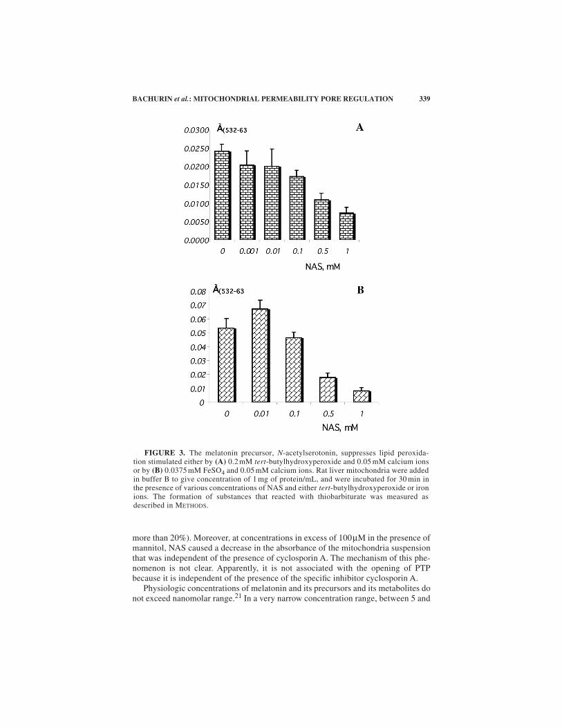
F
IGURE
3 illustrates that NAS suppressed the formation of products of lipid per-oxidation caused by either butylhydroperoxide (F
IG
. 3A) or by iron ions (F
IG
. 3B)in isolated rat liver mitochondria. A dose-dependent inhibition of lipid peroxidationobserved in our experiments shows good correlation with results from other labora-tories. Karbownik and coauthors
19
studied the effect of NAS on lipid peroxidationin microsomal liver membranes and testis membranes, where lipid peroxidation wasgenerated by FeCl
2
, ADP, or NADPH. In the same concentration range, NAS has astabilizing effect on membranes. It does not alter the membrane state in the absenceof the oxidative stress, but it prevents the membrane fluidity induced by the oxidativestress.
20
The effect of NAS on PTP is rather complex. In pharmacologic concentrations, atwhich NAS inhibits lipid peroxidation, NAS demonstrates only marginal protectiveeffect on the opening of PTP generated by various inductors. At NAS concentrationsbetween 1
µ
M and 100
µ
M, the inhibition of PTP is unstable and insignificant (not
339BACHURIN
et al.
: MITOCHONDRIAL PERMEABILITY PORE REGULATION
more than 20
%
). Moreover, at concentrations in excess of 100
µ
M in the presence ofmannitol, NAS caused a decrease in the absorbance of the mitochondria suspensionthat was independent of the presence of cyclosporin A. The mechanism of this phe-nomenon is not clear. Apparently, it is not associated with the opening of PTPbecause it is independent of the presence of the specific inhibitor cyclosporin A.
Physiologic concentrations of melatonin and its precursors and its metabolites donot exceed nanomolar range.
21
In a very narrow concentration range, between 5 and
FIGURE 3. The melatonin precursor, N-acetylserotonin, suppresses lipid peroxida-tion stimulated either by (A) 0.2 mM tert-butylhydroxyperoxide and 0.05 mM calcium ionsor by (B) 0.0375 mM FeSO4 and 0.05 mM calcium ions. Rat liver mitochondria were addedin buffer B to give concentration of 1 mg of protein/mL, and were incubated for 30 min inthe presence of various concentrations of NAS and either tert-butylhydroxyperoxide or ironions. The formation of substances that reacted with thiobarbiturate was measured asdescribed in METHODS.

340 ANNALS NEW YORK ACADEMY OF SCIENCES
50nM, NAS suppressed opening of PTP induced by calcium (100µM) and by phos-phate (0.5mM). The concentration range, where NAS was active toward PTP, wasnot consistent for different mitochondria preparations and, occasionally, could notbe detected at all. However, the inhibition of PTP opening by NAS, which wasinduced by MPP+, was observed in the same nanomolar concentration range (seeFIGURE 4) and was consistent for all the mitochondria preparations.
Melatonin protects against the toxicity induced by MPP+.22 It is believed that theeffect of melatonin is associated with its ability to prevent inhibition by MPP+ of theComplex I of the mitochondrial respiration chain, rather than with its antioxidantproperties. The same is also likely to be true for NAS. Thus, inhibition of PTP open-ing may be one of the physiologic mechanisms of the neuroprotective effect of NAS,and, possibly, of melatonin.
Effect of Tacrine on the Function of PTP
Tacrine (Cognex, 9-amino-1,2,3,4-tetrahydroacridine) is a non-specific cholinest-erase inhibitor, approved in the United States for the therapy of Alzheimer’s disease.Tacrine increases the efficiency of cholinergic neurotransmission and has a numberof other properties that contribute to its therapeutic effect. It was previously demon-strated that Tacrine protected cultured neural cells against β-amyloid toxicity,23
which was not probably associated with its anti-oxidative properties.24
It is possible that the cytoprotective effect of Tacrine is associated with its effecton the opening of PTP. In our experiments, Tacrine inhibited Ca2+-induced mito-chondria swelling at concentrations between 0.5mM and 1.2mM. This concentration
FIGURE 4. The effect of various concentrations of N-acetylserotonin on the openingof mitochondrial pores induced by neurotoxin MPP+. Mitochondria were preincubated forfive minutes in buffer A containing glutamate and malate. Then, a solution containing Ca2+
was added to give a concentration in the cuvette of 0.05 mM. A solution of MPP+ was addedtwo minutes later to give a neurotoxin concentration of 0.4 mM in the cuvette. The reactionwas triggered in two minutes by adding phosphate to give a concentration of 0.5 mM in thecuvette. The traces on the figure correspond to the control reaction (1), when no neurotoxinwas present, the control in the presence of neurotoxin MPP+ (2), and the reaction in thepresence of both neurotoxin (0.4 mM) and NAS in concentrations of 5 nM (3), 10 nM (4),50 nM (5), and 60 nM (6).

341BACHURIN et al.: MITOCHONDRIAL PERMEABILITY PORE REGULATION
range, however, was essentially higher than concentrations required for the cyto-protective effect (1–50µM). These observations suggest that the effect of Tacrine onthe mitochondria pores may contribute to its cytoprotective effect. However, the mainreason is probably associated with the anticholinesterase activity of the drug, sincean activation of cholinesterases is linked to the induction apoptosis.25
Effect of Dimebon on the Function of PTP
Dimebon, 9-[2-(2-methylpyridyl-5)-ethyl]3,6-dimethyl-1,2,3,4-tetrahydro-γ-car-boline, stimulated cognitive functions in the experimental model of AD.26 Dimebonwas successfully tested on Alzheimer’s patients in clinical trials and was patented asa new agent for the therapy of neurodegenerative disorders.
Dimebon remarkably suppressed opening of PTP induced by phosphate ions, bycalcium ions, or by butylhydroxyperoxide (see FIGURE 5). The inductors selected forour experiments generate different intracellular conditions that result in opening ofmitochondrial pores. Calcium ion is an absolute prerequisite for PTP opening. Itbinds to the specific sites of translocase of adenine nucleotides and cyclophiline,thus providing the condition for PTP opening. Phosphate ions induce PTP openingby reducing the ADP concentration in the mitochondria matrix. Butylhydroperoxide
FIGURE 5. The effect of dimebon on the opening of mitochondria pores induced by0.05 mM Ca2+ (�), by 2 mM phosphate ions (�), or by 0.4 mM tert-butylhydroperoxide(�). The ordinate-axis is maximum slope in change of absorbance at 540 nm, which is afunction of the rate of mitochondria swelling. Details are described in the text.

342 ANNALS NEW YORK ACADEMY OF SCIENCES
is metabolized by the glutathione peroxidase system, which is accompanied byNADPH oxidation. This also results in increasing the probability of PTP opening.
Inhibition of PTP opening by Dimebon dramatically depends on the type ofinductor (FIG. 5). Inhibition of the pore opening induced by phosphate ions requiredhigher concentrations of the drug and was complete. The level of inhibition reachedits maximum effect at a drug concentration of 50µM when either calcium ions orbutylhydroperoxide was used as inductors. Moreover, further increase in the drugconcentration resulted in reversing its effect when butylhydroperoxide was used.
Dimebon effectively suppressed lipid peroxidation induced by either butylhydro-peroxide or by the β-amyloid fragment 25–35 (data not shown). Suppression of lipidperoxidation and inhibition of PTP opening are not directly related events.10 Appar-ently, suppression of lipid peroxidation is not sufficient for the inhibition of PTPopening, since 0.4mM of dimebon was sufficient to suppress lipid peroxidation, butdid not have any effect on PTP opening induced by butylhydroperoxide. In addition,lipid peroxidation does not necessary result in PTP opening, because cyclosporin Acompletely inhibited pore opening, but only insignificantly suppressed lipid peroxi-dation in the presence of β-amyloid 25–35.
Thus, dimebon successfully suppressed both lipid peroxidation and PTP openinginduced by physiologic inductors. Both of these properties may play a contributing,or even critical, role in the protection of the mitochondrial function and in the overallneuroprotective effect of the drug. The most important observation, however, wasthe ability of dimebon to prevent PTP opening induced by neurotoxins MPP+ (seeFIGURE 6) and β-amyloid (25–35) (data not shown).
FIGURE 6. Dimebon and cyclosporin A prevent opening of mitochondria poresinduced by neurotoxin MPP+. Mitochondria were preincubated for five minutes in buffer Acontaining glutamate, malate, and 0.2 mM of phosphate in the presence of various concen-trations of MPP+ and in the presence or in the absence of cyclosporin A or dimebon. Theopening of pores was triggered by adding to the reaction mixture a solution containing cal-cium ions, as indicated in the figure. The traces correspond to the control reaction, whenonly calcium and phosphate were present (1), and the reaction in the presence of (2) 0.1 mMMPP+; (3) 0.1 mM MPP+ and 0.2 mM dimebon; (4) 0.5 mM MPP+; (5) 0.5 mM MPP+ and0.2 mM dimebon; and (6) 0.5 mM MPP+ and 2 µM cyclosporin A.

343BACHURIN et al.: MITOCHONDRIAL PERMEABILITY PORE REGULATION
In conclusion, the mitochondrial function associated with the regulation of PTPappears to be an important target for neurotoxins, as well as for the substances thatmay inhibit the effect of such neurotoxins. Compounds that prevent pathologic PTPopening may preserve the mitochondrial function and, thus, may have potential asneuroprotective agents.
REFERENCES
1. BEAL, M.F. 1998. Mitochondrial dysfunction in neurodegenerative diseases. Biochim.Biophys. Acta 1366: 211–223.
2. CASSARINO, D.S. & J.P. BENNETT, JR. 1999. An evaluation of the role of mitochondriain neurodegenerative diseases: mitochondrial mutations and oxidative pathology,protective nuclear responses and cell death in neurodegeneration. Brain Res. Rev.29: 1–25.
3. BONILLA, E., K. TANJI, et al. 1997. Neurologic and neuropatologic features of mito-chondrial encephalomyopathies. In Mitochondria and Free Radicals in Neurodegen-erative Diseases. M.F. Beal, N. Howell & I. Bodis-Wollner, Eds.: 271–281. Wiley-Liss, New York.
4. GREEN, D.R. & J.C. REED. 1998. Mitochondria and apoptosis. Science 281: 1309–1312.5. BERNARDI, P. 1999. Mitochondrial transport of cations: channels, exchangers, and per-
meability transition. Physiol. Rev. 79: 1127–1155.6. STOUT, A.K. & H.M. RAPHAEL, et al. 1998. Glutamate-induced neuron death requires
mitochondrial calcium uptake. Nat. Neurosci. 1: 366–373.7. MATHER, M. & H. ROTTENBERG. 2000. Aging enhances the activation of the permeabil-
ity transition pore in mitochondria. Biochem. Biophys. Res. Comm. 273: 603–608.8. RAMSAY, R., R. MEHLHORN & T. SINGER. 1989. Enhancement by tetraphenylboron of
the interaction of the 1-methyl-4-phenylpyridinium ion (MPP+) with mitochondria.Biochem. Biophys. Res. Comm. 159: 983–990.
9. SIMS, N.R. 1990. Rapid isolation of metabolically active mitochondria from rat brain andsubregions using Percoll density gradient centrifugation. J. Neurochem. 55: 698–707.
10. ERDAHL, W.L., R.J. KREBSBACH & D.R. PFEIFFER. 1991. A comparison of phospholipiddegradation by oxidation and hydrolysis during the mitochondrial permeability tran-sition. Arch. Biochem. Biophys. 285: 252–260.
11. SEYFRIED, J., F. SOLDNER, et al. 2000, Effect of 1-methyl-4-phenylpyridinium on glu-tathione in rat pheochromocytoma PC12 cells. Neurochem. Int. 36: 489–497.
12. CASSARINO, D.S., J.K. PARKS, et al. 1999. The parkinsonian neurotoxin MPP+ opensthe mitochondrial permeability transition pore and releases cytochrome c in isolatedmitochondria via an oxidative mechanism. Biochim. Biophys. Acta 1453: 49–62.
13. BERMAN, S.B., S.C. WATKINS & T.G. HASTINGS. 2000. Quantitative biochemical andultrastructural comparison of mitochondrial permeability transition in isolated brainand liver mitochondria: evidence for reduced sensitivity of brain mitochondria. Exp.Neurol. 164: 415–425.
14. PIKE, C.J., N. RAMEZAN-ARAB & C.W. COTMAN. 1997. β-Amyloid neurotoxicity invitro: evidence of oxidative stress but not protection by antioxidants. J. Neurochem.69: 1601–1611.
15. ZHI-XING, Y., K. DRIEU, L.I. SZWEDA & V. PAPADOPOULOS. 1999. Free radicals andlipid peroxidation do not mediate β-amyloid-induced neuronal cell death. Brain Res.847: 203–210.
16. WOLFLER, A., P.M. ABUJA, et al. 1999. N-Acetylserotonin is a better extra- and intra-cellular antioxidant than melatonin. FEBS Lett. 449: 206–210.
17. OXENKRUG, G., P. REQUINTINA & S. BACHURIN. 2001. Antioxidant and antiaging activ-ity of N-acetylserotonin and melatonin in the in vivo models. Ann. N.Y. Acad. Sci.939: 190–199.
18. BACHURIN, S., G. OXENKRUG, et al. 1999. N-Acetylserotonin, melatonin and theirderivatives improve cognition and protect against β-amyloid–induced neurotoxicity.Ann. N.Y. Acad. Sci. 890: 156–166.

344 ANNALS NEW YORK ACADEMY OF SCIENCES
19. KARBOWNIK, M., E. GITTO, et al. 2001. Relative efficacies of indole antioxidants inreducing autoxidation and iron-induced lipid peroxidation in hamster testes. J. Cell.Biochem. 81: 693–699.
20. GARCYA, J.J., R.J. REITER, et al. 2001. N-Acetylserotonin suppresses hepatic microso-mal membrane rigidity associated with lipid peroxidation. Eur. J. Pharm. 428: 169–175.
21. YOUNGLAI, E.V., S.F. PANG & G.M. BROWN. 1986. Effects of different photoperiodson circulating levels of melatonin and N-acetylserotonin in the female rabbit. ActaEndocrinol. 112: 145–149.
22. ABSI, E., A. AYALA, A. MACHADO & J. PARRADO. 2000. Protective effect of melatoninagainst the 1-methyl-4-phenylpyridinium-induced inhibition of complex I of themitochondrial respiratory chain. J. Pineal Res. 9: 40–47.
23. BACHURIN, S., N. LERMONTOVA, et al. 1998. Prevention of β-amyloid-induced neuro-toxicity by tacrine and dimebon. J. Neurochem. 71(Suppl. 1): S68.
24. XIAO, X.Q., N. TZE-KIN LEE, et al. 2000. Bis(7)-Tacrine, a promising anti-Alzheimer’sagent, reduces hydrogen peroxide-induced injury in rat pheochromocytoma cells:comparison with tacrine. Neurosci. Lett. 290: 197–200.
25. ROBITZKI, A., A. MACK, et al. 1998. Butyrylcholinesterase antisense transfectionincreases apoptosis in differentiating retinal reaggregates of the chick embryo. J.Neurochem. 71: 413–420.
26. BACHURIN, S., E. BUKATINA, et al. 2001. Antihistamine agent Dimebon as a novel neu-roprotector and a cognition enhancer. Ann. N.Y. Acad. Sci. 939: 425–435.
Copyright © 2022 FDOKUMEN