Forced, not voluntary, exercise effectively induces neuroprotection in stroke
Upload
khangminh22Category
view
2download
0
HAL Id: tel-02003607https://tel.archives-ouvertes.fr/tel-02003607
Submitted on 1 Feb 2019
HAL is a multi-disciplinary open accessarchive for the deposit and dissemination of sci-entific research documents, whether they are pub-lished or not. The documents may come fromteaching and research institutions in France orabroad, or from public or private research centers.
L’archive ouverte pluridisciplinaire HAL, estdestinée au dépôt et à la diffusion de documentsscientifiques de niveau recherche, publiés ou non,émanant des établissements d’enseignement et derecherche français ou étrangers, des laboratoirespublics ou privés.
Neuroprotective effects of voluntary exercise andmonoaminergic modulators in stressed male rats
Sarawut Lapmanee
To cite this version:Sarawut Lapmanee. Neuroprotective effects of voluntary exercise and monoaminergic modulators instressed male rats. Neurobiology. Université de Strasbourg; Mahidol University (Bangkok, Thaïlande),2017. English. �NNT : 2017STRAJ058�. �tel-02003607�

UNIVERSITÉ DE STRASBOURG
ÉCOLE DOCTORALE DES SCIENCES DE LA VIE ET DE LA SANTÉ
Institut des Neurosciences Cellulaires et Intégrées CNRS UMR 3212 Université de Strasbourg
Center of Calcium and Bone Research, Department of Physiology, Faculty of Sciences, Mahidol University (Bangkok, Thailand)
THÈSE présentée par :
Sarawut LAPMANEE soutenue le : 7 Juin 2017
pour obtenir le grade de : Docteur de l’université de Strasbourg Discipline/ Spécialité : Neurosciences
Effets neuroprotecteurs de l’exercice volontaire et de modulateurs
monoaminergiques chez le rat mâle stressé
THÈSE dirigée par : Mr KLOSEN Paul
RAPPORTEURS : Mme CHATTIPAKORN Siriporn Mme KALANDAKANOND Sarinee
AUTRES MEMBRES DU JURY : Mr LELIÈVRE Vincent Mr CHAROENPHANDHU Narattaphol
Maître de Conférences (HDR), Université de Strasbourg
Professeur, Chiang Mai University, Thailand Professeur Associé, Chulalongkorn University, Thailand
Professeur, Université de Strasbourg Professeur, Mahidol University, Thailand
Mme KRISHNAMRA Nateetip Professeur, Mahidol University, Thailand

ii
ACKNOWLEDGMENTS
I would like to express my deepest gratitude and sincere appreciation to my major advisors, Prof. Dr. Nateetip Krishnamra and Assoc. Prof. Dr. Paul Klosen for their valuable advice and encouragement during my doctoral studies.
My sincere gratitude and respect are expressed to my co-advisors, Prof. Dr. Narattaphol Charoenphandhu and Asst. Prof. Dr. Jantarima Charoenphandhu as well as the chair of my thesis examination committee, Prof. Vincent Lelièvre, and external committees, Prof. Dr. Siriporn Chattipakorn and Assoc. Prof. Dr. Sarinee Kalandakanond-Thongsong for their valuable criticisms, scientific guidance and suggestions for my research work.
I would like to extend my thanks to Asst. Prof. Dr. Pritsana Piyabhan, Dr. Marie-Paule Felder-Schmittbuhl, Dr. Apron Nuntapornsak for guidance in the study of recognition memory, genotyping, and microdissection techniques. I am also thankful Dr. Panan Suntornsaratoon, Dr. Jarinthorn Teerapornpuntakit, and Miss Nithipak Thammayon for their technical assistance.
I am thankful for the scholarships and co-funding institutions from the Institutional Strengthening Program, Faculty of Science, Mahidol University, the Royal Golden Jubilee Ph.D. Program, Thailand Research Fund, the Embassy of France in Thailand, the Graduate Studies of Mahidol University Alumni Association, Mahidol University, Dr. Paul Pévet, and Dr. Valérie Simonneaux, Department of Neurobiology of Rhythms, Institute of Cellular and Integrative Neurosciences, University of Strasbourg, France.
Furthermore, I would like to express my gratitude to the experimental animals, my appreciation to the staffs at the Animal Facility and the Research Unit of the Faculty of Medicine, Thammasat University, and at the Chronobiotron Animal Facility of the Institute of Cellular and Integrative Neurosciences, University of Strasbourg, France, for animal care and laboratory equipments.
I thank all members of the Department of Physiology, Faculty of Science, Mahidol University and the Melatonin and Seasonal Rhythms team of the Institute of Cellular and Integrative Neurosciences, University of Strasbourg, France and my friends for their moral supports. Finally, I am grateful to my parents and family members for their love and continuous encouragement throughout my study. I dedicate this thesis to my parents, all of my teachers, and the experimental animals.
Sarawut Lapmanee

iii
CONTENTS
Page
ACKNOWLEDGMENTS ii
LIST OF TABLES iv
LIST OF FIGURES v
LIST OF ABBREVIATIONS xix
CHAPTER I INTRODUCTION 1
CHAPTER II LITERATURE REVIEW 5
CHAPTER III MATERIALS AND METHODS 38
CHAPTER IV RESULTS 66
CHAPTER V DISCUSSION 125
CHAPTER VI CONCLUSIONS 139
SUMMARY (FRENCH) 142
REFERENCES 166
APPENDIX 196
BIOGRAPHY 201
iv
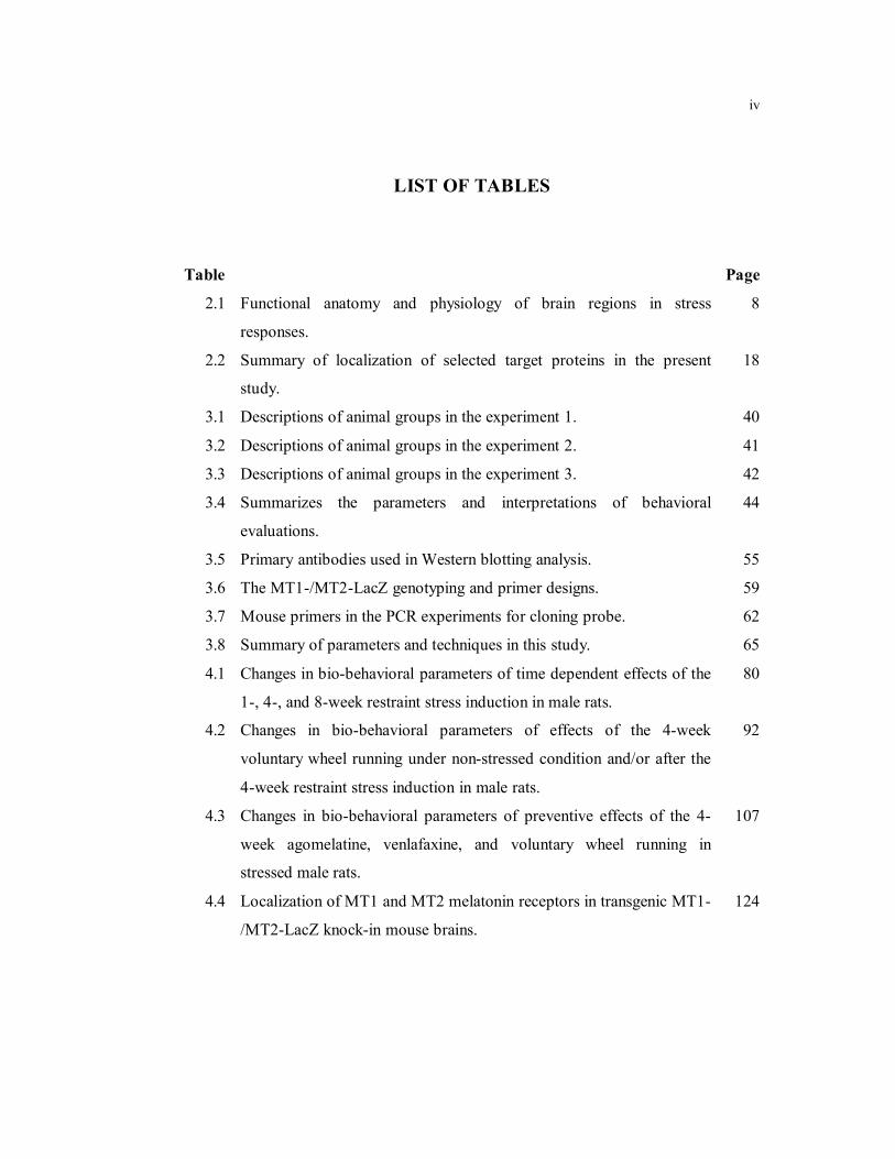
LIST OF TABLES
Table Page
2.1 Functional anatomy and physiology of brain regions in stress
responses.
8
2.2 Summary of localization of selected target proteins in the present
study.
18
3.1 Descriptions of animal groups in the experiment 1. 40
3.2 Descriptions of animal groups in the experiment 2. 41
3.3 Descriptions of animal groups in the experiment 3. 42
3.4 Summarizes the parameters and interpretations of behavioral
evaluations.
44
3.5 Primary antibodies used in Western blotting analysis. 55
3.6 The MT1-/MT2-LacZ genotyping and primer designs. 59
3.7 Mouse primers in the PCR experiments for cloning probe. 62
3.8
4.1
4.2
4.3
4.4
Summary of parameters and techniques in this study.
Changes in bio-behavioral parameters of time dependent effects of the
1-, 4-, and 8-week restraint stress induction in male rats.
Changes in bio-behavioral parameters of effects of the 4-week
voluntary wheel running under non-stressed condition and/or after the
4-week restraint stress induction in male rats.
Changes in bio-behavioral parameters of preventive effects of the 4-
week agomelatine, venlafaxine, and voluntary wheel running in
stressed male rats.
Localization of MT1 and MT2 melatonin receptors in transgenic MT1-
/MT2-LacZ knock-in mouse brains.
65
80
92
107
124

v
LIST OF FIGURES
Figure Page
2.1 The hypothalamic-pituitary-adrenal (HPA) axis in stress responses
showing positive feedbacks (+) and negative feedbacks ().
Elevated glucocorticoids (GCs) are induced by the release of
corticotropin-releasing hormone (CRH) and adrenocorticotropic
hormone (ACTH) from the hypothalamic paraventricular nucleus
(PVN) and anterior pituitary, respectively. Negative feedback
inhibition by GCs acts on the pituitary gland and the hypothalamus.
The release of GCs is also regulated by circadian signals () from
the suprachiasmatic nucleus (SCN) in the hypothalamus.
6
2.2 Neuroanatomical organization of stress responses consist of three
levels, i.e., the cortico-limbic system, the hypothalamus-brain stem
system, and brain-to-adrenal gland system. ACTH,
adrenocorticotropic hormone; ACH, acetylcholine; BNST, bed
nucleus of the stria terminalis; CRH, corticotropin releasing
hormone; EPI, epinephrine; GC, glucocorticoids; NE,
norepinephrine; PFC, prefrontal cortex; and subcortical structures,
amygdala (Gunnar and Quevedo, 2007).
7
2.3 Effects of MT1 and MT2 melatonin receptor agonists and
antagonists in mammals. FSH: follicle-stimulating hormone; GnRH:
gonadotropin-releasing hormone; KLH: Keyhole limpet
hemocyanin; LH: luteinizing hormone; PGE: prostaglandin; PRL:
Prolactin (Modified from Dubocovich and Markowska, 2005).
17
2.4 Chemical structures of monoamine modulators used in the present
study.
27

vi
LIST OF FIGURES (cont.)
Figure Page
2.5 Physiological effects of physical exercise: Adaptive mechanisms are
responsible for a decreased perception of chronic stress. BDNF:
Brain-derived neurotrophic factor; DA: Dopamine; HPA axis:
hypothalamic-pituitary-adrenal axis; iNOS: Inducible nitric oxide
synthase; IL-1: Interleukin 1; IL-6: Interleukin 6; IL-10:
Interleukin 10; NE; Norepinephrine; ROS: Reactive oxygen species;
TNF-: Tumor necrosis factor; 5-HT: Serotonin; VEGF: Vascular
endothelial growth factor (Modified from Sanches et al., 2016).
30
3.1 Schematic diagram showing the time-course of restraint stress
induction and sample collections in the experimental protocol 1.
39
3.2 Schematic diagram showing the time-course of restraint stress
induction, voluntary wheel running, and sample collections in the
experimental protocol 2.
41
3.3 Schematic diagram showing the time-course of treatments and
sample collections in the experimental protocol 3. 42
3.4 Schematic showing, the EPM apparatus that consisted of two open
arms (50 cm long × 10 cm wide) at right angles to two closed arms
(50 cm long × 10 cm wide × 40 cm high). The whole apparatus was
elevated to 50 cm above the floor.
45
3.5 Schematic showing, the ETM apparatus that consisted of three arms
of equal dimensions (50 cm long ×10 cm wide) and elevated 50 cm
above the floor. One arm, enclosed by walls (40 cm high) was
perpendicular to the two opposed open arms.
46

vii
LIST OF FIGURES (cont.)
Figure Page
3.6 Schematic showing, A. The OFT was made from a black painted
wooden box (76 cm long × 57 cm wide × 35 cm high) with a 48
square grid floor (6 × 8 squares, 9.5 cm per side). B. The arena of
maze was divided into two zones, i.e., inner (white area) and outer
zones (gray area).
47
3.7 Schematic showing the MWM pool that consisted of a circular
aluminum pool (150 cm diameter and 60 cm deep) that was filled
with water (25 °C temperature) up to 31 cm. The pool was divided
into four quadrants of equal area. An aluminum platform (10
diameter and 30 cm high) was submerged 1 cm below the water
surface.
49
3.8 Brain cutting block illustrating orientation of brain and placement of
razor blades to obtain coronal brain sections. The numbers on the
right refer to brain sections. Coronal brain sections from which
brain regions were dissected. Dotted lines indicate borders of brain
regions. FC, frontal cortex; S, septum; aH, anterior hypothalamus;
pH, posterior hypothalamus; A, amygdala; VTA, ventral tegmentum
area; and H, hippocampus (Heffner et al., 1980).
54
3.9 Coronal brain sections at the levels of periaqueductal grey and
dorsal raphe nuclei (A) and locus coeruleus (B) (Paxinos and
Watson, 2005).
54
3.10 Schematic diagram showing the experimental protocols in the study
of part 2.
58
viii
LIST OF FIGURES (cont.)
Figure Page
4.1 Time-dependent effects of the 1-, 4-, and 8-week restraint stress
induction on (A) starting body weight, (B) final body weight, (C)
body weight gain, (D) daily food intake, (E) wet adrenal gland
weight, (F) dry adrenal gland weight, (G) relative wet adrenal gland
weights, and (H) relative dry adrenal gland weights. Numbers of
animals are noted in parentheses. *P < 0.05, **P < 0.01, ***P <
0.001 compared to non-stress control group.
67
4.2 Time-dependent effects of the 4-week restraint stress induction on
(A) basal serum and (B) urinary corticosterone levels. Numbers of
animals are noted in parentheses. *P < 0.05 compared to non-stress
control group.
68
4.3 Time-dependent effects of the 1-, 4-, and 8-week restraint stress-
induced anxiety-like behavior as evaluated by EPM, (A) percent
open arm entry, (B) percent open arm time, (C) percent closed arm
entry, (D) percent closed arm time, (E) percent center of arm time,
(F) total arm entry, (G) numbers of rearing, and (H) numbers of
grooming. Numbers of animals are noted in parentheses. *P < 0.05
and **P < 0.01 compared to non-stressed control group.
70
4.4 Time-dependent effects of the 1-, 4-, and 8-week restraint stress-
induced anxiety/learned and innate fear-like behavior as evaluated
by ETM, (A) baseline latency, (B) avoidance latency 1, (C)
avoidance latency 2, and (E) one-way escape latency. Numbers of
animals are noted in parentheses. *P < 0.05 and ***P < 0.001
compared to non-stressed control group.
71

ix
LIST OF FIGURES (cont.)
Figure Page
4.5 Time-dependent effects of the 1-, 4-, and 8-week restraint stress-
induced anxiety-like behavior as evaluated by OFT, (A) number of
lines crossed in the first 30 seconds, (B) total lines crossed, (C)
inner zone time, (D) outer zone time, (E) number of rearing, and (F)
number of grooming. Numbers of animals are noted in parentheses.
*P < 0.05 compared to non-stressed control group.
72
4.6 Time-dependent effects of the 1-, 4-, and 8-week restraint stress-
induced depression-like behaviors as evaluated by forced swimming
and sucrose preference, (A) swimming duration, (B) climbing
duration, (C) immobility duration, (D) number of fecal pellets, and
(E) weekly percent sucrose preference. Numbers of animals are
noted in parentheses. aP < 0.05 and aaP < 0.01 compared to percent
sucrose preference in baseline period. *P < 0.05 and **P < 0.01
compared to non-stressed control group.
73
4.7 Time-dependent effects of the 1-, 4-, and 8-week restraint stress-
induced learning and memory impairment-like behavior as
evaluated by MWM and NOR, (A) escape latency, (B) correct
quadrant time, (C) percent correct quadrant time, and (D)
discrimination ratio. Numbers of animals are noted in parentheses.
*P < 0.05, **P < 0.01, and ***P < 0.001 compared to non-stressed
control group.
74
4.8 Time-dependent effects of the 1-, 4-, and 8-week stress induction on
the BDNF protein levels. Numbers of animals are noted in
parentheses. *P < 0.05 compared to non-stressed control group.
76
x
LIST OF FIGURES (cont.)
Figure Page
4.9 Time-dependent effects of 1-, 4-, and 8-week restraint stress
induction on the hypothalamic and hippocampal GR protein levels.
Numbers of animals are noted in parentheses. *P < 0.05 and ***P <
0.001 compared to non-stressed control group.
76
4.10 Time-dependent effects of the 1-, 4-, and 8-week restraint stress
induction on the NET protein levels. Numbers of animals are noted
in parentheses. *P < 0.05 compared to non-stressed control group.
77
4.11 Time-dependent effects of the 1-, 4-, and 8-week restraint stress
induction on the SERT protein levels. Numbers of animals are noted
in parentheses. *P < 0.05 compared to non-stressed control group.
78
4.12 Time-dependent effects of the 1-, 4-, and 8-week restraint stress
induction on the 5-HT2CR protein levels. Numbers of animals are
noted in parentheses. *P < 0.05, and **P < 0.01 compared to non-
stressed control group.
79
4.13 The effects of the 4-week voluntary wheel running on (A) starting
body weight, (B) final body weight, (C) body weight gain, and (D)
daily food intake. Numbers of animals are noted in parentheses. **P
< 0.01, ***P < 0.001 exercise group compared to non-stressed
sedentary control. #P < 0.05 sedentary stressed group compared to
non-stressed sedentary control group. †P < 0.05 combined stressed
and exercised group compared to sedentary stressed group.
82
xi
LIST OF FIGURES (cont.)
Figure Page
4.14
4.15
The effects of the 4-week voluntary wheel running on (A) running
distance per day, (B) total running distance, (C) wet heart weight,
(D) dry heart weight, (E) relative wet heart weight, and (F) relative
dry heart weight. Numbers of animals are noted in parentheses. **P
< 0.01, ***P < 0.001 exercise group compared to non-stressed
sedentary control. †P < 0.05, †††P < 0.001 combined stressed and
exercised group compared to sedentary stressed group.
The effects of the 4-week voluntary wheel running on (A) wet
adrenal weight, (B) dry adrenal weight, (C) relative wet adrenal
weight, (D) relative dry adrenal gland weight, and (E) serum
corticosterone levels. Numbers of animals are noted in parentheses.
**P < 0.01 exercised group compared to non-stressed control
sedentary group. ##P < 0.05, ##P < 0.01 sedentary stressed group
compared to non-stressed sedentary control group. †P < 0.05
combined stressed and exercised group compared to sedentary
stressed group.
83
84
4.16 Effects of the 4-week voluntary wheel running on restraint stress-
induced depression-like behavior as determined by sucrose
preference test. Numbers of animals are noted in parentheses. ###P <
0.001 sedentary stressed group compared to non-stressed sedentary
control group. †P < 0.05 combined stressed and exercised group
compared to sedentary stressed group.
85
xii
LIST OF FIGURES (cont.)
Figure Page
4.17 Effects of the 4-week voluntary wheel running on BDNF protein
levels. Numbers of animals are noted in parentheses. *P < 0.05
exercised group compared to non-stressed control sedentary group. #P < 0.05 sedentary stressed group compared to non-stressed
sedentary control group. †P < 0.05 combined stressed and exercised
group compared to sedentary stressed group.
86
4.18 Effects of the 4-week voluntary wheel running on the hypothalamic
and hippocampal GR and adrenal MC2R protein levels. Numbers of
animals are noted in parentheses. #P < 0.05 sedentary stressed group
compared to non-stressed sedentary control group. †P < 0.05
combined stressed and exercised group compared to sedentary
stressed group.
88
4.19 Effects of the 4-week voluntary wheel running on NET protein
levels. Numbers of animals are noted in parentheses. *P < 0.05, **P
< 0.01 exercised group compared to non-stressed control sedentary
group. #P < 0.05 sedentary stressed group compared to non-stressed
sedentary control group. †P < 0.05 combined stressed and exercised
group compared to sedentary stressed group.
89
4.20 Effects of the 4-week voluntary wheel running on SERT protein
levels. Numbers of animals are noted in parentheses. **P < 0.01
exercised group compared to non-stressed control sedentary group. #P < 0.05, ##P < 0.01 sedentary stressed group compared to non-
stressed sedentary control group. †P < 0.01 combined stressed and
exercised group compared to sedentary stressed group.
90

xiii
LIST OF FIGURES (cont.)
Figure Page
4.21 Effects of the 4-week voluntary wheel running on the 5-HT2CR
protein levels. Numbers of animals are noted in parentheses. *P <
0.05 exercised group compared to non-stressed control sedentary
group. #P < 0.05 sedentary stressed group compared to non-stressed
sedentary control group. †P < 0.01 combined stressed and exercised
group compared to sedentary stressed group.
91
4.22 Protective effects of agomelatine (Ago), venlafaxine (Vlx) and
voluntary wheel running (Rw) on (A) starting body weight, (B)
running distance, (C) body weight, (D) final body weight, (E) daily
weight gain in pre-treatment, (F) daily weight gain in stress
induction, (G) daily food intake in pre-treatment, and (H) daily
weight gain in stress induction. Numbers of animals are noted in
parentheses. †P < 0.05, ††P < 0.01, and †††P < 0.001compared to
vehicle (Veh)-treated group
94
4.23 Protective effects of agomelatine (Ago), venlafaxine (Vlx) and
voluntary wheel running (Rw) on (A) wet adrenal weight (B) dry
adrenal weight, (C) relative wet adrenal weight, (D) relative dry
adrenal weight, (E) serum corticosterone levels in stressed male
rats. Numbers of animals are noted in parentheses.
95

xiv
LIST OF FIGURES (cont.)
Figure Page
4.24 Protective effects of agomelatine (Ago), venlafaxine (Vlx) and
voluntary wheel running (Rw) on stress-induced anxiety-like
behavior as determined by EPM, (A) percent open arm entry, (B)
percent open arm time, (C) percent closed arm entry, (D) percent
closed arm time, (E) percent center of arm time, (F) total arm
entries, (G) numbers of grooming, and (H) numbers of rearing.
Numbers of animals are noted in parentheses. †P < 0.05 compared
to vehicle (Veh)-treated group.
97
4.25 Protective effects of agomelatine (Ago), venlafaxine (Vlx) and
voluntary wheel running (Rw) on stress-induced anxiety/learned
and innate fear-like behavior as determined by ETM, (A) baseline
latency, (B) avoidance latency 1, (C) avoidance latency 2, and (D)
one-way escape latency. Numbers of animals are noted in
parentheses. †††P < 0.001 compared to vehicle (Veh)-treated group.
98
4.26 Protective effects of agomelatine (Ago), venlafaxine (Vlx) and
voluntary wheel running (Rw) on stress-induced anxiety-like
behavior as determined by OFT, (A) inner zone time, (B) outer zone
time, (C) number of lines crossed in the first 30 seconds, (D) total
lines crossed, (E) numbers of rearing, and (F) numbers of grooming.
Numbers of animals are noted in parentheses. ††P < 0.01, †††P <
0.001 compared to vehicle (Veh)-treated group.
99

xv
LIST OF FIGURES (cont.)
Figure Page
4.27 Protective effects of agomelatine (Ago), venlafaxine (Vlx) and
voluntary wheel running (Rw) on stress-induced depression-like
behavior as determined by FST and sucrose preference test, (A)
swimming duration, (B) climbing duration, (C) immobility duration,
(D) numbers of fecal pellets, and (E) weekly sucrose preference.
Numbers of animals are noted in parentheses. ††P < 0.01, †††P <
0.001 compared to vehicle (Veh)-treated group. aaaP < 0.001
compared to percent sucrose preference after completing 4-week
pre-treatment period.
100
4.28 Protective effects of agomelatine (Ago), venlafaxine (Vlx) and
voluntary wheel running (Rw) on stress-induced learning and
memory impairment-like behavior as determined by MWM and
NOR, (A) escape latency, (B) correct quadrant time, (C) percent
correct quadrant time, and (D) discrimination ratio. Numbers of
animals are noted in parentheses. †P < 0.05, ††P < 0.01, †††P < 0.001
compared to vehicle (Veh)-treated group.
101
4.29 Protective effects of agomelatine (Ago), venlafaxine (Vlx) and
voluntary wheel running (Rw) on the BDNF protein levels.
Numbers of animals are noted in parentheses. †P < 0.05, ††P < 0.01,
and †††P < 0.001 compared to vehicle (Veh)-treated group.
103
4.30 Protective effects of agomelatine (Ago), venlafaxine (Vlx) and
voluntary wheel running (Rw) on the hypothalamic and
hippocampal GR protein levels. Numbers of animals are noted in
parentheses. †P < 0.05 compared to vehicle (Veh)-treated group.
103
xvi
LIST OF FIGURES (cont.)
Figure Page
4.31 Protective effects of agomelatine (Ago), venlafaxine (Vlx) and
voluntary wheel running (Rw) on the NET protein levels. Numbers
of animals are noted in parentheses. ††P < 0.01 compared to vehicle
(Veh)-treated group.
104
4.32 Protective effects of agomelatine (Ago), venlafaxine (Vlx) and
voluntary wheel running (Rw) on the SERT protein levels. Numbers
of animals are noted in parentheses. †P < 0.05 and ††P < 0.01
compared to vehicle (Veh)-treated group.
105
4.33 Protective effects of agomelatine (Ago), venlafaxine (Vlx) and
voluntary wheel running (Rw) on the 5-HT2CR protein levels.
Numbers of animals are noted in parentheses. Veh; vehicle
treatment.
106
4.34 Generation of the MT1/MT2-LacZ knock-in mice. (A) The MT1 or
MT2 receptor gene locus organization before and after site-specific
insertion. (B) Mouse genotyping by PCR for MT1/MT2-LacZ
transgenic knock-in mice.
109
4.35 Comparison MT1-LacZ staining between immunocytochemistry (A
and C) and X-gal histochemistry (B and D) in the suprachiasmatic
nucleus (upper) and pars tuberalis of anterior pituitary (lower) of
transgenic knock-in mice.
111
4.36 MT1-LacZ expressions in neurons as detected by X-gal
histochemical staining compared to 125I- iodomelatonin binding
autoradiography by Liu et al., 1997.
112
4.37 X-gal histochemical mapping of MT1- and MT2-LacZ expression.
113

xvii
LIST OF FIGURES (cont.)
Figure Page
4.38 Immunocytochemical staining of MT1-LacZ expression in (A) the
suprachiasmatic nucleus; SCN and (B) pars tuberalis of anterior
pituitary; PT. Nuclear LacZ staining and lysosomal accumulation of
LacZ can be seen in the SCN (C). In the PT (D), the cytoplasmic
staining is very strong, but sometimes the lysosomal accumulation
can also be seen (arrows).
116
4.39 Immunocytochemical staining of MT2-LacZ expressing neurons in
(A) the paraventicular nucleus of the hypothalamus, (B) the arcuate
nucleus of the hypothalamus, (C) the dorsal hippocampus, (D) the
ventral hippocampus, (E) the CA2 of the hippocampus, and (F) the
molecular layer of the dentate gyrus. DG; dentate gyrus of
hippocampus.
117
4.40 Double-labeling with MT1-LacZ-Immunohistochemistry (green)
and non-radioactive in situ hybridization for neuropeptides (red).
Diagram representing of the cell and fiber distributions of
neuropeptides in the SCN (Abrahamson and Moore, 2001) (A)
AVP, (B), GRP, and (C) VIP. For AVP, the green dots of the MT1-
LacZ cells are clearly located outside of the area of the AVP cells
(D). For VIP (E) and GRP (F) some red neuropeptide cells contain
the green dot indicating co-expression of the MT1-LacZ transgene
(arrows/arrowheads).
120
4.41 Double-labeling with LacZ-immunohistochemistry for MT2 (green)
and non-radioactive in situ hybridization for CRH (red) in the
paraventricular nucleus (PVN) of the hypothalamus. Inset shows a
group of CRH cells that are MT2 positive. Asterisk shows CRH
neurons without MT2 label. Most GRP cells are MT2 positive.
121
xviii
LIST OF FIGURES (cont.)
Figure Page
4.42 Double-labeling with LacZ-Immunohistochemistry and non-
radioactive in situ hybridization for MT2 (green) and GAD (red) co-
localization in the hippocampus (A). Most small horizontal MT2
positive neurons in the external molecular layer (B) are GAD
negative (C). Only a few of these horizontal cells are GAD positive
(arrow in D). Many GAD positive neurons in the dentate gyrus hilus
are also MT2 positive (arrowheads in E).
122
4.43
6.1
A.1
Double-labeling with LacZ-Immunohistochemistry and non-
radioactive in situ hybridization for MT2 (green) and GAD (red) co-
localization. The MT2 positive cells in the CA2 of the hippocampus
are GAD negative. A few non-pyramidal cells of the CA2 are MT2
+ GAD positive (arrowheads).
Diagram showing the summary results in this present study.
Generation of MT1-/MT2-LacZ knock-in mice. The mutant MT1
(Mtnr1a) or MT2 (Mtnr1b) alleles contain a loxP-hUbC-Neomycin-
polyA stop cassette. This Neomycin resistance cassette used for the
selection of recombinant embryonic stem cells is then deleted from
the mutant locus using Cre-mediated recombination to avoid
interference of these sequences with the expression profile of the
LacZ reporter gene.
123
141
199
xix
LIST OF ABBREVIATIONS
A Amygdala
Ach Acetylcholine
ACC Anterior cingulate
ACTH Adrenocorticotropic hormone
AG Adrenal gland
Ago Agomelatine
aH Anterior hypothalamus
AMPA α-amino-3-hydroxy- 5-methyl-4-isoxazolepropionic acid
ANOVA Analysis of variance
AR Adrenergic receptor
AVP Arginine Vasopressin
BCA Bicinchoninic acid
BCIP Bromo-chloro-indolyl phosphate
BDNF Brain-derived neurotrophic factor
BLA Basolateral Amygdala
BNST Bed Nucleus of the Stria Terminalis
BSA Bovine Serum Albumin
BW Body weight
BDZ Benzodiazepine
CA Cornu Ammonis
cDNA Complementary DNA
cGMP cyclic guanosine monophosphate
cm Centimeter
cm3 Cubic centimeter
CO2 Carbon dioxide
COMT Catechol-O-methyltransferase
CRH Corticotropin-releasing hormone

xx
LIST OF ABBRIVATIONS (cont.)
CREB Cyclic adenosine monophosphate response element-binding protein
CYP Cytochrome P450
DA Dopamine
DAB 3,3’-diaminobenzidine
DAG Diacylglycerol
DNA Deoxyribonucleic acid
df Degree of freedom
DFI Daily food intake
DG Dentate gyrus
DHBA 3, 4-dihydroxy-benzyl-amine hydrobromide
DI Discrimination index
DMPC Dimethylpyrocarbonate
DMSO Dimethyl sulfoxide
DNA Deoxyribonucleic acid
dNTP Deoxynucleotide
DR Dorsal Raphe
DSM Diagnostic and Statistical Manual of Mental Disorder
DWG Daily weight gain
DWI Daily water intake
EDTA Ethylenediaminetetraacetic acid
EGF Epidermal growth factor (EGF)
ELISA Enzyme-linked immunosorbent assay
EPI Epinephrine
EPM Elevated plus-maze
ETM Elevated T-maze
FSH Follicle-stimulating hormone
FST Forced swimming test

xxi
LIST OF ABBRIVATIONS (cont.)
g Gram
x g centrifugal force in multiples of the gravitational acceleration
GABA Gamma-aminobutyric acid
GAD Glutamic acid decarboxylase
GC Glucocorticoid
GnRH Gonadotropin-releasing hormone
GR Glucocortocoid receptor
GRP Gastrin releasing peptide
h Hour
H Hippocampus
HCl Hydrochloric acid
H2O2 Hydrogen peroxide
HPA Hypothalamic-pituitary-adrenal
HRP Horseradish peroxidase
ICC Immunocytochemistry
IGF-I Insulin-like growth factor-1
IL Interleukin
iNOS Inducible nitric oxide synthase
ipRGC Intrinsically photosensitive retinal ganglion cell
ISH In situ hybridization
IU International units
K3Fe(CN)6 Potassium hexacyanoferrate
K4Fe(CN)6 Potassium ferrocyanide
kg Kilogram
Ki The inhibitory constant
KLH Keyhole limpet hemocyanin
L Liter
LacZ Beta-galactosidase
LB Luria-Bertani medium

xxii
LIST OF ABBRIVATIONS (cont.)
LC Locus coeruleus
LH Luteinizing hormone
LTD Long-term depression
LTP Long-term potentiation
M Molar
MC2R Melanocortin type 2 receptor
MgCl2 Magnesium chloride
MHPG 3-methoxy-4-hydroxyphenylglycol
min Minute
mL Milliliter
mm Millimeter
MOA Monoamine oxidase enzyme
MOAI Monoamine oxidase inhibitor
mol Mole
MR Mineralocorticoid receptor
mRNA Messenger ribonucleic acid
MT Melatonin
MWM Morris water maze
NA Nucleus accumbens
NaCl Sodium chloride
NaIO4 Sodium periodate
NaOH Sodium hydroxide
NBT Nitroblue tetrazolium
NDRI Norepinephrine-dopamine reuptake inhibitor
NE Norepinephrine
NET Norepinephrine transporter
ng nanogram
NGF Nerve growth factor (NGF)

xxiii
LIST OF ABBRIVATIONS (cont.)
NOR Novel objective recognition
NOS Nitric oxide synthase
NRI Norepinephrine reuptake inhibitor
OFC Orbital frontal cortex
OFT Open field test
PAG Periaqueductal grey
PBS Phosphate buffered saline
PCR Polymerase chain reaction
PD Panic disorder
PEG Polyethylene glycol
PGE Prostaglandin
PFC Prefrontal cortex
p75NR Pan- neurotrophin receptors
pH Posterior hypothalamus
PLP Periodate-lysine-paraformaldehyde
pmol Picomole
p.o. Per os (Oral administration)
PRL Prolactin
PT The pars tuberalis of pituitary
PVN Paraventricular nucleus of hypothalamus
PVT Paraventricular nucleus of thalamus
RIPA Radioimmunoprecipitation assay buffer
ROS Reactive oxygen species
RPM Revolutions per minute of centrifuge rotor speed
s Second
S Septum
SCN Suprachiasmatic nucleus
SDS Sodium dodecyl sulfate

xxiv
LIST OF ABBRIVATIONS (cont.)
SE The standard error
SERT Serotonin reuptake transporter
SN Substantial nigra
SNDRI Serotonin-norepinephrine-dopamine reuptake inhibitor
SNRI Serotonin-norepinephrine reuptake inhibitor
SP Sucrose preference
SSC Saline sodium citrate
SSRI Selective serotonin reuptake inhibitor
TBE Tris-borate-EDTA buffer
TBST Tris buffer saline with Tween 20
TCA Tricyclic antidepressant
TH Tyrosine hydroxylase
TNF Tumor necrosis factor
TPH Tryptophan hydroxylase
TrkB Tyrosine kinase type B receptor
V Volt
VEGF Vascular endothelial growth factor
VIP Vasoactive intestinal polypeptide
Vlx Venlafaxine
VTA Ventral tegmental area
X-gal 5-bromo-4-chloro-3-indolyl-β-d-galactoside
5-HIAA 5-hydroxyindoleacetic acid
5-HT 5-hydroxytryptamine, serotonin
5-HTP 5-hydroxytryptophan
5-HT2CR 5-HT2C receptor
C Degree Celsius
µg Microgram
µl Microliter
University of Strasbourg/Mahidol University Ph.D. (Neurosciences & Physiology) / 1
CHAPTER I
INTRODUCTION
Stress-related mood disorders are common psychiatric illness and are
becoming a global burden of disability and comorbidity (Kessler et al., 2009). Severe
and chronic stress exposures are believed to cause anxiety, depression, and memory
impairment later in life (Sareen et al., 2007; Li et al., 2014; Ikeda et al., 2017), and
therefore, preventive intervention for these stress complications is worth exploring. In
many situations, such as extreme military combat or war, infectious disease outbreak,
and natural disaster, preventive strategies become more important in reducing
incidence of stress-related psychiatric problems in veterans or victims.
The underlying mechanisms of mood disorders in stressed individuals are
associated with deregulation of the hypothalamic-pituitary adrenal (HPA) axis and an
imbalance of the monoamines serotonin (5-HT) and norepinephrine (NE) (Charney,
1998; Bowman et al., 2003; Belmaker and Agam, 2008). Chronic stress increases the
glucocorticoid stress hormones, that glucocorticoids can pass the blood–brain barrier
and induce deleterious changes in the neuronal function in brain regions regulating
memory formation and behavior. Sustained high levels of glucocorticoids decrease
glucocorticoid receptor (GR), brain-derived neurotrophic factor (BDNF), and
neurogenesis in the hippocampus leading to anxiety, depression and memory
impairment (McEwen, 2007; Schmidt and Duman, 2007).
Besides the aberrant HPA axis, stress could disturb the circadian rhythms
and the pineal-adrenal secretory response (Konakchieva et al., 1997; McClung, 2011).
Melatonin (MT) plays a crucial role in the synchronization of circadian rhythmicity.
Actions of MT are mediated by membrane MT receptor subtypes, MT1 and MT2.
Despite the finding that MT levels were decreased in depressed patients (Claustrat et
al., 1984), it is still important to know where MT1 and MT2 are localized because they
are targets for the development of novel antidepressants and MT derivatives in the
HPA axis and hippocampus. So far, the cellular localization of MT1 and MT2 has not

Sarawut Lapmanee Introduction / 2
been visualized clearly despite the use of 125I-iodomelatonin binding, in situ
hybridization, and some developed antibody for immunocytochemistry (Liu et al.,
1997; Fugieda et al., 1999; Poirel et al., 2002). MT1 has been identified in the
suprachiasmatic nucleus (SCN), but site of MT2 remain elusive. Therefore, a genetic
modified rodent model has been generated for mapping and phenotyping MT receptor
expression.
The use of animal models in the stress-related studies provides useful
insights into the behavioral and physiological mechanisms involved in the stress
response. Restraint stress is a simple but effective method that produces both physical
and behavioral changes in mouse and rats. However, the variations of the procedures
such as the intensity and duration of restraint stress have so far failed to produce a
consensus in the results for stress-induced anxiety, depression, and memory
impairment (Buynitsky and Mostofsky, 2009). Although chronic restraint stress could
produce anxiety-and depression-like behavior in male rats (Lapmanee et al., 2012,
2013), but whether total durations of stress induced learning and memory impairment
is still unclear. In addition, the mechanisms triggering the restraint stress-related
behaviors are still not known
The involvement of the dysregulation of 5-HT and NE system in stress-
related mood disorders has been primarily supported by the enhancing effects of
antidepressants on the binding of 5-HT to the serotonin transporter (SERT) and NE to
the noradrenaline transporter (NET), thus increasing storage and release of these
neurotransmitters into the synaptic cleft. A recent study showed that a serotonin-
norepinephrine-dopamine reuptake inhibitor (SNDRI), venlafaxine, showed a greater
potency in the treatment of anxiety-like behavior in stressed male rats than a selective
serotonin reuptake inhibitor (SSRI), fluoxetine or a norepinephrine reuptake inhibitor
(NRI), reboxetine (Lapmanee et al., 2012). However, it is not known whether
venlafaxine can prevent stress-induced anxiety, depression, and memory impairments.
Interestingly, due to the anxiolytic and antidepressant effects (Papp et al.,
2003; Millan et al., 2005; Taylor et al., 2014) of the melatoninergic antidepressant
agomelatine, the a melatonin MT1 and MT2 receptor agonist and 5-HT2C receptor (5-
HT2CR) antagonist could be a candidate drug for the prevention of stress-induced
behavioral change. Unlike venlafaxine, agomelatine is without side effects, such as
University of Strasbourg/Mahidol University Ph.D. (Neurosciences & Physiology) / 3
nausea, diarrhea, or sexual dysfunctions, which can be attributed to excessive
stimulation of some peripheral and central 5-HT receptors (Kennedy and Emsley,
2007). Furthermore, there are no discontinuation symptoms after cessation of
agomelatine treatment compared to paroxetine (Montgomery et al., 2004). However, it
is unclear whether agomelatine acts directly on brain region responsible for stress
responses. Since localization of MT receptors in the rodent central nervous system is
not completely understood, and there is limitation in the use of immunolocalization for
MT1 and MT2 (Wu et al., 2013; Lacoste et al., 2015), the present study attempted to
use transgenic mice with β-galactosidase (LacZ) reporter gene in the study of MT
receptor distribution in in brain regions associated with stress responses.
Since the pharmacological intervention in mood disorders are time-
consuming and might cause side effects, it is better to search for a novel medication in
parallel with alternative interventions. Therefore, non-pharmacological intervention
such as physical exercise has recently become popular, and could be a low cost-stress-
relieving therapy with no side effects for stressed individuals. Exercise has been
reported to be an alternative treatment for a variety of mood disorders. Compared to
forced exercise, e.g., treadmill running and swimming, voluntary exercise such as
voluntary wheel running in rodents should be a motivational exercise with less
associated stress. Moreover, voluntary exercise was found to have both antidepressant
and anxiolytic effects in rodents and human studies (Burghardt et al., 2004; Ströhle,
2009; Lapmanee et al., 2013). Voluntary exercise is a potent stimulator of the HPA
axis (Stranahan et al., 2008). Voluntary wheel running increases hippocampal BDNF
and neurogenesis (Cotman and Berchtold, 2002) and also enhances brain function by
altering monoaminergic neurotransmission (Dishman, 1997). However, the
modulating effects of voluntary wheel running exercise on the chronic stress response
are not well understood, and its preventive effect on stress-induced mood and memory
deficits remains unclear.
Therefore, the objectives of thesis research were:
1. To examine the time-dependent effects of the 1-, 4- and 8-week stress
induction on anxiety-, depression-, and memory impairment-like behaviors and
changes in the expression of selected target proteins in brain regions associated with
stress responses.

Sarawut Lapmanee Introduction / 4
2. To determine the effects of the 4 week-voluntary wheel running on the
HPA axis under two conditions, non-stress condition or after the 4-week exposure to
restraint stress, and changes in the expression of selected target proteins in brain
regions associated with stress responses.
3. To evaluate the effectiveness of agomelatine, venlafaxine, and voluntary
wheel running exercise in the prevention of stress-induced anxiety-, depression-, and
memory impairment-like behaviors and changes in the expression of selected target
proteins in brain regions associated with stress responses.
4. To characterize the phenotype of MT1-/MT2-LacZ expressing neurons
in knock-in mice by analyzing co-expressed neuropeptides and neuronal markers.
The hypothesizes of this study were:
1. Monoaminergic modulators and voluntary wheel running could prevent
stress-related behaviors in male rats by modulating hippocampal neurogenesis and
altering the expression of target proteins of central monoamine neurotransmission in
the brain regions associate with stress response.
2. MT1/MT2-LacZ expressing neurons in brain regions associated with
stress responses were characterized in knock-in mice and might be the sites action of
pharmacological treatment.
University of Strasbourg/Mahidol University Ph.D. (Neurosciences & Physiology) / 5
CHAPTER II
LITERATURE REVIEW
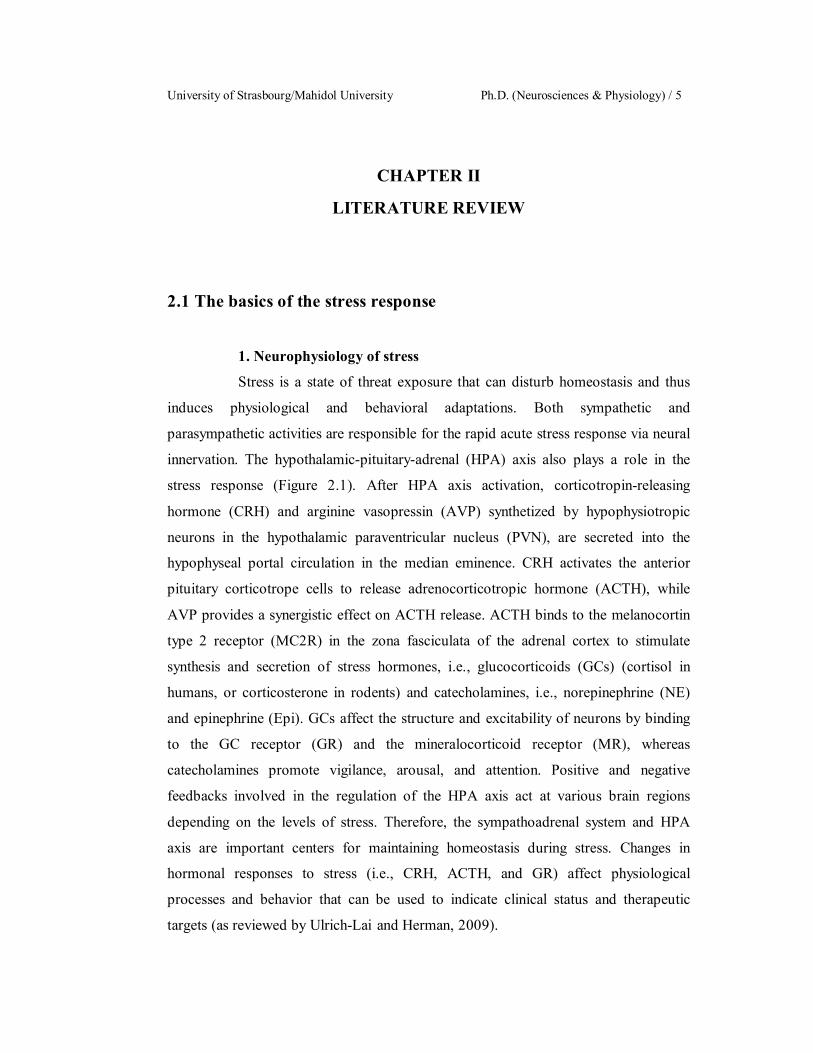
2.1 The basics of the stress response
1. Neurophysiology of stress
Stress is a state of threat exposure that can disturb homeostasis and thus
induces physiological and behavioral adaptations. Both sympathetic and
parasympathetic activities are responsible for the rapid acute stress response via neural
innervation. The hypothalamic-pituitary-adrenal (HPA) axis also plays a role in the
stress response (Figure 2.1). After HPA axis activation, corticotropin-releasing
hormone (CRH) and arginine vasopressin (AVP) synthetized by hypophysiotropic
neurons in the hypothalamic paraventricular nucleus (PVN), are secreted into the
hypophyseal portal circulation in the median eminence. CRH activates the anterior
pituitary corticotrope cells to release adrenocorticotropic hormone (ACTH), while
AVP provides a synergistic effect on ACTH release. ACTH binds to the melanocortin
type 2 receptor (MC2R) in the zona fasciculata of the adrenal cortex to stimulate
synthesis and secretion of stress hormones, i.e., glucocorticoids (GCs) (cortisol in
humans, or corticosterone in rodents) and catecholamines, i.e., norepinephrine (NE)
and epinephrine (Epi). GCs affect the structure and excitability of neurons by binding
to the GC receptor (GR) and the mineralocorticoid receptor (MR), whereas
catecholamines promote vigilance, arousal, and attention. Positive and negative
feedbacks involved in the regulation of the HPA axis act at various brain regions
depending on the levels of stress. Therefore, the sympathoadrenal system and HPA
axis are important centers for maintaining homeostasis during stress. Changes in
hormonal responses to stress (i.e., CRH, ACTH, and GR) affect physiological
processes and behavior that can be used to indicate clinical status and therapeutic
targets (as reviewed by Ulrich-Lai and Herman, 2009).

Sarawut Lapmanee Literature Review / 6
Figure 2.1 The hypothalamic-pituitary-adrenal (HPA) axis in stress responses
showing positive feedbacks (+) and negative feedbacks (). Elevated glucocorticoids
(GCs) are induced by the release of corticotropin-releasing hormone (CRH) and
adrenocorticotropic hormone (ACTH) from the hypothalamic paraventricular nucleus
(PVN) and anterior pituitary, respectively. Negative feedback inhibition by GCs acts
on the pituitary gland and the hypothalamus. The release of GCs is also regulated by
circadian signals () from the suprachiasmatic nucleus (SCN) in the hypothalamus.
2. Neuroanatomy of stress
Various brain regions involved in stress responses can be described at
three levels (Figure 2.2), i.e., the cortico-limbic system, the hypothalamus–brain stem
system, and brain-to-adrenal gland system (as reviewed by Gunnar and Quevedo,
2007). At the level of the cortico-limbic system, the anterior cingulate (ACC) and
orbital frontal cortex (OFC) are connected to the amygdala, which in turn connects
with the hippocampus and bed nucleus of the stria terminalis (BNST). The BNST
signals to the PVN to stimulate the release of CRH and AVP, whereas the
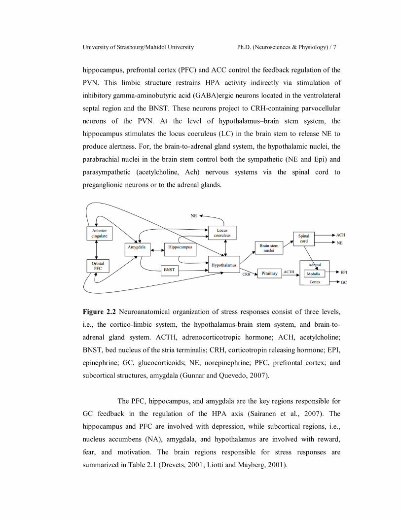
University of Strasbourg/Mahidol University Ph.D. (Neurosciences & Physiology) / 7
hippocampus, prefrontal cortex (PFC) and ACC control the feedback regulation of the
PVN. This limbic structure restrains HPA activity indirectly via stimulation of
inhibitory gamma-aminobutyric acid (GABA)ergic neurons located in the ventrolateral
septal region and the BNST. These neurons project to CRH-containing parvocellular
neurons of the PVN. At the level of hypothalamus–brain stem system, the
hippocampus stimulates the locus coeruleus (LC) in the brain stem to release NE to
produce alertness. For, the brain-to-adrenal gland system, the hypothalamic nuclei, the
parabrachial nuclei in the brain stem control both the sympathetic (NE and Epi) and
parasympathetic (acetylcholine, Ach) nervous systems via the spinal cord to
preganglionic neurons or to the adrenal glands.
Figure 2.2 Neuroanatomical organization of stress responses consist of three levels,
i.e., the cortico-limbic system, the hypothalamus-brain stem system, and brain-to-
adrenal gland system. ACTH, adrenocorticotropic hormone; ACH, acetylcholine;
BNST, bed nucleus of the stria terminalis; CRH, corticotropin releasing hormone; EPI,
epinephrine; GC, glucocorticoids; NE, norepinephrine; PFC, prefrontal cortex; and
subcortical structures, amygdala (Gunnar and Quevedo, 2007).
The PFC, hippocampus, and amygdala are the key regions responsible for
GC feedback in the regulation of the HPA axis (Sairanen et al., 2007). The
hippocampus and PFC are involved with depression, while subcortical regions, i.e.,
nucleus accumbens (NA), amygdala, and hypothalamus are involved with reward,
fear, and motivation. The brain regions responsible for stress responses are
summarized in Table 2.1 (Drevets, 2001; Liotti and Mayberg, 2001).
Sarawut Lapmanee Literature Review / 8
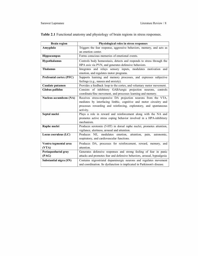
Table 2.1 Functional anatomy and physiology of brain regions in stress responses.
Brain region Physiological roles in stress responses Amygdala
Triggers the fear response, aggressive behaviors, memory, and acts as an emotion center.
Hippocampus Forms conscious memories of emotional events. Hypothalamus Controls body homeostasis, detects and responds to stress through the
HPA axis via PVN, and generates defensive behaviors. Thalamus Integrates and relays sensory inputs, modulates motivation and
emotion, and regulates motor programs. Prefrontal cortex (PFC) Supports learning and memory processes, and expresses subjective
feelings (e.g., nausea and anxiety). Caudate putamen Provides a feedback loop to the cortex, and voluntary motor movement. Globus pallidus
Consists of inhibitory GABAergic projection neurons, controls coordinate/fine movement, and processes learning and memory.
Nucleus accumbens (NA) Receives stress-responsive DA projection neurons from the VTA, mediates by interfacing limbic, cognitive and motor circuitry and processes rewarding and reinforcing, exploratory, and spontaneous activity.
Septal nuclei Plays a role in reward and reinforcement along with the NA and promotes active stress coping behavior involved in a HPA-inhibitory mechanism.
Raphe nuclei
Produces serotonin (5-HT) in dorsal raphe nuclei, promotes attention, vigilance, alertness, arousal and attention.
Locus coeruleus (LC)
Produces NE, modulates emotion, attention, pain, autonomic, respiratory, and cardiovascular functions.
Ventra tegmental area (VTA)
Produces DA, processes for reinforcement, reward, memory, and attention.
Periaqueductal gray (PAG)
Generates defensive responses and strong feeling of fear in panic attacks and promotes fear and defensive behaviors, arousal, hypoalgesia
Substantial nigra (SN) Contains nigrostriatal dopaminergic neurons and regulates movement and coordination. Its dysfunction is implicated in Parkinson's disease.
University of Strasbourg/Mahidol University Ph.D. (Neurosciences & Physiology) / 9
3. Neurochemistry of stress
3.1 Corticotropin-releasing hormone (CRH)
CRH is an amino acid neuropeptide, which plays a major role
in the adaptation to stress (Vale et al., 1981). Release of CRH depends on circulating
GC and ACTH, which are associated with circadian patterns of the HPA axis. CRH is
required for the activation of anterior pituitary ACTH gene expression. CRH is present
throughout the central nervous system, adrenal and sympathetic ganglion of autonomic
nervous system and some fetal organs such as lung and gastrointestinal tract.
Regarding localization of CRH receptors, CRH 1 and CRH 2 are expressed in the
brain. About 50% of CRH neurons colocalize with AVP in the the dorsomedial
parvocellular part of the PVN (Smith and Vale, 2006). CRH neurons directly synapse
with glutamatergic and GABAergic neurons in the PVN and the cerebral cortex, i.e.,
the layers 2–3 of the rat brain (Yan et al., 1998; Kubota et al., 2011). In addition, CRH
neurons and CRH receptors are expressed in the hippocampus, stria terminalis, solitary
nucleus of hypothalamus, amygdala, locus coeruleus, cerebellum and spinal cord
(Aguilera and Liu, 2012). CRH is not only responsible for metabolic, endocrine and
autonomic responses in the perceived acute stress, but CRH also leads to
psychological problems in the perceived chronic stress (Korosi and Baram, 2008).
Therefore, CRH can mediate behavioral responses such as anxiety, arousal,
depression, and learning and memory (Binder and Nemeroff, 2010).
3.2 Glucocorticoid (GC)
GCs are essential for life. Normally, the release of GC is
driven by a circadian rhythm originating in the SCN of the hypothalamus. Signal
inputs from SCN result CRH release, which activates the HPA axis and releases GCs
(Figure 2.1). In addition, sympathetic nerve fibers directly connect the SCN to the
HPA axis; therefore GC release exhibits a circadian pattern. GC promotes energy
mobilization, i.e., increase in blood glucose and free-fatty acids in the fight to flight
response, and also disturbs the functions of the immune, digestive, and reproductive
systems in long-term stress exposure. GRs and MRs are nuclear receptors with
transcription factor activity, which is activated by the binding of GCs to the GR and
MR. GR is highly expressed throughout the whole brain, whereas MR is expressed in
Sarawut Lapmanee Literature Review / 10
specific restricted brain regions such as the amygdala, the hippocampus, the LC, the
PFC, the solitary nucleus and the PVN (de Kloet et al., 2005; McEwen, 2007). GC
mediates the negative feedback to inhibit the secretion of CRH and ACTH.
Hyperactivity or dysregulation of the HPA axis leads to
hypercortisolism contributing to metabolic, cardiovascular, and mental diseases. It has
been well established that chronic stress is a risk factor for mood disorders (e.g.
anxiety disorders and depression). Particularly, GCs cause impaired learning and
memory formation, changes in the distribution pattern of GR and MR, atrophy of
hippocampal cells, reduced hippocampal neurogenesis, reduced synaptic plasticity and
impaired learning ability, resulting in memory problems in stressed individuals
(Sapolsky et al., 1986).
3.3 GABAergic neurotransmission
GABA plays a role in the modulation of emotional behaviors.
GABAergic neurons synthesize GABAs to inhibit neuronal excitability. GABA is
synthesized by glutamic acid decarboxylase (GAD), which exists as two isoforms
based on the protein size of glutamic acid decarboxylase (GAD), i.e., 67 kDa (GAD1)
and 65 kDa (GAD2). In the mouse brain, 80–90% of GAD activity is produced by
GAD1 (Asada et al. 1997, Condie et al. 1997). Changes in GABA, GABA receptor,
and GAD expression can occur in stress exposure. Stress can inhibit the GABAergic
plasticity, which contributes to the development of stress-related disorders (reviewed
by Maguire, 2014).
During stress, increased levels of GCs lead to increased GABA
levels in the hippocampus of rats (de Groote and Linthorst, 2007). Alteration of GAD
expression causes marked inhibitory effects on postsynaptic neurons in the
hippocampus (Stone et al., 2001; Maggio and Segal, 2009). Both GAD1 and GAD2
expressions are increased in the bed nucleus of the stria terminalis, preoptic area of
hypothalamus, PVN and CA3 of the hippocampus after chronic intermittent stress
(Bowers et al., 1998). Moreover, chronic immobilization stress increases the
expression of GABAA receptors in the PFC as indicated by greater binding of 3H-
flunitrazepam (Braestrup et al., 1979). In contrast, the reduction of GAD was reported
in the striatum and olfactory bulb in chronic cold stress (Acosta et al., 1993) leading to
University of Strasbourg/Mahidol University Ph.D. (Neurosciences & Physiology) / 11
reduced levels of GABA. These results indicate that the GABAergic inhibitory control
over glutamatergic cells is reduced (Kalkman and Loetscher, 2003). Therefore, the
reduction of GABAergic inhibitory control in these areas could be implicated in the
pathophysiology of fear, anxiety, and depression.
3.4 Monoaminergic neurotransmission
Monoamines, organic compounds with one amino group, are
associated with mood stabilization. Imbalance of monoaminergic neurotransmitters
contributes to the impairments of structural plasticity and changes in the size of of
specific limbic areas. The members of monoamine neurotransmitters include
catecholamines (dopamine, DA and NE) and 5-hydroxytryptamine (serotonin; 5-HT).
Therefore, dysregulation of monoaminergic neurotransmission has been implicated in
mood disorders such as such as depression, anxiety, and schizophrenia (Kandel et al.,
2000, Siegla et al., 2006)
3.4.1 Catecholamines
The dopaminergic system plays a role in emotional
reward, motivation, desire, addiction, pleasure, learning and motor fine tuning. The
central component of the dopaminergic system is the ventral tegmental area (VTA).
DA is synthesized in the nigrostriatal neurons that are involved in body movement and
mood. In rats, dopaminergic neurons are present in the midbrain, i.e., the substantia
nigra (SN), VTA and related nuclei. DA and the limbic system are essential in forming
behavior, sensory and environmental/emotional-memory connections. The VTA forms
a connection with a key pleasure center known as NA. DA release into the NA is
responsible for rewarding and addictive behaviors. In addition, the NE system plays a
role in wakefulness, alertness, energy, arousal and motivation. The LC, a nucleus
located in the pons of the brainstem, is the central norepinephrine system. The LC
produces NE and sends projections to areas of the hindbrain, forebrain, and spinal cord
through a system of noradrenergic projections. Terminals in the cerebellum and cortex
could be derived from collateral branches of a single neuron's axon. It plays a major
role in the function of the sympathetic nervous system, during states of agitation or
stress. The LC and the adrenergic projections are known as the LC-noradrenergic
system. Activation of the LC induces the fight or flight reaction in the stress response.

Sarawut Lapmanee Literature Review / 12
NE is synthesized by adrenergic neurons in LC in the brain stem while the major site
of Epi synthesis is adrenal medulla.
The catecholamine neurotransmitters are
synthesized from tyrosine. The biosynthetic pathways of catecholamines involve
several enzymes, i.e., tyrosine hydroxylase (TH), pteridine reductase, aromatic amino
acid decarboxylase, dopamine β-hydroxylase, and phenylethanolamine-N-methyl
transferase. TH in the LC is upregulated in response to depletion of NE in normal and
stressed rats. Therefore, elevated TH levels could be an indicator of the stressed status
in the NE system (Melia et al., 1992). Moreover, high NE may facilitate long-term
potential (LTP) induction in the hippocampus by decreasing the threshold to LTP
generation, facilitating memory formation (Hu et al., 2007). DA and NE are stored in
the storage vesicles that are located in the nerve terminal. Since the metabolizing
enzyme monoamine oxidase (MAO) is present in the cytosol, concentrations of both
catecholamines in the cytosol are low. Catecholamines are released by exocytosis from
the nerve terminal. Both DA and NE are inactivated by MOA and catechol-O-
methyltransferase (COMT) after binding to the receptors. Some catecholamines are
transported back into the presynaptic nerve terminal via their respective selective
reuptake transporters such as the DA reuptake transporter or the norepinephrine
transporter (NET).
NETs are located on the presynaptic membrane of
NE nerve terminals. NET expression has not been found in serotonergic and
dopaminergic neurons, but the NET is located on the presynaptic part of an NE
terminal (Barker and Blakely, 1995). Moreover, alterations of NET expression could
contribute to the pathophysiology of depressive symptoms (Klimek et al., 1997;
Haenisch et al., 2009; Haller et al., 2002). Although the role of the NET in NE
signaling is well known, its regulation in response to treatment with NET inhibiting
antidepressants remains uninvestigated. Moreover, reports of effects of stress on NET
expression are inconsistent. Either membrane trafficking or degradation of NET
proteins may contribute to the loss of NET ligand binding and protein levels. Altered
expression of the NET gene may contribute to the loss of NET proteins in response to
antidepressant treatment (Zhu et al., 2002; Chen et al., 2012).

University of Strasbourg/Mahidol University Ph.D. (Neurosciences & Physiology) / 13
3.4.2 Serotonin (5-hydroxytryptamine, 5-HT)
The 5-HT system plays a role in mood, emotion,
social disposition, sleep cycle, wakefulness, dreaming, appetite, spinal pain
modulation and the modulation of the endogenous opioidergic system. The raphe
nuclei are located in the brainstem, and are the primary source of 5-HT in the brain. 5-
HT is a hydrophilic substance and that can pass the blood-brain barrier. Ascending
serotonergic projections innervating the cerebral cortex and other regions of the
forebrain arise from the dorsal raphe (DR) and the median raphe. The median raphe
projects to the hippocampus, the septum and the hypothalamus, whereas the striatum is
innervated predominantly by the DR. Raphe neurons send collateral axons to brain
areas that are functional related, i.e., the amygdala and hippocampus or the SN and
caudate putamen. The serotonergic pathways project to areas of the cerebral cortex,
and the spinal cord. When activated, the ascending projections of the 5-HT neurons in
the DR facilitate neuronal activity in the amygdala and suppress neurons commanding
flight in the periaqueductal grey (PAG). Neurons in the median raphe nucleus could be
stimulated by uncontrollable stress. In order to prevent the occurrence of depression,
5-HT would promote a disconnecting mechanism that results in tolerance to stress that
takes place in the hippocampus. Abnormalities of 5-HT function in the brain can lead
to psychiatric disorders such as schizophrenia, anxiety, panic and depression (Graeff et
al., 1996).
5-HT is synthesized from the amino acid
tryptophan by the rate limiting enzymes tryptophan hydroxylase (TPH) and 5-
hydroxytryptophan (5-HTP) decarboxylase. TPH is similar to TH in term of catalytic
mechanism and the amino acid sequences. 5-HT is taken up from the cytoplasm into
the storage vesicle by active transport. The 5-HT receptors are divided into 5-HT1, 5-
HT2, and 5-HT3 receptors. The 5-HT1 and 5-HT2 receptor families are G-protein
coupled receptor, while the 5-HT3 receptor is a ligand-gated sodium and potassium ion
channel (Mohammad-Zadeh et al., 2008).
Among the 5-HT receptor families, the 5-HT2C
receptor (5-HT2CR) is a prominent serotonin receptor that is distributed throughout the
central nervous system. 5-HT2CRs are widely distributed in the choroid plexus, frontal
cortex, hippocampus, hypothalamus, ventral tegmental area and amygdala (Xu et al.,
Sarawut Lapmanee Literature Review / 14
2008; Greenwood et al., 2012). Activation of 5-HT2CR induces a decrease in food
intake and increases anxiety (Dryden et al., 1996; Gatch, 2003) because 5-HT2CR is
expressed on CRF neurons that regulate ACTH and corticosterone secretion (Heisler
et al., 2007a). Furthermore, 5-HT2CR knockout mice display reduced anxiety when
exposed to novel environments and show decreased neuronal activities, specifically in
the amygdala (Heisler et al., 2007b). In addition, altered RNA editing of the 5-HT2CR
was observed in the PFC of suicide victims with major depressive disorder as well as
in stressed rats (Niswender et al., 2001; Iwamoto et al., 2005). Therefore, the
activation of this receptor could be involved in anxiety and fear responses and could
be a target for anxiolytic drugs, i.e., agomelatine.
3.4.3 Serotonin reuptake transporter (SERT)
In order to regulate the availability of 5-HT in the
synaptic cleft, both binding of 5-HT to its autoreceptor and the activity of the SERT
located on the presynaptic membrane are required. Stimulation of the 5-HT
autoreceptor decreases further release of 5-HT, while the SERT removes 5-HT from
the synaptic cleft back into the presynaptic neuron. Changes in SERT expression and
function are correlated with the pathophysiology of depression and anxiety disorders.
Effects of stress on SERT expression in the raphe nuclei have been studied but the
results are still controversial. For example, single immobilization reduced SERT
mRNA in the raphe pontis (Vollmayr et al. 2000). Single social defeat reduced the
SERT densities in hippocampus, but did not change those in the midbrain where the
DR nuclei are located (Berton et al., 1999). In contrast, chronic restraint plus cold
stress resulted in an increase in SERT mRNA levels in the DR of both male and
female rats (Pare et al., 1999). These studies with inconsistent results clearly
emphasize the need for further studies on the effects of chronic stress on the SERT in
serotoninergic neurons. SERT is the primary target of the clinically successful class of
antidepressant and anxiolytic drugs known as the selective serotonin reuptake
inhibitors (SSRIs). Although the action of the SSRIs is generally understood to
involve an increase in 5-HT concentrations in the synapse, resulting in increased
postsynaptic receptor binding, this has not been clearly established (Schloss and
Williams, 1998).
University of Strasbourg/Mahidol University Ph.D. (Neurosciences & Physiology) / 15
The complex neural pathways of the serotonergic
system allow this transmitter substance to broadly affect behavior and mood. Changes
in the serotonergic system have been shown to be strongly correlated with mental
disorders. At the cellular level, abnormalities may include abnormal regulation of 5-
HT synthesis, release and/or reuptake or abnormal responsiveness to the 5-HT signal.
In addition, alteration of this neurotransmission involves the presynaptic autoreceptors
(5-HT1A), the SERT site, and the postsynaptic 5-HT2CR, in several brain regions, that
are believed to be potentially important in mood and memory disorders.
3.4.4 Melatonin
Melatonin (MT) is an important regulator of
circadian rhythms and seasonal physiology. MT was first isolated and identified by
Lerner and co-workers (1958). MT synthesis in the pineal gland is modulated by the
light/dark information that is detected by the retina. The relative contributions of the
intrinsically photosensitive retinal ganglion cells (ipRGCs) and the classical
photoreceptors in the control of MT synthesis are subject to discussion. IpRGCs alone
are sufficient in photoreceptor deficient animals, but that does not mean that in
animals with functional retinal photoreceptors only the ipRGC control melatonin
production. MT production in the pineal gland and locally in the retina follows a
circadian rhythm, with the highest levels produced during the dark phase. In
mammals, the light/dark information is sent through the retino-hypothalamic tract and
the SCN to the pineal gland, where MT is synthesized and secreted by pinealocytes.
Glutamate released from the retinohypothalamic tract stimulates the SCN GABAergic
neurons, which in turn inhibit the stimulatory action of the PVN on MT secretion by
the pineal gland through its sympathetic innervation at the intermediolateral nucleus of
the spinal cord and the superior cervical ganglion. MT binds to the MT1 and/or MT2
receptors on the target cells (reviewed by Liu et al, 2016).
Stress-induced activation of the HPA axis can
modulate the MT rhythm, and thus the MT production and secretion pattern
(McClung, 2011). There is a reduction in MT secretion in the dark phase after stress
exposure, which probably results from a reduction in tryptophan precursor for MT
synthesis. However, nocturnal illumination did not further suppress MT production in
stressed animals (Persengiev et al., 1991). Corticosterone apparently alters 5-HT and
Sarawut Lapmanee Literature Review / 16
MT synthesis by modulating the mRNA levels of TH (Clark and Russo, 1997).
Although corticosterone reportedly inhibited nuclear factor-B translocation, thereby
enhancing the NE-induced synthesis of MT in the pineal gland (Ferreira et al., 2005),
chronic stress indirectly impaired sympathetic inputs to the pineal gland, leading to
disruption of MT rhythm (Dagnino-Subiabre et al., 2006). It is therefore possible that
derangement of the melatonergic system in stressed rats may stem from inappropriate
MT production and its irregular rhythm. Also, MT levels are decreased in depressed
patients (Claustrat et al., 1984). However, the cellular localization of MT1 and MT2 is
still not fully established and subject to controversies. This localization is important
because MT1 and MT2 receptors are targets for the development of novel
antidepressants and MT derivatives
MT receptors belong to the family of G protein
coupled receptors or the seven-transmembrane domain receptors. In mammals, there
are two types of MT receptors, melatonin receptor 1 (MT1; MTNR1A) and melatonin
receptor 2 (MT2; MTNR1B). The MT1 receptor mediates the acute inhibitory action
on the SCN firing, whereas the MT2 receptor mediates the phase shifting effect on the
SCN activity (Pandi-Perumal et al., 2007). When MT binds to MT receptors, it
activates Gi and Gq proteins, which in turn inhibit the adenylate cyclase/cAMP
pathway and activate the protein kinase C pathway. As a result of the phosphorylating
activity of protein kinases, cAMP response element-binding protein (CREB) and
mitogen-activated protein kinase (MAPK or MAP kinase) regulate the expression of
clock genes and modulate clock gene rhythms (phase advances and delays) (Sharma et
al., 2015). MT actions were mediated via MT1 and MT2 receptors as summarized in
the Figure 2.3. MT receptors are the target for development of pharmacological
agents. However, there is variation in the density and location of MT receptor
expression between species. Radioactive ligand binding, especially 125I-
iodomelatonin receptor binding remains the “gold standard” for the detection of MT
receptors (Vanecek et al., 1987). Although the tissue distribution of MT1 can be
detected by receptor binding, there is no specific mapping for MT2 and this technique
does not allow the study of cellular receptor localization. In situ hybridization has
proven not sensitive enough for the mapping and phenotyping of MT receptor
expressing cells (Poirel et al., 2002).

University of Strasbourg/Mahidol University Ph.D. (Neurosciences & Physiology) / 17
In addition, MT receptor protein localization using
the immunocytochemistry in rodents is controversial because reliable antibodies with
high sensitivity are not available (Fujieda et al., 1999). Therefore, a genetically
modified rodent model has been generated for mapping and phenotyping MT receptor
expression. Tissues characterized functional MT1 and/or MT2 melatonin receptors,
e.g., retina, SCN, pars tuberalis, cerebral, arteries, kidney, pancreas, adrenal cortex,
testes and immune cells (Dubocovich and Markowska, 2005). However, MT receptor
localization is not clearly and controversial finding.
Figure 2.3 Effects of MT1 and MT2 melatonin receptor agonists and antagonists in
mammals. FSH: follicle-stimulating hormone; GnRH: gonadotropin-releasing
hormone; KLH: Keyhole limpet hemocyanin; LH: luteinizing hormone; PGE:
prostaglandin; PRL: Prolactin (Modified from Dubocovich and Markowska, 2005).
3.5. Brain-derived neurotrophic factor (BDNF)
Besides affecting neurotransmitter levels underlying the
regulation of neurogenesis such as GABA, 5-HT, DA, and NE, cannabinoids, opioids
and nitric oxide (Balu and Lucki, 2009), stress also decreases the expression of growth
factors, such as BDNF, insulin-like growth factor-1 (IGF-1), nerve growth factor
(NGF), epidermal growth factor (EGF) and vascular endothelial growth factor (VEGF)
that also influence neurogenesis (Schmidt and Duman, 2007).

Sarawut Lapmanee Literature Review / 18
BDNF is a member of the nerve growth factor family and is
expressed in the hippocampus and the cortex where it modulates neuronal plasticity,
inhibits cell death cascades and increases the cell survival proteins that are responsible
for the proliferation and maintenance of central nervous system neurons (Duman,
2004, Murakami et al., 2005). BDNF mRNA was transcribed and regulated by 8
various promoters containing untranslated exons (I to VIII). Especially, promoter IV is
mediated by calcium and regulatory components, leading to protein translation (Zheng
and Wang et al., 2009). The function of BDNF is mediated by its binding to specific
receptors, i.e., tropomycin receptor kinase B (TrkB) receptors and pan75 neurotrophin
receptor (p75NTR). TrkB signaling promotes neurogenesis while p75NTR activation
mediates proteolytic mechanisms in survival and apoptosis inducing mechanisms (Lee
and Kim, 2010). BDNF is involved in the pathophysiology of mood disorders and
plays an important role in the mechanism of action of antidepressant drugs (Duman
and Monteggia, 2006). Clinical studies have found a decreased BDNF level in the
blood of depressed patients (Aydemir et al., 2005).
Taken together, these proteins involved in neuronal remodeling
of the noradrenergic and serotoninergic systems and which were among the primary
targets of the effects of stress, pharmacological treatment, and voluntary wheel
running were brain-derived neurotrophic factor (BDNF), glucocorticoid receptor (GR),
norepinephrine transporter (NET), serotonin transporter (SERT), and 5-HT type 2C
receptor (5-HT2CR).
Table 2.2 Summary of localization of selected target proteins in the present study
Target protein Study model/Methods Brain regions References Brain-derived neurotrophic factor (BDNF)
Rat/Western blotting, Immunocytochemistry, In situ hybridization
Cerebral cortex, hippocampus, forebrain, striatum, hypothalamus, brainstem, cerebellum, amygdala, claustrum, substantia nigra, septum, bed nucleus of the stria teminalis, preoptic nucleus, olivery pretectal nucleus, lateral paragigantocellular nucleus, trigeminal nuclei, nuclei tractus solitarius, superior colliculus, geniculate nucleus, and olivary pretectal nucleus.
Kawamoto et al., 1996; Yan et al., 1997; Zhou et al., 2004; Avwenagha et al., 2006

University of Strasbourg/Mahidol University Ph.D. (Neurosciences & Physiology) / 19
Table 2.2 Summary of localization of selected target proteins in the present study
(cont.).
Target protein Study model/Methods Brain regions References Norepinephrine transporter (NET)
Rat/(3)H-nisoxetine binding, Western blotting , In situ hybridization
Cerebral cortex, hippocampus, locus coeruleus, amygdala
Zhao et al., 2008; Zhu et al., 2002; Chen et al., 2012; Fan et al., 2014
Serotonin reuptake transporter (SERT)
Rat/ Western blotting, Immunocytochemistry,
Raphe nuclei, olfactory bulb, frontal cortex, locus ceruleus, hypothalamus, frontal cortex, striatal neuroepithelia, sensory thalamic pathways, thalamocortical bundles, reticular nucleus, internal capsule bundle and form barrels in somatosensory cortices, hippocampus, cerebellum, hippocampus, and amygdala
Zhou et al., 1996; 2000, Zhang et al., 2012
Glucocorticoid receptor (GR)
Human/Northern blot, In situ hybridization Rats/ Western blotting In situ hybridization
Prefrontal cortex, cerebellum, medulla, putamen, hippocampus
Choroid plexus, hippocampus, paraventricular and periventricular
hypothalamic nuclei, dorsal thalamic nuclei, layers II and VI of the
cerebral cortex, olfactory nucleus, olfactory cortex, olfactory bulb.
mammillary nuclei, subthalamus, basal ganglia, rhinencephalon, pons, cerebellum, amygdala, subiculum,
and pituitary gland
Sousa et al., 1989 Kitraki et al.,
1996
Serotonin type 2C receptor (5-HT2CR)
Human / RT-PCR, Editing efficiency analyses Rats/microdialysis, In situ hybridization, Immunocytochemistry
Cerebral cortex, substantia nigra, and cerebellum Striatum, prefrontal cortex, nucleus accumbens, amygdala, hippocampus, hypothalamus, choroid plexus. retrosplenial, piriform and entorhinal cortex, olfactory nucleus, septal nucleus, subthalamic nucleus, subiculum and ventral part of CA3, lateral habenula, substantia nigra pars compacta, brainstem nuclei, geniculate thalamic nuclei, caudate-putamen, and dorsal raphe.
Niswender et al., 2001 Pompeiano et al., 1994; Clemett et al., 2000; Alex et al., 2005
Sarawut Lapmanee Literature Review / 20
2.2 Stress-induced mood and memory disorders
1 Stress induced-anxiety disorders
Kim and Gorman (2005) explained that anxiety is a normal response to
threat or stress, and it is an unpleasant emotional state consisting of
psychophysiological responses to the anticipation of an unreal event. According to the
classification system of the American Psychiatric Association’s modified fifth edition
of the Diagnostic and Statistical manual of Mental Disorder (DSM-5), anxiety
disorders are divided into 5 subtypes i.e., panic disorder, generalized anxiety disorder,
social anxiety disorder, post-traumatic stress disorder, and obsessive-compulsive
disorder (American Psychiatric Association, 2013).
Anxiety involves multiple brain regions, namely the BNST, the LC and the
PFC. The activation and regulation of neuroendocrine pathways in fear and anxiety
involve the stress response in the HPA axis and its related pathways, i.e., the CRH-
ACTH-cortisol axis and the LC-axis. Anxiety disorders may result from genetic
factors, threatening environmental factors, life experiences or imbalanced
neurotransmitters, especially, monoamine neurotransmission. Stress-induced changes
in monoamines (5-HT and NE) by anxiety such as immobilization could produce
negative emotions such as anxiety and/or fear and emotional responses such as
defecation, vocalization and struggling (Charney and Drevets, 2002).
Dysregulation of 5-HT release and reuptake or decreased responsiveness
to the 5-HT signal lead to anxiety and mood disorders (Graeff et al., 1996). It was
reported that stress caused a reduction of 5-HT and/or 5-hydroxyindoleacetic acid (5-
HIAA)metabolite of 5-HT in the brain of rats exposed to electrical foot shock
(Malyzko et al., 1994). Maternal deprivation and uncontrollable stress induced the
reductions in the 5-HT1A autoreceptor and 5-HT2CR in the hippocampus and the
basolateral amygdala of rats (Christianson et al., 2010, Maniam and Morris, 2010).
Apart from the serotoninergic system, the central noradrenergic system has been
implicated in both anxiety and regulation of the HPA axis (Sullivan et al., 1999). After
being activated by stress, this system modulates the activity of forebrain regions
involved in behavioral and neuroendocrine responses to stress, such as the BNST that
receives dense noradrenergic innervations. Tanaka and co-workers (2000)

University of Strasbourg/Mahidol University Ph.D. (Neurosciences & Physiology) / 21
demonstrated that immobilization stress led to a marked increase in NE release in
several brain regions. In addition, stressed rats exhibited increased anxiety in the
elevated plus-maze (EPM) and a high dendritic arborization in the basolateral
amygdala neurons (Mitra et al., 2005). They also displayed aggressive behaviors and
an increase learned fear in elevated T-maze (ETM) (Wood et al., 2008, Lapmanee et
al., 2012, 2013).
2. Stress induced-depression
Depression is a common feeling that disturbs daily life if present for long
periods. The risk factors for depression include personal or family history, physical
illness and stress exposure. Besides the genetic background, dysfunction of HPA axis
can also cause depression. Maladaptive responses to stress and GC hypersecretion can
induce extreme emotional imbalance, mood dysfunction and cognitive impairments in
depressed patients. PFC and hippocampus mediate memory impairments and feelings
of worthlessness, helplessness, guilt and suicidal thoughts. The volume of these brain
structures was reduced in severe depression (Cerqueira et al., 2008, Kempton et al.,
2011). In addition, the striatum and amygdala control the emotional memory and could
be the cause of anhedonia. The CRH expressing neurons and the amount of CRH-
mRNA in the PVN are increased and disrupted CRH signaling by CRH1 receptors
promotes stress-related depressive conditions (Raadsheer et al., 1995).
Concerning the monoamine deficiency hypothesis, many studies suggested
that there is a decrease in 5-HT, as indicated by its metabolite (5-HIAA) in the
cerebrospinal fluid of depressed suicide victims (Fisher et al., 2001). Moreover, SERT
mRNA levels were upregulated in PFC, hippocampus, amygdala, and PGA of
ovariectomized rats subjected to chronic mild stress (Charoenphandhu et al., 2013).
Alteration of the noradrenergic system in depression is associated with a synaptic
deficiency of NE that is consistent with up-regulation of TH and down-regulation of
the NET observed in postmortem studies (Klimek et al., 1997). Therefore, monoamine
modulators, such as monoamine oxidase inhibitor (MAOI), SSRIs, norepinephrine
reuptake inhibitors (NRIs) and selective norepinephrine reuptake inhibitors (SNRIs),
are widely used to alleviate the symptoms by elevating the levels of monoamines in
Sarawut Lapmanee Literature Review / 22
the synaptic cleft. This suggests that availability of 5-HT and NE plays an important
role in the pathogenesis of depression.
3. Stress induced-memory impairment
Memory is ability to recall or recognize that can be divided into
declarative and non-declarative memory. Declarative memory processes consciously
recollect events and facts, whereas non-declarative memory processes implicitly
through performance and skill without consciousness (Squire, 1992). The
hippocampus consolidates information while the amygdala plays a role in emotional
declarative memory. The basal ganglia and cerebellum are associated with non-
declarative memory, such as procedural or habit learning and motor skill learning
(Foerde and Shohamy, 2011). Declarative memory is associated with the storage of
consciously learned facts, and includes both general knowledge (e.g., figures, word
meanings, object uses, scientific facts) and episodic knowledge in the specific events.
The hippocampus, amygdala, entorhinal cortex, and areas of the cerebral cortex
participate in declarative (explicit) memory. Non-declarative memory also includes
new skills through repeated trials and learning (Searleman and Herrmann, 1994).
Learning refers to the encoding of information. For example, learning new words is
defined as one type of non-declarative memory. The formation and storage of long-
term memory are derived from transient changes in neuronal efficiency followed by
long-lasting functional and morphological modifications in the hippocampus, related
cortical regions and different nuclei of the amygdala (Izquierdo et al., 2006). The
cellular and molecular cascade of memory involves glutamate release from
presynaptic neurons and activation of the postsynaptic α-amino-3-hydroxy- 5-methyl-
4-isoxazolepropionic acid (AMPA) and the N-Methyl-Daspartic acid (NMDA)
receptors. These events are associated with increased protein levels of BDNF and
activation of TrkB which is coupled to mitogen-activated protein kinases
(MAPK/ERK/Ark), which in turn regulate the phosphorylation of the CREB (Yamada
and Nabeshima, 2003, Izquierdo et al., 2008).
A study on physiological stress responses demonstrated that GCs
potentially act on the hippocampus to exert their effect on memory (Sadni, 1996).
High levels of GCs from stress exposure affect LTP processes, facilitate long-term

University of Strasbourg/Mahidol University Ph.D. (Neurosciences & Physiology) / 23
depression (LTD) and also induce cognitive impairment (Sapolsky et al., 1986).
Studies in rats with hippocampal damage showed impairment in learning the location
of the hidden platform in the radial arm water maze, whereas chronic restraint stress
caused object-recognition memory defects. Similarly, the infusion of NMDA receptor
antagonists, which block hippocampal LTP, impaired object-recognition memory in
rats (Beck and Luine, 1999; Dimond et al., 1999). Memory impairment may also occur
in the onset and progression of Alzheimer’s disease (Huang et al., 2009).
2.3 Pharmacological treatment of mood and memory disorders Current treatments involve pharmacological approaches and psychological
interventions. However, pharmacological treatments are largely alleviative and may
potentially offer superior benefits in the management of patients with mood and
memory disorders (Garner et al., 2009; Li et al., 2012).
1. GABA receptor agonist
Benzodiazepines (BDZs) are the most widely used anxiolytic drugs. The
target for BDZs actions is GABA neurotransmission, an inhibitory neurotransmitter in
central nervous system). BDZs, such as flurazepam, temazepam, triazolam,
alprazolam, clonazepam and diazepam are usually considered as a second line
treatment because of their side effects, risk of dependence and withdrawal problems.
BDZs are frequently used for the treatment of anxiety associated with panic disorder
and generalized anxiety disorder. Since anxiety co-exists with depression,
combinations of BDZs and antidepressant drugs are more effective than
antidepressants alone. The side effects of BDZs are related to their sedating action,
i.e., cognitive impairment, impaired coordination of muscle movement, and muscle
relaxation.
2. Antidepressants
MAOIs and tricyclic antidepressants (TCAs) were the effective first-line
antidepressants in clinical use during the early period of psychotropic drug discovery
(Youdim and Bakhle, 2006). Recently SSRIs, SNRIs, and other antidepressants, such
Sarawut Lapmanee Literature Review / 24
as mirtazapine and nefazodone, have improved side-effect profiles but are no more
effective than MAOIs and TCAs. All of these drugs share similar mechanisms of
enhancing or modulating 5-HT and NE neurotransmission in order to alleviate the
symptoms.
2.1 Monoamine oxidase inhibitors (MAOI)
These drugs inhibit the MAO, which destroys excessive
monoamine within the axon terminals. MAOs are a group of enzymes that metabolize
and subsequently inactivate monoamine and indolamine neurotransmitters, i.e., DA,
NE, and 5-HT (Amsterdam and Chopra, 2001). The precursor amine, similar to
catecholamines, stimulates the sympathetic nervous system. Thus a depressed patient
who is treated with MAOI may develop a serious sympathetic reaction after eating
food containing the precursor amine. MAOIs, such as phenelzine, isocarboxazid, and
tranylcypromine show effectiveness in anxiety disorders, e.g., panic disorder and
social anxiety disorder in cases in which no success was found with BDZs. Phenelzine
prevents degradation and accumulation of neurotransmitters. Hypotension, dizziness,
drowsiness, insomnia, nausea, weight gain, edema, muscle soreness, paresthesias,
sexual dysfunction and abnormal sensation such as tingling, tickling, pricking,
numbness have been reported as the side effects of MAOIs (Evans et al., 1982;
Fiedorowicz and Swartz, 2004).
2.2 Tricyclic antidepressants (TCA)
TCAs are antidepressant drugs that increase the availability of
5-HT and NE in the post-synaptic cleft by inhibiting reuptake of these
neurotransmitters. TCAs mediate nonselective interaction between 5-HT and NE
neurotransmitter systems, anticholinergic action, direct alpha-adrenergic blockade and
membrane-stabilizing effects on the myocardium resulting from over-dosage (Biggs et
al., 1977). TCAs such as imipramine also have beneficial effects in depression, panic
attack, generalized anxiety disorder and obsessive-compulsive disorder.
University of Strasbourg/Mahidol University Ph.D. (Neurosciences & Physiology) / 25
2.3 Selective antidepressants
Selective reuptake inhibitor-antidepressants promote
availability of monoamines in the synaptic cleft by inhibiting the specific reuptake
transporters. SSRIs and SNRIs cause adverse effects resulting from drug-induced
spillover of 5-HT and NE. SNRIs block central 2-AR, hetero-, 5-HT2 and 5-HT3
receptors. NE/DA reuptake inhibitors (NDRIs) are effective in bipolar depression.
However, these antidepressant drugs are effective only after several weeks of
continued administration.
2.3.1 SSRIs selectively block the reuptake of 5-HT,
leading to increased 5-HT levels in the synaptic cleft and thus greater postsynaptic
neuronal activity. SSRIs such as fluoxetine, sertraline, paroxetine, and escitalopram
are generally recommended as first line agents. SSRIs are also used in the acute and
long-term treatment of patients with major depression, anxiety disorders. SSRIs have
less risk of overdose with fewer side effects when compared with TCAs and MAOIs.
But SSRIs can cause gastrointestinal discomfort, weakness and sleep disturbances
(Isbister et al., 2004).
2.3.2 NRIs such as atomoxetine, daledalin,
edivoxetine, mazindol, and viloxazine have a positive effect on concentration and
motivation. Reboxetine, in particular, is efficacious in the acute treatment of manic
disorder and panic disorder (Eyding et al., 2010).
2.3.3 SNRIs selectively inhibit the reuptake of both
5-HT and NE. SNRIs are used in the treatment of depressions, and anxiety disorders
(i.e., obsessive-compulsive disorder), neuropathic pain, menopausal-related symptoms,
and are also used as an appetite suppressant for body weight control (Spina et al.,
2008). SNRIs in general use are venlafaxine, sibutramine, duloxetine, desvenlafaxine,
milnacipran, and levomilnacipran.
Interestingly, venlafaxine inhibits 5-HT reuptake at
a low dose. And at high dose, it inhibits both, 5-HT and NE reuptake. Venlafaxine was
the first to be introduced in this class and has been estimated to be more effective than
the SSRIs (Bauer et al., 2010). Venlafaxine and duloxetine are effective in panic
disorder, generalized anxiety disorder, and social phobia. Their adverse effects include
nausea, dizziness, insomnia, sedation and constipation. After oral administration, this
Sarawut Lapmanee Literature Review / 26
drug is absorbed from the gut and undergoes hepatic first-pass metabolism. The peak
plasma concentration is obtained within 2 h. The oral bioavailability of venlafaxine is
45%, and its biotransformation pathway is mediated by cytochrome P450 (CYP) 2D6.
The active metabolite is o-desmethylvenlafaxine. The elimination half-life of
venlafaxine is at least 4 h. Both venlafaxine and its metabolites have little affinity for
muscarinic, cholinergic, H1-histaminergic or -adrenergic receptors (AR), and have
no MAOA or B inhibitory activity. Venlafaxine is available in both immediate-release
and extended release formulations, and its metabolites are effective in depression,
panic disorder, generalized anxiety disorder, and social anxiety disorder. Its adverse
effects include nausea, dizziness, insomnia, sedation and constipation (Dell’Osso et
al., 2010). Therefore, venlafaxine has a greater potency in the treatment of anxiety-like
behavior in stressed male rats than fluoxetine or reboxetine (Lapmanee et al., 2012).
However, further investigation is required to demonstrate whether venlafaxine could
protect and directly modulate neurotransmission in the stress circuit of male rats.
2.4 Melatonergic antidepressants
Agomelatine (N-[2-(7-methoxy-1-naphthyl)ethyl] acetamide)
is the first melatonergic antidepressant in an alternative new pharmacological
approach for depression with no side effects. Even though the role of the different
melatonin (MT) receptors in the treatment of depression is still unknown, it is
suggested that the antidepressant action of agomelatine is caused by its agonistic
action at MT receptors and antagonistic action at 5-HT2CRs. It shows a high affinity
for the cloned human MT1 and MT2 subtypes (Ki = 6.15×10⁻¹¹ mol/L and 2.68×10–
10 mol/L, respectively) and displaces iodomelatonin from its binding sites in the
central SCN and peripheral organs, the biological clock (Bonnefond et al., 1993). This
drug is more than 80% absorbed after oral administration. The peak plasma
concentration is obtained within 1–2 hours. It is metabolized by the CYP1A2,
CYP2C9, and CYP2C19 isoenzymes. The elimination is rapid as the mean plasma
half-life is 1–2 hours and clearance is about 1,100 mL/min with excretion of 80% in
urine. Previous studies reported that agomelatine had antidepressant-like effects in the
learned helplessness test, chronic mild stress and in a transgenic mouse model of
depression, as well as anxiety. Agomelatine-treated rats showed reduced duration of
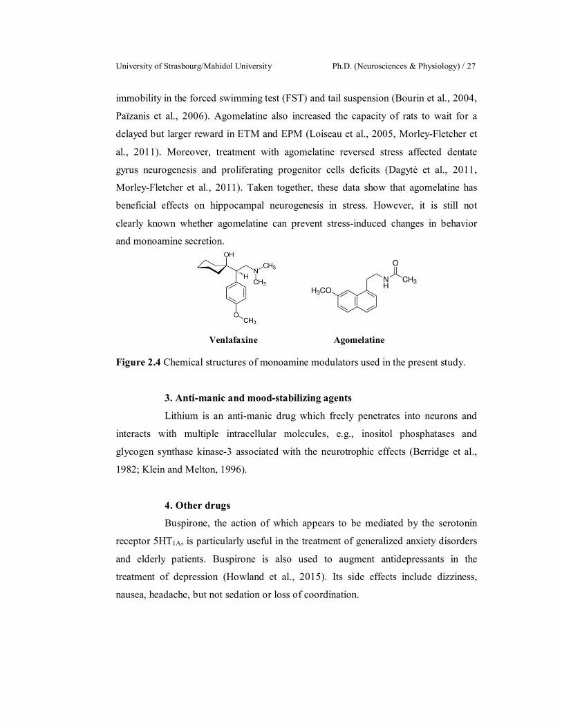
University of Strasbourg/Mahidol University Ph.D. (Neurosciences & Physiology) / 27
immobility in the forced swimming test (FST) and tail suspension (Bourin et al., 2004,
Païzanis et al., 2006). Agomelatine also increased the capacity of rats to wait for a
delayed but larger reward in ETM and EPM (Loiseau et al., 2005, Morley-Fletcher et
al., 2011). Moreover, treatment with agomelatine reversed stress affected dentate
gyrus neurogenesis and proliferating progenitor cells deficits (Dagytè et al., 2011,
Morley-Fletcher et al., 2011). Taken together, these data show that agomelatine has
beneficial effects on hippocampal neurogenesis in stress. However, it is still not
clearly known whether agomelatine can prevent stress-induced changes in behavior
and monoamine secretion.
Venlafaxine Agomelatine
Figure 2.4 Chemical structures of monoamine modulators used in the present study.
3. Anti-manic and mood-stabilizing agents
Lithium is an anti-manic drug which freely penetrates into neurons and
interacts with multiple intracellular molecules, e.g., inositol phosphatases and
glycogen synthase kinase-3 associated with the neurotrophic effects (Berridge et al.,
1982; Klein and Melton, 1996).
4. Other drugs
Buspirone, the action of which appears to be mediated by the serotonin
receptor 5HT1A, is particularly useful in the treatment of generalized anxiety disorders
and elderly patients. Buspirone is also used to augment antidepressants in the
treatment of depression (Howland et al., 2015). Its side effects include dizziness,
nausea, headache, but not sedation or loss of coordination.
Sarawut Lapmanee Literature Review / 28
2.4 Beneficial effects of exercise on stress-induced mood and memory
disorders It is generally accepted that physical activity and exercise have positive
effects on mental health reducing the incidence and severity of psychiatric disorders
such as anxiety and depression. These interventions could relieve symptoms and are as
effective as meditation or psychotherapy.
Physical exercise is cost-efficient and also provides protection against
harmful consequences of stress in healthy people and patients (Salmon, 2001). Light-
to-moderate intensity of aerobic exercise produces antidepressant-and anxiety-like
action (de Moor et al., 2006). In human studies, aerobic exercise training shows a
comparable effect when compared to antidepressant drugs or placebo in middle-age
and older adults with major depression disorder (Blumenthal et al., 2007). Carta and
co-workers (2008) showed that long term-exercise provided better physical quality of
life in depressed patients who received antidepressant and/or physical exercise as
evaluated by self-report survey questionnaire. In addition, both high and low exercise
intensities could reduce anxiety sensitivity as indicated by an anxiety sensitivity index.
On the other hand, exercise may paradoxically induce anxiety in some groups of
patients, e.g., inducing acute panic attacks or increasing the anxiety levels in patients
with panic disorder (Ströhle, 2009). The aforementioned human studies indicate the
strong effects of aerobic exercise on the emotional states, such as anxiety, depression
and mood disorders. Although, the mechanism responsible for the exercise-induced
anxiolytic-like effect is not completely understood, the improvement could result from
the released endorphins and some monoamines.
Benefits of physical exercise as an alternative to psychological
interventions have been demonstrated in several animal models. The mechanisms of
action of exercise probably involve increased expression of neurotrophic factors,
increased neurogenesis and increased monoamine neurotransmission in various brain
regions. The possible mechanism underlying of beneficial effects of physical exercise
are summarized in Figure 2.5. However, some investigators found controversial
results, presumably due to the use of different exercise programs and intensities.
Leasure and Jones (2008) reported that the increased emotional state (as indicated by
anxiety-like behaviors, defecation and increased circulating levels of corticosterone)
University of Strasbourg/Mahidol University Ph.D. (Neurosciences & Physiology) / 29
were found in forced exercising rats as compared to those subjected to wheel running.
Indeed, most exercise models, such as treadmill and swimming may be considered as
forced exercise. Swimming might induce additional stress on animals since the
swimming tank represents an inescapable environment (Charoenphandhu et al., 2011).
Therefore, voluntary exercise is better for the study of behavioral and neuroendocrine
changes.
The effects of voluntary wheel running on brain NE and its metabolites in
male rats have been previously investigated. Two major metabolites of NE, 3,4-
dihydroxyphenylglycol (DHPG) and 3-methoxy-4-hydroxyphenylglycol (MHPG), as
indicators of neuronal NE release (Scheinin et al., 1991). For example, during running,
there was an increase in NE in the cell bodies of the pons, the LC, the DR and the
spinal cord MHPG, whereas DHPG levels were unaltered in spinal cord, pons
medulla, hippocampus, and PFC, which was correlated with increased freezing in
contextual fear conditioning (Dunn et al., 1996; Dishman, 1997). The enhancement of
-1B adrenoceptor mRNA expression in DR depended on exercise duration, but not
the distance ran (Greenwood et al., 2005). Moreover, previous studies also reported
that wheel running exercise increased 5-HT release and metabolism. The effects of
exercise on the 5-HT system appeared to be highly dependent on the receptor subtypes
and brain regions investigated, and exercise effects may not be as immediate as in the
NE system (Garcia et al., 2008). The levels of 5-HIAA were reduced in the amygdala
and CA1 only after uncontrollable stress. The ratio of 5-HIAA to 5-HT was also
reduced in the PVN (Dishman et al., 1997). SERT mRNA in the MR and DRN
subregions was reduced but this effect was not correlated with the distance ran.
Furthermore, the increase in 5-HT1A receptor in the MR and in DR was dependent on
the duration of exercise (Greenwood et al., 2003, 2005). Voluntary exercise reversed
maternal deprivation induced reductions in 5-HT1A mRNA in the hippocampus
(Maniam and Morris, 2010). On other hand, 5-HT1B receptor mRNA in the DR was
decreased by exercise and this decrease was dependent on exercise length (Greenwood
et al., 2005).

Sarawut Lapmanee Literature Review / 30
Figure 2.5 Physiological effects of physical exercise: Adaptive mechanisms are
responsible for a decreased perception of chronic stress. BDNF: Brain-derived
neurotrophic factor; DA: Dopamine; HPA axis: hypothalamic-pituitary-adrenal axis;
iNOS: Inducible nitric oxide synthase; IL-1: Interleukin 1; IL-6: Interleukin 6; IL-
10: Interleukin 10; NE; Norepinephrine; ROS: Reactive oxygen species; TNF-:
Tumor necrosis factor; 5-HT: Serotonin; VEGF: Vascular endothelial growth factor
(Modified from Sanches et al., 2016).
As mentioned before, NE and 5-HT, the two classical central monoamine
neurotransmitters, can regulate synaptic plasticity, enhance neuronal survival, promote
neurogenesis and improve mental health. Therefore, NE and/or 5-HTcould be
important in the exercise-induced protective effects on the brain, and may participate
in the up-regulation of BDNF in the response to voluntary exercise.
The effects of exercise on depressive behavior and spatial performance in
stressed rats were examined by determining the alterations of corticosterone,
hippocampal BDNF mRNA expression and hippocampal GRs. Voluntary wheel
running should reverse the adverse effects of variable stressors on both behavior and
hippocampal BDNF mRNA level. Although, exercise alone increased corticosterone,
exercise did not affect the hippocampal GR mRNA expression, whereas stress showed

University of Strasbourg/Mahidol University Ph.D. (Neurosciences & Physiology) / 31
increased corticosterone and decreased hippocampal GR mRNA. These results
suggested that exercise had beneficial effect on depressive behavior that might be
associated with corticosterone and BDNF mRNA levels (Zheng et al., 2006). In
hyperactive rats, there were higher extracellular NE levels and 2-ARs but lower
MAOA activity. Wheel running activity reduced the level of depression (as indicated
by immobility time in FST) by increasing the number of 2-ARs and elevating the
extracellular NE via a reduction of MAOA (Morishima et al., 2006).
The anxiolytic effects of voluntary wheel running associated with
adaptation of mental and physiological effects on brain and behavior in laboratory
rodents and their implications to humans have been well studied (Pietropaolo et al.,
2008). After being trained with a running wheel for 4 weeks, mice spent more time in
the open arm, had a higher percentage of open arm entries in the EPM test and
increased open-field locomotion. Furthermore, chronic wheel running exercise
resulted in antidepressant-like behavior, which was associated with increased BDNF
levels (Duman et al., 2008). van Praag and co-workers (2005) reported that voluntary
wheel running enhanced the number of hippocampal DG cells and LTP, thus
improving performance of mice in the Morris water maze (MWM). This result
suggested a close relationship between neurogenesis and the hippocampal function
involved in the behavioral performance. Voluntary exercise also led to a reduction in
anxiety-like behaviors in mice, and the experimental outcome was consistent with
those measured with acoustic startle, stress-induced hyperthermia, social interaction
and light-enhanced startle (Salam et al., 2009).
Therefore, it is possible that voluntary exercise can be an alternative
treatment for mood and memory disorders in humans or animals. However, little is
known regarding the preventive effect of exercise on behavior and brain
neurochemical changes in stressed individuals.
Sarawut Lapmanee Literature Review / 32
2.5 Behavioral testing of depression, anxiety, cognition, and memory
1. Experimental models of anxiety
van der Staay (2006) grouped animal models of anxiety into the
conditioned response test or conflict test (e.g., exposure to electric foot shock,
punished drinking, defensive burying tests) and the unconditional response test (e.g.
exploration test or social test) that includes ethologically based paradigms and
involves the animal’s natural reactions such as fight, avoidance and freezing. The
behavioral paradigms are generally probed as the influencing parameter of anxiety.
The methods such as, open field can simultaneously measure locomotion, exploration,
and anxiety when exposed to an open field arena, while the EPM test is based upon the
conflict between an innate fear to exposed spaces and the elevation of the maze. These
studies are designed to indicate anxiety related behaviors that parallel anxiety behavior
in humans.
1.1 Elevated plus-maze (EPM) test
The EPM test has become one of the most popular behavioral
tests for investigations on anxiety (Hogg, 1996; Belzung and Griebel, 2001). The EPM
test is based on the conflict between an innate fear to exposed spaces and the elevation
of the maze. This test is typically used for the investigation of anxiolytic drugs. The
EPM consists of 4 arms in a crossed arrangement, where 2 open arms represent the
unsafe places and the 2 closed arms represent the safer places. Anxiety related
behaviors include increases in closed arm activity, grooming, rearing, freezing,
defecation and urination. A greater number of these behaviors indicate a greater level
of anxiety or fear. Risk assessment behaviors, e.g., stretches-attend postures-forward
elongation of the head and shoulders followed by retraction to the normal position, are
an index of a greater level of anxiety. Decreased open arm activities and increased risk
assessment behaviors in locomotion and exploration indicate increased anxiety in the
EPM test. Behavioral and pharmacological validation of the EPM has reported that
BDZs and 5-HT2A/CR antagonist (e.g., mianserin, ritanserin, and ketanserin) increase
the open arm activities (Pinheiro et al., 2007).

University of Strasbourg/Mahidol University Ph.D. (Neurosciences & Physiology) / 33
1.2. Elevated T-maze (ETM) test
The ETM test is developed to discriminate between
conditioned and unconditioned fear in animals. It is derived from the elevated X- or
plus-maze by closing the entrance to one of the enclosed arms (Graeff et al., 1993;
1998). Based on 2 strategiesavoidance of open arms when the rat is in one of the
closed arms, and escape from an open arm to enter a safer (closed arm) the
constructing ETM has been succeeded. The maze consists of 2 elevated arms that are 1
enclosed arm and 2 open with equal dimensional arms. Conditioned fear results from
inhibitory avoidance that is measured by recording the time taken to leave the
enclosed arm, whereas unconditioned fear is measured by recording the time to escape
from the open arm. An open arm seems to generate an unpleasant feeling with its
elevation and the openness of the maze, such that an animal has an innate or
unconditioned fear. This will allow the rats to learn inhibitory avoidance if frequently
placed within the enclosed arm to explore the maze. Therefore, learning is indicated
by the increase of the withdrawal latency along the trials. When the rat is placed at the
end of the enclosed arm, it does not see the open arms until it extends its head further
from the walls of the closed arm. On the other hand, when the rat is placed at the end
of one of the open arms, it can shift to the closed arm, which is like performing an
escape response. Behavioral and pharmacological validation of the ETM has reported
that TCAs (e.g., imipramine), SSRIs (e.g., sibutramine, fenfluramine, and paroxetine),
5-HT1AR partial agonist (e.g., buspirone, and isapirone), 5-HT2A/B/CR antagonist (e.g.,
SR46349B, SER082, and SB200646A) impaired inhibitory avoidance and one-way
escape latencies (Pinheiro et al., 2007).
1.3 Open field test (OFT)
The OFT test is used to determine locomotion, exploration,
and anxiety by exposure to an open area (Prut and Belzung, 2003). The number of
crossed lines and the frequency of rearing (i.e. lifting the forepaws from the floor) are
used as measures of locomotor activity. A high frequency of these behaviors indicates
increased locomotion, while the duration of time spent in the inner zone are a measure
of exploration and anxiety. A high frequency or duration of these behaviors indicates
high exploratory behavior and low anxiety levels. Behavioral and pharmacological
Sarawut Lapmanee Literature Review / 34
validation of the OFT has reported that BDZs but not SSRIs or 5-HT1AR partial
agonist enhanced time spent in the inner zone (Prut and Belzung, 2003).
1.4 Assessment of emotionality in the anxiety of the animal
In the assessment of changes in emotion, the number of fecal
pellet is related to the affective or anxiety state of the animal. An increased number of
fecal pellets are correlated with other measures of anxiety. The excretion of
corticosteroid metabolites in fecal pellets is correlated with serum corticosterone
levels. The validity of defecation as a measure of anxiety remains clear, i.e., increased
defecation indicates increased anxiety. Risk assessment behavior seems to be
defensive behavior and is indicative of anxiety when the animal faces an unknown or
threatening situation. Animals express stretched-attend postures which when occurring
a high frequency indicate a higher level of anxiety. Grooming includes licking the fur,
beginning from the muzzle, going to the ears, and down to the rest of the body.
Rearing and grooming behaviors are a displacement response and are expected to be
displayed in a novel environment (Thanos et al., 2009).
2. Experimental models of depression
Preclinical studies in experimental animals have been an important part of
the development of antidepressants. Equipments used to assess the behavior of rodents
include swimming tanks, drinking tubes and an open field apparatus. Forced
swimming test and the sucrose preference test are used to evaluate helplessness,
anhedonia, behavioral despair and other neurovegetative changes such as changes in
sleep and appetite patterns. The use of animal models for behavioral tests can reveal
depression-like behavioral profiles.
2.1 Forced swimming test (FST)
The FST is high the predictive validity models of depression
that is most commonly used to evaluate antidepressant-like action. The FST described
by Porsolt and co-workers (1979), aimed to measure the effects of antidepressant
compounds in mice. This procedure involves placing an animal in a water-filled
cylinder which it is unable to exit. At the beginning, the animal will try to escape, and
then it adopts a posture of immobility defined by a lack of movements except for
keeping the body afloat in the water. The test for rats consists of 2 swimming sessions.
University of Strasbourg/Mahidol University Ph.D. (Neurosciences & Physiology) / 35
The first session lasts 15 minutes and the second is performed 24 h after the first
session, with a test period of 5 minutes. The FST is used for the selection of new
antidepressant drugs. Various antidepressants reduce immobility time by increasing
the swimming and/or climbing time. Antidepressant drugs affecting noradrenergic
neurotransmission such as imipramine and reboxetine increase the climbing behavior,
whereas antidepressant drugs affecting serotonergic neurotransmission such as
fluoxetine, sertraline, paroxetine and citalopram increase swimming time (Castagné et
al., 2011, Lapmanee et al., 2013). The swimming time is measured by the horizontal
and vertical movements as animals try to scale the cylinder walls with their paws. The
effects of antidepressants on FST behavior are relatively specific, since they do not
increase spontaneous motor activity, unlike stimulants such as amphetamine and
cocaine (Kang et al., 2010). Besides the effects of antidepressant drugs, the FST can
also be used to evaluate the type of depressive behavior; for example, it has been
demonstrated that animals subjected to a protocol of maternal deprivation and chronic
restrain stress exhibit increased immobility time in the FST (Reus et al., 2011,
Lapmanee et al., 2013).
2.2 Sucrose preference test
The sucrose preference test is used for the assessment of
hedonic sensitivity whereby animals learn to associate an environment with a
rewarding experience. Measurement of consumption of sweetened fluids (sucrose or
saccharin) is commonly conducted for assessing stress-induced anhedonia in
depression models. Rats habituated to sucrose are typically given a choice of drinks
between sucrose and water in a two-bottle test. While control rats typically show a
preference for drinking weak sucrose solutions, rats exposed to chronic stress such as
chronic mild stress lose this preference. The effects of an antidepressant treatment
could reverse this symptom in a time dependent manner. However, the factors
affecting sucrose consumption or preference include sucrose concentration,
temperature, test duration and body weight. Anhedonic measures after chronic mild
stress are sensitive to strain effects for both rats and mice (Willner et al., 1996, Pothion
et al., 2004). In addition, Strekalova and co-workers (2011) have reported that a four
week chronic stress paradigm resulted in a decrease in the preference for sucrose

Sarawut Lapmanee Literature Review / 36
solutions by ≤ 65% in 50–70% of mice and rats without any changes at the beginning
of the stress paradigm.
3. Experimental models of learning and memory
Animal models have been widely used to study the physiology and the
cellular and molecular mechanisms of learning and memory and human cognition.
Spatial memory involves processes such as encoding, retaining and retrieving
information in the hippocampus, whereas non-spatial memory (implicit) relies on the
cerebellum and the amygdala.
3.1 Morris water maze (MWM) test
The MWM test is one of the most commonly used methods in
assessing hippocampal-dependent spatial memory in rodents (Morris et al., 1981,
1984). Spatial learning is assessed across repeated trials and reference memory is
determined by preference for the platform area when the platform is absent. The test
includes a pool of water that is made opaque, and surrounded by a wall with different
patterns at various directions. The principle of the test is that rodents can learn to swim
towards a hidden escape platform from any starting position. They do this using distal
extra-maze cues that are remote from the actual place in the pool to which the animal
is heading. Therefore, the room containing the tank should have permanently
positioned distinctive objects such as posters placed outside of the pool and/or on the
walls. Animals are expected to find the hidden platform under the water using these
visual cues. After several trials, the platform is removed, and experimenter further
measures the time spent by animals in the corrected quadrant to evaluate their spatial
memories (Paul et al., 2009).
3.2 Novel objective recognition (NOR) test
The NOR test has a large field of application not only to test
the pharmacological effects of a drug, but also to characterize which brain regions are
involved in memory and learning, as well as to understand Alzheimer’s disease (e.g.,
hippocampus and perirhinal cortex). In 1988, Ennaceur and Delacour developed the
NOR test to evaluate the differences in the exploration time of novel and familiar
objects. The NOR test is a simple behavioral assessment of memory that relies
primarily on a rodent’s innate exploratory behavior in the absence of externally
University of Strasbourg/Mahidol University Ph.D. (Neurosciences & Physiology) / 37
applied rules or reinforcement, motivation, reward, or punishment (Antunes and Biala,
2012). However, training or habituation is required. The exploration is the orientation
of animal’s snout toward the object, sniffing or touching with the snout. Running
around the object, sitting or climbing on it is not included as exploration. If animals
showed a lack of exploration activity, they are excluded from the test. The NOR test
consists of the habituation phase (exploring the empty open field arena), the
familiarization phase (exploring only one object) and finally the test phase (exploring
the familiar object and a novel one). The objects used in this test vary in shape, size,
texture, material, color and appearance. From the familiarization to the test phase,
object features change when a novel object that is different from the familiar one is
presented. The discrimination index (DI) indicates discrimination between the novel
and familiar objects as the difference in exploration time for familiar objects divided
by the total amount of exploration of the novel and familiar objects. A positive score
indicates more time spent with the novel object, a negative score indicates more time
spent with the familiar object, and a zero score indicates a no preference (Redrobe et
al., 2010; 2012; Antunes and Biala, 2012).
Sarawut Lapmanee Materials and Methods / 38
CHAPTER III
MATERIALS AND METHODS
Part I: Effects of restraint stress induction, monoamine modulators
(i.e., agomelatine and venlafaxine), and voluntary wheel running in
male rats.
3.1 Animals
Eight-week-old Wistar male rats, weighing 180–220 g, were obtained from
the National Animal Center of Thailand, Mahidol University. Rats were individually
housed in standard stainless steel shoe box at 25 ± 2 °C in a humidity-controlled room
at 55 ± 5% humidity with a 12-h (06:0018:00) light/dark cycle (average illuminance
of 200 lux). Rats were acclimatized for 1 week with food (CP Co., Ltd., Thailand) and
water available ad libitum before the start of the experiments. Body weight (BW) and
food intake were measured daily. All procedures were approved by the Animal Care
and Use Committee of the Faculty of Medicine, Thammasat University, Thailand.
3.2 Experimental protocols
Experiment 1. To examine the time-dependent effect of the 1-, 4- and 8-
week stress induction on anxiety-, depression-, and memory impairment-like
behaviors and changes in the expression of selected target proteins in the brain regions
associated with stress responses.
Rats were subdivided into 2 groups as follows.
1. Non-stress control group (16 rats/group)
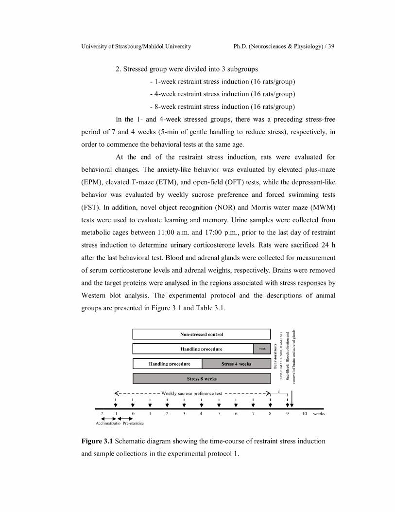
University of Strasbourg/Mahidol University Ph.D. (Neurosciences & Physiology) / 39
2. Stressed group were divided into 3 subgroups
- 1-week restraint stress induction (16 rats/group)
- 4-week restraint stress induction (16 rats/group)
- 8-week restraint stress induction (16 rats/group)
In the 1- and 4-week stressed groups, there was a preceding stress-free
period of 7 and 4 weeks (5-min of gentle handling to reduce stress), respectively, in
order to commence the behavioral tests at the same age.
At the end of the restraint stress induction, rats were evaluated for
behavioral changes. The anxiety-like behavior was evaluated by elevated plus-maze
(EPM), elevated T-maze (ETM), and open-field (OFT) tests, while the depressant-like
behavior was evaluated by weekly sucrose preference and forced swimming tests
(FST). In addition, novel object recognition (NOR) and Morris water maze (MWM)
tests were used to evaluate learning and memory. Urine samples were collected from
metabolic cages between 11:00 a.m. and 17:00 p.m., prior to the last day of restraint
stress induction to determine urinary corticosterone levels. Rats were sacrificed 24 h
after the last behavioral test. Blood and adrenal glands were collected for measurement
of serum corticosterone levels and adrenal weights, respectively. Brains were removed
and the target proteins were analysed in the regions associated with stress responses by
Western blot analysis. The experimental protocol and the descriptions of animal
groups are presented in Figure 3.1 and Table 3.1.
Figure 3.1 Schematic diagram showing the time-course of restraint stress induction
and sample collections in the experimental protocol 1.
Non-stressed control
Handling procedure 1 week
Handling procedure Stress 4 weeks
Stress 8 weeks
-2 -1 0 1 2 3 4 5 6 7 8 9 10 weeks
Acclimatizatio Pre-exercise
Beh
avio
ral t
ests
(E
PM, E
TM, O
FT, N
OR,
MW
M, F
ST)
Weekly sucrose preference test
Sacr
ifice
d: B
lood
colle
ctio
n an
d
rem
oval
of b
rain
s and
adre
nal g
land
s.

Sarawut Lapmanee Materials and Methods / 40
Table 3.1 Descriptions of animal groups in the experiment 1.
Experimental group Definition Control Rats were subjected to regular handling for 8 weeks. 1-week stress Rats were subjected to regular handling for 7 weeks and exposed to
restraint stress for 1 week. 4-week stress Rats were subjected to regular handling for 4 weeks and exposed to
restraint stress for 4 weeks. 8-week stress Rats were exposed to restraint stress for 8 weeks.
Experiment 2. To determine the effect of the 4 weeks of voluntary wheel
running on the hypothalamic-pituitary-adrenal (HPA) axis under non-stress condition
or after the 4-week exposure to restraint stress and changes in the expression of
selected target proteins in brain regions associated with stress responses.
Rats were subdivided into 2 groups as follows.
1. Non-stressed control group was divided into 2 subgroups
- Non-stressed sedentary (10 rats/group)
- Non-stressed voluntary wheel running (10 rats/group)
2. Stressed group was divided into 2 subgroups
- Stressed sedentary (10 rats/group)
- Stressed and voluntary wheel running (10 rats/group)
All rats were weekly subjected to a sucrose preference test for the
evaluation of stress-induced anhedonia. After the last stress or exercise sessions, rats
were euthanized immediately. Blood was collected in the following morning (8:00–
11:00 a.m.) for basal serum corticosterone levels by cardiac puncture. Heart and
adrenal glands were collected and weighed. Brains were removed and analyzed for
target proteins in the brain regions associated with stress responses by Western blot
analysis. Adrenal glands were also analyzed for melanocortin receptor 2 (MC2R;
ATCH receptor) protein. The experimental protocol and descriptions of animal groups
are presented in Figure 3.2 and Table 3.2.

University of Strasbourg/Mahidol University Ph.D. (Neurosciences & Physiology) / 41
Figure 3.2 Schematic diagram showing the time-course of restraint stress induction,
voluntary wheel running, and sample collections in the experimental protocol 2.
Table 3.2 Descriptions of animal groups in the experiment 2.
Experimental group Definition Sedentary control Rats were kept undisturbed and subjected to regular handling for
4 weeks. Exercise Rats were subjected to voluntary wheel running for 4 weeks. Stress control Rats were exposed to restraint stress for 4 weeks. Stress + Exercise Rats were exposed to restraint stress and subjected to voluntary
wheel running for 4 weeks.
Experiment 3. To evaluate the effectiveness of agomelatine, venlafaxine,
and voluntary wheel running exercise in the prevention of stress-induced anxiety-,
depression-, and memory impairment-like behaviors and changes in the expression of
selected target proteins in brain regions associated with stress responses.
Rats were subdivided into 2 groups as follows.
1. Monoamine modulators-treated group were divided into
3 subgroups
- Vehicle (Veh, 5 mL/kgBW, p.o., 16 rats/group)
- Agomelatine (Ago, 10 mg/kgBW, p.o., 16 rats/group)
- Venlafaxine (Vlx, 10 mg/kgBW, p.o., 16 rats/group)
2. Voluntary wheel running group (16 rats/group)
-2 -1 0 1 2 3 4 5 weeks
Acclimatization Pre-exercise
Non-stressed control
Stress
Wheel running
Stress + Wheel running
Weekly sucrose preference test
Sacrificed: Blood collection
and removal of brains and
adrenal glands.
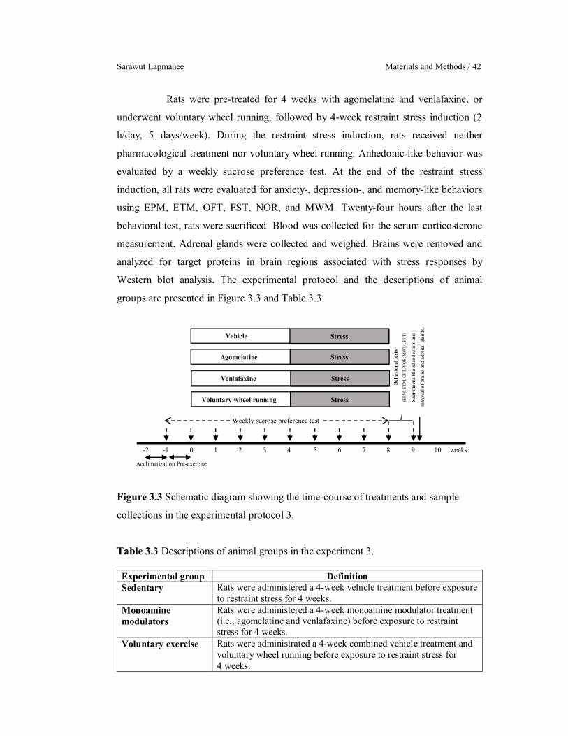
Sarawut Lapmanee Materials and Methods / 42
Rats were pre-treated for 4 weeks with agomelatine and venlafaxine, or
underwent voluntary wheel running, followed by 4-week restraint stress induction (2
h/day, 5 days/week). During the restraint stress induction, rats received neither
pharmacological treatment nor voluntary wheel running. Anhedonic-like behavior was
evaluated by a weekly sucrose preference test. At the end of the restraint stress
induction, all rats were evaluated for anxiety-, depression-, and memory-like behaviors
using EPM, ETM, OFT, FST, NOR, and MWM. Twenty-four hours after the last
behavioral test, rats were sacrificed. Blood was collected for the serum corticosterone
measurement. Adrenal glands were collected and weighed. Brains were removed and
analyzed for target proteins in brain regions associated with stress responses by
Western blot analysis. The experimental protocol and the descriptions of animal
groups are presented in Figure 3.3 and Table 3.3.
Figure 3.3 Schematic diagram showing the time-course of treatments and sample
collections in the experimental protocol 3.
Table 3.3 Descriptions of animal groups in the experiment 3.
Experimental group Definition Sedentary Rats were administered a 4-week vehicle treatment before exposure
to restraint stress for 4 weeks. Monoamine modulators
Rats were administered a 4-week monoamine modulator treatment (i.e., agomelatine and venlafaxine) before exposure to restraint stress for 4 weeks.
Voluntary exercise Rats were administrated a 4-week combined vehicle treatment and voluntary wheel running before exposure to restraint stress for 4 weeks.
Vehicle
Agomelatine Stress
Venlafaxine Stress
Voluntary wheel running
-2 -1 0 1 2 3 4 5 6 7 8 9 10 weeks
Acclimatization Pre-exercise
Beh
avio
ral t
ests
(E
PM, E
TM, O
FT, N
OR,
MW
M, F
ST)
Weekly sucrose preference test Sa
crifi
ced:
Blo
od co
llect
ion
and
rem
oval
of b
rain
s and
adre
nal g
land
s.
Stress
Stress
University of Strasbourg/Mahidol University Ph.D. (Neurosciences & Physiology) / 43
3.3 Experimental procedures
1. Restraint stress induction
The stress protocol was followed from the methods of Lapmanee et al.,
2013. Briefly, stressed rats were immobilized 2 h/day (8:0010:00 a.m.) in a quiet
room, 5 days/week for 1-, 4-, or 8-weeks. Restraint technique involved placing the rat
in a transparent plastic cylinder (24 cm long × 6 cm wide) with a 1-cm hole at the end
of the cylinder for breathing. Rat was fixed with plastic tape. The restraint procedure
was performed after 4-weeks of pharmacological treatments or voluntary wheel
running.
2. Monoamine modulator administration
Drugs were daily administrated orally by gavage between 15:00 p.m. and
16:00 p.m. every day for 4 weeks. The dosage was based on those reported previously
(Bourin et al., 2004; Lapmanee et al., 2012). Drugs used were agomelatine (10
mg/kgBw, Les Laboratoires Servier Industrie, GidyServier, Paris, France) and
venlafaxine (10 mg/kgBW, venlafaxine hydrochloride; Pfizer Ireland pharmaceuticals,
Newbridge, Co. Kildare, Ireland). Sterile saline (5 mL/kg/day) was used as vehicle.
3. Voluntary wheel running
Mild to moderate intensity aerobic voluntary exercise provides beneficial
effects on both physical and psychological health. The voluntary exercise protocol was
adapted from the methods of Droste et al., 2007 and Lapmanee et al., 2013. Rats were
individually housed in polycarbonate living chamber (50 cm long 40 cm wide 20
cm high) with a running wheel system. The running distance was calculated by the
number of turns multiplied by the circumference (144 cm) of the running wheel
(model 86060; Lafayette Instrument Company, Lafayette, IN, USA). The number of
turns was automatically recorded by an electronic counter. In preparation for wheel
running, rats were housed in cages with a running wheel for 1 week to familiarize
them with the running wheel. Exercise on the running wheel was voluntary (7
days/week for 4 weeks). Rats not completely familiarized with the running wheel were
Sarawut Lapmanee Materials and Methods / 44
excluded from the study. As for the sedentary control groups, rats remained in their
cages without a wheel for the duration of the experiment.
4. Behavioral tests
In all experiments, rats were subjected to a weekly sucrose preference test
and evaluated for anxiety-, depression-, and memory impairment-like behaviors at the
end of the restraint stress induction (8:00–12:00 a.m.). Each rat was brought to the
behavioral testing room with average illuminance of 20 lux, and left undisturbed in a
quiet environment for at least 5 min. Behavioral profiles were recorded by an infrared
video camera (model HDR-XR200E; Sony, Tokyo, Japan). The behavioral test
apparatus and the surrounding area were cleaned after each test with a wet towel and
70% ethanol to eliminate odorants, feces and urine. Each rat was subjected to one test
at a time without the presence of other rats in the room. The EPM, ETM, OFT, NOR,
MWM, and FST tests were performed, respectively as described in the following
section. All measured parameters and interpretation are noted in Table 3.4.
Table 3.4 Summarizes the parameters and interpretations of behavioral evaluations.
Test Parameter Interpretation Definition References EPM Opened arm time/entry Anxiety-like behavior Hogg, 1996; Rodgers et
al., 1997; Pandaranandaka et al., 2006
Closed arm entry Locomotor activity Total arm entry
ETM Inhibitory avoidance latency
Conditioned fear/ Generalized anxiety
Graeff et al., 1998; Pandaranandaka et al., 2009 Escape latency Unconditioned fear/
Panic-like symptom OFT Inner zone time Anxiety-like behavior Treit and Fundytus,
1988; McCarthy et al., 1995; Pandaranandaka et al., 2009
The first 30 s line crossings
fear reaction-thigmotaxis, hyperarousal
Total line crossings Locomotor activity NOR Discrimination ratio Cognitive and memory
impairment Antunes and Biala, 2012; Redrobe et al., 2010; 2012
MWM Escape latency Time reach to correct quadrant
Spatial learning Spatial memory
Morris et al., 1981, 1984
EPM: elevated plus-maze; ETM: elevated T- maze; NOR: novel objective recognition;
MWM: Morris water maze; OFT: open-field test.
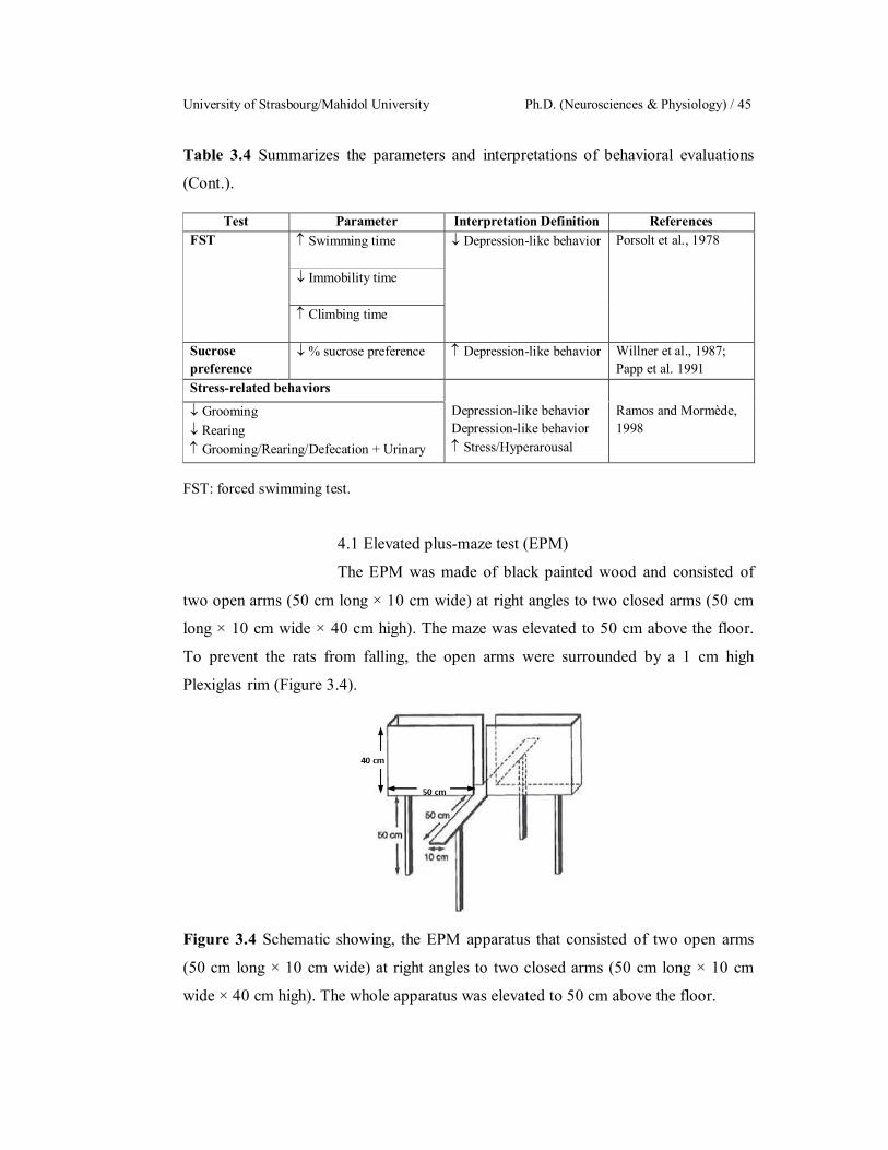
University of Strasbourg/Mahidol University Ph.D. (Neurosciences & Physiology) / 45
Table 3.4 Summarizes the parameters and interpretations of behavioral evaluations
(Cont.).
Test Parameter Interpretation Definition References FST Swimming time Depression-like behavior Porsolt et al., 1978
Immobility time
Climbing time
Sucrose preference
% sucrose preference Depression-like behavior Willner et al., 1987; Papp et al. 1991
Stress-related behaviors
Grooming Rearing Grooming/Rearing/Defecation + Urinary
Depression-like behavior Depression-like behavior Stress/Hyperarousal
Ramos and Mormède, 1998
FST: forced swimming test.
4.1 Elevated plus-maze test (EPM)
The EPM was made of black painted wood and consisted of
two open arms (50 cm long × 10 cm wide) at right angles to two closed arms (50 cm
long × 10 cm wide × 40 cm high). The maze was elevated to 50 cm above the floor.
To prevent the rats from falling, the open arms were surrounded by a 1 cm high
Plexiglas rim (Figure 3.4).
Figure 3.4 Schematic showing, the EPM apparatus that consisted of two open arms
(50 cm long × 10 cm wide) at right angles to two closed arms (50 cm long × 10 cm
wide × 40 cm high). The whole apparatus was elevated to 50 cm above the floor.
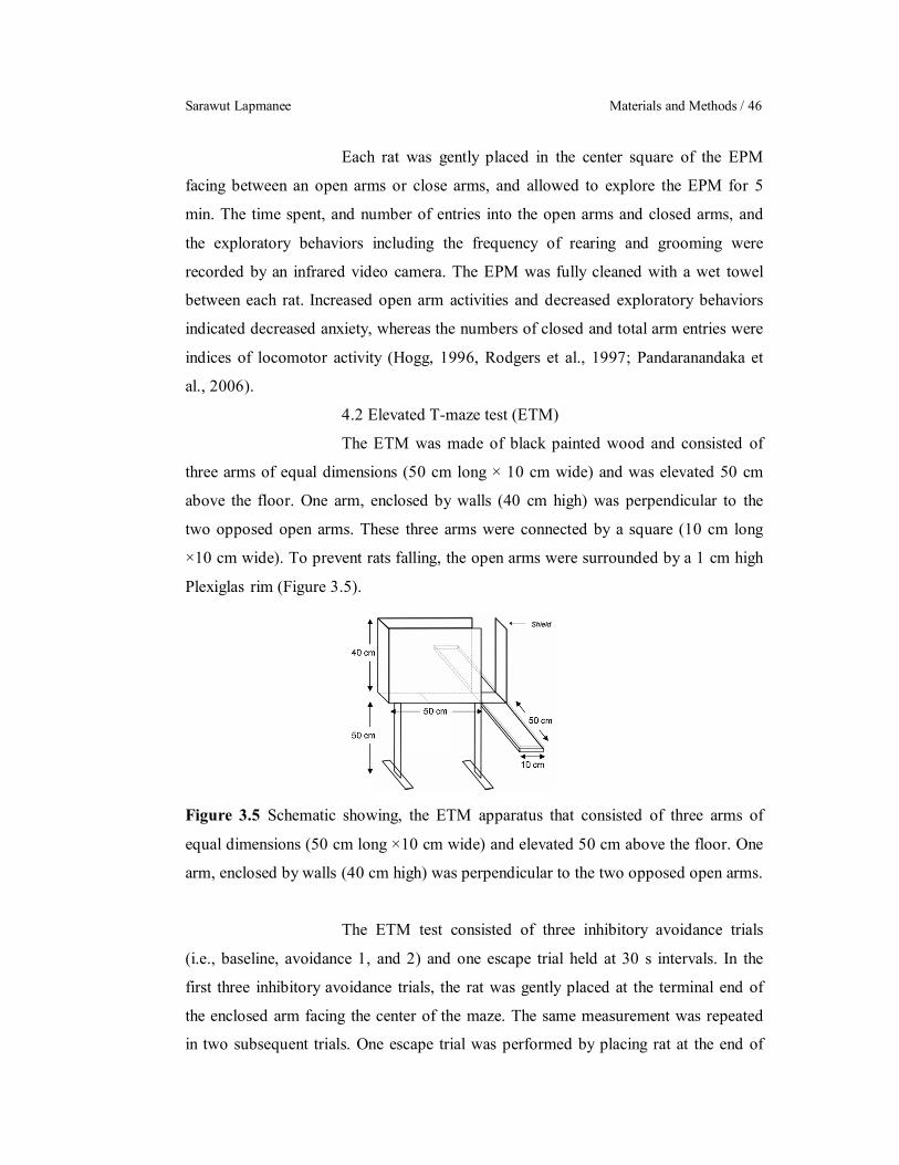
Sarawut Lapmanee Materials and Methods / 46
Each rat was gently placed in the center square of the EPM
facing between an open arms or close arms, and allowed to explore the EPM for 5
min. The time spent, and number of entries into the open arms and closed arms, and
the exploratory behaviors including the frequency of rearing and grooming were
recorded by an infrared video camera. The EPM was fully cleaned with a wet towel
between each rat. Increased open arm activities and decreased exploratory behaviors
indicated decreased anxiety, whereas the numbers of closed and total arm entries were
indices of locomotor activity (Hogg, 1996, Rodgers et al., 1997; Pandaranandaka et
al., 2006).
4.2 Elevated T-maze test (ETM)
The ETM was made of black painted wood and consisted of
three arms of equal dimensions (50 cm long × 10 cm wide) and was elevated 50 cm
above the floor. One arm, enclosed by walls (40 cm high) was perpendicular to the
two opposed open arms. These three arms were connected by a square (10 cm long
×10 cm wide). To prevent rats falling, the open arms were surrounded by a 1 cm high
Plexiglas rim (Figure 3.5).
Figure 3.5 Schematic showing, the ETM apparatus that consisted of three arms of
equal dimensions (50 cm long ×10 cm wide) and elevated 50 cm above the floor. One
arm, enclosed by walls (40 cm high) was perpendicular to the two opposed open arms.
The ETM test consisted of three inhibitory avoidance trials
(i.e., baseline, avoidance 1, and 2) and one escape trial held at 30 s intervals. In the
first three inhibitory avoidance trials, the rat was gently placed at the terminal end of
the enclosed arm facing the center of the maze. The same measurement was repeated
in two subsequent trials. One escape trial was performed by placing rat at the end of

University of Strasbourg/Mahidol University Ph.D. (Neurosciences & Physiology) / 47
the right open arm facing the center of the maze and recording the latent time the rats
took to exit this arm with all four paws. The ETM was fully cleaned with a wet towel
after each test. Increased inhibitory avoidance latency represented learned or
conditioned fear, whereas one-way escape latency represented innate or unconditioned
fear (Graeff et al., 1998; Pandaranandaka et al., 2009).
4.3 Open-field test (OFT)
The OFT was used to evaluate both locomotor activity and
anxiety-like behavior. The apparatus was made of black painted wood (76 cm long ×
57 cm wide × 35 cm high) with a 48-square grid floor (6 × 8 squares, 9.5 cm per side).
The arena was divided into two zones, i.e., inner and outer zones (24 peripheral
squares) as shown in Figure 3.6.
A.
B.
Figure 3.6 Schematic showing, A. The OFT was made from a black painted wooden
box (76 cm long × 57 cm wide × 35 cm high) with a 48 square grid floor (6 × 8
squares, 9.5 cm per side). B. The arena of maze was divided into two zones, i.e., inner
(white area) and outer zones (gray area).
Each rat was subjected to OFT after EPM and ETM tests. The
rat was gently placed in one of the four corner squares facing the wall and allowed 5
min to explore the apparatus. The time spent in each zone, the number of lines crossed
in the 1st 30-s and 5-min test, and numbers of rearing, grooming, and fecal pellets
were recorded by an infrared video camera. The OFT was fully cleaned with a wet

Sarawut Lapmanee Materials and Methods / 48
towel between tests. An increase in time spent in the inner zone or a decrease in time
spent in the outer zone indicated anxiolysis or increased exploration. A change in the
number of lines crossed represented change in locomotor activity (McCarthy et al.,
1995; Treit and Fundytus, 1988; Pandaranandaka et al., 2009).
4.4 Novel object recognition test (NOR)
NOR was performed in a black rectangular plastic box (63 cm
long × 63 cm wide × 45 cm high) in 360 lux room light. The procedure was modified
from the method of Redrobe et al., 2010; 2012. A video camera was installed on a
movable trolley above the box to record behaviors. The objects to be discriminated
were made of glass or ceramic. On the day before the NOR test, each rat was allowed
to habituate the empty box (2 sessions, 10 min/session). During the test, each rat was
gently placed in the box and exposed for 3 min to an acquisition session with identical
objects (two ceramic pepper bottles; 3 cm long × 3 cm wide × 7 cm high) placed,
approximately 10 cm apart at the center of the box. Rats were then transferred to their
home cages for a 1 h inter-trial interval. Meanwhile, the box and objects were cleaned.
One object in the box was replaced with a novel object (glass paperweight; 5 cm long
× 5 cm wide × 12 cm high). The same rat was then returned to the box and allowed to
explore the new object for 3 min. Object-exploring behaviors included sniffing,
licking, or touching each object. Rats that did not reach a total exploration time of 15
s, after exploring both objects for a minimum of 2 s, were excluded from the data
analysis. The discrimination ratio was calculated from the following equation:
Time exploring novel object (s) – Time exploring familiar object (s)
Total exploration time (s)
A decrease in discrimination ratio indicated cognitive and
memory impairment in stressed rats (Redrobe et al., 2010, 2012; Antunes and Biala,
2012;).
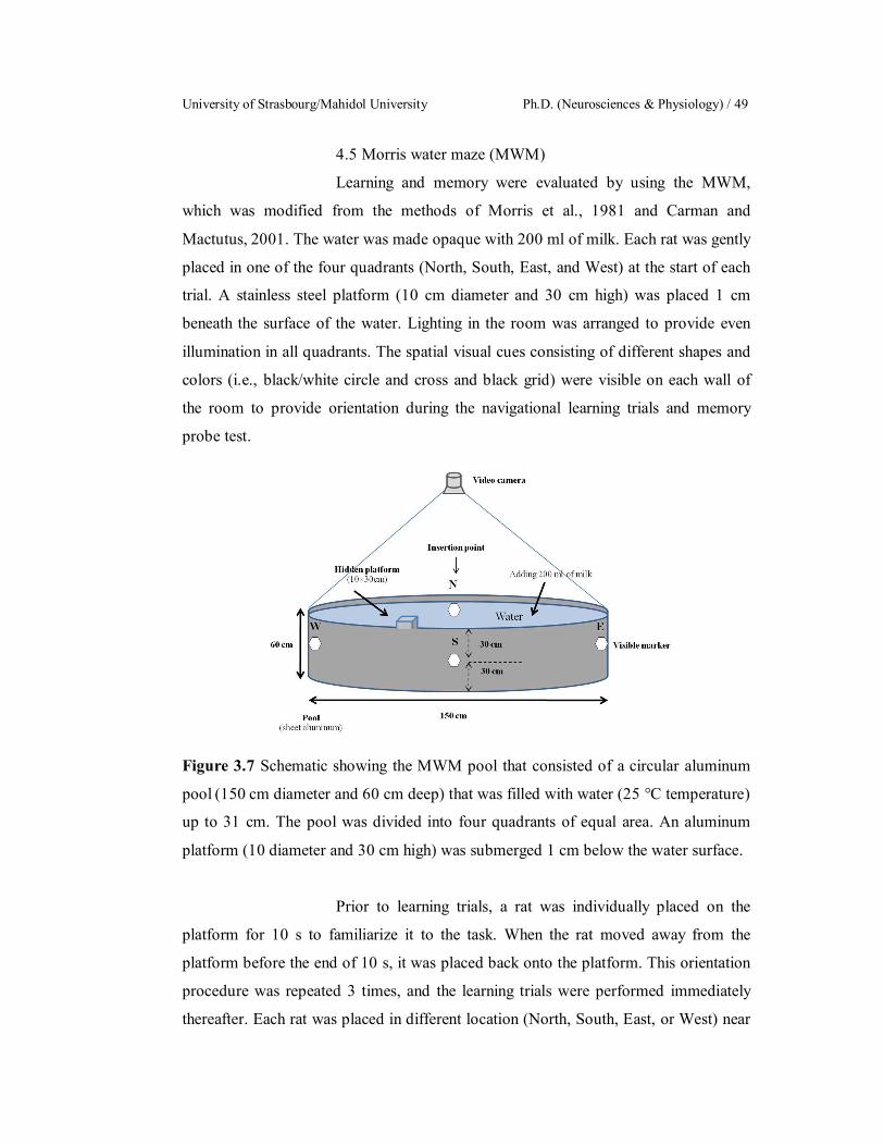
University of Strasbourg/Mahidol University Ph.D. (Neurosciences & Physiology) / 49
4.5 Morris water maze (MWM)
Learning and memory were evaluated by using the MWM,
which was modified from the methods of Morris et al., 1981 and Carman and
Mactutus, 2001. The water was made opaque with 200 ml of milk. Each rat was gently
placed in one of the four quadrants (North, South, East, and West) at the start of each
trial. A stainless steel platform (10 cm diameter and 30 cm high) was placed 1 cm
beneath the surface of the water. Lighting in the room was arranged to provide even
illumination in all quadrants. The spatial visual cues consisting of different shapes and
colors (i.e., black/white circle and cross and black grid) were visible on each wall of
the room to provide orientation during the navigational learning trials and memory
probe test.
Figure 3.7 Schematic showing the MWM pool that consisted of a circular aluminum
pool (150 cm diameter and 60 cm deep) that was filled with water (25 °C temperature)
up to 31 cm. The pool was divided into four quadrants of equal area. An aluminum
platform (10 diameter and 30 cm high) was submerged 1 cm below the water surface.
Prior to learning trials, a rat was individually placed on the
platform for 10 s to familiarize it to the task. When the rat moved away from the
platform before the end of 10 s, it was placed back onto the platform. This orientation
procedure was repeated 3 times, and the learning trials were performed immediately
thereafter. Each rat was placed in different location (North, South, East, or West) near
Sarawut Lapmanee Materials and Methods / 50
the edge of the pool with its front paws first touching the wall. Then, it was allowed to
find the hidden platform, and time spent for locating the platform (escape latency) was
recorded. The hidden platform was placed at a fixed location, i.e., in the Southeast
quadrant. When the rat was unable to locate the platform within 60 s, it was placed on
the platform and an escape latency of 60 s was recorded. Finally, the rat was given 10
s to remain on the platform to familiarize itself with the surrounding visual cues. Rats
performed 8 trials on day 1 and 2, and 4 trials on day 3 (total 20 trials). There was a 5-
min inter-trial interval, during which each rat was kept warm in a cage under a heating
lamp. Spatial memory was evaluated by a probe test 1 h after the last learning trial on
day 3. The rat was individually placed in the North quadrant in the absence of a
platform. Each rat was allowed to swim for 60 s, and time spent in each quadrant was
recorded by a video camera. Percentage time spent in the correct quadrant was
calculated as follows.
( )
( ) 100
An increase in escape latency indicated poor spatial learning,
whereas an increase in correct quadrant time (i.e., time to reach the correct quadrant)
or a decrease in percentage time spent in the correct quadrant indicated spatial
memory impairment. To eliminate confounding factors (e.g., urine, fecal pellets, stress
odors, and pheromones), the pool was drained and refilled with fresh tap water before
the training of each group of rats. Finally, all learning trials and memory probes were
recorded by an experimenter unaware of the grouping of the animals.
4.6 Forced swimming test (FST)
All rats were individually forced to swim in a cylindrical
container (25 cm diameter and 45 cm high) filled with 25 C tap water up to 35 cm. In
the first session, rats were placed in water for a 15-min assessment. Then, they were
removed from water, dried and cleaned with towel before returning to their cages.
Twenty-four hours later, the second session was performed in which rats were put
back into the swimming tank for 5 min. Duration of immobility behavior (floating in
water with only movement necessary to keep the head above water), swimming
University of Strasbourg/Mahidol University Ph.D. (Neurosciences & Physiology) / 51
behavior (active movement of the forepaws with goal-directed horizontal actions, such
as crossing between quadrants of the cylinder and turning), climbing (upward goal-
directed movements of the forepaws along the side of the cylindrical container), and
number of fecal pellets were recorded. An increase in immobility duration or
decreases in swimming duration and climbing duration indicated depression-like
behaviors in rats (Porsolt et al., 1979).
4.7. Sucrose preference test
The sucrose preference test was modified from the methods of
Charoenphandhu et al., 2013 and performed once weekly throughout the experimental
periods. To confirm that sucrose neophobia was not a confounding factor in this
behavioral test (Strekalova and Steinbusch, 2010), rats were given free access to the
sucrose preference test prior to the start of the experiment to make sure that baseline
sucrose intake did not differ among groups. For the determination of sucrose
preference, rats were deprived of water and food for 4 h (08:00–12:00 h). Then rats
were presented for 1 h with two bottles, one containing water and the other containing
2% sucrose with the density of 1.0098 g/cm3 (Ajax Finechem, NSW, Australia). The
volumes of liquid were measured by weighing pre-weighed bottles before and after
testing. To prevent possible effects of side-preference in drinking behavior, the
position of the bottle in the cage was switched weekly. The amount of water and
sucrose intake was measured. The relative sucrose intake per body weight (BW)
(g/kgBW) was the absolute sucrose intake per gram of rat body weight, whereas
sucrose preference was calculated according to the following ratio:
( )
( ) ( ) 100
Decreased sucrose preference represents anhedonia-like
behavior in rodents (Willner et al., 1987; Papp et al., 1991).
5. Measurement of body, organ weights, and food intake
Body weights (BW), food intake and water intake were measured daily.
Pre-weighed food or water was provided and 24 h later the amount of remaining food
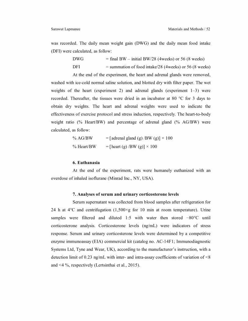
Sarawut Lapmanee Materials and Methods / 52
was recorded. The daily mean weight gain (DWG) and the daily mean food intake
(DFI) were calculated, as follow:
DWG = final BW – initial BW/28 (4weeks) or 56 (8 weeks)
DFI = summation of food intake/28 (4weeks) or 56 (8 weeks)
At the end of the experiment, the heart and adrenal glands were removed,
washed with ice-cold normal saline solution, and blotted dry with filter paper. The wet
weights of the heart (experiment 2) and adrenal glands (experiment 1–3) were
recorded. Thereafter, the tissues were dried in an incubator at 80 °C for 3 days to
obtain dry weights. The heart and adrenal weights were used to indicate the
effectiveness of exercise protocol and stress induction, respectively. The heart-to-body
weight ratio (% Heart/BW) and percentage of adrenal gland (% AG/BW) were
calculated, as follow:
% AG/BW = adrenal gland (g) /BW (g) × 100
% Heart/BW = heart (g) /BW (g) × 100
6. Euthanasia
At the end of the experiment, rats were humanely euthanized with an
overdose of inhaled isoflurane (Minrad Inc., NY, USA).
7. Analyses of serum and urinary corticosterone levels
Serum supernatant was collected from blood samples after refrigeration for
24 h at 4°C and centrifugation (1,500×g for 10 min at room temperature). Urine
samples were filtered and diluted 1:5 with water then stored −80°C until
corticosterone analysis. Corticosterone levels (ng/mL) were indicators of stress
response. Serum and urinary corticosterone levels were determined by a competitive
enzyme immunoassay (EIA) commercial kit (catalog no. AC-14F1; Immunodiagnostic
Systems Ltd, Tyne and Wear, UK), according to the manufacturer’s instruction, with a
detection limit of 0.23 ng/mL with inter- and intra-assay coefficients of variation of <8
and <4 %, respectively (Lertsinthai et al., 2015).

University of Strasbourg/Mahidol University Ph.D. (Neurosciences & Physiology) / 53
8. Brain dissection
Brain was rapidly removed after decapitation, frozen in liquid nitrogen,
and stored at –80 C. On the analysis day, the brain regions associated with stress
responses (i.e., frontal cortex, septum, amygdala, hippocampus, periaqueductal gray,
dorsal raphe, locus coeruleus, and ventral tegmental area) were microscopically
dissected according to the methods of Heffner et al., 1980 and the atlas of Paxinos and
Watson, 2005 (Figure 3.83.9). The isolation procedure was done on an ice-cold plate.
Razor blades were carefully inserted through the cutting channels slicing the brain at
right angles to the sagittal axis. The initial razor blade sliced through the coronal plane
of the brain at the level of the anterior commissure. The position of the initial razor
blade served as a reference point other brain sections. A total of eight razor blades
were inserted anteriorly or posteriorly to the first blade and thus divided the brains into
8 sections. The razor blades were removed from the block leaving coronal brain slices
adhere to their surfaces. Brain sections were placed on a glass plate on ice. Brain
regions were then bilaterally dissected from these slices. Besides the frontal cortex,
hippocampus, and amygdala, two independent samples from relatively tiny brain areas
(i.e., septum, periaqueductal gray, dorsal raphe, locus coeruleus, and ventral
tegmental) were pooled together before protein extraction (Charoenphandhu et al.,
2013).
9. Western blot analysis
The brain regions under study were separately lysed and homogenized with
a handheld pestle and mortar in the radioimmunoprecipitation assay (RIPA) buffer
containing 150 mM NaCl, 1.0% IGEPAL® CA-630, 0.5% sodium deoxycholate, 0.1%
sodium dodecyl sulfate (SDS), 50 mM Tris, pH 8.0 supplemented with protease
inhibitors and a phosphatase inhibitor cocktail (sodium pyrophosphate and sodium
orthovanadate) (Sigma, St. Louis, MO, USA). Therefore, membrane-bound proteins
should thus be in the supernatant. Brain tissues were then sonicated at power 6 for 25-s
pulses (60-s pause between pulses) at 4°C and centrifuged (13,000 rpm for 15 min) at
4 °C.
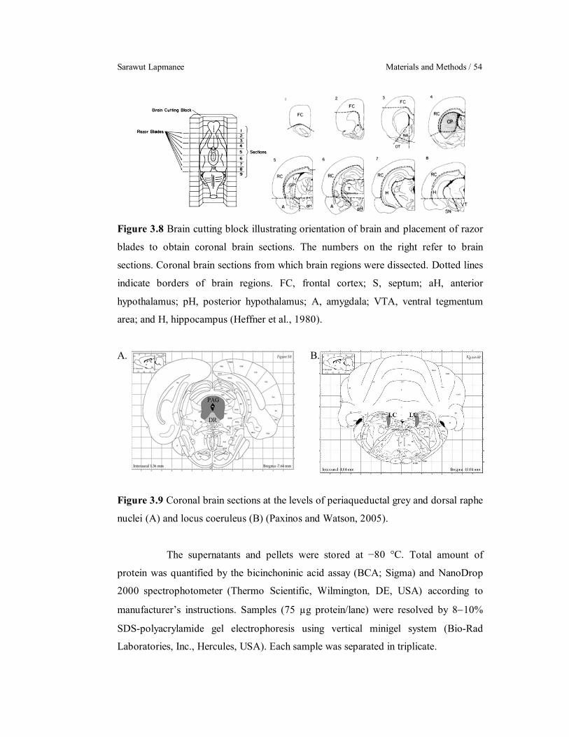
Sarawut Lapmanee Materials and Methods / 54
Figure 3.8 Brain cutting block illustrating orientation of brain and placement of razor
blades to obtain coronal brain sections. The numbers on the right refer to brain
sections. Coronal brain sections from which brain regions were dissected. Dotted lines
indicate borders of brain regions. FC, frontal cortex; S, septum; aH, anterior
hypothalamus; pH, posterior hypothalamus; A, amygdala; VTA, ventral tegmentum
area; and H, hippocampus (Heffner et al., 1980).
A. B.
Figure 3.9 Coronal brain sections at the levels of periaqueductal grey and dorsal raphe
nuclei (A) and locus coeruleus (B) (Paxinos and Watson, 2005).
The supernatants and pellets were stored at −80 °C. Total amount of
protein was quantified by the bicinchoninic acid assay (BCA; Sigma) and NanoDrop
2000 spectrophotometer (Thermo Scientific, Wilmington, DE, USA) according to
manufacturer’s instructions. Samples (75 µg protein/lane) were resolved by 810%
SDS-polyacrylamide gel electrophoresis using vertical minigel system (Bio-Rad
Laboratories, Inc., Hercules, USA). Each sample was separated in triplicate.
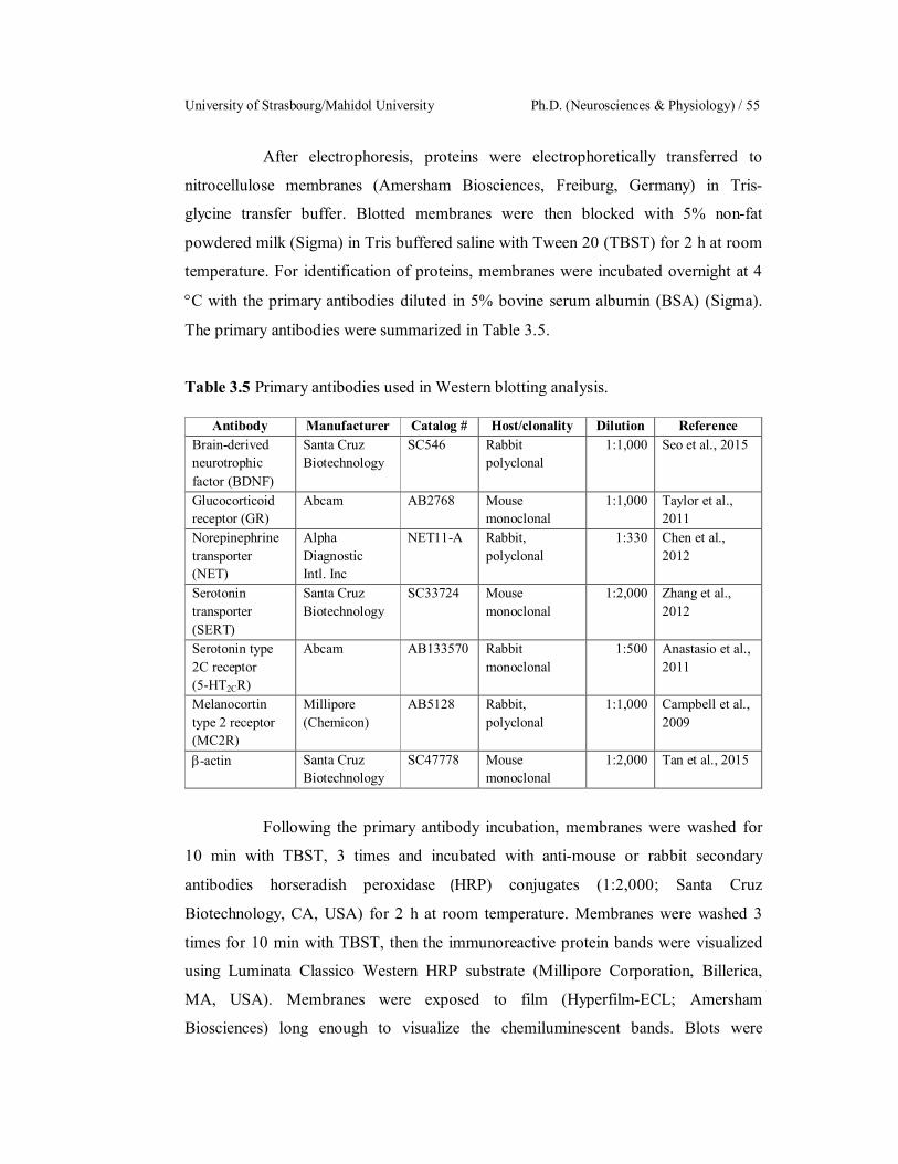
University of Strasbourg/Mahidol University Ph.D. (Neurosciences & Physiology) / 55
After electrophoresis, proteins were electrophoretically transferred to
nitrocellulose membranes (Amersham Biosciences, Freiburg, Germany) in Tris-
glycine transfer buffer. Blotted membranes were then blocked with 5% non-fat
powdered milk (Sigma) in Tris buffered saline with Tween 20 (TBST) for 2 h at room
temperature. For identification of proteins, membranes were incubated overnight at 4
C with the primary antibodies diluted in 5% bovine serum albumin (BSA) (Sigma).
The primary antibodies were summarized in Table 3.5.
Table 3.5 Primary antibodies used in Western blotting analysis.
Antibody Manufacturer Catalog # Host/clonality Dilution Reference Brain-derived neurotrophic factor (BDNF)
Santa Cruz Biotechnology
SC546 Rabbit polyclonal
1:1,000 Seo et al., 2015
Glucocorticoid receptor (GR)
Abcam AB2768 Mouse monoclonal
1:1,000 Taylor et al., 2011
Norepinephrine transporter (NET)
Alpha Diagnostic Intl. Inc
NET11-A Rabbit, polyclonal
1:330 Chen et al., 2012
Serotonin transporter (SERT)
Santa Cruz Biotechnology
SC33724 Mouse monoclonal
1:2,000 Zhang et al., 2012
Serotonin type 2C receptor (5-HT2CR)
Abcam AB133570 Rabbit monoclonal
1:500 Anastasio et al., 2011
Melanocortin type 2 receptor (MC2R)
Millipore (Chemicon)
AB5128 Rabbit, polyclonal
1:1,000 Campbell et al., 2009
-actin Santa Cruz Biotechnology
SC47778 Mouse monoclonal
1:2,000 Tan et al., 2015
Following the primary antibody incubation, membranes were washed for
10 min with TBST, 3 times and incubated with anti-mouse or rabbit secondary
antibodies horseradish peroxidase (HRP) conjugates (1:2,000; Santa Cruz
Biotechnology, CA, USA) for 2 h at room temperature. Membranes were washed 3
times for 10 min with TBST, then the immunoreactive protein bands were visualized
using Luminata Classico Western HRP substrate (Millipore Corporation, Billerica,
MA, USA). Membranes were exposed to film (Hyperfilm-ECL; Amersham
Biosciences) long enough to visualize the chemiluminescent bands. Blots were
Sarawut Lapmanee Materials and Methods / 56
reprobed with mouse monoclonal anti-β-actin (1:2,000; Santa Cruz) and normalized to
verify equivalent protein loading. Comparisons were made with known molecular
weight standards using prestained protein ladder (Thermo Scientific, Walthem,
MA, USA). Blots were scanned and band density was quantified using ImageJ
(Rasband, 19972016). The area of the curve in each gel band were quantified and
normalized relative to the β-actin levels of control or vehicle-treated groups.
3.4 Statistical analysis Results were expressed as means ± SE. Statistical analyses for
comparisons of two sets of data were performed by unpaired Student’s t-test. Multiple
comparisons were made by one-way analysis of variance (ANOVA) with Dunnett
post-test. The t-value, F-value and degree of freedom (df) values are presented. The
level of significance for all statistical tests was P < 0.05. Data were analyzed by
GraphPad Prism 6.0 for Windows XP (GraphPad Software, San Diego, CA, USA).

University of Strasbourg/Mahidol University Ph.D. (Neurosciences & Physiology) / 57
Part II: Characterization of melatonin receptor expressing cells in the
rodent brain using MT1 and MT2 melatonin receptor LacZ knock-in
reporter mice: Identification of potential action sites of agomelatine
on brain structures involved in stress.
3.5 Animals
Adult male and female wild-type and transgenic mice with knocked-in β-
galactosidase (LacZ) reporter into melatonin (MT) receptor 1A (MTNR1A, MT1) and
1B (MTNR1B, MT2) gene locus of MT-proficient C3H mice and MT-deficient
C57BL/6 mice (1.518 months, 2060 g) were obtained from the Institute of Cellular
and Integrative Neurosciences, University of Strasbourg, France. Mice were housed in
the control room of the conventional zone of the Chronobiotron, UMS3415 with
12:12-h light:dark cycle with food and water ad libitum. All efforts were made to
minimize the number of animals used. Experimental procedures were approved by the
Animal Care and Use Committee of Institute of Cellular and Integrative
Neurosciences, University of Strasbourg, France (Figure 3.10).
3.6 Experimental protocols
Experiment 1. To validate and map MT1/MT2-LacZ expressing neurons
in transgenic knock-in mice.
Both MT1 and MT2 transgenic knock-in mice had their genotypes
confirmed by polymerase chain reaction (PCR) technique. Wild-type and transgenic
mice of different age groups were euthanized by CO2 inhalation and intracardially
perfused with fixative solutions. The free-floating sections were stained either for
immunocytochemistry or LacZ enzymatic activity detection.
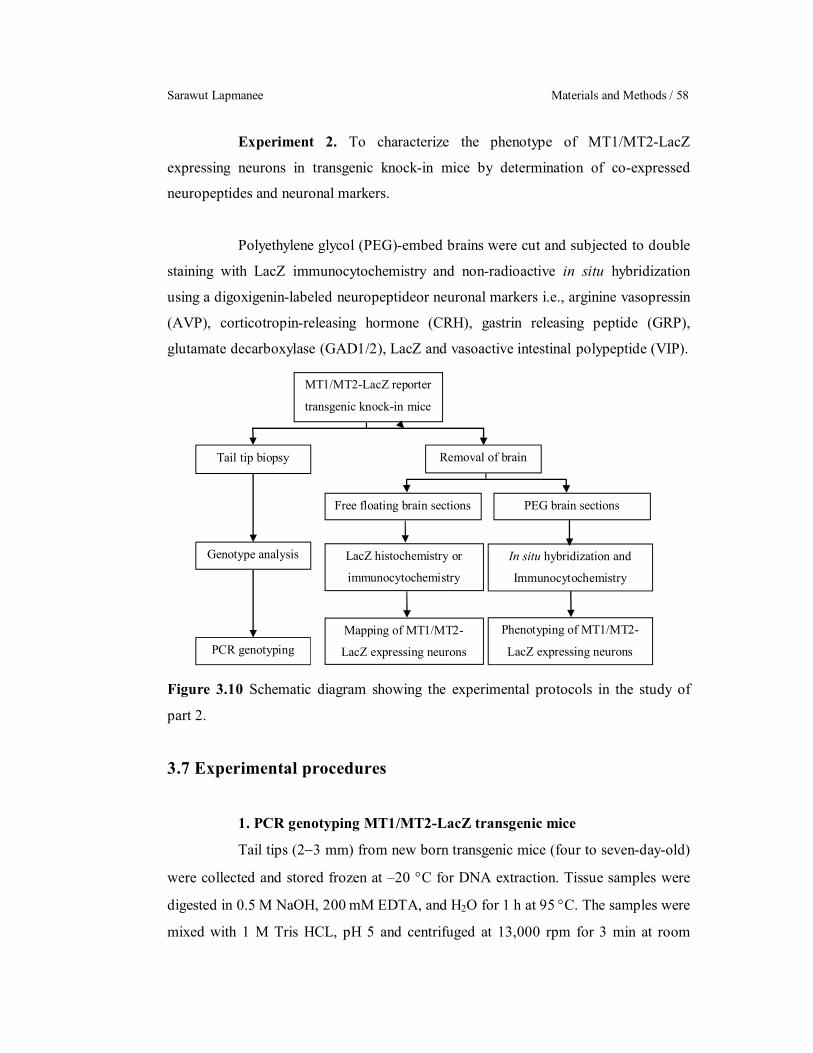
Sarawut Lapmanee Materials and Methods / 58
MT1/MT2-LacZ reporter
transgenic knock-in mice
Removal of brain Tail tip biopsy
LacZ histochemistry or
immunocytochemistry
Mapping of MT1/MT2-
LacZ expressing neurons
Genotype analysis
Free floating brain sections PEG brain sections
In situ hybridization and
Immunocytochemistry
Phenotyping of MT1/MT2-
LacZ expressing neurons PCR genotyping
Experiment 2. To characterize the phenotype of MT1/MT2-LacZ
expressing neurons in transgenic knock-in mice by determination of co-expressed
neuropeptides and neuronal markers.
Polyethylene glycol (PEG)-embed brains were cut and subjected to double
staining with LacZ immunocytochemistry and non-radioactive in situ hybridization
using a digoxigenin-labeled neuropeptideor neuronal markers i.e., arginine vasopressin
(AVP), corticotropin-releasing hormone (CRH), gastrin releasing peptide (GRP),
glutamate decarboxylase (GAD1/2), LacZ and vasoactive intestinal polypeptide (VIP).
Figure 3.10 Schematic diagram showing the experimental protocols in the study of
part 2.
3.7 Experimental procedures
1. PCR genotyping MT1/MT2-LacZ transgenic mice
Tail tips (23 mm) from new born transgenic mice (four to seven-day-old)
were collected and stored frozen at –20 C for DNA extraction. Tissue samples were
digested in 0.5 M NaOH, 200 mM EDTA, and H2O for 1 h at 95 C. The samples were
mixed with 1 M Tris HCL, pH 5 and centrifuged at 13,000 rpm for 3 min at room

University of Strasbourg/Mahidol University Ph.D. (Neurosciences & Physiology) / 59
temperature, and the supernatants were used for PCR genotyping. One (1) µL
supernatant was transferred to 15-µL PCR reaction mixture containing 1.5 µl Taq
DNA polymerase 10X buffer without MgCl2 and detergent (Euromedex,
Souffelweyersheim, France), 1.5 µl 25 mM MgCl2, 0.75 µl DMSO (Hybri-Max,
Sigma D2650), 0.5 µl 10 mM dNTP, 0.25 µl 50 µM forward primers, 0.25 µl 50 µM
reverse primers, 0.2 µl Taq DNA polymerase (Euromedex), and 9.05 µl molecular
biology grade H2O. The corresponding primers are shown in Table 3.6.
Table 3.6 The MT1-/MT2-LacZ genotyping and primer designs.
Genotyping Primer sequence 5’3’ (Forward/Reverse) MT1-LacZ
Wild-type CAAGTTGCTGGGCAGTGGACAGCAG CTTCTTGTTGCGGTACACAGACAGG
Knock-in CTAGTCTGTTCAGCTGTGTCACACC GCGGGAGGGCCATAAAAAGTGGC
MT2-LacZ
Wild-type AAGCACCAGCAATTTCCCATTCTCC TCTTGCCTGAGGAAGGTCAGAATCC
Knock-in ACTTGCTTTAAAAAACCTCCCACA ATGGGTCCCTGGAAGTCACTCACC
Each PCR reaction was performed as follows: cycling touchdown PCR
program, 94 °C for a 5-min initial strand separation, 94 °C for 30 s, 58°C for 30 s, at
72 °C for 45 min (repeat for a total of 35 cycles) , and a 5-min final elongation step at
72°C. Five µl of each PCR product and mass ruler DNA ladder Mix (Euromedex) was
electrophoresed through a 1.5% agarose gel in Tris-borate-EDTA buffer (TBE) that
was then stained with 1g/ml ethidium bromide. DNA fragments were visualized
under ultraviolet light and photographed (Sony camera). DNA fragment phenotypes
were identified by 425 bp (wild-type) or 196 bp (MT1-LacZ transgene), whereas
MT2-LacZ DNA fragment phenotypes were identified by 143 bp (wild-type) or 666 bp
(MT2-LacZ transgene).
2. Euthanasia
Mice were euthanized during the light phase (9.0012.00 a.m.) by CO2
inhalation. Heparin (500 IU, Héparine Choay; Sanofi, Gentilly, France) was injected
into the heart before transcardial perfusion first with PBS, and then with fixative
Sarawut Lapmanee Materials and Methods / 60
solution (4% phosphate-buffered formaldehyde for LacZ histochemistry or periodate-
lysine-paraformaldehyde (PLP) fixative for immunocytochemistry and in situ
hybridization).
3. Preparation of brain tissues
For LacZ histochemistry, the brains were removed from the skulls and
post-fixed with 4% paraformaldehyde in 100 mM phosphate buffer, pH 7.4,
cryoprotected in 30% sucrose solution at 4 C for at least 24 hours. After sinking in
the 30% sucrose cryoprotectant, the brains were frozen in isopentane, chilled to 78°C
with mixed dry ice and 95% ethanol. The brains which were cut on a cryo-microtome
(Microm HM 560, Thermo Scientific) into 40-m-thick slices. Free floating sections
were collected in PBS with 0.1% sodium azide (PBS-azide) and processed for
detection of LacZ activity by X-gal staining.
For immunohistochemistry and in situ hybridization, we used PLP fixative
(4% formaldehyde, 10 mM NaIO4 in 75 mM phosphate-lysine buffer, pH 7.4). After
transcardial perfusion, the brains were removed from the skulls and post-fixed in PLP
fixative for 10 hours. Then the brains were rinsed in 50% ethanol and dehydrated with
ethanol (70% for 2 times 1 hour, 95% for 2 times 1 hour), ethoxyethanol for 2 times 1
hour and finally n-butanol overnight. Thereafter, brains were embedded in PEG as
described by Klosen et al., 1993. PEG-embedded brains were cut by microtome
(Leica, Germany) into 8-μm-thick coronal sections and mounted onto slides.
4. LacZ histochemistry
The expression of the LacZ reporter can be detected by enzyme
histochemical staining using the chromogenic substrate, 5-bromo-4-chloro-3-indolyl-
β-d-galactoside (X-gal). The free-floating sections were washed for 3 times 15 min
each in LacZ wash buffer (PBS, 2 mM MgCl2, 0.01% sodium deoxycholate, 0.02%
nonidet-P40) at room temperature. The sections were stained overnight at 37 °C with
an X-gal development solution (K4Fe(CN)6, K3Fe(CN)6, and X-gal in LacZ wash
buffer). On the next day, the sections were washed for 3 times 5 min each in LacZ
wash buffer and the floating sections were mounted on gelatinized slides from 0.5X
PBS and 0.5% polyvinylacohol. Slides were dehydrated by 5 min incubations in 70%,

University of Strasbourg/Mahidol University Ph.D. (Neurosciences & Physiology) / 61
95%, and 100% (twice) ethanol and cleared in toluene (twice). Coverslips were
mounted using Eukitt (Chem Lab) and allowed to dry. Blue LacZ expressing cells
were examined on a Leica DMRB microscope (Leica Microsystems, Rueil-
Malmaison, France) equipped with an Olympus DP50 digital camera (Olympus
France, Rungis, France).
5. Riboprobe synthesis for in situ hybridization
5.1 Preparation of plasmid DNA
For cloning probe sequences into plasmid, mouse brain
complementary DNAs (cDNA) were amplified with target primers (Table 3.7) in PCR
reaction mixtures. The 1 µL cDNA was transferred to 20-µL PCR reaction mixtures
containing 2 µl AmpliTaq® 360 DNA polymerase 10X buffer (Applied Biosystems,
CA, USA), 0.1 µl Taq DNA polymerase, 1.6 µl 20 mM MgCl2, 0.4 µl 10 mM dNTP,
1.6 µl 20 µM forward primers, 1.6 µl 20 µM reverse primers, and 11.7 µl molecular
biology grade H2O. Touchdown PCR cycling program was selected that the annealing
temperature was gradually reduced. The PCR products were kept at 4 °C after
amplification. Ten µl of each PCR product and mass ruler DNA ladder Mix
(Euromedex) was electrophoresed through a 1.5% agarose gel in TBE that was then
stained with 1g/ml ethidium bromide. DNA fragments were visualized and extracted
the correct band using a Zymoclean™ gel DNA recovery kit (Zymo Research Corp,
CA, USA). DNA quantity and quality were measured OD260/OD280 ratio by
NanoDrop 2000 spectrophotometer. DNA fragments were ligated and cloned into the
pT7-Blue 1 vector (Perfectly BluntTM Cloning Kits, Novagen, Germany). The ligation
products were transformed into bacteria (E. coli strains) and colonies were grown
overnight at 37 C. Bacterial colonies containing DNA fragments were screened and
performed colony PCR. The primer sets of 5’TTTTCCCAGTCACGACGTT3’
(primer U19) and 5’ATGACCATGATTACGC3’ (primer R20) were used to
determine the size and orientation of the inserted cDNA. The expected sizes of PCR
products for pT7-Blue 1 vector in both the presence and absence of the 212 bp positive
control insert were 468 bp and 256 bp, respectively. The selected colony was then
dropped it into Luria-Bertani (LB) broth medium in a 10 mL tube and incubated
overnight with shaking at 37 C. Liquid cultures were collected and processed for

Sarawut Lapmanee Materials and Methods / 62
plasmid DNA isolation (Qiagen Miniprep kit, Qiagen, Valencia, CA, USA). Bacteria
containing the correct plasmid were stored in glycerol stocks and frozen at 80 C.
Plasmid DNA was digested by restricted enzymes (e.g., BamH I, EcoR I, and Hind III)
and its examined sequence by AGOWA Sequencing Service, Germany, in order to
confirm the orientation and identity of the plasmid DNA fragments.
5.2 Preparation of probes
The transcribed messenger RNA (mRNAs) coded for LacZ,
neuropeptides (i.e., AVP, CRH, VIP, and GRP), or enzymes GAD1/2 in neuronal cells
that express the corresponding genes were detected. Brain sections were incubated
with labeled cDNA probes that form hybrids with the cellular mRNA, and the location
of labeled cells was then determined by hapten labeled probes with alkaline
phosphatase detection. Neuropeptides AVP and VIP probes were the gifts from Assoc.
Prof. Paul Klosen, Team Melatonin and Seasonal Rhythms, Department of
Neurobiology of Rhythms, Institute of Cellular and Integrative Neurosciences,
University of Strasbourg, France. In the probe synthesis, mouse CRH, GRP, GAD1,
GAD2, and LacZ cDNA primers (Sigma-Aldrich, France) underwent PCR
amplification using the following oligonucleotides (Table 3.7).
Table 3.7 Mouse primers in the PCR experiments for cloning probe.
Gene Name (Abbreviated) Primer sequence 5’3’ (Forward/Reverse)
Product Length, bp
Corticotropin-releasing hormone (CRH)
AGAAACTCAGAGCCCAAGTACG GATGACTCCCATCTGCTTTTTC
1,062
Gastrin-releasing peptide (GRP) CTGAGAACCGTCCTCCGTC GGAAAAAGCGTTCATTGGAA
766
Glutamic acid decarboxylase 1 (GAD1)
TGTGCCCAAACTGGTCCT TGGCCGATGATTCTGGTT
982
Glutamic acid decarboxylase 2 (GAD2)
TCTTTTCTCCTGGTGGCG TTGAGAGGCGGCTCATTC
869
-galactosidas (LacZ) CCAACGTGACCTATCCCATTAC CAAAGACCAGACCGTTCATACA
1,524
The plasmids were fully linearized with suitable restriction
enzymes overnight and the restriction mixtures were phenol–chloroform extracted and
ethanol-precipitated. Probe labeling and hybridization were performed using
digoxigenin (Roche Biochemicals, Germany). DNA templated RNA transcription was
University of Strasbourg/Mahidol University Ph.D. (Neurosciences & Physiology) / 63
carried out in a reaction mixture containing templates and RNA polymerase e.g., SP6
(2,00010,000 units), T7 (5,00025,000 units), or T3 (5,000 units). These promoters
recognized the 5′ end of the reverse primer (Anti-sense RNA). The size of the probes
was confirmed by formaldehyde-(N-2-morpholino) propane sulfonic acid agarose gel
electrophoresis and Northern blotting, respectively. Non-hydrolyzed (full-length)
probes were used. Specificity controls for single in situ hybridizations were performed
using sense riboprobes transcribed from the same vectors.
6. Non-radioactive in situ hybridization (ISH)
ISH was performed as described by Klosen et al., 2002. PEG brain
sections were postfixed in 4% phosphate-buffered formaldehyde for 10 min at room
temperature, washed in PBS, and then digested with 0.5 μg/ml proteinase K (Roche;
Meylan, France) in PBS for 30 min at 37 C. Proteinase K digestion was stopped
incubating in 2% cold phosphate-buffered formaldehyde for 5 min. After the sections
were washed for 3 times 5 min with PBS and 0.2% dimethylpyrocarbonate (DMPC),
the sections were acetylated twice for 10 min each in 100 mM triethanolamine with
0.25% acetic anhydride. After they were rinsed for 5 min in PBS and 0.2% DMPC
twice, the sections were rinsed in 5X saline sodium citrate (SSC), 0.05% Tween-20,
and 0.02% DMPC before hybridization.
Both sense and antisense probes were denatured for 5 min at 90 C. The
sections were then incubated with 200 ng/ml (1:100 dilution) labeled probes in
hybridization buffer (50% formamide, 5X SSC, 5X Denhardt's solution, and 500
μg/ml salmon sperm DNA) and placed in the incubator at 62 C for 40 h. After
hybridization, the sections were washed in 5X SSC for 10 min at room temperature.
Stringency rinses with 0.1X SSC were performed for 6 times 10 min at 72C followed
by A-Dig buffer rinses for 5 min, twice. After 1 h incubation in blocking buffer,
hapten-labeled bound probes in the sections were detected with alkaline phosphatase-
labeled sheep anti-digoxigenin antibodies (Roche). Alkaline phosphatase activity was
visualized by bromo-chloro-indolyl phosphate (BCIP) and nitroblue tetrazolium
(NBT).

Sarawut Lapmanee Materials and Methods / 64
7. Immunocytochemistry
For antigen retrieval, the slide-mounted brain sections were incubated in
Tris-Citrate buffer, pH 8.0 for 1 h at 95C, followed by a tap water rinse. After
blocking with 3% dry skimmed milk and 0.02% NaN3 in Tris-buffered saline, 0.05%
Tween 20 (TBST) buffer for 2 h, brain sections were incubated with chicken anti-
LacZ (1:5,000; ab9361, Abcam, UK) in antibody diluting buffer (TBS, 0.05% Tween
20, 0.02% NaN3, and 1% fetal calf serum) overnight at room temperature. After 3
times 10 min TBST rinses, the brain sections were incubated with the secondary
antibody, biotinylated donkey anti-chicken (1:2,000; Jackson ImmunoResearch, PA,
USA) in antibody dilution buffer. After a further 3 times 10 min TBST rinses, the
slides were finally incubated with a streptavidin-peroxidase conjugate (Pierce) in
diluting buffer (TBS, 0.05% Tween 20, and 0.2% cold water fish skin gelatin) for 1 h.
Peroxidase activity was detected using 0.5 mg/ml diaminobenzidine (DAB, Sigma) in
50 mM Tris buffer (pH 7.60) with 10 mM imidazole, and 0.003% H2O2 for 15 min.
The slides were dehydrated and cover-slipped with Eukitt (Chem Lab, Zedelgem,
Belgium). The peroxidase-immunostained sections were photographed with a Leica
DMRB microscope (Leica Microsystems, Rueil-Malmaison, France) equipped with an
Olympus DP50 digital camera (Olympus France, Rungis, France).
8. Combined ISH with ICC
The brain sections were first hybridized. After visualization of phosphatase
activity with NBT/BCIP, the slides were washed with TBST for 3 times for 5 min and
then selected for double labeling. These slides were rinsed in 95% ethanol for 5 min to
elute background deposits and to change the color of the in situ labeling from dark
purple to blue color. Brain sections were then immunolabeled using the standard
protocol described above, but with fluorescein, green fluorescent tyramide substrate in
PBS-Imidazole buffer instead of DAB. The absence of interactions between the ISH
and the ICC was verified by omission of the primary antibody in the ICC detection.
Further tests included ICC on sections that were mock-hybridized to check for antigen
inactivation or decloaking by the hybridization procedure. Slides were visualized
under bright field or green fluorescence filters. Images were processed using Adobe
Photoshop 6 (Adobe Systems, Mountain View, CA, USA).
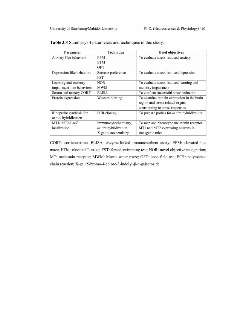
University of Strasbourg/Mahidol University Ph.D. (Neurosciences & Physiology) / 65
Table 3.8 Summary of parameters and techniques in this study.
Parameter Technique Brief objectives Anxiety-like behaviors EPM
ETM OFT
To evaluate stress-induced anxiety.
Depression-like behaviors Sucrose preference FST
To evaluate stress-induced depression.
Learning and memory impairment-like behaviors
NOR MWM
To evaluate stress-induced learning and memory impairment.
Serum and urinary CORT ELISA To confirm successful stress induction. Protein expression Western blotting To examine protein expression in the brain
region and stress-related organs contributing to stress responses.
Riboprobe synthesis for in situ hybridization
PCR cloning To prepare probes for in situ hybridization.
MT1/ MT2-LacZ localization
Immunocytochemistry, in situ hybridization, X-gal histochemistry
To map and phenotype melatonin receptor MT1 and MT2 expressing neurons in transgenic mice
CORT: corticosterone; ELISA: enzyme-linked immunosorbent assay; EPM: elevated-plus
maze; ETM: elevated T-maze; FST: forced swimming test; NOR: novel objective recognition;
MT: melatonin receptor; MWM: Morris water maze; OFT: open-field test; PCR: polymerase
chain reaction; X-gal: 5-bromo-4-chloro-3-indolyl-β-d-galactoside.

Sarawut Lapmanee Results / 66
CHAPTER IV
RESULTS
Part I: Effects of restraint stress induction, monoamine modulators
(i.e., agomelatine and venlafaxine), and voluntary wheel running in
male rats.
Experiment 1. To examine the time-dependent effect of the 1-, 4- and 8-
week stress induction on anxiety-, depression-, and memory impairment-like
behaviors and changes in the expression of selected target proteins in the brain regions
associated with stress responses.
1. Restraint stress induced a change in body weight, adrenal gland
weight, food intake, and corticosterone levels.
There was no difference in the starting body weight (Figure 4.1A).
Following the 1-, 4-, and 8-week restraint stress induction, the final body weights of
stressed rats were decreased (Figure 4.1B), resulting from less body weight gain when
compared to non-stress control rats (Figure 4.1C). Daily food intake was decreased in
the 4-week stressed rats (Figure 4.1D). In addition, the adrenal gland weights (dry
weight and dry weight-body weight ratio) in the 4-and 8-week stressed rats were
increased when compared with non-stress control rats (Figure 4.1F and 4.1H). In
addition, the results from experimental 2 showed that the 4-week stressed rats had
increased serum and urinary corticosterone levels (Figure 4.2A−B). These could be
correlated with adrenal hypertrophy. Therefore, the 4-weeks restraint stress induction
effectively activated the HPA axis in male rats.

University of Strasbourg/Mahidol University Ph.D. (Neurosciences & Physiology) / 67
Control 1 4 8100
150
200
250
300
(16) (16) (16)
F(3,60) = 0.281 P = 0.839
(16)
Stress (weeks)
Sta
rtin
g B
W (
g)
Control 1 4 8200
300
400
500
600
(16)
(16)(16)
F(3,60) = 16.236 P < 0.001
(16)
Stress (weeks)
***** **
Fin
al B
W (
g)Control 1 4 8
0.0
1.5
3.0
4.5
6.0
(16)(16) (16)
F(3,60) = 13.487 P < 0.001
(16)
Stress (weeks)
**** *
Dai
ly b
ody
wei
ght g
ain
(g)
Control 1 4 80
10
20
30
40
50
(16)(16) (16)
F(3,60) = 9.337 P < 0.001
(16)
Stress (weeks)
*
Dai
ly f
ood
inta
ke (
g)
Control 1 4 80.00
0.05
0.10
0.15
0.20
0.25
(16) (16) (16)
F(3,60) = 0.813, P = 0.492
(16)
Stress (weeks)
Wet
adr
enal
wei
ght (
mg)
Control 1 4 80.00
0.05
0.10
0.15
0.20
0.25
(16)(16) (16)
F(3,60) = 3.6838 P = 0.017
*
(16)
*
Stress (weeks)
Dry
adr
enal
wei
ght (
mg)
Control 1 4 80.00
0.10
0.20
0.30
0.40
0.50
(16)(16)(16)
F(3,60) = 2.037 P = 0.118
(16)
Stress (weeks)
Wet
adr
enal
wei
ght (
mg)
/BW
(kg
)
Control 1 4 80.00
0.10
0.20
0.30
0.40
0.50
(16)
(16)
F(3,60) = 5.637 P = 0.002
(16)*
(16)
**
Stress (weeks)
Dry
adr
enal
wei
ght (
mg)
/BW
(kg
)
A. B.
C. D.
E. F.
G. H.
Figure 4.1 Time-dependent effects of the 1-, 4-, and 8-week restraint stress induction
on (A) starting body weight, (B) final body weight, (C) body weight gain, (D) daily
food intake, (E) wet adrenal gland weight, (F) dry adrenal gland weight, (G) relative
wet adrenal gland weights, and (H) relative dry adrenal gland weights. Numbers of
animals are noted in parentheses. *P < 0.05, **P < 0.01, ***P < 0.001 compared to
non-stress control group.

Sarawut Lapmanee Results / 68
Control Stress0
50
100
150
200
(7)
(7)
t(12) = 2.142 P = 0.027
*
Serum
Cor
ticos
tero
ne (
ng/m
L)
Control Stress150
300
450
600
750
(4)
(4)
t(6) = 2.076 P = 0.042
*
Urinary
Cor
ticos
tero
ne (
ng/m
L)
A. B.
Figure 4.2 Time-dependent effects of the 4-week restraint stress induction on (A)
basal serum and (B) urinary corticosterone levels. Numbers of animals are noted in
parentheses. *P < 0.05 compared to non-stress control group.
2. Restraint stress-induced the changes in behavioral responses.
2.1 Anxiety-like behavior
In the EPM test, the 1-, 4-, and 8-week restraint stress resulted
in decreased open arm entry and had a tendency to decrease open arm time (Figure
4.3A−B). Stressed rats increased closed arm entry and closed arm time without
changes in center of arm time, total number of entries, or exploration (i.e., rearing and
grooming) (Figure 4.3C−H). In the ETM test, there was no difference in baseline
latency in the inhibitory avoidance test (Figure 4.4A). The 4-week stressed rats had
prolonged latency in inhibitory avoidance 1, while 1- and 4-week, but not 8-week
stress, showed prolonged latencies in inhibitory avoidance 2 (Figure 4.4B−C).
However, the restraint stress induction did not alter the one-way escape latency
(Figure 4.4D). In the OFT test, the 1- and 4-week stressed rats were hyperarousal to
the sudden change from a familiar environment to an open area as shown by an
increase in the number of lines crossed in the first 30 seconds but no changes in total
lines crossed in 5 min (Figure 4.5A−B). Stressed rats spent less time in the inner zone
and more time in the outer zone (Figure 4.5C−D). Increased numbers of grooming and
rearing were observed in the 1- and 4-week stressed rats (Figure 4.5E−F). Therefore,
the 1-, 4-, and 8-week restraint stress induction could induce anxiety-like behavior and
did not alter the locomotor activities, i.e., no change in total arm entry in the EPM and
total lines crossed in the OFT.
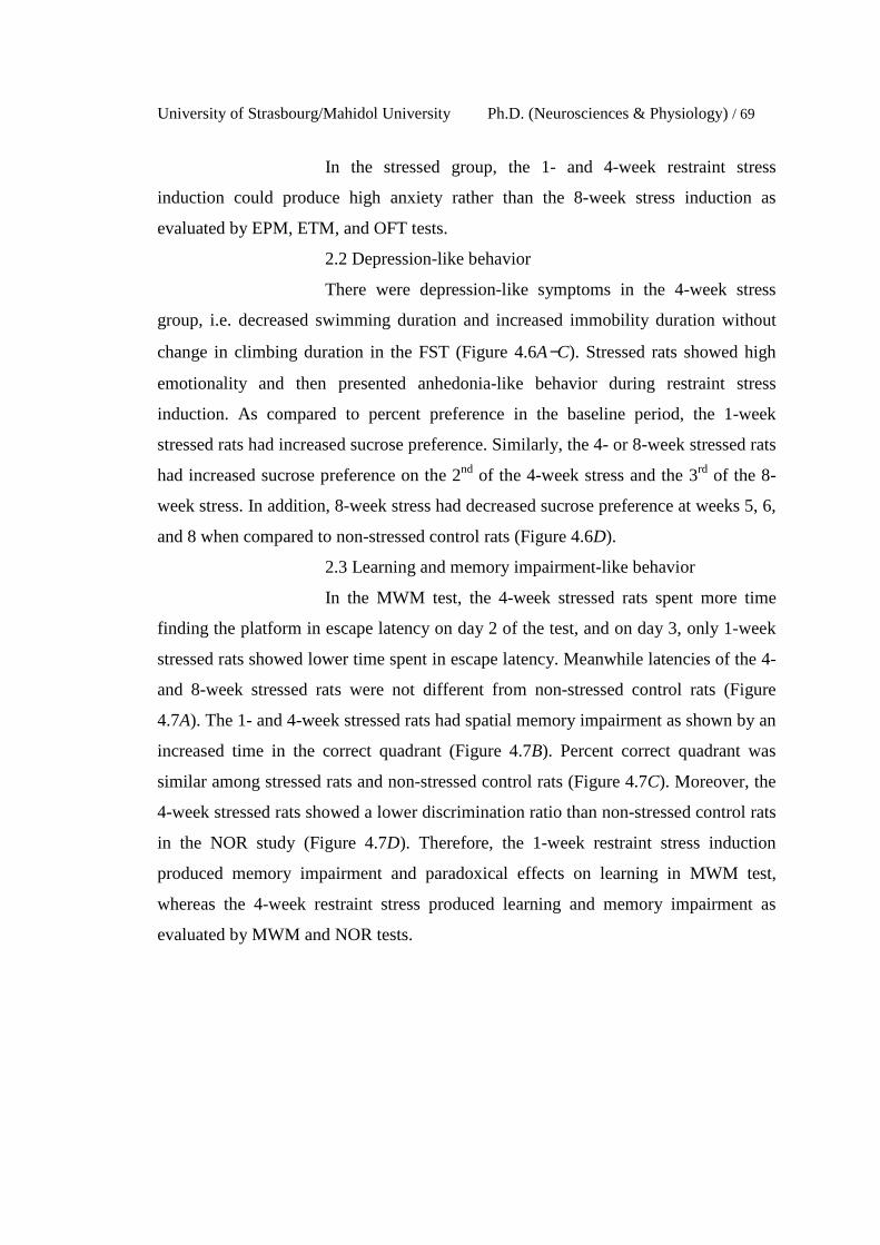
University of Strasbourg/Mahidol University Ph.D. (Neurosciences & Physiology) / 69
In the stressed group, the 1- and 4-week restraint stress
induction could produce high anxiety rather than the 8-week stress induction as
evaluated by EPM, ETM, and OFT tests.
2.2 Depression-like behavior
There were depression-like symptoms in the 4-week stress
group, i.e. decreased swimming duration and increased immobility duration without
change in climbing duration in the FST (Figure 4.6A−C). Stressed rats showed high
emotionality and then presented anhedonia-like behavior during restraint stress
induction. As compared to percent preference in the baseline period, the 1-week
stressed rats had increased sucrose preference. Similarly, the 4- or 8-week stressed rats
had increased sucrose preference on the 2nd of the 4-week stress and the 3rd of the 8-
week stress. In addition, 8-week stress had decreased sucrose preference at weeks 5, 6,
and 8 when compared to non-stressed control rats (Figure 4.6D).
2.3 Learning and memory impairment-like behavior
In the MWM test, the 4-week stressed rats spent more time
finding the platform in escape latency on day 2 of the test, and on day 3, only 1-week
stressed rats showed lower time spent in escape latency. Meanwhile latencies of the 4-
and 8-week stressed rats were not different from non-stressed control rats (Figure
4.7A). The 1- and 4-week stressed rats had spatial memory impairment as shown by an
increased time in the correct quadrant (Figure 4.7B). Percent correct quadrant was
similar among stressed rats and non-stressed control rats (Figure 4.7C). Moreover, the
4-week stressed rats showed a lower discrimination ratio than non-stressed control rats
in the NOR study (Figure 4.7D). Therefore, the 1-week restraint stress induction
produced memory impairment and paradoxical effects on learning in MWM test,
whereas the 4-week restraint stress produced learning and memory impairment as
evaluated by MWM and NOR tests.
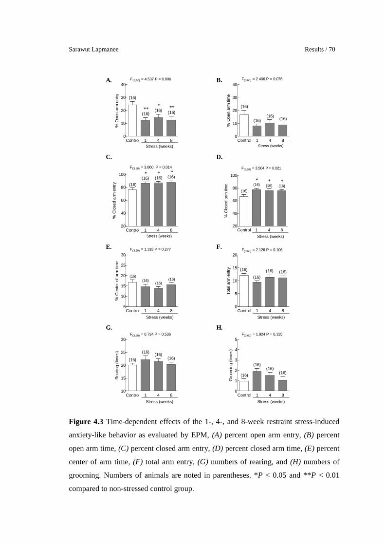
Sarawut Lapmanee Results / 70
Control 1 4 80
10
20
30
40
(16)
(16)
F(3,60) = 4.537 P = 0.006
(16)(16)**
* **
Stress (weeks)
% O
pen
arm
ent
ry
Control 1 4 80
10
20
30
40
(16)
(16)
F(3,60) = 2.406 P = 0.076
(16)(16)
Stress (weeks)
% O
pen
arm
tim
eControl 1 4 8
20
40
60
80
100
(16)
(16)
F(3,60) = 3.860, P = 0.014
(16)(16)* * *
Stress (weeks)
% C
lose
d ar
m e
ntry
Control 1 4 820
40
60
80
100
(16)(16) (16)
F(3,60) = 3.504 P = 0.021
(16)* * *
Stress (weeks)
% C
lose
d ar
m ti
me
Control 1 4 85
10
15
20
25
30
(16)
(16)(16)
F(3,60) = 1.318 P = 0.277
(16)
Stress (weeks)
% C
ente
r of
arm
tim
e
Control 1 4 80
5
10
15
20
(16) (16)
F(3,60) = 2.126 P = 0.106
(16)
(16)
Stress (weeks)
Tota
l arm
ent
ry
Control 1 4 810
15
20
25
30
(16) (16)
F(3,60) = 0.734 P = 0.536
(16)(16)
Stress (weeks)
Rea
ring
(tim
es)
Control 1 4 80
1
2
3
4
5
(16)(16)
F(3,60) = 1.924 P = 0.135
(16)(16)
Stress (weeks)
Gro
omin
g (t
imes
)
A. B.
C. D.
E. F.
G. H.
Figure 4.3 Time-dependent effects of the 1-, 4-, and 8-week restraint stress-induced
anxiety-like behavior as evaluated by EPM, (A) percent open arm entry, (B) percent
open arm time, (C) percent closed arm entry, (D) percent closed arm time, (E) percent
center of arm time, (F) total arm entry, (G) numbers of rearing, and (H) numbers of
grooming. Numbers of animals are noted in parentheses. *P < 0.05 and **P < 0.01
compared to non-stressed control group.
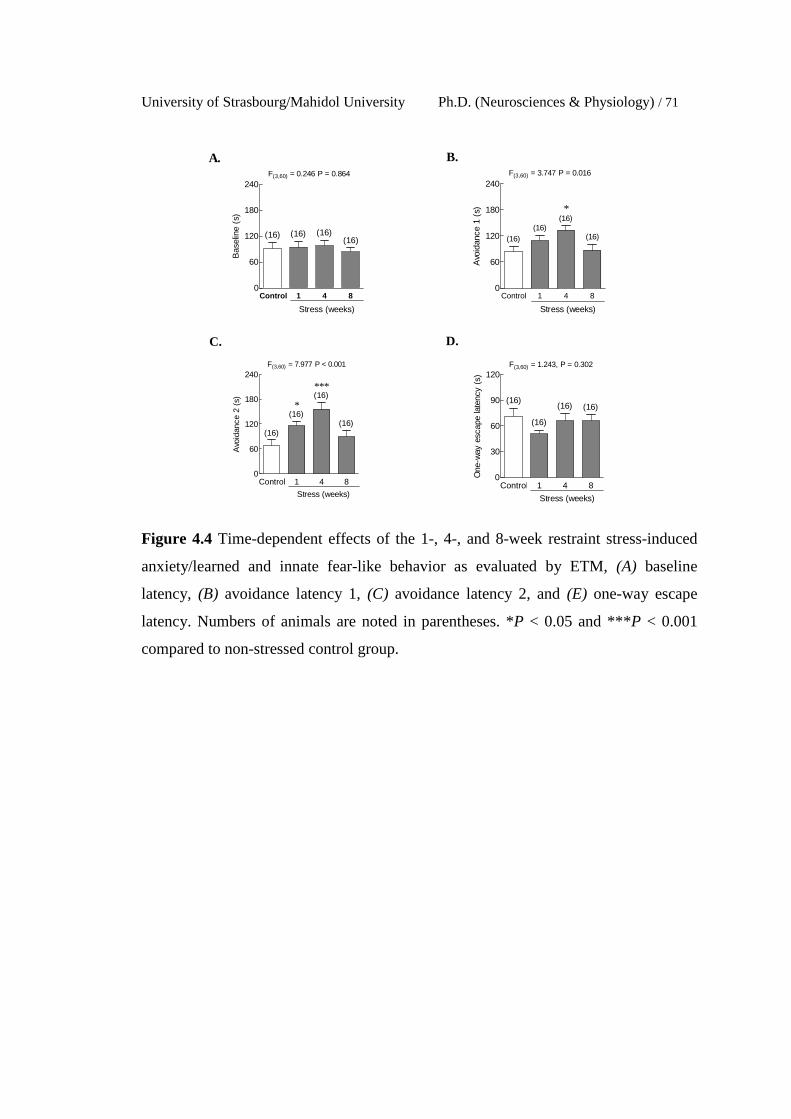
University of Strasbourg/Mahidol University Ph.D. (Neurosciences & Physiology) / 71
Control 1 4 80
60
120
180
240
(16) (16)(16)
F(3,60) = 0.246 P = 0.864
(16)
Stress (weeks)
Bas
elin
e (s
)
Control 1 4 80
60
120
180
240
(16)
(16)
(16)
F(3,60) = 3.747 P = 0.016
(16)
*
Stress (weeks)
Avo
idan
ce 1
(s)
Control 1 4 80
60
120
180
240
(16)
(16)
(16)
F(3,60) = 7.977 P < 0.001
(16)
***
*
Stress (weeks)
Avo
idan
ce 2
(s)
Control 1 4 80
30
60
90
120
(16)
(16)
(16)
F(3,60) = 1.243, P = 0.302
(16)
Stress (weeks)
One
-way
esc
ape
late
ncy
(s)
A. B.
C. D.
Figure 4.4 Time-dependent effects of the 1-, 4-, and 8-week restraint stress-induced
anxiety/learned and innate fear-like behavior as evaluated by ETM, (A) baseline
latency, (B) avoidance latency 1, (C) avoidance latency 2, and (E) one-way escape
latency. Numbers of animals are noted in parentheses. *P < 0.05 and ***P < 0.001
compared to non-stressed control group.
Sarawut Lapmanee Results / 72
Control 1 4 820
30
40
50
60
(16)
(16)
(16)
F(3,60) = 4.550 P = 0.006
(16)* *
Stress (weeks)
No.
of
lines
cro
ssed
in 1
st 3
0 s
Control 1 4 80
100
200
300
400
(16) (16)(16)
F(3,60) = 0.865 P = 0.465
(16)
Stress (weeks)
Tota
l lin
es c
ross
ed in
5 m
in
Control 1 4 80
30
60
90
120
(16)
(16) (16)
F(3,60) = 3.423 P = 0.023
(16)* * *
Stress (weeks)
Inne
r zo
ne ti
me
(s)
Control 1 4 8100
150
200
250
300
(16)(16) (16)
F(3,60) = 3.423 P = 0.023
(16)* * *
Stress (weeks)
Out
er z
one
time
(s)
Control 1 4 80
10
20
30
40
(16)
(16) (16)
F(3,60) = 2.900 P = 0.042
(16)
*
Stress (weeks)
Rea
ring
(tim
es)
Control 1 4 80
2
4
6
8
(16)
(16)
(16)
F(3,60) = 3.291 P = 0.027
(16)* *
Stress (weeks)
Gro
omin
g (t
imes
)
A. B.
C. D.
E. F.
Figure 4.5 Time-dependent effects of the 1-, 4-, and 8-week restraint stress-induced
anxiety-like behavior as evaluated by OFT, (A) number of lines crossed in the first 30
seconds, (B) total lines crossed, (C) inner zone time, (D) outer zone time, (E) number
of rearing, and (F) number of grooming. Numbers of animals are noted in parentheses.
*P < 0.05 compared to non-stressed control group.
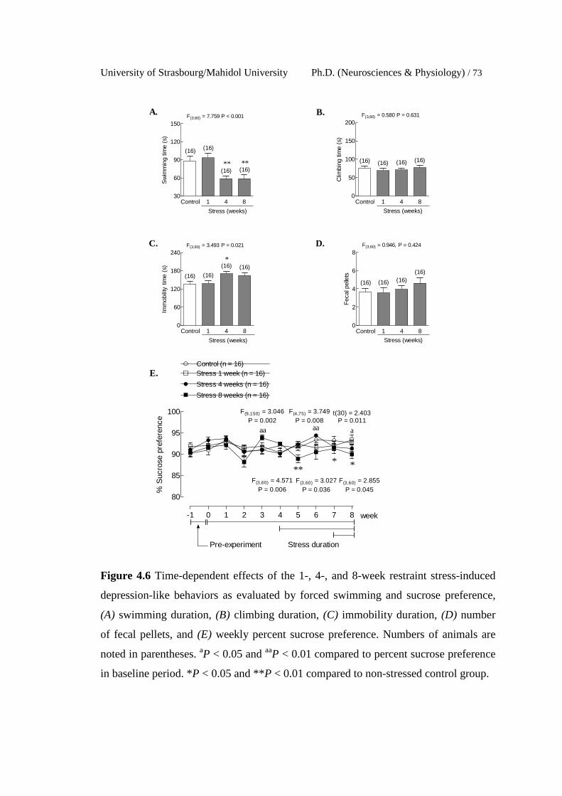
University of Strasbourg/Mahidol University Ph.D. (Neurosciences & Physiology) / 73
Control 1 4 830
60
90
120
150
(16)
(16) (16)
F(3,60) = 7.759 P < 0.001
(16)
****
Stress (weeks)
Sw
imm
ing
time
(s)
Control 1 4 80
50
100
150
200
(16) (16) (16)
F(3,60) = 0.580 P = 0.631
(16)
Stress (weeks)
Clim
bing
tim
e (s
)Control 1 4 8
0
60
120
180
240
(16)
(16) (16)
F(3,60) = 3.493 P = 0.021
(16)
*
Stress (weeks)
Imm
obili
ty ti
me
(s)
Control 1 4 80
2
4
6
8
(16) (16)(16)
F(3,60) = 0.946, P = 0.424
(16)
Stress (weeks)F
ecal
pel
lets
A. B.
C. D.
E.
80
85
90
95
100
Control (n = 16)
Stress 4 weeks (n = 16)
Stress 8 weeks (n = 16)
-1 0 1 2 3 4 5 6 7 8
Stress durationPre-experiment
Stress 1 week (n = 16)
**F(3,60) = 4.571
P = 0.006F(3,60) = 2.855
P = 0.045
week
F(3,60) = 3.027P = 0.036
aa
*
F(9,150) = 3.046P = 0.002
% S
ucro
se p
refe
renc
e
aa
F(4,75) = 3.749P = 0.008
t(30) = 2.403P = 0.011
a
*
Figure 4.6 Time-dependent effects of the 1-, 4-, and 8-week restraint stress-induced
depression-like behaviors as evaluated by forced swimming and sucrose preference,
(A) swimming duration, (B) climbing duration, (C) immobility duration, (D) number
of fecal pellets, and (E) weekly percent sucrose preference. Numbers of animals are
noted in parentheses. aP < 0.05 and aaP < 0.01 compared to percent sucrose preference
in baseline period. *P < 0.05 and **P < 0.01 compared to non-stressed control group.
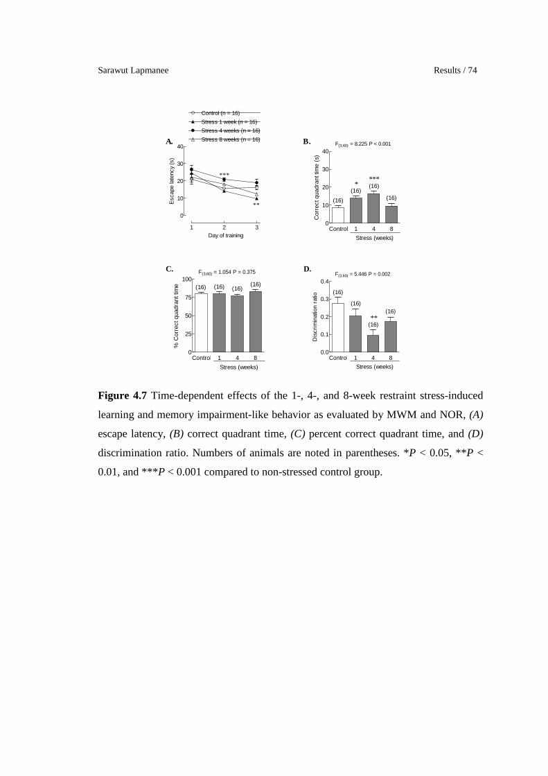
Sarawut Lapmanee Results / 74
1 2 3
0
10
20
30
40
Control (n = 16)
Stress 1 week (n = 16)
Stress 4 weeks (n = 16)
Stress 8 weeks (n = 16)
***
**
Day of training
Esc
ape
late
ncy
(s)
Control 1 4 80
10
20
30
40
(16)(16)
(16)(16)
F(3,60) = 8.225 P < 0.001
****
Stress (weeks)
Cor
rect
qua
dran
t tim
e (s
)
Control 1 4 80
25
50
75
100(16)(16) (16)(16)
F(3,60) = 1.054 P = 0.375
Stress (weeks)
% C
orre
ct q
uadr
ant t
ime
Control 1 4 80.0
0.1
0.2
0.3
0.4
(16)
(16)
(16)
(16)
F(3,60) = 5.446 P = 0.002
Stress (weeks)
**D
iscr
imin
atio
n ra
tio
A. B.
C. D.
Figure 4.7 Time-dependent effects of the 1-, 4-, and 8-week restraint stress-induced
learning and memory impairment-like behavior as evaluated by MWM and NOR, (A)
escape latency, (B) correct quadrant time, (C) percent correct quadrant time, and (D)
discrimination ratio. Numbers of animals are noted in parentheses. *P < 0.05, **P <
0.01, and ***P < 0.001 compared to non-stressed control group.

University of Strasbourg/Mahidol University Ph.D. (Neurosciences & Physiology) / 75
3. Restraint stress induced changes in target protein expression in
brain regions associated with stress responses.
3.1 Levels of BDNF
The 1- and 8-week restraint stress induction decreased BDNF
protein levels in periaqueductal gray and ventral tegmentum area (Figure 4.8F and G).
3.2 Levels of GR in the hypothalamus and hippocampus
Hypothalamic GR protein was increased in the 1-week
restraint stress induction, whereas hippocampal GR protein was decreased in the 4-
week stress induction (Figure 4.9). These results suggest stress responses of GR
proteins, resulting from hyperactivity of the HPA axis. GR could act back on the
hippocampus to exert GR-mediated negative feedback inhibition on the HPA axis
3.3 Levels of NET
The 4-week restraint stress induction increased NET proteins
in the locus coeruleus when compared to non-stressed controls (Figure 4.10D).
3.4 Levels of SERT
The 1- and 8-week restraint induction decreased SERT protein
in the ventral tegmental area and hippocampus, respectively (Figure 4.11B and F).
3.5 Levels of 5-HT2CR
The 1-, 4-, and 8-week restraint stress induction increased 5-
HT2CR protein levels in the amygdala and locus coeruleus (Figure 4.12A and E). In
contrast, the 4- or 8-week stress induction decreased 5-HT2CR protein expression in
periaqueductal gray and the ventral tegmental area when compared to non-stressed
control rats (Figure 4.12G).
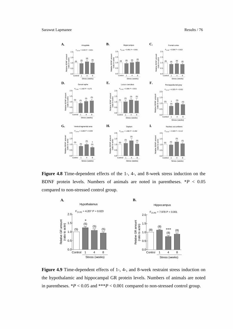
Sarawut Lapmanee Results / 76
Control 1 4 80.0
0.5
1.0
1.5
2.0
Stress (weeks)
(8) (8)(8)
(8)
Amygdala
F(3,28) = 0.320 P = 0.811
Rel
ativ
e B
DN
F a
mou
nt(r
atio
to a
ctin
)
Control 1 4 80.0
0.5
1.0
1.5
2.0
Stress (weeks)
(8) (8) (8) (8)
Hippocampus
F(3,28) = 0.491 P = 0.691
Rel
ativ
e B
DN
F a
mou
nt(r
atio
to a
ctin
)
Control 1 4 80.0
0.5
1.0
1.5
2.0
Stress (weeks)
(8) (8) (8) (8)
Frontal cortex
F(3,28) = 0.596 P = 0.623
Rel
ativ
e B
DN
F a
mou
nt(r
atio
to a
ctin
)
Control 1 4 80.0
0.5
1.0
1.5
2.0
Stress (weeks)
(5) (5)(5)
(5)
Dorsal raphe
F(3,16) = 1.429 P = 0.271
Rel
ativ
e B
DN
F a
mou
nt(r
atio
to a
ctin
)
Control 1 4 80.0
0.5
1.0
1.5
2.0
Stress (weeks)
(5)(5)
(5) (5)
Locus coeruleus
F(3,16) = 0.306 P = 0.821
Rel
ativ
e B
DN
F a
mou
nt(r
atio
to a
ctin
)
Control 1 4 80.0
0.5
1.0
1.5
2.0
Stress (weeks)
(5)(5)
(5)(5)
Periaqueductal gray
*
F(3,16) = 4.325 P = 0.021
Rel
ativ
e B
DN
F a
mou
nt(r
atio
to a
ctin
)Control 1 4 8
0.0
0.5
1.0
1.5
2.0
Stress (weeks)
(5)(5)
(5)
(5)
Ventral tegmental area
*
F(3,16) = 3.266 P = 0.049
Rel
ativ
e B
DN
F a
mou
nt(r
atio
to a
ctin
)
Control 1 4 80.0
0.5
1.0
1.5
2.0
Stress (weeks)
(5)(5)
(5)
(5)
Septum
F(3,16) = 1.390 P = 0.282
Rel
ativ
e B
DN
F a
mou
nt(r
atio
to a
ctin
)
Control 1 4 80.0
0.5
1.0
1.5
2.0
Stress (weeks)
(5) (5)
(5)(5)
Nucleus accumbens
F(3,16) = 2.340 P = 0.112
Rel
ativ
e B
DN
F a
mou
nt(r
atio
to a
ctin
)
A. B. C.
D. E. F.
G. H. I.
Figure 4.8 Time-dependent effects of the 1-, 4-, and 8-week stress induction on the
BDNF protein levels. Numbers of animals are noted in parentheses. *P < 0.05
compared to non-stressed control group.
Control 1 4 80.0
0.5
1.0
1.5
2.0
Stress (weeks)
(5)
(5)(5)
(5)
Hypothalamus
*
F(3,16) = 4.207 P = 0.023
Rel
ativ
e G
R a
mou
nt(r
atio
to a
ctin
)
Control 1 4 80.0
0.5
1.0
1.5
2.0
Stress (weeks)
(8)(8)
(8)(8)
Hippocampus
***
F(3,28) = 7.978 P < 0.001
Rel
ativ
e G
R a
mou
nt(r
atio
to a
ctin
)
A. B.
Figure 4.9 Time-dependent effects of 1-, 4-, and 8-week restraint stress induction on
the hypothalamic and hippocampal GR protein levels. Numbers of animals are noted
in parentheses. *P < 0.05 and ***P < 0.001 compared to non-stressed control group.
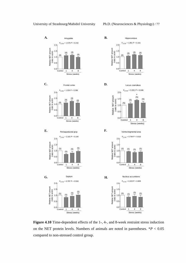
University of Strasbourg/Mahidol University Ph.D. (Neurosciences & Physiology) / 77
Control 1 4 80.0
0.5
1.0
1.5
2.0
Stress (weeks)
(8)(8) (8)
(8)
Amygdala
F(3,28) = 1.575 P = 0.218
Rel
ativ
e N
ET
amou
nt(r
atio
to a
ctin
)
Control 1 4 80.0
0.5
1.0
1.5
2.0
Stress (weeks)
(8)(8) (8) (8)
Hippocampus
F(3,28) = 1.851 P = 0.161
Rel
ativ
e N
ET
amou
nt(r
atio
to a
ctin
)
Control 1 4 80.0
0.5
1.0
1.5
2.0
Stress (weeks)
(8)(8)
(8)(8)
Frontal cortex
F(3,28) = 1.324 P = 0.286
Rel
ativ
e N
ET
amou
nt(r
atio
to a
ctin
)
Control 1 4 80.0
0.5
1.0
1.5
2.0
Stress (weeks)
(5)
(5)(5)
(5)
Locus coeruleus
*
F(3,16) = 3.331 P = 0.046
Rel
ativ
e N
ET
amou
nt(r
atio
to a
ctin
)
Control 1 4 80.0
0.5
1.0
1.5
2.0
Stress (weeks)
(5)
(5)(5) (5)
Periaqueductal gray
F(3,16) = 2.101 P = 0.140
Rel
ativ
e N
ET
amou
nt(r
atio
to a
ctin
)
Control 1 4 80.0
0.5
1.0
1.5
2.0
Stress (weeks)
(5) (5) (5) (5)
Ventral tegmental area
F(3,16) = 0.794 P = 0.515
Rel
ativ
e N
ET
amou
nt(r
atio
to a
ctin
)
Control 1 4 80.0
0.5
1.0
1.5
2.0
Stress (weeks)
(5) (5)(5)
(5)
Septum
F(3,16) = 0.787 P = 0.518
Rel
ativ
e N
ET
amou
nt(r
atio
to a
ctin
)
Control 1 4 80.0
0.5
1.0
1.5
2.0
Stress (weeks)
(5) (5)(5) (5)
Nucleus accumbens
F(3,16) = 0.323 P = 0.809
Rel
ativ
e N
ET
amou
nt(r
atio
to a
ctin
)
A. B.
C. D.
E. F.
G. H.
Figure 4.10 Time-dependent effects of the 1-, 4-, and 8-week restraint stress induction
on the NET protein levels. Numbers of animals are noted in parentheses. *P < 0.05
compared to non-stressed control group.

Sarawut Lapmanee Results / 78
Control 1 4 80.0
0.5
1.0
1.5
2.0
Stress (weeks)
(8)(8)
(8) (8)
Amygdala
F(3,28) = 2.620 P = 0.070
Rel
ativ
e S
ER
T am
ount
(rat
io to
act
in)
Control 1 4 80.0
0.5
1.0
1.5
2.0
Stress (weeks)
(8) (8) (8) (8)
Hippocampus
*
F(3,28) = 2.991 P = 0.048
Rel
ativ
e S
ER
T am
ount
(rat
io to
act
in)
Control 1 4 80.0
0.5
1.0
1.5
2.0
Stress (weeks)
(8) (8) (8) (8)
Frontal cortex
F(3,16) = 2.991 P = 0.048
Rel
ativ
e S
ER
T am
ount
(rat
io to
act
in)
Control 1 4 80.0
0.5
1.0
1.5
2.0
Stress (weeks)
(5) (5) (5) (5)
Dorsal raphe
F(3,16) = 1.070 P = 0.378
Rel
ativ
e S
ER
T am
ount
(rat
io to
act
in)
Control 1 4 80.0
0.5
1.0
1.5
2.0
Stress (weeks)
(5) (5) (5) (5)
Periaqueductal gray
F(3,16) = 0.493 P = 0.692
Rel
ativ
e S
ER
T am
ount
(rat
io to
act
in)
Control 1 4 80.0
0.5
1.0
1.5
2.0
Stress (weeks)
(5)(5)
(5) (5)
Ventral tegmental area
*
F(3,16) = 3.317 P = 0.047
Rel
ativ
e S
ER
T am
ount
(rat
io to
act
in)
Control 1 4 80.0
0.5
1.0
1.5
2.0
Stress (weeks)
(5)(5) (5) (5)
Septum
F(3,16) = 2.392 P = 0.107
Rel
ativ
e S
ER
T am
ount
(rat
io to
act
in)
Control 1 4 80.0
0.5
1.0
1.5
2.0
Stress (weeks)
(5) (5) (5) (5)
Nucleus accumbens
F(3,16) = 1.425 P = 0.272
Rel
ativ
e S
ER
T am
ount
(rat
io to
act
in)
A. B.
C. D.
E. F.
G. H.
Figure 4.11 Time-dependent effects of the 1-, 4-, and 8-week restraint stress induction
on the SERT protein levels. Numbers of animals are noted in parentheses. *P < 0.05
compared to non-stressed control group.
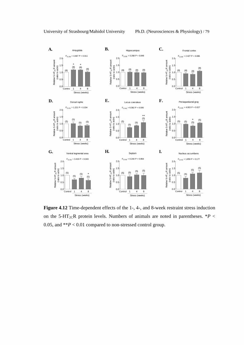
University of Strasbourg/Mahidol University Ph.D. (Neurosciences & Physiology) / 79
Control 1 4 80.0
0.5
1.0
1.5
2.0
Stress (weeks)
(8)
(8) (8)(8)
Amygdala
* *
F(3,28) = 4.467 P = 0.011R
elat
ive
5-H
T2C
R a
mou
nt(r
atio
to a
ctin
)
Control 1 4 80.0
0.5
1.0
1.5
2.0
Stress (weeks)
(8) (8) (8) (8)
Hippocampus
F(3,28) = 0.268 P = 0.848
Rel
ativ
e 5-
HT
2CR
am
ount
(rat
io t
o ac
tin)
Control 1 4 80.0
0.5
1.0
1.5
2.0
Stress (weeks)
(8) (8) (8)(8)
Frontal cortex
F(3,28) = 2.427 P = 0.086
Rel
ativ
e 5-
HT
2CR
am
ount
(rat
io t
o ac
tin)
Control 1 4 80.0
0.5
1.0
1.5
2.0
Stress (weeks)
(5)(5)
(5) (5)
Dorsal raphe
F(3,16) = 1.221 P = 0.334
Rel
ativ
e 5-
HT
2CR
am
ount
(rat
io t
o ac
tin)
Control 1 4 80.0
0.5
1.0
1.5
2.0
Stress (weeks)
(5)(5)
(5)
(5)
Locus coeruleus
**
F(3,16) = 6.082 P = 0.006R
elat
ive
5-H
T2C
R a
mou
nt(r
atio
to
actin
)
Control 1 4 80.0
0.5
1.0
1.5
2.0
Stress (weeks)
(5) (5) (5)(5)
Periaqueductal gray
F(3,16) = 4.003 P = 0.027
*
Rel
ativ
e 5-
HT
2CR
am
ount
(rat
io to
act
in)
Control 1 4 80.0
0.5
1.0
1.5
2.0
Stress (weeks)
(5)(5)
(5)(5)
Ventral tegmental area
*
F(3,16) = 3.419 P = 0.043
Rel
ativ
e 5-
HT
2CR
am
ount
(rat
io t
o ac
tin)
Control 1 4 80.0
0.5
1.0
1.5
2.0
Stress (weeks)
(5) (5)(5) (5)
Septum
F(3,16) = 0.244 P = 0.864
Rel
ativ
e 5-
HT
2CR
am
ount
(rat
io to
act
in)
Control 1 4 80.0
0.5
1.0
1.5
2.0
Stress (weeks)
(5) (5)
(5)(5)
Nucleus accumbens
F(3,16) = 1.859 P = 0.177
Rel
ativ
e 5-
HT
2CR
am
ount
(rat
io t
o ac
tin)
A. B. C.
D. E. F.
G. H. I.
Figure 4.12 Time-dependent effects of the 1-, 4-, and 8-week restraint stress induction
on the 5-HT2CR protein levels. Numbers of animals are noted in parentheses. *P <
0.05, and **P < 0.01 compared to non-stressed control group.
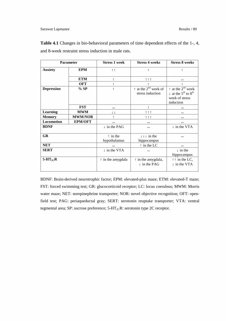
Sarawut Lapmanee Results / 80
Table 4.1 Changes in bio-behavioral parameters of time dependent effects of the 1-, 4,
and 8-week restraint stress induction in male rats.
Parameter Stress 1 week Stress 4 weeks Stress 8 weeks
Anxiety EPM ↑↑ ↑ ↑
ETM ↑ ↑↑↑ ↔ OFT ↑ ↑ ↑
Depression % SP ↑ ↑ at the 2nd week of stress induction
↑ at the 2nd week ↓ at the 5th to 8th week of stress induction
FST ↔ ↑ ↔ Learning MWM ↓↓ ↑↑↑ ↔ Memory MWM/NOR ↑ ↑↑↑ ↔ Locomotion EPM/OFT ↔ ↔ ↔ BDNF ↓ in the PAG ↔ ↓ in the VTA
GR ↑ in the hypothalamus
↓↓↓ in the hippocampus
↔
NET ↔ ↑ in the LC ↔ SERT ↓ in the VTA ↔ ↓ in the
hippocampus 5-HT2CR ↑ in the amygdala ↑ in the amygdala,
↓ in the PAG ↑↑ in the LC, ↓ in the VTA
BDNF: Brain-derived neurotrophic factor; EPM: elevated-plus maze; ETM: elevated-T maze;
FST: forced swimming test; GR: glucocorticoid receptor; LC: locus coeruleus; MWM: Morris
water maze; NET: norepinephrine transporter; NOR: novel objective recognition; OFT: open-
field test; PAG: periaqueductal gray; SERT: serotonin reuptake transporter; VTA: ventral
tegmental area; SP: sucrose preference; 5-HT2CR: serotonin type 2C receptor.

University of Strasbourg/Mahidol University Ph.D. (Neurosciences & Physiology) / 81
Experiment 2. To determine the effect of 4 weeks of voluntary wheel
running on the hypothalamic-pituitary-adrenal (HPA) axis under non-stressed
conditions, or after a 4-week exposure to restraint stress, and changes in the expression
of selected target proteins in brain regions associated with stress responses.
1. The 4-week voluntary wheel running induced changes in body
weight, heart weight, adrenal gland weight, food intake, corticosterone levels, and
running distance.
There was no difference in starting body weight (Figure 4.13A). The
sedentary stressed rats decreased body weight when compared to the non-stressed
sedentary control rats. Both non-stressed exercised rats and the combined stressed and
exercised rats had decreased body weight when compared to the non-stressed
sedentary rats and stressed sedentary rats, respectively (Figure 4.13B). While the non-
stressed sedentary rats had gained weight, non-stressed exercised and sedentary
stressed rats had less weight gain. The combined stressed and exercised rats also
displayed less weight gain when compared to sedentary stressed rats (Figure 4.13C).
Sedentary stressed rats decreased daily food intake when compared to non-stressed
control rats (Figure 4.13D). Therefore, the 4-week voluntary wheel running exercise
could alter the energy balance including body weight and food intake.
In the exercise group, there was a similar running distance between non-
stressed and stressed groups (Figure 4.14A−B). Both non-stressed and stressed rats
gradually increased running distance through experimental period suggesting that
stress did not affect the general locomotor activity in exercise training. Voluntary
wheel running increased wet and dry heart weights in both non-stressed sedentary
control and stressed rats (Figure 4.14C−F) indicating that the aerobic exercise protocol
induced cardiac hypertrophy.
In addition, only the non-stressed exercised control rats, but not stressed
exercised rats, had an increase in dry adrenal gland weight when compared with non-
stressed sedentary control rats (Figure 4.15A−D). Therefore, voluntary wheel running-
induced increase in adrenal weights might represent a modulation of sympatho-adrenal
systems. Sedentary stressed rats had increased serum corticosterone levels when
compared with non-stressed sedentary control rats. The combined stressed and
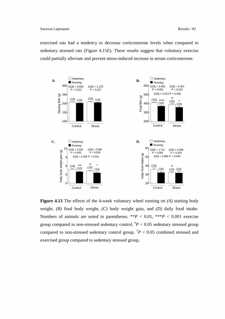
Sarawut Lapmanee Results / 82
exercised rats had a tendency to decrease corticosterone levels when compared to
sedentary stressed rats (Figure 4.15E). These results suggest that voluntary exercise
could partially alleviate and prevent stress-induced increase in serum corticosterone.
100
150
200
250
300
(10) (10)(10) (10)
t(18) = 0.820P = 0.212
Sedentary
Running
t(18) = 1.178P = 0.127
StressControl
Sta
rtin
g B
W (
g)
200
300
400
500
600
(10)(10)
(10)(10)
t(18) = 4.403P < 0.001
Sedentary
Runningt(18) = 2.457
P = 0.012
StressControl
*** †#
t(18) = 2.013 P = 0.030
Fin
al B
W (
g)
0
3
6
9
12
(10)(10)
(10)(10)
t(18) = 3.326P = 0.002
Sedentary
Running
t(18) = 2.082P = 0.026
StressControl
** †
t(18) = 2.199 P = 0.021
#
Dai
ly b
ody
wei
ght g
ain
(g)
20
30
40
50
60
(10) (10)(10)
(10)
t(18) = 1.711P = 0.052
Sedentary
Running
t(18) = 0.206P = 0.420
StressControl
t(18) = 1.856 P = 0.040
#
Dai
ly f
ood
inta
ke (
g)
A. B.
C. D.
Figure 4.13 The effects of the 4-week voluntary wheel running on (A) starting body
weight, (B) final body weight, (C) body weight gain, and (D) daily food intake.
Numbers of animals are noted in parentheses. **P < 0.01, ***P < 0.001 exercise
group compared to non-stressed sedentary control. #P < 0.05 sedentary stressed group
compared to non-stressed sedentary control group. †P < 0.05 combined stressed and
exercised group compared to sedentary stressed group.
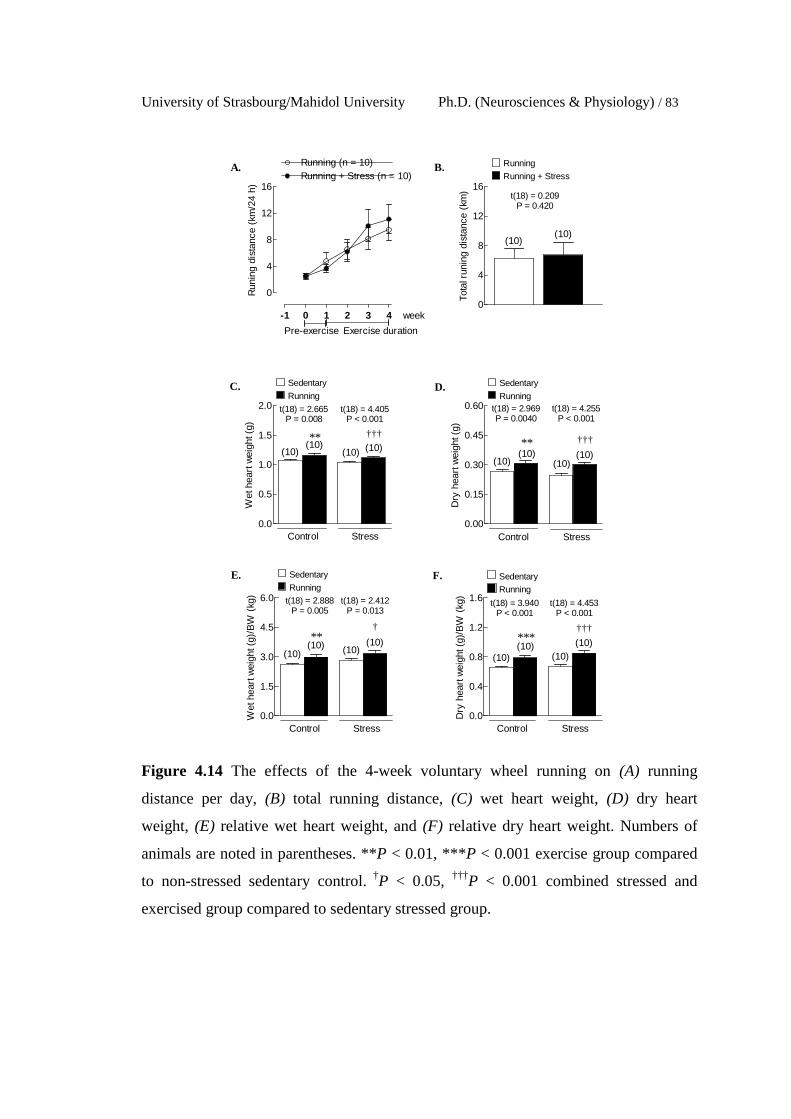
University of Strasbourg/Mahidol University Ph.D. (Neurosciences & Physiology) / 83
0
4
8
12
16
Running (n = 10)Running + Stress (n = 10)
Exercise durationPre-exercise
week-1 0 1 2 3 4
Run
ing
dist
ance
(km
/24
h)
0
4
8
12
16
(10)(10)
Running
Running + Stress
t(18) = 0.209P = 0.420
Tota
l run
ing
dist
ance
(km
)
0.0
0.5
1.0
1.5
2.0
(10)(10)
t(18) = 2.665P = 0.008
(10) (10)
Sedentary
Running
StressControl
†††**
t(18) = 4.405P < 0.001
Wet
hea
rt w
eigh
t (g)
0.00
0.15
0.30
0.45
0.60
(10)(10)
t(18) = 2.969P = 0.0040
(10)(10)
Sedentary
Running
StressControl
†††**
t(18) = 4.255P < 0.001
Dry
hea
rt w
eigh
t (g)
0.0
1.5
3.0
4.5
6.0
(10)(10)
t(18) = 2.888P = 0.005
(10)(10)
Sedentary
Running
StressControl
†**
t(18) = 2.412P = 0.013
Wet
hea
rt w
eigh
t (g)
/BW
(kg
)
0.0
0.4
0.8
1.2
1.6
(10)(10)
t(18) = 3.940P < 0.001
(10)(10)
Sedentary
Running
StressControl
†††***
t(18) = 4.453P < 0.001
Dry
hea
rt w
eigh
t (g)
/BW
(kg
)
A. B.
C. D.
E. F.
Figure 4.14 The effects of the 4-week voluntary wheel running on (A) running
distance per day, (B) total running distance, (C) wet heart weight, (D) dry heart
weight, (E) relative wet heart weight, and (F) relative dry heart weight. Numbers of
animals are noted in parentheses. **P < 0.01, ***P < 0.001 exercise group compared
to non-stressed sedentary control. †P < 0.05, †††P < 0.001 combined stressed and
exercised group compared to sedentary stressed group.
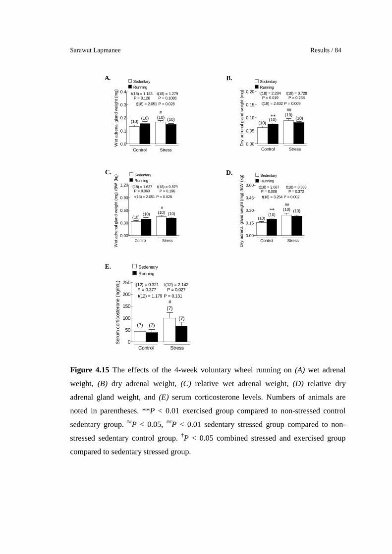
Sarawut Lapmanee Results / 84
0.0
0.1
0.2
0.3
0.4
(10)(10)
t(18) = 1.183P = 0.126
(10) (10)
Sedentary
Running
StressControl
t(18) = 1.279P = 0.1086
t(18) = 2.051 P = 0.028
#W
et a
dren
al g
land
wei
ght (
mg)
0.00
0.05
0.10
0.15
0.20
(10)(10)
t(18) = 2.234P = 0.019
(10)(10)
Sedentary
Running
StressControl
**
t(18) = 0.729P = 0.238
##
t(18) = 2.632 P = 0.009
Dry
adr
enal
gla
nd w
eigh
t (m
g)0.00
0.30
0.60
0.90
1.20
(10)(10)
t(18) = 1.637P = 0.060
(10) (10)
Sedentary
Running
StressControl
t(18) = 0.879P = 0.196
t(18) = 2.051 P = 0.028
#
Wet
adr
enal
gla
nd w
eigh
t (m
g) /
BW
(kg
)
0.00
0.15
0.30
0.45
0.60
(10)(10)
t(18) = 2.687P = 0.008
(10) (10)
Sedentary
Running
StressControl
**
t(18) = 0.333P = 0.372
t(18) = 3.254 P = 0.002
##
Dry
adr
enal
gla
nd w
eigh
t (m
g) /
BW
(kg
)
0
50
100
150
200
250
(7) (7)
t(12) = 0.321P = 0.377
(7)
(7)
Sedentary
Running
StressControl
t(12) = 2.142P = 0.027
t(12) = 1.179 P = 0.131#
Ser
um c
ortic
oste
rone
(ng
/mL)
A. B.
C. D.
E.
Figure 4.15 The effects of the 4-week voluntary wheel running on (A) wet adrenal
weight, (B) dry adrenal weight, (C) relative wet adrenal weight, (D) relative dry
adrenal gland weight, and (E) serum corticosterone levels. Numbers of animals are
noted in parentheses. **P < 0.01 exercised group compared to non-stressed control
sedentary group. ##P < 0.05, ##P < 0.01 sedentary stressed group compared to non-
stressed sedentary control group. †P < 0.05 combined stressed and exercised group
compared to sedentary stressed group.

University of Strasbourg/Mahidol University Ph.D. (Neurosciences & Physiology) / 85
2. The 4-week voluntary wheel running induced the changes in
behavioral responses
All groups of rats had increased sucrose preference as compared to the
baseline period. There were no significant differences between groups as analyzed by
one-way ANOVA. When compared to non-stress sedentary control by using unpaired
t-test, stressed rats exhibited reduced sucrose preference in the 4th week of stress
induction. Interestingly, the combined stress and voluntary exercise could prevent
stress-induced decrease in sucrose preference in stressed male rats (Figure 4.16).
88
89
90
91
92
93
Control (n = 10)
Stress (n = 10)
Running + Stress (n = 10)
-1 0 1 2 3 4
Stress/Exercise durationPre-exercise
Running (n = 10)
week
###
† t(18) = 2.463P = 0.012
t(18) = 4.754P < 0.001
% S
ucro
se p
refe
renc
e
Figure 4.16 Effects of the 4-week voluntary wheel running on restraint stress-induced
depression-like behavior as determined by sucrose preference test. Numbers of
animals are noted in parentheses. ###P < 0.001 sedentary stressed group compared to
non-stressed sedentary control group. †P < 0.05 combined stressed and exercised
group compared to sedentary stressed group.
3. The 4-week wheel running induced changes in target protein
expression in the brain regions associated with stress responses.
3.1 Levels of BDNF
Decreased hippocampal and septal BDNF protein expression
in male rats was observed following the 4-week restraint stress induction (Figure
4.17B and H). The protein levels of BDNF in the nucleus accumbens were decreased
in the exercise group. In the combined stressed and exercised rats BDNF protein was
increased in the hippocampus and septum but decreased in the nucleus accumbens
(Figure 4.17H-I).

Sarawut Lapmanee Results / 86
0.0
0.5
1.0
1.5
2.0
(8) (8) (8) (8)
SedentaryRunning
StressControl
Amygdala
t(14) = 0.249P = 0.404
t(14) = 0.179P = 0.430
Rel
ativ
e B
DN
F a
mou
nt(r
atio
to a
ctin
)
0.0
0.5
1.0
1.5
2.0
(8)(8)
(8)(8)
SedentaryRunning
StressControl
Hippocampus
†
#
t(14) = 2.341P = 0.017
t(14) = 1.372P = 0.096
t(14) = 1.985 P = 0.034
Rel
ativ
e B
DN
F a
mou
nt(r
atio
to a
ctin
)
0.0
0.5
1.0
1.5
2.0
(8) (8) (8)
(8)
SedentaryRunning
StressControl
Frontal cortex
t(14) = 1.594P = 0.067
t(14) = 0.121P = 0.453
Rel
ativ
e B
DN
F a
mou
nt(r
atio
to a
ctin
)
0.0
0.5
1.0
1.5
2.0
(5) (5) (5)(5)
SedentaryRunning
StressControl
Dorsal raphe
t(8) = 1.120P = 0.148
t(8) = 0.453P = 0.121
Rel
ativ
e B
DN
F a
mou
nt(r
atio
to a
ctin
)
0.0
0.5
1.0
1.5
2.0
(5) (5)(5)
(5)
SedentaryRunning
StressControl
Locus coeruleus
t(8) = 0.808P = 0.221
t(8) = 0.237P = 0.410
Rel
ativ
e B
DN
F a
mou
nt(r
atio
to a
ctin
)
0.0
0.5
1.0
1.5
2.0
(5)(5)
(5)(5)
SedentaryRunning
StressControl
Periaqueductal gray
t(8) = 1.279P = 0.118
t(8) = 0.237P = 0.410
Rel
ativ
e B
DN
F a
mou
nt(r
atio
to a
ctin
)
0.0
0.5
1.0
1.5
2.0
(5)(5) (5) (5)
SedentaryRunning
StressControl
Ventral tegmental area
t(8) = 0.555P = 0.297
t(8) = 1.616P = 0.072
Rel
ativ
e B
DN
F a
mou
nt(r
atio
to a
ctin
)
0.0
0.5
1.0
1.5
2.0
(5) (5) (5)
(5)
SedentaryRunning
StressControl
Septum
†
#
t(8) = 2.499P = 0.019
t(8) = 0.559P = 0.296
t(8) = 2.560 P = 0.017
Rel
ativ
e B
DN
F a
mou
nt(r
atio
to a
ctin
)
0.0
0.5
1.0
1.5
2.0
(5)(5)
(5)
(5)
SedentaryRunning
StressControl
Nucleus accumbens
* †
t(8) = 2.364P = 0.023
t(8) = 2.089P = 0.035
Rel
ativ
e B
DN
F a
mou
nt(r
atio
to a
ctin
)
A. B. C.
D. E. F.
G. H. I.
Figure 4.17 Effects of the 4-week voluntary wheel running on BDNF protein levels.
Numbers of animals are noted in parentheses. *P < 0.05 exercised group compared to
non-stressed control sedentary group. #P < 0.05 sedentary stressed group compared to
non-stressed sedentary control group. †P < 0.05 combined stressed and exercised
group compared to sedentary stressed group.
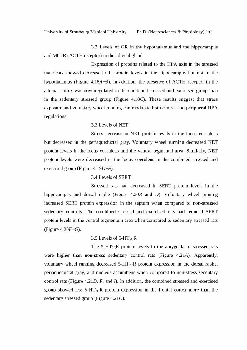
University of Strasbourg/Mahidol University Ph.D. (Neurosciences & Physiology) / 87
3.2 Levels of GR in the hypothalamus and the hippocampus
and MC2R (ACTH receptor) in the adrenal gland.
Expression of proteins related to the HPA axis in the stressed
male rats showed decreased GR protein levels in the hippocampus but not in the
hypothalamus (Figure 4.18A−B). In addition, the presence of ACTH receptor in the
adrenal cortex was downregulated in the combined stressed and exercised group than
in the sedentary stressed group (Figure 4.18C). These results suggest that stress
exposure and voluntary wheel running can modulate both central and peripheral HPA
regulations.
3.3 Levels of NET
Stress decrease in NET protein levels in the locus coeruleus
but decreased in the periaqueductal gray. Voluntary wheel running decreased NET
protein levels in the locus coeruleus and the ventral tegmental area. Similarly, NET
protein levels were decreased in the locus coeruleus in the combined stressed and
exercised group (Figure 4.19D−F).
3.4 Levels of SERT
Stressed rats had decreased in SERT protein levels in the
hippocampus and dorsal raphe (Figure 4.20B and D). Voluntary wheel running
increased SERT protein expression in the septum when compared to non-stressed
sedentary controls. The combined stressed and exercised rats had reduced SERT
protein levels in the ventral tegmentum area when compared to sedentary stressed rats
(Figure 4.20F−G).
3.5 Levels of 5-HT2CR
The 5-HT2CR protein levels in the amygdala of stressed rats
were higher than non-stress sedentary control rats (Figure 4.21A). Apparently,
voluntary wheel running decreased 5-HT2CR protein expression in the dorsal raphe,
periaqueductal gray, and nucleus accumbens when compared to non-stress sedentary
control rats (Figure 4.21D, F, and I). In addition, the combined stressed and exercised
group showed less 5-HT2CR protein expression in the frontal cortex more than the
sedentary stressed group (Figure 4.21C).
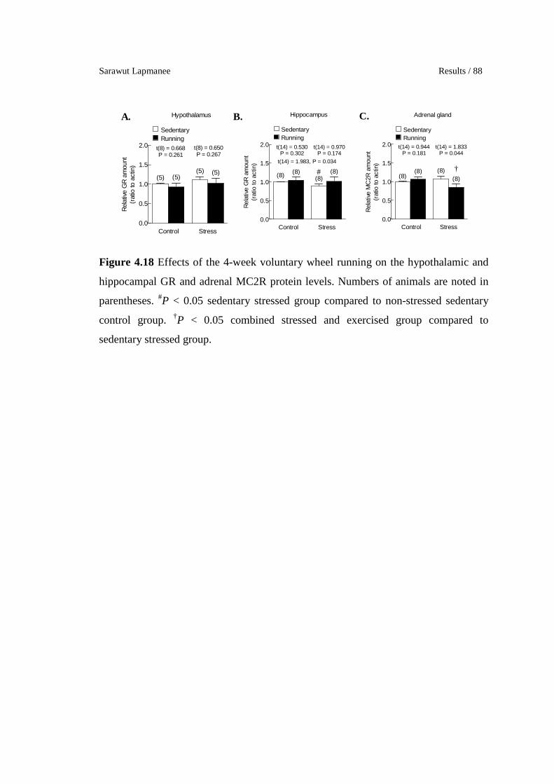
Sarawut Lapmanee Results / 88
0.0
0.5
1.0
1.5
2.0
(5) (5)(5) (5)
SedentaryRunning
StressControl
Hypothalamus
t(8) = 0.650P = 0.267
t(8) = 0.668P = 0.261
Rel
ativ
e G
R a
mou
nt(r
atio
to a
ctin
)
0.0
0.5
1.0
1.5
2.0
(8) (8)(8)
(8)
SedentaryRunning
StressControl
Hippocampus
#
t(14) = 0.970P = 0.174
t(14) = 0.530P = 0.302
t(14) = 1.983, P = 0.034
Rel
ativ
e G
R a
mou
nt(r
atio
to a
ctin
)
0.0
0.5
1.0
1.5
2.0
(8)(8) (8)
(8)
SedentaryRunning
StressControl
†
Adrenal gland
t(14) = 1.833P = 0.044
t(14) = 0.944P = 0.181
Rel
ativ
e M
C2R
am
ount
(rat
io to
act
in)
A. B. C.
Figure 4.18 Effects of the 4-week voluntary wheel running on the hypothalamic and
hippocampal GR and adrenal MC2R protein levels. Numbers of animals are noted in
parentheses. #P < 0.05 sedentary stressed group compared to non-stressed sedentary
control group. †P < 0.05 combined stressed and exercised group compared to
sedentary stressed group.

University of Strasbourg/Mahidol University Ph.D. (Neurosciences & Physiology) / 89
0.0
0.5
1.0
1.5
2.0
(8) (8) (8) (8)
SedentaryRunning
StressControl
Amygdala
t(14) = 0.173P = 0.433
t(14) = 1.530P = 0.074
Rel
ativ
e N
ET
amou
nt(r
atio
to a
ctin
)
0.0
0.5
1.0
1.5
2.0
(8) (8) (8) (8)
SedentaryRunning
StressControl
Hippocampus
t(14) = 0.487P = 0.317
t(14) = 0.009P = 0.496
Rel
ativ
e N
ET
amou
nt(r
atio
to a
ctin
)
0.0
0.5
1.0
1.5
2.0
(8) (8) (8) (8)
SedentaryRunning
StressControl
Frontal cortex
t(14) = 0.358P = 0.363
t(14) = 0.504P = 0.311
Rel
ativ
e N
ET
amou
nt(r
atio
to a
ctin
)
0.0
0.5
1.0
1.5
2.0
(5)(5)
(5)(5)
SedentaryRunning
StressControl
Locus coeruleus
*†#
t(8) = 2.587 P = 0.016
t(8) = 2.135P = 0.033
t(8) = 2.685P = 0.014
Rel
ativ
e N
ET
amou
nt(r
atio
to a
ctin
)
0.0
0.5
1.0
1.5
2.0
(5) (5)(5) (5)
SedentaryRunning
StressControl
Periaqueductal gray
#t(8) = 1.892 P = 0.048
t(8) = 0.265P = 0.7978
t(8) = 0.415P = 0.345
Rel
ativ
e N
ET
amou
nt(r
atio
to a
ctin
)
0.0
0.5
1.0
1.5
2.0
(5)(5)
(5)(5)
SedentaryRunning
StressControl
Ventral tegmental area
**
t(8) = 0.722P = 0.246
t(8) = 3.015P = 0.008
Rel
ativ
e N
ET
amou
nt(r
atio
to a
ctin
)
0.0
0.5
1.0
1.5
2.0
(5) (5) (5)
(5)
SedentaryRunning
StressControl
Septum
t(8) = 1.186P = 0.135
t(8) = 0.388P = 0.354
Rel
ativ
e N
ET
amou
nt(r
atio
to a
ctin
)
0.0
0.5
1.0
1.5
2.0
(5) (5)(5) (5)
SedentaryRunning
StressControl
Nucleus accumbens
t(8) = 0.366P = 0.362
t(8) = 0.652P = 0.266
Rel
ativ
e N
ET
amou
nt(r
atio
to a
ctin
)
A. B.
C. D.
E. F.
G. H.
Figure 4.19 Effects of the 4-week voluntary wheel running on NET protein levels.
Numbers of animals are noted in parentheses. *P < 0.05, **P < 0.01 exercised group
compared to non-stressed control sedentary group. #P < 0.05 sedentary stressed group
compared to non-stressed sedentary control group. †P < 0.05 combined stressed and
exercised group compared to sedentary stressed group.

Sarawut Lapmanee Results / 90
0.0
0.5
1.0
1.5
2.0
(8)(8)
(8) (8)
SedentaryRunning
StressControl
Amygdala
t(14) = 0.108P = 0.458
t(14) = 0.931P = 0.184
Rel
ativ
e S
ER
T am
ount
(rat
io to
act
in)
0.0
0.5
1.0
1.5
2.0
(8) (8)(8)
(8)
SedentaryRunning
StressControl
Hippocampus
##
t(14) = 1.623P = 0.063
t(14) = 0.555P = 0.294
t(14) = 3.072, P = 0.004
Rel
ativ
e S
ER
T am
ount
(rat
io to
act
in)
0.0
0.5
1.0
1.5
2.0
(8) (8) (8) (8)
SedentaryRunning
StressControl
Frontal cortex
t(14) = 0.252P = 0.805
t(14) = 1.037P = 0.159
Rel
ativ
e S
ER
T am
ount
(rat
io to
act
in)
0.0
0.5
1.0
1.5
2.0
(5) (5)(5) (5)
SedentaryRunning
StressControl
Dorsal raphe
#
t(8) = 0.231P = 0.412
t(8) = 0.965P = 0.182
t(8) = 2.235, P = 0.028
Rel
ativ
e S
ER
T am
ount
(rat
io to
act
in)
0.0
0.5
1.0
1.5
2.0
(5) (5) (5) (5)
SedentaryRunning
StressControl
Periaqueductal gray
t(8) = 0.220P = 0.412
t(8) = 1.010P = 0.171
Rel
ativ
e S
ER
T am
ount
(rat
io to
act
in)
0.0
0.5
1.0
1.5
2.0
(5) (5) (5)(5)
SedentaryRunning
StressControl
Ventral tegmental area
†
t(8) = 2.021P = 0.039
t(8) = 0.167P = 0.436
Rel
ativ
e S
ER
T am
ount
(rat
io to
act
in)
0.0
0.5
1.0
1.5
2.0
(5)(5)
(5) (5)
SedentaryRunning
StressControl
Septum
**
t(8) = 0.265P = 0.399
t(8) = 3.476P = 0.004
Rel
ativ
e S
ER
T am
ount
(rat
io to
act
in)
0.0
0.5
1.0
1.5
2.0
(5) (5) (5)(5)
SedentaryRunning
StressControl
Nucleus accumbens
t(8) = 0.830P = 0.215
t(8) = 0.669P = 0.261
Rel
ativ
e S
ER
T am
ount
(rat
io to
act
in)
A. B.
C. D.
E. F.
G. H.
Figure 4.20 Effects of the 4-week voluntary wheel running on SERT protein levels.
Numbers of animals are noted in parentheses. **P < 0.01 exercised group compared to
non-stressed control sedentary group. #P < 0.05, ##P < 0.01 sedentary stressed group
compared to non-stressed sedentary control group. †P < 0.01 combined stressed and
exercised group compared to sedentary stressed group.

University of Strasbourg/Mahidol University Ph.D. (Neurosciences & Physiology) / 91
0.0
0.5
1.0
1.5
2.0
(8) (8)(8)
(8)
SedentaryRunning
StressControl
Amygdala
#t(14) = 1.878 P = 0.041
t(14) = 1.340P = 0.101
t(14) = 0.146P = 0.443
Rel
ativ
e 5-
HT
2CR
am
ount
(rat
io t
o ac
tin)
0.0
0.5
1.0
1.5
2.0
(8) (8)
(8)(8)
SedentaryRunning
StressControl
Hippocampus
t(14) = 0.693P = 0.250
t(14) = 1.263P = 0.114
Rel
ativ
e 5-
HT
2CR
am
ount
(rat
io t
o ac
tin)
0.0
0.5
1.0
1.5
2.0
(8) (8)(8)
(8)
SedentaryRunning
StressControl
Frontal cortex
†
t(14) = 1.783P = 0.048
t(14) = 1.317P = 0.105
Rel
ativ
e 5-
HT
2CR
am
ount
(rat
io to
act
in)
0.0
0.5
1.0
1.5
2.0
(5)(5) (5) (5)
SedentaryRunning
StressControl
Dorsal raphe
*
t(8) = 0.0162P = 0.494
t(8) = 2.012P = 0.040
Rel
ativ
e 5-
HT
2CR
am
ount
(rat
io to
act
in)
0.0
0.5
1.0
1.5
2.0
(5) (5) (5) (5)
SedentaryRunning
StressControl
Locus coeruleus
t(8) = 0.940P = 0.187
t(8) = 0.999P = 0.174
Rel
ativ
e 5-
HT
2CR
am
ount
(rat
io to
act
in)
0.0
0.5
1.0
1.5
2.0
(5)(5)
(5)(5)
SedentaryRunning
StressControl
Periaqueductal gray
*
t(8) = 1.114P = 0.149
t(8) = 2.762P = 0.012
Rel
ativ
e 5-
HT
2CR
am
ount
(rat
io t
o ac
tin)
0.0
0.5
1.0
1.5
2.0
(5) (5) (5)
(5)
SedentaryRunning
StressControl
Ventral tegmental area
t(8) = 128P = 0.118
t(8) = 0.224P = 0.414
Rel
ativ
e 5-
HT
2CR
am
ount
(rat
io t
o ac
tin)
0.0
0.5
1.0
1.5
2.0
(5)(5) (5)
(5)
SedentaryRunning
StressControl
Septum
t(8) =1.162P = 0.139
t(8) = 1.213P = 0.130
Rel
ativ
e 5-
HT
2CR
am
ount
(rat
io to
act
in)
0.0
0.5
1.0
1.5
2.0
(5)(5) (5) (5)
SedentaryRunning
StressControl
Nucleus accumbens
*
t(8) = 0.244P = 0.407
t(8) = 1.923P = 0.045
Rel
ativ
e 5-
HT
2CR
am
ount
(rat
io t
o ac
tin)
A. B. C.
D. E. F.
G. H. I.
Figure 4.21 Effects of the 4-week voluntary wheel running on the 5-HT2CR protein
levels. Numbers of animals are noted in parentheses. *P < 0.05 exercised group
compared to non-stressed control sedentary group. #P < 0.05 sedentary stressed group
compared to non-stressed sedentary control group. †P < 0.01 combined stressed and
exercised group compared to sedentary stressed group.
Sarawut Lapmanee Results / 92
Table 4.2 Changes in bio-behavioral parameters of effects of the 4-week voluntary
wheel running under non-stressed condition and/or after the 4-week restraint stress
induction in male rats.
Parameter Ex vs control
Stress vs control
Stress + Ex vs stress
Depression % SP ↔ ↓↓↓ ↑ Serum corticosterone ↔ ↑ ↔ BDNF ↓ in the NA ↓↓ in the
hippocampus and the septum
↑ in the hippocampus
and the septum, ↓ in the NA
GR ↔ ↓ in the hippocampus ↔
Adrenal MC2R ↔ ↔ ↓ NET ↓↓ in the LC and
the VTA ↑ in the LC, ↓ in the
PAG ↓ in the LC
SERT ↑↑ in the septum ↓↓ in the hippocampus and the
DR
↓ in the VTA
5-HT2CR ↓ in the DR, the PAG, and the NA
↑ in the amygdala ↓ in the PFC
BDNF: Brain-derived neurotrophic factor; DR: dorsal raphe; EPM: elevated-plus maze; ETM:
elevated-T maze; FST: forced swimming test; GR: glucocorticoid receptor; LC: locus
coeruleus; MWM: Morris water maze; NA: nucleus accumbens; NET: norepinephrine
transporter; NOR: novel objective recognition; OFT: open-field test; PAG: periaqueductal
gray; SERT: serotonin reuptake transporter; VTA: Ventral tegmental area; SP: sucrose
preference; 5-HT2CR: serotonin type 2C receptor.

University of Strasbourg/Mahidol University Ph.D. (Neurosciences & Physiology) / 93
Experiment 3. To evaluate the effectiveness of agomelatine, venlafaxine,
and voluntary wheel running exercise in the prevention of stress-induced anxiety-,
depression-, and memory impairment-like behaviors and changes in the expression of
selected target proteins in brain regions associated with stress responses.
1. The 4-week pre-treatment of agomelatine, venlafaxine, and
voluntary wheel running induced changes in body weight, adrenal gland weight,
food intake, and corticosterone levels.
There was no difference in the starting body weight. Following the 4-week
pre-treatments, voluntary wheel running animals had lower body weight gain and food
intake. Exercising rats gradually increased their running distance throughout the
exercise training (Figure 4.22A−D). Agomelatine-and venlafaxine-treated rats did not
exhibit changes in their body weight gain but their food intake was higher than that of
the vehicle-treated sedentary stressed rats (Figure 4.22C). Despite gaining body weight
after stopping voluntary wheel running (Figure 4.22F), exercising rats subjected to
stress induction had a decrease in the final body weight (Figure 4.22D), thus,
demonstrating detraining effects of exercise. Therefore, pre-treatments with
monoamine modulators and voluntary exercise could prevent the stress-induced
decrease in body weight and food intake. Pre-treatment of agomelatine, venlafaxine or
voluntary wheel running did not alter adrenal weights (Figure 4.23A−D) but serum
corticosterone levels of agomelatine-treated rats was higher than non-stressed controls
and vehicle-treated sedentary stressed rats (Figure 4.23E).
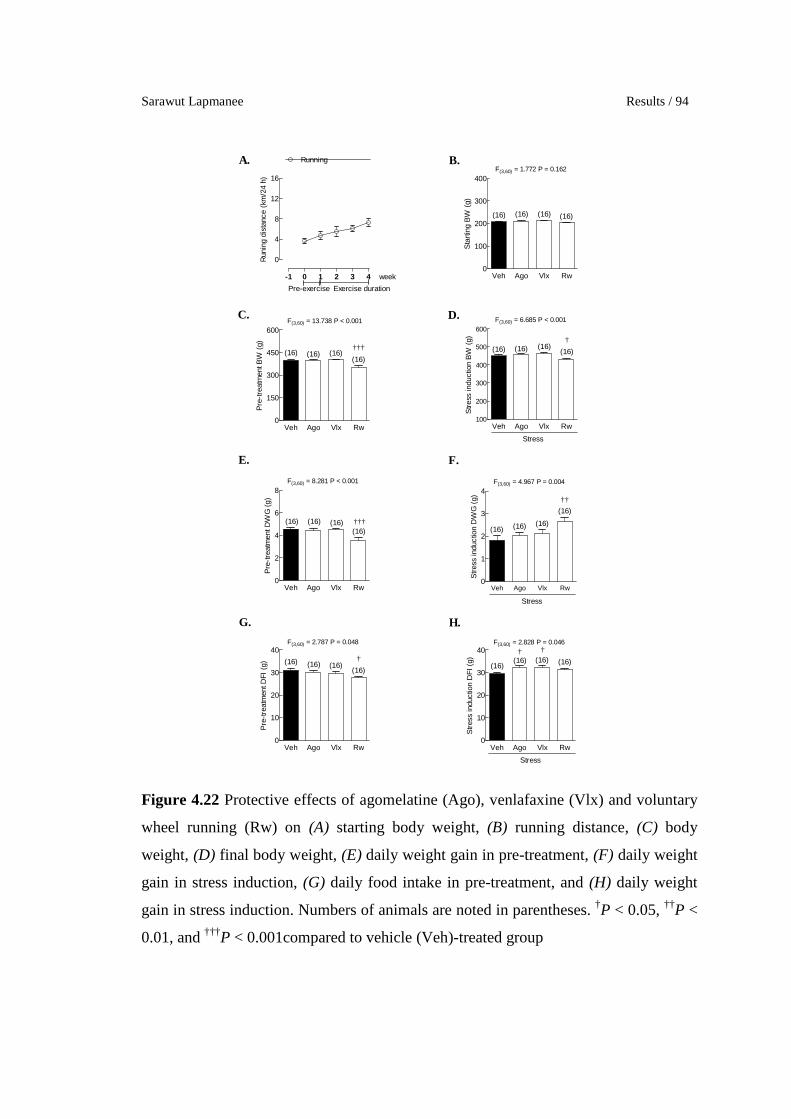
Sarawut Lapmanee Results / 94
0
4
8
12
16
Running
Exercise durationPre-exercise
week-1 0 1 2 3 4
Run
ing
dist
ance
(km
/24
h)
Veh Ago Vlx Rw0
100
200
300
400
(16) (16)(16)
F(3,60) = 1.772 P = 0.162
(16)
Sta
rtin
g B
W (
g)
Veh Ago Vlx Rw0
150
300
450
600
(16)(16)
F(3,60) = 13.738 P < 0.001
(16)(16)
†††
Pre
-tre
atm
ent B
W (
g)
Veh Ago Vlx Rw100
200
300
400
500
600
(16)(16)
F(3,60) = 6.685 P < 0.001
(16)(16)
Stress
†
Stre
ss in
duct
ion
BW
(g)
Veh Ago Vlx Rw0
2
4
6
8
(16) (16)(16)
F(3,60) = 8.281 P < 0.001
(16)†††
Pre
-tre
atm
ent D
WG
(g)
Veh Ago Vlx Rw0
1
2
3
4
(16) (16)(16)
F(3,60) = 4.967 P = 0.004
(16)
Stress
††
Stre
ss in
duct
ion
DW
G (
g)
Veh Ago Vlx Rw0
10
20
30
40
(16) (16)(16)
F(3,60) = 2.787 P = 0.048
(16)
†
Pre
-tre
atm
ent D
FI (
g)
Veh Ago Vlx Rw0
10
20
30
40(16) (16)
(16)
F(3,60) = 2.828 P = 0.046
(16)
Stress
† †
Str
ess
indu
ctio
n D
FI (
g)
A. B.
C. D.
E. F.
G. H.
Figure 4.22 Protective effects of agomelatine (Ago), venlafaxine (Vlx) and voluntary
wheel running (Rw) on (A) starting body weight, (B) running distance, (C) body
weight, (D) final body weight, (E) daily weight gain in pre-treatment, (F) daily weight
gain in stress induction, (G) daily food intake in pre-treatment, and (H) daily weight
gain in stress induction. Numbers of animals are noted in parentheses. †P < 0.05, ††P <
0.01, and †††P < 0.001compared to vehicle (Veh)-treated group
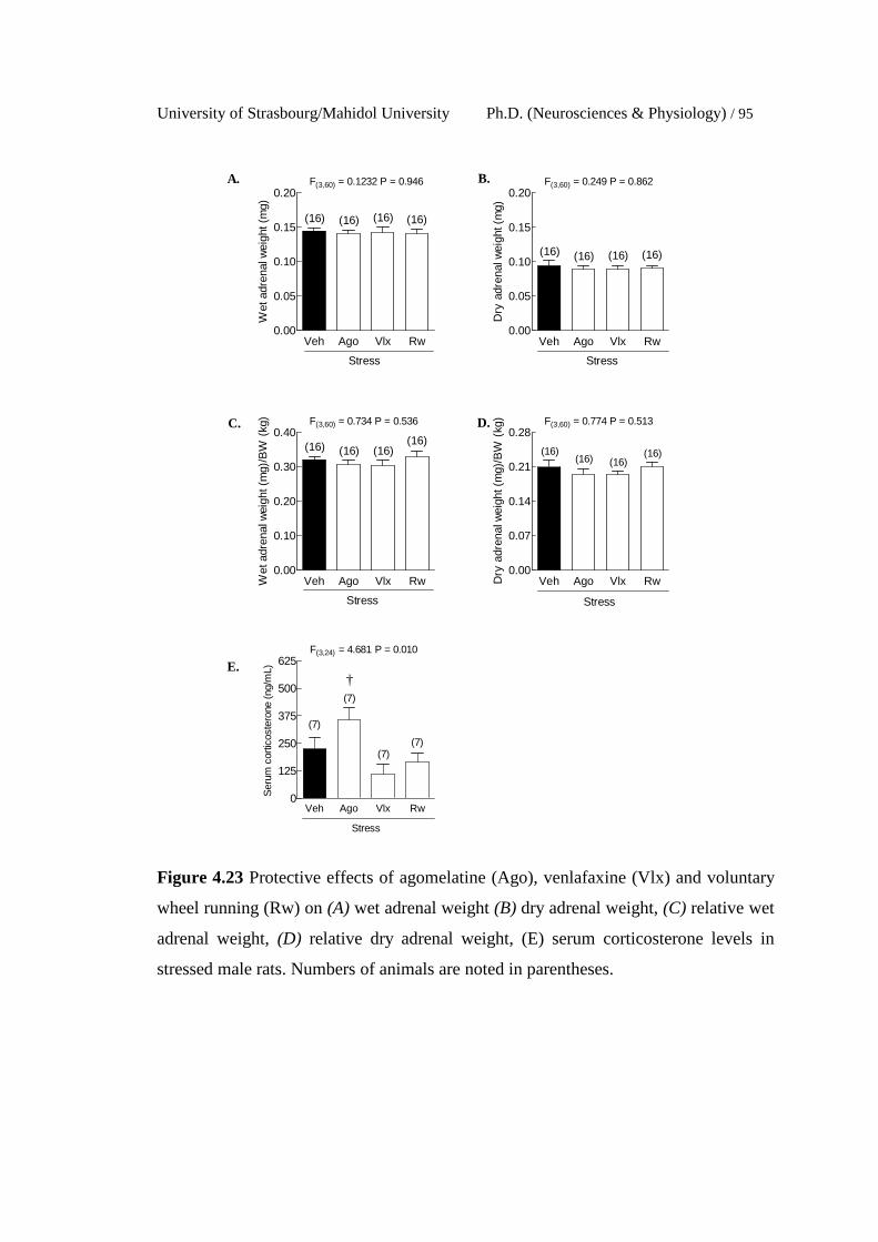
University of Strasbourg/Mahidol University Ph.D. (Neurosciences & Physiology) / 95
Veh Ago Vlx Rw0.00
0.05
0.10
0.15
0.20
(16) (16)
F(3,60) = 0.1232 P = 0.946
(16)(16)
Stress
Wet
adr
enal
wei
ght (
mg)
Veh Ago Vlx Rw0.00
0.05
0.10
0.15
0.20
(16) (16)
F(3,60) = 0.249 P = 0.862
(16)(16)
Stress
Dry
adr
enal
wei
ght (
mg)
Veh Ago Vlx Rw0.00
0.10
0.20
0.30
0.40
(16) (16)
F(3,60) = 0.734 P = 0.536
(16)(16)
Stress
Wet
adr
enal
wei
ght (
mg)
/BW
(kg
)
Veh Ago Vlx Rw0.00
0.07
0.14
0.21
0.28
(16) (16)
F(3,60) = 0.774 P = 0.513
(16)(16)
Stress
Dry
adr
enal
wei
ght (
mg)
/BW
(kg
)
Veh Ago Vlx Rw0
125
250
375
500
625
(7)
(7)
F(3,24) = 4.681 P = 0.010
(7)
(7)
Stress
†
Ser
um c
ortic
oste
rone
(ng/
mL)
A. B.
C. D.
E.
Figure 4.23 Protective effects of agomelatine (Ago), venlafaxine (Vlx) and voluntary
wheel running (Rw) on (A) wet adrenal weight (B) dry adrenal weight, (C) relative wet
adrenal weight, (D) relative dry adrenal weight, (E) serum corticosterone levels in
stressed male rats. Numbers of animals are noted in parentheses.

Sarawut Lapmanee Results / 96
2. The 4-week pre-treatment of agomelatine, venlafaxine, and
voluntary wheel running induced changes in behavioral responses.
2.1 Anxiety-like behaviors
In the EPM test, pre-treatment with venlafaxine, but not
agomelatine, or voluntary wheel running, increased the percent open arm time in
stressed rats when compared to vehicle-treated stressed rats (Figure 4.24B). However,
both pharmacological treatments and voluntary wheel running did not alter the percent
open arm entry, percent closed arm entry, percent closed arm time, total arm entries or
stress-related behaviors, i.e., rearing and grooming (Figure 4.24A and C−H). In the
ETM test, pre-treatment with agomelatine, venlafaxine or voluntary wheel running
decreased latencies in the inhibitory avoidance 1 and 2 in stressed rats when compared
to vehicle-treated stressed rats (Figure 4.25B−C), while having no effect on baseline
latency (Figure 4.25A) or one-way escape latency (Figure 4.25D). In the OFT test,
agomelatine and voluntary wheel running, but not venlafaxine increased time spent in
the inner zone and decreased time spent in the outer zone (Figure 4.26A−B). No
change in the number of lines crossed in the first 30 seconds or total lines crossed in 5
min were observed in agomelatine-, venlafaxine-, and exercise-treated rats (Figure
4.26C−D). In addition, stress-increased grooming behavior was reduced by pre-
treatment with agomelatine, venlafaxine, or with voluntary wheel running (Figure
4.26F).
2.2 Depression-like behaviors
Both agomelatine- and venlafaxine-treated stressed rats but not
exercised rats showed longer swimming duration and less immobility duration than
vehicle-treated stressed rats (Figure 4.27A−C). The number of fecal pellets could be
used as an index of the stress in the forced swimming test; however, there were no
difference among these groups (Figure 4.27D). No change in weekly sucrose
preference was also observed in pre-treatment or exercise period. When comparing
percent sucrose preference after completing the pre-treatment section, vehicle–treated
rats increased sucrose preference at the 2nd week of stress. However, pre-treatment did
not alter percent sucrose preference in stressed rats compared to the baseline period
and to the vehicle-treated group (Figure 4.27E).
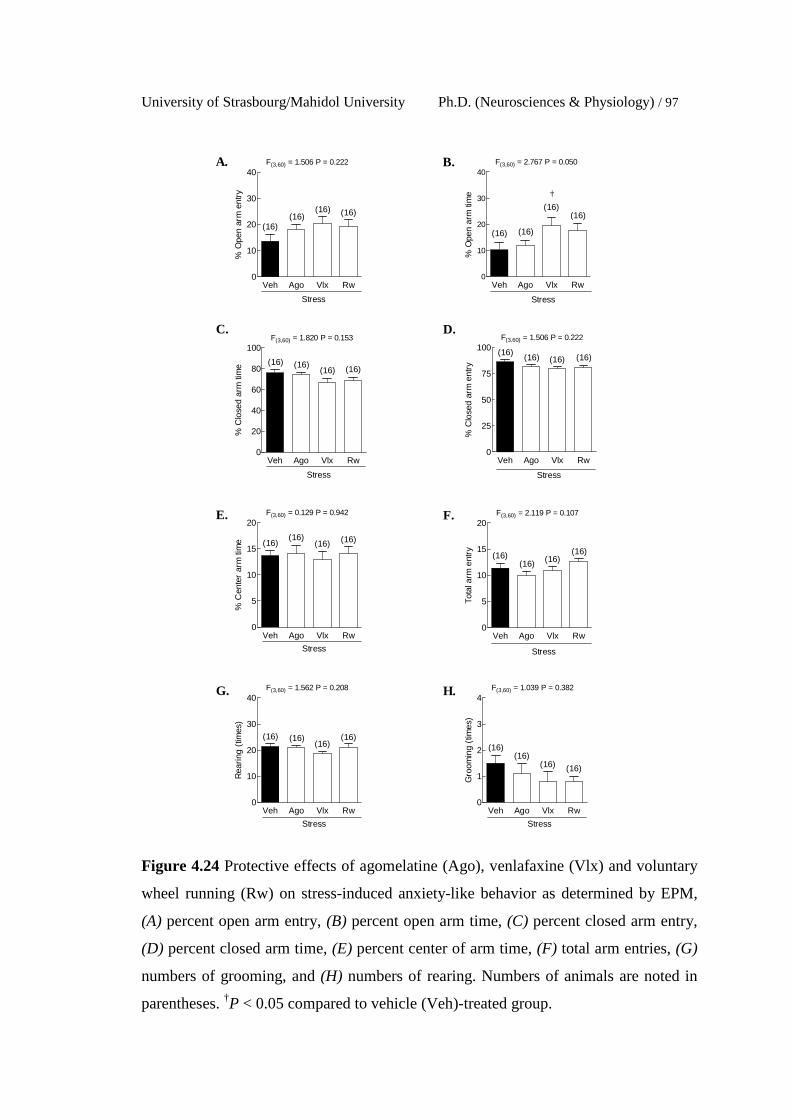
University of Strasbourg/Mahidol University Ph.D. (Neurosciences & Physiology) / 97
Veh Ago Vlx Rw0
10
20
30
40
(16)(16)
F(3,60) = 1.506 P = 0.222
(16)
(16)
Stress
% O
pen
arm
ent
ry
Veh Ago Vlx Rw0
10
20
30
40
(16)
(16)
F(3,60) = 2.767 P = 0.050
(16)
(16)
Stress
†
% O
pen
arm
tim
eVeh Ago Vlx Rw
0
20
40
60
80
100
(16)(16)
(16)
F(3,60) = 1.820 P = 0.153
(16)
Stress
% C
lose
d ar
m ti
me
Veh Ago Vlx Rw0
25
50
75
100(16) (16)
F(3,60) = 1.506 P = 0.222
(16)(16)
Stress%
Clo
sed
arm
ent
ry
Veh Ago Vlx Rw0
5
10
15
20
(16)(16)(16)
F(3,60) = 0.129 P = 0.942
(16)
Stress
% C
ente
r ar
m ti
me
Veh Ago Vlx Rw0
5
10
15
20
(16) (16)(16)
F(3,60) = 2.119 P = 0.107
(16)
Stress
Tota
l arm
ent
ry
Veh Ago Vlx Rw0
10
20
30
40
(16)(16)
(16)
F(3,60) = 1.562 P = 0.208
(16)
Stress
Rea
ring
(tim
es)
Veh Ago Vlx Rw0
1
2
3
4
(16)(16)
(16)
F(3,60) = 1.039 P = 0.382
(16)
Stress
Gro
omin
g (t
imes
)
A. B.
C. D.
E. F.
G. H.
Figure 4.24 Protective effects of agomelatine (Ago), venlafaxine (Vlx) and voluntary
wheel running (Rw) on stress-induced anxiety-like behavior as determined by EPM,
(A) percent open arm entry, (B) percent open arm time, (C) percent closed arm entry,
(D) percent closed arm time, (E) percent center of arm time, (F) total arm entries, (G)
numbers of grooming, and (H) numbers of rearing. Numbers of animals are noted in
parentheses. †P < 0.05 compared to vehicle (Veh)-treated group.
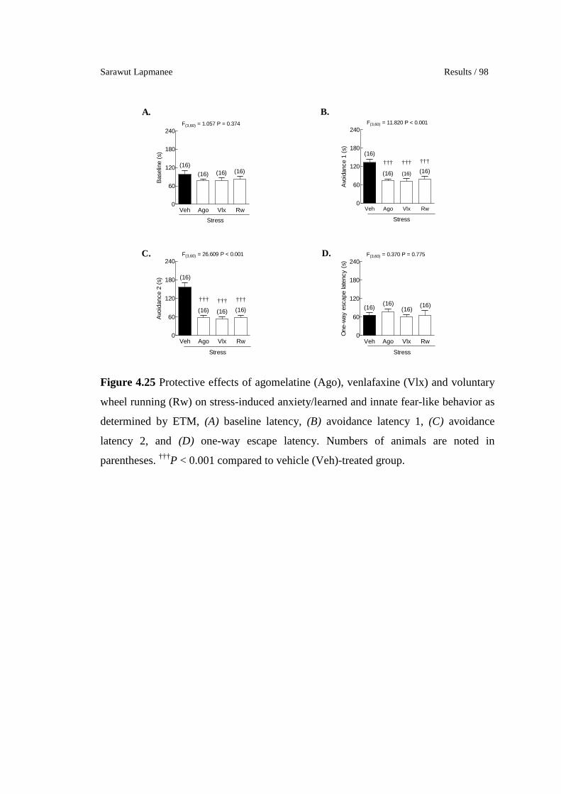
Sarawut Lapmanee Results / 98
Veh Ago Vlx Rw0
60
120
180
240
(16)(16)
F(3,60) = 1.057 P = 0.374
(16)(16)
Stress
Bas
elin
e (s
)
Veh Ago Vlx Rw0
60
120
180
240
(16) (16)
(16)
F(3,60) = 11.820 P < 0.001
(16)
Stress
††† ††† †††
Avo
idan
ce 1
(s)
Veh Ago Vlx Rw0
60
120
180
240
(16) (16)
F(3,60) = 26.609 P < 0.001
(16)
(16)
Stress
††† ††† †††
Avo
idan
ce 2
(s)
Veh Ago Vlx Rw0
60
120
180
240
(16)(16)
F(3,60) = 0.370 P = 0.775
(16)(16)
Stress
One
-way
esc
ape
late
ncy
(s)
A. B.
C. D.
Figure 4.25 Protective effects of agomelatine (Ago), venlafaxine (Vlx) and voluntary
wheel running (Rw) on stress-induced anxiety/learned and innate fear-like behavior as
determined by ETM, (A) baseline latency, (B) avoidance latency 1, (C) avoidance
latency 2, and (D) one-way escape latency. Numbers of animals are noted in
parentheses. †††P < 0.001 compared to vehicle (Veh)-treated group.
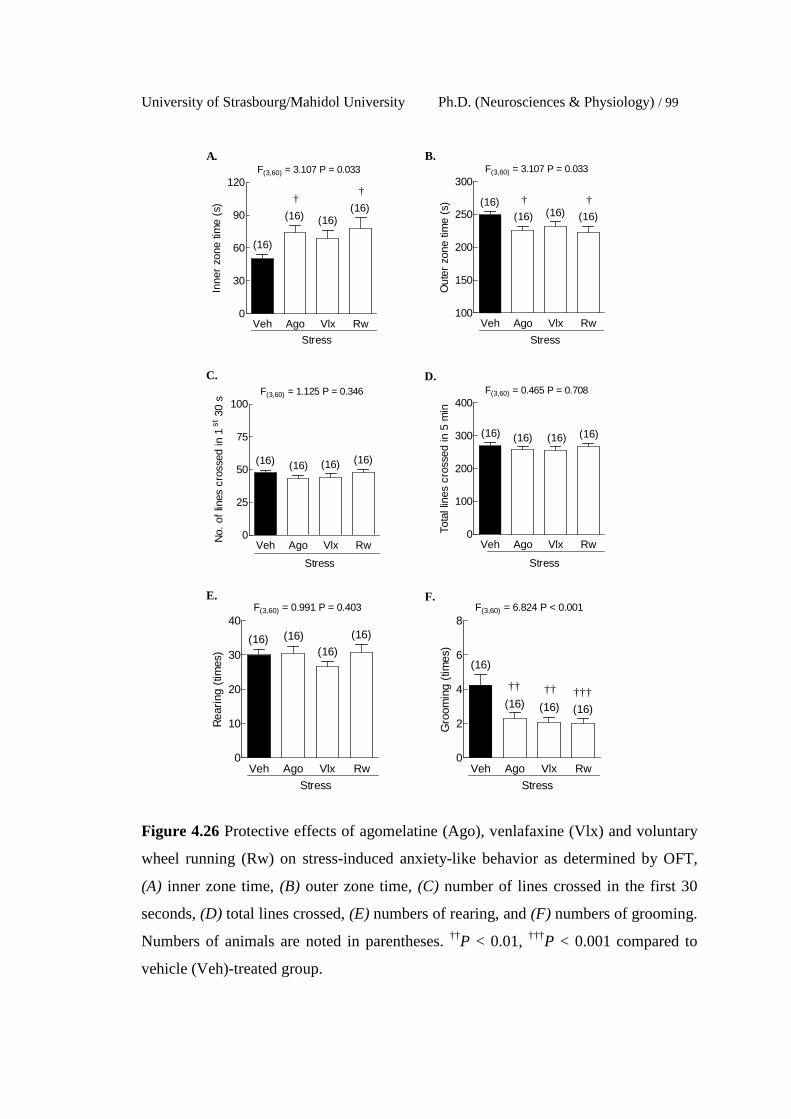
University of Strasbourg/Mahidol University Ph.D. (Neurosciences & Physiology) / 99
Veh Ago Vlx Rw0
30
60
90
120
(16) (16)
(16)
F(3,60) = 3.107 P = 0.033
(16)
Stress
††
Inne
r zo
ne ti
me
(s)
Veh Ago Vlx Rw100
150
200
250
300
(16) (16)(16)
F(3,60) = 3.107 P = 0.033
(16)
Stress
† †
Out
er z
one
time
(s)
Veh Ago Vlx Rw0
25
50
75
100
(16) (16)
F(3,60) = 1.125 P = 0.346
(16)(16)
Stress
No.
of
lines
cro
ssed
in 1
st 3
0 s
Veh Ago Vlx Rw0
100
200
300
400
(16) (16)
F(3,60) = 0.465 P = 0.708
(16)(16)
Stress
Tota
l lin
es c
ross
ed in
5 m
in
Veh Ago Vlx Rw0
10
20
30
40(16)
(16)(16)
F(3,60) = 0.991 P = 0.403
(16)
Stress
Rea
ring
(tim
es)
Veh Ago Vlx Rw0
2
4
6
8
(16) (16)
(16)
F(3,60) = 6.824 P < 0.001
(16)
Stress
†† †††††
Gro
omin
g (t
imes
)
A. B.
C. D.
E. F.
Figure 4.26 Protective effects of agomelatine (Ago), venlafaxine (Vlx) and voluntary
wheel running (Rw) on stress-induced anxiety-like behavior as determined by OFT,
(A) inner zone time, (B) outer zone time, (C) number of lines crossed in the first 30
seconds, (D) total lines crossed, (E) numbers of rearing, and (F) numbers of grooming.
Numbers of animals are noted in parentheses. ††P < 0.01, †††P < 0.001 compared to
vehicle (Veh)-treated group.

Sarawut Lapmanee Results / 100
Veh Ago Vlx Rw0
60
120
180
240
(16) (16)
F(3,60) = 6.201 P = 0.001
(16)
(16)
Stress
†† †††
Sw
imm
ing
time
(s)
Veh Ago Vlx Rw0
60
120
180
240
(16) (16)
F(3,60) = 6.201 P = 0.001
(16)
(16)
Stress
†† †††
Sw
imm
ing
time
(s)
Veh Ago Vlx Rw0
60
120
180
240
(16)(16)
F(3,60) = 5.633 P = 0.002
(16)(16)
Stress
†††††
Imm
obili
ty ti
me
(s)
Veh Ago Vlx Rw0
2
4
6
8
(16)(16)
F(3,60) = 0.309 P = 0.819
(16)(16)
Stress
Fec
al p
elle
ts
A. B.
C. D.
E.
84
88
92
96
100
Veh/Stress 4 weeks (n = 16)
Vlx/Stress 4 weeks (n = 16)
Rw/Stress 4 weeks (n = 16)
-1 0 1 2 3 4 5 6 7 8
Stress durationPre-experiment
Ago/Stress 4 weeks (n = 16)
week
Pre-treatment
aaa
F(4,75) = 5.499P = 0.007
% S
ucro
se p
refe
renc
e
Figure 4.27 Protective effects of agomelatine (Ago), venlafaxine (Vlx) and voluntary
wheel running (Rw) on stress-induced depression-like behavior as determined by FST
and sucrose preference test, (A) swimming duration, (B) climbing duration, (C)
immobility duration, (D) numbers of fecal pellets, and (E) weekly sucrose preference.
Numbers of animals are noted in parentheses. ††P < 0.01, †††P < 0.001 compared to
vehicle (Veh)-treated group. aaaP < 0.001 compared to percent sucrose preference after
completing 4-week pre-treatment period.

University of Strasbourg/Mahidol University Ph.D. (Neurosciences & Physiology) / 101
2.3 Learning and memory impairment-like behaviors
The MWM test showed that the agomelatine-, venlafaxine-,
and exercise-treated stressed rats exhibited less escape latencies (on day 1, 2 and 3)
and correct quadrant time than vehicle-treated stressed rats (Figure 4.28A−B), but
percent correct quadrant was not different among these groups (Figure 4.28C). In the
NOR, there was an improvement in the discrimination ratio in agomelatine- and
venlafaxine-treated rats when compared to vehicle-treated stressed rats. However, the
exercise-treated rats and vehicle-treated rats showed similar discrimination ratios
(Figure 4.28D). These results suggest that pre-treatment of agomelatine, venlafaxine,
and voluntary wheel running could improve learning and memory in stressed rats.
1 2 3
0
10
20
30
40
Veh/Stress 4 weeks (n = 16)
Ago/Stress 4 weeks (n = 16)
Vlx/Stress 4 weeks (n = 16)
Rw/Stress 4 weeks (n = 16)
***
*** ***
Day of training
Esc
ape
late
ncy
(s)
Veh Ago Vlx Rw0
10
20
30
40F(3,60) = 15.790 P < 0.001
(16)
(16) (16)(16)
Stress
††† ††† †††
Cor
rect
qua
dran
t tim
e (s
)
Veh Ago Vlx Rw0
25
50
75
100F(3,60) =0.977 P = 0.410
(16)(16)
(16)(16)
Stress
% C
orre
ct q
uadr
ant t
ime
Veh Ago Vlx Rw0.0
0.1
0.2
0.3
0.4F(3,60) = 3.647 P = 0.018
(16)
(16)
(16)
(16)
Stress
† †
Dis
crim
inat
ion
ratio
A. B.
C. D.
Figure 4.28 Protective effects of agomelatine (Ago), venlafaxine (Vlx) and voluntary
wheel running (Rw) on stress-induced learning and memory impairment-like behavior
as determined by MWM and NOR, (A) escape latency, (B) correct quadrant time, (C)
percent correct quadrant time, and (D) discrimination ratio. Numbers of animals are
noted in parentheses. †P < 0.05, ††P < 0.01, †††P < 0.001 compared to vehicle (Veh)-
treated group.

Sarawut Lapmanee Results / 102
3. The 4-week pre-treatment of agomelatine, venlafaxine, and
voluntary wheel running induced changes in target protein expression in the
brain regions associated with stress responses.
3.1 Levels of BDNF
Venlafaxine and voluntary wheel running resulted in higher
levels of hippocampal BDNF as compared to vehicle-treated rats. In contrast,
agomelatine or venlafaxine pre-treatment decreased BDNF protein levels in the locus
coeruleus and the septum (Figure 4.29B, E, and H).
3.2 Levels of GR in hypothalamus and hippocampus
The hypothalamic GR protein expression levels did not change
in any of the groups. The hippocampal GR protein levels were increased in
venlafaxine-treated stressed rats (Figure 4.30B).
3.3 Levels of NET
When compared to vehicle-treated stressed rats, NET protein
levels in the locus coeruleus were increased in exercise treated-stressed rats. However,
NET protein levels were decreased in the hippocampus and the hippocampus and
periaqueductal gray in agomelatine- and venlafaxine-treated rats (Figure 4.31B and E).
3.4 Levels of SERT
Pre-treatment of agomelatine and venlafaxine decreased the
septal SERT protein levels when compared to vehicle-treated rats (Figure 4.32G).
3.5 Levels of 5-HT2CR
No significant differences in the 5-HT2CR protein expression
were observed among these groups (Figure 4.33). However, there was a tendency
towards a downregulation of the 5-HT2CR in the amygdala of the exercise group. This
result might suggest partially anxiolytic effects of voluntary exercise on stress-induced
5-HT2CR upregulation in stressed rats.
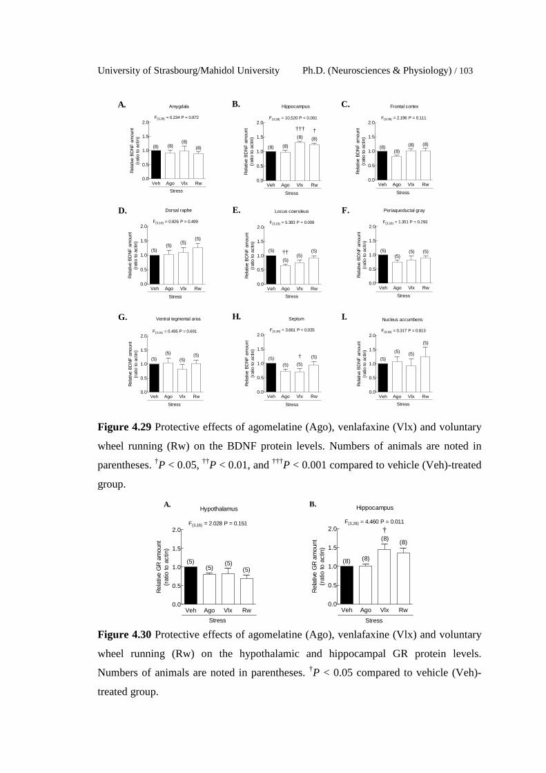
University of Strasbourg/Mahidol University Ph.D. (Neurosciences & Physiology) / 103
Figure 4.29 Protective effects of agomelatine (Ago), venlafaxine (Vlx) and voluntary
wheel running (Rw) on the BDNF protein levels. Numbers of animals are noted in
parentheses. †P < 0.05, ††P < 0.01, and †††P < 0.001 compared to vehicle (Veh)-treated
group.
Figure 4.30 Protective effects of agomelatine (Ago), venlafaxine (Vlx) and voluntary
wheel running (Rw) on the hypothalamic and hippocampal GR protein levels.
Numbers of animals are noted in parentheses. †P < 0.05 compared to vehicle (Veh)-
treated group.
Veh Ago Vlx Rw0.0
0.5
1.0
1.5
2.0
Stress
(8) (8)(8)
(8)
Amygdala
F(3,28) = 0.234 P = 0.872
Rel
ativ
e B
DN
F a
mou
nt(r
atio
to a
ctin
)
Veh Ago Vlx Rw0.0
0.5
1.0
1.5
2.0
Stress
(8) (8)
(8) (8)
Hippocampus
††† †
F(3,28) = 10.520 P < 0.001
Rel
ativ
e B
DN
F a
mou
nt(r
atio
to a
ctin
)
Veh Ago Vlx Rw0.0
0.5
1.0
1.5
2.0
Stress
(8)(8)
(8) (8)
Frontal cortex
F(3,28) = 2.196 P = 0.111
Rel
ativ
e B
DN
F a
mou
nt(r
atio
to a
ctin
)
Veh Ago Vlx Rw0.0
0.5
1.0
1.5
2.0
Stress
(5)(5)
(5)(5)
Dorsal raphe
F(3,16) = 0.826 P = 0.499
Rel
ativ
e B
DN
F a
mou
nt(r
atio
to a
ctin
)
Veh Ago Vlx Rw0.0
0.5
1.0
1.5
2.0
Stress
(5)
(5)(5)
(5)
Locus coeruleus
††
F(3,16) = 5.383 P = 0.009
Rel
ativ
e B
DN
F a
mou
nt(r
atio
to a
ctin
)
Veh Ago Vlx Rw0.0
0.5
1.0
1.5
2.0
Stress
(5)(5)
(5) (5)
Periaqueductal gray
F(3,16) = 1.351 P = 0.293
Rel
ativ
e B
DN
F a
mou
nt(r
atio
to a
ctin
)
Veh Ago Vlx Rw0.0
0.5
1.0
1.5
2.0
Stress
(5)(5)
(5)(5)
Ventral tegmental area
F(3,16) = 0.495 P = 0.691
Rel
ativ
e B
DN
F a
mou
nt(r
atio
to a
ctin
)
Veh Ago Vlx Rw0.0
0.5
1.0
1.5
2.0
Stress
(5)(5)
(5)
(5)
Septum
F(3,16) = 3.661 P = 0.035
†
Rel
ativ
e B
DN
F a
mou
nt(r
atio
to a
ctin
)
Veh Ago Vlx Rw0.0
0.5
1.0
1.5
2.0
Stress
(5)
(5) (5)
(5)
Nucleus accumbens
F(3,16) = 0.317 P = 0.813
Rel
ativ
e B
DN
F a
mou
nt(r
atio
to a
ctin
)
A. B. C.
D. E. F.
G. H. I.
Veh Ago Vlx Rw0.0
0.5
1.0
1.5
2.0
Stress
(5) (5)(5)(5)
Hypothalamus
F(3,16) = 2.028 P = 0.151
Rel
ativ
e G
R a
mou
nt(r
atio
to a
ctin
)
Veh Ago Vlx Rw0.0
0.5
1.0
1.5
2.0
Stress
(8) (8)
(8)(8)
Hippocampus
†F(3,28) = 4.460 P = 0.011
Rel
ativ
e G
R a
mou
nt(r
atio
to a
ctin
)
A. B.

Sarawut Lapmanee Results / 104
Veh Ago Vlx Rw0.0
0.5
1.0
1.5
2.0
Stress
(8) (8) (8)(8)
Amygdala
F(3,28) = 0.376 P = 0.771
Rel
ativ
e N
ET
amou
nt(r
atio
to a
ctin
)
Veh Ago Vlx Rw0.0
0.5
1.0
1.5
2.0
Stress
(8) (8) (8)(8)
Hippocampus
††
F(3,28) = 6.330 P = 0.002
Rel
ativ
e N
ET
amou
nt(r
atio
to a
ctin
)Veh Ago Vlx Rw
0.0
0.5
1.0
1.5
2.0
Stress
(8) (8) (8)(8)
Frontal cortex
F(3,28) = 0.937 P = 0.436
Rel
ativ
e N
ET
amou
nt(r
atio
to a
ctin
)
Veh Ago Vlx Rw0.0
0.5
1.0
1.5
2.0
Stress
(5)(5) (5)
(5)
Locus coeruleus
††
F(3,16) = 5.333 P = 0.010
Rel
ativ
e N
ET
amou
nt(r
atio
to a
ctin
)
Veh Ago Vlx Rw0.0
0.5
1.0
1.5
2.0
Stress
(5)(5) (5)
(5)
Periaqueductal gray
†† ††
F(3,16) = 6.401 P = 0.005
Rel
ativ
e N
ET
amou
nt(r
atio
to a
ctin
)
Veh Ago Vlx Rw0.0
0.5
1.0
1.5
2.0
Stress
(5)
(5)
(5) (5)
Ventral tegmental area
F(3,16) = 0.451 P = 0.720
Rel
ativ
e N
ET
amou
nt(r
atio
to a
ctin
)
Veh Ago Vlx Rw0.0
0.5
1.0
1.5
2.0
Stress
(5)
(5) (5)
(5)
Septum
F(3,16) = 0.197 P = 0.897
Rel
ativ
e N
ET
amou
nt(r
atio
to a
ctin
)
Veh Ago Vlx Rw0.0
0.5
1.0
1.5
2.0
Stress
(5) (5)
(5) (5)
Nucleus accumbens
F(3,16) = 1.214 P = 0.337
Rel
ativ
e N
ET
amou
nt(r
atio
to a
ctin
)
A. B.
C. D.
E. F.
G. H.
Figure 4.31 Protective effects of agomelatine (Ago), venlafaxine (Vlx) and voluntary
wheel running (Rw) on the NET protein levels. Numbers of animals are noted in
parentheses. ††P < 0.01 compared to vehicle (Veh)-treated group.
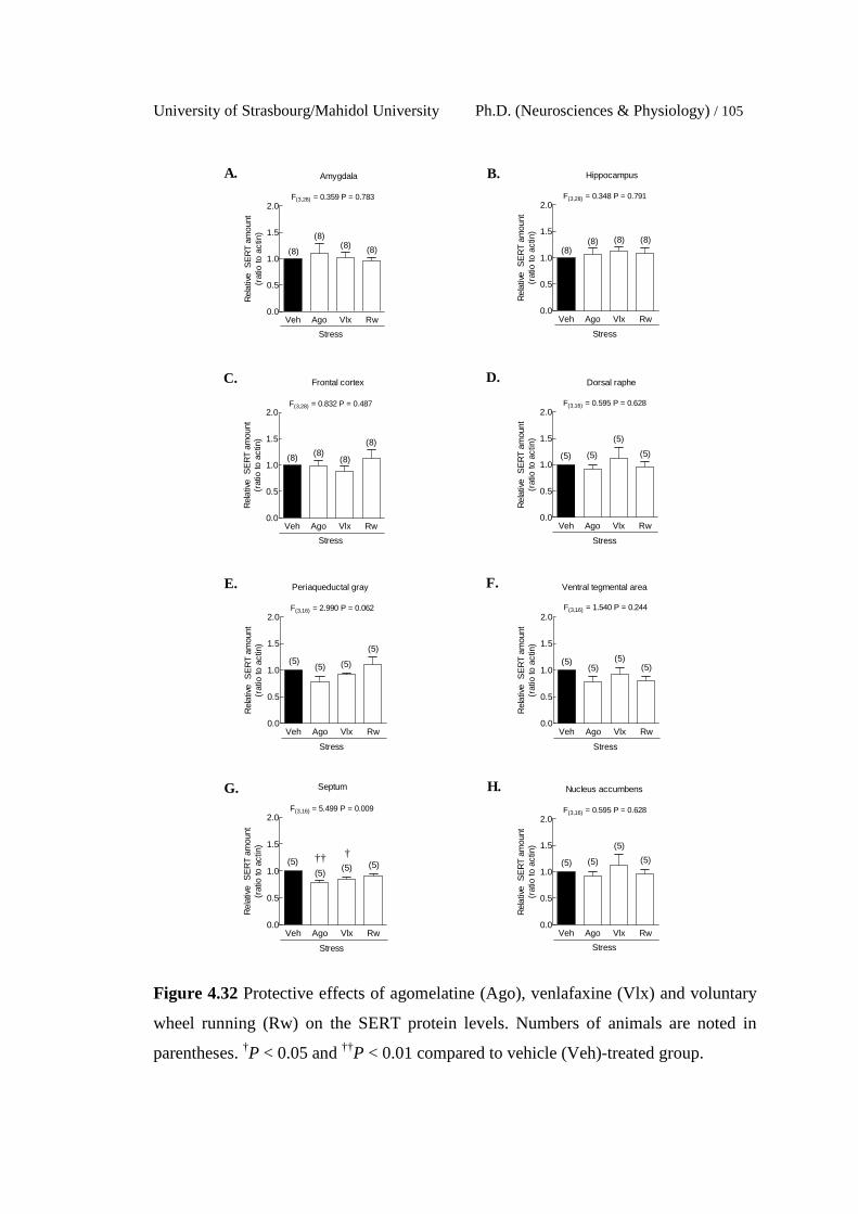
University of Strasbourg/Mahidol University Ph.D. (Neurosciences & Physiology) / 105
Veh Ago Vlx Rw0.0
0.5
1.0
1.5
2.0
Stress
(8)(8) (8)
(8)
Amygdala
F(3,28) = 0.359 P = 0.783
Rel
ativ
e S
ER
T am
ount
(rat
io to
act
in)
Veh Ago Vlx Rw0.0
0.5
1.0
1.5
2.0
Stress
(8)(8) (8) (8)
Hippocampus
F(3,28) = 0.348 P = 0.791
Rel
ativ
e S
ER
T am
ount
(rat
io to
act
in)
Veh Ago Vlx Rw0.0
0.5
1.0
1.5
2.0
Stress
(8) (8)(8)
(8)
Frontal cortex
F(3,28) = 0.832 P = 0.487
Rel
ativ
e S
ER
T am
ount
(rat
io to
act
in)
Veh Ago Vlx Rw0.0
0.5
1.0
1.5
2.0
Stress
(5) (5)
(5)
(5)
Dorsal raphe
F(3,16) = 0.595 P = 0.628
Rel
ativ
e S
ER
T am
ount
(rat
io to
act
in)
Veh Ago Vlx Rw0.0
0.5
1.0
1.5
2.0
Stress
(5)(5) (5)
(5)
Periaqueductal gray
F(3,16) = 2.990 P = 0.062
Rel
ativ
e S
ER
T am
ount
(rat
io to
act
in)
Veh Ago Vlx Rw0.0
0.5
1.0
1.5
2.0
Stress
(5) (5)(5)(5)
Ventral tegmental area
F(3,16) = 1.540 P = 0.244
Rel
ativ
e S
ER
T am
ount
(rat
io to
act
in)
Veh Ago Vlx Rw0.0
0.5
1.0
1.5
2.0
Stress
(5)(5)
(5) (5)
Septum
F(3,16) = 5.499 P = 0.009
†††
Rel
ativ
e S
ER
T am
ount
(rat
io to
act
in)
Veh Ago Vlx Rw0.0
0.5
1.0
1.5
2.0
Stress
(5) (5)
(5)
(5)
Nucleus accumbens
F(3,16) = 0.595 P = 0.628
Rel
ativ
e S
ER
T am
ount
(rat
io to
act
in)
A. B.
C. D.
E. F.
G. H.
Figure 4.32 Protective effects of agomelatine (Ago), venlafaxine (Vlx) and voluntary
wheel running (Rw) on the SERT protein levels. Numbers of animals are noted in
parentheses. †P < 0.05 and ††P < 0.01 compared to vehicle (Veh)-treated group.
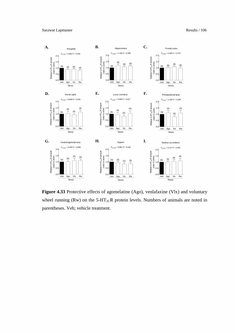
Sarawut Lapmanee Results / 106
Veh Ago Vlx Rw0.0
0.5
1.0
1.5
2.0
Stress
(8) (8) (8) (8)
Amygdala
F(3,28) = 2.854 P = 0.055
Rel
ativ
e 5-
HT
2CR
am
ount
(rat
io to
act
in)
Veh Ago Vlx Rw0.0
0.5
1.0
1.5
2.0
Stress
(8)
(8)(8) (8)
Hippocampus
F(3,28) = 1.318 P = 0.288
Rel
ativ
e 5-
HT
2CR
am
ount
(rat
io t
o ac
tin)
Veh Ago Vlx Rw0.0
0.5
1.0
1.5
2.0
Stress
(8) (8)(8) (8)
Frontal cortex
F(3,28) = 0.425 P = 0.737
Rel
ativ
e 5-
HT
2CR
am
ount
(rat
io t
o ac
tin)
Veh Ago Vlx Rw0.0
0.5
1.0
1.5
2.0
Stress
(5)(5)
(5)
(5)
Dorsal raphe
F(3,16) = 0.589 P = 0.631
Rel
ativ
e 5-
HT
2CR
am
ount
(rat
io t
o ac
tin)
Veh Ago Vlx Rw0.0
0.5
1.0
1.5
2.0
Stress
(5)(5)
(5)
(5)
Locus coeruleus
F(3,16) = 0.596 P = 0.627R
elat
ive
5-H
T2C
R a
mou
nt(r
atio
to
actin
)
Veh Ago Vlx Rw0.0
0.5
1.0
1.5
2.0
Stress
(5)
(5)
(5)
(5)
Periaqueductal gray
F(3,16) = 1.216 P = 0.336
Rel
ativ
e 5-
HT
2CR
am
ount
(rat
io t
o ac
tin)
Veh Ago Vlx Rw0.0
0.5
1.0
1.5
2.0
Stress
(5) (5)(5) (5)
Ventral tegmental area
F(3,16) = 0.293 P = 0.830
Rel
ativ
e 5-
HT
2CR
am
ount
(rat
io to
act
in)
Veh Ago Vlx Rw0.0
0.5
1.0
1.5
2.0
Stress
(5) (5) (5)(5)
Septum
F(3,16) = 0.981 P = 0.426
Rel
ativ
e 5-
HT
2CR
am
ount
(rat
io to
act
in)
Veh Ago Vlx Rw0.0
0.5
1.0
1.5
2.0
Stress
(5) (5)(5)
(5)
Nucleus accumbens
F(3,16) = 0.127 P = 0.943
Rel
ativ
e 5-
HT
2CR
am
ount
(rat
io to
act
in)
A. B. C.
D. E. F.
G. H. I.
Figure 4.33 Protective effects of agomelatine (Ago), venlafaxine (Vlx) and voluntary
wheel running (Rw) on the 5-HT2CR protein levels. Numbers of animals are noted in
parentheses. Veh; vehicle treatment.

University of Strasbourg/Mahidol University Ph.D. (Neurosciences & Physiology) / 107
Table 4.3 Changes in bio-behavioral parameters of preventive effects of the 4-week
agomelatine, venlafaxine, and voluntary wheel running in stressed male rats.
Parameter Ago Vlx Rw
Anxiety EPM ↔ ↓ ↔
ETM ↓↓↓ ↓↓↓ ↓↓↓ OFT ↓ ↔ ↓
Depression % SP ↔ ↔ ↔ FST ↓↓ ↓↓ ↔
Learning MWM ↑↑↑ ↑↑ ↑↑↑ Memory MWM/NOR ↑↑↑ ↑↑↑ ↑↑↑ Locomotion EPM/OFT ↔ ↔ ↔ Serum corticosterone ↑ ↔ ↔
BDNF ↓↓ in the LC ↑↑↑ in the hippocampus, ↓ in
the septum
↑ in the hippocampus
GR ↔ ↑ in the hippocampus
↔
NET ↓↓ in the PAG ↓↓ in the hippocampus and
the PAG
↑↑ in the hippocampus and
the LC
SERT ↓↓ in the septum ↓↓ in the septum ↔
5-HT2CR ↔ ↔ ↔
Ago: agomelatine; BDNF: Brain-derived neurotrophic factor; EPM: elevated-plus maze;
ETM: elevated-T maze; FST: forced swimming test; GR: glucocorticoid receptor; LC: locus
coeruleus; MWM: Morris water maze; NET: norepinephrine transporter; NOR: novel
objective recognition; OFT: open-field test; PAG: periaqueductal gray; SERT: serotonin
reuptake transporter; SP: sucrose preference; Vlx: venlafaxine; Rw: wheel running; 5-HT2CR:
serotonin type 2C receptor.

Sarawut Lapmanee Results / 108
Part II: Characterization of melatonin receptor expressing cells in the
rodent brain using MT1 and MT2 melatonin receptor LacZ knock-in
reporter mice: Identification of potential action sites of agomelatine
on brain structures involved in stress.
Experiment 1. To validate and map MT1/MT2-LacZ expressing neurons
in transgenic knock-in mice.
1. Genotyping of MT1/MT2-LacZ knock-in mice
The knock-in strategy is shown in the Figure 4.34A. The LacZ reporter
gene was inserted at the MT1 or MT2 locus in order to investigate the expression
pattern of MT1/MT2 gene. For both MT1 and MT2 loci, the LacZ transgene was
inserted starting at the original ATG start codon, thus retaining all upstream and
downstream regulatory sequences. Only potential regulatory elements contained in the
intron of either the MT1 or the MT2 gene have been deleted by this strategy.
Therefore, MT1/MT2-LacZ reporter knock-in mice were considered ideal tools to
investigate the sites of expression of MT1/MT2 melatonin receptors in the absence of
reliable specific antibodies for the immunocytochemical localization of these
receptors.
We first selected heterozygous (MT1- or MT2-LacZKI+) for the initial
investigations. These heterozygous animals have one intact MT1 or MT2 allele, thus
reducing the risk of loss of the expression of the MT1/2 locus, or ectopic expression of
the MT1/2 locus, because of the absence of functional MT1/2 receptors. Heterozygous
carriers were also interbred to produce homozygous (MT1- or MT2-LacZKIKI )
offsprings that were identified by genotyping PCR (Figure 4.34B).
In the genotyping of MT1-LacZ knock-in mice, a product of ∼425 bp
indicated the wild-type MT1 allele and a product of ∼196 bp indicated the recombinant
MT1-LacZ allele. Similarly, in the genotyping of MT2-LacZ knock-in mice, a product
of ∼143 bp indicated the wide-type allele and a product of ∼666 bp indicated the
recombinant allele.

University of Strasbourg/Mahidol University Ph.D. (Neurosciences & Physiology) / 109
Figure 4.34 Generation of the MT1-/MT2-LacZ knock-in mice. (A) The MT1 or MT2
receptor gene locus organization before and after site-specific insertion. (B) Mouse
genotyping by PCR for MT1-/MT2-LacZ transgenic knock-in mice.
2. LacZ mapping of MT1 and MT2 gene expression in the brain
Detection of LacZ enzymatic activity and LacZ protein expressing neurons
were examined by X-gal histochemistry and immunocytochemistry respectively.
Immunocytochemistry detected LacZ protein mostly in lysosomal accumulations as
strongly stained dot-like structures, but also in the nucleus and sometimes in the
cytoplasm (Figure 4.35A and C). There was also some diffuse background staining in
immunocytochemistry that reduced the signal to noise ratio. X-gal histochemistry only
stained the lysosomal accumulations of LacZ, and the absence of background resulted
in a highly contrasted image (Figure 4.35B and D). Therefore, the staining of LacZ
enzymatic activity was selected to map the patterns of MT1 and MT2 expression in
these transgenic mice. Immunocytochemical detection of LacZ was used for the fine
characterization of the MT1 and MT2 expressing cells by colabeling for phenotypic
markers either by immunocytochemistry or by in situ hybridization.
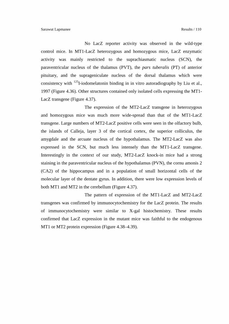
Sarawut Lapmanee Results / 110
No LacZ reporter activity was observed in the wild-type
control mice. In MT1-LacZ heterozygous and homozygous mice, LacZ enzymatic
activity was mainly restricted to the suprachiasmatic nucleus (SCN), the
paraventricular nucleus of the thalamus (PVT), the pars tuberalis (PT) of anterior
pituitary, and the suprageniculate nucleus of the dorsal thalamus which were
consistency with 125I-iodomelatonin binding in in vitro autoradiography by Liu et al.,
1997 (Figure 4.36). Other structures contained only isolated cells expressing the MT1-
LacZ transgene (Figure 4.37).
The expression of the MT2-LacZ transgene in heterozygous
and homozygous mice was much more wide-spread than that of the MT1-LacZ
transgene. Large numbers of MT2-LacZ positive cells were seen in the olfactory bulb,
the islands of Calleja, layer 3 of the cortical cortex, the superior colliculus, the
amygdale and the arcuate nucleus of the hypothalamus. The MT2-LacZ was also
expressed in the SCN, but much less intensely than the MT1-LacZ transgene.
Interestingly in the context of our study, MT2-LacZ knock-in mice had a strong
staining in the paraventricular nucleus of the hypothalamus (PVN), the cornu amonis 2
(CA2) of the hippocampus and in a population of small horizontal cells of the
molecular layer of the dentate gyrus. In addition, there were low expression levels of
both MT1 and MT2 in the cerebellum (Figure 4.37).
The pattern of expression of the MT1-LacZ and MT2-LacZ
transgenes was confirmed by immunocytochemistry for the LacZ protein. The results
of immunocytochemistry were similar to X-gal histochemistry. These results
confirmed that LacZ expression in the mutant mice was faithful to the endogenous
MT1 or MT2 protein expression (Figure 4.38–4.39).

University of Strasbourg/Mahidol University Ph.D. (Neurosciences & Physiology) / 111
Figure 4.35 Comparison MT1-LacZ staining between immunocytochemistry (A and
C) and X-gal histochemistry (B and D) in the suprachiasmatic nucleus (upper) and
pars tuberalis of anterior pituitary (lower) of transgenic knock-in mice.

Sarawut Lapmanee Results / 112
Figure 4.36 MT1-LacZ expressions in neurons as detected by X-gal histochemical
staining compared to 125I- iodomelatonin binding autoradiography by Liu et al., 1997.

University of Strasbourg/Mahidol University Ph.D. (Neurosciences & Physiology) / 113
Figure 4.37 X-gal histochemical mapping of MT1- and MT2-LacZ expression.
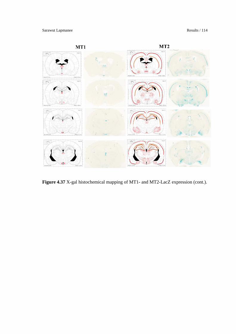
Sarawut Lapmanee Results / 114
Figure 4.37 X-gal histochemical mapping of MT1- and MT2-LacZ expression (cont.).
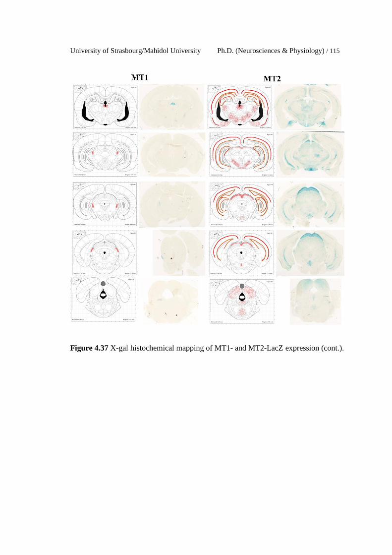
University of Strasbourg/Mahidol University Ph.D. (Neurosciences & Physiology) / 115
Figure 4.37 X-gal histochemical mapping of MT1- and MT2-LacZ expression (cont.).

Sarawut Lapmanee Results / 116
Figure 4.38 Immunocytochemical staining of MT1-LacZ expression in (A) the
suprachiasmatic nucleus; SCN and (B) pars tuberalis of anterior pituitary; PT. Nuclear
LacZ staining and lysosomal accumulation of LacZ can be seen in the SCN (C). In the
PT (D), the cytoplasmic staining is very strong, but sometimes the lysosomal
accumulation can also be seen (arrows).

University of Strasbourg/Mahidol University Ph.D. (Neurosciences & Physiology) / 117
Figure 4.39 Immunocytochemical staining of MT2-LacZ expressing neurons in (A)
the paraventicular nucleus of the hypothalamus, (B) the arcuate nucleus of the
hypothalamus, (C) the dorsal hippocampus, (D) the ventral hippocampus, (E) the CA2
of the hippocampus, and (F) the molecular layer of the dentate gyrus. DG; dentate
gyrus of hippocampus.

Sarawut Lapmanee Results / 118
Experiment 2. To characterize the phenotype of MT1/MT2-LacZ
expressing neurons in transgenic knock-in mice by determination of co-expressed
neuropeptides and neuronal markers.
2.1 Co-localization of MT1-LacZ expressing neurons
Both X-gal histochemical and immunocytochemical staining in the
previous experiment showed expression of the MT1-LacZ transgene in the SCN.
Generally, neurons in the SCN subregions are distinguished by their neuropeptide
contents. The SCN contains neurons containing AVP, GRP, and VIP in specific
subregions. Therefore, these neuropeptides were selected for identifying MT1
expression in the SCN. Combined staining showed that MT1-LacZ expressing neurons
co-localized with some, but not all, GRP and VIP neurons in the SCN, but did not co-
localize with AVP neurons (Figure 4.40).
2.2 Co-localization of MT2-LacZ expressing neurons
MT2-LacZ expression was much more widespread than that of MT1-LacZ.
We did not attempt to identify the phenotype of all MT2-LacZ expressing cells, but
concentrated on those structures that are involved in the modulation of stress
responses, in this case the PVN and the hippocampus.
The PVN contains CRH neurons, which are an important component of the
HPA axis. CRH probes were cloned and applied on brain sections. MT2-LacZ
expressing neurons co-localized with CRH in the PVN (Figure 4.41). Furthermore,
MT2-LacZ expressing neurons co-localized with CRH in the layer III of cerebral
cortex. Therefore, the co-localization of MT1 and CRH suggests that melatonin and
agomelatine might directly modulate the HPA axis in the PVN, which may have
implications for stress conditions such as depression.
Hippocampal interneurons express both the GAD1 and the GAD2
isoforms which synthesize GABA. Both isoforms are co-localized in 95% of GABA
cells in hippocampus. Therefore, GAD 1 and 2 probes were cloned and double
staining was performed with in situ hybridization and LacZ immunocytochemistry.
MT2-LacZ expression was present in some GAD2 neurons in the hippocampus
(Figure 4.42). GABAergic interneurons are inhibitory neurons. Melatonin signaling
University of Strasbourg/Mahidol University Ph.D. (Neurosciences & Physiology) / 119
through MT1 or MT2 receptors is mostly inhibitory. Thus MT action (and agomelatine
action) should result in the inhibition of these inhibitory interneurons, and thus an
activation of hippocampal activity. But in the CA2, MT2-LacZ expression is seen in
the pyramidal neurons, which are excitatory glutamatergic neurons (Figure 4.43). Thus
melatonin (and agomelatine) action should result in the selective inhibition of these
CA2 neurons and thus an inhibition of CA2-mediated hippocampal action. The
neurons staining most intensely for MT2-LacZ were small horizontal neurons located
in the outer molecular layer of the dentate gyrus. No description of this population
could be found in the literature and we could not colocalize these neurons with any of
the neuropeptide markers we tested (i.e., calbindin, choline acetyltransferase, galanin,
neuropeptide Y, and somatostatin). Only a very few of these cells colocalized with
GAD.

Sarawut Lapmanee Results / 120
Figure 4.40 Double-labeling with MT1-LacZ-Immunohistochemistry (green) and non-
radioactive in situ hybridization for neuropeptides (red). Diagram representing of the
cell and fiber distributions of neuropeptides in the SCN (Abrahamson and Moore,
2001) (A) AVP, (B), GRP, and (C) VIP. For AVP, the green dots of the MT1-LacZ
cells are clearly located outside of the area of the AVP cells (D). For VIP (E) and GRP
(F) some red neuropeptide cells contain the green dot indicating co-expression of the
MT1-LacZ transgene (arrows/arrowheads).

University of Strasbourg/Mahidol University Ph.D. (Neurosciences & Physiology) / 121
Figure 4.41 Double-labeling with LacZ-immunohistochemistry for MT2 (green) and
non-radioactive in situ hybridization for CRH (red) in the paraventricular nucleus
(PVN) of the hypothalamus. Inset shows a group of CRH cells that are MT2 positive.
Asterisk shows CRH neurons without MT2 label. Most GRP cells are MT2 positive.
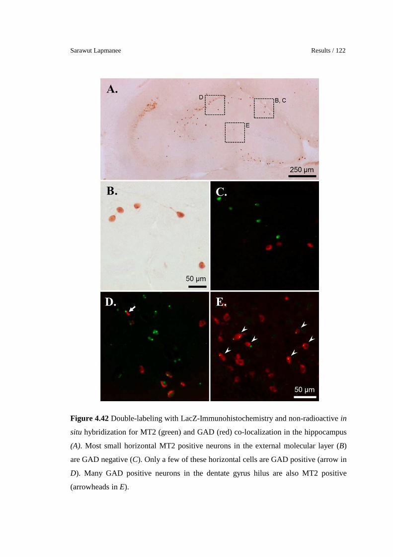
Sarawut Lapmanee Results / 122
Figure 4.42 Double-labeling with LacZ-Immunohistochemistry and non-radioactive in
situ hybridization for MT2 (green) and GAD (red) co-localization in the hippocampus
(A). Most small horizontal MT2 positive neurons in the external molecular layer (B)
are GAD negative (C). Only a few of these horizontal cells are GAD positive (arrow in
D). Many GAD positive neurons in the dentate gyrus hilus are also MT2 positive
(arrowheads in E).

University of Strasbourg/Mahidol University Ph.D. (Neurosciences & Physiology) / 123
Figure 4.43 Double-labeling with LacZ-Immunohistochemistry and non-radioactive in
situ hybridization for MT2 (green) and GAD (red) co-localization. The MT2 positive
cells in the CA2 of the hippocampus are GAD negative. A few non-pyramidal cells of
the CA2 are MT2 + GAD positive (arrowheads).

Sarawut Lapmanee Results / 124
Table 4.4 Localization of MT1 and MT2 melatonin receptors in transgenic MT1-
/MT2-LacZ knock-in mouse brains.
Brain regions MT1 MT2 Cerebral cortex isolated cells �� in layer III Basal forebrain - Olfactory tubercle - Islands of Calleja
– ��
Hypothalamus - Pre-optic area - SCN - PVN - Arcuate nucleus - Tuberal nucleus
–
�� (Co-localized with
GRP and VIP) –
a few isolated cells – –
� �
�� (Co-localized with CRH)
� �
Thalamus - PVT - Posterior limiting nucleus - Dorsal anteromedial nucleus - Lateral habenula - Intermediodorsal nucleus - Ventral medial nucleus
� �
� � � �
Hippocampus - CA2 - Dentate gyrus – outer molecular layer - Dentate gyrus − hilus
– �� ��
�
(Co-localized with GAD) Amygdala – � Midbrain - Superior colliculus (optic tectum) - Inferior colliculus - Interpeduncular nucleus - Dorsal raphe
– ��
� �� �
Hindbrain - Nucleus incertus - Parabrachial nucleus
� �
Cerebellum – – Pituitary gland - pars tuberalis
��
–
CA2: Cornu Ammonis; CRH: Corticotropin-releasing hormone; GAD: glutamic acid
decarboxylase; GRP: gastrin-releasing peptide; MT: melatonin receptor; PVN: paraventicular
nucleus of hypothalamus; PVT: paraventicular nucleus of the thalamus; SCN: suprachiasmatic
nucleus; VIP: vasoactive intestinal peptide.
University of Strasbourg/Mahidol University Ph.D. (Neurosciences & Physiology) / 125
CHAPTER V
DISCUSSION
5.1 Validation of restraint stress induction
Because restraint stress induces both physical and behavioral changes in
animals, and it is often used to model mental illness in human (Keim and Sigg, 1977;
Buynitsky and Mostofsky, 2009). The sustained circulating level of corticosterone
induces a negative feedback that diminishes the release of CRH in the hypothalamus
and ACTH from the pituitary gland. Restraint stress induction increases heart rate and
blood pressure in rats (Irvine et al., 1997; McDougall et al., 2005). Stress-induced
hyperactivation of HPA axis leads to adrenal hyperplasia and elevated serum and
urine corticosterone levels (Ulrich-Lai et al., 2006).
In the present study, 4-and 8-week chronic restraint stress (2 h/day, 5
days/week) induced an increase in the adrenal gland weight in male rats. However,
the absence of increased adrenal gland weight in the 1-week restraint stressed rats
suggested that the 1-week duration was too short to produce long-lasting effects.
Elevated corticosterone levels have been shown to cause a catabolic state and muscle
wasting via lipolysis and glycogenolysis (Hasselgren et al., 1999; Adam and Epel,
2007). Corticosterone was reported to reduce appetite and body weight by
downregulating hypothalamic neuropeptide Y/agouti gene-related protein and
amphetamine-regulated transcript peptide mRNA expression levels resulting in a
reduction in food intake and body weight gain (Liu et al., 2011). In addition, the
intensity and duration of restraint stress on the body weight in stressed rats were
associated with decreased levels of central monoamines and opioid peptides (Ely et
al., 1997). Therefore, a decrease in food intake and an increase in adrenal gland
weight could be used as appropriate indicators of physical change in stress induction.
Sarawut Lapmanee Discussion / 126
5.2 Behavioral response to different durations of restrain induction
It has been shown that acute stress exposure led to anxiety in human and
animal studies. Intense fear is present in posttraumatic stress disorders (Garner et al.,
2009). However, behavioral responses in male rats were found to depend on the
intensity of the stressors (Mercier et al., 2003). Apart from the acute stress reaction,
chronic stress-induced increase in corticosterone levels disturbed the balance of
monoamine neurotransmitters and neuronal function in several brain regions, thus
contributing to the development of mood disorders and memory problems (Jezova and
Hlavacova, 2008; Christoffel et al., 2011).
Herein, the study of different durations of restraint stress induction that
stressed rats exhibited anxiety-, depression-, and memory impairment-like behaviors.
Stressed rats had high anxiety as indicated by decreased open arm activity in the EPM
test and decreased time spent in the inner zone of open arena with hyperarousal in the
first 30s and stress-related behaviors (i.e., rearing and grooming) in the OFT test. The
1- and 4-week stressed rats further showed learned (conditioned) fear as indicated by
increased avoidance latency, but not innate fear in one-way escape latency of the
ETM test, suggesting that the subchronic and chronic restraint stress could produce a
generalized anxiety disorder-like condition in rats without panic which was similar in
human (Treit and Fundytus, 1988; Wood et al., 2008; Lapmanee et al., 2012; 2013).
Besides the anxiety-like behavior, the 4- and 8-week stressed rats showed
strong emotionality and depression-like behavior as indicated by increased immobility
time in the FST and altered sucrose preference. Reduction of sucrose preference in
stressed rats indicated a decreased sensitivity to reward and may be homologous to
anhedonia (Willner et al., 1996). These results were consistent with previous reports
(Lapmanee et al., 2012; 2013). These changes are related to the hopelessness and
anhedonic-like symptoms (a decreased ability to experience pleasures) in major
depression (Strekalova et al., 2004; Cryan and Holmes, 2005). In addition, chronic
stress in rats and mice has been shown to display several physiological changes that
are also observed in patients with depression such as abnormal sleep cycle, reduced
sexual function, and a disturbed circadian clock (Strekalova et al., 2004)
University of Strasbourg/Mahidol University Ph.D. (Neurosciences & Physiology) / 127
Changes in learning and memory were more complicated to apprehend.
As shown in the MWM test, an increase in escape latency (an indicator of impaired
spatial learning) was observed in the 4-week stressed rats (day 2), but the spatial
learning was apparently improved in the 1-week stressed rats (day 3). These results
suggest that the transitional period of stress promotes memory formation, but the
memory does not last long. This was consistent with a previous finding that acute
exposure to mild-to-moderate stress led to better learning in radial arm maze test
(Luine et al., 1996). These results imply that the first role of stress is to improve the
response to a stressful situation. The spatial memory or ability to retrieve previous
consolidated information, as indicated by an increase in the time spent to reach the
correct quadrant, was found to be impaired in the 1- and 4-week stressed rats.
Furthermore, the NOR test showed that the 4-week stressed rats had poor ability to
discriminate a previously explored object from a new object, a sign of cognitive or
memory impairment (Redrobe et al., 2010, 2012; Antunes and Biala, 2012). Such
stress on a short timescale is supposed to be “beneficial” because it improves the
responsiveness to stressors, thus increasing the chance of the organism’s survival in
dangerous situations. But this “reprogramming” of the physiological phenomena by
stress does not last long. That is why long term stress becomes detrimental.
However, there was no alteration of motor skills or locomotor activities
following chronic stress induction in the present study as suggested by the absence of
change in total arm entries in the EPM, total lines crossed in OFT, or climbing
duration in FST. Regarding approximately 80% change in the correct quadrant in all
studied group, this means that both control and stressed rats spent a similar time in the
correct quadrant (i.e., quadrant with a platform). Nevertheless, stressed rats required
more time swimming around the correct quadrant before reaching the platform, as
indicated by increased in the correct quadrant time. In other words, stressed rats
remembered where the correct quadrant was, but required additional time to actually
reach the platform.
Interestingly, it was not clear why some behavioral responses observed in
the 1- or 4-week stressed groups were absent in the 8-week stressed group. However,
it might be explained by habituation to prolonged restraining (Stamp and Herbert,
2001). The process of habituation involves the reduction of HPA axis activity and
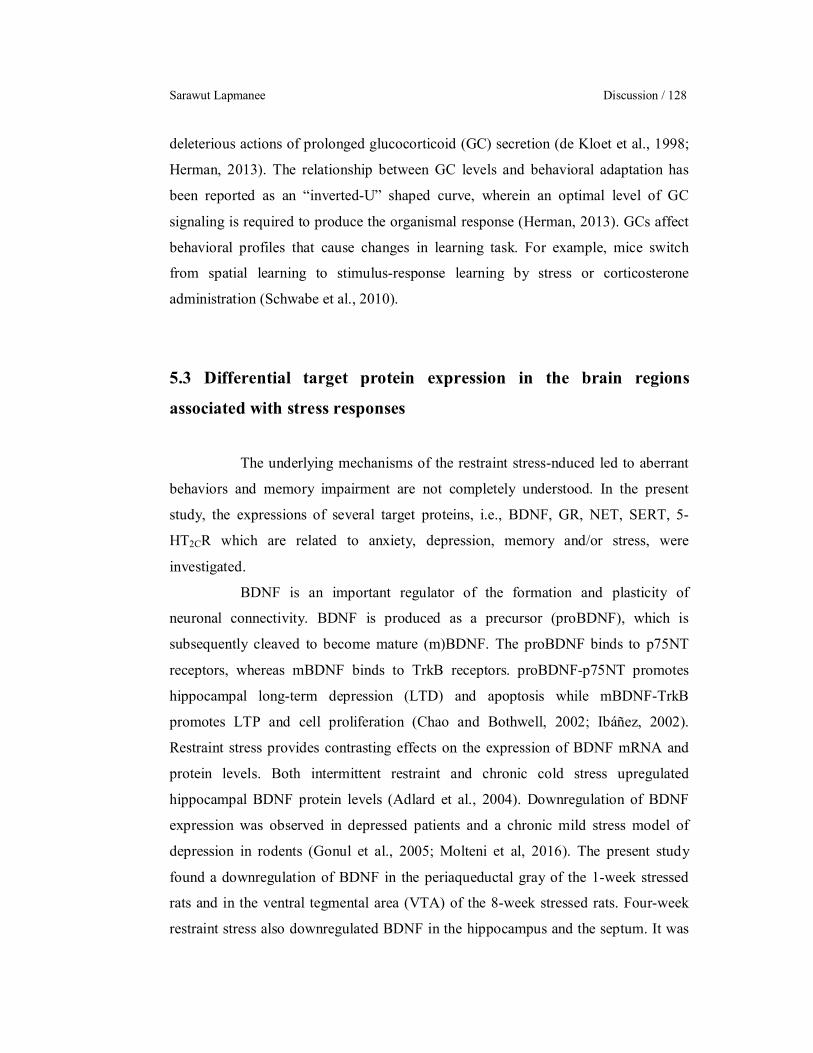
Sarawut Lapmanee Discussion / 128
deleterious actions of prolonged glucocorticoid (GC) secretion (de Kloet et al., 1998;
Herman, 2013). The relationship between GC levels and behavioral adaptation has
been reported as an “inverted-U” shaped curve, wherein an optimal level of GC
signaling is required to produce the organismal response (Herman, 2013). GCs affect
behavioral profiles that cause changes in learning task. For example, mice switch
from spatial learning to stimulus-response learning by stress or corticosterone
administration (Schwabe et al., 2010).
5.3 Differential target protein expression in the brain regions
associated with stress responses
The underlying mechanisms of the restraint stress-nduced led to aberrant
behaviors and memory impairment are not completely understood. In the present
study, the expressions of several target proteins, i.e., BDNF, GR, NET, SERT, 5-
HT2CR which are related to anxiety, depression, memory and/or stress, were
investigated.
BDNF is an important regulator of the formation and plasticity of
neuronal connectivity. BDNF is produced as a precursor (proBDNF), which is
subsequently cleaved to become mature (m)BDNF. The proBDNF binds to p75NT
receptors, whereas mBDNF binds to TrkB receptors. proBDNF-p75NT promotes
hippocampal long-term depression (LTD) and apoptosis while mBDNF-TrkB
promotes LTP and cell proliferation (Chao and Bothwell, 2002; Ibáñez, 2002).
Restraint stress provides contrasting effects on the expression of BDNF mRNA and
protein levels. Both intermittent restraint and chronic cold stress upregulated
hippocampal BDNF protein levels (Adlard et al., 2004). Downregulation of BDNF
expression was observed in depressed patients and a chronic mild stress model of
depression in rodents (Gonul et al., 2005; Molteni et al, 2016). The present study
found a downregulation of BDNF in the periaqueductal gray of the 1-week stressed
rats and in the ventral tegmental area (VTA) of the 8-week stressed rats. Four-week
restraint stress also downregulated BDNF in the hippocampus and the septum. It was

University of Strasbourg/Mahidol University Ph.D. (Neurosciences & Physiology) / 129
reported that stress-induced inflammatory cytokine production downregulated BDNF
in the hippocampus and the prefrontal cortex, but upregulated BDNF in the nucleus
accumbens (NA) leading to depression-like behavior in stressed male rats (Zhang et
al., 2014). BDNF expression in the VTA–NA pathway plays a crucial role in
resilience to social defeat stress (Krishnan et al. 2007). Antidepressant treatment for at
least 4 weeks can restore the decreased BDNF levels to normal levels in stressed rats
(Lee and Kim, 2010).
Prolonged high GC levels also down-regulate GR expression in the
hippocampus and the prefrontal cortex controlling negative feedback (Chiba et al.,
2012) leading to an impaired ability to regulate GC homeostasis. GR expression is
another indicator of stress response (Boyle et al., 2005; de Kloet et al., 2005). There
was the hyperactivity of the HPA axis in the 1-week stressed rats as shown by
increased hypothalamic GR protein levels. On the other hand, the 4-week stressed rats
had decreased levels of hippocampal GR indicating hyperactivity HPA axis in
depression (de Kloet et al., 1998; Zhu et al., 2014). Moreover, the 4-week restraint
stress markedly increased the MC2R protein levels suggesting an increased adrenal
sensitivity to ACTH (Liu et al., 2013).
NET is a key protein regulating noradrenergic transmission and a site of
action of TCA and NRIs. The 4-week stress induction upregulated NET protein levels
in the LC similar to chronic social defeat (Chen et al., 2012; Fan et al., 2015).
However, the upregulated NET in the locus should reduce noradrenergic transmission
in the LC, but the reduction of NET in the PAG should act in this structure much like
antidepressants, and thus stimulated noradrenergic transmission. Therefore, NET of
the 4-week stressed rats showed adaptive change by downregulating its projections
PAG (van Bockstaele and Aston-Jones, 1992). These results indicated that chronic
stress activated the LC–noradrenergic system and caused depression-like behaviors.
Serotonergic transmission is critically regulated by serotonin reuptake via
the SERT. Dysfunction of the SERT causes depression and anxiety disorders (Owens
and Nemeroff, 1994). The 1-and 8-week restraint stress induction downregulated
SERT protein levels in the VTA and the hippocampus. The 4-week restraint stress
tended to downregulate SERT in the dorsal raphe. Acute restraint stress and social
defeat decreased SERT in the raphe pontis and hippocampus (Berton et al., 1999;
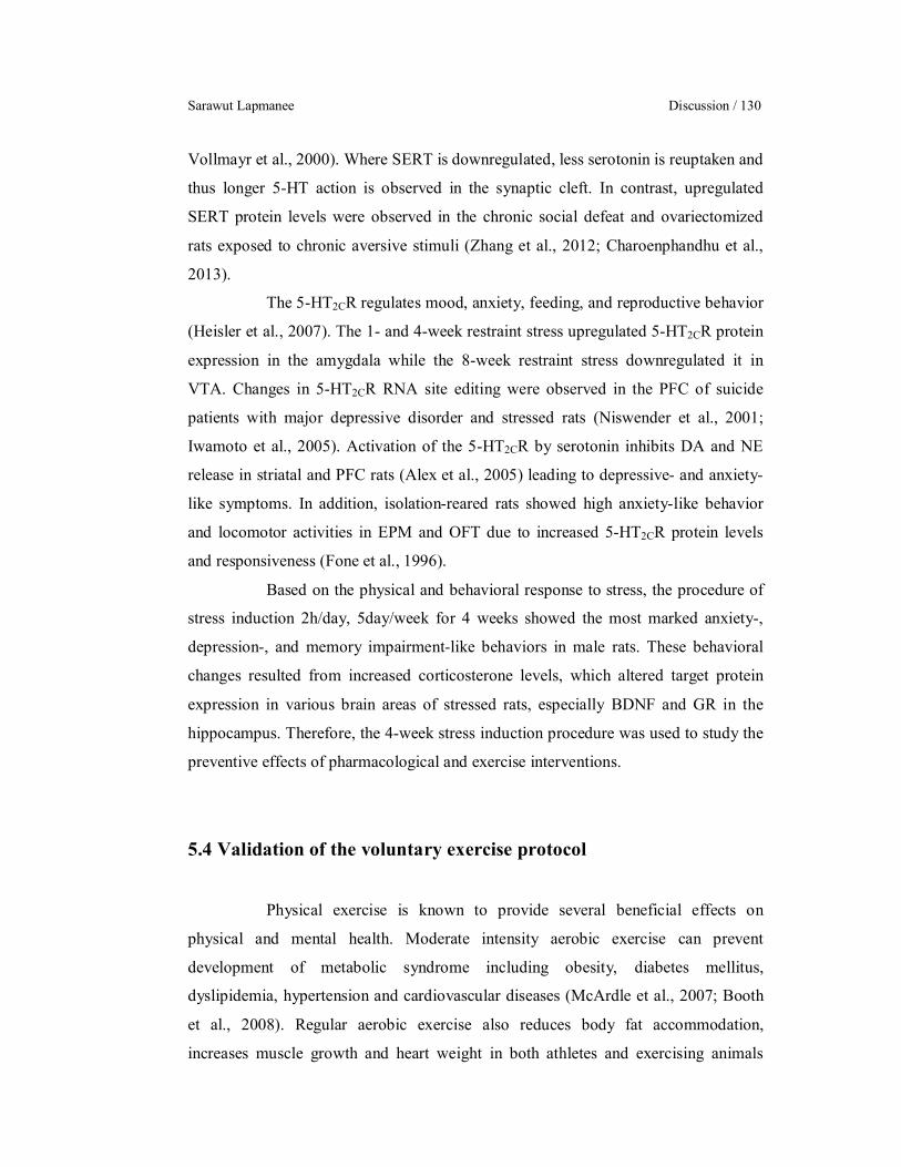
Sarawut Lapmanee Discussion / 130
Vollmayr et al., 2000). Where SERT is downregulated, less serotonin is reuptaken and
thus longer 5-HT action is observed in the synaptic cleft. In contrast, upregulated
SERT protein levels were observed in the chronic social defeat and ovariectomized
rats exposed to chronic aversive stimuli (Zhang et al., 2012; Charoenphandhu et al.,
2013).
The 5-HT2CR regulates mood, anxiety, feeding, and reproductive behavior
(Heisler et al., 2007). The 1- and 4-week restraint stress upregulated 5-HT2CR protein
expression in the amygdala while the 8-week restraint stress downregulated it in
VTA. Changes in 5-HT2CR RNA site editing were observed in the PFC of suicide
patients with major depressive disorder and stressed rats (Niswender et al., 2001;
Iwamoto et al., 2005). Activation of the 5-HT2CR by serotonin inhibits DA and NE
release in striatal and PFC rats (Alex et al., 2005) leading to depressive- and anxiety-
like symptoms. In addition, isolation-reared rats showed high anxiety-like behavior
and locomotor activities in EPM and OFT due to increased 5-HT2CR protein levels
and responsiveness (Fone et al., 1996).
Based on the physical and behavioral response to stress, the procedure of
stress induction 2h/day, 5day/week for 4 weeks showed the most marked anxiety-,
depression-, and memory impairment-like behaviors in male rats. These behavioral
changes resulted from increased corticosterone levels, which altered target protein
expression in various brain areas of stressed rats, especially BDNF and GR in the
hippocampus. Therefore, the 4-week stress induction procedure was used to study the
preventive effects of pharmacological and exercise interventions.
5.4 Validation of the voluntary exercise protocol
Physical exercise is known to provide several beneficial effects on
physical and mental health. Moderate intensity aerobic exercise can prevent
development of metabolic syndrome including obesity, diabetes mellitus,
dyslipidemia, hypertension and cardiovascular diseases (McArdle et al., 2007; Booth
et al., 2008). Regular aerobic exercise also reduces body fat accommodation,
increases muscle growth and heart weight in both athletes and exercising animals
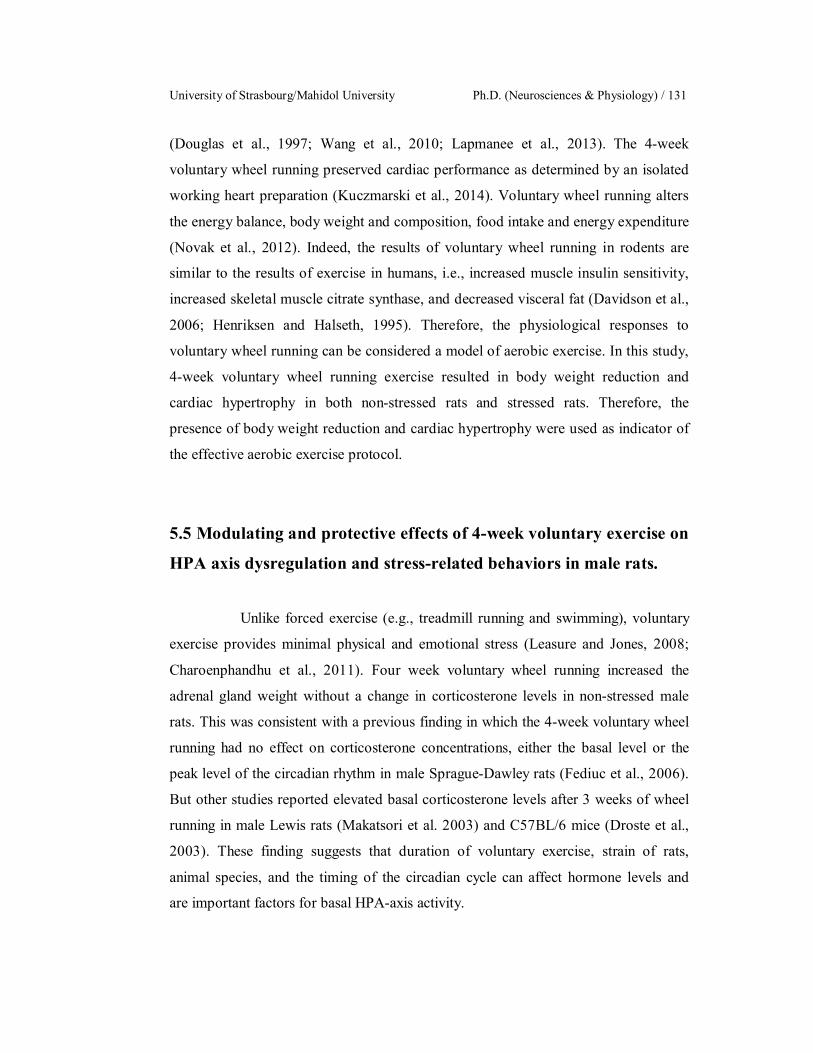
University of Strasbourg/Mahidol University Ph.D. (Neurosciences & Physiology) / 131
(Douglas et al., 1997; Wang et al., 2010; Lapmanee et al., 2013). The 4-week
voluntary wheel running preserved cardiac performance as determined by an isolated
working heart preparation (Kuczmarski et al., 2014). Voluntary wheel running alters
the energy balance, body weight and composition, food intake and energy expenditure
(Novak et al., 2012). Indeed, the results of voluntary wheel running in rodents are
similar to the results of exercise in humans, i.e., increased muscle insulin sensitivity,
increased skeletal muscle citrate synthase, and decreased visceral fat (Davidson et al.,
2006; Henriksen and Halseth, 1995). Therefore, the physiological responses to
voluntary wheel running can be considered a model of aerobic exercise. In this study,
4-week voluntary wheel running exercise resulted in body weight reduction and
cardiac hypertrophy in both non-stressed rats and stressed rats. Therefore, the
presence of body weight reduction and cardiac hypertrophy were used as indicator of
the effective aerobic exercise protocol.
5.5 Modulating and protective effects of 4-week voluntary exercise on
HPA axis dysregulation and stress-related behaviors in male rats.
Unlike forced exercise (e.g., treadmill running and swimming), voluntary
exercise provides minimal physical and emotional stress (Leasure and Jones, 2008;
Charoenphandhu et al., 2011). Four week voluntary wheel running increased the
adrenal gland weight without a change in corticosterone levels in non-stressed male
rats. This was consistent with a previous finding in which the 4-week voluntary wheel
running had no effect on corticosterone concentrations, either the basal level or the
peak level of the circadian rhythm in male Sprague-Dawley rats (Fediuc et al., 2006).
But other studies reported elevated basal corticosterone levels after 3 weeks of wheel
running in male Lewis rats (Makatsori et al. 2003) and C57BL/6 mice (Droste et al.,
2003). These finding suggests that duration of voluntary exercise, strain of rats,
animal species, and the timing of the circadian cycle can affect hormone levels and
are important factors for basal HPA-axis activity.
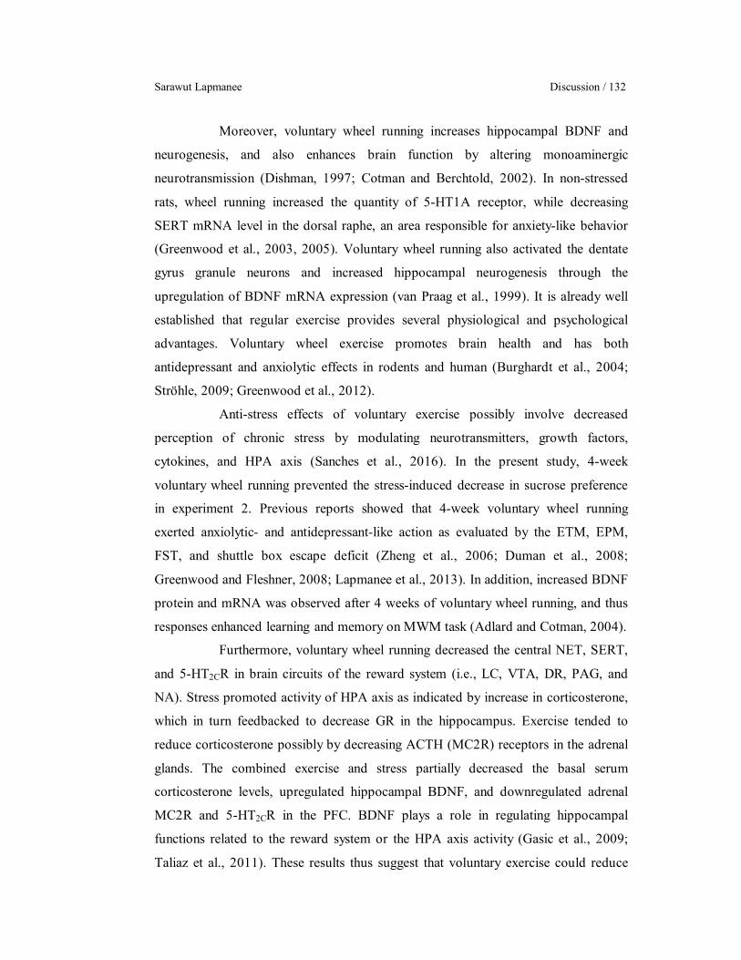
Sarawut Lapmanee Discussion / 132
Moreover, voluntary wheel running increases hippocampal BDNF and
neurogenesis, and also enhances brain function by altering monoaminergic
neurotransmission (Dishman, 1997; Cotman and Berchtold, 2002). In non-stressed
rats, wheel running increased the quantity of 5-HT1A receptor, while decreasing
SERT mRNA level in the dorsal raphe, an area responsible for anxiety-like behavior
(Greenwood et al., 2003, 2005). Voluntary wheel running also activated the dentate
gyrus granule neurons and increased hippocampal neurogenesis through the
upregulation of BDNF mRNA expression (van Praag et al., 1999). It is already well
established that regular exercise provides several physiological and psychological
advantages. Voluntary wheel exercise promotes brain health and has both
antidepressant and anxiolytic effects in rodents and human (Burghardt et al., 2004;
Ströhle, 2009; Greenwood et al., 2012).
Anti-stress effects of voluntary exercise possibly involve decreased
perception of chronic stress by modulating neurotransmitters, growth factors,
cytokines, and HPA axis (Sanches et al., 2016). In the present study, 4-week
voluntary wheel running prevented the stress-induced decrease in sucrose preference
in experiment 2. Previous reports showed that 4-week voluntary wheel running
exerted anxiolytic- and antidepressant-like action as evaluated by the ETM, EPM,
FST, and shuttle box escape deficit (Zheng et al., 2006; Duman et al., 2008;
Greenwood and Fleshner, 2008; Lapmanee et al., 2013). In addition, increased BDNF
protein and mRNA was observed after 4 weeks of voluntary wheel running, and thus
responses enhanced learning and memory on MWM task (Adlard and Cotman, 2004).
Furthermore, voluntary wheel running decreased the central NET, SERT,
and 5-HT2CR in brain circuits of the reward system (i.e., LC, VTA, DR, PAG, and
NA). Stress promoted activity of HPA axis as indicated by increase in corticosterone,
which in turn feedbacked to decrease GR in the hippocampus. Exercise tended to
reduce corticosterone possibly by decreasing ACTH (MC2R) receptors in the adrenal
glands. The combined exercise and stress partially decreased the basal serum
corticosterone levels, upregulated hippocampal BDNF, and downregulated adrenal
MC2R and 5-HT2CR in the PFC. BDNF plays a role in regulating hippocampal
functions related to the reward system or the HPA axis activity (Gasic et al., 2009;
Taliaz et al., 2011). These results thus suggest that voluntary exercise could reduce

University of Strasbourg/Mahidol University Ph.D. (Neurosciences & Physiology) / 133
the negative effect of chronic stress by modulating sympathetic activity and the HPA
axis, which subsequently results in decreased corticosterone secretion (Droste et al.,
2007; Patki et al., 2014).
Therefore, the sympathoadrenomedullary input is likely to play a role in
glucocorticoid responses to the 4-week restraint stress. Four-week voluntary wheel
running was thus effective in preventing the stress-induced anhedonia by modulating
central target proteins in the brain structures and neural pathway of the reward
systems.
5.6 Effectiveness of monoamine modulators and voluntary exercise in
the prevention of stress-induced anxiety-, depression-, and memory
impairment in male rats
Both pharmacological treatments (i.e., agomelatine and venlafaxine) and
voluntary wheel running successfully prevented anxiety-like behavior, especially the
learned fear (i.e., generalized anxiety disorder-like), as well as the impairment of
spatial learning and memory in restraint stressed rats. Drugs and exercise had no
impact on locomotor activity in the EPM and OFT. However, only pharmacological
treatments, but not voluntary wheel running in rats showed a protective action against
depression-like behavior and impaired memory in FST and NOR test, even though
running concurrently with stress could reduce depression-like behaviors (Lapmanee et
al., 2013). The finding of agomelatine-induced reduction in depression-like behavior
was consistent with the previous reports using other behavioral tests. For example,
Païzanis and co-workers (2010) reported that, after agomelatine treatment, immobility
was significantly reduced in mice subjected to the tail suspension test.
The corticosterone levels were above basal non-stressed levels in all 4
groups (even worse for the Ago group), thus indeed they all had high corticosterone
levels. At the cellular level, stressed rats pre-treated with agomelatine or venlafaxine
seemed to be sensitive to sustained high corticosterone levels, which resulted in a
downregulation of BDNF through stress in the hippocampus and the septum, and in
Sarawut Lapmanee Discussion / 134
both locations, this was counteracted and improved by wheel running. The GR was
also downregulated but only in the hippocampus. As for SERT and NET, significant
changes by stress are solely limited to the hippocampus and dorsal raphe for SERT
and to the LC and the PAG for NET. Furthermore, venlafaxine and voluntary wheel
running increased the levels of hippocampal BDNF protein in stressed male rats.
Thus, the present findings demonstrate that both venlafaxine and exercise exert a
protective effect against anxiety, depression, and memory impairment by restoring
hippocampal GR and upregulating BDNF expression in stressed rats. These results
suggest that change in corticosterone levels is not associated with the preventive
effects of agomelatine treatments. Presumably, the blockade of 5-HT2CR receptors
causes enhanced release of both NE and DA at the fronto-cortical dopaminergic and
noradrenergic pathways and promotes hippocampal cell proliferation, which in turn
could be the underlying mechanism of the preventive action against stress-induced
behavioral abnormality (Millan et al., 2000; 2003; Dagytė et al., 2010). Although pre-
treatment did not alter 5-HT2CR protein levels, there was a tendency towards a
downregulation of the 5-HT2CR in the amygdala of the exercise group. This result
might suggest anxiolytic effects of voluntary exercise on stress-induced 5-HT2CR
upregulation in stressed rats (Greenwood et al., 2012).
Since the beneficial effects of the studied drugs and exercise were
observed without changes in serum corticosterone levels, it is plausible that
agomelatine, venlafaxine, and exercise might have already induced a long-lasting
adaptation in monoaminergic neurotransmission or even neurogenesis in the brain,
which lasted throughout the entire stress periods. For instance, agomelatine is known
to stimulate cell proliferation within the dentate gyrus of rodents (Banasr et al., 2006).
Venlafaxine also enduringly modulated monoaminergic neurotransmission by
increasing 5-HT, DA and NE levels in the PFC and striatum (Higashino et al., 2014),
and increasing the expression of BDNF protein in rat hippocampus (Cooke et al.,
2009). BDNF possibly promoted the function and survival of dopaminergic,
GABAergic, noradrenergic, and serotonergic neurons (Connor and Dragunow, 1998).
Improvement of memory by venlafaxine and running exercise could be explained by
increased BDNF expression in the hippocampus (Li et al., 2011; Liu et al., 2009),
which is one of the utmost important brain structures for learning and memory.

University of Strasbourg/Mahidol University Ph.D. (Neurosciences & Physiology) / 135
Furthermore, voluntary wheel running exhibited SSRI and/or NRI-like actions
(Engesser-Cesar et al., 2007; Lapmanee et al., 2013). During exercise, NE in the cell
body and its metabolites were also increased in the pons, medulla oblongata, and
spinal cord of rats (Dunn et al., 1996). The mRNA expression of 5-HT1B and 1
adrenergic receptors was upregulated in the locus coeruleus and dorsal raphe
(Greenwood et al., 2005), which could in turn alter adrenergic and serotonergic
activities, respectively.
As aforementioned, it is possible that agomelatine, venlafaxine and
voluntary wheel running can normalize and coordinate the balance of glucocorticoids
and neurotransmitter metabolism, and induce long-lasting adaptations in
monoaminergic neurotransmission or neurogenesis in the brain (Cotman and
Berchtold, 2002; Dagytè et al., 2010; Huang et al., 2015), throughout the entire stress
period.
Finally, the present findings suggest that, in a situation in which future
exposure to stress is anticipated such as extreme military combat or war, infectious
disease outbreak, and natural disaster, administration of melatonergic modulator
(agomelatine), SNDRI (venlafaxine), and voluntary moderate-intensity exercise could
be useful in the prevention against the posttraumatic development of mood disorders
and memory impairment.
5.7 Mapping and characterization of MT1-/MT2-LacZ expressing
neurons in transgenic knock-in mouse models
Melatonin (MT) not only plays a role in the mammalian circadian rhythm
but also modulates mood, emotion, and memory. It also acts as a neuromodulator by
interacting with its central and peripheral MT receptors (Dubocovich, 2007; Pévet,
2016). Thus, it is important to demonstrate the localization of MT receptor in the
central nervous system. In the present study, the mouse brain was examined for the
expression of the MT gene at the cellular level, using the LacZ reporter gene to the
Sarawut Lapmanee Discussion / 136
targeting on MT1 or MT2 receptor gene locus downstream of the endogenous MT
receptor regulatory elements.
Previous studies described the distribution of high-affinity binding sites
for 125I-iodomelatonin or localized the mRNA expression of MT receptors
(Dubocovich et al., 2010). These investigations identified numerous structures
containing neurons expressing MT receptor genes (Masson-Pévet et al., 1994).
Unfortunately, there are no reliable specific antibodies for MT1 and MT2
immunocytochemical detection. Therefore, we used X-gal histochemistry and
immunohistochemistry to map the distribution of both MT1 and MT2 receptors in
these transgenic reporter mice. MT1-LacZ positive cells were restricted to a few
structures such as the SCN, the paraventricular nucleus of the thalamus, the posterior
limiting nucleus of the thalamus and the pars tuberalis (PT) of the adenohypophysis.
This MT1-LacZ labeling is perfectly superposable to the 125I-iodomelatonin binding
sites, thus validating our approach (Liu et al., 1997). On the other hand, MT2-LacZ
positive cells were detected in many structures throughout the brain such as the
olfactory bulb, the islands of Calleja, the SCN, the cerebral cortex, the superior
colliculus, the amygdala and the arcuate nucleus of the hypothalamus. MT2-LacZ
knock-in mice also had a strong staining in the PVN, the CA2 and a population of
small horizontal cells in the outer molecular layer of dentate gyrus of the
hippocampus.
MT1 and/or MT2 receptors are present in the SCN and PT which have
high concentration of MT binding sites (Vanĕcek, 1987; Morgan et al., 1989; Weaver
et al., 1989). The distinct presence of MT1 and MT2 receptors in different brain areas
suggests distinctive functions for each receptor in neurophysiology and
neuropathology (Comai and Gobbi, 2014; Lacoste et al., 2015). MT binding sites are
found predominantly in the median eminence, SCN, area postrema, paraventricular
thalamic nucleus, hypothalamus, hippocampus, parietal cortex, striatum, PT, medial
region of the lateral habenula, arcuate nucleus, and amygdala of the rats and mice
(Laudon et al., 1988; Weaver et al., 1989; Masson-Pévet et al., 1994; Williams et al.,
1995). MT1 and MT2 receptors are localized in regions known to contribute to
anxiety and stress, e.g., the septal area, the habenula, and the dentate gyrus of
hippocampus (Yadin et al., 1993; Karakaş et al., 2011). Regarding MT receptor

University of Strasbourg/Mahidol University Ph.D. (Neurosciences & Physiology) / 137
mRNA expression in rodent brain, MT1 receptor mRNA was detected in the rodent
dorsal striatum, NA, olfactory tubercle, substantia nigra, and VTA. Also, MT1 and
MT2 receptor mRNA expressions have been found in the SCN and vestibular nuclei
(Us et al., 2005; Imbesi et al., 2008; Ahn et al., 2012). In human studies, MT1 mRNA
and protein were detected in the hypothalamus, the cerebellum, the PFC, the NA, the
amygdala, and hippocampus (Weaver et al., 1993; Mazzucchelli et al., 1996; Wu et
al., 2006), and MT2 receptor transcripts were detected in the hippocampus (Musshoff
et al., 2002).
The SCN is the master circadian clock and a target the drug discovery in
circadian-dependent pathogeneses. Neurons in the SCN subregions are distinguished
by their neuropeptide contents, i.e., AVP, GRP, and VIP, while AVP cells are the
output of the SCN central clock, VIP cells are the input and GRP cells the core of the
clock (Abrahamson and Moore, 2001; Welsh et al., 2010). MT1-LacZ expressing
neurons co-localized with some GRP and VIP neurons in order to synchronize the
endogenous circadian rhythms. Therefore, MT can act through MT1 receptors on the
input and the inner working of the clock. MT2-LacZ expressing neurons were located
more diffusely throughout the SCN and around it.
MT2-LacZ expressing neurons were also located in brain regions
associated with stress responses, particularly the PVN and the hippocampus, both of
which are important components of the HPA axis (Smith and Vale, 2006). The
present study found that MT2-LacZ expressing neurons co-localized with CRH in the
PVN, suggesting that melatonin and agomelatine might directly modulate the HPA
axis at the PVN. As MT2 receptor signaling is mostly inhibitory, agomelatine action
on the CRH neurons should thus reduce the activity of these neurons and reduce the
secretion of CRH. Reduced secretion of CRH should result in a decrease in
corticosterone levels. We observed however higher levels of corticosterone in the
agomelatine-treated animals. Agomelatine being a combined MT1/M2 agonist and 5-
HT2C receptor antagonist, further studies of this complex pharmacological profile
might help us to understand this apparently contradictory result.
Furthermore, MT2-LacZ expressing neurons were co-localized with
GAD2 in the hippocampus (Stone et al., 1999). These results could imply that the
actions of MT and agomelatine result in the inhibition of these inhibitory

Sarawut Lapmanee Discussion / 138
interneurons, and thus an activation of hippocampal activity (Chamberland and
Topolnik, 2012). However, MT2 receptors also seem to be very selectively placed on
pyramidal neurons of the CA2. These pyramidal neurons are known to be
glutamatergic, and thus MT or agomelatine action on these neurons should result in
the selective inhibition of CA2 mediated functions. Thus MT and agomelatine should
activate the hippocampus, but selectively inhibit CA2. How this dual action impacts
hippocampal tasks and the hippocampal action on the HPA axis remains to be
established.
In addition, MT2-LacZ expressing cells were observed in the dorsal raphe.
This brain region apparently does not contain 5-HT2CR, but the 5-HT2CR is involved
in negative serotonin feedback to the dorsal raphe through GABAergic neurons
around the dorsal raphe, which express 5-HT2CR (Serrats et al., 2005). Thus
agomelatine might act directly on dorsal raphe and indirectly through this GABAergic
5-HT2C feedback. As an agonist on MT2 receptors in the dorsal raphe, it might inhibit
these serotonergic neurons. As a 5-HT2C antagonist, it could block 5-HT negative
feedback, which in turn could therefore stimulate the dorsal raphe. Thus this action is
probably quite complex because it would both directly inhibit the dorsal raphe and
indirectly stimulate the same structure. Therefore, the dorsal raphe is another target of
interest for agomelatine.
University of Strasbourg/Mahidol University Ph.D. (Neurosciences & Physiology) / 139
CHAPTER VI
CONCLUSIONS
The aims of the present study were first, to examine the effects of restraint
stress on anxiety-, depression-, and memory impairment-like behaviors at different
time points, and second, to determine the preventive therapeutic effects of monoamine
modulators (agomelatine and venlafaxine) and voluntary exercise in stressed male rats.
Changes in the physical and behavioral parameters and protein expression levels in the
brain regions associated with stress responses were investigated. In addition, possible
sites of action for agomelatine administration were identified in melatonin (MT) MT1-
and MT2-LacZ knocked-in mouse.
The conclusions are as follow with a summary in the Figure 6.1.
Four week restraint stress in male rats resulted in decreases in food intake
and body weight gain, adrenal hypertrophy, and increased serum and urine levels of
corticosterone. Increased levels of corticosterone could alter central the target proteins,
i.e., brain-derived neurotrophic factor (BDNF), glucocorticoid receptor (GR),
norepinephrine transporter (NET), and serotonin type 2C receptor (5-HT2CR) in the
amygdala, hippocampus, locus coeruleus, and periaqueductal gray (PAG) leading to
manifestation of anxiety-, depression-, and memory impairment-like behaviors.
Four week voluntary aerobic wheel running exercise protocol were found
to partly prevent the stress-induced increase in serum corticosterone levels and
anhedonia-like behavior, most probably by deregulating adrenal adrenocorticotropic
hormone (ACTH) receptors, and altering target protein expression levels (i.e., BDNF
and NET) in the brain structures specially involved in stress response, i.e.,
hippocampus, septum nuclei, locus coeruleus, and periaqueductal gray. Therefore,
voluntary exercise could reduce the negative effect of chronic stress by modulating
sympathetic activity in the central and peripheral hypothalamic-pituitary-adrenal
(HPA) axis.

Sarawut Lapmanee Conclusions / 140
The study also revealed that agomelatine, venlafaxine as well as voluntary
wheel running prevented the restraint stress-induced anxiety/depression-like behaviors
and memory impairment in male rats. Upregulation of hippocampal BDNF protein
levels might be a possible mechanism underlying the protective effects of venlafaxine
and voluntary exercise in stressed male rats.
Regarding the applications of the present findings for clinical use,
voluntary exercise should be recommended as an alternative treatment or used in
concert with pharmacological treatments (i.e., agomelatine or venlafaxine) for the
treatment of anxiety-, depression-, and memory impairment-like symptoms in patients
and stressed individuals.
Indeed, mapping and phenotyping of MT1 and MT2 receptors expressing
neurons indicate possible sites of action for melatonin or melatonin derivatives. MT1-
LacZ expressing neurons were found in some studied areas, i.e., suprachiasmatic
nucleus (SCN), paraventicular nucleus of thalamus (PVT), and pars tuberalis (PT) of
anterior pituitary gland, whereas MT2-LacZ expressing neurons were more widely
distributed throughout the whole brain, e.g., SCN, paraventicular nucleus of
hypothalamus (PVN), dorsal raphe (DR), amygdala, and CA2 of hippocampus. Further
studies are required to clarify the mode of action of agomelatine.
Taken together, the 4-week stress caused hyperactivity of HPA axis and
increased glucocorticoid/corticosterone levels leading to hippocampal GR and BDNF
downregulation. Four-week voluntary wheel running modulated the HPA axis and
partially decreased corticosterone levels by downregulating adrenal ACTH receptor
and also promoted hippocampal BDNF upregulation resulting in decreased anhedonic-
like behaviors in stressed male rats. Furthermore, voluntary exercise prevented stress-
induced anxiety, depression, and memory impairment. Pre-treatment with venlafaxine
directly inhibited NET to increase norepinephrine (NE) in the synaptic cleft and
mediated antidepressant and anxiolytic effects via hippocampal BDNF production. In
addition, pre-treatment with agomelatine, a MT receptor agonist, effectively prevented
stress-related behaviors. Potential sites of action for these effects through MT1 and
MT2 receptors are the SCN, the PVN, the dorsal raphe (DR), and the hippocampus.
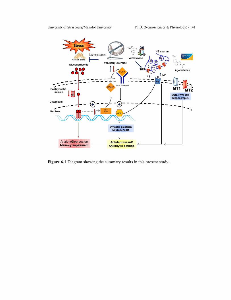
University of Strasbourg/Mahidol University Ph.D. (Neurosciences & Physiology) / 141
Figure 6.1 Diagram showing the summary results in this present study.

Sarawut Lapmanee Résumé français / 142
Effets neuroprotecteurs de l’exercice volontaire et de modulateurs
monoaminergiques chez le rat mâle stressé
A. Introduction et objectifs
Le stress est un facteur important dans le développement de troubles de
l’humeur tels que l’anxiété, la dépression et des troubles de la mémoire. Les troubles
de l’humeur liés au stress sont des maladies psychiatriques en train de devenir un
fardeau mondial d’invalidité et de comorbidité (Kessler et al. 2009). Les mécanismes
sous-tendant les troubles de l’humeur chez l’individu stressé sont supposés être un
dérèglement de l’axe hypothalamus-pituito-surrénalien (HPS) et un déséquilibre des
monoamines sérotonine (5-HT) et noradrénaline (NA) (Bowman et al. 2003, Belmaker
& Agam 2008). L’implication de 5-HT et NA dans la dépression est confirmée par
l’action antidépresseur d’inhibiteurs de la recapture de ces molécules par les
transporteurs de la sérotonine (SERT) et de la noradrénaline (NET). Ces inhibiteurs de
recapture augmentent la concentration et la durée d’action de 5-HT et de NA dans la
fente synaptique (Charney 1998, Bowman et al. 2003, Belmaker & Agam 2008).
Outre la dérégulation de l’axe HPS, le stress perturbe aussi les rythmes circadiens et la
sécrétion de mélatonine par la pinéale (Konakchieva et al. 1997, McClung 2011).
Aussi, des niveaux réduits de mélatonine (MT) ont été rapportés chez des patients
dépressifs (Claustrat et al. 1984). De ce fait, les deux types de récepteurs de la
mélatonine, MT1 et MT2, sont devenus des cibles thérapeutiques pour le
développement de nouveaux antidépresseurs. Cependant, la localisation cellulaire
précise des récepteurs MT1 et MT2 n’est pas encore bien connue du fait de l’absence
d’anticorps bien caractérisés et parfaitement spécifiques pour l’immunocytochimie.
Basé sur ses effets anxiolytiques et antidépresseurs (Milan et al. 2005, Papp et al.
2006, Taylor et al. 2014), l’agomélatine, un agoniste MT1/MT2 et antagoniste 5-
HT2C pourrait être un candidat nouveau pour la prévention de changements
comportementaux induits par le stress.
Outre les perturbations de l’axe HPS et des systèmes monoaminergiques,
le stress chronique augmente les glucocorticoïdes, ce qui décroit la production de
University of Strasbourg/Mahidol University Ph.D. (Neurosciences & Physiology) / 143
récepteur aux glucocorticoïdes (GR) et de facteur neurotrophique dérivé du cerveau
(Brain Derived Neurotropic Factor, BDNF) dans l’hippocampe, ainsi qu’une réduction
de la neurogenèse dans cette structure, le tout conduisant à des troubles de l’humeur
tels que l’anxiété et la dépression, ainsi que des perturbations de la mémoire (Schmidt
& Duncan 2007, Autry & Monteggia 2012).
A côté de ces agents pharmacologiques, l’exercice physique est connu
pour stimuler la neurotransmission monoaminergique et la production de BDNF
(Cotman & Berchtold 2002). L’exercice physique volontaire possède des propriétés
antidépressives et anxiolytiques chez les rongeurs et chez l’homme (Burghardt et al.
2004, Ströhle et al. 2009, Lapmanee et al. 2013). L’activité locomotrice volontaire
dans une roue augmente la production de BDNF et la neurogenèse dans l’hippocampe
(Cotman & Berchtold 2002), et améliore le fonctionnement cérébral en modifiant la
neurotransmission monoaminergique (Dishman 1997). Certains patients ne répondent
pas ou peu aux traitements pharmacologiques ou présentent une longue phase de
latence avant de présenter des effets anxiolytiques ou antidépresseurs. L’exercice
physique ou d’autres interventions pourraient donc s’avérer bénéfiques pour ces
individus. Précédemment, nous avions montré qu’un inhibiteur de la recapture
sérotonine-noradrénaline-dopamine, la venlafaxine, ainsi que l’activité locomotrice
volontaire dans une roue pouvaient réduire des comportements de type anxiété et
dépression chez des rats stressés (Lapmanee et al. 2012, 2013).
Cependant, les effets modulatoires de l’exercice de roue volontaire sur la
réponse à un stress chronique ne sont pas encore bien compris, et les effets préventifs
de cet exercice sur les troubles de l’humeur et les perturbations de la mémoire induits
par le stress restent à préciser. Enfin, les mécanismes cellulaires des effets protecteurs
de l’agomélatine, de la venlafaxine et de l’exercice physique dans les neurones 5-HT
et NA vis-à-vis des changements comportementaux induits par le stress ne sont pas
encore connus.
Ainsi, les objectifs de ce travail étaient
1. d’établir un modèle de stress de contention chronique chez le rat pour
des études comportementales et des analyses biochimiques des changements
d’expression de protéines cibles dans le cerveau.

Sarawut Lapmanee Résumé français / 144
2. de déterminer les bénéfices de l’activité de roue volontaire sur l’axe
hypothalamo-pituito-surrénalien (HPS) et sur l’expression de protéines cibles dans
diverses régions cérébrales chez le rat.
3. de comparer l’efficacité de l’Agomélatine (Ago), d’un inhibiteur de la
recapture de la 5-HT et de la NA (Venlafaxine, Vlx) et de l’exercice physique
volontaire dans la prévention de l’anxiété et de la dépression, ainsi que des
perturbations de la mémoire induits par le stress.
4. de déterminer la localisation des récepteurs MT1 et MT2 chez des souris
transgéniques rapportrices afin d’identifier des sites potentiels d’action de
l’Agomélatine, en particulier dans le contexte du stress.
Les hypothèses centrales de ce travail étaient :
1. Les modulateurs de la neurotransmission monoaminergique et l’activité
de roue volontaire peuvent prévenir les comportements induits par le stress chez le rat
mâle en modulant la neurogenèse hippocampique et en altérant l’expression de
protéines cibles de la neurotransmission monoaminergique centrale dans des régions
associées avec la réponse au stress.
2. Des neurones exprimant les récepteurs de la mélatonine MT1 et MT2
localisées dans des régions associées à la réponse au stress pourraient être des cibles
de l’action antidépressive et anxiolytique du traitement pharmacologique par
l’Agomélatine.
B. Matériel et Méthodes
Partie 1 : Effets du stress de contention, des modulateurs pharmacologiques
(Agomélatine, Venlafaxine) et de l’activité de roue volontaire chez des rats mâles
Animaux
Des rats mâles de souche Wistar (âgés de 8 semaines, 180200 g) étaient
obtenus du Centre National d’Animaux de Thaïlande (Université Mahidol, Thaïlande).
A la fin des expériences, tous les rats étaient évalués pour des comportements de type
anxiété, dépression ou perturbations de la mémoire. Après, le sang et des urines ont été
collectés pour des mesures de la corticostérone. Le cœur, les surrénales et le cerveau
University of Strasbourg/Mahidol University Ph.D. (Neurosciences & Physiology) / 145
ont été prélevés dans toutes les expériences. Des régions cérébrales ont été
microdisséquées pour étudier l’expression de protéines cibles : BDNF, récepteur au
glucocorticoïdes (GR), transporteur de la NA (NET), transporteur de la 5-HT (SERT)
et le récepteur de la sérotonine 5-HT2c. Les niveaux d’expression des protéines ont été
évalués par Western blotting dans des régions cérébrales contribuant à la réponse au
stress : cortex frontal, amygdale, hypothalamus, hippocampe, raphé dorsal, locus
coeruleus, gris périacquéducal, septum, noyau accumbens et aire tegmentale ventrale.
Toutes les procédures animales ont été validées par le Comité de Soins et d’Utilisation
d’Animaux de la Faculté de Médecine de l’Université Thammasat, Thaïlande.
Stress de contention
Les rats ont été placés dans des tubes de contention modifiés pendant 2
heures/jour, 5 jours/semaine pendant 1, 4 ou 8 semaines. Les tubes de contention
avaient une ouverture d’1 cm de diamètre à un bout pour permettre aux animaux de
respirer.
Études comportementales
Les comportements anxieux ont été évaluées par le labyrinthe élevé en
croix (elevated plus maze EPM), le labyrinthe élevé en T (elevated T maze ETM) et le
test « Open Field » (OFT). Les comportements dépressifs ont été évalués par des tests
hebdomadaires de préférence pour le sucrose et de nage forcée (forced swimming test
FST). La piscine de Morris (Morris water maze MWM) et le test de reconnaissance
d’objets nouveaux (novel object recognition NOR) ont été utilisés pour évaluer
l’apprentissage et la mémoire.
Protocoles expérimentaux
Expérience 1 : Cinétique de l’induction par le stress de contention de comportements
anxieux ou dépressifs, et de perturbations de la mémoire.
Les rats ont été séparés en 4 groupes : 1 groupe contrôle (non stressé) et 3
groupes de rats soumis au stress de contention pendant 1, 4 ou 8 semaines
Sarawut Lapmanee Résumé français / 146
Expérience 2 : Effets de 4 semaines d’activité locomotrice volontaire sur l’axe HPS
en conditions basales et après 4 semaines d’exposition à un stress de contention.
Les rats ont été séparés en 4 groupes : 1 groupe non-stressé sans roue, 1
groupe non-stressé avec exercice de roue, 1 groupe stressé sans roue et 1 groupe
stressé avec exercice de roue. Tous les rats ont été évalués pour des comportements
anhédoniques par le test de préférence pour le sucrose. Le sang et les urines ont été
collectés pour la détermination du niveau de corticostérone. Le cœur et les glanes
surrénales ont été prélevés et pesés. Les cerveaux ont été prélevés et analysés pour les
protéines cibles. Les glandes surrénales ont aussi analysées pour le niveau
d’expression protéique du récepteur aux mélanocortines 2 (MC2R, récepteur de
l’ACTH).
Expérience 3 : Effets de l’agomélatine, de la venlafaxine et de l’activité locomotrice
volontaire dans la prévention de perturbations de la mémoire et de comportements de
type anxiété et dépression.
Les rats ont été séparés en 4 groupes : 1 groupe contrôle recevant la
solution véhicule (5 ml/kg de saline 9‰ per os), 1 groupe Agomélatine (10 mg/kg per
os), Venlafaxine (10 mg/kg per os) et 1 groupe avec exercice de roue volontaire. Les
animaux ont été soumis à ces traitements pendant 4 semaines (administrations per os
tous les jours). Ensuite, ces traitements ont été arrêtés et les animaux ont été soumis
pendant 4 semaines au stress de contention (2 h/jour, 5 jours/semaine). Les
comportements anhédoniques ont été évalués par un test hebdomadaire de préférence
pour le sucrose. A la fin des 4 semaines de soumission au stress, tous les rats ont été
évalués pour des comportements anxieux ou dépressifs, ainsi que pour l’apprentissage
et la mémoire.
Partie 2 : Caractérisation des cellules exprimant les récepteurs MT1 ou MT2 dans le
cerveau de souris rapportrices LacZ knock-in : Identification de sites d’action
potentiels de l’Agomélatine dans des structures impliquées dans le stress
University of Strasbourg/Mahidol University Ph.D. (Neurosciences & Physiology) / 147
Animaux
Des souris (1,5 à 18 mois d’âge, 20-60 g) mâles et femelles adultes
transgéniques avec le rapporteur LacZ introduit dans le locus MT1 (MTNR1A) ou
MT2 (MTNR2A) ont été obtenues à la plateforme Chronobiotron (CNRS UMS 3415)
de l’Institut des Neurosciences Cellulaires et Intégratives (INCI CNRS UPR 3212) de
l’Université de Strasbourg. Ces mutations étaient introduites dans la lignée C57/BL6,
qui ne produit pas de mélatonine. Nous avons utilisé des souris hétérozygotes (KI/+) et
des souris homozygotes (KI/KI) pour nos études. Des souris non-mutantes (+/+) des
mêmes portées ont été utilisées comme contrôles. Pour obtenir des souris mutantes
produisant de la mélatonine, nous avons utilisé des souris hétérozygotes (KI/+) issues
d’un croisement F1 avec la lignée C3H, qui produit de la mélatonine. L’euthanasie des
souris a été effectuée selon les protocoles agrémentés pour la plateforme «
Chronobiotron » (UMS 3415) par le Comité Régional d'Ethique en Matière
d'Expérimentation Animale de Strasbourg.
Analyses histologiques :
Les souris ont été euthanasiées par inhalation de CO2 et perfusées par des
solutions de fixateur à travers le cœur. Nous avons utilisé le fixateur Periodate-Lysine-
Paraformaldéhyde (PLP) pour les analyses par immunocytochimie et par hybridation
in situ. Pour la détection histoenzymologique de l’activité LacZ, les animaux ont été
fixés par du formaldéhyde 4% dans du tampon phosphate 100 mM. Après dissection,
le cerveau a été postfixé par immersion dans le fixateur et soit préparé pour des coupes
flottantes en congélation après cryoprotection au sucrose (Histoenzymologie LacZ) ou
enrobé en Polyéthylène Glycol (PEG) (immunocytochimie et hybridation in situ).
Des coupes flottantes (30-40 m d’épaisseur) ont été colorés pour l’activité beta-
galactosidase (LacZ) en utilisant un substrat chromogénique (5-bromo-4-chloro-3-
indolyl-beta-D-galactopyranoside, X-Gal).
Pour la détection immunocytochimique de la protéine LacZ, des coupes en
PEG ont été pratiquées à 10 m d’épaisseur et montées sur lames. Après une
réactivation antigénique, la présence de la protéine LacZ a été révélée par un anticorps
anti-LacZ (Abcam), suivi d’une amplification immunoperoxidase.
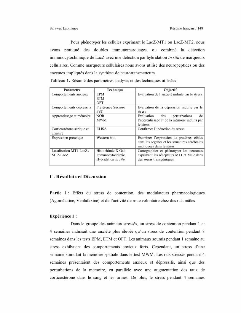
Sarawut Lapmanee Résumé français / 148
Pour phénotyper les cellules exprimant le LacZ-MT1 ou LacZ-MT2, nous
avons pratiqué des doubles immunomarquages, ou combiné la détection
immunocytochimique de LacZ avec une détection par hybridation in situ de marqueurs
cellulaires. Comme marqueurs cellulaires nous avons utilisé des neuropeptides ou des
enzymes impliqués dans la synthèse de neurotransmetteurs.
Tableau 1. Résumé des paramètres analyses et des techniques utilisées
Paramètre Technique Objectif Comportements anxieux EPM
ETM OFT
Evaluation de l’anxiété induite par le stress
Comportements dépressifs Préférence Sucrose FST
Evaluation de la dépression induite par le stress
Apprentissage et mémoire NOR MWM
Evaluation des perturbations de l’apprentissage et de la mémoire induits par le stress
Corticostérone sérique et urinaire
ELISA Confirmer l’induction du stress
Expression protéique Western blot Examiner l’expression de protéines cibles dans les organes et les structures cérébrales impliquées dans le stress
Localisation MT1-LacZ / MT2-LacZ
Histochimie X-Gal, Immunocytochimie, Hybridation in situ
Cartographier et phénotyper les neurones exprimant les récepteurs MT1 et MT2 dans des souris transgéniques
C. Résultats et Discussion
Partie 1 : Effets du stress de contention, des modulateurs pharmacologiques
(Agomélatine, Venlafaxine) et de l’activité de roue volontaire chez des rats mâles
Expérience 1 :
Dans le groupe des animaux stressés, un stress de contention pendant 1 et
4 semaines induisait une anxiété plus élevée qu’un stress de contention pendant 8
semaines dans les tests EPM, ETM et OFT. Les animaux soumis pendant 1 semaine au
stress exhibaient des comportements anxieux forts. Cependant, un stress d’une
semaine stimulait la mémoire spatiale dans le test MWM. Les rats stressés pendant 4
semaines présentaient des comportements anxieux et dépressifs, ainsi que des
perturbations de la mémoire, en parallèle avec une augmentation des taux de
corticostérone dans le sang et les urines. De plus, le stress pendant 4 semaines

University of Strasbourg/Mahidol University Ph.D. (Neurosciences & Physiology) / 149
augmentait l’expression du NET et du 5-HT2c dans le locus coeruleus et dans
l’amygdale, et diminuait l’expression hypothalamique du GR. Ces résultats indiquent
une perturbation du rétrocontrôle de l’axe HPS (Smith & Vale 2006). L’augmentation
de l’expression du NET et du 5-HT2C dans le cerveau antérieur et le locus coeruleus
pourrait causer des comportements liés au stress (Kimura et al. 2009, Chen et al.
2012). Cependant, un stress de 8 semaines induisait moins de comportements liés au
stress dans les tests comportementaux. Un stress d’une telle durée pourrait induire des
adaptations compensatoires (Dhabar et al. 1997). Ces résultats montrent qu’un stress
de 4 semaines est suffisant pour induire de l’anxiété et de la dépression, ainsi que des
perturbations de la mémoire. Nous avons donc utilisé ce protocole de stress de 4
semaines pour la suite de nos études.
Tableau 2. Cinétique des changements comportementaux et biochimiques après un stress de contention pendant 1, 4 ou 8 semaines chez le rat male.
Paramètre Stress 1 semaine Stress 4 semaines Stress 8 semaines
Anxiété EPM
ETM OFT
Dépression % Préférence Sucrose
à la deuxième semaine de stress
à la deuxième semaine de stress de la 5ème à la 8ème semaine de stress
FST Apprentissage MWM Mémoire MWM/NOR Locomotion EPM/OFT BDNF dans le PAG dans la VTA
GR dans l’hypothalamus
dans l’hippocampe
NET dans le LC SERT dans la VTA dans
l’hippocampe 5-HT2C dans l’amygdale dans l’amygdale,
dans le PAG dans le LC, dans la VTA
LC locus coeruleus, PAG gris périacquéducal, VTA aire tegmentale ventrale

Sarawut Lapmanee Résumé français / 150
Expérience 2 :
L’activité de roue volontaire a induit une réduction du poids corporel et
une augmentation du poids sec du cœur autant chez les rats contrôles que chez les rats
stressés. La présence d’une hypertrophie cardiaque indique le succès du protocole
d’exercice physique aérobique comme montré précédemment par Wang et al. (2010)
et Lapmanee et al. (2013). L’exercice physique volontaire a aussi augmenté le poids
des glandes surrénales sans changement des niveaux de corticostérone sériques chez
les rats non-stressés. Ceci est en accord avec une étude précédente qui a montrée que 4
semaines d’activité de roue volontaire n’avaient aucun effet sur les niveaux de
corticostérone circulante, que ce soit au niveau basal ou au niveau maximal du rythme
circadien chez le rat mâle (Fedicu et al. 2006). De plus, l’activité de roue volontaire à
diminué les niveaux du NET, du SERT et du 5-HT2C dans le circuit de la récompense
dans le système nerveux central (p.ex. locus coeruleus, aire tegmentale ventrale, raphé
dorsal, gris périacquéducal et noyau accumbens). La combinaison du stress avec
l’exercice physique volontaire a partiellement diminué le niveau basal de
corticostérone, a augmenté le niveau de BDNF dans l’hippocampe et réduit le
récepteur MC2R dans la surrénale et le récepteur 5-HT2C dans le cortex frontal. Ces
résultats suggèrent que l’exercice physique volontaire peut réduire l’effet négatif du
stress chronique en modulant l’activité sympathique de l’axe HPS tant au niveau
central qu’au niveau périphérique, ce qui par conséquent a réduit la sécrétion de
corticostérone (Droste et al. 2007, Patki et al. 2014).
Tableau 3. Changements comportementaux et biochimiques chez des rats mâles contrôles ou après 4 semaines d’activité de roue volontaire et / ou soumis pendant 4 semaines à un stress de contention.
Paramètre Exercice vs contrôle
Stress vs contrôle
Stress + Exercice vs stress
Dépression % Préférence Sucrose
Corticostérone sérique BDNF dans le NA dans
l’hippocampe et le septum
dans l’hippocampe et le
septum, dans le NA
GR dans l’hippocampe

University of Strasbourg/Mahidol University Ph.D. (Neurosciences & Physiology) / 151
Paramètre Exercice vs contrôle
Stress vs contrôle
Stress + Exercice vs stress
NET dans le LC et la VTA
dans le LC, dans le PAG
dans le LC
SERT dans le septum dans l’hippocampe et le
DR
dans la VTA
5-HT2CR dans le DR, le PAG, et le NA
dans l’amygdale dans le PFC
DR raphé dorsal, LC locus coeruleus, NA noyau accumbens, PAG gris périacquéducal, PFC cortex
préfrontal, VTA aire tegmentale ventrale
Expérience 3 :
L’agomélatine, la venlafaxine et l’exercice volontaire de roue peuvent
prévenir les comportements anxieux et les perturbations de la mémoire. Les
prétraitements par l’agomélatine, la venlafaxine ou l’exercice volontaire n’ont pas
changé le poids des surrénales, mais les niveaux de corticostérone sérique chez les
animaux traités à l’agomélatine étaient plus élevés que ceux d’animaux contrôles sans
exercice de roue et traités uniquement avec le véhicule. Les rats traités par
l’agomélatine et la venlafaxine étaient sensible aux niveaux élevés de corticostérone
de manière prolongée, ce qui a résulté en une diminution du SERT, du NET, du BDNF
et du GR dans les régions contribuant aux réponses autonomiques et
comportementales, p.ex. le septum, le locus coeruleus, le gris périacquéducal et
l’hippocampe. La venlafaxine et l’activité volontaire de roue ont significativement
augmenté les niveaux de BDNF chez des rats stressés. Il est donc possible que les
prétraitements par l’agomélatine, la venlafaxine et l’exercice physique volontaire
puissent normaliser et coordonner la balance des glucocorticoïdes et du métabolisme
des neurotransmetteurs, et induire des adaptations au long terme de la
neurotransmission monoaminergique et même de la neurogenèse dans le cerveau
(Cotman & Berchtold 2002, Dagyte et al. 2010, Huang et al. 2014), adaptations qui
ont été maintenues pendant l’intégralité de la période de stress.
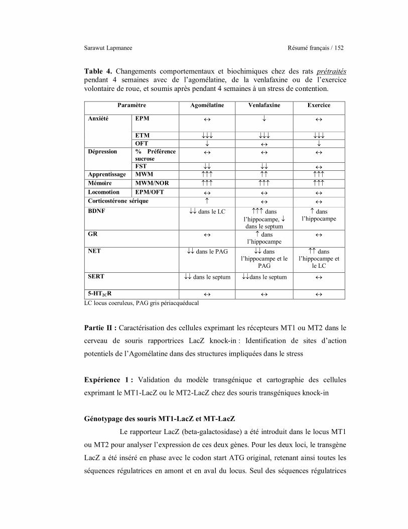
Sarawut Lapmanee Résumé français / 152
Table 4. Changements comportementaux et biochimiques chez des rats prétraités pendant 4 semaines avec de l’agomélatine, de la venlafaxine ou de l’exercice volontaire de roue, et soumis après pendant 4 semaines à un stress de contention.
Paramètre Agomélatine Venlafaxine Exercice
Anxiété EPM
ETM OFT
Dépression % Préférence sucrose
FST Apprentissage MWM Mémoire MWM/NOR Locomotion EPM/OFT Corticostérone sérique BDNF dans le LC dans
l’hippocampe, dans le septum
dans l’hippocampe
GR dans l’hippocampe
NET dans le PAG dans l’hippocampe et le
PAG
dans l’hippocampe et
le LC
SERT dans le septum dans le septum
5-HT2CR LC locus coeruleus, PAG gris périacquéducal
Partie II : Caractérisation des cellules exprimant les récepteurs MT1 ou MT2 dans le
cerveau de souris rapportrices LacZ knock-in : Identification de sites d’action
potentiels de l’Agomélatine dans des structures impliquées dans le stress
Expérience 1 : Validation du modèle transgénique et cartographie des cellules
exprimant le MT1-LacZ ou le MT2-LacZ chez des souris transgéniques knock-in
Génotypage des souris MT1-LacZ et MT-LacZ
Le rapporteur LacZ (beta-galactosidase) a été introduit dans le locus MT1
ou MT2 pour analyser l’expression de ces deux gènes. Pour les deux loci, le transgène
LacZ a été inséré en phase avec le codon start ATG original, retenant ainsi toutes les
séquences régulatrices en amont et en aval du locus. Seul des séquences régulatrices
University of Strasbourg/Mahidol University Ph.D. (Neurosciences & Physiology) / 153
potentielles situées dans l’intron unique des gènes MT1 ou MT2 ont été délétées par
cette stratégie. Ainsi ces souris rapportrices knock-in MT1-LacZ ou MT2-LacZ sont
considérées un outil idéal pour investiguer les sites d’expression des récepteurs de la
mélatonine MT1 et MT2 en absence d’anticorps spécifiques fiables pour la détection
immunocytochimique de ces récepteurs.
Nous avons d’abord sélectionné des animaux hétérozygotes MT1-LacZKI+
ou MT2-LacZKI+ pour les études initiales. Ces animaux hétérozygotes disposent d’un
allèle MT1 ou MT2 intact, réduisant ainsi les risques d’expression ectopique du
transgène du fait d’une perte de fonction complète du récepteur MT1 ou MT2. L’étude
de Liu et al. (1997) a montré que la délétion d’un allèle du locus MT1 réduisait
effectivement le niveau de liaison de la 125I-iodomélatonine de 50%, mais n’affectait
en rien le patron d’expression. Les animaux hétérozygotes ont aussi été croisés entre
eux pour produire des animaux homozygotes MT1-LacZKIKI et MT2-LacZKIKI, qui
sont de fait des animaux invalidés (knockout KO) soit pour le MT1 ou le MT2 comme
alors les deux allèles sont remplacés par LacZ.
Les animaux transgéniques ont été génotypés par PCR. Chez les animaux
MT1-LacZ, un produit de 425 bp indique un allèle MT1 sauvage, donc intact, et un
produit de 196 bp indique un allèle MT1-LacZ recombinant. Chez les animaux MT2-
LacZ un produit de 143 bp indique l’allèle sauvage intact, tandis qu’un produit de 666
bp indique l’allèle MT2-LacZ recombinant.
Cartographie de l’expression MT1-LacZ et MT2-LacZ dans le cerveau
Nous avons utilisé deux techniques pour visualiser l’expression de LacZ
sous le contrôle du promoteur MT1 ou du promoteur MT2. La première technique est
la visualisation histoenzymologique de l’activité enzymatique LacZ en utilisant le
substrat chromogénique X-Gal (5-bromo-4-chloro-3-indolyl-beta-D-
galactopyranoside) pour cette enzyme. La deuxième technique est la détection
immunocytochimique de LacZ à l’aide d’un anticorps spécifique pour cette enzyme.
Dans les deux cas, des animaux sauvages non-transgéniques ont été utilisés comme
contrôles négatifs. Les deux techniques ont toujours fourni des résultats identiques
quant à la localisation de LacZ, même si l’apparence du signal n’était pas toujours
identique.

Sarawut Lapmanee Résumé français / 154
Le signal enzymatique LacZ se présentait sous forme de points très
fortement colorés contre un fond pratiquement sans signal. Le signal
immunocytochimique présentait aussi les mêmes points très fortement marqués, mais
aussi du signal dans le noyau et parfois dans diffus dans le cytoplasme des cellules
exprimant MT1-LacZ ou MT2-LacZ. Les points fortement marqués à l’intérieur des
cellules par les deux techniques sont probablement des accumulations lysosomiales de
LacZ. La détection immunocytochimique présentait aussi plus de bruit de fonds que la
détection enzymologique. Nous avons utilisé la détection enzymologique pour la
cartographie de l’expression MT1-LacZ et MT2-LacZ du fait du rapport signal/bruit
très élevé. Tous les résultats obtenus par détection enzymologique ont été confirmés
par détection immunocytochimique. Malheureusement, la détection enzymologique ne
permet pas une combinaison avec une détection par hybridation in situ pour le
phénotypage des cellules marquées. Nous avons donc utilisé l’immunocytochimie
LacZ pour le phénotypage des cellules exprimant MT1-LacZ ou MT2-LacZ en le
combinant avec un autre signal immunocytochimique ou d’hybridation in situ.
Les cellules MT1-LacZ étaient restreintes à quelques structures bien
distinctes : le noyau suprachiasmatique (SCN), le noyau paraventriculaire du thalamus
(PVT), le noyau postérieur limitant du thalamus et la pars tuberalis de
l’adénohypophyse (PT). Ce marquage est parfaitement superposable à la liaison de la 125I-iodomélatonine décrite entre autres par Liu et al. (1997), validant ainsi la
spécificité de notre modèle transgénique (Figure 1 & 2).
Figure 1 Expression MT1-LacZ détectée par coloration histoenzymologique utilisant le X-gal comme substrat, comparé à la liaison 125I- iodomelatonine rapportée par Liu et al. (1997).
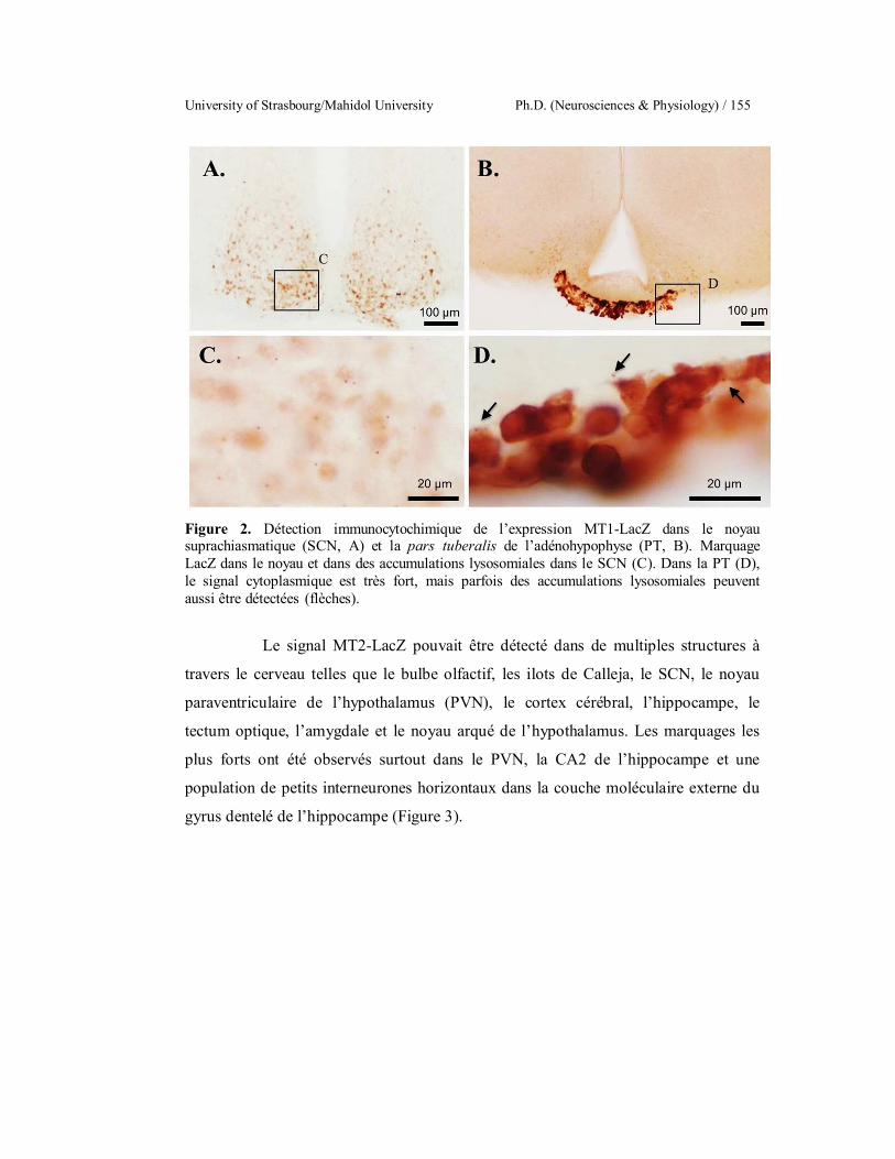
University of Strasbourg/Mahidol University Ph.D. (Neurosciences & Physiology) / 155
Figure 2. Détection immunocytochimique de l’expression MT1-LacZ dans le noyau suprachiasmatique (SCN, A) et la pars tuberalis de l’adénohypophyse (PT, B). Marquage LacZ dans le noyau et dans des accumulations lysosomiales dans le SCN (C). Dans la PT (D), le signal cytoplasmique est très fort, mais parfois des accumulations lysosomiales peuvent aussi être détectées (flèches).
Le signal MT2-LacZ pouvait être détecté dans de multiples structures à
travers le cerveau telles que le bulbe olfactif, les ilots de Calleja, le SCN, le noyau
paraventriculaire de l’hypothalamus (PVN), le cortex cérébral, l’hippocampe, le
tectum optique, l’amygdale et le noyau arqué de l’hypothalamus. Les marquages les
plus forts ont été observés surtout dans le PVN, la CA2 de l’hippocampe et une
population de petits interneurones horizontaux dans la couche moléculaire externe du
gyrus dentelé de l’hippocampe (Figure 3).
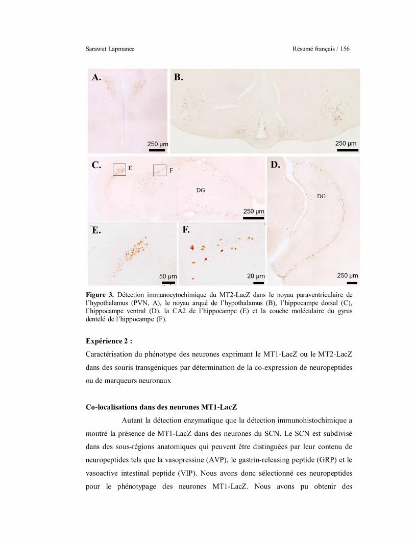
Sarawut Lapmanee Résumé français / 156
Figure 3. Détection immunocytochimique du MT2-LacZ dans le noyau paraventriculaire de l’hypothalamus (PVN, A), le noyau arqué de l’hypothalamus (B), l’hippocampe dorsal (C), l’hippocampe ventral (D), la CA2 de l’hippocampe (E) et la couche moléculaire du gyrus dentelé de l’hippocampe (F).
Expérience 2 :
Caractérisation du phénotype des neurones exprimant le MT1-LacZ ou le MT2-LacZ
dans des souris transgéniques par détermination de la co-expression de neuropeptides
ou de marqueurs neuronaux
Co-localisations dans des neurones MT1-LacZ
Autant la détection enzymatique que la détection immunohistochimique a
montré la présence de MT1-LacZ dans des neurones du SCN. Le SCN est subdivisé
dans des sous-régions anatomiques qui peuvent être distinguées par leur contenu de
neuropeptides tels que la vasopressine (AVP), le gastrin-releasing peptide (GRP) et le
vasoactive intestinal peptide (VIP). Nous avons donc sélectionné ces neuropeptides
pour le phénotypage des neurones MT1-LacZ. Nous avons pu obtenir des

University of Strasbourg/Mahidol University Ph.D. (Neurosciences & Physiology) / 157
colocalisations pour le GRP et le VIP, mais pas du tout pour l’AVP. Dans le cas du
GRP et du VIP, seules certaines cellules GRP ou VIP positives contenaient aussi le
MT1-LacZ (Figure 4).
Figure 4. Double marquages avec du MT1-LacZ par immunocytochimie (fluorescence verte) et de l’hybridation in situ pour des neuropeptides (fluorescence rouge) dans le SCN. Diagrammes représentant la distribution des corps cellulaires et des fibres des neuropeptides dans le SCN (Abrahamson and Moore, 2001) AVP (A), GRP (B) et VIP (C). Les accumulations lysosomiales du MT1-LacZ (points verts) sont clairement situées en-dehors de la zone des neurones à AVP (D). Pour les colocalisations avec le VIP (E) ou le GRP (F), quelques neurones peptidergiques rouges contiennent les points verts des accumulations lysosomiales du MT1-LacZ (flèches).
Les neurones vasopressinergiques de l’SCN sont considérés être la
principale voie de sortie de cette structure qui est l’horloge biologique centrale de
l’organisme. Les neurones à VIP reçoivent les afférences en provenance de la rétine et
les neurones à GRP sont considérés faire partie du mécanisme central de l’horloge
biologique. Par le récepteur MT1, la mélatonine peut donc moduler les afférences
rétiniennes et le fonctionnement central de l’horloge, mais pas directement les voies
efférentes de l’SCN.
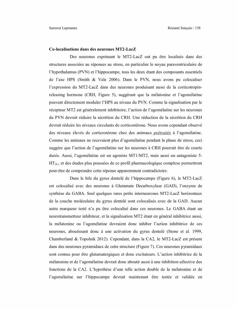
Sarawut Lapmanee Résumé français / 158
Co-localisations dans des neurones MT2-LacZ
Des neurones exprimant le MT2-LacZ ont pu être localisés dans des
structures associées au réponses au stress, en particulier le noyau paraventriculaire de
l’hypothalamus (PVN) et l’hippocampe, tous les deux étant des composants essentiels
de l’axe HPS (Smith & Vale 2006). Dans le PVN, nous avons pu colocaliser
l’expression du MT2-LacZ dans des neurones produisant aussi de la corticotropin-
releasing hormone (CRH, Figure 5), suggérant que la mélatonine et l’agomélatine
peuvent directement moduler l’HPS au niveau du PVN. Comme la signalisation par le
récepteur MT2 est généralement inhibitoire, l’action de l’agomélatine sur les neurones
du PVN devrait réduire la sécrétion du CRH. Une réduction de la sécrétion du CRH
devrait réduire les niveaux circulants de corticostérone. Nous avons cependant observé
des niveaux élevés de corticostérone chez des animaux prétraités à l’agomélatine.
Comme les animaux ne recevaient plus d’agomélatine pendant la phase de stress, ceci
suggère que l’action de l’agomélatine sur les neurones à CRH pourrait être de courte
durée. Aussi, l’agomélatine est un agoniste MT1/MT2, mais aussi un antagoniste 5-
HT2C, et des études plus poussées de ce profil pharmacologique complexe permettront
peut-être de comprendre cette réponse apparemment contradictoire.
Dans le hile du gyrus dentelé de l’hippocampe (Figure 6), le MT2-LacZ
est colocalisé avec des neurones à Glutamate Decarboxylase (GAD), l’enzyme de
synthèse du GABA. Seul quelques rares petits interneurones MT2-LacZ horizontaux
de la couche moléculaire du gyrus dentelé sont colocalisés avec de la GAD. Aucun
autre marqueur testé n’a pu être colocalisé dans ces neurones. Le GABA étant un
neurotransmetteur inhibiteur, et la signalisation MT2 étant en général inhibitrice aussi,
la mélatonine ou l’agomélatine devraient donc inhiber l’action inhibitrice de ces
neurones, aboutissant donc à une activation du gyrus dentelé (Stone et al. 1999,
Chamberland & Topolnik 2012). Cependant, dans la CA2, le MT2-LacZ est présent
dans des neurones pyramidaux de cette structure (Figure 7). Ces neurones pyramidaux
sont connus pour être glutamatergiques et donc excitateurs. L’action inhibitrice de la
mélatonine et de l’agomélatine devrait donc aboutir aussi à une inhibition sélective des
fonctions de la CA2. L’hypothèse d’une telle action double de la mélatonine et de
l’agomélatine sur l’hippocampe devrait maintenant être testée et validée en

University of Strasbourg/Mahidol University Ph.D. (Neurosciences & Physiology) / 159
électrophysiologie et par des études comportementales. Comme un telle action
impacterait en particulier le fonctionnement de l’HPS reste à déterminer.
De plus, des neurones à MT2-LacZ sont aussi observés dans le raphé
dorsal, qui contient une des principales populations de neurones à 5-HT. Le raphé
dorsal ne contient apparemment pas de 5-HT2C, mais le 5-HT2C est impliqué dans le
rétrocontrôle négatif de la 5-HTsur le raphé dorsal par l’intermédiaire de neurones
GABAergiques localisés autour du raphé dorsale et qui eux expriment le 5-HT2C
(Serrats et al. 2005). Ainsi, l’agomélatine pourrait agir directement sur le raphé dorsal
par le MT2 et indirectement par le 5-HT2C par l’intermédiaire de ces neurones
GABAergiques. En tant qu’agoniste MT1/MT2, l’agomélatine pourrait inhiber les
neurones sérotoninergiques du raphé dorsal. En tant qu’antagoniste 5-HT2C,
l’agomélatine pourrait bloquer le rétrocontrôle négatif des neurones GABAergiques.
Ainsi l’action de l’agomélatine sur le raphé dorsal est probablement très complexe
parce qu’elle inhiberait directement le raphé dorsal tout en le stimulant de manière
indirecte. Néanmoins, le raphé dorsal est donc certainement une cible d’intérêt pour
l’agomélatine.
Figure 5. Double marquages avec de l’immunomarquage pour le MT2-LacZ (brun à gauche, fluorescence verte à droite) et de l’hybridation in situ non-radioactive pour le CRH (fluorescence rouge) dans le noyau paraventriculaire de l’hypothalamus (PVN). L’agrandissement montre un groupe de neurones à CRH (rouge) dont la majeure partie exprime aussi le MT2-LacZ (vert). L’astérisque montre un neurone à CRH ne contenant pas de MT2-LacZ.
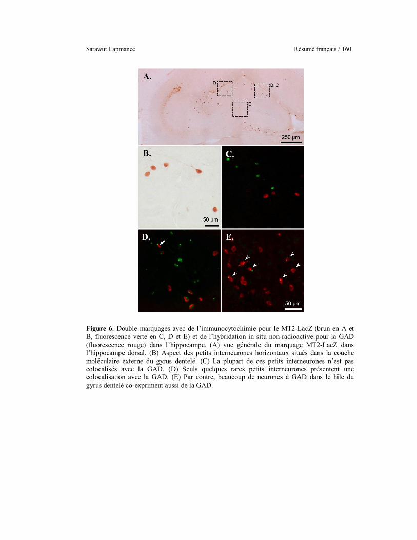
Sarawut Lapmanee Résumé français / 160
Figure 6. Double marquages avec de l’immunocytochimie pour le MT2-LacZ (brun en A et B, fluorescence verte en C, D et E) et de l’hybridation in situ non-radioactive pour la GAD (fluorescence rouge) dans l’hippocampe. (A) vue générale du marquage MT2-LacZ dans l’hippocampe dorsal. (B) Aspect des petits interneurones horizontaux situés dans la couche moléculaire externe du gyrus dentelé. (C) La plupart de ces petits interneurones n’est pas colocalisés avec la GAD. (D) Seuls quelques rares petits interneurones présentent une colocalisation avec la GAD. (E) Par contre, beaucoup de neurones à GAD dans le hile du gyrus dentelé co-expriment aussi de la GAD.
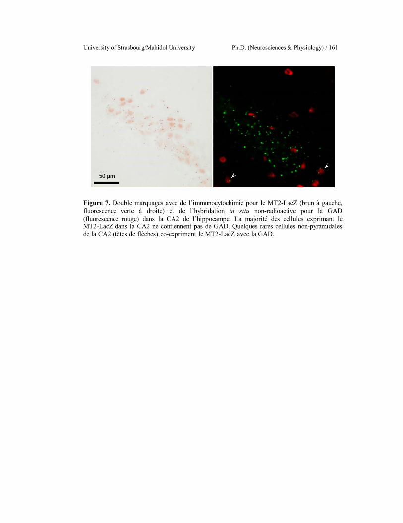
University of Strasbourg/Mahidol University Ph.D. (Neurosciences & Physiology) / 161
Figure 7. Double marquages avec de l’immunocytochimie pour le MT2-LacZ (brun à gauche, fluorescence verte à droite) et de l’hybridation in situ non-radioactive pour la GAD (fluorescence rouge) dans la CA2 de l’hippocampe. La majorité des cellules exprimant le MT2-LacZ dans la CA2 ne contiennent pas de GAD. Quelques rares cellules non-pyramidales de la CA2 (tètes de flèches) co-expriment le MT2-LacZ avec la GAD.
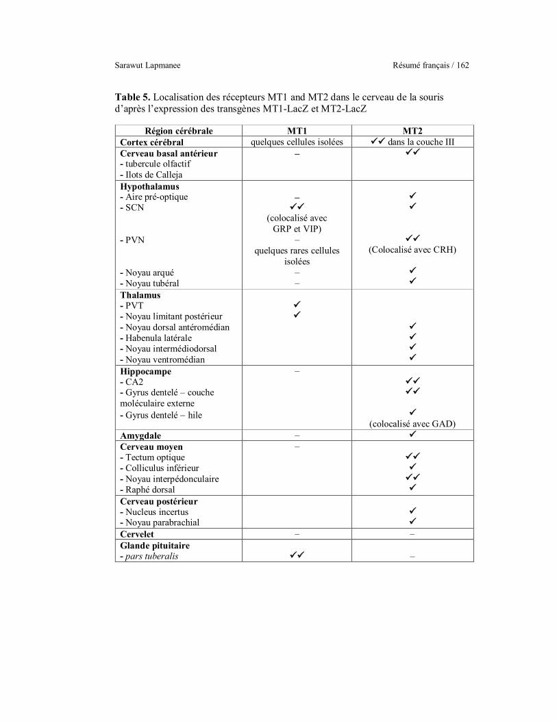
Sarawut Lapmanee Résumé français / 162
Table 5. Localisation des récepteurs MT1 and MT2 dans le cerveau de la souris d’après l’expression des transgènes MT1-LacZ et MT2-LacZ
Région cérébrale MT1 MT2 Cortex cérébral quelques cellules isolées dans la couche III Cerveau basal antérieur - tubercule olfactif - Ilots de Calleja
–
Hypothalamus - Aire pré-optique - SCN - PVN - Noyau arqué - Noyau tubéral
–
(colocalisé avec GRP et VIP)
– quelques rares cellules
isolées – –
(Colocalisé avec CRH)
Thalamus - PVT - Noyau limitant postérieur - Noyau dorsal antéromédian - Habenula latérale - Noyau intermédiodorsal - Noyau ventromédian
Hippocampe - CA2 - Gyrus dentelé – couche moléculaire externe - Gyrus dentelé hile
–
(colocalisé avec GAD) Amygdale – Cerveau moyen - Tectum optique - Colliculus inférieur - Noyau interpédonculaire - Raphé dorsal
–
Cerveau postérieur - Nucleus incertus - Noyau parabrachial
Cervelet – – Glande pituitaire - pars tuberalis
–
University of Strasbourg/Mahidol University Ph.D. (Neurosciences & Physiology) / 163
D. Conclusions
Les buts de ce travail étaient d’abord d’examiner la cinétique des effets
d’un stress de contention sur les comportements d’anxiété et de dépression, ainsi que
les perturbations de la mémoire chez des rats mâles. En deuxième lieu, nous voulions
déterminer les effets thérapeutiques préventifs d’un traitement pharmacologique par
des modulateurs monoamingergiques (agomélatine ou venlafaxine) ou d’un exercice
physique volontaire sur ces comportements. Outre des paramètres comportementaux,
nous avons aussi investigué des paramètres physiques et physiologiques, ainsi que
l’expression de protéines cibles dans les régions associées aux réponses de stress.
Enfin, nous avons identifié des sites d’action potentiels pour l’agomélatine dans des
souris transgéniques rapportrices MT1-LacZ et MT2-LacZ.
Les conclusions sont les suivantes, résumées dans la Figure 8 :
Un stress de contention de 4 semaines chez le rat mâle réduit la prise de
nourriture et le poids corporel, induit une hypertrophie des surrénales et augmente les
niveaux sériques et urinaires de la corticostérone. Ces niveaux élevés de corticostérone
pourraient altérer l’expression des protéines cibles, telles que le facteur neurotrophique
dérivé du cerveau (BDNF), le récepteur aux glucocorticoïdes (GR), le transporteur de
la noradrénaline (NET) et le récepteur 5-HT2C de la sérotonine dans l’amygdale,
l’hippocampe, le locus coeruleus et le gris périacquéducal menant à des manifestations
d’anxiété, de dépression et des troubles de la mémoire.
Un exercice volontaire d’activité de roue de 4 semaines peut partiellement
prévenir l’élévation de la corticostérone sérique et les comportements anhédoniques
induits par le stress, probablement par une modulation des récepteurs surrénaliens
pour l’hormone adrénocorticotrope (ACTH) et en altérant l’expression de protéines
cibles (BDNF et NET) dans des structures impliquées dans la réponse au stress telles
que l’hippocampe, le septum, le locus coeruleus et le gris périacquéducal. Ainsi,
l’exercice physique volontaire pourrait réduire les effets néfastes d’un stress chronique
en modulant l’activité sympathique tant au niveau central qu’au niveau périphérique
de l’axe HPS.
L’agomélatine, la venlafaxine et l’activité volontaire dans une roue
peuvent prévenir les comportements anxieux et dépressifs, ainsi que les perturbations
Sarawut Lapmanee Résumé français / 164
de la mémoire, induits par un stress de contention. L’augmentation de l’expression du
BDNF dans l’hippocampe pourrait être un mécanisme des effets protecteurs de la
venlafaxine et de l’exercice physique chez le rat mâle stressé.
Concernant les applications cliniques de nos observations, l’exercice
physique volontaire peut être recommandé comme traitement alternatif ou de concert
avec un traitement pharmacologique (p.ex. agomélatine ou venlafaxine) pour le
traitement des symptômes d’anxiété, de dépression et de perturbation de la mémoire
chez des patients et des individus stressés.
La cartographie et le phénotypage des neurones exprimant les récepteurs
de la mélatonine MT1 et M2 indique des sites d’action potentiels pour la mélatonine et
des agents mélatoninergiques tels que l’agomélatine. L’expression du MT1 est
restreinte à quelques structures clés, en particulier le noyau suprachiasmatique (SCN),
le noyau paraventriculaire du thalamus (PVT) et la pars tuberalis de la glande
pituitaire. Le récepteur MT2 quant à lui est beaucoup plus largement exprimé dans le
cerveau, avec notablement le noyau suprachiasmatique (SCN), le noyau
paraventriculaire de l’hypothalamus (PVN), l’hippocampe, l’amygdale et le raphé
dorsal (DR). Des études supplémentaires seront nécessaires pour clarifier le mode
d’action de l’agomélatine.
En résumé, un stress de contention de 4 semaines cause une hyperactivité
de l’axe HPS et augmente le niveau des glucocorticoïdes menant à une réduction
d’expression du récepteur aux glucocorticoïdes et du BDNF dans l’hippocampe.
Quatre semaines d’activité de roue volontaire module l’activité de l’axe HPS et
diminue partiellement les niveaux de glucocorticoïdes en diminuant l’expression du
récepteur surrénalien de l’ACTH. Cette activité augmente aussi l’expression du BDNF
réduisant les comportements anhédoniques chez le rat mâle stressé. De plus, l’exercice
physique volontaire est capable de prévenir l’anxiété, la dépression et les troubles de
la mémoire induits par le stress. Un prétraitement par la venlafaxine inhibe
directement le NET pour augmenter la noradrénaline dans la fente synaptique et
exerce des effets antidépresseurs et anxiolytiques en promouvant la production de
BDNF par l’hippocampe. Un prétraitement par l’agomélatine, un agoniste MT1/MT2,
peut prévenir les comportements liés au stress en agissant par les récepteurs MT1 et
MT2 dans le SCN, le PVN, le raphé dorsal et l’hippocampe.

University of Strasbourg/Mahidol University Ph.D. (Neurosciences & Physiology) / 165
Figure 8. Résumé graphique des résultats de cette étude.

Sarawut Lapmanee References / 166
REFERENCES
Abrahamson EE, Moore RY. Suprachiasmatic nucleus in the mouse: retinal
innervation, intrinsic organization and efferent projections. Brain Res.
2001;916(12):17291.
Acosta GB, Otero Losada ME, Rubio MC. Area-dependent changes in GABAergic
function after acute and chronic cold stress. Neurosci Lett. 1993;154,
175–8.
Adam TC, Epel ES. Stress, eating and the reward system. Physiol Behav.
2007;91(4):44958.
Adlard PA, Cotman CW. Voluntary exercise protects against stress-induced decreases
in brain-derived neurotrophic factor protein expression. Neuroscience.
2004;124(4):985–92.
Adlard PA, Perreau VM, Engesser-Cesar C, Cotman CW. The timecourse of induction
of brain-derived neurotrophic factor mRNA and protein in the rat
hippocampus following voluntary exercise. Neurosci Lett.
2004;363(1):438.
Aguilera G, Liu Y. The molecular physiology of CRH neurons. Front
Neuroendocrinol. 2012;33(1):6784.
Ahn SK, Khalmuratova R, Hah YS, Jeon SY, Hur DG, Kang HS, Balaban CD.
Immunohistochemical and biomolecular identification of melatonin 1a and
1b receptors in rat vestibular nuclei. Auris Nasus Larynx.
2012;39(5):47983.
Alex KD, Yavanian GJ, McFarlane HG, Pluto CP, Pehek EA. Modulation of
dopamine release by striatal 5-HT2C receptors. Synapse.
2005;55(4):24251.
American Psychiatric Association: DSM-5 (the Diagnostic and Statistical manual of
Mental Disorders, 5th edition). American Psychiatric Association, 2013.
Amsterdam JD, Chopra M. Monoamine oxidase inhibitors revisited. Psychiatric
Annals. 2001;31:361–70.
University of Strasbourg/Mahidol University Ph.D. (Neurosciences & Physiology) / 167
Anastasio NC, Lanfranco MF, Bubar MJ, Seitz PK, Stutz SJ, McGinnis AG, Watson
CS, Cunningham KA. Serotonin 5-HT2C receptor protein expression is
enriched in synaptosomal and post-synaptic compartments of rat cortex.
J Neurochem. 2010;113(6):150415.
Antunes M, Biala G. The novel object recognition memory: neurobiology, test
procedure, and its modifications. Cogn Process 2012;13(2):93–110.
Asada H, Kawamura Y, Maruyama K, Kume H, Ding RG, Kanbara N, Kuzume H,
Sanbo M, Yagi T, Obata K. Cleft palate and decreased brain gamma-
aminobutyric acid in mice lacking the 67-kDa isoform of glutamic acid
decarboxylase. Proc Natl Acad Sci U S A. 1997;94(12):64969.
Avwenagha O, Bird MM, Lieberman AR, Yan Q, Campbell G. Patterns of expression
of brain-derived neurotrophic factor and tyrosine kinase B mRNAs and
distribution and ultrastructural localization of their proteins in the visual
pathway of the adult rat. Neuroscience. 2006;140(3):91328.
Aydemir C, Deveci A, Taneli F. The effect of chronic antidepressant treatment of
serum brain-derived neurotrophic factor levels in depressed patients: a
preliminary study. Prog Neuropsychopharmacol Biol Psychiatry.
2005;29:261–5.
Balu DT, Lucki I. Adult hippocampal neurogenesis: regulation, functional
implications, and contribution to disease pathology. Neurosci Biobehav
Rev. 2009;33:232–52.
Banasr M, Soumier A, Hery M, Mocaër E, Daszuta A. Agomelatine, a new
antidepressant, induces regional changes in hippocampal neurogenesis.
Biol Psychiatry. 2006;59(11):108796.
Barker EL, Blakely RD. Norepinephrine and serotonin transporters: molecular targets
of antidepressant drugs. J Biol Chem. 1995; 264:2195–8.
Bauer M, El-Khalili N, Datto C, Szamosi J, Eriksson H. A pooled analysis of two
randomised, placebo-controlled studies of extended release quetiapine
fumarate adjunctive to antidepressant therapy in patients with major
depressive disorder. J Affect Disord. 2010;127(13):1930.

Sarawut Lapmanee References / 168
Beck KD, Luine VN. Food deprivation modulates chronic stress effects on object
recognition in male rats: Role of monoamines and amino acids. Brain Res.
1999;830:56–71.
Belmaker RH, Agam G. Major depressive disorder. N Engl J Med.
2008;358(1):5568.
Belzung C, Griebel G. Measuring normal and pathological anxiety-like behaviour in
mice: a review. Behav Brain Res. 2001;125(1–2):141–9.
Berridge MJ, Downes CP, Hanley MR. Lithium amplifies agonist-dependent
phosphatidylinositol responses in brain and salivary glands. Biochem J.
1982;206(3):58795.
Berton O, Durand M, Aguerre S, Mormede P, Chaouloff F. Behavioral,
neuroendocrine and serotonergic consequences of single social defeat and
repeated fluoxetine pretreatment in the Lewis rat strain. Neuroscience.
1999;92:327–41.
Biggs JT, Spiker DG, Petit JM, Ziegler VE. Tricyclic antidepressant overdose:
incidence of symptoms. JAMA. 1977;238(2):1358.
Binder EB, Nemeroff CB. The CRF system, stress, depression and anxiety-insights
from human genetic studies. Mol Psychiatry. 2010;15(6):57488.
Blumenthal JA, Babyak MA, Doraiswamy PM, Watkins L, Hoffman BM, Barbour
KA, Herman S, Craighead WE, Brosse AL, Waugh R, Hinderliter A,
Sherwood A. Exercise and pharmacotherapy in the treatment of major
depressive disorder. Psychosom Med. 2007;69(7):58796.
Bonnefond C, Monnerie R, Richard JP, Martinet L. Melatonin and the circadian clock
in mink: effects of daily injections of melatonin on circadian rhythm of
locomotor activity and autoradiographic localization of melatonin binding
sites. J Neuroendocrinol. 1993;5(3):2416.
Booth FW, Laye MJ, Lees SJ, Rector RS, Thyfault JP. Reduced physical activity and
risk of chronic disease: the biology behind the consequences. Eur. J. Appl.
Physiol. 2008;102(4):381–90.
Bourin M, Mocaër E, Porsolt R. Antidepressant-like activity of S20098 (agomelatine)
in the forced swimming test in rodents: involvement of melatonin and
serotonin receptors. J Psychiatry Neurosci. 2004;29(2):126–33.
University of Strasbourg/Mahidol University Ph.D. (Neurosciences & Physiology) / 169
Bowers G, Cullinan WE, Herman JP. Region-specific regulation of glutamic acid
decarboxylase (GAD) mRNA expression in central stress circuits. J
Neurosci. 1998;18(15):593847.
Bowman RE, Beck KD, Luine VN. Chronic stress effects on memory: sex differences
in performance and monoaminergic activity. Horm Behav.
2003;43(1):4859.
Boyle MP, Brewer JA, Funatsu M, Wozniak DF, Tsien JZ, Izumi Y, Muglia LJ.
Acquired deficit of forebrain glucocorticoid receptor produces depression-
like changes in adrenal axis regulation and behavior. Proc Natl Acad Sci U
S A. 2005;102(2):4738.
Braestrup C, Nielsen M, Nielsen EB, Lyon M. Benzodiazepine receptors in the brain
as affected by different experimental stresses: the changes are small and
not undirectional. Psychopharmacology (Berl). 1979;65(3):2737.
Burghardt PR, Fulk LJ, Hand GA, Wilson MA. The effects of chronic treadmill and
wheel running on behavior in rats. Brain Res. 2004;1019(12):8496.
Buynitsky T, Mostofsky DI. Restraint stress in biobehavioral research: Recent
developments. Neurosci Biobehav Rev. 2009;33(7):108998.
Campbell JE, Rakhshani N, Fediuc S, Bruni S, Riddell MC. Voluntary wheel running
initially increases adrenal sensitivity to adrenocorticotrophic hormone,
which is attenuated with long-term training. J Appl Physiol (1985).
2009;106(1):6672.
Carman HM, Mactutus CF. Proximal versus distal cue utilization in spatial navigation:
the role of visual acuity? Neurobiol Learn Mem. 2002;78(2):33246.
Carta MG, Hardoy MC, Pilu A, Sorba M, Floris AL, Mannu FA, Baum A, Cappai A,
Velluti C, Salvi M. Improving physical quality of life with group physical
activity in the adjunctive treatmment of major depression disorder. Clin
Pract Epidemiol Ment Health. 2008;4:1–6.
Castagné V, Moser P, Roux S, Porsolt RD. Rodent models of depression: forced swim
and tail suspension behavioral despair tests in rats and mice. Curr Protoc
Neurosci. 2011;55:8.10A.
Sarawut Lapmanee References / 170
Cerqueira JJ, Almeida OF, Sousa N. The stressed prefrontal cortex. Left? Right! Brain
Behav Immun. 2008;22(5):630–8.
Chamberland S, Topolnik L. Inhibitory control of hippocampal inhibitory neurons.
Front Neurosci. 2012;165(6):1–13.
Chao MV, Bothwell M. Neurotrophins: To cleave or not to cleave. Neuron.
2002;33:9–12.
Charney DS, Drevets WC. The neurobiological basis of anxiety disorders. In: Davis
KL, Charney DS, Coyle JT, Nemeroff C, editors.
Neuropsychopharmacology: The fifth generation of progress. Philadelphia:
Lippincott Williams and Wilkins; 2002.
Charney DS. Monoamine dysfunction and the pathophysiology and treatment of
depression. J Clin Psychiatry. 1998;59 Suppl 14:114.
Charoenphandhu N, Nuntapornsak A, Wongdee K, Krishnamra N, Charoenphandhu J.
Upregulated mRNA levels of SERT, NET, MAOB, and BDNF in various
brain regions of ovariectomized rats exposed to chronic aversive stimuli.
Mol Cell Biochem. 2013;375(1–2):49–58.
Charoenphandhu N, Teerapornpuntakit J, Lapmanee S, Dorkkam N, Krishnamra N,
Charoenphandhu J. Long-term swimming in an inescapable stressful
environment attenuates the stimulatory effect of endurance swimming on
duodenal calcium absorption in rats. J Physiol Sci. 2011;61(6):47386.
Chen P, Fan Y, Li Y, Sun Z, Bissette G, Zhu MY. Chronic social defeat up-regulates
expression of norepinephrine transporter in rat brains. Neurochem Int.
2012;60:920.
Chiba S, Numakawa T, Ninomiya M, Richards MC, Wakabayashi C, Kunugi H.
Chronic restraint stress causes anxiety- and depression-like behaviors,
downregulates glucocorticoid receptor expression, and attenuates
glutamate release induced by brain-derived neurotrophic factor in the
prefrontal cortex. Prog Neuropsychopharmacol Biol Psychiatry.
2012;39(1):1129.
Christianson JP, Ragole T, Amat J, Greenwood BN, Strong PV, Paul ED, Fleshner M,
Watkins LR, Maier SF. 5-hydroxytryptamine 2C receptors in the
University of Strasbourg/Mahidol University Ph.D. (Neurosciences & Physiology) / 171
basolateral amygdala are involved in the expression of anxiety after
uncontrollable traumatic stress. Biol Psychiatry. 2010;67:339–45.
Christoffel DJ, Golden SA, Russo SJ. Structural and synaptic plasticity in stress-
related disorders. Rev Neurosci. 2011;22(5):53549.
Clark MS, Russo AF. Tissue-specific glucocorticoid regulation of tryptophan
hydroxylase mRNA levels. Brain Res Mol Brain Res. 1997;48:346–54.
Claustrat B, Chazot G, Brun J, Jordan D, Sassolas G. A chronobiological study of
melatonin and cortisol secretion in depressed subjects: plasma melatonin, a
biochemical marker in major depression. Biol Psychiatry.
1984;19(8):121528.
Clemett DA, Punhani T, Duxon MS, Blackburn TP, Fone KC. Immunohistochemical
localisation of the 5-HT2C receptor protein in the rat CNS.
Neuropharmacology. 2000;39(1):12332.
Comai S, Gobbi G. Unveiling the role of melatonin MT2 receptors in sleep, anxiety
and other neuropsychiatric diseases: a novel target in
psychopharmacology. J Psychiatry Neurosci. 2014;39(1):621.
Condie BG, Bain G, Gottlieb DI, Capecchi MR. Cleft palate in mice with a targeted
mutation in the gamma-aminobutyric acid-producing enzyme glutamic
acid decarboxylase 67. Proc Natl Acad Sci U S A. 1997;94(21):114515.
Connor B, Dragunow M. The role of neuronal growth factors in neurodegenerative
disorders of the human brain. Brain Res Brain Res Rev. 1998;27(1):139.
Cooke JD, Grover LM, Spangler PR. Venlafaxine treatment stimulates expression of
brain-derived neurotrophic factor protein in frontal cortex and inhibits
long-term potentiation in hippocampus. Neuroscience.
2009;162(4):14119.
Cotman CW, Berchtold NC. Exercise: a behavioral intervention to enhance brain
health and plasticity. Trends Neurosci. 2002;25(6):295301
Cryan JF, Holmes A. The ascent of mouse: advances in modelling human depression
and anxiety. Nat Rev Drug Discov. 2005;4(9):77590.
Dagnino-Subiabre A, Orellana JA, Carmona-Fontaine C, Montiel J, Díaz-Velíz G,
Serón-Ferré M, Wyneken U, Concha ML, Aboitiz F. Chronic stress
Sarawut Lapmanee References / 172
decreases the expression of sympathetic markers in the pineal gland and
increases plasma melatonin concentration in rats. J Neurochem.
2006;97:1279–87.
Dagytè G, Crescente I, Postema F, Seguin L, Gabriel C, Mocaër E, Boer JA, Koolhaas
JM. Agomelatine reverses the decrease in hippocampal cell survival
induced by chronic mild stress. Behav Brain Res. 2011;218(1):121–8.
Dagytè G, Trentani A, Postema F, Luiten PG, Den Boer JA, Gabriel C, Mocaër E,
Meerlo P, Van der Zee EA. The novel antidepressant agomelatine
normalizes hippocampal neuronal activity and promotes neurogenesis in
chronically stressed rats. CNS Neurosci Ther. 2010;16(4):195207.
Davidson SR, Burnett M, Hoffman-Goetz L. Training effects in mice after long-term
voluntary exercise. Med. Sci. Sports Exerc. 2006;38(2):250–5.
de Groote L, Linthorst AC. Exposure to novelty and forced swimming evoke stressor-
dependent changes in extracellular GABA in the rat hippocampus.
Neuroscience. 2007;148(3):794805.
de Kloet ER, Joëls M, Holsboer F. Stress and the brain: from adaptation to disease.
Nat Rev Neurosci. 2005;6(6):46375.
de Kloet ER, Vreugdenhil E, Oitzl MS, Joëls M. Brain corticosteroid receptor balance
in health and disease. Endocr Rev. 1998;19(3):269301.
de Moor MH, Beem AL, Stubbe JH, Boomsma DI, De Geus EJ. Regular exercise,
anxiety, depression and personality: a population-based study. Prev Med.
2006;42(4):2739.
Dell’Osso B, Buoli M, Baldwin DS, Altamura AC. Serotonin norepinephrine reuptake
inhibitors (SNRIs) in anxiety disorders: a comprehensive review of their
clinical efficacy. Hum Psychopharmacol. 2010;25(1):1729.
Diamond DM, Park CR, Heman KL, Rose GM. Exposing rats to a predator impairs
spatial working memory in the radial arm water maze. Hippocampus.
1999;9:542–52.
Dishman RK, Renner KJ, Youngstedt SD, Reigle TG, Bunnell BN, Burke KA, Yoo
HS, Mougey EH, Meyerhoff JL. Activity wheel running reduces escape
latency and alters brain monoamine levels after footshock. Brain Res Bull.
1997;42(5):399–406.

University of Strasbourg/Mahidol University Ph.D. (Neurosciences & Physiology) / 173
Dishman RK. Brain monoamines, exercise, and behavioral stress: animal models. Med
Sci Sports Exerc. 1997;29(1):63–74.
Douglas PS, O’Toole ML, Katz SE, Ginsburg GS, Hiller WD, Laird RH. Left
ventricular hypertrophy in athletes. Am J Cardiol. 1997;80(10):13848.
Drevets WC. Neuroimaging and neuropathological studies of depression: implications
for the cognitive-emotional features of mood disorders. Curr Opin
Neurobiol. 2001;11(2):240–9.
Droste SK, Chandramohan Y, Hill LE, Linthorst AC, Reul JM. Voluntary exercise
impacts on the rat hypothalamic-pituitary-adrenocortical axis mainly at the
adrenal level. Neuroendocrinology. 2007;86(1):2637.
Droste SK, Gesing A, Ulbricht S, Muller MB, Linthorst ACE, Reul JMHM. Effects of
long-term voluntary exercise on the mouse hypothalamic–pituitary–
adrenocortical axis. Endocrinology. 2003;144(7):3012–23.
Dryden S, Wang Q, Frankish HM, Williams G. Differential effects of the 5-HT 1B/2C
receptor agonist mCPP and the 5-HT1A agonist flesinoxan on
hypothalamic neuropeptide Y in the rat: evidence that NPY may mediate
serotonin's effects on food intake. Peptides. 1996;17(6):9439.
Dubocovich ML, Delagrange P, Krause DN, Sugden D, Cardinali DP, Olcese J.
International Union of Basic and Clinical Pharmacology. LXXV.
Nomenclature, classification, and pharmacology of G protein-coupled
melatonin receptors. Pharmacol Rev. 2010;62(3):34380.
Dubocovich ML, Markowska M. Functional MT1 and MT2 melatonin receptors in
mammals. Endocrine. 2005;27(2):10110.
Dubocovich ML. Melatonin receptors: role on sleep and circadian rhythm regulation.
Sleep Med. 2007;8 Suppl 3:3442.
Duman CH, Schlesinger L, Russell DS, Duman RS. Voluntary exercise produces
antidepressant and anxiolytic behavioral effects in mice. Brain Res.
2008;1199:148–58.
Duman RS, Monteggia LM. A neurotrophic model for stress-related mood disorders.
Biol Psychiatry. 2006;59(12):1116–27.
Duman RS. Role of neurotrophic factors in the etiology and treatment of mood
disorders. Neuromolecular Med. 2004;5(1):11–25.
Sarawut Lapmanee References / 174
Dunn AL, Reigle TG, Youngstedt SD, Armstrong RB, Dishman RK. Brain
norepinephrine and metabolites after treadmill training and wheel running
in rats. Med Sci Sports Exerc. 1996;28(2):204–9.
Ely DR, Dapper V, Marasca J, Corrêa JB, Gamaro GD, Xavier MH, Michalowski MB,
Catelli D, Rosat R, Ferreira MB, Dalmaz C. Effect of restraint stress on
feeding behavior of rats. Physiol Behav. 1997;61:395–8.
Engesser-Cesar C, Anderson AJ, Cotman CW. Wheel running and fluoxetine
antidepressant treatment have differential effects in the hippocampus and
the spinal cord. Neuroscience. 2007;144(3):103344.
Ennaceur A, Delacour J. A new one-trial test for neurobiological studies of memory in
rats. 1: Behavioral data. Behav Brain Res. 1988;31(1):4759.
Evans DL, Davidson J, Raft T. Early and late side-effects of phenelzine. J Clin
Psychopharmacol. 1982;2:208–10.
Eyding D, Lelgemann M, Grouven U, Härter M, Kromp M, Kaiser T, Kerekes MF,
Gerken M, Wieseler B. Reboxetine for acute treatment of major
depression: systematic review and meta-analysis of published and
unpublished placebo and selective serotonin reuptake inhibitor controlled
trials. BMJ. 2010;341(c4737):114.
Fan Y, Chen P, Li Y, Cui K, Noel DM, Cummins ED, Peterson DJ, Brown RW, Zhu
MY. Corticosterone administration up-regulated expression of
norepinephrine transporter and dopamine β-hydroxylase in rat locus
coeruleus and its terminal regions. J Neurochem. 2014;128(3):44558.
Fan Y, Chen P, Li Y, Ordway GA, Zhu MY. Effects of desipramine treatment on
stress-induced up-regulation of norepinephrine transporter expression in
rat brains. Psychopharmacology (Berl). 2015;232(2):37990.
Fediuc S, Campbell JE, Riddell MC. Effect of voluntary wheel running on circadian
corticosterone release and on HPA axis responsiveness to restraint stress in
Sprague-Dawley rats. J Appl Physiol. 2006;100:1867–75.
Ferreira ZS, Fernandes PA, Duma D, Assreuy J, Avellar MC, Markus RP.
Corticosterone modulates noradrenaline-induced melatonin synthesis
through inhibition of nuclear factor kappa B. J Pineal Res. 2005;38:182–8.

University of Strasbourg/Mahidol University Ph.D. (Neurosciences & Physiology) / 175
Fiedorowicz JG, Swartz KL. The role of monoamine oxidase inhibitors in current
psychiatric practice. J Psychiatr Pract. 2004;10(4):23948.
Fisher L, Chesla CA, Mullan JT, Skaff MM, Kanter RA. Contributors to depression in
Latino and European-American patients with type 2 diabetes. Diabetes
Care. 2001;24(10):1751–57.
Foerde K, Shohamy D. Feedback timing modulates brain systems for learning in
humans. J Neurosci. 2011;31(37):1315767
Fone KC, Shalders K, Fox ZD, Arthur R, Marsden CA. Increased 5-HT2C receptor
responsiveness occurs on rearing rats in social isolation.
Psychopharmacology (Berl). 1996;123(4):34652.
Fujieda H, Hamadanizadeh SA, Wankiewicz E, Pang SF, Brown GM. Expression of
mt1 melatonin receptor in rat retina: evidence for multiple cell targets for
melatonin. Neuroscience. 1999;93(2):7939.
Garcia R, Spennato G, Nilsson-Todd L, Moreau JL, Deschaux O. Hippocampal low-
frequency stimulation and chronic mild stress similarly disrupt fear
extinction memory in rats. Neurobiol Learn Mem. 2008;89(4):560–6.
Garner M, Möhler H, Stein DJ, Mueggler T, Baldwin DS. Research in anxiety
disorders: from the bench to the bedside. Eur Neuropsychopharmacol.
2009;19(6):38190.
Gasic GP, Smoller JW, Perlis RH, Sun M, Lee S, Kim BW, Lee MJ, Holt DJ, Blood
AJ, Makris N, Kennedy DK, Hoge RD, Calhoun J, Fava M, Gusella JF,
Breiter HC. BDNF, relative preference, and reward circuitry responses to
emotional communication. Am J Med Genet B Neuropsychiatr Genet.
2009;150B(6):76281.
Gatch MB. Discriminative stimulus effects of m-chlorophenylpiperazine as a model of
the role of serotonin receptors in anxiety. Life Sci. 2003;73(11):134767.
Gonul AS, Akdeniz F, Taneli F, Donat O, Eker C, Vahip S. Effect of treatment on
serum brain-derived neurotrophic factor levels in depressed patients. Eur
Arch Psychiatry Clin Neurosci. 2005;255:381–6.
Graeff FG, Guimarães FS, De Andrade TG, Deakin JF. Role of 5-HT in stress,
anxiety, and depression. Pharmacol Biochem Behav. 1996;54(1):129–41.
Sarawut Lapmanee References / 176
Graeff FG, Netto CF, Zangrossi H Jr. The elevated T-maze as an experimental model
of anxiety. Neurosci Biobehav Rev. 1998;23(2):237–46.
Graeff FG, Viana MB, Tomaz C. The elevated T maze, a new experimental model of
anxiety and memory: effect of diazepam. Braz J Med Biol Res.
1993;26(1):6770.
Greenwood BN, Fleshner M. Exercise, learned helplessness, and the stress-resistant
brain. Neuromolecular Med. 2008;10(2):8198.
Greenwood BN, Foley TE, Day HE, Burhans D, Brooks L, Campeau S, Fleshner M.
Wheel running alters serotonin (5-HT) transporter, 5-HT1A, 5-HT1B, and
alpha 1b-adrenergic receptor mRNA in the rat raphe nuclei. Biol
Psychiatry. 2005;57(5):55968.
Greenwood BN, Foley TE, Day HE, Campisi J, Hammack SH, Campeau S, Maier SF,
Fleshner M. Freewheel running prevents learned helplessness/behavioral
depression: role of dorsal raphe serotonergic neurons. J Neurosci.
2003;23(7):2889–98.
Greenwood BN, Strong PV, Loughridge AB, Day HE, Clark PJ, Mika A, Hellwinkel
JE, Spence KG, Fleshner M. 5-HT2C receptors in the basolateral
amygdala and dorsal striatum are a novel target for the anxiolytic and
antidepressant effects of exercise. PLoS One. 2012;7(9):110.
Gunnar M, Quevedo K. The neurobiology of stress and development. Annu Rev
Psychol. 2007;58:14573.
Haenisch B, Bilkei-Gorzo A, Caron MG, Bönisch H. Knockout of the norepinephrine
transporter and pharmacologically diverse antidepressants prevent
behavioral and brain neurotrophin alterations in two chronic stress models
of depression. J Neurochem. 2009;111(2):40316.
Haller J, Bakos N, Rodriguiz RM, Caron MG, Wetsel WC, Liposits Z. Behavioral
responses to social stress in noradrenaline transporter knockout mice:
effects on social behavior and depression. Brain Res Bull.
2002;58:279–84.
Hasselgren PO. Glucocorticoids and muscle catabolism. Curr Opin Clin Nutr Metab
Care. 1999;2(3):2015.

University of Strasbourg/Mahidol University Ph.D. (Neurosciences & Physiology) / 177
Heffner TG, Hartman JA, Seiden LS. A rapid method for the regional dissection of the
rat brain. Pharmac Biochem Behav. 1980;13:453–6.
Heisler LK, Pronchuk N, Nonogaki K, Zhou L, Raber J, Tung L, Yeo GS, O'Rahilly S,
Colmers WF, Elmquist JK, Tecott LH. Serotonin activates the
hypothalamic-pituitary-adrenal axis via serotonin 2C receptor stimulation.
J Neurosci. 2007;27(26):695664.
Heisler LK, Zhou L, Bajwa P, Hsu J, Tecott LH. Serotonin 5-HT(2C) receptors
regulate anxiety-like behavior. Genes Brain Behav. 2007;6(5):4916.
Henriksen EJ, Halseth AE. Adaptive responses of GLUT-4 and citrate synthase in fast-
twitch muscle of voluntary running rats. Am J Physiol.
1995;268(1 Pt 2):R1304.
Herman JP. Neural control of chronic stress adaptation. Front Behav Neurosci.
2013;61(7):112.
Higashino K, Ago Y, Umehara M, Kita Y, Fujita K, Takuma K, Matsuda T. Effects of
acute and chronic administration of venlafaxine and desipramine on
extracellular monoamine levels in the mouse prefrontal cortex and
striatum. Eur J Pharmacol. 2014;729:8693.
Hogg S. A review of the validity and variability of the elevated plus-maze as an
animal model of anxiety. Pharmacol Biochem Behav. 1996;54(1):21– 30.
Howland RH. Buspirone: Back to the Future. J Psychosoc Nurs Ment Health Serv.
2015;53(11):214.
Hu H, Real E, Takamiya K, Kang M, Ledoux JE, Huganir RL, Malinow R. Emotion
enhances learning via norepinephrine regulation of AMPA-receptor
trafficking. Cell 2007;131(1):160–73.
Huang CW, Lui CC, Chang WN, Lu CH, Wang YL, Chang CC. Elevated basal
cortisol level predicts lower hippocampal volume and cognitive decline in
Alzheimer’s disease. J Clin Neurosci 2009;16:1283–6.
Huang RR, Hu W, Yin YY, Wang YC, Li WP, Li WZ. Chronic restraint stress
promotes learning and memory impairment due to enhanced neuronal
endoplasmic reticulum stress in the frontal cortex and hippocampus in
male mice. Int J Mol Med. 2015;35(2):5539.
Sarawut Lapmanee References / 178
Ibáñez CF. Jekyll-Hyde neurotrophins: The story of proNGF. Trends Neurosci.
2002;25:284–6.
Ikeda A, Tanigawa T, Charvat H, Wada H, Shigemura J, Kawachi I. Longitudinal
effects of disaster-related experiences on mental health among Fukushima
nuclear plant workers: The Fukushima NEWS Project Study. Psychol
Med. 20174:111.
Imbesi M, Uz T, Dzitoyeva S, Giusti P, Manev H. Melatonin signaling in mouse
cerebellar granule cells with variable native MT1 and MT2 melatonin
receptors. Brain Res. 2008;1227:1925.
Irvine RJ, White J, Chan R. The influence of restraint on blood pressure in the rat.
J Pharmacol Toxicol Methods. 1997;38(3):15762.
Isbister GK, Bowe SJ, Dawson A, Whyte IM. Relative toxicity of selective serotonin
reuptake inhibitors (SSRIs) in overdose. J Toxicol Clin Toxicol.
2004;42(3):27785.
Iwamoto K, Nakatani N, Bundo M, Yoshikawa T, Kato T. Altered RNA editing of
serotonin 2C receptor in a rat model of depression. Neurosci Res.
2005;53(1):6976.
Izquierdo I, Bevilaqua LR, Rossato JI, Bonini JS, Medina JH, Cammarota M.
Different molecular cascades in different sites of the brain control memory
consolidation. Trends Neurosci. 2006;29(9):496–505.
Izquierdo I, Bevilaqua LR, Rossato JI, da Silva WC, Bonini J, Medina JH, Cammarota
M. The molecular cascades of long-term potentiation underlie memory
consolidation of one-trial avoidance in the CA1 region of the dorsal
hippocampus, but not in the basolateral amygdala or the neocortex.
Neurotox Res. 2008;14(2–3):273–94.
Jezova D, Hlavacova N. Endocrine factors in stress and psychiatric disorders: focus on
anxiety and salivary steroids. Ann N Y Acad Sci. 2008;1148:495503.
Kalkman HO, Loetscher E. GAD(67): the link between the GABA-deficit hypothesis
and the dopaminergic- and glutamatergic theories of psychosis. J Neural
Transm (Vienna). 2003;110(7):80312.
University of Strasbourg/Mahidol University Ph.D. (Neurosciences & Physiology) / 179
Kandel ER, Schwartz J, Jessell TM. Principles of neural science. 4th ed. USA:
McGraw-Hill companies;2000.
Kang S, Kim HJ, Kim HJ, Shin SK, Choi SH, Lee MS, et al. Effects of reboxetine and
citalopram pretreatment on changes in cocaine and amphetamine regulated
transcript (CART) expression in rat brain induced by the forced swimming
test. Eur J Pharmacol. 2010; 647:110–6.
Karakaş A, Coşkun H, Kaya A, Kücük A, Gündüz B. The effects of the
intraamygdalar melatonin injections on the anxiety like behavior and the
spatial memory performance in male Wistar rats. Behav Brain Res.
2011;222(1):14150.
Kawamoto Y, Nakamura S, Nakano S, Oka N, Akiguchi I, Kimura J.
Immunohistochemical localization of brain-derived neurotrophic factor in
adult rat brain. Neuroscience. 1996;74(4):120926.
Keim KL, Sigg EB. Plasma corticosterone and brain catecholamines in stress: Effect
of psychotropic drugs. Pharmacol Biochem Behav. 1977;6(1):79–85.
Kempton MJ, Salvador Z, Munafò MR, Geddes JR, Simmons A, Frangou S, Williams
SC. Structural neuroimaging studies in major depressive disorder: meta-
analysis and comparison with bipolar disorder. Arch Gen Psychiatry.
2011;68(7):675–90.
Kennedy SH, Eisfeld BS. Agomelatine and its therapeutic potential in the depressed
patient. Neuropsychiatr Dis Treat. 2007;3(4):4238.
Kessler RC, Aguilar-Gaxiola S, Alonso J, Chatterji S, Lee S, Ormel J, Ustün TB,
Wang PS. The global burden of mental disorders: an update from the
WHO World Mental Health (WMH) surveys. Epidemiol Psichiatr Soc.
2009;18(1):2333.
Kim J, Gorman J. The psychobiology of anxiety. Clin Neurosci Res. 2005;4:335–47.
Kitraki E, Alexis MN, Papalopoulou M, Stylianopoulou F. Glucocorticoid receptor
gene expression in the embryonic rat brain. Neuroendocrinology. 1996;63:
30517.
Klein PS, Melton DA. A molecular mechanism for the effect of lithium on
development. Proc Natl Acad Sci U S A. 1996;93(16):84559.
Sarawut Lapmanee References / 180
Klimek V, Stockmeier C, Overholser J, Meltzer HY, Kalka S, Dilley G, Ordway GA.
Reduced levels of norepinephrine transporters in the locus coeruleus in
major depression. J Neurosci. 1997;17(21):84518.
Klosen P, Bienvenu C, Demarteau O, Dardente H, Guerrero H, Pévet P, Masson-Pévet
M. The mt1 melatonin receptor and RORbeta receptor are co-localized in
specific TSH-immunoreactive cells in the pars tuberalis of the rat pituitary.
J Histochem Cytochem. 2002;50(12):164757.
Klosen P, Maessen X, van den Bosch de Aguilar P. PEG embedding for
immunocytochemistry: application to the analysis of immunoreactivity
loss during histological processing. J Histochem Cytochem.
1993;41(3):45563.
Konakchieva R, Mitev Y, Almeida OF, Patchev VK. Chronic melatonin treatment and
the hypothalamo-pituitary-adrenal axis in the rat: attenuation of the
secretory response to stress and effects on hypothalamic neuropeptide
content and release. Biol Cell. 1997;89(9):58796.
Korosi A, Baram TZ. The central corticotropin releasing factor system during
development and adulthood. Eur J Pharmacol. 2008;583(23):20414.
Krishnan V, Han MH, Graham DL, Berton O, Renthal W, Russo SJ, Laplant Q,
Graham A, Lutter M, Lagace DC, Ghose S, Reister R, Tannous P, Green
TA, Neve RL, Chakravarty S, Kumar A, Eisch AJ, Self DW, Lee FS,
Tamminga CA, Cooper DC, Gershenfeld HK, Nestler EJ. Molecular
adaptations underlying susceptibility and resistance to social defeat in
brain reward regions. Cell. 2007;131:391–404.
Kubota Y, Shigematsu N, Karube F, Sekigawa A, Kato S, Yamaguchi N, Hirai Y,
Morishima M, Kawaguchi Y. Selective coexpression of multiple chemical
markers defines discrete populations of neocortical GABAergic neurons.
Cereb Cortex. 2011;21(8):180317.
Kuczmarski JM, Martens CR, Kim J, Lennon-Edwards SL, Edwards DG. Cardiac
function is preserved following 4 weeks of voluntary wheel running in a
rodent model of chronic kidney disease. J Appl Physiol (1985).
2014;117(5):48291.

University of Strasbourg/Mahidol University Ph.D. (Neurosciences & Physiology) / 181
Lacoste B, Angeloni D, Dominguez-Lopez S, Calderoni S, Mauro A, Fraschini F,
Descarries L, Gobbi G. Anatomical and cellular localization of melatonin
MT1 and MT2 receptors in the adult rat brain. J Pineal Res.
2015;58(4):397417.
Lapmanee S, Charoenphandhu J, Charoenphandhu N. Beneficial effects of fluoxetine,
reboxetine, venlafaxine, and voluntary running exercise in stressed male
rats with anxiety- and depression-like behaviors. Behav Brain Res.
2013;250:31625.
Lapmanee S, Charoenphandhu N, Krishnamra N, Charoenphandhu J. Anxiolytic-like
actions of reboxetine, venlafaxine and endurance swimming in stressed
male rats. Behav Brain Res. 2012;231(1):208.
Laudon M, Nir I, Zisapel N. Melatonin receptors in discrete brain areas of the male
rat. Impact of aging on density and on circadian rhythmicity.
Neuroendocrinology. 1988;48(6):57783.
Leasure JL, Jones M. Forced and voluntary exercise differentially affect brain and
behavior. Neuroscience. 2008;156(3):45665.
Lee BH, Kim YK. The roles of BDNF in the pathophysiology of major depression and
in antidepressant treatment. Psychiatry Investig. 2010;7(4):2315.
Lerner AB, Case JD, Takahashi Y, Lee TH, Mori W. Isolation of melatonin, the pineal
gland factor that lightens melanocytes. J Am Chem Soc. 1958;80:2587.
Lertsinthai P, Charoenphandhu J, Suntornsaratoon P, Krishnamra N, Charoenphandhu
N. Voluntary wheel running mitigates the stress-induced bone loss in
ovariectomized rats. J Bone Miner Metab. 2015;33(3):2619.
Li JJ, Yuan YG, Hou G, Zhang XR. Dose-related effects of venlafaxine on pCREB
and brain-derived neurotrophic factor (BDNF) in the hippocampus of the
rat by chronic unpredictable stress. Acta Neuropsychiatr.
2011;23(1):2030.
Li WY, Chang YC, Lee LJ, Lee LJ. Prenatal infection affects the neuronal architecture
and cognitive function in adult mice. Dev Neurosci. 2014;36(5):35970.

Sarawut Lapmanee References / 182
Li X, Frye MA, Shelton RC. Review of pharmacological treatment in mood disorders
and future directions for drug development. Neuropsychopharmacology.
2012;37(1):77101.
Liotti M, Mayberg HS. The role of functional neuroimaging in the neuropsychology of
depression. J Clin Exp Neuropsychol 2001;23(1):121–36.
Liu C, Weaver DR, Jin X, Shearman LP, Pieschl RL, Gribkoff VK, Reppert SM.
Molecular dissection of two distinct actions of melatonin on the
suprachiasmatic circadian clock. Neuron. 1997;19(1):91102.
Liu J, Clough SJ, Hutchinson AJ, Adamah-Biassi EB, Popovska-Gorevski M,
Dubocovich ML. MT1 and MT2 Melatonin Receptors: A Therapeutic
Perspective. Annu Rev Pharmacol Toxicol. 2016;56:36183.
Liu XY, Shi JH, DU WH, Fan YP, Hu XL, Zhang CC, Xu HB, Miao YJ, Zhou HY,
Xiang P, Chen FL. Glucocorticoids decrease body weight and food intake
and inhibit appetite regulatory peptide expression in the hypothalamus of
rats. Exp Ther Med. 2011;2(5):97784.
Liu Y, Smith LI, Huang V, Poon V, Coello A, Olah M, Spiga F, Lightman SL,
Aguilera G. Transcriptional regulation of episodic glucocorticoid
secretion. Mol Cell Endocrinol. 2013;371(12):6270.
Liu YF, Chen HI, Wu CL, Kuo YM, Yu L, Huang AM, Wu FS, Chuang JI, Jen CJ.
Differential effects of treadmill running and wheel running on spatial or
aversive learning and memory: roles of amygdalar brain-derived
neurotrophic factor and synaptotagmin I. J Physiol. 2009;587(Pt 13):
322131.
Loiseau F, Le Bihan C, Hamon M, Thiébot MH. Antidepressant-like effects of
agomelatine, melatonin and the NK1 receptor antagonist GR205171 in
impulsive-related behaviour in rats. Psychopharmacology (Berl).
2005;182(1):2432.
Luine V, Martinez C, Villegas M, Magariños AM, McEwen BS. Restraint stress
reversibly enhances spatial memory performance. Physiol Behav.
1996;59(1):2732.

University of Strasbourg/Mahidol University Ph.D. (Neurosciences & Physiology) / 183
Maggio N, Segal M. Differential corticosteroid modulation of inhibitory synaptic
currents in the dorsal and ventral hippocampus. J Neurosci.
2009;29(9):285766.
Maguire J. Stress-induced plasticity of GABAergic inhibition. Front Cell Neurosci.
2014;157(8):18.
Makatsori A, Duncko R, Schwendt M, Moncek F, Johansson BB, Jezova D. Voluntary
wheel running modulates glutamate receptor subunit gene expression and
stress hormone release in Lewis rats. Psychoneuroendocrinology.
2003;28:702–14.
Malyszko J, Urano T, Takada Y, Takada A. Stress-dependent changes in fibrinolysis,
serotonin and platelet aggregation in rats. Life Sci. 1994;54(17):1275– 80.
Maniam J, Morris MJ. Voluntary exercise and palatable high-fat diet both improve
behavioural profile and stress responses in male rats exposed to early life
stress: role of hippocampus. Psychoneuroendocrinology.
2010;35(10):1553–64.
Masson-Pévet M, Naimi F, Canguilhem B, Saboureau M, Bonn D, Pévet P. Are the
annual reproductive and body weight rhythms in the male European
hamster (Cricetus cricetus) dependent upon a photoperiodically entrained
circannual clock? J Pineal Res. 1994;17(4):15163.
Mazzucchelli C, Pannacci M, Nonno R, Lucini V, Fraschini F, Stankov BM. The
melatonin receptor in the human brain: cloning experiments and
distribution studies. Brain Res Mol Brain Res. 1996;39(12):11726.
McArdle WD, Katch FI, Katch VL. Training for anaerobic and aerobic power. In:
McArdle WD, Katch FI, Katch VL, editors. Exercise Physiology: Energy,
Nutrition and Human Performance. Philadelphia: Lippincott Williams and
Wilkins; 2007. p. 469–507.
McCarthy MM, Felzenberg E, Robbins A, Pfaff DW, Schwartz-Giblin S. Infusions of
diazepam and allopregnanolone into the midbrain central gray facilitate
open-field behavior and sexual receptivity in female rats. Horm Behav.
1995;29(3):27995.
McClung CA. Circadian rhythms and mood regulation: insights from pre-clinical
models. Eur Neuropsychopharmacol. 2011;21 Suppl 4:S68393.

Sarawut Lapmanee References / 184
McDougall SJ, Lawrence AJ, Widdop RE. Differential cardiovascular responses to
stressors in hypertensive and normotensive rats. Exp Physiol.
2005;90(1):14150.
McEwen BS. Physiology and neurobiology of stress and adaptation: central role of the
brain. Physiol Rev. 2007;87(3):873904.
Melia KR, Rasmussen K, Terwilliger RZ, Haycock JW, Nestler EJ, Duman RS.
Coordinate regulation of the cyclic AMP system with firing rate and
expression of tyrosine hydroxylase in the ratlocus coeruleus: effects of
chronic stress and drug treatments. J Neurochem.1992;58(2):494–502.
Mercier S, Frédéric, Canini, Buguet A, Cespuglio R, Martin S, Bourdon L.
Behavioural changes after an acute stress: stressor and test types
influences. Behav Brain Res. 2003;139(12):16775.
Millan MJ, Brocco M, Gobert A, Dekeyne A. Anxiolytic properties of agomelatine, an
antidepressant with melatoninergic and serotonergic properties: role of
5-HT2C receptor blockade. Psychopharmacology (Berl).
2005;177(4):44858.
Millan MJ, Gobert A, Lejeune F, Dekeyne A, Newman-Tancredi A, Pasteau V, Rivet
JM, Cussac D. The novel melatonin agonist agomelatine (S20098) is an
antagonist at 5-hydroxytryptamine 2C receptors, blockade of which
enhances the activity of frontocortical dopaminergic and adrenergic
pathways. J Pharmacol Exp Ther. 2003;306(3):95464.
Millan MJ, Gobert A, Rivet JM, Adhumeau-Auclair A, Cussac D, Newman-Tancredi
A, Dekeyne A, Nicolas JP, Lejeune F. Mirtazapine enhances frontocortical
dopaminergic and corticolimbic adrenergic, but not serotonergic,
transmission by blockade of alpha2-adrenergic and serotonin 2C receptors:
a comparison with citalopram. Eur J Neurosci. 2000;12(3):107995.
Mitra R, Jadhav S, McEwen BS, Vyas A, Chattarji S. Stress duration modulates the
spatiotemporal patterns of spine formation in the basolateral amygdala.
Proc Natl Acad Sci USA. 2005;102:9371–76.
Mohammad-Zadeh LF, Moses L, Gwaltney-Brant SM. Serotonin: a review. J Vet
Pharmacol Ther. 2008;31(3):187–99.
University of Strasbourg/Mahidol University Ph.D. (Neurosciences & Physiology) / 185
Molteni R, Rossetti AC, Savino E, Racagni G, Calabrese F. Chronic Mild Stress
Modulates Activity-Dependent Transcription of BDNF in Rat
Hippocampal Slices. Neural Plast. 2016;2016:2592319.
Montgomery SA, Kennedy SH, Burrows GD, Lejoyeux M, Hindmarch I. Absence of
discontinuation symptoms with agomelatine and occurrence of
discontinuation symptoms with paroxetine: a randomized, double-blind,
placebo-controlled discontinuation study. Int Clin Psychopharmacol.
2004;19(5):27180.
Morgan PJ, Williams LM, Davidson G, Lawson W, Howell E. Melatonin receptors on
ovine pars tuberalis: characterization and autoradiographicai localization. J
Neuroendocrinol. 1989;1(1):14.
Morishima M, Harada N, Hara S, Sano A, Seno H, Takahashi A, Morita Y, Nakaya Y.
Monoamine oxidase A activity and norepinephrine level in hippocampus
determine hyperwheel running in SPORTS rats.
Neuropsychopharmacology. 2006;31(12):262738.
Morley-Fletcher S, Mairesse J, Soumier A, Banasr M, Fagioli F, Gabriel C, Mocaer E,
Daszuta A, McEwen B, Nicoletti F, Maccari S. Chronic agomelatine
treatment corrects behavioral, cellular, and biochemical abnormalities
induced by prenatal stress in rats. Psychopharmacology (Berl).
2011;217(3):301–13.
Morris R. Developments of a water-maze procedure for studying spatial-learning in
the rat. J of Neurosci Methods. 1984;11:47–60.
Morris R. Spatial localization does not require the presence of local cues. Learning
and Motivation. 1981;12:239–60.
Murakami S, Imbe H, Morikawa Y, Kubo C, Senba E. Chronic stress, as well as acute
stress, reduces BDNF mRNA expression in the rat hippocampus but less
robustly. Neurosci Res. 2005;53:129–39.
Musshoff U, Riewenherm D, Berger E, Fauteck JD, Speckmann EJ. Melatonin
receptors in rat hippocampus: molecular and functional investigations.
Hippocampus. 2002;12(2):16573.
Niswender CM, Herrick-Davis K, Dilley GE, Meltzer HY, Overholser JC, Stockmeier
CA, Emeson RB, Sanders-Bush E. RNA editing of the human serotonin 5-
Sarawut Lapmanee References / 186
HT2C receptor: alterations in suicide and implications for serotonergic
pharmacotherapy. Neuropsychopharmacology. 2001;24(5):47891.
Novak CM, Burghardt PR, Levine JA. The use of a running wheel to measure activity
in rodents: relationship to energy balance, general activity, and reward.
Neurosci Biobehav Rev. 2012;36(3):100114.
Owens MJ, Nemeroff CB. Role of serotonin in the pathophysiology of depression:
focus on the serotonin transporter. Clin Chem. 1994;40:288–95.
Païzanis E, Renoir T, Lelievre V, Saurini F, Melfort M, Gabriel C, Barden N, Mocaër
E, Hamon M, Lanfumey L. Behavioural and neuroplastic effects of the
new-generation antidepressant agomelatine compared to fluoxetine in
glucocorticoid receptor-impaired mice. Int J Neuropsychopharmacol.
2010;13(6):75974.
Pandaranandaka J, Poonyachoti S, Kalandakanond-Thongsong S. Anxiolytic property
of estrogen related to the changes of the monoamine levels in various brain
regions of ovariectomized rats. Physiol Behav. 2006;87(4):828–35.
Pandaranandaka J, Poonyachoti S, Kalandakanond-Thongsong S. Differential effects
of exogenous and endogenous estrogen on anxiety as measured by
elevated T-maze in relation to the serotonergic system. Behav Brain Res.
2009;198(1):142–8.
Pandi-Perumal SR, Srinivasan V, Poeggeler B, Hardeland R, Cardinali DP. Drug
Insight: the use of melatonergic agonists for the treatment of insomnia-
focus on ramelteon. Nat Clin Pract Neurol. 2007;3(4):2218.
Papp M, Gruca P, Boyer PA, Mocaër E. Effect of agomelatine in the chronic mild
stress model of depression in the rat. Neuropsychopharmacology.
2003;28(4):694703.
Papp M, Willner P, Muscat R. An animal model of anhedonia: attenuation of sucrose
consumption and place preference conditioning by chronic unpredictable
mild stress. Psychopharmacology (Berl). 1991;104:255–9.
Pare WP, Blair GR, Kluczynski J, Tejani-Butt S. Gender differences in acute and
chronic stress in Wistar Kyoto (WKY) rats. Integr Physiol Behav Sci.
1999;34:227–41.

University of Strasbourg/Mahidol University Ph.D. (Neurosciences & Physiology) / 187
Patki G, Solanki N, Atrooz F, Ansari A, Allam F, Jannise B, Maturi J, Salim S. Novel
mechanistic insights into treadmill exercise based rescue of social defeat-
induced anxiety-like behavior and memory impairment in rats. Physiol
Behav. 2014;130:13544.
Paul C, Magda G, Abel S. Spatial memory: theoretical basis and comparative review
on experimental methods in rodents. Behav Brain Res. 2009;203:151–164.
Paxinos G, Watson C. The rat brain in stereotaxic coordinates. San Diego (CA):
Elsevier Academic Press. 2005.
Persengiev S, Kanchev L, Vezenkova G. Circadian patterns of melatonin,
corticosterone, and progesterone in male rats subjected to chronic stress:
effect of constant illumination. J Pineal Res. 1991;11:57–62.
Pévet P. Melatonin receptors as therapeutic targets in the suprachiasmatic nucleus.
Expert Opin Ther Targets. 2016;20(10):120918.
Pietropaolo S, Sun Y, Li R, Brana C, Feldon J, Yee BK. The impact of voluntary
exercise on mental health in rodents: a neuroplasticity perspective. Behav
Brain Res. 2008;192(1):4260.
Pinheiro SH1, Zangrossi H Jr, Del-Ben CM, Graeff FG. Elevated mazes as animal
models of anxiety: effects of serotonergic agents. An Acad Bras Cienc.
2007;79(1):7185.
Poirel VJ, Masson-Pévet M, Pevét P, Gauer F. MT1 melatonin receptor mRNA
expression exhibits a circadian variation in the rat suprachiasmatic nuclei.
Brain Res. 2002;946(1):6471.
Pompeiano M, Palacios JM, Mengod G. Distribution of the serotonin 5-HT2 receptor
family mRNAs: comparison between 5-HT2A and 5-HT2C receptors.
Brain Res Mol Brain Res. 1994;23(12):16378.
Porsolt RD, Bertin A, Blavet N, Deniel M, Jalfre M. Immobility induced by forced
swimming in rats: effects of agents which modify central catecholamine
and serotonin activity. Eur J Pharmacol. 1979;57(2–3):201–10.
Pothion S, Bizot JC, Trovero F, Belzung C. Strain differences in sucrose preference
and in the consequences of unpredictable chronic mild stress. Behav Brain
Res. 2004;155(1):135–46.
Sarawut Lapmanee References / 188
Prut L, Belzung C. The open field as a paradigm to measure the effects of drugs on
anxiety-like behaviors: a review. Eur J Pharmacol 2003;463(13):3–33.
Raadsheer FC, van Heerikhuize JJ, Lucassen PJ, Hoogendijk WJ, Tilders FJ, Swaab
DF. Corticotropin-releasing hormone mRNA levels in the paraventricular
nucleus of patients with Alzheimer's diseaseand depression. Am J
Psychiatry. 1995;152(9):1372–6.
Ramos A, Mormède P. Stress and emotionality: a multidimensional and genetic
approach. Neurosci Biobehav Rev. 1998;22(1):33–57.
Rasband, WS., ImageJ, U. S. National Institutes of Health, Bethesda, Maryland, USA,
http://imagej.nih.gov/ij/, 19972016. [Accessed: 09/02/2016].
Redrobe JP, Bull S, Plath N. Translational Aspects of the Novel Object Recognition
Task in Rats Abstinent Following Sub-Chronic Treatment with
Phencyclidine (PCP): Effects of Modafinil and Relevance to Cognitive
Deficits in Schizophrenia. Front Psychiatry. 2010;146(1):17.
Redrobe JP, Elster L, Frederiksen K, Bundgaard C, de Jong IE, Smith GP, Bruun AT,
Larsen PH, Didriksen M. Negative modulation of GABAA α5 receptors by
RO4938581 attenuates discrete sub-chronic and early postnatal
phencyclidine (PCP)-induced cognitive deficits in rats.
Psychopharmacology (Berl). 2012;221(3):451–68.
Reus GZ, Stringari RB, Ribeiro KF, Cipriano AL, Panizzutti BS, Stertz L, et al.
Maternal deprivation induces depressive-like behaviour and alters
neurotrophin levels in the rat brain. Neurochem Res. 2011;36:460–6.
Rodgers RJ, Cao BJ, Dalvi A, Holmes A. Animal models of anxiety: an ethological
perspective. Braz J Med Biol Res. 1997;30(3):289304.
Sairanen M, O'Leary OF, Knuuttila JE, Castrén E. Chronic antidepressant treatment
selectively increases expression of plasticity-related proteins in the
hippocampus and medial prefrontal cortex of the rat. Neuroscience.
2007;144(1):36874.
Salam JN, Fox JH, Detroy EM, Guignon MH, Wohl DF, Falls WA. Voluntary
exercise in C57 mice is anxiolytic across several measures of anxiety.
Behav Brain Res. 2009;197(1):3140.

University of Strasbourg/Mahidol University Ph.D. (Neurosciences & Physiology) / 189
Salmon P. Effects of physical exercise on anxiety, depression, and sensitivity to stress:
a unifying theory. Clin Psychol Rev. 2001;21(1):33–61.
Sanches A, Costa R, Marcondes FK, Cunha TS. Relationship among stress,
depression, cardiovascular and metabolic changes and physical exercise.
Fisioter mov. 2016;29(1):2336.
Sandi C, Venero C, Guaza C. Novelty-related rapid locomotor effects of
corticosterone in rats. Eur J Neurosci. 1996;8:794–800.
Sapolsky RM, Krey LC, McEwen BS. The neuroendocrinology of stress and aging:
the glucocorticoid cascade hypothesis. Endocr Rev. 1986;7(3):284–301.
Sareen J, Cox BJ, Afifi TO, Stein MB, Belik SL, Meadows G, Asmundson GJ.
Combat and peacekeeping operations in relation to prevalence of mental
disorders and perceived need for mental health care: findings from a large
representative sample of military personnel. Arch Gen Psychiatry.
2007;64(7):84352.
Scheinin M, Karhuvaara S, Ojala-Karlsson P, Kallio A, Koulu M. Plasma 3,4-
dihydroxyphenylglycol (DHPG) and 3-methoxy-4-hydroxyphenylglycol
(MHPG) are insensitive indicators of alpha 2-adrenoceptor mediated
regulation of norepinephrine release in healthy human volunteers. Life Sci.
1991;49(1):7584.
Schloss P, Williams DC. The serotonin transporter: a primary target for antidepressant
drugs. J Psychopharmacol. 1998;12(2):11521.
Schmidt HD, Duman RS. The role of neurotrophic factors in adult hippocampal
neurogenesis, antidepressant treatments and animal models of depressive-
like behavior. Behav Pharmacol. 2007;18:391–418.
Schwabe L, Schächinger H, de Kloet ER, Oitzl MS. Corticosteroids operate as a
switch between memory systems. J Cogn Neurosci. 2010;22(7):136272.
Searleman A, Herrmann, D. Memory from a broader perspective. New York:McGraw-
Hill, Inc. 1994.
Seo MK, Lee CH, Cho HY, You YS, Lee BJ, Lee JG, Park SW, Kim YH. Effects of
antipsychotic drugs on the expression of synapse-associated proteins in the
frontal cortex of rats subjected to immobilization stress. Psychiatry Res.
2015;229(3):96874.
Sarawut Lapmanee References / 190
Serrats J, Mengod G, Cortés R. Expression of serotonin 5-HT2C receptors in
GABAergic cells of the anterior raphe nuclei. J Chem Neuroanat.
2005;29(2):8391.
Sharma S, Singh H, Ahmad N, Mishra P, Tiwari A. The role of melatonin in diabetes:
therapeutic implications. Arch Endocrinol Metab. 2015;59(5):3919.
Siegla GJ, Albers RW, Brady ST, Price DL Basic neurochemistry moleclar, cellular,
and medical aspects. 7th ed. London: Elsevier Academic press; 2006.
Smith SM, Vale WW. The role of the hypothalamic-pituitary-adrenal axis in
neuroendocrine responses to stress. Dialogues Clin Neurosci.
2006;8(4):38395.
Sousa RJ, Tannery NH, Lafer EM. In situ hybridization mapping of glucocorticoid
receptor messenger ribonucleic acid in rat brian. Mol Endocrinol. 1989;3:
48194.
Spina E, Santoro V, D'Arrigo C. Clinically relevant pharmacokinetic drug interactions
with second-generation antidepressants: an update. Clin Ther.
2008;30(7):120627.
Squire LR. Declarative and nondeclarative memory: multiple brain systems supporting
learning and memory. J Cogn Neurosci. 1992;4(3):23243.
Stamp J, Herbert J. Corticosterone modulates autonomic responses and adaptation of
central immediate-early gene expression to repeated restraint stress.
Neuroscience. 2001;107:465–79.
Stone DJ, Walsh JP, Benes FM. Localization of cells preferentially expressing
GAD(67) with negligible GAD(65) transcripts in the rat hippocampus. A
double in situ hybridization study. Brain Res Mol Brain Res.
1999;71(2):2019.
Stone DJ, Walsh JP, Sebro R, Stevens R, Pantazopolous H, Benes FM. Effects of pre-
and postnatal corticosterone exposure on the rat hippocampal GABA
system. Hippocampus. 2001;11(5):492507.
Stranahan AM, Lee K, Mattson MP. Central mechanisms of HPA axis regulation by
voluntary exercise. Neuromolecular Med. 2008;10(2):11827.
University of Strasbourg/Mahidol University Ph.D. (Neurosciences & Physiology) / 191
Strekalova T, Couch Y, Kholod N, Boyks M, Malin D, Leprince P, Steinbusch HM.
Update in the methodology of the chronic stress paradigm: internal control
matters. Behav Brain Funct. 2011;9(7):118.
Strekalova T, Spanagel R, Bartsch D, Henn FA, Gass P. Stress-induced anhedonia in
mice is associated with deficits in forced swimming and exploration.
Neuropsychopharmacology. 2004;29(11):200717.
Strekalova T, Steinbusch HW. Measuring behavior in mice with chronic stress
depression paradigm. Prog Neuropsychopharmacol Biol Psychiatry.
2010;34(2):34861.
Ströhle A. Physical activity, exercise, depression and anxiety disorders. J Neural
Transm. 2009;116(6):777–84.
Sullivan GM, Coplan JD, Kent JM, Gorman JM. The noradrenergic system in
pathological anxiety: a focus on panic with relevance to generalized
anxiety and phobias. Biol Psychiatry. 1999;46(9):1205–18.
Taliaz D, Loya A, Gersner R, Haramati S, Chen A, Zangen A. Resilience to chronic
stress is mediated by hippocampal brain-derived neurotrophic factor.
J Neurosci. 2011;31(12):447583.
Tan F, Fu W, Cheng N, Meng D, Gu Y. Ligustrazine reduces blood-brain barrier
permeability in a rat model of focal cerebral ischemia and reperfusion. Exp
Ther Med. 2015;9:175762.
Tanaka M, Yoshida M, Emoto H, Ishii H. Noradrenaline systems in the hypothalamus,
amygdala and locus coeruleus are involved in the provocation of anxiety:
basic studies. Eur J Pharmacol. 2000;405(1–3):397–406.
Taylor D, Sparshatt A, Varma S, Olofinjana O. Antidepressant efficacy of
agomelatine: meta-analysis of published and unpublished studies. BMJ.
2014;348(g1888):119.
Taylor SB, Markham JA, Taylor AR, Kanaskie BZ, Koenig JI. Sex-specific
neuroendocrine and behavioral phenotypes in hypomorphic Type II
Neuregulin 1 rats. Behav Brain Res. 2011;224(2):22332.
Thanos PK, Cavigelli SA, Michaelides M, Olvet DM, Patel U, Diep MN, et al. A non-
invasive method for detecting the metabolic stress response in rodents:

Sarawut Lapmanee References / 192
Characterization and disruption of the circadian corticosterone rhythm.
Physiol Res. 2009;58(2):21928.
Treit D, Fundytus M. Thigmotaxis as a test for anxiolytic activity in rats.
Pharmacol Biochem Behav. 1988;31(4):95962.
Ulrich-Lai YM, Figueiredo HF, Ostrander MM, Choi DC, Engeland WC, Herman JP.
Chronic stress induces adrenal hyperplasia and hypertrophy in a
subregion-specific manner. Am J Physiol Endocrinol Metab.
2006;291(5):E96573.
Ulrich-Lai YM, Herman JP. Neural regulation of endocrine and autonomic stress
responses. Nat Rev Neurosci. 2009;10(6):397409.
Uz T, Arslan AD, Kurtuncu M, Imbesi M, Akhisaroglu M, Dwivedi Y, Pandey GN,
Manev H. The regional and cellular expression profile of the melatonin
receptor MT1 in the central dopaminergic system. Brain Res Mol Brain
Res. 2005;136(12):4553.
Vale W, Spiess J, Rivier C, Rivier J. Characterization of a 41-residue ovine
hypothalamic peptide that stimulates secretion of corticotropin and beta-
endorphin. Science. 1981;213(4514):13947.
van Bockstaele EJ, Aston-Jones G. Collateralized projections from neurons in the
rostral medulla to the nucleus locus coeruleus, the nucleus of the solitary
tract and the periaqueductal gray. Neuroscience. 1992;49:653–68
van der Staay FJ. Animal models of behavioral dysfunctions: basic concepts and
classifications, and an evaluation strategy. Brain Res Rev.
2006;52(1):13159.
van Praag H, Kempermann G, Gage FH. Running increases cell proliferation and
neurogenesis in the adult mouse dentate gyrus. Nat Neurosci.
1999;2(3):26670.
van Praag H, Shubert T, Zhao C, Gage FH. Exercise enhances learning and
hippocampal neurogenesis in aged mice. J Neurosci.
2005;25(38):868085.
Vanĕcek J, Pavlík A, Illnerová H. Hypothalamic melatonin receptor sites revealed by
autoradiography. Brain Res. 1987;435(12):35962.

University of Strasbourg/Mahidol University Ph.D. (Neurosciences & Physiology) / 193
Vollmayr B, Keck S, Henn FA, Schloss P. Acute stress decreases serotonin transporter
mRNA in the raphe pontis but not in other raphe nuclei of the rat. Neurosci
Lett. 2000;290:109–12.
Wang Y, Wisloff U, Kemi OJ. Animal models in the study of exercise-induced cardiac
hypertrophy. Physiol Res. 2010;59(5):63344.
Weaver DR, Rivkees SA, Reppert SM. Localization and characterization of melatonin
receptors in rodent brain by in vitro autoradiography. J Neurosci.
1989;9(7):258190.
Weaver DR, Stehle JH, Stopa EG, Reppert SM. Melatonin receptors in human
hypothalamus and pituitary: implications for circadian and reproductive
responses to melatonin. J Clin Endocrinol Metab. 1993;76(2):295301.
Welsh DK, Takahashi JS, Kay SA. Suprachiasmatic nucleus: cell autonomy and
network properties. Annu Rev Physiol. 2010;72:55177.
Williams LM, Hannah LT, Hastings MH, Maywood ES. Melatonin receptors in the rat
brain and pituitary. J Pineal Res. 1995;19(4):1737.
Willner P, Moreau JL, Nielsen CK, Papp M, Sluzewska A. Decreased hedonic
responsiveness following chronic mild stress is not secondary to loss of
body weight. Physiol Behav. 1996;60(1):12934.
Willner P, Towell A, Sampson D, Sophokleous S, Muscat R. Reduction of sucrose
preference by chronic unpredictable mild stress, and its restoration by a
tricyclic antidepressant. Psychopharmacology (Berl). 1987;93(3):358–64.
Wood GE, Norris EH, Waters E, Stoldt JT, McEwen BS. Chronic immobilization
stress alters aspects of emotionality and associative learning in the rat.
Behav Neurosci. 2008;122(2):28292.
Wu YH, Ursinus J, Zhou JN, Scheer FA, Ai-Min B, Jockers R, van Heerikhuize J,
Swaab DF. Alterations of melatonin receptors MT1 and MT2 in the
hypothalamic suprachiasmatic nucleus during depression. J Affect Disord.
2013;148(23):35767.
Wu YH, Zhou JN, Balesar R, Unmehopa U, Bao A, Jockers R, Van Heerikhuize J,
Swaab DF. Distribution of MT1 melatonin receptor immunoreactivity in
the human hypothalamus and pituitary gland: colocalization of MT1 with
Sarawut Lapmanee References / 194
vasopressin, oxytocin, and corticotropin-releasing hormone. J Comp
Neurol. 2006;499(6):897910.
Xu Y, Jones JE, Kohno D, Williams KW, Lee CE, Choi MJ, Anderson JG, Heisler
LK, Zigman JM, Lowell BB, Elmquist JK. 5-HT2CRs expressed by pro-
opiomelanocortin neurons regulate energy homeostasis. Neuron.
2008;60(4):5829.
Yadin E, Thomas E, Grishkat HL, Strickland CE. The role of the lateral septum in
anxiolysis. Physiol Behav. 1993;53(6):107783.
Yamada K, Nabeshima T. Brain-derived neurotrophic factor/TrkB signaling in
memory processes. J Pharmacol Sci. 2003;91(4):267–70.
Yan Q, Rosenfeld RD, Matheson CR, Hawkins N, Lopez OT, Bennett L, Welcher AA.
Expression of brain-derived neurotrophic factor protein in the adult rat
central nervous system. Neuroscience. 1997;78(2):43148.
Yan XX, Baram TZ, Gerth A, Schultz L, Ribak CE. Co-localization of corticotropin-
releasing hormone with glutamate decarboxylase and calcium-binding
proteins in infant rat neocortical interneurons. Exp Brain Res.
1998;123(3):33440.
Youdim MB, Bakhle YS. Monoamine oxidase: isoforms and inhibitors in Parkinson's
disease and depressive illness. Br J Pharmacol. 2006;147 Suppl
1:S28796.
Zhang J, Fan Y, Li Y, Zhu H, Wang L, Zhu MY. Chronic social defeat up-regulates
expression of the serotonin transporter in rat dorsal raphe nucleus and
projection regions in a glucocorticoid-dependent manner. J Neurochem.
2012;123(6):105468.
Zhang JC, Wu J, Fujita Y, Yao W, Ren Q, Yang C, Li SX, Shirayama Y, Hashimoto
K. Antidepressant effects of TrkB ligands on depression-like behavior and
dendritic changes in mice after inflammation. Int J
Neuropsychopharmacol. 2014;18(4):112.
Zhao Z, Baros AM, Zhang HT, Lapiz MD, Bondi CO, Morilak DA, O'Donnell JM.
Norepinephrine transporter regulation mediates the long-term behavioral

University of Strasbourg/Mahidol University Ph.D. (Neurosciences & Physiology) / 195
effects of the antidepressant desipramine. Neuropsychopharmacology.
2008;33(13):3190200.
Zheng F, Wang H. NMDA-mediated and self-induced bdnf exon IV transcriptions are
differentially regulated in cultured cortical neurons. Neurochem Int.
2009;54(56):38592.
Zheng H, Liu Y, Li W, Yang B, Chen D, Wang W, Jiang Z, Wang Z, Cornelisson G,
Halberg F. Beneficial effects of exercise and its molecular mechanism on
depression in rats. Behav Brain Res. 2006;168:47–55.
Zhou FC, Sari Y, Zhang JK. Expression of serotonin transporter protein in developing
rat brain. Brain Res Dev Brain Res. 2000;119(1):3345.
Zhou FC, Xu Y, Bledsoe S, Lin R, Kelley MR. Serotonin transporter antibodies:
production, characterization, and localization in the brain. Brain Res Mol
Brain Res. 1996;43(12):26778.
Zhou XF, Song XY, Zhong JH, Barati S, Zhou FH, Johnson SM. Distribution and
localization of pro-brain-derived neurotrophic factor-like
immunoreactivity in the peripheral and central nervous system of the adult
rat. J Neurochem. 2004;91(3):70415.
Zhu LJ, Liu MY, Li H, Liu X, Chen C, Han Z, Wu HY, Jing X, Zhou HH, Suh H, Zhu
DY, Zhou QG. The different roles of glucocorticoids in the hippocampus
and hypothalamus in chronic stress-induced HPA axis hyperactivity. PLoS
One. 2014;9(5):114.
Zhu MY, Kim CH, Hwang DY, Baldessarini RJ, Kim KS. Effects of desipramine
treatment on norepinephrine transporter gene expression in the cultured
SK-N-BE(2)M17 cells and rat brain tissue. J Neurochem.
2002;82(1):14653.

Sarawut Lapmanee Appendix / 196
APPENDIX

University of Strasbourg/Mahidol University Ph.D. (Neurosciences & Physiology) / 197
TRANSGENIC MOUSE MODELS
Strategies for gene transfer into early embryonic stages have been
developed to generate animal models to study human physiology and the
pathophysiology of diseases. Technically, the process of generating transgenic mice
consists in the purification of a transgenic construct, harvesting donor zygotes,
microinjection of the transgenic construct into these zygotes, implantation of the
microinjected zygotes into pseudo-pregnant recipient mice, and genotyping and
analysis of transgene expression in founder mice (Cho et al., 2009).
Such transgenic models also include knock-out models, where genes are
inactivated by the deletion of all or only parts of the coding sequence of a gene to be
studied. Similarly, knock-in models allow the introduction of either specific mutations
or foreign coding sequences into the locus of a gene. For these knock-in and knock-out
models, the transgenic constructs are introduced directly into the locus of the gene of
interest by homologous recombination in embryonic stem cells. These modified
embryonic stem cells are then introduced into blastocysts, which then generate
chimeric animals. These chimeric animals are then bred to generate first heterozygous
founder mice, and further into homozygous mutant knock-in or knock-out mice if
desired. Classical transgenic animals, knock-in and knock-out animals are tools to
study the underlying mechanisms of candidate genes at cellular and molecular levels.
The Cre/LoxP system is an additional tool to generate mutant mice.
Briefly, the Cre recombinase enzyme deletes a part of DNA located between two loxP
sites (locus of crossing over of the P1 bacteriophage). The loxP sites can be introduced
around an essential exon in a gene by the knock-in technique described above. The
mouse strain containing the floxed allele is phenotypically identical to wild type
animals. However, when these mice are then crossed with a transgenic mouse line
expressing the enzyme Cre recombinase, the Cre recombinase will delete the sequence
between the two loxP sites, but only in the cells that express the Cre recombinase.
Sarawut Lapmanee Appendix / 198
Thus the Cre/LoxP system allows the targeting of gene deletions to selective cell
types, according to the regulatory sequences that drive the expression of the Cre
recombinase (reviewed by Kos, 2004; Hall et al., 2009).
In the transgenic animals used in this study, the LacZ reporter gene was
introduced into either the MT1 or the MT2 receptor gene locus in such a way that the
ATG start codon of LacZ is located at the position of the original ATG start codon of
the MT1 or MT2 coding sequence. The MT1 or MT2 coding sequences are deleted in
this process, while leaving intact all upstream and downstream regulatory elements
driving the expression of the MT1 or MT2 gene (Figure A.1). Only potential
regulatory sequences located in the intron of the MT1 or MT2 sequence are lost in this
process. Thus the expression pattern of the reporter LacZ beta-galactosidase (from the
bacterium Escherichia coli) should reflect the expression pattern of the original MT1
or MT2 genes. LacZ beta-galactosidase (encoded by the lacZ gene in the lacZ operon)
is an enzyme converting lactose into glucose and galactose. When presented with the
substrate analog X-gal (5-bromo-4-chloro-3-indolyl-beta-D-galactopyranoside), the
LacZ enzyme activity results in the formation of a blue precipitate.
Heterogeneous (KI+/-) MT1 or MT2 knock-in mice conserve one intact
MT1 or MT2 allele. Thus these KI+/- mice still express this one intact allele. Liu and
co-workers (1997) reported that the expression of MT1 in Mtnr1a+/- mice is about half
that of wild-type animals, as assessed by 125I-iodomelatonin binding. Except for this
reduction in the quantity of 125I-iodomelatonin binding, the qualitative pattern of 125I-
iodomelatonin binding in the Mtnr1a+/- animals is identical to that of wild type
Mtnr1a+/+ animals. Thus deletion of one Mtnr1a allele does not seem to affect the
expression pattern of the MT1 receptor, and replacing one of the Mtnr1a alleles with a
reporter gene construct should not modify the expression pattern due to the regulatory
elements. Deletion of both Mtnr1a alleles in Mtnr1a-/- knockout animals resulted in the
complete abolishment of 125I-iodomelatonin binding, indicating that this 125I-
iodomelatonin binding on tissue sections was entirely due to the MT1 receptor (Liu et
al., 1997).
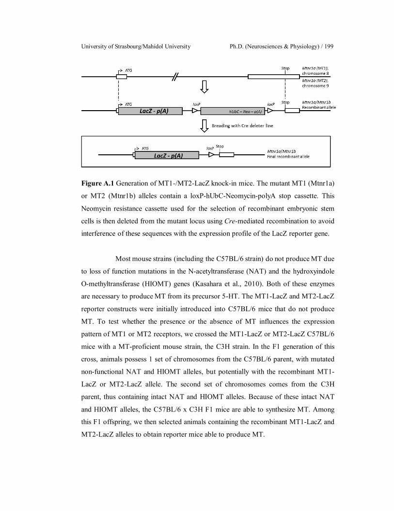
University of Strasbourg/Mahidol University Ph.D. (Neurosciences & Physiology) / 199
Figure A.1 Generation of MT1-/MT2-LacZ knock-in mice. The mutant MT1 (Mtnr1a)
or MT2 (Mtnr1b) alleles contain a loxP-hUbC-Neomycin-polyA stop cassette. This
Neomycin resistance cassette used for the selection of recombinant embryonic stem
cells is then deleted from the mutant locus using Cre-mediated recombination to avoid
interference of these sequences with the expression profile of the LacZ reporter gene.
Most mouse strains (including the C57BL/6 strain) do not produce MT due
to loss of function mutations in the N-acetyltransferase (NAT) and the hydroxyindole
O-methyltransferase (HIOMT) genes (Kasahara et al., 2010). Both of these enzymes
are necessary to produce MT from its precursor 5-HT. The MT1-LacZ and MT2-LacZ
reporter constructs were initially introduced into C57BL/6 mice that do not produce
MT. To test whether the presence or the absence of MT influences the expression
pattern of MT1 or MT2 receptors, we crossed the MT1-LacZ or MT2-LacZ C57BL/6
mice with a MT-proficient mouse strain, the C3H strain. In the F1 generation of this
cross, animals possess 1 set of chromosomes from the C57BL/6 parent, with mutated
non-functional NAT and HIOMT alleles, but potentially with the recombinant MT1-
LacZ or MT2-LacZ allele. The second set of chromosomes comes from the C3H
parent, thus containing intact NAT and HIOMT alleles. Because of these intact NAT
and HIOMT alleles, the C57BL/6 x C3H F1 mice are able to synthesize MT. Among
this F1 offspring, we then selected animals containing the recombinant MT1-LacZ and
MT2-LacZ alleles to obtain reporter mice able to produce MT.

Sarawut Lapmanee Appendix / 200
References
Cho A, Haruyama N, Kulkarni AB. Generation of transgenic mice. Curr Protoc Cell
Biol. 2009;Chapter 19:Unit 19.11.
Hall B, Limaye A, Kulkarni AB. Overview: generation of gene knockout mice. Curr
Protoc Cell Biol. 2009 Sep;Chapter 19:Unit 19.12 19.12.117.
Kasahara T, Abe K, Mekada K, Yoshiki A, Kato T. Genetic variation of melatonin
productivity in laboratory mice under domestication. Proc Natl Acad Sci
U S A. 2010;107(14):64127.
Kos CH. Cre/loxP system for generating tissue-specific knockout mouse models. Nutr
Rev. 2004;62(6 Pt 1):2436.
Liu C, Weaver DR, Jin X, Shearman LP, Pieschl RL, Gribkoff VK, Reppert SM.
Molecular dissection of two distinct actions of melatonin on the
suprachiasmatic circadian clock. Neuron. 1997;19(1):91102.
University of Strasbourg/Mahidol University Ph.D. (Neurosciences & Physiology) / 201
BIOGRAPHY
NAME Mr. Sarawut Lapmanee
DATE OF BIRTH June 7, 1985
PLACE OF BIRTH Nakhon Si Thammarat, Thailand
INSTITUTIONS ATTENTED Mahidol University, 2004–2007
Bachelor of Science (Physiotherapy)
Mahidol University, 2008–2011
Master of Science (Exercise Physiology)
Mahidol University, 2012–2017
Doctor of Philosophy
(Neurosciences & Physiology)
SCHOLARSHIPS 2012–2013
Institutional Strengthening Program
Scholarship, Faculty of Science,
Mahidol University
2013–2015
Royal Golden Jubilee Ph.D. Program,
Thailand Research Fund
2015–2016
- Embassy of France, Thailand
- Graduate Studies of Mahidol
University Alumni Association,
Mahidol University
HOME ADDRESS 79/105 moo22, Robwiang, Muang ,
Chiang Rai, Thailand, 57000
Tel. 0979355055
E-mail: [email protected]
Sarawut Lapmanee Biography / 202
PUBPLICATIONS
Lapmanee S, Charoenphandhu J, Teerapornpuntakit J, Krishnamra N,
Charoenphandhu N. (2017): Agomelatine, venlafaxine, and running
exercise effectively prevent anxiety- and depression-like behaviors and
memory impairment in restraint stressed rats. (Edited and submitted to
PLoS ONE)
Kraidith K, Svasti S, Teerapornpuntakit J, Vadolas J, Chaimana R, Lapmanee S,
Suntornsaratoon P, Krishnamra N, Fucharoen S, Charoenphandhu N.
Hepcidin and 1,25(OH)2D3 effectively restore Ca2+ transport in β
thalassemic mice: reciprocal phenomenon of Fe2+ and Ca2+ absorption. Am
J Physiol Endocrinol Metab. 2016;311(1):E21423.
Lapmanee S, Charoenphandhu N, Aeimlapa R, Suntornsaratoon P, Wongdee K,
Tiyasatkulkovit W, Kengkoom K, Chaimongkolnukul K, Seriwatanachai
D, Krishnamra N. High dietary cholesterol masks type 2 diabetes-induced
osteopenia and changes in bone microstructure in rats. Lipids.
2014;49(10):97586.
POSTER PRESENTATIONS
Lapmanee S, Charoenphandhu J, Krishnamra N, Charoenphandhu N.
“Neuroprotective effects of venlafaxine and voluntary wheel running
against stress-induced anxiety, depression and cognitive impairment in
male rats” at Royal Golden Jubilee (RGJ)-Ph.D. Congress 18, Nonthaburi,
Thailand, June 810, 2017.
Lapmanee S, Felder-Schmittbuhl MP, Schuster-Klein C, Guardiola B , Pévet P,
Klosen P. “Transgenic reporter mice as a tool to identify MT1 and MT2
melatonin receptor expressing cells” at 45ème Congrès de la Société
Francophone de Chronobiologie, Strasbourg, France, September 1416,
2016.
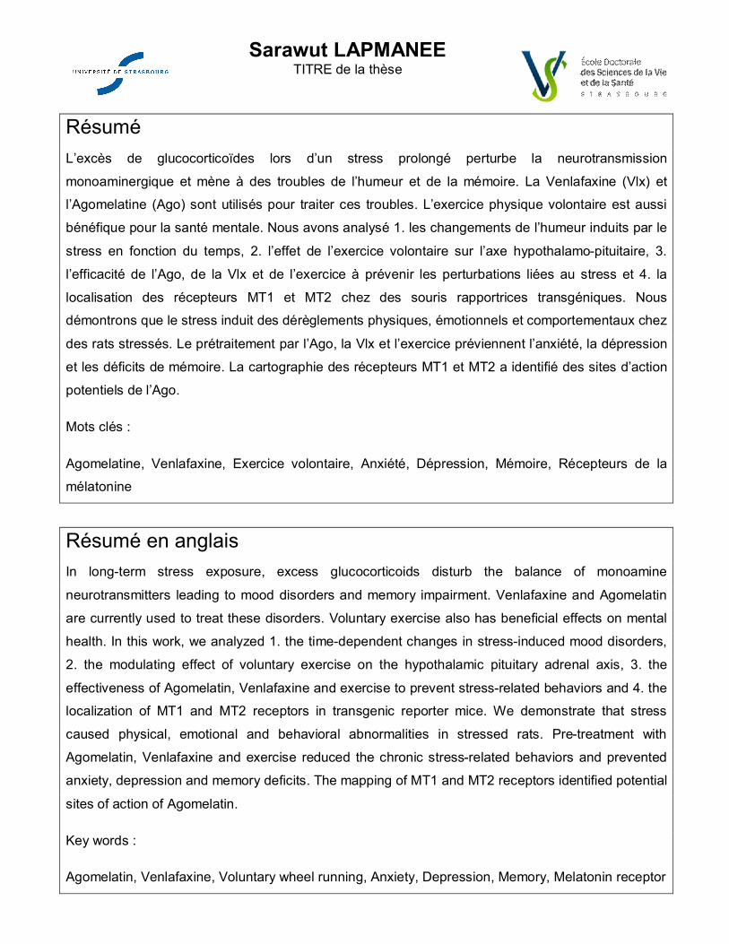
Sarawut LAPMANEE TITRE de la thèse
Résumé L’excès de glucocorticoïdes lors d’un stress prolongé perturbe la neurotransmission
monoaminergique et mène à des troubles de l’humeur et de la mémoire. La Venlafaxine (Vlx) et l’Agomelatine (Ago) sont utilisés pour traiter ces troubles. L’exercice physique volontaire est aussi
bénéfique pour la santé mentale. Nous avons analysé 1. les changements de l’humeur induits par le
stress en fonction du temps, 2. l’effet de l’exercice volontaire sur l’axe hypothalamo-pituitaire, 3.
l’efficacité de l’Ago, de la Vlx et de l’exercice à prévenir les perturbations liées au stress et 4. la
localisation des récepteurs MT1 et MT2 chez des souris rapportrices transgéniques. Nous démontrons que le stress induit des dérèglements physiques, émotionnels et comportementaux chez
des rats stressés. Le prétraitement par l’Ago, la Vlx et l’exercice préviennent l’anxiété, la dépression
et les déficits de mémoire. La cartographie des récepteurs MT1 et MT2 a identifié des sites d’action
potentiels de l’Ago.
Mots clés :
Agomelatine, Venlafaxine, Exercice volontaire, Anxiété, Dépression, Mémoire, Récepteurs de la mélatonine
Résumé en anglais In long-term stress exposure, excess glucocorticoids disturb the balance of monoamine
neurotransmitters leading to mood disorders and memory impairment. Venlafaxine and Agomelatin
are currently used to treat these disorders. Voluntary exercise also has beneficial effects on mental
health. In this work, we analyzed 1. the time-dependent changes in stress-induced mood disorders,
2. the modulating effect of voluntary exercise on the hypothalamic pituitary adrenal axis, 3. the
effectiveness of Agomelatin, Venlafaxine and exercise to prevent stress-related behaviors and 4. the localization of MT1 and MT2 receptors in transgenic reporter mice. We demonstrate that stress
caused physical, emotional and behavioral abnormalities in stressed rats. Pre-treatment with
Agomelatin, Venlafaxine and exercise reduced the chronic stress-related behaviors and prevented
anxiety, depression and memory deficits. The mapping of MT1 and MT2 receptors identified potential
sites of action of Agomelatin.
Key words :
Agomelatin, Venlafaxine, Voluntary wheel running, Anxiety, Depression, Memory, Melatonin receptor
Copyright © 2022 FDOKUMEN