Tenofovir renal toxicity targets mitochondria of renal proximal tubules
11
Tenofovir renal toxicity targets mitochondria of renal proximal tubules James J Kohler, Seyed H Hosseini, Amy Hoying-Brandt, Elgin Green, David M Johnson, Rodney Russ, Dung Tran, C Michael Raper, Robert Santoianni, and William Lewis Department of Pathology, Emory University, Atlanta, GA, USA Abstract Tenofovir disoproxil fumarate (TDF) is an analog of adenosine monophosphate that inhibits HIV reverse transcriptase in HIV/AIDS. Despite its therapeutic success, renal tubular side effects are reported. The mechanisms and targets of tenofovir toxicity were determined using ‘2 × 2’ factorial protocols, and HIV transgenic (TG) and wild-type (WT) littermate mice with or without TDF (5 weeks). A parallel study used didanosine (ddI) instead of TDF. At termination, heart, kidney, and liver samples were retrieved. Mitochondrial DNA (mtDNA) abundance, and histo- and ultrastructural pathology were analyzed. Laser-capture microdissection (LCM) was used to isolate renal proximal tubules for molecular analyses. Tenofovir increased mtDNA abundance in TG whole kidneys, but not in their hearts or livers. In contrast, ddI decreased mtDNA abundance in the livers of WTs and TGs, but had no effect on their hearts or kidneys. Histological analyses of kidneys showed no disruption of glomeruli or proximal tubules with TDF or ddI treatments. Ultrastructural changes in renal proximal tubules from TDF-treated TGs included an increased number and irregular shape of mitochondria with sparse fragmented cristae. LCM-captured renal proximal tubules from TGs showed decreased mtDNA abundance with tenofovir. The results indicate that tenofovir targets mitochondrial toxicity on the renal proximal tubule in an AIDS model. Pyrimidine and purine analogs called ‘nucleoside reverse transcriptase inhibitors’ (NRTIs) are drugs used in the treatment of HIV/AIDS. Tenofovir is an acyclic nucleotide phosphonate diester analog of adenosine monophosphate. 1 Similar to many NRTIs, tenofovir inhibits HIV-1 reverse transcriptase by competing with the natural substrate deoxyadenosine 5′-triphosphate, 2 which are part of the nucleotide pools used by virus in generating cDNA. Despite the distinct benefits of NRTI-based therapies, toxicity is a limiting factor. In particular, a number of in vitro and in vivo studies showed cardiomyopathy and hepatic failure associated with specific NRTIs. 3-6 NRTI toxicities seem to be tissue specific. 7,8 Zidovudine and stavudine are toxic to striated muscle. 4,9 Didanosine (ddI) is toxic to the liver and pancreas. 6,10 An important form of tenofovir toxicity is tubular dysfunction, 11-14 but its mechanism or precise target has not been elucidated. Although the mechanisms of these specific antiretroviral drug-related toxicities remain unclear, it has been hypothesized that as analogs to native nucleosides, tenofovir and other NRTIs may potentially inhibit mammalian DNA polymerases, including mitochondrial DNA (mtDNA) polymerase-γ, and induce oxidative stress. 15-17 Tenofovir is not a substrate of CYP450 enzymes, and is eliminated by glomerular filtration and active tubular secretion. 18 Correspondence: Dr JJ Kohler, PhD, Department of Pathology, Emory University School of Medicine, 7126 Woodruff Memorial Building, 101 Woodruff Circle, Atlanta, GA 30322, USA. E-mail: E-mail: [email protected]. NIH Public Access Author Manuscript Lab Invest. Author manuscript; available in PMC 2009 May 1. Published in final edited form as: Lab Invest. 2009 May ; 89(5): 513–519. doi:10.1038/labinvest.2009.14. NIH-PA Author Manuscript NIH-PA Author Manuscript NIH-PA Author Manuscript
-
Upload
independent -
Category
Documents
-
view
1 -
download
0
Transcript of Tenofovir renal toxicity targets mitochondria of renal proximal tubules

Tenofovir renal toxicity targets mitochondria of renal proximaltubules
James J Kohler, Seyed H Hosseini, Amy Hoying-Brandt, Elgin Green, David M Johnson,Rodney Russ, Dung Tran, C Michael Raper, Robert Santoianni, and William LewisDepartment of Pathology, Emory University, Atlanta, GA, USA
AbstractTenofovir disoproxil fumarate (TDF) is an analog of adenosine monophosphate that inhibits HIVreverse transcriptase in HIV/AIDS. Despite its therapeutic success, renal tubular side effects arereported. The mechanisms and targets of tenofovir toxicity were determined using ‘2 × 2’ factorialprotocols, and HIV transgenic (TG) and wild-type (WT) littermate mice with or without TDF (5weeks). A parallel study used didanosine (ddI) instead of TDF. At termination, heart, kidney, andliver samples were retrieved. Mitochondrial DNA (mtDNA) abundance, and histo- and ultrastructuralpathology were analyzed. Laser-capture microdissection (LCM) was used to isolate renal proximaltubules for molecular analyses. Tenofovir increased mtDNA abundance in TG whole kidneys, butnot in their hearts or livers. In contrast, ddI decreased mtDNA abundance in the livers of WTs andTGs, but had no effect on their hearts or kidneys. Histological analyses of kidneys showed nodisruption of glomeruli or proximal tubules with TDF or ddI treatments. Ultrastructural changes inrenal proximal tubules from TDF-treated TGs included an increased number and irregular shape ofmitochondria with sparse fragmented cristae. LCM-captured renal proximal tubules from TGsshowed decreased mtDNA abundance with tenofovir. The results indicate that tenofovir targetsmitochondrial toxicity on the renal proximal tubule in an AIDS model.
Pyrimidine and purine analogs called ‘nucleoside reverse transcriptase inhibitors’ (NRTIs) aredrugs used in the treatment of HIV/AIDS. Tenofovir is an acyclic nucleotide phosphonatediester analog of adenosine monophosphate.1 Similar to many NRTIs, tenofovir inhibits HIV-1reverse transcriptase by competing with the natural substrate deoxyadenosine 5′-triphosphate,2 which are part of the nucleotide pools used by virus in generating cDNA.
Despite the distinct benefits of NRTI-based therapies, toxicity is a limiting factor. In particular,a number of in vitro and in vivo studies showed cardiomyopathy and hepatic failure associatedwith specific NRTIs.3-6 NRTI toxicities seem to be tissue specific.7,8 Zidovudine andstavudine are toxic to striated muscle.4,9 Didanosine (ddI) is toxic to the liver and pancreas.6,10 An important form of tenofovir toxicity is tubular dysfunction,11-14 but its mechanismor precise target has not been elucidated. Although the mechanisms of these specificantiretroviral drug-related toxicities remain unclear, it has been hypothesized that as analogsto native nucleosides, tenofovir and other NRTIs may potentially inhibit mammalian DNApolymerases, including mitochondrial DNA (mtDNA) polymerase-γ, and induce oxidativestress.15-17 Tenofovir is not a substrate of CYP450 enzymes, and is eliminated by glomerularfiltration and active tubular secretion.18
Correspondence: Dr JJ Kohler, PhD, Department of Pathology, Emory University School of Medicine, 7126 Woodruff MemorialBuilding, 101 Woodruff Circle, Atlanta, GA 30322, USA. E-mail: E-mail: [email protected].
NIH Public AccessAuthor ManuscriptLab Invest. Author manuscript; available in PMC 2009 May 1.
Published in final edited form as:Lab Invest. 2009 May ; 89(5): 513–519. doi:10.1038/labinvest.2009.14.
NIH
-PA Author Manuscript
NIH
-PA Author Manuscript
NIH
-PA Author Manuscript

HIV-associated nephropathy (HIVAN) is an AIDS-related disease of the kidney. NRTIs(similar to tenofovir) are used to treat HIV/AIDS. Tenofovir is the only NRTI that is associatedwith renal disease. Nevertheless, tenofovir tubular toxicity may be additive or synergistic withHIVAN. These studies parametrically assess the individual and combined effects of NRTIs(similar to tenofovir or ddI) using a genetically homogenous (albeit phenotypicallyheterogenous, possibly because of differences in transgene expression level) murine HIVtransgenic (TG) model.19 Laser-capture microdissection (LCM) was used to anatomicallyisolate renal proximal tubules to define the effects of tenofovir on tubules in this HIV TGmodel.
MATERIALS AND METHODSAnimals
Hemizygous HIV-1 TG mice established by Dickie20 were provided as compliments of PaulKlotman. Originally on FVB/n background, this TG line was bred congenically to C57/BL6(and given the trivial name: MSB). The resultant TG that was made congenic to C57/BL6 didnot develop HIVAN, compared with the original TG on FVB/n. This absence of HIVAN wasan experimental feature that allowed us to detect changes related to drug toxicity, whichotherwise might be overshadowed by HIVAN. This study used a ‘2 × 2’ factorial protocol andincluded TG and wild-type (WT) mice treated with vehicle or NRTI. Murine TG authenticitywas confirmed for each generation using dot blot analysis and real-time PCR.21
Treatment ProtocolsProcedures complied with the Emory IACUC and NIH guidelines. WT and TG littermates ofboth genders (male and female) were age matched (8-12 weeks) at the start of treatment. Foodand water were provided ad libitum. Antiretroviral drugs were provided by the manufacturers(compliments of Professor Raymond Schinazi, VA Medical Center, Decatur, GA, USA andEmory Center for AIDS Research Pharmacology Core). To remain clinically relevant, dosingwas done by daily gavage (morning) at doses that resembled human therapy on a mg/kg/daybasis (eg, human tenofovir disoproxil fumarate (TDF) daily dose: 300 mg/60 kg human = 5mg/kg; ∼25 g mouse = 0.125 mg/day). Treatments included TDF (0.125 mg/day), ddI (0.14mg/day), or vehicle (buffered with 0.1 M NaOH). Each cohort consisted of ∼20 mice,12-22ultimately depending on actual litter sizes from three replicate experiments. Mortality from theprocedure was nil (100% survival). On the basis of a number of NRTI treatment protocols usedin our laboratory,4,9,21-25 a treatment duration of 5 weeks was used in these initial studies.After 5 weeks of treatment, body weights were measured, animals terminated, and samplesretrieved and stored for DNA extraction and analysis.
mtDNA and Nuclear DNA (nDNA) Quantitation in the Heart, Kidney, and Liver Tissues UsingReal-Time PCR
Methods employed were modifications used by others,26 as employed by us in the past.21,25 Total DNA was extracted from select tissues (∼10 mg wet weight) using a MagNA PureSystem and reagents (Roche Life Sciences, Indianapolis, IN, USA). Alternatively, DNA wasextracted from proximal tubules isolated with LCM (see procedure below).
DNA sequences for primers and probes used for quantitation of mtDNA and nDNA have beendescribed earlier.25 Real-time PCR was performed in duplicate for each amplicon.Amplification was performed using LC 480 (Roche). Standard DNA curves for quantitationof the LC products were used. PCR products of mtDNA and nDNA were quantified using thecorresponding external standard.
Kohler et al. Page 2
Lab Invest. Author manuscript; available in PMC 2009 May 1.
NIH
-PA Author Manuscript
NIH
-PA Author Manuscript
NIH
-PA Author Manuscript
Histological ExaminationTissue samples were processed routinely. Sections (6 μm) were stained with hematoxylin andeosin (H&E) and examined microscopically by a blinded, trained pathologist (WL).Photomicrographs of glass slides were obtained using a Nikon photomicroscope (Nikon,Garden City, NY, USA).
Fine Structure of Kidney Tissues Using Electron Microscopy (EM)EM was evaluated using reported methods.4 Sections (0.5 μm) were cut with glass knives andstained with Toluidine Blue for orientation. Ultrathin (900 Å) sections were cut with a diamondknife, stained with uranyl acetate and lead citrate and examined by EM (Philips MorgagniModel 201, Philips, Eindhoven, Amsterdam, The Netherlands), and evaluated andphotographed. Each EM photomicrograph was reviewed independently by two investigators.Parameters included the presence of structurally abnormal mitochondria, increased numbersof mitochondrial profiles per field, intra-mitochondrial lamellar bodies, abnormal cristaedensity, cristae reduplication, mitochondrial swelling, and intra-mitochondrial paracrystals.27
Laser-Capture MicrodissectionThe inherent heterogeneity of kidney tissues (including glomeruli, proximal and distal tubuleepithelial cells, blood vessels, and interstitium with its mix of resident and infiltrating cells)can affect the outcome and interpretation of molecular studies. LCM, a novel techniquedeveloped at the National Cancer Institute,28 allows for specific, single-cell isolation fromfixed heterogenous tissues. Specifically, proximal renal tubular cells were isolated using theArcturus LCM 1110 system (Arcturus Biosciences Inc., Mountain View, CA, USA). DNA wasextracted from pooled renal tubular epithelial cells (m=~\100 cells) isolated from eachembedded sample using the PicoPure™ DNA Extraction Kit (MDS Analytical Technologies,Sunnyvale, CA, USA).
Statistical AnalysisData were expressed as the ratio of mean values for mtDNA to those of nDNA × 10-3. Resultantvalues were expressed as mean ± s.e., normalized to the mean of vehicle-treated WTs (set at1.0). A value of P<0.05 was considered statistically significant. Results from vehicle-treatedTGs were combined statistically from the respective TDF and ddI ‘2 × 2’ studies to increasethe power. Results from all groups were compared using one-way ANOVA.9 Alternatively,the Mann-Whitney unpaired t-test was applied to the data. In some cohorts, outliers (asdetermined using Grubb’s test with P<0.05)29 were excluded from the cohort data set.
RESULTSGeneral
All the TG and WT cohorts maintained overall health with normal body weights and levels ofactivity throughout the study duration, regardless of treatment.
mtDNA Abundance in Various Tissues after TreatmentTo determine mitochondrial toxicity with tenofovir or ddI, mtDNA abundance was assessedusing DNA extracts from various tissues of both TGs and WTs after 5 weeks of treatment withTDF, ddI, or vehicle. mtDNA abundance was determined in the heart, kidney, and liver tissues.
TDF treatment had no effect on mtDNA abundance in the hearts or livers from all cohorts(Figure 1a). Kidneys from TDF-treated TGs showed increased mtDNA abundance, comparedwith those of vehicle-treated TGs and WTs (Figure 1a). Treatment with ddI yielded differentresults. mtDNA abundance remained unchanged in all hearts and kidneys after ddI treatment,
Kohler et al. Page 3
Lab Invest. Author manuscript; available in PMC 2009 May 1.
NIH
-PA Author Manuscript
NIH
-PA Author Manuscript
NIH
-PA Author Manuscript
whereas liver mtDNA abundance decreased significantly in both TGs and WTs, compared withtheir vehicle-treated littermates (Figure 1b).
Histological Analysis of Kidney TissuesKidneys from TGs and WTs were histologically assessed to determine whether treatment withTDF yielded microscopic changes in renal tubules or glomeruli. Vehicle- and ddI-treatedkidney tissues were used comparatively. As expected, vehicle and ddI treatments resulted inno renal microscopic changes (Figure 2, top and bottom panels). Glomeruli and tubules alsoappeared to be intact in both TDF-treated WTs and in vehicle-treated TGs (Figure 2, middlepanels). After 5 weeks of treatment with TDF or ddI, detectable renal tubular or glomerulardamage was not found.
EM Features of Mitochondria in Tubular Epithelium with TenofovirTo investigate the organelle-specific effect of tenofovir on renal proximal tubules,ultrastructural changes in renal tubular epithelial mitochondria were defined parametrically.Vehicle-treated WTs showed renal proximal tubular epithelial cells with characteristic, ovalmitochondria having densely packed cristae (Figure 3, upper left panel). Renal tubule EMprofiles from WTs treated with TDF or ddI also appeared unchanged from vehicle-treatedlittermates (Figure 3, top center and right panels).
Mitochondria from renal proximal tubular cells of vehicle-treated TGs were oblong, butresembled WTs with respect to the mitochondrial cristae (Figure 3, lower left panel). Renaltubular epithelia of TDF-treated TGs showed an increased number of mitochondria, withirregular mitochondrial shape, and sparse, fragmented cristae (Figure 3, lower middle panel,arrows). ddI caused no changes in the size and number of mitochondria in renal tubular epithelia(Figure 3, lower right panel).
LCM-Isolated Proximal Tubular mtDNA AbundanceProximal tubules were microdissected from kidney sections using LCM (Figure 4a) todetermine the specific effect of tenofovir on this histological compartment. Proximal tubuleswere defined histopathologically by staining with PAS to highlight the glycoprotein-rich brushborder.
mtDNA abundance in renal proximal tubules decreased in TGs treated with TDF, comparedwith that in vehicle-treated TGs and WT controls (Figure 4b, left graph). TDF treatment ofWTs also resulted in decreased mtDNA abundance compared with that of vehicle-treated WTs,although not enough to be significant. In contrast, treatment with ddI had no effect on proximaltubular epithelial mtDNA abundance (Figure 4b, right graph). Thus, tenofovir toxicity depletedmtDNA in proximal tubules in comparison to its effects on total mtDNA in the homogenizedtissue.
DISCUSSIONThese studies offer novel and mechanistic data to clarify subcellular events in clinical tenofovirrenal toxicity. They provide evidence that tenofovir causes renal proximal tubularmitochondrial ultrastructural abnormalities that are parallel to mtDNA depletion in the samecells. Isolation of renal proximal tubules using LCM provided tissue-specific evidence of thecompartmentalized toxic effects of tenofovir on renal tubules, and localized the toxic siteanatomically and molecularly. To our knowledge, this is the first time that mitochondrialtoxicity is localized to an anatomically and functionally distinct tissue in a complex organ.
Kohler et al. Page 4
Lab Invest. Author manuscript; available in PMC 2009 May 1.
NIH
-PA Author Manuscript
NIH
-PA Author Manuscript
NIH
-PA Author Manuscript
A paradoxical increase in steady-state mtDNA abundance was found in homogenized wholekidneys. As mtDNA abundance, as reported here, is a ratio between mtDNA and nDNA, theresultant change in mtDNA abundance may be the result of a change in the level of mtDNAor in the level of nDNA. Thus, an increase in mtDNA or decrease in nDNA could yield anincrease in that ratio. Histologically, nuclei appeared to be as intact and as abundant in theTDF-treated TG renal tissue as was seen in the littermate controls. Renal tubules arecharacteristically multinucleated, and a detailed enumeration of nuclei was determined in thesestudies. Alternatively, there was direct EM evidence of an increase in mitochondria withirregular shape and fragmented cristae in the TDF-treated TGs, compared with that in the WTcontrols. Thus, it is possible to suggest the mtDNA in these studies were altered (mtDNA levelincreased), whereas the nDNA remained intact (nDNA level remained constant), resulting inthe identified change in mtDNA abundance. This result extends earlier studies17,21,24,25 thatfocused on disruption of mtDNA biogenesis, potentially at the level of the mitochondrialnucleotide pools.
The compartmentalized toxicity of tenofovir at the organelle level of mitochondria in renalproximal tubules suggests that tenofovir toxicity may be directly related to its metabolicprocessing. As tenofovir is eliminated by active secretion through the renal tubules, impairedor delayed elimination of tenofovir would lead to its accumulation. Increased tenofovirabundance in the proximal tubules and its phosphorylation in those cells could create animbalance in nucleotide pools,30 thereby disrupting mitochondrial biogenesis.31
HIVAN was first described in 1984 with HIV infection,32 before any antiretroviral agentswere available. Therefore, it seems likely that tenofovir tubular disease and HIVAN areseparate events with distinct mechanisms, because HIVAN was reported over a decade beforeTDF was used. In both human and experimental HIVAN, all tissue compartments are affected,including glomeruli with collapsing focal segmental glomerulosclerosis and tubular andinterstitial cells with tubulointerstitial disease.33-35 In contrast, tenofovir primarily targetsproximal tubules. An HIV TG model was used here to delineate the individual and combinedeffects of TG and tenofovir.
Neither TG nor TDF treatment alone induced detectable molecular or ultrastructural changeswithin the short-term study of 5 weeks. Vehicle-treated TGs had no change in mtDNAabundance in whole-kidney tissues from WTs. These data confirm the absence of HIVAN ina C57/BL6 background. TDF treatment of WTs caused no change in mtDNA abundance. OnlyTDF treatment in TGs resulted in a significant change in mtDNA abundance, compared withthat in vehicle-treated WTs. A logical conclusion is that this observed synergy is the result oflocal HIV gene expression and tenofovir.
It may be possible that neither TG nor TDF treatment alone is sufficient within 5 weeks (shortterm) to yield detectable changes. Expanding studies of longer treatment duration may resultin detectable changes in TDF-treated WTs, supporting cumulative toxicity. Moreover,coexistence of the TG with TDF treatment may cause the phenotype to occur more robustly orearlier.
The NRTI toxicity in these studies was both tissue and treatment specific (ie, renal proximaltubule and TDF). It must be noted that TDF treatment had no effect on either cardiac or livermtDNA abundance, and toxicity of TDF is not reported in those organs. In contrast, ddIhepatotoxicity occurred. The outcome with ddI treatment supports earlier studies.36,37
In summary, tenofovir caused renal proximal tubular ultrastructural defects (mitochondria) anddepleted mtDNA in that specialized tissue. Tenofovir caused no such change in the heart orliver, and ddI caused no such change in the kidney. The data of these studies support thehypothesis that specific NRTIs for HIV/AIDS show toxicity to mitochondria in specific tissues.
Kohler et al. Page 5
Lab Invest. Author manuscript; available in PMC 2009 May 1.
NIH
-PA Author Manuscript
NIH
-PA Author Manuscript
NIH
-PA Author Manuscript
The findings provide a nexus between tissue and organelle toxicity. Additional studies mayhelp define why individual NRTIs show relatively selective organ toxicity, yet seem to involvemitochondria in diverse tissues.
ACKNOWLEDGEMENTSThe authors thank the Emory Center for AIDS Research and the Department of Pathology Experimental CoreLaboratory for their support. Dr Vanessa Bijol is also thanked for her expertise and guidance in analyzing the renaltissues.
The studies were supported by Grants R01 HL059798 and R01 HL079867 to WL. JJK was supported by Grant K01DK078513.
References1. Lyseng-Williamson KA, Reynolds NA, Plosker GL. Tenofovir disoproxil fumarate: a review of its use
in the management of HIV infection. Drugs 2005;65:413–432. [PubMed: 15669881]2. Vivet-Boudou V, Didierjean J, Isel C, et al. Nucleoside and nucleotide inhibitors of HIV-1 replication.
Cell Mol Life Sci 2006;63:163–186. [PubMed: 16389458]3. Lewis W, Gonzalez B, Chomyn A, et al. Zidovudine induces molecular, biochemical, and
ultrastructural changes in rat skeletal muscle mitochondria. J Clin Invest 1992;89:1354–1360.[PubMed: 1556193]
4. Lewis W, Grupp IL, Grupp G, et al. Cardiac dysfunction occurs in the HIV-1 transgenic mouse treatedwith zidovudine. Lab Invest 2000;80:187–197. [PubMed: 10701688]
5. Lewis W. Mitochondrial DNA replication, nucleoside reverse-transcriptase inhibitors, and AIDScardiomyopathy. Prog Cardiovasc Dis 2003;45:305–318. [PubMed: 12638094]
6. Sulkowski MS, Mehta SH, Torbenson M, et al. Hepatic steatosis and antiretroviral drug use amongadults coinfected with HIV and hepatitis C virus. Aids 2005;19:585–592. [PubMed: 15802977]
7. Lund KC, Peterson LL, Wallace KB. Absence of a universal mechanism of mitochondrial toxicity bynucleoside analogs. Antimicrob Agents Chemother 2007;51:2531–2539. [PubMed: 17470651]
8. Bruggeman LA, Thomson MM, Nelson PJ, et al. Patterns of HIV-1 mRNA expression in transgenicmice are tissue-dependent. Virology 1994;202:940–948. [PubMed: 7518165]
9. Lewis W, Haase CP, Raidel SM, et al. Combined antiretroviral therapy causes cardiomyopathy andelevates plasma lactate in transgenic AIDS mice. Lab Invest 2001;81:1527–1536. [PubMed:11706060]
10. Price CJ, George JD, Marr MC, et al. Prenatal developmental toxicity evaluation of 2′,3′-dideoxyinosine (ddI) and 2′,3′-didehydro-3′-deoxythymidine (d4T) co-administered to Swiss Albino(CD-1) mice. Birth Defects Res B Dev Reprod Toxicol 2006;77:207–215. [PubMed: 16767756]
11. Fux CA, Christen A, Zgraggen S, et al. Effect of tenofovir on renal glomerular and tubular function.Aids 2007;21:1483–1485. [PubMed: 17589197]
12. Mocroft A, Kirk O, Gatell J, et al. Chronic renal failure among HIV-1-infected patients. Aids2007;21:1119–1127. [PubMed: 17502722]
13. Cote HC, Magil AB, Harris M, et al. Exploring mitochondrial nephrotoxicity as a potential mechanismof kidney dysfunction among HIV-infected patients on highly active antiretroviral therapy. AntivirTher 2006;11:79–86. [PubMed: 16518963]
14. Vidal F, Domingo JC, Guallar J, et al. In vitro cytotoxicity and mitochondrial toxicity of tenofoviralone and in combination with other antiretrovirals in human renal proximal tubule cells. AntimicrobAgents Chemother 2006;50:3824–3832. [PubMed: 16940060]
15. Birkus G, Hajek M, Kramata P, et al. Tenofovir diphosphate is a poor substrate and a weak inhibitorof rat DNA polymerases alpha, delta, and epsilon*. Antimicrob Agents Chemother 2002;46:1610–1613. [PubMed: 11959615]
16. Lewis W. Mitochondrial dysfunction and nucleoside reverse transcriptase inhibitor therapy:experimental clarifications and persistent clinical questions. Antiviral Res 2003;58:189–197.[PubMed: 12767466]
Kohler et al. Page 6
Lab Invest. Author manuscript; available in PMC 2009 May 1.
NIH
-PA Author Manuscript
NIH
-PA Author Manuscript
NIH
-PA Author Manuscript

17. Lewis W, Copeland WC, Day B. Mitochondrial DNA depletion, oxidative stress and mutation:mechanisms of nucleoside reverse transcriptase inhibitor toxicity. Lab Invest 2001;81:777–790.[PubMed: 11406640]
18. Delaney, WEt; Ray, AS.; Yang, H., et al. Intracellular metabolism and in vitro activity of tenofoviragainst hepatitis B virus. Antimicrob Agents Chemother 2006;50:2471–2477. [PubMed: 16801428]
19. Lu TC, He JC, Klotman P. Animal models of HIV-associated nephropathy. Curr Opin NephrolHypertens 2006;15:233–237. [PubMed: 16609288]
20. Dickie P, Felser J, Eckhaus M, et al. HIV-associated nephropathy in transgenic mice expressing HIV-1genes. Virology 1991;185:109–119. [PubMed: 1926769]
21. Lewis W, Day BJ, Kohler JJ, et al. Decreased mtDNA, oxidative stress, cardiomyopathy, and deathfrom transgenic cardiac targeted human mutant polymerase gamma. Lab Invest 2007;87:326–335.[PubMed: 17310215]
22. Lewis W, Haase CP, Miller YK, et al. Transgenic expression of the deoxynucleotide carrier causesmitochondrial damage that is enhanced by NRTIs for AIDS. Lab Invest 2005;85:972–981. [PubMed:15951836]
23. Lewis W, Kohler JJ, Hosseini SH, et al. Antiretroviral nucleosides, deoxynucleotide carrier andmitochondrial DNA: evidence supporting the DNA pol gamma hypothesis. Aids 2006;20:675–684.[PubMed: 16514297]
24. Kohler JJ, Hosseini SH, Green E, et al. Cardiac-targeted transgenic mutant mitochondrial enzymes:mtDNA defects, antiretroviral toxicity and cardiomyopathy. Cardiovasc Toxicol 2008;8:57–69.[PubMed: 18446447]
25. Hosseini SH, Kohler JJ, Haase CP, et al. Targeted transgenic overexpression of mitochondrialthymidine kinase (TK2) alters mitochondrial DNA (mtDNA) and mitochondrial polypeptideabundance: transgenic TK2, mtDNA, and antiretrovirals. Am J Pathol 2007;170:865–874. [PubMed:17322372]
26. Cote HC, Yip B, Asselin JJ, et al. Mitochondrial:nuclear DNA ratios in peripheral blood cells fromhuman immunodeficiency virus (HIV)-infected patients who received selected HIV antiretroviraldrug regimens. J Infect Dis 2003;187:1972–1976. [PubMed: 12792876]
27. Dalakas MC, Illa I, Pezeshkpour GH, et al. Mitochondrial myopathy caused by long-term zidovudinetherapy. N Engl J Med 1990;322:1098–1105. [PubMed: 2320079]
28. Emmert-Buck MR, Bonner RF, Smith PD, et al. Laser capture microdissection. Science1996;274:998–1001. [PubMed: 8875945]
29. Barnett, V.; Lewis, T. Outliers in Statistical Data. Wiley; Hoboken: 1994.30. Bianchi V. Nucleotide pool unbalance induced in cultured cells by treatments with different
chemicals. Toxicology 1982;25:13–18. [PubMed: 6760464]31. Mercy L, Pauw A, Payen L, et al. Mitochondrial biogenesis in mtDNA-depleted cells involves a Ca2
+-dependent pathway and a reduced mitochondrial protein import. FEBS J 2005;272:5031–5055.[PubMed: 16176275]
32. Herman ES, Klotman PE. HIV-associated nephropathy: epidemiology, pathogenesis, and treatment.Semin Nephrol 2003;23:200–208. [PubMed: 12704580]
33. Shah SN, He CJ, Klotman P. Update on HIV-associated nephropathy. Curr Opin Nephrol Hypertens2006;15:450–455. [PubMed: 16775461]
34. Pope SD, Johnson MD, May DB. Pharmacotherapy for human immunodeficiency virus-associatednephropathy. Pharmacotherapy 2005;25:1761–1772. [PubMed: 16305296]
35. Wyatt CM, Rosenstiel PE, Klotman PE. HIV-associated nephropathy. Contrib Nephrol2008;159:151–161. [PubMed: 18391591]
36. Youssef JA, Badr MZ. Disruption of mitochondrial energetics and DNA synthesis by the anti-AIDSdrug dideoxyinosine. Toxicol Lett 1992;60:197–202. [PubMed: 1570633]
37. Walker UA, Setzer B, Venhoff N. Increased long-term mitochondrial toxicity in combinations ofnucleoside analogue reverse-transcriptase inhibitors. Aids 2002;16:2165–2173. [PubMed:12409738]
Kohler et al. Page 7
Lab Invest. Author manuscript; available in PMC 2009 May 1.
NIH
-PA Author Manuscript
NIH
-PA Author Manuscript
NIH
-PA Author Manuscript
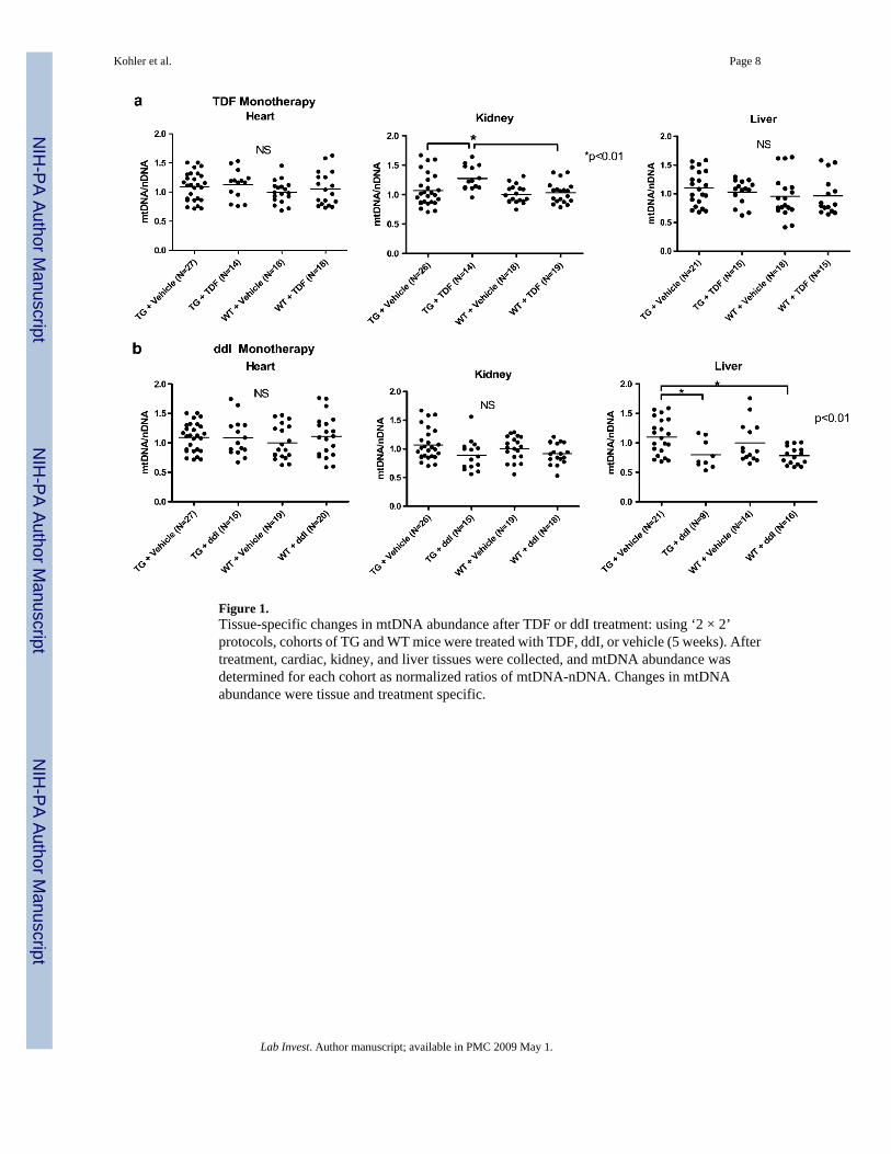
Figure 1.Tissue-specific changes in mtDNA abundance after TDF or ddI treatment: using ‘2 × 2’protocols, cohorts of TG and WT mice were treated with TDF, ddI, or vehicle (5 weeks). Aftertreatment, cardiac, kidney, and liver tissues were collected, and mtDNA abundance wasdetermined for each cohort as normalized ratios of mtDNA-nDNA. Changes in mtDNAabundance were tissue and treatment specific.
Kohler et al. Page 8
Lab Invest. Author manuscript; available in PMC 2009 May 1.
NIH
-PA Author Manuscript
NIH
-PA Author Manuscript
NIH
-PA Author Manuscript
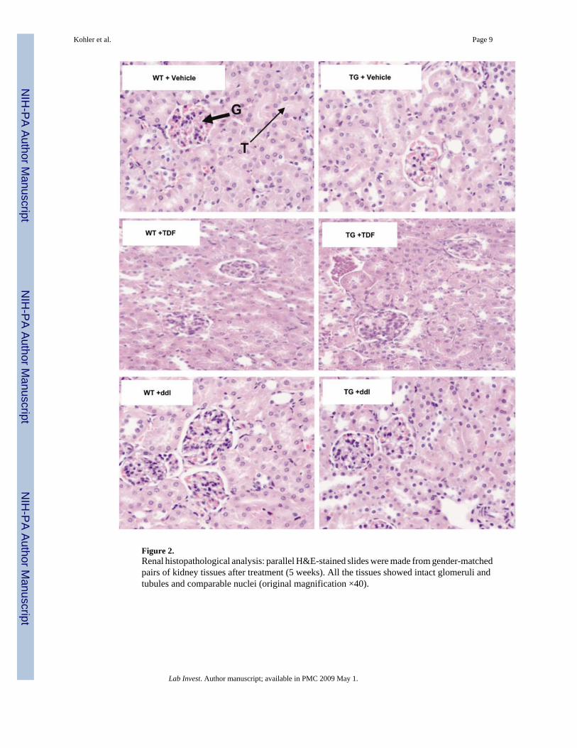
Figure 2.Renal histopathological analysis: parallel H&E-stained slides were made from gender-matchedpairs of kidney tissues after treatment (5 weeks). All the tissues showed intact glomeruli andtubules and comparable nuclei (original magnification ×40).
Kohler et al. Page 9
Lab Invest. Author manuscript; available in PMC 2009 May 1.
NIH
-PA Author Manuscript
NIH
-PA Author Manuscript
NIH
-PA Author Manuscript

Figure 3.Mitochondrial ultrastructural changes in renal tubular epithelial cells with tenofovir: renaltubular epithelial cells from TDF-treated TGs showed increased mitochondrial number,irregular shape, and fragmented cristae. (EM, original magnification ×22 400).
Kohler et al. Page 10
Lab Invest. Author manuscript; available in PMC 2009 May 1.
NIH
-PA Author Manuscript
NIH
-PA Author Manuscript
NIH
-PA Author Manuscript
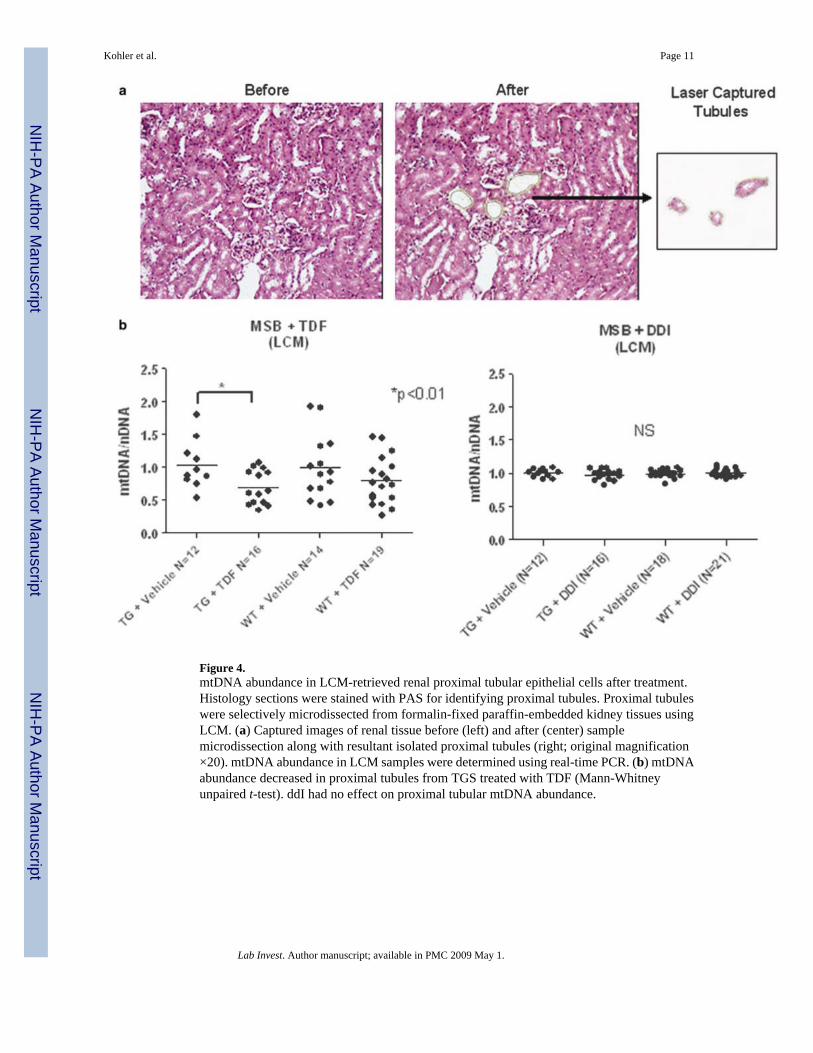
Figure 4.mtDNA abundance in LCM-retrieved renal proximal tubular epithelial cells after treatment.Histology sections were stained with PAS for identifying proximal tubules. Proximal tubuleswere selectively microdissected from formalin-fixed paraffin-embedded kidney tissues usingLCM. (a) Captured images of renal tissue before (left) and after (center) samplemicrodissection along with resultant isolated proximal tubules (right; original magnification×20). mtDNA abundance in LCM samples were determined using real-time PCR. (b) mtDNAabundance decreased in proximal tubules from TGS treated with TDF (Mann-Whitneyunpaired t-test). ddI had no effect on proximal tubular mtDNA abundance.
Kohler et al. Page 11
Lab Invest. Author manuscript; available in PMC 2009 May 1.
NIH
-PA Author Manuscript
NIH
-PA Author Manuscript
NIH
-PA Author Manuscript