The Czech registry of renal biopsies. Occurrence of renal diseases in the years 1994-2000
10
Nephrol Dial Transplant (2004) 19: 3040–3049 doi:10.1093/ndt/gfh521 Advance Access publication 19 October 2004 Original Article The Czech registry of renal biopsies. Occurrence of renal diseases in the years 1994–2000 Ivan Rychlı´k 1 , Eva Jancˇova´ 1 , Vladimı´r Tesarˇ 1 , Alexander Kolsky´ 2 , Jirˇı´ La´cha 3 , Josef Stejskal 4 , Alena Stejskalova´ 5 , Jirˇı´ Dusˇek 6 and Vladimı´r Herout 7 , on behalf of the Czech Registry of Renal Biopsies 1 1st Department of Medicine and 5 1st Department of Pathology, 1st Faculty of Medicine, 4 Department of Pathology and 6 1st Department of Paediatrics, 2nd Faculty of Medicine, Charles University, 2 Department of Paediatrics, Thomayer Teaching Hospital, 3 Department of Nephrology, Transplant Centre, Institute for Clinical and Experimental Medicine, Prague and 7 1st Department of Metabolic Care, Faculty of Medicine, Charles University, Hradec Kra´love´, The Czech Republic Abstract Background. This report describes data collected by the Czech Registry of Renal Biopsies (CRRB). Methods. Twenty-eight centres provided data on all biopsies of native kidneys performed in the Czech Republic (population 10.3 million) over the period 1994–2000. Data on serum creatinine concentration (sCr), 24 h proteinuria, haematuria, serum albumin level, arterial hypertension, diabetes mellitus, histo- logical diagnosis and complications after renal biopsy were collected. Results. Altogether 4004 biopsies in 3874 patients were performed (males 57.9%, children 15 years 17.7%, elderly >60 years 14.3%). Microhaematuria was present in 65.9%, macrohaematuria in 9.2%, nephrotic proteinuria (3.5 g/24 h) in 39.3%, and low- grade proteinuria (<3.5 g/24 h) in 41.4%. Among adults, hypertension was present in 45.2%, mild renal insufficiency in 23% (sCr 111–200 mmol/l) and advanced renal insufficiency in 13.7% (sCr 201–400), while 11.5% of patients had sCr >400 mmol/l. The most frequent renal diseases were primary (59.8%) and secondary (25.4%) glomerulonephritis (GN). Tubulointerstitial nephritis (TIN) was observed in 4.4% and hypertensive nephroangiosclerosis in 3.4%. The samples were non-diagnostic in 4.6%. Among primary GNs, the most frequent diagnoses were: IgA nephropathy (IgAN) 34.5%, minimal change disease (MCD) 12.4%, non-IgA mesangioproliferative GN (MesGN) 11.3%, focal segmental glomerulosclerosis (FSGS) 10.8% and membranous GN (MGN) 9.3%. Among secondary GNs, systemic lupus erythematosus (SLE) represented 23.0%, necrotizing vasculitis (NV) 15.5%, Henoch–Scho¨nlein purpura 5.7%, thin base- ment membrane glomerulopathy (TBN) 19.3%, Alport syndrome 6.9%, renal amyloidosis 9.9% and myeloma kidney 2.9%. Among children, the most common were IgAN (19.2%), MCD (17.6%) and TBM glomerulo- pathy (12.3%), while among the elderly the most common were MGN (11.0%), NV (10.7%) and amyloidosis (9.6%). The most common in patients with nephrotic proteinuria were MCD (50.5%) among children, but IgAN (24.6%) in adults aged 16–60 years and MGN (16.8%) among the elderly. IgAN (21.3%) and FSGS (8.3%) were the most common diagnoses among patients with mild renal insufficiency, but TIN (11.6%) and NV (11.3%) were the most common in more advanced renal insufficiency. Since 1999, dia- betic patients represented 12.2% of adults, with mean proteinuria 8.9 g/24 h; diabetic glomerulosclerosis was found in 42.4% (with microhaematuria present in 66%) and non-diabetic renal diseases in 47.5% (IgAN in 17.5%, MGN and NAS in 11.1% and NV in 9.5%). The mean annual incidence (per million population) was: primary GN 32.4, secondary GN 13.8, IgAN 11.2, MCD 4.0, MesGN 3.7, FSGS 3.5, SLE 3.2, MGN 3.0, TBM 2.7, TIN 2.4 and NV 2.1. Ultrasound needle guidance was used in 56%, preferably in children (79%). The frequency of serious complications (gross haematuria, symptomatic haematoma, blood transfu- sion) remained at 3%. Conclusion. The CRRB provides important data on the epidemiology of GN based on a whole country population. Keywords: epidemiology; glomerulonephritis; nephropathy; renal biopsy; registry Correspondence and offprint requests to: I. Rychlı´k, MD, PhD, Associate Professor, S ˇ roba´rova 50, 10034 Prague 10, The Czech Republic. Email: [email protected] Nephrol Dial Transplant Vol. 19 No. 12 ß ERA–EDTA 2004; all rights reserved by guest on May 23, 2011 ndt.oxfordjournals.org Downloaded from
Transcript of The Czech registry of renal biopsies. Occurrence of renal diseases in the years 1994-2000
Nephrol Dial Transplant (2004) 19: 3040–3049
doi:10.1093/ndt/gfh521
Advance Access publication 19 October 2004
Original Article
The Czech registry of renal biopsies. Occurrence of renal diseases
in the years 1994–2000
Ivan Rychlık1, Eva Jancova1, Vladimır Tesar1, Alexander Kolsky2, Jirı Lacha3, Josef Stejskal4,Alena Stejskalova5, Jirı Dusek6 and Vladimır Herout7, on behalf of the Czech Registry ofRenal Biopsies
11st Department of Medicine and 51st Department of Pathology, 1st Faculty of Medicine, 4Department of Pathology
and 61st Department of Paediatrics, 2nd Faculty of Medicine, Charles University, 2Department of Paediatrics,
Thomayer Teaching Hospital, 3Department of Nephrology, Transplant Centre, Institute for Clinical and Experimental
Medicine, Prague and 71st Department of Metabolic Care, Faculty of Medicine, Charles University, Hradec Kralove,
The Czech Republic
Abstract
Background. This report describes data collected bythe Czech Registry of Renal Biopsies (CRRB).Methods. Twenty-eight centres provided data on allbiopsies of native kidneys performed in the CzechRepublic (population 10.3 million) over the period1994–2000. Data on serum creatinine concentration(sCr), 24 h proteinuria, haematuria, serum albuminlevel, arterial hypertension, diabetes mellitus, histo-logical diagnosis and complications after renal biopsywere collected.Results. Altogether 4004 biopsies in 3874 patientswere performed (males 57.9%, children �15 years17.7%, elderly >60 years 14.3%). Microhaematuriawas present in 65.9%, macrohaematuria in 9.2%,nephrotic proteinuria (�3.5 g/24 h) in 39.3%, and low-grade proteinuria (<3.5 g/24 h) in 41.4%. Amongadults, hypertension was present in 45.2%, mild renalinsufficiency in 23% (sCr 111–200 mmol/l) andadvanced renal insufficiency in 13.7% (sCr 201–400),while 11.5% of patients had sCr >400 mmol/l. Themost frequent renal diseases were primary (59.8%)and secondary (25.4%) glomerulonephritis (GN).Tubulointerstitial nephritis (TIN) was observed in4.4% and hypertensive nephroangiosclerosis in 3.4%.The samples were non-diagnostic in 4.6%. Amongprimary GNs, the most frequent diagnoses were: IgAnephropathy (IgAN) 34.5%, minimal change disease(MCD) 12.4%, non-IgA mesangioproliferative GN(MesGN) 11.3%, focal segmental glomerulosclerosis(FSGS) 10.8% and membranous GN (MGN) 9.3%.Among secondary GNs, systemic lupus erythematosus
(SLE) represented 23.0%, necrotizing vasculitis (NV)15.5%, Henoch–Schonlein purpura 5.7%, thin base-ment membrane glomerulopathy (TBN) 19.3%, Alportsyndrome 6.9%, renal amyloidosis 9.9% and myelomakidney 2.9%. Among children, the most common wereIgAN (19.2%), MCD (17.6%) and TBM glomerulo-pathy (12.3%), while among the elderly the mostcommon were MGN (11.0%), NV (10.7%) andamyloidosis (9.6%). The most common in patientswith nephrotic proteinuria were MCD (50.5%) amongchildren, but IgAN (24.6%) in adults aged 16–60 yearsand MGN (16.8%) among the elderly. IgAN (21.3%)and FSGS (8.3%) were the most common diagnosesamong patients with mild renal insufficiency, but TIN(11.6%) and NV (11.3%) were the most commonin more advanced renal insufficiency. Since 1999, dia-betic patients represented 12.2% of adults, with meanproteinuria 8.9 g/24 h; diabetic glomerulosclerosis wasfound in 42.4% (with microhaematuria present in66%) and non-diabetic renal diseases in 47.5% (IgANin 17.5%, MGN and NAS in 11.1% and NV in 9.5%).The mean annual incidence (per million population)was: primary GN 32.4, secondary GN 13.8, IgAN 11.2,MCD 4.0, MesGN 3.7, FSGS 3.5, SLE 3.2, MGN 3.0,TBM 2.7, TIN 2.4 and NV 2.1. Ultrasound needleguidance was used in 56%, preferably in children(79%). The frequency of serious complications (grosshaematuria, symptomatic haematoma, blood transfu-sion) remained at 3%.Conclusion. The CRRB provides important data onthe epidemiology of GN based on a whole countrypopulation.
Keywords: epidemiology; glomerulonephritis;nephropathy; renal biopsy; registry
Correspondence and offprint requests to: I. Rychlık, MD, PhD,Associate Professor, Srobarova 50, 10034 Prague 10, The CzechRepublic. Email: [email protected]
Nephrol Dial Transplant Vol. 19 No. 12 � ERA–EDTA 2004; all rights reserved
by guest on May 23, 2011
ndt.oxfordjournals.orgD
ownloaded from
Introduction
Glomerulonephritis (GN) is a relatively rare diseasewith numerous subtypes. Most regional nephrologycentres see only a limited number of patients with eachtype of GN every year. Information about the preva-lence and incidence of GN in the general populationis rather scarce; comprehensive epidemiological sur-veys are difficult to undertake, especially since theonset of most cases of GN is ‘silent’ so the diagnosisis often incidental, made by urine testing during aroutine medical examination. Developing a regionaland then a national registry gave us the opportunityto learn more about the epidemiology of GN.
Current epidemiological data of renal disease inEurope are available from large national renal biopsyregistries from Italy [1], Denmark [2] and Spain [3].Information derived from local or limited nationalregistries of renal biopsy has been also reported fromEuropean [4] and other countries [5–7]. Many otherreports have also been published dealing with spe-cific population groups (elderly, native Indians, etc.) orwith specific diagnoses (nephrotic syndrome, rapidlyprogressive GN, vasculitis, etc.) or with local one-centre experience.
In 1993, nephrologists from 12 leading centres inthe Czech Republic established a national registry ofbiopsies of native kidneys recording histopathological,clinical and laboratory data. The Czech Registry ofRenal Biopsies (CRRB) has been working continuouslysince 1994 and the number of participating renal unitshas risen to 28, representing nearly all Czech renal unitscurrently performing renal biopsy.
The aims of CRRB are: (i) to study the epidemi-ology of renal disease based on histological diagnosisin the region of Central Europe covered by the CzechRepublic; (ii) to identify the most frequent clinicalsyndromes; and (iii) to evaluate renal function at thetime of renal biopsy.
Materials and methods
Over a period of 7 years (1994–2000), renal biopsy recordswere collected from the Czech renal units. Using a simplequestionnaire, the following data were collected at the timeof renal biopsy: serum creatinine concentration (sCr), 24 hproteinuria, presence of haematuria (micro or macro), pres-ence of arterial hypertension defined as a blood pressure>140/90mmHg or permanent treatment with antihyperten-sive medication, clinical and histological diagnosis, clinicalcomplications after renal biopsy (with serious complicationsdefined as: clinically symptomatic subcapsular or perirenalhaematoma, gross haematuria, presence of hypovolaemicshock and need for blood transfusion) and biopsy needleguidance technique [ultrasonography (USG), X-ray, com-puted tomography (CT), scintigraphy or transjugular route].Since 1999, the serum albumin level and the presence ofdiabetes mellitus (DM) have also been recorded. To allowcomparison with previous reports, we defined children as�15 years, adults as age 16–60 years, and the elderly asage >60 years.
The clinical and laboratory conditions observed at the timeof renal biopsy were reported as follows: (i) nephrotic pro-teinuria: �3.5 g/24 h; (ii) urinary abnormalities: persistent low-grade proteinuria (<3.5 g/24 h) with or without microhae-maturia; (iii) isolated haematuria: presence of micro- ormacrohaematuria, without any proteinuria; (iv) nephriticsyndrome: combination of haematuria, arterial hypertensionand reduced renal function (sCr >110mmol/l); (v) mild renalinsufficiency was defined as sCr 111–200 mmol/l; and (vi)advanced renal insufficiency was sCr >200 mmol/l. More thanone of these six syndromes overlapped in some patients.
The indications for renal biopsy varied among centresaccording to local practice. Histological evaluation by lightmicroscopy and immunofluorescence was performed rou-tinely, combined with electron microscopy in a number ofcases. Histological classification of renal diseases used theWHO recommendations (1995), but the slightly modifiedscheme of Schena [1] was used for statistical analysis. Renaldiseases were divided into five groups. (i) Primary GNincluding minimal change disease (MCD), minor glomerularabnormalities (MGA), focal segmental glomerulosclerosis(FSGS), membranous GN (MGN), IgA nephropathy(IgAN), mesangioproliferative GN (MesGN), membrano-proliferative GN (MPGN), crescentic GN (CGN) (defined asCGN not fulfilling the criteria for systemic disease),proliferative endocapillary GN (PEGN), sclerosing GN(ScGN) and unclassified GN. (ii) Secondary GN:(a) immune-mediated GN such as systemic lupus erythema-tosus (SLE), Henoch–Schonlein purpura (HSP), necrotizingvasculititis (NV) and Goodpasture’s syndrome (GPS); (b) GNcaused by dysgammaglobulinaemia or paraproteinaemia,such as renal amyloidosis (AM), light-chain deposit disease(LCDD), myeloma kidney (MM) and essential mixedcryoglobulinaemia; (c) GN associated with infectious diseases(non-streptoccocal GN, endocarditis, shunt GN and others);(d) metabolic disorders, particularly diabetic nephropathy(DN); (e) hereditary nephropathies, i.e. Alport syndrome(AS), Fabry disease, thin basement membrane glomerulo-pathy (TBM) or other hereditary diseases. (iii) Acute andchronic tubulointerstitial nephritis (TIN) and acute tubularnecrosis. (iv) Vascular diseases including benign andmalignant nephroangiosclerosis (NAS), haemolytic–uraemicsyndrome and thrombotic thrombocytopenic purpura(HUS/TTP), renal scleroderma and cortical necrosis.(v) Others, including end-stage renal disease (ESRD) ofundetermined cause, miscellaneous (pre-eclampsia, rarenephropathies, etc.), unclassified nephropathies and normalhistopathological findings.
Data analysis
One clinician in each participating renal unit was responsiblefor supplying data to the Registry quarterly by mail. Data werestored on a personal computer, and for statistical analysis, thestandard FoxPro2 database and statistics processor was used.The annual incidence was defined as the number of new casesper year related to the mean total population, expressed as permillion population (p.m.p) per year.
Results
The total population of the Czech Republic in the years1994–2000 was �10.3 million inhabitants, 98% were
The Czech registry of renal biopsies 1994–2000 3041
by guest on May 23, 2011
ndt.oxfordjournals.orgD
ownloaded from

Caucasians, with a male/female ratio of 48.7/51.3. Thepercentage of children (<15 years) decreased from18.9% in 1994 to 16.3% in 2000, and the elderlypopulation (�60 years) increased from 13.1% in 1994to 14.0% in 2000. Mean age also increased, from 37.0years in 1994 to 38.8 years in 2000. As far as theurban–rural distribution pattern is concerned, two-thirds lived in cities.
Over the 7 year study, a total of 4004 renal biopsyrecords were referred to the CRRB; these were per-formed in 3874 patients. A total of 130 records wereidentified as re-biopsy, with 40 of these carried outearly due to inadequate sampling during the firstbiopsy, and 90 for therapeutic/diagnostic reasons atdifferent time points of follow-up (range 2–70 months);a third renal biopsy was done in six patients.
During the study, the number of referring centresincreased from 19 in 1994 (with five being paediatricnephrology departments) to 28 in 1997 (nine paedi-atric). Since 1997 the registry has included almost allnative kidney renal biopsies performed in the CzechRepublic. The number of renal biopsies performedhas increased year by year, being 44.1 p.m.p./yearin 1994 and 69.3 p.m.p./year in 2000. We observedconsiderable variation in biopsy rate between centres:53.9% of adult and 59.4% of paediatric renal biopsieswere performed in Prague-based centres.
A total of 3294 biopsies (82.3% of the total) inadults and 710 biopsies in children age � 15 years(17.7%) were registered. If children are consideredto be those aged <18 years, this age group had 1073biopsies (26.4%). The age distribution is given inTable 1, with mean age being 10 years for children(range 0.5–15) and 42 years for adults (range 16–89).Mean age according to diagnosis was lowest in AS(17 years) and highest in dysgammaglobulinaemias(60 years). Male gender was associated most fre-quently with most diagnostic categories (Table 8),with only SLE (77.2%) and TBM (55.2%) beingmore frequent among females.
All biopsy samples were evaluated by light micros-copy and immunofluorescence. However, immuno-fluorescence was not performed in the case of the lackof or poor quality of a tissue sample. The numberassessed by electron microscopy varied from 15.0 to75.5% of all samples; electron microscopy was usedat the discretion of the pathologist.
Table 2 shows the annual percentage of each groupof nephropathies, Tables 3 and 4 show the annualfrequency of different forms of primary GN andsecondary GN, respectively. Specimens were inade-quate for diagnosis in 185 cases (4.6%). IgAN was themost frequent GN seen (34.5%).
The clinical and laboratory findings are displayed inTable 1. Proteinuria was less often present in childrencompared with adults: any proteinuria (64.1 vs 84.3%)and nephrotic range proteinuria (26.8 vs 42.0%).Arterial hypertension was present more commonlyin adults (45.2%), and most often among patientssuffering with malignant NAS (97.6%) and DN(78.1%), whereas hypertension was least common inpatients with hereditary GN (9.6%) and with minorglomerular abnormalities (8.9%).
The sCr distribution pattern in patients withimpaired renal function is given in detail in Table 1.
Table 1. Distribution according to age, gender, basic laboratoryfindings and arterial hypertension: comparison of children andadults (%)
Total Children(�15 years)
Adults
Total number (100%) 4004 710 3294
Age (years)0–5 2.7 15.2 –6–10 5.9 33.1 –11–15 9.1 51.6 –16–30 26.7 – 32.531–45 19.3 – 23.546–60 21.9 – 26.761–75 12.6 – 15.3>75 1.8 – 2.1
Male gender 57.9 53.2 59.0Macrohaematuria 9.2 13.0 8.3Microhaematuria 65.9 60.2 67.1
ProteinuriaLow (<3.5 g/24 h) 41.4 37.3 42.3Nephrotic (�3.5 g/24 h) 39.3 26.8 42.0
sCr (mmol/l)�110 59.5 94.2 51.8111–200 19.4 3.4 23.0201–400 11.5 1.7 13.7401–600 4.4 0.1 5.3>600 5.2 0.6 6.2
Arterial hypertension 38.7 9.1 45.2
Table 2. Annual percentage of major groups of renal diseases (n¼ 4004)
1994 1995 1996 1997 1998 1999 2000 Total
Primary glomerulonephritis 64.9 65.5 58.2 61.3 58.9 55.5 57.0 59.8Secondary glomerulonephritis 21.7 24.0 23.5 24.3 24.4 28.0 29.3 25.4Tubulointerstitial nephritis 4.5 2.0 6.9 3.5 6.2 5.0 2.9 4.4Hypertensive nephroangiosclerosis 3.4 5.2 3.5 3.1 3.0 2.8 3.0 3.4End-stage renal disease 2.2 1.0 1.6 1.4 0.4 1.5 0.6 1.2Miscellaneous 0.4 0.1 0.8 0.9 0.3 0.1 0.1 0.4Non-diagnostic sample 2.5 1.2 4.6 5.2 5.1 6.4 6.5 4.6Normal findings 0.7 1.0 0.9 0.3 1.7 0.7 0.6 0.8Total no. of cases (100%) 454 517 563 586 547 623 714 4004
3042 Rychlık et al.
by guest on May 23, 2011
ndt.oxfordjournals.orgD
ownloaded from

Data were not available to interpret renal function inchildren according to size, so we simply report that5.8% of children had an sCr >110 mmol/l.
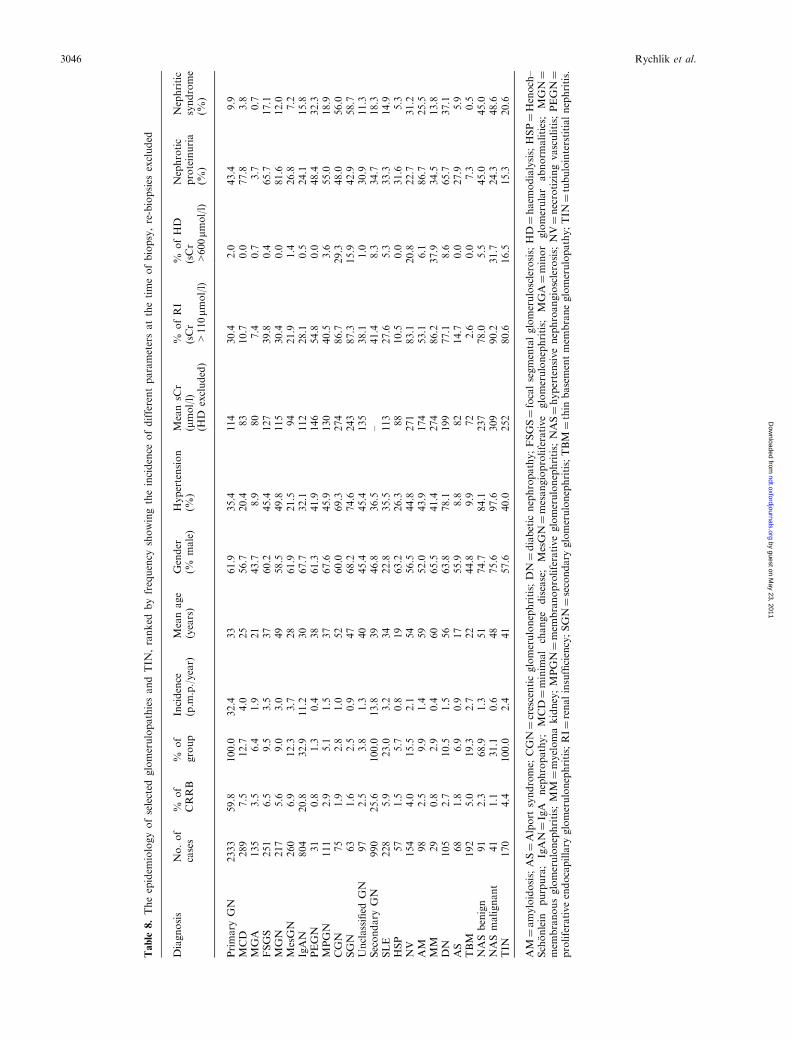
Among adults with normal renal function (meansCr¼ 70 mmol/l), mean age was 35 years, while 78.8%had proteinuria (mean 6.0 g/24 h), 75.8% also hadhaematuria at presentation and 28.5% were hyper-tensive. Among the group with renal insufficiency(sCr >110 mmol/l), a similar percentage had haema-turia while mean age (50 years), presence of proteinuria(90.5%) and hypertension (63.4%) were higher. Dataconcerning dialysis treatment at the time of renalbiopsy were not available, but 203 patients (5.2%) hadsCr >600 mmol/l. The highest percentage of patientswith sCr >600 mmol/l was found in GPS (58.4%), MM(37.9%) and CGN (29.3%). After excluding patientswith sCr >600 mmol/l, the highest mean sCr levelswere observed in CGN (274 mmol/l), NV (271 mmol/l)and TIN (252 mmol/l). The incidence of GN related toage is shown in Table 5.
More than one clinical syndrome was found insome patients and so a total of 6071 clinical syndromeswere registered (Table 6). The most frequent
indications for performing renal biopsy in adultswere nephrotic range proteinuria (39.3%) and urinaryabnormalities (36.2%), but among children it was iso-lated haematuria.
Nephrotic proteinuria (Figure 1A) was the mostcommon clinical presentation (overall frequency39.3%) particularly among the elderly (59.0%). Thedistribution pattern of diagnoses differed with age(Table 7). Those with proteinuria >10 g/24 h comprised23.5% of all patients and 34.6% of adults. Micro-haematuria (66%), hypertension (52%) and renalinsufficiency (46%) were found frequently with nephro-tic range proteinuria.
Among the patients with urinary abnormalities(Figure 1B), all had low-grade proteinuria, 77.7%also had microhaematuria, with hypertension in40.5%, and renal insufficiency in 44.5%. Mean agewas 36 years. The most common disease was IgAN(23.1%) followed by SLE (7.4%).
Nephritic syndrome (Figure 1C) was observed in19.1% of all cases. Mean age was 51 years and morethan one-third of patients (36%) were in the age range46–60 years. sCr >600 mmol/l was found in 13.2%.
Table 3. Annual incidence (%) of primary glomerulonephritis (n¼ 2333), re-biopsies excluded
1994 1995 1996 1997 1998 1999 2000 Total
MCD 10.0 14.2 12.8 10.0 16.9 10.7 12.4 12.5MGA 3.8 8.3 5.6 8.0 5.7 4.5 4.5 5.8FSGS 9.3 8.9 11.0 10.7 8.0 17.3 10.0 10.8MGN 7.2 7.7 9.4 12.0 8.6 9.2 10.3 9.3MesGN 17.9 10.7 14.1 14.3 13.4 8.3 2.8 11.3IgAN 33.1 30.5 33.8 29.7 38.0 33.0 41.8 34.5PEGN 2.4 0.3 1.6 1.4 0.6 1.2 1.8 1.3MPGN 6.6 8.0 3.1 4.9 2.6 4.8 3.0 4.6CGN 2.4 3.4 2.5 4.3 1.0 3.3 5.0 3.2SGN 3.5 1.8 3.5 1.4 2.6 3.9 2.0 2.7Unclassified GN 3.8 6.2 2.5 3.4 2.6 3.8 6.3 4.1Total no. of cases (100%) 290 325 319 350 313 336 400 2333
CGN¼ crescentic glomerulonephritis; FSGS¼ focal segmental glomerulosclerosis; IgAN¼ IgA nephropathy; MCD¼minimal changedisease; MesGN¼mesangioproliferative glomerulonephritis; MGA¼minor glomerular abnormalities; MGN¼membranous glomerulone-phritis; MPGN¼membranoproliferative glomerulonephritis; PEGN¼ proliferative endocapillary glomerulonephritis; SGN¼ secondaryglomerulonephritis.
Table 4. Annual incidence (%) of secondary glomerulopathies (n¼ 990), re-biopsies excluded
1994 1995 1996 1997 1998 1999 2000 Total
Immune-mediated glomerulonephritis 62.3 48.7 46.5 48.2 44.6 41.2 43.2 48.6Lupus nephritis 24.7 28.6 23.3 23.0 21.5 21.2 21.4 23.0Necrotizing vasculitides 23.7 13.5 13.9 17.3 14.6 12.3 16.0 15.6Henoch–Schonlein purpura 10.3 4.2 4.6 6.5 6.9 4.7 4.9 5.7Goodpasture’s syndrome 2.1 0.8 2.3 0 0.8 1.8 1.0 1.2Dysgammaglobulinaemias 12.4 18.5 17.8 15.8 14.6 13.5 13.1 14.9Renal amyloidosis 7.2 10.1 10.8 7.9 11.5 10.6 10.2 9.9Myeloma kidney 4.1 7.6 3.1 3.6 2.3 1.2 1.0 2.9Light-chain deposit disease 0 0 2.3 2.9 0 0 1.9 1.1Essential mixed cryoglobulinaemia 1.0 0.8 1.6 1.4 0.8 1.8 0 1.0Diabetic glomerulosclerosis 9.3 3.4 7.0 10.8 6.9 17.0 14.6 10.6Hereditary disorders 15.5 29.4 28.7 25.2 33.8 28.2 29.1 27.7Alport syndrome 2.1 11.8 5.4 4.3 12.3 4.7 7.3 6.9Thin membrane disease 10.3 16.8 22.5 19.4 20.0 22.3 20.4 19.3Total no. of cases (100%) 97 119 129 139 130 170 206 990
The Czech registry of renal biopsies 1994–2000 3043
by guest on May 23, 2011
ndt.oxfordjournals.orgD
ownloaded from

The most frequent diagnosis was IgAN, but it is ofinterest thatNASandFSGSwere also found frequently.
Finally, IgAN was also found most frequently(26.1%) in patients with isolated haematuria(Figure 1D) followed by hereditary nephropathies(23.4%). Most patients in this group had normal sCr(93.6%), normotension (87.7%); mean age (25 years)was the lowest of all evaluated groups.
IgAN (21.3%) and FSGS (8.3%) were the mostfrequent diagnosis in mild renal insufficiency(Figure 1E), while TIN (11.6%) and NV (11.3%)were the most frequent in advanced renal insufficiencies(Figure 1F). Nephrotic proteinuria was common inboth groups with renal insufficiency, 58.5 and 52.3%,respectively.
Since 1999, the presence of DM was registered, anddiabetic patients represented 12.2% of adult renalbiopsies in 1999–2000 with a mean age of 58 years(range 18–80). Only five patients (3.6%) were youngerthan 30 years, but none of them was in the paediatricage group. DN was found in 42.4% and non-diabeticrenal diseases (NDRD) in 47.5%. It is also of interestthat a large number of samples were non-diagnostic(10.1%). Mean proteinuria was 8.9 g/24 h. Comparingthe DN and NDRD groups, there were no significantdifferences in predominance of male gender (61/69%),hypertension (83/79.8%), microhaematuria (66/62%),low-grade proteinuria (28/36%) or nephrotic protein-uria (68/62.5%). Mean sCr was 244/207 mmol/l (range54–1002). Normal renal function was reported in18.5/33.0% and sCr 111–200 mmol/l in 27.8/27.3%.However, more advanced renal insufficiency wasmore common in DN compared with NDRD: sCr201–400 mmol/l in 33.4/24.2% and sCr >400 mmol/lin 19.0/7.5%. Among NDRD, the most frequentdiagnoses were IgAN in 17.5%, MGN and NAS in11.1%, and NV in 9.5%.
The most important clinical and laboratory datarelated to each diagnostic entity are summarized inTable 8.
USG needle guidance was used most commonly(56.4%), followed by X-ray guidance (35.7%). Theother modalities were used rarely: CT in 2.4%, scintig-raphy in 4.5% and transjugular biopsy in 0.8%. USGwas used more among children (78.9%). The biopsygun has been used since 1994 with an increasingfrequency, from 2.0% in 1994 to 50.3% in 2000. Theuse of USG compared with X-ray was likewiseincreasing over the years, being 40.6/51.2% in 1994and 58.4/35.5% in 2000. Ninety-four clinically seriouscomplications (Table 9) were recorded (3.12% of allbiopsies), being more frequent in children (4.37%) thanin adults (2.85%). Serious complications were observedmost frequently in CT (5.15%) and least frequentlyin USG without a gun (2.33%). Use of the biopsy gunwas not associated with a lower complication rate(2.80 vs 2.88%).
Discussion
This report provides information about the occurrenceof renal diseases diagnosed by renal biopsy duringa period of 7 years covering the population of a wholecountry. Our preliminary results have been presentedelsewhere [8–10]. To our knowledge, only three papershave been published dealing systematically with renalbiopsy data for a whole country with a populationexceeding 10 million [1,3,6]. Since a national registrymay fail particularly due to incomplete data collec-tion, we decided to minimize the required data set toachieve a high response rate and ensure it was trulynationally representative.
Records collected by the CRRB showed thatmales were over-represented in biopsy-proven renaldiseases, with the exception of SLE and TBM.
Table 5. Incidence of glomerulopathies and TIN related to age(given as a percentage of the group, re-biopsies excluded)
Diagnosis �15years
16–30years
31–45years
46–60years
>60years
Total no. of cases 693 1041 750 851 554Primary GN 67.0 68.7 60.5 53.2 44.4MCD 26.2 10.2 9.0 6.4 9.8MGA 12.0 6.3 5.3 1.3 1.6FSGS 8.4 9.8 11.0 12.1 15.0MGN 1.9 3.6 9.3 17.4 24.8MesGN 11.8 11.9 9.7 6.1 4.1IgAN 28.6 46.6 38.3 30.0 10.6PEGN 1.3 0.7 2.0 1.3 2.0MPGN 3.0 4.1 5.1 6.4 5.3CGN 0.7 1.7 1.3 5.5 11.8SGN 0.4 1.1 3.5 4.8 6.1Unclassified GN 3.4 2.2 4.4 6.4 6.4Secondary GN 26.4 22.6 21.3 24.8 36.3Immune mediated 31.1 56.2 57.5 47.9 39.8Dysgammaglobulinaemia 0.6 2.6 10.6 22.8 38.3DM 0.0 2.6 9.4 20.3 20.4Hereditary 68.3 38.7 22.5 9.0 1.5Hypertensive nephrosclerosis 0.0 0.6 7.3 8.5 6.3TIN 3.6 3.0 4.9 4.8 6.0
CGN¼ crescentic glomerulonephritis; DM¼ diabetes mellitus; FSGS¼focal segmental glomerulosclerosis; IgAN¼ IgA nephropathy; MCD¼
minimal change disease; MesGN¼mesangioproliferative glomerulone-phritis; MGA¼minor glomerular abnormalities; MGN¼membranousglomerulonephritis; MPGN¼membranoproliferative glomerulonephri-tis; PEGN¼ proliferative endocapillary glomerulonephritis; SGN¼
secondary glomerulonephritis; TIN¼ tubulointerstitial nephritis.
Table 6. Clinical syndromes (n¼ 6071) at the time of renal biopsy(given as a percentage of all renal biopsies)
Total(n¼ 4004)
Children(n¼ 710)
Adults(n¼ 3294)
Nephrotic proteinuria 39.3 26.8 42.0Urinary abnormalities 36.2 31.7 37.1Isolated haematuria 16.5 32.7 12.5Nephritic syndrome 19.1 1.8 22.3Mild renal insufficiency 19.4 3.4 23.0Advanced renal insufficiency 21.1 2.4 25.2
In some patients, more than one clinical syndrome was found; forinstance, nephrotic proteinuria associated with renal insufficiency.
3044 Rychlık et al.
by guest on May 23, 2011
ndt.oxfordjournals.orgD
ownloaded from

Other investigators have also observed this finding in65 [1] and 66% [3], respectively.
More than a quarter of renal biopsies wereperformed in paediatric units. We used an age limitfor children of 15 years to allow comparison with
other studies. There are few epidemiological dataconcerning renal biopsy in children in Europeanregistries. Coppo et al. [11] reported 432 children�15 years old undergoing renal biopsy in 1992–1994,representing only 5.7% of the all-Italian Registry
Fig. 1. Percentage of different forms of renal diseases according to clinical presentations: (A) patients with nephrotic proteinuria (n¼ 1557);(B) patients with urinary abnormalities (n¼ 1451); (C) patients with nephritic syndrome (n¼ 747); (D) patients with isolated haematuria(n¼ 643); (E) patients with mild renal insufficiency (n¼ 762); (F) patients with advanced renal insufficiency (n¼ 829). For abbreviationssee text.
Table 7. Distribution of nephrotic proteinuria according to selected diagnosis and age (given as a percentage of allpatients with nephrotic proteinuria in each age group)
Diagnosis All Children(�15 years)
Adults
16–30 years 31–45 years 46–60 years >60 years All
MCD 14.9 50.5 17.0 9.7 5.7 6.7 9.4IgAN 12.9 7.8 21.7 18.1 10.5 3.7 13.1MGN 11.8 2.1 4.7 11.3 16.3 16.8 12.7FSGS 11.0 12.5 13.0 11.3 8.9 8.9 10.3AM 5.7 0.5 0.7 3.8 5.5 14.4 6.2SLE 5.1 3.6 9.3 5.6 3.6 2.5 5.1MesGN 4.7 6.8 7.0 4.4 4.1 1.8 4.3DM 4.6 0.0 0.7 2.8 6.9 8.9 5.1MPGN 4.1 1.6 5.3 4.7 4.5 2.5 4.3NAS 3.4 0.0 0.7 5.0 5.0 3.7 3.7CGN 2.4 0.0 2.0 0.6 3.1 4.6 2.6% of all cases 39.3 26.8 28.8 42.6 49.1 59.0 42.0
AM¼ amyloidosis; CGN¼ crescentic glomerulonephritis; DM¼diabetes mellitus; FSGS¼ focal segmental glomerulo-sclerosis; IgAN¼ IgA nephropathy; MCD¼minimal change disease; MesGN¼mesangioproliferative glomerulo-nephritis; MGN¼membranous glomerulonephritis; MPGN¼membranoproliferative glomerulonephritis; NAS¼hypertensive nephroangiosclerosis SLE¼ systemic lupus erythematosus.
The Czech registry of renal biopsies 1994–2000 3045
by guest on May 23, 2011
ndt.oxfordjournals.orgD
ownloaded from
Table
8.Theepidem
iologyofselected
glomerulopathiesandTIN
,ranked
byfrequency
showingtheincidence
ofdifferentparametersatthetimeofbiopsy,re-biopsies
excluded
Diagnosis
No.of
cases
%of
CRRB
%of
group
Incidence
(p.m
.p./year)
Meanage
(years)
Gender
(%male)
Hypertension
(%)
MeansC
r(mmol/l)
(HD
excluded)
%ofRI
(sCr
>110mm
ol/l)
%ofHD
(sCr
>600mm
ol/l)
Nephrotic
proteinuria
(%)
Nephritic
syndrome
(%)
Primary
GN
2333
59.8
100.0
32.4
33
61.9
35.4
114
30.4
2.0
43.4
9.9
MCD
289
7.5
12.7
4.0
25
56.7
20.4
83
10.7
0.0
77.8
3.8
MGA
135
3.5
6.4
1.9
21
43.7
8.9
80
7.4
0.7
3.7
0.7
FSGS
251
6.5
9.5
3.5
37
60.2
45.4
127
39.8
0.4
65.7
17.1
MGN
217
5.6
9.0
3.0
49
58.5
49.8
115
30.4
0.0
81.6
12.0
MesGN
260
6.9
12.3
3.7
28
61.9
21.5
94
21.9
1.4
26.8
7.2
IgAN
804
20.8
32.9
11.2
30
67.7
32.1
112
28.1
0.5
24.1
15.8
PEGN
31
0.8
1.3
0.4
38
61.3
41.9
146
54.8
0.0
48.4
32.3
MPGN
111
2.9
5.1
1.5
37
67.6
45.9
130
40.5
3.6
55.0
18.9
CGN
75
1.9
2.8
1.0
52
60.0
69.3
274
86.7
29.3
48.0
56.0
SGN
63
1.6
2.5
0.9
47
68.2
74.6
243
87.3
15.9
42.9
58.7
Unclassified
GN
97
2.5
3.8
1.3
40
45.4
45.4
135
38.1
1.0
30.9
11.3
Secondary
GN
990
25.6
100.0
13.8
39
46.8
36.5
–41.4
8.3
34.7
18.3
SLE
228
5.9
23.0
3.2
34
22.8
35.5
113
27.6
5.3
33.3
14.9
HSP
57
1.5
5.7
0.8
19
63.2
26.3
88
10.5
0.0
31.6
5.3
NV
154
4.0
15.5
2.1
54
56.5
44.8
271
83.1
20.8
22.7
31.2
AM
98
2.5
9.9
1.4
59
52.0
43.9
174
53.1
6.1
86.7
25.5
MM
29
0.8
2.9
0.4
60
65.5
41.4
274
86.2
37.9
34.5
13.8
DN
105
2.7
10.5
1.5
56
63.8
78.1
199
77.1
8.6
65.7
37.1
AS
68
1.8
6.9
0.9
17
55.9
8.8
82
14.7
0.0
27.9
5.9
TBM
192
5.0
19.3
2.7
22
44.8
9.9
72
2.6
0.0
7.3
0.5
NASbenign
91
2.3
68.9
1.3
51
74.7
84.1
237
78.0
5.5
45.0
45.0
NASmalignant
41
1.1
31.1
0.6
48
75.6
97.6
309
90.2
31.7
24.3
48.6
TIN
170
4.4
100.0
2.4
41
57.6
40.0
252
80.6
16.5
15.3
20.6
AM
¼amyloidosis;
AS¼Alport
syndrome;
CGN¼crescenticglomerulonephritis;
DN¼diabetic
nephropathy;FSGS¼focalsegmentalglomerulosclerosis;
HD¼haem
odialysis;
HSP¼Henoch–
Schonlein
purpura;
IgAN¼IgA
nephropathy;
MCD¼minim
al
change
disease;
MesGN¼mesangioproliferative
glomerulonephritis;
MGA¼minor
glomerular
abnorm
alities;
MGN¼
mem
branousglomerulonephritis;
MM
¼myelomakidney;MPGN¼mem
branoproliferativeglomerulonephritis;
NAS¼hypertensivenephroangiosclerosis;
NV¼necrotizingvasculitis;
PEGN¼
proliferativeendocapillary
glomerulonephritis;RI¼renalinsufficiency;SGN¼secondary
glomerulonephritis;TBM
¼thin
basementmem
braneglomerulopathy;TIN
¼tubulointerstitialnephritis.
3046 Rychlık et al.
by guest on May 23, 2011
ndt.oxfordjournals.orgD
ownloaded from

of renal biopsy. Rivera et al. [3] and Briganti et al. [7]reported 487 and 104 renal biopsies performed amongchildren <15 years old, which represented 7.0 and5.2% of all biopsies, respectively. Our registry reportsalmost twice the total number of paediatric renalbiopsies (n¼ 710, i.e. 17.7% of all biopsies), apparentlyindicating more liberal criteria for renal biopsyamong our paediatric nephrologists. Higher propor-tions of paediatric biopsies have also been reported inAsia, with 40.5% out of all renal biopsies, in Korea [5]and in Japan with 20% [6].
It is rather difficult to report definitive epidemio-logical data on the frequency of the various forms ofGN for several reasons. First, the renal biopsy indi-cation policy varies from centre to centre. Secondly,renal biopsy is often not performed when the likelihoodof a therapeutic consequence is low (e.g. steroid-sensitive nephrotic syndrome in children, intermittenthaematuria without proteinuria, bilateral small kid-neys, post-infection GN). For this reason, the trueincidence of MCD, PEGN and IgAN will be under-represented. Using the experience of biopsy registrieswith a similar design [2], we estimate that the CRRBincludes up to 70–85% of these patients. Thirdly,insufficient tissue did not always allow complete evalu-ation, i.e. by immunofluorescence, so that a correcthistological diagnosis could not be established (e.g.IgAN). Other reports are similarly incomplete; forexample, only 78% out of all samples were evaluatedby immunofluorescence in a Danish study [2] and56.5% in Japan [6], where 42.5% cases of MesGN werenot evaluated by immunofluorescence. During the7 year survey, we could observe a ‘crossover’ phenom-enon with decreasing incidence of MesGN andincreasing incidence of IgAN, suggesting increasinglycomplete evaluation by immunofluorescence. Electronmicroscopy is rather expensive and was not used in thisstudy for all biopsies, although it is a crucial diagnos-tic tool in some circumstances, e.g. establishing thediagnosis ofMCD and TBM. The frequency of electronmicroscopy did not differ compared with other reportsworldwide (�30% of all samples). Newer techniques,e.g. polymerase chain reaction (PCR) or in situ hybrid-ization, would seem to be helpful in the future [12], butcertainly their routine use is not intended.
Our report showed that renal biopsy was predomi-nantly performed in patients with primary and second-ary GN. Despite any possible local influence of renal
biopsy policy, the incidence of GN (47 p.m.p./year)was comparable with that seen in other Europeancountries: 34, 39, 63 and 86 cases p.m.p./year [2–4,11].The distribution pattern of renal diagnosis also cor-responded to other European series. IgAN was themost frequent disease among all biopsied patients aswell as among primary GN, reaching an incidenceof 11.2 p.m.p./year. A similar frequency has been alsoreported from Italy, 8.4 p.m.p./year [1], but is higherin France at 25–31 p.m.p./year [4], and lower in Spainat 7.9 p.m.p./year [3], Denmark at 1.8 p.m.p./year [2]and Central Kentucky, USA at 5.4 p.m.p./year [13].The true incidence of IgAN is probably higher sinceanother 11.3% of primary GN was diagnosed asMesGN, and not all these cases were evaluated byimmunofluorescence.
Our incidence of unclassified GN is also higher(4.1%) than reported in other studies, e.g. 0.9% inKorea [5] and 1.2% in Japan [6]; data are not avail-able in European registries.
GN induced by immunological factors was themost frequent among secondary GNs. The incidenceof SLE was relatively high, and comparable data havebeen reported from Italy [1] with 2.6 p.m.p./yearand Spain [3] with 5.6 p.m.p./year. AM was themost frequent dysgammaglobulinaemia-associatedGN, especially in the elderly, as has been repeatedlyreported by other investigators [17].
Among those with nephrotic proteinuria, MGN,IgAN, MCD and FSGS were the most frequent diag-noses in CRRB. With the exception of IgAN, this isin accordance with other experience [1–4]. IgAN waspresent in a similar percentage of adult nephroticsyndrome patients (14%) in the report of Haas et al.[14], while others found lower frequencies, 8 [15]and 2.4% [1]. Since 1999 when the serum albuminlevel was recorded, the incidence of nephrotic syndromeamong IgAN patients decreased to only 6.5%, indicat-ing a large proportion of IgAN patients with nephroticrange proteinuria but without frank nephrosis. Thisfigure is very similar to that (6.0%) reported fromKorea [5]. This may be because IgAN patients oftenundergo renal biopsy at a more advanced stage ofdisease.
Persistent urinary abnormalities represented thesecond most frequent clinical syndrome (36.2%),rather higher than that observed in Italy (30.8%) [1]but much lower than reported from Japan (48.1%) [6].
Table 9. Clinically significant complications after renal biopsy
Type No ofcases
% of allrenal biopsis
Concomitant co-morbidity (%)
Renalinsufficiency
Hypertension Both Total
Haemorrhage and hypotension 35 0.87 20.0 13.3 28.6 65.7Gross haematuria 45 1.12 17.8 8.9 31.1 57.8Symptomatic haematoma 45 1.12 6.7 8.9 26.7 42.3Total 125 3.12 14.4 11.2 28.8 53.6Children 31 4.37Adults 94 2.85
The Czech registry of renal biopsies 1994–2000 3047
by guest on May 23, 2011
ndt.oxfordjournals.orgD
ownloaded from

IgAN was the disease diagnosed predominantly in thisgroup (23.1%), similar to data from Italy (29.8%);however, in our study, the next most common was SLE(7.4%), which was quite different from the Italianexperience (28.2%).
The presence of nephritic syndrome is usually takenas a sign of acute renal inflammation. It is thereforenot surprising that IgAN, NV and CGN were foundcommonly among those with nephritic symdrome.However, it is of interest that NAS, FSGS andalso DN were recorded frequently among those withnephritic syndrome even though haematuria is notusually regarded as a typical feature of these diseases.The experience from Italy [1] was quite different, IgANrepresenting 14.0%of patients presentingwith nephriticsyndrome but SLE 20.1 and PEGN 16.1%.
IgAN and TBM glomerulopathy were the mostfrequent diagnoses in patients with isolated haema-turia, which is not significantly different from otherreports in Europe [1–3] and elsewhere [5,6].
Concerning diabetic patients, it is not surprisingthat the incidence of DN was low, since isolatedproteinuria in patients with long-standing diabetes isnot an indication for renal biopsy, and those diabeticswho have a renal biopsy are likely to have atypicalclinical features suggesting a cause other than DN.However, since 1999, we observed an apparentdoubling of the incidence of DN [10], which we ascribeto local policies on the indications for renal biopsy.Recently, the influence of varying renal biopsy policieshas been stressed by Mazzucco et al. [16]. Since 1999,diabetic patients represented 12.2% of all adult biopsiesbut about half had non-diabetic renal disease. In thisgroup, the percentages of low-grade proteinuria,normal renal function, the number of patients onhaemodialysis and mean age were significantly highercompared with those with DN. There was a surpris-ingly high number of diabetic patients with renalvasculitis (9.5%). On the other hand, in 1999–2000,a high percentage of microhaematuria (66%) was alsofound among patients with DN; comparable datafrom other registries in this respect are not available.
With regard to post-biopsy complications, werecorded lower rates of clinically serious complicationscompared with the experience of other authors [17] whoreported gross haematuria in 5–7% or severe perirenalhaemorrhage in 0.2–1.4%. The presence of hyper-tension and renal insufficiency was related to the occur-rence of haemorrhage with hypotension. Contrary toother reports [18], we did not observe a significantdecrease in complication rate when the biopsy gunwas used, but we are not aware whether a thinnerneedle gauge was used in the gun. Although someauthors [19] did not observe any serious complicationsusing the biopsy gun, we believe that despite thesenewer devices, the risk of renal biopsy as an invasiveprocedure remains. USG-guided biopsy was predomi-nant in our series, particularly among children, but stilla large number biopsies were performed under X-rayguidance despite the wide availability of USG. Thismay be the result of local policy, perhaps influenced
by good personal experience with particular techniques.USG guidance was also reported as predominant(�85%) in Europe [20].
CRRB continues to collect data, and a numberof new features are planned: (i) the CRRB recordsare now available on the Internet (www.nefrol.cz);(ii) 10 year follow-up data are being collected, partic-ularly in patients with GN; and (iii) comparison ofCRRB records is being made with the Czech Registryof Renal Replacement Therapy, which uses ESRD asthe identifier.
In conclusion, CRRB, which includes renal biopsiesof the native kidney performed in almost all Czechrenal units over a period of 7 years, represents animportant contribution to the epidemiology of renaldiseases in the Czech Republic, and permits a valuablecomparison with other renal biopsy registries world-wide. Our results show that the frequency of themain groups of renal diseases and the distributionpattern of different forms of GN change only slightlyover a long period. Two groups of disease were partic-ularly frequent: primary GN (59.8%) and secondaryGN (25.6%) which showed incidences of 32.4 and13.8 p.m.p./year, respectively. Particular attentionshould also be paid to TIN and vascular diseases, inwhich a significant percentage of patients showed renalinsufficiency. This report is intended to serve as asource for nephrologists, researchers and health careproviders to stimulate new analysis and investigation,to improve treatment of renal diseases, to designnationwide trials, and to help national and regionalgovernments to develop protocols for preventivemedicine.
Acknowledgements. The authors thank all colleagues from theparticipating renal units listed below for supplying the requesteddata. We gratefully acknowledge the invaluable computer andstatistical assistance of Dr Martin Vyhnanovsky and Dr Jirı Gallas,and the linguistic text revision of Dr Rene Prahl.
Brno: Faculty of Medicine: Department of Medicine, Hospital Sv.Anna (D. Sobotova); Department of Medicine, Hospital Bohunice(H. Hertlova); Department of Pathology (Dr Husek); 2ndDepartment of Paediatrics (Z. Dolezal, J. Starha). CeskeBudejovice: Regional Hospital, Department of Medicine (J. Hana);Department of Pathology (Dr Pradna); Department of Paediatrics(V. Smrcka). Hradec Kralove: Faculty of Medicine: 1st Departmentof Medicine (P. Fixa, V. Herout, J. Zahradnık); 2nd Department ofMedicine (J. Kacerovsky, E. Pinterova); Department of Pathology(M. Podhola); Department of Paediatrics (Z. Pellantova, S.Skalova). Litomerice: District Hospital, Department of Paediatrics(J. Rambousek). Karvina: District Hospital, Department ofMedicine (J. Kubatko). Olomouc: Faculty of Medicine: 3rdDepartment of Medicine (K. Konecny, K. Krejcı, J. Zadrazil);Department of Pathology (Dr Tichy). Ostrava: Department ofMedicine (I. Valkovsky); Department of Pathology (R. Curık).Ostrava Poruba: Centre for Paediatric Dialysis and Nephrology,University Hospital (M. Hladık). Plzen: Army Hospital, Depart-ment of Medicine (M. Pojer, K. Smetana); Faculty of Medicine: 1stDepartment of Medicine (I. Malkusova, K. Opatrny); Departmentof Pathology (Dr Hess); Department of Paediatrics (Dr Huml).Prague: 1st Faculty of Medicine: 1st Department of Medicine(E. Jancova, M. Merta, I. Rychlık, R. Rysava, V. Tesar, J. Zabka,);2nd Department of Medicine (M. Havrda, V. Knotkova);1st Department of Pathology (E. Honsova, A. Stejskalova,Z. Vernerova); 2nd Faculty of Medicine: Department of Medicine
3048 Rychlık et al.
by guest on May 23, 2011
ndt.oxfordjournals.orgD
ownloaded from
(V. Martınek, K. Matousovic, J. Osten, J. Vrzanova,);1st Department of Paediatrics (J. Dusek, J. Feber, J. Janda,E. Simkova, K. Vondrak); Department of Pathology (J. Stejskal);3rd Faculty of Medicine: 1st Department of Medicine(M. Horackova, M. Jaros); Central Army Hospital (V. Monhart,K. Petru); Hospital Homolka (L. Stranık, L. Svoboda); ThomayerTeaching Hospital, Department of Paediatrics (A. Kolsky);Institut for Clinical and Experimental Medicine: Department ofNephrology (P. Bubenıcek, J. Lacha, O. Viklicky); Department ofPathology (M. Chadimova). Svitavy: Department of Medicineand Dialysis Unit (M. Nyvlt). Ustı/Labem: Masaryk Hospital,Department of Paediatrics (Z. Hynkova, J. Laubova).
Conflict of interest statement. None declared.
References
1. Schena FP. Survey of the Italian Registry of renal Biopsies.Frequency of the renal diseases for 7 consecutive years. NephrolDial Transplant 1997; 12: 418–426
2. Heaf JG, Løkkegaard H, Larsen S. The epidemiology andprognosis of glomerulonephritis in Denmark 1985–97. NephrolDial Transplant 1999; 14: 1889–1897
3. Rivera F, Lopez-Gomez JM, Perez-Garcıa R. Frequency ofrenal pathology in Spain 1994–1999. Nephrol Dial Transplant2002; 17: 1594–1602
4. Simon P, Ramee MP, Autuly V et al. Epidemiology of primaryglomerular disease in a French region. Variations accordingto period and age. Kidney Int 1994; 46: 1192–1198
5. Choi IJ, Jeong HJ, Han DS et al. An analysis of 4514 cases ofrenal biopsy in Korea. Yonsei Med J 2001; 42: 247–254
6. Research Group on Progressive Chronic Renal Disease. Nation-wide and long-term survey of primary glomerulonephritis in Japanas observed in 1850 biopsied cases. Nephron 1999; 82: 205–213
7. Briganti EM, Dowlin J, Finlay M et al. The incidence of biopsy-proven glomerulonephritis in Australia. Nephrol Dial Transplant2001; 16: 1364–1367
8. Rychlık I, Jancova E, Tesar V et al., on behalf ofCRRB. The Czech Registry of Renal Biopsies (CRRB)—fiveyears experience (1994–1998). Nephrol Dial Transplant 2001;16: A68
9. Rychlık I, Jancova E, Tesar V, on behalf of CRRB. IgAnephropathy (IgAN) in the Czech registry of renal biopsies(CRRB) in years 1995–1998. Nephrol Dial Transplant 2000;15: A88
10. Rychlık I, Jancova E, Tesar V, on behalf of CRRB. Diabeticand non-diabetic renal disease among diabetic patients under-going renal biopsy in the Czech Republic in years 1999–2000.J Am Soc Nephrol 2002; 13: 636A
11. Coppo R, Gianoglio B, Porcellini MG, Maringhini S.Frequency of renal diseases and clinical indications for renalbiopsy in children (Report of the Italian National Registryof Renal Biopsies in Children). Nephrol Dial Transplant 1998;13: 293–297
12. Schena P, Gesualdo L. Renal biopsy beyond histology andimmunofluorescence.Nephrol Dial Transplant 1994; 9: 1541–1544
13. Wyatt RJ, Julian BA, Baehler RW et al. Epidemiology of IgAnephropathy in central and eastern Kentucky for the period1975 through 1994. Central Kentucky Region of the South-eastern United States IgA Nephropathy DATABANK Project.J Am Soc Nephrol 1998; 9: 853–858
14. Haas M, Meehan SM, Karrison TG, Spargo BH. Changingetiologies of unexplained adult nephrotic syndrome. Am JKidney Dis 1997; 30: 621–631
15. Korbet SM, Genchi RM, Borok RZ, Schwartz MM. Theracial prevalence of glomerular lesion in nephrotic adults. Am JKidney Dis 1996; 27: 647–651
16. Mazzucco G, Bertani T, Fortunato M et al. Different patternsof renal damage in type 2 diabetes mellitus: a multicentric studyon 393 biopsies. Am J Kidney Dis 2002; 39: 713–720
17. Ponticelli C, Mihatsch MJ, Imbasciati E. Renal biopsy: per-formance and interpretation. In: Davison AM et al., eds. OxfordTextbook of Clinical Nephrology, 2nd ed., Oxford UniversityPress, Oxford; 1999: 158–159
18. Riehl J, Maigatter S, Kierdorf H et al. Percutaneous renalbiopsy: comparison of manual and automated puncture tech-niques with native and transplanted kidneys. Nephrol DialTransplant 1994; 9: 1568–1574
19. Hergesell O, Felten H, Andrassy K et al. Safety of ultrasound-guided percutanous renal biopsy—retrospective analysis of 1090consecutive cases. Nephrol Dial Transplant 1998; 13: 975–977
20. Fuiano G, Mazza G, Comi N et al. Current indications forrenal biopsy: a questionnaire-based survey. Am J Kidney Dis2000; 35: 448–457
Received for publication: 5.5.03Accepted in revised form: 6.9.04
The Czech registry of renal biopsies 1994–2000 3049
by guest on May 23, 2011
ndt.oxfordjournals.orgD
ownloaded from